title sept4 and myofibroblastic transformation of...
TRANSCRIPT
TitleDownregulation of the Wnt antagonist Dkk2 links the loss ofSept4 and myofibroblastic transformation of hepatic stellatecells.
Author(s)Yanagida, Atsuko; Iwaisako, Keiko; Hatano, Etsuro; Taura,Kojiro; Sato, Fumiaki; Narita, Masato; Nagata, Hiromitsu;Asechi, Hiroyuki; Uemoto, Shinji; Kinoshita, Makoto
Citation Biochimica et biophysica acta (2011), 1812(11): 1403-1411
Issue Date 2011-11
URL http://hdl.handle.net/2433/149268
Right
© 2011 Elsevier B.V.; この論文は出版社版でありません。引用の際には出版社版をご確認ご利用ください。This isnot the published version. Please cite only the publishedversion.
Type Journal Article
Textversion author
Kyoto University

1
Downregulation of the Wnt antagonist Dkk2 links the loss of Sept4 and myofibroblastic
transformation of hepatic stellate cells
Atsuko Yanagidaa,b,f
, Keiko Iwaisakoa,b,c,f
, Etsuro Hatanoa, Kojiro Taura
a,c, Fumiaki
Satod, Masato Narita
a, Hiromitsu Nagata
a, Hiroyuki Asechi
a, Shinji Uemoto
a, Makoto
Kinoshitab,e
aDepartment of Surgery, Graduate School of Medicine, Kyoto University, 54
Kawahara-cho, Shogoin, Sakyo-ku, Kyoto 606 8507, Japan
bBiochemistry and Cell Biology Unit, Graduate School of Medicine, Kyoto University,
Yoshida-Konoe, Sakyo-ku, Kyoto 606 8501, Japan
cDepartment of Medicine, University of California, San Diego, 9500 Gilman DR #0702,
La Jolla, CA 92093, USA
dDepartment of Nanobio Drug Discovery, Graduate School of Pharmacology, Kyoto
University, 46-29 Shimoadachi-cho, Yoshida, Sakyo-ku, Kyoto 606 8501, Japan
eDivision of Biological Science, Graduate School of Science, Nagoya University,
Furo-cho, Chikusa-ku, Nagoya 464 8602, Japan
fThese authors contributed equally to this paper.

2
E-mail addresses
Atsuko Yanagida: [email protected]
Keiko Iwaisako: [email protected]
Etsuro Hatano: [email protected]
Kojiro Taura: [email protected]
Fumiaki Sato: [email protected]
Masato Narita: [email protected]
Hiromitsu Nagata: [email protected]
Hiroyuki Asechi: [email protected]
Shinji Uemoto: [email protected]
Makoto Kinoshita: [email protected]
Corresponding author: Dr. Etsuro Hatano
Department of Surgery, Graduate School of Medicine, Kyoto University, 54
Kawahara-cho, Shogoin, Sakyo-ku, Kyoto 606 8507, Japan
Telephone: +81-75-751-4323
Fax: +81-75-751-4348
E-mail: [email protected]

3
ABSTRACT
Background/Aims: Sept4, a subunit of the septin cytoskeleton specifically expressed
in quiescent hepatic stellate cells (HSCs), is downregulated through transdifferentiation
to fibrogenic and contractile myofibroblastic cells. Since Sept4−/−
mice are prone to
liver fibrosis, we aimed to identify the unknown molecular network underlying liver
fibrosis by probing the association between loss of Sept4 and accelerated
transdifferentiation of HSCs. Methods: We compared the transcriptomes of Sept4+/+
and
Sept4−/−
HSCs undergoing transdifferentiation by DNA microarray and quantitative
reverse transcription polymerase chain reaction (RT-PCR) analysis. Because Dickkopf2
(Dkk2) gene expression is reduced in Sept4−/−
HSCs, we tested whether supplementing
Dkk2 could suppress myofibroblastic transformation of Sept4−/−
HSCs. We tested the
involvement of the canonical Wnt pathway in this process by using a lymphoid
enhancer-binding factor/transcription factor-luciferase reporter assay. Results: We
observed consistent upregulation of Dkk2 in primary cultured HSCs and in a carbon
tetrachloride liver fibrosis in mice, which was decreased in the absence of Sept4.
Supplementation with Dkk2 suppressed the induction of pro-fibrotic genes (-smooth
muscle actin and 2 collagen genes) and induced an anti-fibrotic gene (Smad7) in
Sept4−/−
HSCs. In human liver specimens with inflammation and fibrosis, Dkk2
4
immunoreactivity appeared to be positively correlated with the degree of fibrotic
changes. Conclusions: Pro-fibrotic transformation of HSCs through the loss of Sept4 is,
in part, due to reduced expression of Dkk2 and its homologues, and the resulting
disinhibition of the canonical Wnt pathway.
Keywords: septin, hepatic stellate cell, canonical Wnt pathway, Dkk (Dickkopf),
myofibroblastic transformation, liver fibrosis
Abbreviations: HSCs (hepatic stellate cells), RT-PCR (reverse transcription polymerase
chain reaction), -Sma (-smooth muscle actin), Dkk (Dickkopf), Fzd (Frizzled), LRP
(low density lipoprotein receptor-related protein), Lef/Tcf (lymphoid enhancer-binding
factor/transcription factor), DMEM (Dulbecco’s Modified Eagle’s Medium), FBS (fetal
bovine serum), CCl4 (carbon tetrachloride), DIV (day in vitro), sFRPs (secreted
frizzled-related proteins), TGF- (transforming growth factor ), SOCS7 (suppressor of
cytokine signaling 7), NCK (non-catalytic region of tyrosine kinase adaptor protein)

5
1. Introduction
Chronic liver inflammation caused by viral infections, alcohol and other toxic
chemicals, and metabolic disorders is accompanied by tissue remodeling and fibrosis.
These interrelated pathological processes are major issues in hepatology because they
are implicated in major clinical disorders, namely liver cirrhosis and carcinoma.
Previous studies have highlighted hepatic stellate cells (HSCs) as pivotal players
contributing to liver fibrosis [1, 2]. Upon liver damage, HSCs transdifferentiate into
myofibroblastic cells by inflammatory cytokines released mainly from Kupffer cells
(KCs). Transformed HSCs are distinct from quiescent HSCs in terms of their
proliferative activity, contractility facilitated by -smooth muscle actin (-SMA)
expression, and production of collagens and other extracellular matrices. Since these
properties contribute to the pathogenesis of liver fibrosis and cirrhosis, the molecular
mechanism underlying myofibroblastic transformation of HSCs has been a research
focus in this field.
Septins are a family of cytoskeletal/scaffold proteins ubiquitously expressed in
eukaryotic cells. Among the 13 septin genes shared by mice and humans, Sept4 is
unique in that it is expressed in specific cells such as Bergmann glial cells in the

6
cerebellum, dopamine neurons in the midbrain, and spermatozoa. Loss of Sept4 causes
disparate phenotypes in these systems [3, 4].
We previously reported that Sept4 is expressed exclusively in quiescent HSCs in
the liver, it is strongly downregulated through transdifferentiation of HSCs in vitro, and
Sept4 protein is undetectable in HSCs in human specimens with liver fibrosis. Since
Sept4−/− mice are prone to liver fibrosis, the presence of Sept4 contributes to the
suppression of myofibroblastic transformation of HSCs by unknown mechanisms [5]. In
this study, we examined the accelerated transdifferentiation of HSCs in Sept4−/− mice
attempting to identify the unknown molecular network underlying liver fibrosis and the
physiological role of Sept4 in HSCs.
By comparing the transcriptomes of HSCs collected from Sept4+/+
and Sept4−/−
mice, we found reproducible differences. Here, we focus on Dickkopf2 (Dkk2) and its
homologues. We found that Dkk2 is expressed specifically in HSCs, that it is
upregulated through myofibroblastic transformation in vivo and in vitro, and that the
Dkk upregulation is diminished in Sept4−/− HSCs. Previous studies from other groups
have shown that the canonical Wnt pathway promotes fibrosis of the liver, kidney, lung,
and other tissues [6-9], which is countered by Dkks that interfere with the interaction
between Wnt proteins, the cognate membrane-bound receptor Frizzled (Fzd), and
7
LRP5/6 (low-density lipoprotein receptor-related protein 5/6) [10-12]. On the basis of
the above information and our own data, we conclude that pro-fibrotic transformation of
HSCs in the absence of Sept4 is, at least in part, due to the reduced upregulation of Dkk
genes and the resulting disinhibition of the canonical Wnt pathway.
2. Materials and Methods
2.1. Animals
Characterization of Sept4−/−
mice with a C57BL/6J background has been reported
previously [3-5]. The protocol for animal handling was reviewed and approved by the
Animal Care and Use Committee of Kyoto University.
2.2. Liver cell fractionation and cell culture
We fractionated HSCs from Sept4+/+
or Sept4−/−
male mice using a standard
method with some modifications [5, 13]. In brief, we dispersed liver cells by infusing
0.025% Pronase E (Kaken Pharmaceutical, Tokyo, Japan) and 0.025% collagenase
(Wako, Osaka, Japan) in SC-2 solution (137 mM NaCl, 5.4 mM KCl, 0.57 mM
NaH2PO4, 0.85 mM Na2HPO4, 10 mM HEPES, 4.2 mM NaHCO3, and 3.8 mM CaCl2
[pH 7.25]). The collected liver cells were centrifuged on an 8.0% Nycodenz (Nycomed
8
Pharma, Oslo, Norway) cushion. The density-separated HSCs were grown on uncoated
dishes in Dulbecco’s Modified Eagle’s Medium (DMEM) supplemented with 10% fetal
bovine serum (FBS) and antibiotics in 5% CO2 at 37ºC. The purity of HSCs was
consistently more than 98%, as assessed by the presence of lipid droplets and
autofluorescence of vitamin A. We isolated hepatocytes as previously described [14]. In
brief, the liver cells dispersed by infusing 0.03% collagenase in SC-2 solution were
centrifuged at 50 g for 1 min. The cell pellet was washed twice by resuspending in
GBSS-B solution (137 mM NaCl, 5.0 mM KCl, 1.0 mM MgCl2, 0.28 mM MgSO4, 0.84
mM NaH2PO4, 0.22 mM KH2PO4, 5.5 mM glucose, 2.7 mM NaHCO3, and 1.5 mM
CaCl [pH 7.25]) and pelleting at 50 g for 1 min. We grew the fractionated hepatocytes
using the same method as for HSCs, except that the dishes were coated with type I
collagen.
2.3. Experimental liver fibrosis induced by carbon tetrachloride
As previously reported [5], we injected carbon tetrachloride (CCl4) (1.0 l/g body
weight, 25% [v/v] in olive oil) or vehicle alone intraperitoneally into 6-week-old, male
Sept4+/+
and Sept4−/−
mice, biweekly for 10 weeks.

9
2.4. Total RNA extraction and DNA microarray analysis
We prepared total RNA from HSCs at the 3rd
day in vitro (DIV3) using a
phenol–chloroform extraction method (TRIzol; Invitrogen, Carlsbad, CA) and
monitored the quality of total RNA from the elution profile of 18S and 28S ribosomal
RNA [15] using the Bioanalyzer 2100 (Agilent Technologies, Palo Alto, CA). We
amplified RNA (aRNA) samples using a T7-RNA polymerase system (TargetAmp
1-Round Aminoallyl aRNA amplification kit; Epicentre, Madison, WI) and labeled the
aRNA samples using Cy5 Mono-Reactive Dye Pack (GE Healthcare, England). We
hybridized DNA microarray chips (3D-Gene Mouse Oligo chip 24k; Toray, Tokyo,
Japan) with Cy-5-labeled aRNA samples at 37ºC for 16 h and then washed the chips
thoroughly. We scanned the hybridized chips and acquired chip image data using
ProScanArray (Perkin Elmer, Waltham, MA). The scanned image data were processed
using Genepix Pro 4.0 software (Molecular Devices, Sunnyvale, CA) to extract gene
expression data, which were normalized using MATLAB software (Mathworks, Natick,
MA) and a quantile normalization method [16]. For the above procedures, we followed
the manufacturers’ protocols with some modifications. We have registered the
microarray data with NCBI’s Gene Expression Omnibus (GEO) database under the
accession number GSE24588.

10
2.5. Quantitative reverse transcription polymerase chain reaction analysis
Total RNA was extracted from liver samples or fractioned liver cells using TRIzol
and reverse transcribed into cDNA using Omniscript RT Kit (QIAGEN, Germantown,
MD) with random primers (Invitrogen). We conducted quantitative reverse transcription
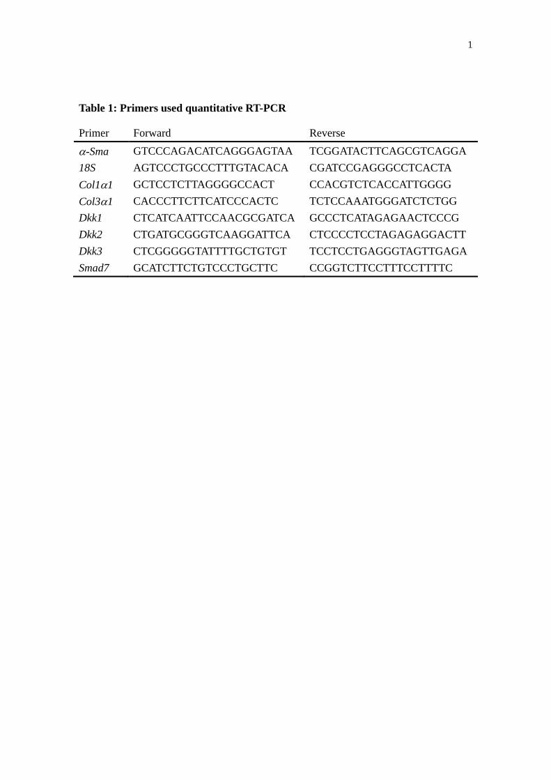
polymerase chain reaction (qRT-PCR) with gene-specific PCR primers (Table 1) and
SYBR Green I Master reaction mix on a LightCycler 480 II (Roche Diagnostics, Basel,
Switzerland). We quantified the relative abundance of each gene product against 18S as
internal controls with the LightCycler 480 software (ver. 1.5).
2.6. Immunoblot analysis
We homogenized and sonicated cells or tissues in lysis buffer (50 mM Tris-HCl,
2% sodium dodecyl sulfate, and 10% glycerol), and centrifuged the homogenates at
15,000 × g for 10 min. After measuring the protein concentration and adding
bromophenol blue (final concentration, 0.1%) and 2-mercaptoethanol (5%), each sample
was separated by sodium dodecyl sulfate-polyacrylamide gel electrophoresis
(SDS-PAGE), transferred onto a polyvinylidene fluoride (PVDF) membrane (Hybond-P,
GE Healthcare), and probed with primary antibodies (anti-Dkk2, anti-desmin,
11
anti-FLAG M2; SIGMA-Aldrich, St. Louis, MO; anti-albumin, BETHYL; Montgomery,
TX, anti-PECAM-1, anti--actin; Santa Cruz Biotechnology, Santa Cruz, CA). After
incubation with horseradish peroxidase-conjugated secondary antibodies (Santa Cruz
Biotechnology) and chemiluminescence reaction with the ECL kit (GE Healthcare), we
quantified the luminescence using an image analyzer, LAS-4000 (FujiFilm, Tokyo,
Japan).
2.7. Construction of an expression plasmid for FLAG-Dkk2
We amplified the coding region of a mouse Dkk2 cDNA clone from the FANTOM
collection (RIKEN) by PCR with a set of oligonucleotide primers, i.e.,
5′-GAGAGAAGCTTTCACAGCTAGGCAGCTCG-3′ and
5′-CGGGATCCTCAGATCTTCTGGCATAC-3′. The HindIII-BamHI-digested fragment
was inserted in-frame into the cognate sites of an expression plasmid, pFLAG-CMV1,
(SIGMA-Aldrich) and the DNA sequence was confirmed. The FLAG-tag of this vector
is preceded by a signal sequence for secretion and a signal peptidase cleavage site. We
used the empty pFLAG-CMV1 plasmid as a control.
2.8. LX-2 cell culture, transfection, and FLAG-Dkk2 conditioned medium

12
We cultured 5.0 105 LX-2 cells [17] (a generous gift from Dr. Friedman and Dr.
Ikeda) per 60-mm dish in DMEM supplemented with 10% FBS. After about 24 h, we
transfected the cells with the FLAG-Dkk2 or empty plasmid using FuGENE6 (Roche
Diagnostics). We collected the culture supernatant (conditioned medium) containing
recombinant FLAG-Dkk2 or FLAG-tag ~72 h after transfection.
2.9. Immunoprecipitation
We incubated the culture supernatant of the transfected LX-2 cells with
anti-FLAG M2 antibody (SIGMA-Aldrich), followed by adsorption with Protein G
Sepharose beads (GE Healthcare) for 2 h. After washing the beads 3 times with TNE
buffer (10 mM Tris-HCl at pH 7.8, 1% NP40, 0.15 M NaCl, and 1 mM EDTA), we
subjected the immunoprecipitates to immunoblot analysis as described above (section
2.6.).
2.10. Luciferase assay
As described above (section 2.8.), we seeded 5.0 104 LX-2 cells into each well
of 12-well plates and co-transfected the cells with the reporter plasmid TOP-Flash
(Upstate; Millipore), which is designed to monitor the Lef/Tcf-mediated transactivation

13
of firefly luciferase (emission, 556 nm) [18], and the pGL4.74 [hRluc/TK] vector
(Promega, Madison, WI) encoding Renilla luciferase (emission, 480 nm) to evaluate the
transfection efficiency. After 24 h, we added the conditioned medium (final
concentration, >90%) containing FLAG-tagged mouse Dkk2 or FLAG-tag alone, or
recombinant Dkk2 (nominal concentration, 5 g/ml [184 nM]; R&D SYSTEMS, Inc.,
Minneapolis, MN). At 48 h after addition of the conditioned medium, we measured the
activities of the 2 luciferases expressed in LX-2 cells using a Dual-Luciferase Reporter
Assay System (Promega) and a Fluoroskan Ascent FL (Thermo Scientific, Waltham,
MA), according to the manufacturers’ instructions. We normalized the data by dividing
the luminescence of the firefly luciferase by that of the Renilla luciferase. The
experiments were performed in triplicate.
2.11. Immunohistochemistry of human liver tissues
We procured human tissue specimens of livers afflicted with chronic hepatitis,
liver fibrosis, or liver cirrhosis from the Department of Surgery, Kyoto University
Hospital. All samples were obtained with the patients’ informed consent. We
immunostained paraffin sections with anti-Dkk2 antibody (Abcam, Cambridge, UK) as
described previously [5, 14] and observed them under a digital microscope (BZ-9000;

14
KEYENCE, Japan).
2.12. Statistical analysis
Quantitative data are expressed as mean ± SEM. For statistical analysis, we used
Mann–Whitney’s U test and a general linear model with repeated measures.

15
3. Results
3.1. Comparative transcriptome analysis revealed significant reduction of Dkk2
expression in Sept4−/−
HSCs
To investigate the molecular mechanism underlying the accelerated
transdifferentiation of Sept4−/−
HSCs [5], we compared the expression profiles of
Sept4+/+
and Sept4−/−
HSCs cultured in vitro for 3 days (DIV3). DNA microarray
analysis showed that the 2 expression profiles did not show a marked difference, but
several genes were expressed aberrantly in Sept4−/−
HSCs (Fig. 1, Tables 2A and 2B,
Suppl. Tables 2A, 2B). Among the genes whose differential expression was confirmed
by qRT-PCR (see below), we focused on Dkk2 whose expression was significantly
reduced in Sept4−/−
HSCs. Dkk2 is a member of the Dkk gene family (Dkk1-5), each
encoding a secretory Wnt antagonist that interacts with the Wnt-Fzd complex by
binding to and internalizing LRP5/6, a co-receptor of Fzd [10-12]. Previous studies
showed that some Dkks are implicated in the negative regulation of Wnt-mediated cell
proliferation, inflammation, and/or pro-fibrotic reactions in diverse tissues, including
the liver [8, 9, 19].
3.2. Loss of Sept4 suppresses upregulation of Dkk1, 2, 3 in an in vivo model of
16
hepatitis/fibrosis
We tested whether the reduced Dkk2 expression in cultured Sept4−/−
HSCs is an in
vitro artifact or a pathologically relevant recapitulation of a molecular event during
hepatitis and fibrosis. We applied the CCl4 model of hepatitis/fibrosis to Sept4−/− and
Sept4+/+
mice as reported previously [5], and quantified the expression levels of 3 major
Dkk genes expressed in the liver by qRT-PCR. While the basal expression levels of
Dkk1–3 were comparable between the 2 genotypes (controls in Fig. 2A-C), their
upregulation after CCl4-induced liver damage was diminished in the absence of Sept4.
Concordantly, the reduction of Dkk2 upregulation in Sept4−/− liver was observed at the
protein level (Fig. 2D). Since Dkks are responsible for the suppressive modulation of
Wnt-mediated inflammatory/fibrotic processes [8], their reduction in Sept4–/–
liver may
contribute to the enhanced pathological reaction shown previously [5].
3.3. HSCs are the major source of Dkk1–3 in the liver
To identify the origin of upregulated Dkk1–3 mRNA in vivo, we fractionated
wildtype mouse liver cells into 4 populations (i.e., hepatocytes, HSCs, endothelial cells
[ECs,] and KCs) and measured the expression of Dkk1–3 at DIV3 by qRT-PCR. Dkk1–3
were almost exclusively expressed in HSCs, indicating that HSCs are the major source

17
of Dkk1–3 secreted in the liver, but hepatocytes and the other cell types are not (Fig.3
A–C, and data not shown). Concordantly, immunoblot analysis showed that Dkk2
protein was almost exclusively expressed in HSCs, but not in hepatocytes, ECs, and
KCs (Fig. 3D). Since Sept4 is also expressed exclusively in HSCs in the liver [5], the
diminished upregulation of Dkk1–3 is likely to be a cell-autonomous, if not a direct,
consequence of the loss of Sept4.
3.4. Loss of Sept4 suppresses robust upregulation of Dkk2/3 through
myofibroblastic transformation of HSCs in vitro
So far, we have demonstrated that the upregulation of Dkk1–3 is an HSC-specific,
pathologically relevant event that may antagonize the inflammatory/fibrotic processes
mediated by the Wnt pathway. To test whether the 3 major Dkk genes are differentially
regulated during myofibroblastic transformation, we examined the time course of their
expression in HSCs cultured in vitro for 1–5 days by qRT-PCR (Fig. 4). Consistent with
the in vivo results (Fig. 2), Dkk2 and Dkk3 were remarkably upregulated in this in vitro
model, and more importantly, the upregulation was significantly diminished in Sept4–/–
HSCs.
18
3.5. Dkk2 interferes with the canonical Wnt pathway in human HSC-derived cells
Because Dkks are secretory proteins that interact with the Wnt–Fzd complex [7,
20], it is important to test whether or not Dkks produced by HSCs can act on HSCs in
an autocrine/paracrine manner. Since primary cultured HSCs are unsuitable for the
following assay which requires a high transfection efficiency, we employed a human
HSC-derived cell line, LX-2 [17]. We transfected LX-2 cells either with a FLAG-tagged
mouse Dkk2 expression plasmid or with an empty (FLAG-tag alone) plasmid, which
served as a control. The culture supernatant from each condition was collected 3 days
after transfection, immunoprecipitated with an anti-FLAG M2 antibody, and
immunoblotted for Dkk2. The immunoblot showed a 28-kDa polypeptide for
FLAG-Dkk2 secreted into the medium (Fig. 5A).
To test whether Dkk2 could interfere with the canonical Wnt signaling pathway in
HSCs, we transfected LX-2 cells with a TOP-Flash reporter plasmid, which is designed
to monitor the activity of the canonical Wnt pathway through Lef/Tcf-mediated
luciferase gene expression [18]. The reporter-transfected LX-2 cells were supplemented
with the aforementioned conditioned medium with or without FLAG-Dkk2. Compared
with the control medium, the FLAG-Dkk2-containing medium significantly suppressed
the luciferase activity of LX-2 cells measured 2 days later (Fig. 5B). Taken together,
19
these data indicate that Dkks secreted from HSCs act on HSCs in an autocrine/paracrine
manner and interfere with the canonical Wnt pathway, which is known to drive
myofibroblastic transformation [9]. However, these data do not exclude the possibility
that Dkks suppress the non-canonical Wnt signaling pathways in HSCs or act on other
cell types in the liver.
3.6. Dkk2 interferes with pro-fibrotic transformation of Sept4–/–
HSCs
On the basis of the above results, we examined whether supplementing exogenous
Dkk2 could mitigate the accelerated fibrotic transformation of Sept4–/–
HSCs. We
isolated HSCs from Sept4–/–
mice and cultured them with FLAG-Dkk2-containing or
control medium. After 3 days, we measured the expression of representative markers by
qRT-PCR: the pro-fibrotic genes -Sma, Col 1, and Col31 (the major collagen genes
in the liver) and an anti-fibrotic gene, Smad7, which encodes an inhibitor of the
transforming growth factor (TGF)- /Smad3 pathway. The FLAG-Dkk2-containing
medium downregulated the expression of -Sma, Col11, and Col31, and upregulated
the expression of Smad7 in Sept4–/–
HSCs (Fig. 6). Concordantly, one-shot
supplementation of recombinant Dkk2 (nominal concentration, 5 g/ml [184 nM]) gave
similar but less significant results, partly due to its low actual potency and/or rapid

20
clearance (Suppl. Fig 1). These data indicate that continuous supplementation with
Dkk2 can partially suppress the myofibroblastic transformation of Sept4–/–
HSCs by
interfering with the canonical Wnt signaling pathway that transactivates pro-fibrotic
genes and suppresses the expression of anti-fibrotic genes.
3.7. Status of Dkk2 in human HSCs under pathological conditions
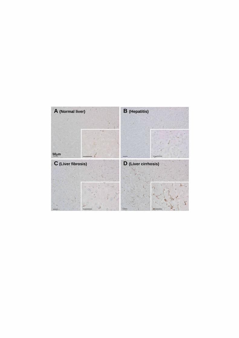
We assessed the clinical relevance of our findings by determining the status of
Dkk2 in pathological human liver specimens using immunohistochemistry. There was a
gradual increment of Dkk2-immunoreactivity from normal liver tissue, hepatitis, mild
fibrosis through to cirrhosis (Fig. 7A–D). Dkk2-positive cells were detected almost
exclusively in the perisinusoidal spaces where HSCs reside, and appeared to increase in
number as fibrosis progressed. These data conform to the hypothesis that the
upregulation of Dkk2 in HSCs is a common counteractive mechanism against fibrotic
processes in liver diseases.
21
4. Discussion
This study derived from an open question raised by our previous findings [5],
which included the determination of the mechanism that renders Sept4–/–
mice prone to
liver fibrosis. Genome-wide expression profiling and qRT-PCR analysis revealed that a
number of genes in HSCs are expressed differently in the absence of Sept4. We
hypothesized that these include genes responsible for the fibrosis-prone phenotype. In
this report, we focused on the reduced expression of the anti-fibrotic genes Dkk1–3 in
Sept4–/–
HSCs. Supplementation with recombinant Dkk2 partially corrected the aberrant
gene expression profile in Sept4–/–
HSCs by downregulating the pro-fibrotic genes
-Sma, Col11 and Col31 and upregulating the anti-fibrotic gene Smad7 (Fig. 6,
Suppl. Fig. 1). These data indicate that the scarcity of Dkks is a major, if not the sole,
determinant of the fibrosis-prone phenotype of Sept4–/–
mice, partly resolving the initial
question. Other differentially expressed genes include downstream targets of the
canonical Wnt pathway (Suppl. Table 2A and B), although their contribution has not
been assessed in this study.
The molecular network of Wnt signaling consists of 3 major pathways, i.e., the
canonical Wnt/-catenin pathway, the Wnt/Ca2+
pathway, and the planar cell
polarity–convergent extension pathway. The canonical Wnt pathway plays a critical role

22
in developmental morphogenesis and homeostatic tissue remodeling by controlling cell
proliferation and differentiation [7, 11]. Hence, dysregulation of this pathway is known
to cause cancer [21], aberrant tissue remodeling [22, 23], and fibrosis [6]. This pathway
is activated when Wnt binds to its cognate receptor Fzd, which interacts with the
glycogen synthase kinase-3 -mediated phosphorylation and the subsequent degradation
of -catenin. When -catenin translocates to the nucleus, it activates the transcription
factor Lef/Tcf and alters the gene expression pattern. Dkks interact with the canonical
Wnt pathway by binding to and facilitating the internalization of LRP5/6, a co-receptor
of Fzd [10]. Among the 3 major Dkk family members expressed in HSCs, Dkk1
consistently antagonizes the canonical Wnt pathway, while Dkk2 can be either an
antagonist or an agonist, depending on the presence or absence of another Wnt
co-receptor, Kremen1/2 [24]. The expression of Kremen1/2 in HSCs (Suppl. Fig. 2) is
consistent with the antagonistic function of Dkk2 in the canonical Wnt pathway (Fig. 5).
Thus, Dkk1/2 may antagonize the canonical Wnt pathway in HSCs, though the function
of Dkk3 is currently unclear.
Open questions also include the molecular mechanism that links loss of Sept4 and
reduced transactivation of the Dkk genes (Fig. 8). Since Sept4 is a subunit of the septin
heteropolymers associated mainly with other cytoskeletal and submembranous
23
components (see Fig. 1 [5]), its direct interaction with the transcription machinery is
unlikely. In some human cell lines, however, depletion of the Sept7 subunit can release
suppressor of cytokine signaling 7 (SOCS7) from septin heteropolymers in the
cytoplasm, which promotes the translocation of the non-catalytic region of tyrosine
kinase adaptor protein (NCK) to the nucleus and activates the DNA damage checkpoint
machinery [25]. Thus, genetic loss of Sept4 could also indirectly alter nuclear events.
Another scenario similar to the scarcity of the dopamine transporter in Sept4−/−
dopamine neurons [3] is that some proteins responsible for anti-fibrotic signaling could
be destabilized by the loss of septin-based scaffolds in Sept4−/−
HSCs. Narrowing down
the molecular mechanism from these and other possibilities should give us a clearer
picture of myofibroblastic transformation of HSCs in liver fibrosis.
Supplementary materials related to this article can be found online 412 at
doi:10.1016/j.bbadis.2011.06.015.
24
5. Acknowledgements
We are grateful to Dr. K. Ikeda (Nagoya City University) and Dr. S.L. Friedman
(Mount Sinai School of Medicine) for the generous gift of LX-2, Dr. Y. Wakamatsu
(Tohoku University) for the TOP-Flash plasmid, and Ms. A. Tanigaki and Ms. R.
Hikawa for technical assistance.
6. Grants
This study was supported in part by grants-in-aid from MEXT of Japan.

25
References
[1] S.L. Friedman, Hepatic fibrosis - overview, Toxicology 254 (2008) 120-129.
[2] S.L. Friedman, Mechanisms of hepatic fibrogenesis, Gastroenterology 134
(2008) 1655-1669.
[3] M. Ihara, N. Yamasaki, A. Hagiwara, A. Tanigaki, A. Kitano, R. Hikawa, H.
Tomimoto, M. Noda, M. Takanashi, H. Mori, N. Hattori, T. Miyakawa, M.
Kinoshita, Sept4, a component of presynaptic scaffold and Lewy bodies, is
required for the suppression of alpha-synuclein neurotoxicity, Neuron 53 (2007)
519-533.
[4] M. Ihara, A. Kinoshita, S. Yamada, H. Tanaka, A. Tanigaki, A. Kitano, M. Goto,
K. Okubo, H. Nishiyama, O. Ogawa, C. Takahashi, S. Itohara, Y. Nishimune, M.
Noda, M. Kinoshita, Cortical organization by the septin cytoskeleton is essential
for structural and mechanical integrity of mammalian spermatozoa, Dev Cell 8
(2005) 343-352.
[5] K. Iwaisako, E. Hatano, K. Taura, A. Nakajima, M. Tada, S. Seo, N. Tamaki, F.
Sato, I. Ikai, S. Uemoto, M. Kinoshita, Loss of Sept4 exacerbates liver fibrosis
through the dysregulation of hepatic stellate cells, J Hepatol 49 (2008) 768-778.
[6] I. Hwang, E.Y. Seo, H. Ha, Wnt/beta-catenin signaling: a novel target for

26
therapeutic intervention of fibrotic kidney disease, Arch Pharm Res 32 (2009)
1653-1662.
[7] C. Prunier, B.A. Hocevar, P.H. Howe, Wnt signaling: physiology and pathology,
Growth Factors 22 (2004) 141-150.
[8] J.H. Cheng, H. She, Y.P. Han, J. Wang, S. Xiong, K. Asahina, H. Tsukamoto,
Wnt antagonism inhibits hepatic stellate cell activation and liver fibrosis, Am J
Physiol Gastrointest Liver Physiol 294 (2008) G39-49.
[9] S.J. Myung, J.H. Yoon, G.Y. Gwak, W. Kim, J.H. Lee, K.M. Kim, C.S. Shin, J.J.
Jang, S.H. Lee, S.M. Lee, H.S. Lee, Wnt signaling enhances the activation and
survival of human hepatic stellate cells, FEBS Lett 581 (2007) 2954-2958.
[10] Y. Kawano, R. Kypta, Secreted antagonists of the Wnt signalling pathway, J Cell
Sci 116 (2003) 2627-2634.
[11] W. Wu, A. Glinka, H. Delius, C. Niehrs, Mutual antagonism between dickkopf1
and dickkopf2 regulates Wnt/beta-catenin signalling, Curr Biol 10 (2000)
1611-1614.
[12] A. Niida, T. Hiroko, M. Kasai, Y. Furukawa, Y. Nakamura, Y. Suzuki, S. Sugano,
T. Akiyama, DKK1, a negative regulator of Wnt signaling, is a target of the
beta-catenin/TCF pathway, Oncogene 23 (2004) 8520-8526.
27
[13] R. Weiskirchen, A.M. Gressner, Isolation and culture of hepatic stellate cells,
Methods Mol Med 117 (2005) 99-113.
[14] N. Tamaki, E. Hatano, K. Taura, M. Tada, Y. Kodama, T. Nitta, K. Iwaisako, S.
Seo, A. Nakajima, I. Ikai, S. Uemoto, CHOP deficiency attenuates
cholestasis-induced liver fibrosis by reduction of hepatocyte injury, Am J
Physiol Gastrointest Liver Physiol 294 (2008) G498-505.
[15] B.A. Centeno, S.A. Enkemann, D. Coppola, S. Huntsman, G. Bloom, T.J.
Yeatman, Classification of human tumors using gene expression profiles
obtained after microarray analysis of fine-needle aspiration biopsy samples,
Cancer 105 (2005) 101-109.
[16] B.M. Bolstad, R.A. Irizarry, M. Astrand, T.P. Speed, A comparison of
normalization methods for high density oligonucleotide array data based on
variance and bias, Bioinformatics 19 (2003) 185-193.
[17] L. Xu, A.Y. Hui, E. Albanis, M.J. Arthur, S.M. O'Byrne, W.S. Blaner, P.
Mukherjee, S.L. Friedman, F.J. Eng, Human hepatic stellate cell lines, LX-1 and
LX-2: new tools for analysis of hepatic fibrosis, Gut 54 (2005) 142-151.
[18] D. Sakai, Y. Tanaka, Y. Endo, N. Osumi, H. Okamoto, Y. Wakamatsu, Regulation
of Slug transcription in embryonic ectoderm by beta-catenin-Lef/Tcf and

28
BMP-Smad signaling, Dev Growth Differ 47 (2005) 471-482.
[19] M.D. Thompson, S.P. Monga, WNT/beta-catenin signaling in liver health and
disease, Hepatology 45 (2007) 1298-1305.
[20] A.M. Zorn, Wnt signalling: antagonistic Dickkopfs, Curr Biol 11 (2001)
R592-595.
[21] B.T. MacDonald, K. Tamai, X. He, Wnt/beta-catenin signaling: components,
mechanisms, and diseases, Dev Cell 17 (2009) 9-26.
[22] R. Baron, G. Rawadi, Targeting the Wnt/beta-catenin pathway to regulate bone
formation in the adult skeleton, Endocrinology 148 (2007) 2635-2643.
[23] X. Li, P. Liu, W. Liu, P. Maye, J. Zhang, Y. Zhang, M. Hurley, C. Guo, A.
Boskey, L. Sun, S.E. Harris, D.W. Rowe, H.Z. Ke, D. Wu, Dkk2 has a role in
terminal osteoblast differentiation and mineralized matrix formation, Nat Genet
37 (2005) 945-952.
[24] B. Mao, C. Niehrs, Kremen2 modulates Dickkopf2 activity during Wnt/LRP6
signaling, Gene 302 (2003) 179-183.
[25] B.E. Kremer, L.A. Adang, I.G. Macara, Septins regulate actin organization and
cell-cycle arrest through nuclear accumulation of NCK mediated by SOCS7, Cell 130
(2007) 837-850.

29
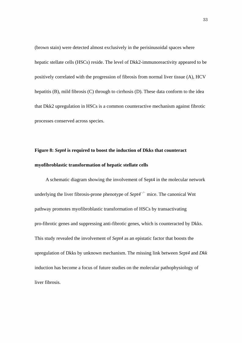
Figure legends
Figure 1: Comparative genome-wide expression profiling between Sept4+/+
and
Sept4−/−
hepatic stellate cells
Two-dimensional scatter plot showing comparative transcriptome data obtained
using a DNA microarray system (23,474 probes; Mouse Oligo chip, Toray, Tokyo,
Japan). Each dot represents the amount of mRNA obtained from Sept4+/+
hepatic
stellate cells (HSCs) (horizontal axis) and Sept4−/−
HSCs (vertical axis) on a log2-based
scale. The quality of mRNA and the reproducibility of these experiments are
corroborated by compact clustering of the dots along the diagonal. The results were
confirmed by triplicate measurements. The genes that prominently deviated from the
diagonal are listed in Tables 2A and 2B. This study focused on Dkk2 whose reduced
expression in Sept4−/−
HSCs was confirmed by quantitative reverse transcription
polymerase chain reaction (qRT-PCR) (see below).
Figure 2: Genetic loss of Sept4 affects induced expression patterns of Dkk genes in
an in vivo model of hepatitis and liver fibrosis
(A–C) Quantitative reverse transcription polymerase chain reaction (RT-PCR)
showed that the basal expression level of the 3 major Dkk genes in the liver, Dkk1-3,
were comparable between Sept4+/+
and Sept4−/−
hepatic stellate cells (HSCs), whereas
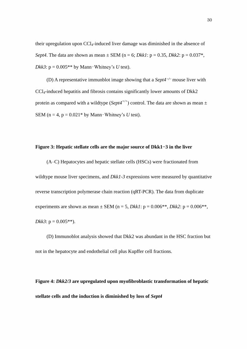
30
their upregulation upon CCl4-induced liver damage was diminished in the absence of
Sept4. The data are shown as mean ± SEM (n = 6; Dkk1: p = 0.35, Dkk2: p = 0.037*,
Dkk3: p = 0.005** by Mann–Whitney’s U test).
(D) A representative immunblot image showing that a Sept4−/− mouse liver with
CCl4-induced hepatitis and fibrosis contains significantly lower amounts of Dkk2
protein as compared with a wildtype (Sept4+/+
) control. The data are shown as mean ±
SEM (n = 4, p = 0.021* by Mann–Whitney’s U test).
Figure 3: Hepatic stellate cells are the major source of Dkk1−3 in the liver
(A–C) Hepatocytes and hepatic stellate cells (HSCs) were fractionated from
wildtype mouse liver specimens, and Dkk1-3 expressions were measured by quantitative
reverse transcription polymerase chain reaction (qRT-PCR). The data from duplicate
experiments are shown as mean ± SEM (n = 5, Dkk1: p = 0.006**, Dkk2: p = 0.006**,
Dkk3: p = 0.005**).
(D) Immunoblot analysis showed that Dkk2 was abundant in the HSC fraction but
not in the hepatocyte and endothelial cell plus Kupffer cell fractions.
Figure 4: Dkk2/3 are upregulated upon myofibroblastic transformation of hepatic
stellate cells and the induction is diminished by loss of Sept4

31
The time course of Dkk1−3 expression in hepatic stellate cells (HSCs) cultured for
1−5 days was estimated by quantitative reverse transcription polymerase chain reaction
(qRT-PCR). The remarkable upregulation of Dkk2 and Dkk3 found in Sept4+/+
HSCs
was significantly diminished in Sept4−/−
HSCs. Data from duplicate experiments are
shown as mean ± SEM (Sept4+/+
: n = 6, Sept4−/−: n = 4; Dkk1: p = 0.49, Dkk2: p <
0.020*, Dkk3: p = 0.057 by a general linear model with repeated measures).
Figure 5: Dkk2 can interfere with the canonical Wnt pathway of human hepatic
stellate cell-derived LX-2 cells
(A) FLAG-tagged Dkk2 expressed in LX-2 cells and released to the culture
supernatant was immunoprecipitated with anti-FLAG M2 antibody and immunoblotted
for Dkk2.
(B) LX-2 cells transfected with the TOP-Flash reporter plasmid were incubated
with the conditioned medium containing FLAG-Dkk2 or FLAG alone and analyzed in a
dual luciferase assay. FLAG-Dkk2 significantly suppressed the transactivation of the
reporter gene encoding firefly luciferase, indicative of its interaction with the canonical
Wnt pathway. Data from duplicate experiments are shown as mean ± SEM (n = 5, p =
0.033*).

32
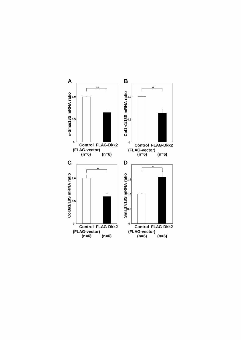
Figure 6: Dkk2 interferes with myofibroblastic transformation of Sept4−/−
hepatic
stellate cells
Sept4−/−
hepatic stellate cells (HSCs) were supplemented with FLAG-Dkk2, and
the expressions of representative pro-fibrotic and anti-fibrotic markers were measured
by quantitative reverse transcription polymerase chain reaction (qRT-PCR). The
pro-fibrotic markers -Sma (p = 0.001), Col11 (p = 0.005), and Col31 (p = 0.001)
were significantly downregulated in the presence of FLAG-Dkk2, while Smad7 (p =
0.011) was significantly upregulated. Data from duplicate experiments are shown as
mean ± SEM (n = 6, *: p < 0.05, **: p < 0.01). In conjunction with the above results
(Fig. 5), these data indicate that supplementation with Dkk2 can mitigate
myofibroblastic transformation of Sept4−/−
HSCs by interaction with the canonical Wnt
signaling pathway that transactivates pro-fibrotic genes and suppresses the expression
of anti-fibrotic genes.
Figure 7: Dkk2 is upregulated in human hepatic stellate cells under pathological
conditions
Immunohistochemistry of Dkk2 in human liver specimens. Dkk2-positive cells
33
(brown stain) were detected almost exclusively in the perisinusoidal spaces where
hepatic stellate cells (HSCs) reside. The level of Dkk2-immunoreactivity appeared to be
positively correlated with the progression of fibrosis from normal liver tissue (A), HCV
hepatitis (B), mild fibrosis (C) through to cirrhosis (D). These data conform to the idea
that Dkk2 upregulation in HSCs is a common counteractive mechanism against fibrotic
processes conserved across species.
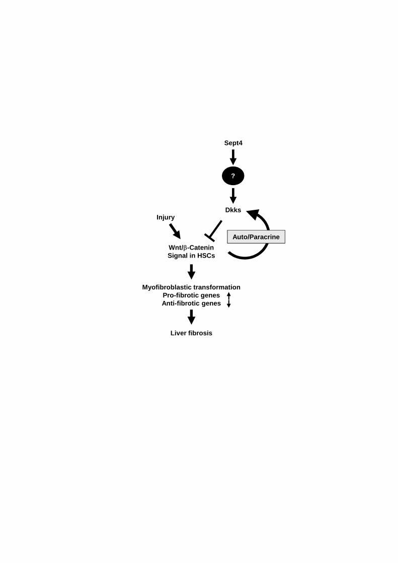
Figure 8: Sept4 is required to boost the induction of Dkks that counteract
myofibroblastic transformation of hepatic stellate cells
A schematic diagram showing the involvement of Sept4 in the molecular network
underlying the liver fibrosis-prone phenotype of Sept4−∕−
mice. The canonical Wnt
pathway promotes myofibroblastic transformation of HSCs by transactivating
pro-fibrotic genes and suppressing anti-fibrotic genes, which is counteracted by Dkks.
This study revealed the involvement of Sept4 as an epistatic factor that boosts the
upregulation of Dkks by unknown mechanism. The missing link between Sept4 and Dkk
induction has become a focus of future studies on the molecular pathophysiology of
liver fibrosis.
Sept4+/+(Log2)
Sep
t4-/
- (Lo
g2)
Dkk20 2 4 6 8 10 12 14 16 18
2
0
18
16
14
12
10
8
6
4 Sept4

Dkk
1/18
Sm
RN
A r
atio
Dkk
2/18
Sm
RN
A r
atio
Dkk
3/18
Sm
RN
A r
atio
Control CCl4Control CCl4 Control CCl4
Sept4+/+
Sept4-/-
A B C
1
2
0 0
2
4
1.0
0.5
0
*
(n=6) (n=6) (n=6) (n=6) (n=6) (n=6)
Sept4+/+
Sept4-/-
Sept4+/+
Sept4-/-
**1.5
Dkk2
β-Actin
MW(kDa)
28
42
DSept4+/+ Sept4-/-
*
Dkk
2/β-
Act
in r
atio
(a.u
.)
(n=4) (n=4)
CCl4-induced fibrotic liver
1.0
0
0.5

A B C
0.8
0.4
0
0.6
0.4
0.2
0 0
10
20
****
**
Dkk
1/18
Sm
RN
A r
atio
Dkk
2/18
Sm
RN
A r
atio
Dkk
3/18
Sm
RN
A r
atio
(n=5) (n=6) (n=5) (n=6) (n=5) (n=6)
D
Dkk2
Desmin
Albumin
CD31
MW(kDa)
28
55
66
131
42β-Actin

Dkk
1/18
Sm
RN
A r
atio
Dkk
2/18
S m
RN
A r
atio
Dkk
3/18
S m
RN
A r
atio
A B C
0
2
41.0
0.5
0 0
10
20
DIV1 DIV3 DIV5 DIV1 DIV3 DIV5 DIV1 DIV3 DIV5
61.5
Sept4+/+
Sept4-/-
Sept4+/+
Sept4-/-
Sept4+/+
Sept4-/-30
(Sept4+/+:n=6, Sept4-/-:n=4)
*

Control(FLAG-vector)
FLAG-Dkk2
B
Lu
min
esce
nce
(R
LU
)
Dkk2(28kDa)
A
*
0
0.5
1.0
(n=5) (n=5)
α-S
ma/
18S
mR
NA
rat
io
Co
l1α
1/18
S m
RN
A r
atio
Co
l3a1
/18S
mR
NA
rat
io
Sm
ad7/
18S
mR
NA
rat
io
Control(FLAG-vector)
FLAG-Dkk2
A B
C D
**
Control(FLAG-vector)
FLAG-Dkk2
Control(FLAG-vector)
FLAG-Dkk2 Control(FLAG-vector)
FLAG-Dkk2
0
0.5
1.0
0
0.5
1.0
**
0
0.5
1.0
0
0.5
1.0
** *
1.5
(n=6) (n=6) (n=6) (n=6)
(n=6) (n=6) (n=6) (n=6)
A (Normal liver)
50µm
B (Hepatitis)
C (Liver fibrosis) D (Liver cirrhosis)
InjuryDkks
Liver fibrosis
Sept4
Myofibroblastic transformationPro-fibrotic genesAnti-fibrotic genes
Wnt/β-CateninSignal in HSCs
Auto/Paracrine
?
1
Table 1: Primers used quantitative RT-PCR
Primer Forward Reverse
-Sma GTCCCAGACATCAGGGAGTAA TCGGATACTTCAGCGTCAGGA
18S AGTCCCTGCCCTTTGTACACA CGATCCGAGGGCCTCACTA
Col11 GCTCCTCTTAGGGGCCACT CCACGTCTCACCATTGGGG
Col31 CACCCTTCTTCATCCCACTC TCTCCAAATGGGATCTCTGG
Dkk1 CTCATCAATTCCAACGCGATCA GCCCTCATAGAGAACTCCCG
Dkk2 CTGATGCGGGTCAAGGATTCA CTCCCCTCCTAGAGAGGACTT
Dkk3 CTCGGGGGTATTTTGCTGTGT TCCTCCTGAGGGTAGTTGAGA
Smad7 GCATCTTCTGTCCCTGCTTC CCGGTCTTCCTTTCCTTTTC
2
Table 2A: Genes downregulated in Sept4−/−
hepatic stellate cells (10 from the top)
Gene
Symbol
Control (Sept4+/+
)
(Log2)
Sept4−/−
(Log2)
Fold
Change Gene Annotation
1 Dkk2 4.79 0.820 0.0640 dickkopf homolog 2
2 Ivl 6.22 2.93 0.102 involucrin
3 Sept4 7.24 4.02 0.107 septin 4
4 Ptgs2 10.9 7.88 0.127 prostaglandin-endoperoxide synthase 2
5 Acta1 6.04 3.06 0.127 actin, alpha 1, skeletal muscle
6 Tll1 7.72 4.76 0.128 tolloid-like
7 Kctd11 3.46 0.556 0.133 potassium channel tetramerisation
domain containing 11
8 Sprrl9 5.72 2.82 0.135 small proline rich-like 9
9 Ahrr 4.77 1.90 0.137 aryl-hydrocarbon receptor repressor
10 Kcnj5 3.17 0.317 0.139 potassium inwardly-rectifying channel J5
Table 2B: Genes upregulated in Sept4−/−
hepatic stellate cells (10 from the top)
Gene
Symbol
Control (Sept4+/+
)
(Log2)
Sept4−/−
(Log2)
Fold
Change Gene Annotation
1 Nfat5 0.838 4.60 13.5 nuclear factor of activated T-cells 5
2 Saa3 3.81 7.29 11.2 serum amyloid A 3
3 Gm839 2.75 6.11 10.2 gene model 839, (NCBI)
4 Cuedc1 4.99 8.26 9.70 CUE domain containing 1
5 Gpr160 3.29 6.52 9.38 G protein-coupled receptor 160
6 Gpr103 0.113 3.30 9.12 G protein-coupled receptor 103
7 Ntf3 0.886 4.05 8.98 neurotrophin 3
8 Akp2 7.74 10.9 8.74 alkaline phosphatase 2, liver
9 Slc10a6 1.08 4.14 8.38 solute carrier family 10, member 6
10 Hsd3b2 1.05 4.02 7.85 hydroxysteroid dehydrogenase-2,
delta<5>-3-beta
Legend of Tables 2A and 2B
Comparative DNA microarray data of Sept4−/−
and Sept4+/+
hepatic stellate cells
(HSCs), showing representative genes whose expression were up- (A) and

3
downregulated (B) in Sept4−/−
HSCs. All the data have been deposited to NCBI GEO
under an accession number of GSE24588.

1
Supplemental Figure 1: Marginal suppression of myofibroblastic transformation
of Sept4−/− hepatic stellate cells after supplementation with recombinant Dkk2
Sept4−/− hepatic stellate cells (HSCs) at day 1 in vitro (DIV1) were supplemented
with recombinant Dkk2 (5 g/ml [184 nM]) and the expression of representative
pro-fibrotic and anti-fibrotic markers was measured 3 days later by quantitative reverse
transcription polymerase chain reaction (qRT-PCR). The pro-fibrotic genes -Sma (p =
0.62), Col11 (p = 0.67), and Col31 (p = 0.46) were downregulated in the presence of
recombinant Dkk2, while the anti-fibrotic gene Smad7 (p = 0.47) was upregulated.
Although the differences were not statistically significant, the results were concordant
with the FLAG-Dkk2 conditioned media experiment (Fig. 6). Data from duplicate
experiments are shown as mean ± SEM (n = 6).
Supplemental Figure 2: Expression of Kremen1/2 in cultured hepatic stellate cells
(A, B) The expression of Kremen1/2 genes in Sept4+/+
and Sept4−/−
mouse hepatic
stellate cells (HSCs) at days 1 to 5 in vitro (DIV1−5) was assessed by quantitative
reverse transcription polymerase chain reaction (qRT-PCR). Kremen1 was upregulated
during myofibroblastic transformation, which was slightly blunted in the absence of
Sept4 (p = 0.12, a general linear model with repeated measures). Kremen2 was not
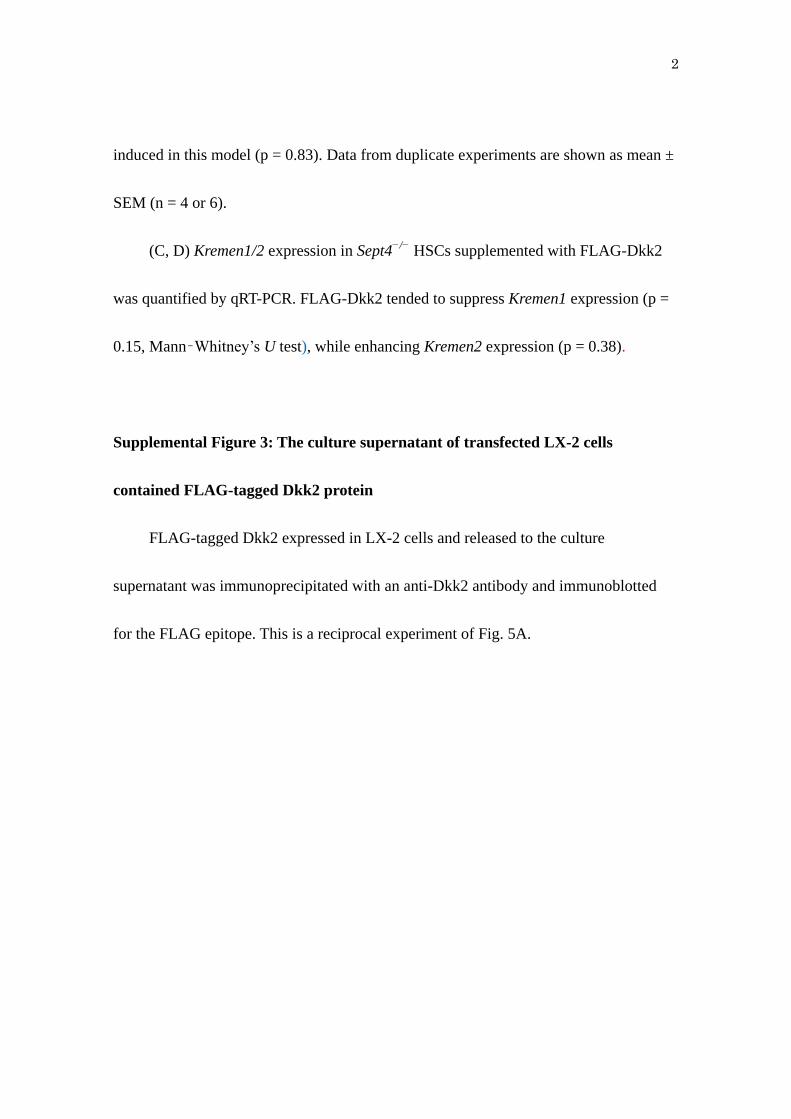
2
induced in this model (p = 0.83). Data from duplicate experiments are shown as mean ±
SEM (n = 4 or 6).
(C, D) Kremen1/2 expression in Sept4−/−
HSCs supplemented with FLAG-Dkk2
was quantified by qRT-PCR. FLAG-Dkk2 tended to suppress Kremen1 expression (p =
0.15, Mann–Whitney’s U test), while enhancing Kremen2 expression (p = 0.38).
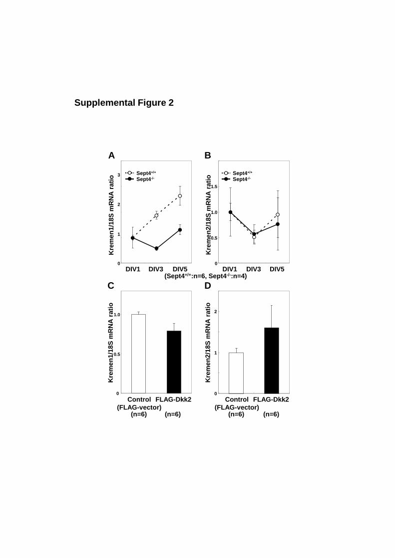
Supplemental Figure 3: The culture supernatant of transfected LX-2 cells
contained FLAG-tagged Dkk2 protein
FLAG-tagged Dkk2 expressed in LX-2 cells and released to the culture
supernatant was immunoprecipitated with an anti-Dkk2 antibody and immunoblotted
for the FLAG epitope. This is a reciprocal experiment of Fig. 5A.

α-S
ma/
18S
mR
NA
rat
io
Control Re-Dkk2
0.5
0
1.0
1.5
Co
l1α
1/18
Sm
RN
A r
atio
0
0.5
1.0
Co
l3α
1/18
Sm
RN
A r
atio
Sm
ad7/
18S
mR
NA
rat
io
Control Re-Dkk2 Control Re-Dkk20
0.5
1.0
0
0.5
1.0
A B
C D
(n=6) (n=6) (n=6) (n=6)
Control Re-Dkk2(n=6) (n=6) (n=6) (n=6)
Supplemental Figure 1
DIV1 DIV3 DIV5
Sept4+/+
Sept4-/-
DIV1 DIV3 DIV5
Sept4+/+
Sept4-/-
1
0
2
3
Kre
men
1/18
Sm
RN
A r
atio
Kre
men
2/18
Sm
RN
A r
atio
0
0.5
1.0
1.5
Kre
men
1/18
Sm
RN
A r
atio
Kre
men
2/18
Sm
RN
A r
atio
Control(FLAG-vector)
FLAG-Dkk2 Control(FLAG-vector)
FLAG-Dkk20
0.5
1.0
0
1
2
A B
C D
(n=6) (n=6) (n=6) (n=6)
(Sept4+/+:n=6, Sept4-/-:n=4)
Supplemental Figure 2
Dkk2(28kDa)
IP:anti-Dkk2 + IB:anti-FLAG M2
Supplemental Figure 3