title new loci and coding variants confer risk for age ... · pdf filetitle new loci and...
TRANSCRIPT

Title New loci and coding variants confer risk for age-relatedmacular degeneration in East Asians
Author(s)
Cheng, Ching Yu; Yamashiro, Kenji; Jia Chen, Li; Ahn,Jeeyun; Huang, Lulin; Huang, Lvzhen; Cheung, Chui Ming G;Miyake, Masahiro; Cackett, Peter D.; Yeo, Ian Y.; Laude,Augustinus; Lee, Ji Eun; Li, Yi; Liu, Jianjun; Teo, Yik Ying;Heng, Chew Kiat; Lim, Tock Han; Yang, Suk Kyun; Song,Kyuyoung; Vithana, Eranga N.; Aung, Tin; Bei, Jin Xin; Zeng,Yi Xin; Tai, E. Shyong; Li, Xiao Xin; Yang, Zhenglin; Park,Kyu Hyung; Pang, Chi Pui; Yoshimura, Nagahisa; Yin Wong,Tien; Khor, Chiea Chuen; Mathur, Ranjana; Pang, Junxiong;Sim, Kar Seng; Koh, Adrian H.; Chen, Peng; Lee, Shu Yen;Wong, Doric; Chan, Choi Mun; Loh, Boon Kwang; Sun,Yaoyao; Davila, Sonia; Nakata, Isao; Nakanishi, Hideo; Akagi-Kurashige, Yumiko; Gotoh, Norimoto; Tsujikawa, Akitaka;Matsuda, Fumihiko; Mori, Keisuke; Yoneya, Shin; Sakurada,Yoichi; Iijima, Hiroyuki; Iida, Tomohiro; Honda, Shigeru; Lai,Timothy Yuk Yau; Tam, Pancy Oi Sin; Chen, Haoyu; Tang,Shibo; Ding, Xiaoyan; Wen, Feng; Lu, Fang; Zhang, Xiongze;Shi, Yi; Zhao, Peiquan; Zhao, Bowen; Sang, Jinghong; Gong,Bo; Dorajoo, Rajkumar; Yuan, Jian Min; Koh, Woon Puay;Van Dam, Rob M.; Friedlander, Yechiel; Lin, Ying; Hibberd,Martin L.; Foo, Jia Nee; Wang, Ningli; Wong, Chang Hua;Tan, Gavin S.; Park, Sang Jun; Bhargava, Mayuri; Gopal,Lingam; Naing, Thet; Liao, Jiemin; Guan Ong, Peng; Mitchell,Paul; Zhou, Peng; Xie, Xuefeng; Liang, Jinlong; Mei, Junpu;Jin, Xin; Saw, Seang Mei; Ozaki, Mineo; Mizoguchi, Takanori;Kurimoto, Yasuo; Woo, Se Joon; Chung, Hum; Yu, HyeongGon; Shin, Joo Young; Park, Dong Ho; Kim, In Taek; Chang,Woohyok; Sagong, Min; Lee, Sang Joon; Kim, Hyun Woong
Citation Nature Communications (2015), 6
Issue Date 2015-01-28
URL http://hdl.handle.net/2433/210236
Right
This work is licensed under a Creative Commons Attribution4.0 International License. The images or other third partymaterial in this article are included in the article’s CreativeCommons license, unless indicated otherwise in the credit line;if the material is not included under the Creative Commonslicense, users will need to obtain permission from the licenseholder to reproduce the material. To view a copy of thislicense, visit http://creativecommons.org/licenses/by/4.0/
Type Journal Article
Kyoto University

Textversion publisher
Kyoto University

ARTICLE
Received 21 May 2014 | Accepted 9 Dec 2014 | Published 28 Jan 2015
New loci and coding variants confer risk forage-related macular degeneration in East AsiansChing-Yu Cheng1,2,3,4,*, Kenji Yamashiro5,*, Li Jia Chen6,*, Jeeyun Ahn7,*, Lulin Huang8,9,*, Lvzhen Huang10,11,12,*, Chui Ming G. Cheung1,4, Masahiro
Miyake5,13, Peter D. Cackett4,14, Ian Y. Yeo4, Augustinus Laude1,15, Ranjana Mathur4, Junxiong Pang16, Kar Seng Sim16, Adrian H. Koh4,17, Peng Chen18,
Shu Yen Lee4, Doric Wong4, Choi Mun Chan4, Boon Kwang Loh4, Yaoyao Sun10,11,12, Sonia Davila3,16, Isao Nakata5,13, Hideo Nakanishi5,
Yumiko Akagi-Kurashige5,13, Norimoto Gotoh5, Akitaka Tsujikawa5, Fumihiko Matsuda13, Keisuke Mori19, Shin Yoneya19, Yoichi Sakurada20,
Hiroyuki Iijima20, Tomohiro Iida21, Shigeru Honda22, Timothy Yuk Yau Lai6, Pancy Oi Sin Tam6, Haoyu Chen6,23, Shibo Tang24,25, Xiaoyan Ding24,
Feng Wen24, Fang Lu8,9, Xiongze Zhang24, Yi Shi8,9, Peiquan Zhao26, Bowen Zhao27, Jinghong Sang27, Bo Gong8,9, Rajkumar Dorajoo16,
Jian-Min Yuan28,29, Woon-Puay Koh2,18, Rob M. van Dam18, Yechiel Friedlander30, Ying Lin8,9, Martin L. Hibberd16, Jia Nee Foo16, Ningli Wang27,
Chang Hua Wong16, Gavin S. Tan4, Sang Jun Park31, Mayuri Bhargava1,3, Lingam Gopal3, Thet Naing3, Jiemin Liao1,3, Peng Guan Ong1, Paul Mitchell32,
Peng Zhou33, Xuefeng Xie34, Jinlong Liang34, Junpu Mei34, Xin Jin34, Seang-Mei Saw1,2,3,18, Mineo Ozaki35, Takanori Mizoguchi36, Yasuo Kurimoto37,
Se Joon Woo31,38, Hum Chung38, Hyeong-Gon Yu38, Joo Young Shin38, Dong Ho Park39, In Taek Kim39, Woohyok Chang40, Min Sagong40,
Sang-Joon Lee41, Hyun Woong Kim42, Ji Eun Lee43,44, Yi Li16, Jianjun Liu16,18, Yik Ying Teo16,18, Chew Kiat Heng45, Tock Han Lim15, Suk-Kyun Yang46,
Kyuyoung Song47, Eranga N. Vithana1,2,3, Tin Aung1,3,4, Jin Xin Bei48,49, Yi Xin Zeng48,49,50, E. Shyong Tai2,18,51,y, Xiao Xin Li10,11,12,y, Zhenglin Yang8,9,y,
Kyu-Hyung Park31,y, Chi Pui Pang6,y, Nagahisa Yoshimura5,y, Tien Yin Wong1,2,3,4 & Chiea Chuen Khor1,16,18
Age-related macular degeneration (AMD) is a major cause of blindness, but presents differently in Europeans and
Asians. Here, we perform a genome-wide and exome-wide association study on 2,119 patients with exudative AMD
and 5,691 controls, with independent replication in 4,226 patients and 10,289 controls, all of East Asian descent, as
part of The Genetics of AMD in Asians (GAMA) Consortium. We find a strong association between CETP Asp442Gly
(rs2303790), an East Asian-specific mutation, and increased risk of AMD (odds ratio (OR)¼ 1.70, P¼ 5.60� 10� 22).
The AMD risk allele (442Gly), known to protect from coronary heart disease, increases HDL cholesterol levels by
0.17 mmol l� 1 (P¼ 5.82� 10� 21) in East Asians (n¼ 7,102). We also identify three novel AMD loci: C6orf223
Ala231Ala (OR¼0.78, P¼ 6.19� 10� 18), SLC44A4 Asp47Val (OR¼ 1.27, P¼ 1.08� 10� 11) and FGD6 Gln257Arg
(OR¼0.87, P¼ 2.85� 10�8). Our findings suggest that some of the genetic loci conferring AMD susceptibility in
East Asians are shared with Europeans, yet AMD in East Asians may also have a distinct genetic signature.
DOI: 10.1038/ncomms7063 OPEN
1 Singapore Eye Research Institute, Singapore 169856, Singapore. 2 Duke-NUS Graduate Medical School, National University of Singapore, Singapore 169857, Singapore. 3 Department of Ophthalmology, NationalUniversity of Singapore and National University Health System, Singapore 119228, Singapore. 4 Singapore National Eye Center, Singapore 168751, Singapore. 5 Department of Ophthalmology and Visual Sciences,Kyoto University Graduate School of Medicine, Kyoto 6068507, Japan. 6 Department of Ophthalmology and Visual Sciences, The Chinese University of Hong Kong, Hong Kong, China. 7 Department ofOphthalmology, Seoul Metropolitan Government Seoul National University Boramae Medical Center, Seoul 156-707, Korea. 8 Sichuan Provincial Key Laboratory for Human Disease Gene Study, Hospital of theUniversity of Electronic Science and Technology of China and Sichuan Provincial People’s Hospital, Chengdu 610072, China. 9 School of Medicine, University of Electronic Science and Technology of China,Chengdu 610072, China. 10 Key Laboratory of Vision Loss and Restoration, Ministry of Education of China, Beijing 100044, China. 11 Beijing Key Laboratory of Diagnosis and Therapy of Retinal and ChoroidDiseases, Beijing 100871, China. 12 Department of Ophthalmology, People’s Hospital, Peking University, Beijing 100871, China. 13 Center for Genomic Medicine/Inserm U.852, Kyoto University Graduate School ofMedicine, Kyoto 6068507, Japan. 14 Princess Alexandra Eye Pavilion, Edinburgh EH3 9HA, UK. 15 National Healthcare Group Eye Institute, Tan Tock Seng Hospital, Singapore 308433, Singapore. 16 Division ofHuman Genetics, Genome Institute of Singapore, Singapore 138672, Singapore. 17 Eye and Retinal Surgeons, Camden Medical Centre, Singapore 248649, Singapore. 18 Saw Swee Hock School of Public Health,National University of Singapore and National University Health System, Singapore 117549, Singapore. 19 Department of Ophthalmology, Saitama Medical University, Iruma 3500495, Japan. 20 Department ofOphthalmology, Faculty of Medicine, University of Yamanashi, Yamanashi 4093898, Japan. 21 Department of Ophthalmology, Tokyo Women’s Medical University Hospital, Tokyo 1628666, Japan. 22 Departmentof Surgery, Division of Ophthalmology, Kobe University Graduate School of Medicine, Kobe 6500017, Japan. 23 Shantou University/Chinese University of Hong Kong Joint Shantou International Eye Center,Shantou 515041, China. 24 Zhongshan Ophthalmic Center, Sun Yat-Sen University, Guangzhou 510060, China. 25 Aier School of Ophthalmology, Central South University, Changsha 410000, China.26 Department of Ophthalmology, Xin Hua Hospital affiliated to Shanghai Jiao Tong University, School of Medicine, Shanghai 200025, China. 27 Beijing Tongren Eye Center, Beijing Tongren Hospital, CapitalMedical University, Beijing Institute of Ophthalmology, Beijing 100730, China. 28 Cancer Control and Population Sciences, University of Pittsburgh Cancer Institute, Pittsburgh, Pennsylvania 15260, USA.29 Department of Epidemiology, Graduate School of Public Health, University of Pittsburgh, Pittsburgh, Pennsylvania 15260, USA. 30 Hebrew University, School of Public Health, Jerusalem 91120, Israel.31 Department of Ophthalmology, Seoul National University Bundang Hospital, Gyeonggi 463-707, Korea. 32 Department of Ophthalmology, University of Sydney and Westmead Millennium Institute, Sydney2145, Australia. 33 Eye and ENT Hospital of Fudan University, Shanghai 200433, China. 34 BGI-Shenzhen, Shenzhen 518083, China. 35 Ozaki Eye Hospital, Miyazaki 8830066, Japan. 36 Mizoguchi Eye Hospital,Nagasaki 8570016, Japan. 37 Department of Ophthalmology, Kobe City General Hospital, Kobe 6500046, Japan. 38 Department of Ophthalmology, Seoul National University Hospital, Seoul National UniversityCollege of Medicine, Seoul 110-744, Korea. 39 Department of Ophthalmology, School of Medicine, Kyungpook National University, Daegu 700-721, Korea. 40 Department of Ophthalmology, Yeungnam UniversityCollege of Medicine, Daegu 705-802, Korea. 41 Department of Ophthalmology, College of Medicine, Kosin University, Pusan 606-701, Korea. 42 Department of Ophthalmology, Pusan Paik Hospital, InjeUniversity College of Medicine, Pusan 614-735, Korea. 43 Department of Ophthalmology, Pusan National University Hospital, Pusan 602-739, Korea. 44 Medical Research Institute, Pusan National University,Pusan 602-739, Korea. 45 Department of Pediatrics, National University Health System and National University of Singapore, Singapore 119228, Singapore. 46 Department of Gastroenterology, Asan MedicalCenter and University of Ulsan College of Medicine, Seoul 138-736, Korea. 47 Department of Biochemistry and Molecular Biology, University of Ulsan College of Medicine, Seoul 138-736, Korea. 48 State KeyLaboratory of Oncology in Southern China, Guangzhou 510060, China. 49 Department of Experimental Research, Sun Yat-Sen University Cancer Center, Guangzhou 510080, China. 50 Peking Union MedicalCollege, Chinese Academy of Medical Science, Beijing 100730, China. 51 Department of Medicine, National University Health System and National University of Singapore, Singapore 119228, Singapore. * Theseauthors contributed equally to this work. y These authors jointly supervised this work. Correspondence and requests for materials should be addressed to C.-Y.C. (email: [email protected]) or toT.Y.W. (email: [email protected]) or to C.C.K.(email: [email protected]).
NATURE COMMUNICATIONS | 6:6063 | DOI: 10.1038/ncomms7063 | www.nature.com/naturecommunications 1
& 2015 Macmillan Publishers Limited. All rights reserved.

Age-related macular degeneration (AMD) is a progressive,blinding disease affecting millions of elderly individualsworldwide1,2. Several genome-wide association studies
(GWAS) have identified common variants associated with AMDin European-ancestry populations3–6, and recently, rare geneticvariation at CFH, CFI, C3 and C9 were also shown to stronglyassociate with AMD in Europeans7–10. However, there are fewsuch studies in Asians11. Importantly, Asians appear to have adistinct clinical presentation of the disease (for example, absenceof drusen and minimal fibrous scarring in polypoidal choroidalvasculopathy, a variant of AMD accounting for 20–55% of Asianpatients with exudative AMD) and different responses totreatment (for example, poorer response to inhibitors ofvascular endothelial growth factor (VEGF) compared withpatients of European ancestry)12,13. It remains unclear whetherthere are differences in underlying genetic characteristics of AMDbetween patients of Asian versus European ancestry.
Concurrently, previous GWAS studies provide limited cover-age of low-frequency coding variants, which may result in the lossof function and are often ethnic-specific. There is thus interest ingenetic studies of AMD and other diseases beyond standard-content GWAS to discover potentially causative coding variantsin different ethnic groups.
To address these questions, the Genetics of AMD in AsiansConsortium perform a genome-wide (GWAS) and exome-wideassociation study (EWAS) of advanced AMD solely on theexudative (neovascular) disease subtype in East Asians. Com-pared with standard-content GWAS arrays, the exome array hassignificantly increased marker density across the coding humanexome, thus increasing power to detect disease associationslocated within the coding frame. EWAS of AMD have not beenpreviously conducted in either Europeans or Asians. In thispaper, we present data from eight independent AMD case–control collections enrolled across multiple sites in East Asia,totalling 6,345 exudative AMD cases and 15,980 controls. This isthe largest sample, to our knowledge, of East Asians everassembled for genetic studies of AMD.
ResultsAssociation with previously identified AMD variants. Aftergenotype imputation, synchronization and stringent quality filterswere performed, a total of 4,471,719 SNPs were assessed for theGWAS and 120,027 autosomal coding-frame SNPs for EWASfrom 2,119 AMD cases and 5,691 controls (Table 1). Overall
genomic inflation was very low (lgc¼ 1.031; SupplementaryFig. 1), suggesting minimal confounding of the disease associationanalysis by population stratification or other systematic studydesign biases. Data from the discovery stage analysis confirmedpreviously identified AMD variants in ARMS2-HTRA1rs10490924 (P¼ 1.20� 10� 103), CFH rs10737680 (P¼ 7.54�10� 38), CETP rs3764261 (P¼ 1.66� 10� 12), ADAMTS9rs6795735 (P¼ 1.13� 10� 5), C2-CFB rs429608 (P¼ 1.06�10� 4), as well as CFI rs4698775 (P¼ 7.5� 10� 4; SupplementaryTable 1 and Supplementary Fig. 2). Our data also showednominal evidence of replication in the same direction as theinitial study for a further three previously reported variants(TGFBR1 rs334353, APOE rs4420638, and VEGFA rs943080;Po0.05 for each). The remaining 8 out of 17 previously describedSNPs that were non-monomorphic in our East Asiancollections did not show evidence of replication in our study(Supplementary Table 1). A recently described rare, functionaland highly penetrant genetic mutation within CFI (G119R,rs141853578) shown to confer markedly elevated risk of AMD inEuropeans14 was observed to be non-polymorphic in our EastAsian samples (Supplementary Table 1). Similarly, recentlydescribed rare mutations in C3 (K155Q, rs147859257) and C9(P167S, rs34882957) were also shown to be non-polymorphic inour East Asian samples (Supplementary Table 1).
Discovery of new SNP variants associated with AMD. Apartfrom verifying previous observations, our discovery analysis alsorevealed genome-wide significant association at C6orf223(rs2295334 encoding for A231A, P¼ 1.41� 10� 8; Table 2,Supplementary Table 2 and Supplementary Fig. 2), a novel SNPmarker not previously reported to associate with AMD risk. Weobserved a further 21 independent SNPs from distinct loci notpreviously implicated with susceptibility to AMD showingevidence of association surpassing Po1� 10� 4. We thenbrought forward all the 22 markers (Table 2) for replicationgenotyping in independent sample collections comprising 4,226exudative AMD cases and 10,289 controls (Table 1). Replicationevidence was compelling for CETP rs2303790 (encoding D442G;odds ratio (OR)¼ 1.73, P¼ 2.95� 10� 16), as well as for C6orf223rs2295334 (A231A; OR¼ 0.80, P¼ 5.25� 10� 11), SLC44A4rs12661281 (D47V; OR¼ 1.22, P¼ 5.13� 10� 6) and FGD6rs10507047 (Q257R; OR¼ 0.88, P¼ 7.69� 10� 5), leading togenome-wide significant findings in the meta-analysis of all 6,345
Table 1 | Baseline characteristics of exudative age-related macular degeneration cases and controls in the discovery andreplication sample collections.
Sample collection Ethnicity No. of cases* No. of controls* AMD phenotyping Genotyping platform
DiscoverySingapore Chinese 631 1,967 Dilated fundoscopy, FA, ICG & OCT Illumina OmniExpress, 610 K & Exome ChipsHong Kong Chinese 507 2,967 Dilated fundoscopy, FA & ICG Illumina OmniExpress & Exome ChipsJapan Japanese 981 757 Dilated fundoscopy, FA, ICG & OCT Illumina OmniExpress & Exome Chips
Subtotal 2,119 5,691
ReplicationKorea Korean 757 1,829 Dilated fundoscopy, FA, ICG & OCT Sequenom MassArray & TaqmanJapan Japanese 1,213 4,035 Dilated fundoscopy, FA, ICG & OCT Sequenom MassArray & TaqmanGuangdong, China Chinese 398 2,478 Dilated fundoscopy, FA & ICG Sequenom MassArray & TaqmanSichuan, China Chinese 1,055 1,089 Dilated fundoscopy, FA & ICG SNAPSHOTBeijing, China Chinese 803 858 Dilated fundoscopy, FA, ICG & OCT Sequenom MassArray & Taqman
Subtotal 4,226 10,289All samples 6,345 15,980
AMD, age-related macular degeneration; FA, fluorescein angiography; ICG, indocyanine green angiography; OCT, optical coherence tomography.*No. of samples reflect those passing quality checks.
ARTICLE NATURE COMMUNICATIONS | DOI: 10.1038/ncomms7063
2 NATURE COMMUNICATIONS | 6:6063 | DOI: 10.1038/ncomms7063 | www.nature.com/naturecommunications
& 2015 Macmillan Publishers Limited. All rights reserved.

AMD cases and 15,980 controls (Po5.0� 10� 8 for each of thefour loci; Table 2 and Supplementary Table 2). Genotypingclusters were directly visualized for the top SNPs and confirmedto be of good quality (Supplementary Fig. 3).
Of note, we did not observe any substantial difference in theassociation signals of the most significant SNPs in the subgroupanalysis of our AMD cases by typical neovascular AMD (n¼ 1,083cases) and polypoidal choroidal vasculopathy (n¼ 1,015 cases;Supplementary Table 3). The effect size for each of the top SNPswas similar between the two AMD subgroups.
Conditional analysis. The presence of the mutant CETP 442G(rs2303790) allele is seen only in East Asians (for example, Chinese,Japanese and Koreans; minor allele frequency (MAF) o5% in ourcontrols) and not in South Asians, Europeans or Africans. Thismutation is independent from all previously described common,non-coding polymorphisms near the CETP locus (r2o0.1;Supplementary Fig. 4). Regional association analysis conditioningon other known common AMD variants in CETP confirmed theindependence of D442G from other nearby common variants(Supplementary Tables 4 and 5). We also genotyped and assessedmultiple, rare, protein-changing mutations at CETP, includingY74Stop, G331S, N358S and A390P (Fig. 1). None of themshowed association with AMD (Table 3). Mutational load andhaplotypic analysis considering all amino-acid changes withinCETP confirmed that the D442G mutation drove all signals ofassociation between CETP and AMD (Table 4).
C6orf223 is located B220,000 base pairs downstream ofVEGFA and B150,000 base pairs from rs943080, a markerpreviously shown to strongly associate with AMD inEuropeans5,6. In this study of AMD in East Asians, theevidence of association for VEGFA rs943080 was onlynominally significant (P¼ 0.041 in the discovery stage;Supplementary Table 1). Linkage disequilibrium analysisrevealed no correlation between C6orf223 rs2295334 andVEGFA rs943080 (r2¼ 0.0), with both markers separated bysignificant recombination events (Fig. 2a). Logistic regressionadjusting for the allele dosage at VEGFA rs943080 did not revealany attenuation of the association signal for C6orf223 rs2295334(P¼ 1.66� 10� 8; Supplementary Table 6). Regional association
Table 2 | Summary of results of the genome-wide and exome-wide association study on exudative age-related maculardegeneration.
SNP Chr Position Nearest gene Minor allele Discovery (2,119/5,691)*
Replication (4,226/10,289)*
Combined (6,345/15,980)*
OR Pdiscovery OR Preplication OR Pcombined
Loci reaching Pdiscoveryo1� 10�4 and Pcombinedo5� 10� 8
rs2303790 16 57,017,292 CETP G 1.69 3.36� 10� 7 1.73 2.95� 10� 16 1.70 5.60� 10� 22w
rs2295334 6 43,970,827 C6orf223 A 0.75 1.41� 10� 8 0.80 5.25� 10� 11 0.78 6.19� 10� 18z
rs12661281 6 31,842,598 SLC44A4 T 1.38 1.23� 10� 7 1.22 5.13� 10�6 1.27 1.08� 10� 11y
rs10507047 12 95,604,290 FGD6 G 0.83 4.75� 10� 5 0.88 7.69� 10� 5 0.87 2.85� 10� 8w
Loci reaching Pdiscoveryo1� 10�4 but PcombinedZ5� 10� 8
rs62191056 2 227,779,676 RHBDD1 A 1.33 2.27� 10� 5 1.14 0.046 1.23 1.10� 10� 5
rs7274811 20 32,333,181 ZNF341 T 0.83 5.72� 10� 5 0.92 0.011 0.89 1.26� 10� 5
rs3894326 19 5,843,784 FUT3 T 0.75 6.22� 10�6 0.91 0.27 0.81 1.85� 10� 5
rs117581914 19 9,236,724 OR7G3 G 2.02 3.27� 10� 5 1.36 0.19 1.78 3.05� 10� 5
rs2287921 19 49,228,272 RASIP1 C 1.74 7.70� 10� 6 1.20 0.13 1.43 3.56� 10� 5
rs17143419 7 70,829,578 WBSCR17 T 1.49 2.28� 10� 5 1.12 0.34 1.34 8.66� 10� 5
rs4280803 4 57,760,424 REST|| T 0.79 4.87� 10� 5 0.65 0.76 0.79 4.68� 10� 5
rs202018816 19 10,445,066 ICAM3 A 5.13 1.39� 10� 5 1.42 0.28 2.47 2.46� 10�4
rs7165901 15 102,021,219 PCSK6 C 0.80 1.46� 10� 5 0.96 0.44 0.88 3.21� 10�4
rs1891359 10 127,495,153 UROS G 1.77 5.42� 10� 5 0.95 0.74 1.36 0.0044rs215736 7 32,443,119 PDE1C-LSM5|| T 3.28 3.82� 10� 5 0.71 0.38 1.93 0.0049rs2221338 2 68,224,745 C1D|| A 1.22 2.60� 10� 5 1.02 0.65 1.13 5.83� 10�4
rs1538240 13 101,968,310 NALCN T 1.24 2.92� 10� 5 1.03 0.59 1.14 6.54� 10�4
rs7560053 2 228,220,769 MFF T 0.83 7.00� 10� 5 0.98 0.76 0.90 0.0023rs73509026 19 12,059,467 ZNF700 G 1.65 1.25� 10� 5 0.85 0.34 1.32 0.0039rs1241050 4 141,410,099 LOC152586 T 0.72 3.35� 10� 5 1.02 0.79 0.86 0.0068rs7612209 3 177,596,989 AK056252 A 1.21 6.45� 10� 5 0.93 0.11 1.06 0.087
Summary of SNPs that exceeded the threshold of Po1� 10�4 on the basis of score-based tests using logistic regression in the discovery stage and were brought forward to the replication stage. In thecombined meta-analysis of the discovery and replication samples, four new variants (the first four SNPs in the table) reached the threshold of genome-wide significance (Po5� 10�8). All of the fournew variants are coding variants (CETP rs2303790 encoding D442G; C6orf223 rs2295334 encoding A231A; SLC44A4 rs12661281 encoding D47V; and FGD6 rs10507047 encoding Q257R). Physicalpositions and nearest genes are based on NCBI build 37 of the human genome.SNP, single-nucleotide polymorphism; Chr, chromosome; OR, odds ratio for per copy of the minor allele.*Number in parentheses presents the no. of cases and no. of controls, respectively.wHeterogeneity I2¼0% for CETP rs2303790 and FGD6 rs10507047.zHeterogeneity I2¼41.5%, random-effects P in the combined analysis¼ 1.61� 10� 13.yHeterogeneity I2¼ 23.1%, random-effects P in the combined analysis¼ 6.91� 10�9.||These index SNPs are located in intergenic regions. Other index SNPs are located within the genes.
Y74*rs201790757
G331Srs5881
N358Srs19018756
A390Prs5880
D442Grs2303790
3′5′
Figure 1 | Genomic organization of the CETP gene. The position of five
rare (minor allele frequency o0.05) amino-acid changes observed in the
discovery samples are shown as indicated by the arrows. Horizontal bars
represent the position of exons in the CETP gene.
NATURE COMMUNICATIONS | DOI: 10.1038/ncomms7063 ARTICLE
NATURE COMMUNICATIONS | 6:6063 | DOI: 10.1038/ncomms7063 | www.nature.com/naturecommunications 3
& 2015 Macmillan Publishers Limited. All rights reserved.

analysis including all markers from the genome and exome arraydata conditioning on C6orf223 rs2295334 also did not reveal anysecondary signal of association within its 1 Mb flanking region(Supplementary Table 7), thus pointing to C6orf223 rs2295334 asa novel and uncharacterized genetic risk factor for exudativeAMD in East Asians.
SLC44A4 is located B116,000 base pairs away from apreviously reported AMD locus, C2-CFB (rs429608)6.Nevertheless, SLC44A4 rs12661281 has no correlation with C2-CFB rs429608 (r2¼ 0.01) and showed the strongest evidence ofassociation with AMD within the genomic region (Fig. 2b),suggesting it to be also a new and uncharacterized risk factor forAMD. Logistic regression analysis adjusting for allele dosage atC2-CFB rs429608 did not result in any significant change inmagnitude of the association either at SLC44A4 rs12661281,testifying to their mutual independence (ORunconditioned¼ 1.38,Punconditioned¼ 1.10� 10� 7; ORconditioned for rs429608¼ 1.35,Pconditioned for rs429608¼ 1.49� 10� 6; Supplementary Table 8).
Gene-based tests on mutational load. We next proceeded toconduct gene-based tests on mutational load to further investigatethe role of low-frequency variants in exudative AMD for all thepatient collections in the discovery stage. Gene-based tests are analternative to single-marker tests for association, which are oftenunderpowered to detect association with rare variants. We per-formed our tests as previously described15. To more directlyaddress the impact of low-frequency, non-synonymous geneticvariants, we considered only 109,296 such variants with MAFo5%. As a result, we were able to assess a total of 10,736 geneshaving at least two such variants using the sequence kernelassociation optimal (SKAT-O) test16. We did not detect significantevidence of association (Po5� 10� 8) between mutational loadand AMD at any of the 10,736 genes tested, which are consistentacross all three discovery sample collections. Nonetheless, while
looking up on previously reported 22 genes within 17 distinct loci(Supplementary Table 1) associated with AMD in Europeanpopulations, we note nominal evidence of association betweengenetic load at CETP (Punconditioned¼ 5.38� 10� 6), whereby theassociation was almost entirely driven by D442G (Pconditioned for
D442G¼ 0.96; Table 4) as well as C2 and AMD (P¼ 1.83� 10� 6,Supplementary Table 9). All the observations exceedingPo1� 10� 4 for gene-based tests on mutational loadsummarized across the three discovery collections are appendedas Supplementary Table 9.
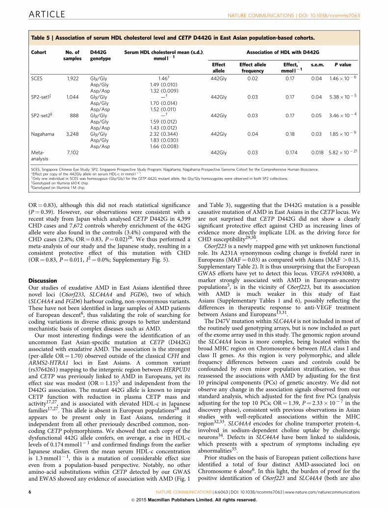
CETP 442G, HDL and coronary heart disease. The mutantCETP 442G allele was shown to result in an abnormally func-tioning CETP protein17. As CETP is a critical component of thepathways that regulate high-density lipoprotein cholesterol(HDL-c)18, we assessed this mutation for associations withserum HDL-c levels using linear regression, with adjustment forage, gender and body mass index, in three population-basedcohorts of Singaporean Chinese19–21 and Japanese22 (n¼ 7,102,see details on the study cohorts in Supplementary Methods)where GWAS data were available. We noted a strong associationbetween 442G allele and increased HDL-c levels(b¼ 0.174 mmol l� 1 per copy of 442G allele; reflecting anB10% shift within the normal HDL range, P¼ 5.82� 10� 21;Table 5). This effect size is at least twice that observed for otherCETP variants reported in European populations (SupplementaryTable 10)23,24.
Due to its strong effect on serum HDL-c, we assessed whetherthe mutant CETP 442G allele conferred any effect on individualsusceptibility to coronary heart disease (CHD) in East Asians.Using 683 CHD cases and 1,281 controls from the SingaporeChinese Health Study (Supplementary Methods)25, we notedsome degree of enrichment of the HDL-increasing, mutant 442Gallele in the controls (2.89%) compared with the cases (2.42%,
Table 3 | Single variant analysis (score-based tests using logistic regression) of the five observed CETP rare variants withexudative age-related macular degeneration in the discovery stage.
Sample collections SNP Amino-acid change A1 A2 Genotype* MAF OR P value
Cases Controls Cases Controls
Hong Kong rs201790757 Y74* C A 0/0/507 0/3/2,962 0 0.0005 N/A 1Hong Kong rs5881 G331S A G 0/5/502 12/1/52 0.0049 0.0024 1.94 0.18Japan rs5881 G331S A G 0/3/970 0/4/759 0.0015 0.0026 0.59 0.49Singapore rs5881 G331S A G 0/5/635 0/21/1,935 0.0039 0.0054 0.73 0.52Hong Kong rs190187567 N358S G A 0/0/507 0/1/2,964 0 0.0002 N/A 1Japan rs190187567 N358S G A 0/1/972 0/0/763 0.0005 0 N/A 1Hong Kong rs5880 A390P C G 0/5/502 0/36/2,926 0.0049 0.0061 0.81 0.66Singapore rs5880 A390P C G 0/13/627 0/28/1,925 0.0102 0.0072 1.43 0.30Hong Kong rs2303790 D442G G A 4/45/458 2/172/2,791 0.0523 0.0297 1.80 2.8� 10�4
Japan rs2303790 D442G G A 4/84/885 0/41/722 0.0473 0.0269 1.79 0.0025Singapore rs2303790 D442G G A 1/44/595 0/98/1,858 0.0359 0.0251 1.48 0.034
SNP, single-nucleotide polymorphism; A1, minor allele; A2, major allele; MAF, minor allele frequency; OR, odds ratio for per copy of the minor allele.*Data are number of genotypes A1A1/A1A2/A2A2.
Table 4 | Results of association of the five observed CETP rare variants with exudative age-related macular degeneration usinggene-based tests in 2,119 cases and 5,691 controls in the discovery stage.
Gene No. of variants Variants (minor allele counts) Unconditionalanalysis*
Conditionalanalysis*
CETP 5 Tyr74*(3), Gly331Ser (56), Asn358Ser (2), Ala390Pro (81), Asp442Gly(536)
P¼ 5.38� 10� 6 P¼0.96
*Gene-based tests on mutational load (additive allele based using Burden tests) at CETP, unconditioned and conditioned for CETP Asp442Gly.
ARTICLE NATURE COMMUNICATIONS | DOI: 10.1038/ncomms7063
4 NATURE COMMUNICATIONS | 6:6063 | DOI: 10.1038/ncomms7063 | www.nature.com/naturecommunications
& 2015 Macmillan Publishers Limited. All rights reserved.

C6orf223 rs 2395334
VEGFA rs943080
Meta-analysis P=6.19 × 10⁻¹⁸
Discovery P=1.41 × 10⁻⁸P=5 ×10⁻⁸
Meta-analysis P=1.08 × 10⁻¹¹
Discovery P=1.23 × 10⁻⁷
Meta-analysis P=2.85 × 10⁻⁸
Discovery P=4.75 × 10⁻⁵
P=5 ×10⁻⁸
P=5 ×10⁻⁸
20
43
34.4
25.8
17.2
8.6
0
0
84
67.2
50.4
33.6
16.8
0
4.8
9.6
14.4
19.2
24
191817161514131211109876543210
12
11
10
9
8
7
6
5
4
3
2
1
0
9
8
7
6
5
4
3
2
1
0
43,639
31,507 31,840 32,173
95,937 95,60495,271
43,971
Chromosome 6 position (kb)
Chromosome 6 position (kb)
Chromosome 12 position (kb)
44,303
POLR1C
YIPF3
XPO5
MICA
MICB
KRT19P2
MIR492 NR2C1
FGD6
VEZT
MIR331
MIR3685 NTN4
USP44
METAP2NDUFA12
SLC44A4
C2 C4A
C6orf10
C4B
CFB
POLH
MAD2L1BP
MRPS18A
RSPH9
VEGFA
LOC100132354
C6orf223
MRPL14 SLC29A1
CAPN11
AARS2
TCTE1
MIR4642
CDC5L
SPATS1TMEM63B SLC35B2
–Log
(P
)–L
og (
P)
–Log
(P
)
Rec
ombi
natio
n ra
te (
cMM
b)R
ecom
bina
tion
rate
(cM
Mb)
Rec
ombi
natio
n ra
te (
cMM
b)
C2-CFB rs429608
SLC44A4 rs12661281
FGD6 rs10507047
a
b
c
Figure 2 | Regional association plots at the three risk loci for exudative age-related macular degeneration. (a) The C6orf223 locus (rs2295334): VEGFA
rs943080, which is located in the intergenic, non-coding region, was very strongly associated with AMD in Europeans, but this study of exudative AMD in
East Asians could not reveal significant association with it (P¼0.041 by score-based tests using logistic regression in 2,119 cases and 5,691 controls). The
two markers are independent from each other (r2¼0.0), as well as separated by strong recombination events in Asians. (b) The SLC44A4 locus
(rs12661281): C2-CFB rs429608 was very strongly associated with AMD in Europeans, but revealed modest evidence of association in our discovery stage
(P¼ 1.06� 10�4). The two markers are independent from one another (r2¼0.01). (c) The FGD6 locus (rs10507047).
NATURE COMMUNICATIONS | DOI: 10.1038/ncomms7063 ARTICLE
NATURE COMMUNICATIONS | 6:6063 | DOI: 10.1038/ncomms7063 | www.nature.com/naturecommunications 5
& 2015 Macmillan Publishers Limited. All rights reserved.
OR¼ 0.83), although this did not reach statistical significance(P¼ 0.39). However, our observations were consistent with arecent study from Japan which analysed CETP D442G in 4,399CHD cases and 7,672 controls whereby enrichment of the 442Gallele were also found in the controls (3.4%) compared with theCHD cases (2.8%; OR¼ 0.83, P¼ 0.02)26. We thus performed ameta-analysis of our study and the Japanese study, resulting in aconsistent protective effect of this mutation with CHD(OR¼ 0.83, P¼ 0.011, I2¼ 0.0%; Supplementary Fig. 5).
DiscussionOur studies of exudative AMD in East Asians identified threenovel loci (C6orf233, SLC44A4 and FGD6), two of which(SLC44A4 and FGD6) harbour coding, non-synonymous variants.These have not been identified in large samples of AMD patientsof European descent6, thus validating the role of searching forcoding variations in diverse ethnic groups to better understandmechanistic basis of complex diseases such as AMD.
Our most interesting findings were the identification of anuncommon East Asian-specific mutation at CETP (D442G)associated with exudative AMD. The association is the strongest(per-allele OR¼ 1.70) observed outside of the classical CFH andARMS2-HTRA1 loci in East Asians. A common variant(rs3764261) mapping to the intergenic region between HERPUD1and CETP was previously linked to AMD in Europeans, yet itseffect size was modest (OR¼ 1.15)5 and independent from theD442G association. The mutant 442G allele is known to impairCETP function with reduction in plasma CETP mass andactivity17,27, and is associated with elevated HDL-c in Japanesefamilies17,27. This allele is absent in European populations28 andappears to be present only in East Asians, rendering itindependent from all other previously described common, non-coding CETP polymorphisms. We showed that each copy of thedysfunctional 442G allele confers, on average, a rise in HDL-clevels of 0.174 mmol l� 1 and confirmed findings from the earlierJapanese studies. Given the mean serum HDL-c concentrationis 1.3 mmol l� 1, this is a mutation of considerable effect sizeeven from a population-based perspective. Notably, no otheramino-acid substitutions within CETP detected by our GWASand EWAS showed any evidence of association with AMD (Fig. 1
and Table 3), suggesting that the D442G mutation is a possiblecausative mutation of AMD in East Asians in the CETP locus. Weare not surprised that CETP D442G did not show a clearlysignificant protective effect against CHD as increasing lines ofevidence more directly implicate LDL as the driving force forCHD susceptibility29,30.
C6orf223 is a newly mapped gene with yet unknown functionalrole. Its A231A synonymous coding change is fivefold rarer inEuropeans (MAF¼ 0.03) as compared with Asians (MAF 40.15,Supplementary Table 2). It is thus unsurprising that the EuropeanGWAS efforts have yet to detect this locus. VEGFA rs943080, amarker strongly associated with AMD in European-ancestrypopulations5, is in the vicinity of C6orf223, but its associationwith AMD is much weaker in this study of EastAsians (Supplementary Tables 1 and 6), possibly reflecting thedifferences in therapeutic response to anti-VEGF treatmentbetween Asians and Europeans13,31.
The D47V mutation within SLC44A4 is not included in most ofthe routinely used genotyping arrays, but is now included as partof the exome array used in this study. The genomic region aroundthe SLC44A4 locus is more complex, being located within thebroad MHC region on Chromosome 6 between HLA class I andclass II genes. As this region is very polymorphic, and allelefrequency differences between cases and controls could beconfounded by even minor population stratification, we thusreassessed the associations with AMD by adjusting for the first10 principal components (PCs) of genetic ancestry. We did notobserve any change in the association signals observed from ourstandard analysis, which adjusted for the first five PCs (analysisadjusting for the top 10 PCs; OR¼ 1.39, P¼ 2.33� 10� 7 in thediscovery phase), consistent with previous observations in Asianstudies with well-replicated associations within the MHCregion32,33. SLC44A4 encodes for choline transporter protein-4,involved in sodium-dependent choline uptake by cholinergicneurons34. Defects in SLC44A4 have been linked to sialidosis,which presents with a spectrum of symptoms including eyeabnormalities35.
Prior studies on the basis of European patient collections haveidentified a total of four distinct AMD-associated loci onChromosome 6 alone6. In this light, the burden of proof for thepositive identification of C6orf223 and SLC44A4 (both are also
Table 5 | Association of serum HDL cholesterol level and CETP D442G in East Asian population-based cohorts.
Cohort No. ofsamples
D442Ggenotype
Serum HDL cholesterol mean (s.d.),mmol l� 1
Association of HDL with D442G
Effectallele
Effect allelefrequency
Effect,*
mmol l� 1s.e.m. P value
SCES 1,922 Gly/Gly 1.46w 442Gly 0.02 0.17 0.04 1.46� 10� 6
Asp/Gly 1.49 (0.010)Asp/Asp 1.32 (0.009)
SP2-set1z 1,044 Gly/Gly —w 442Gly 0.03 0.17 0.04 5.38� 10� 5
Asp/Gly 1.70 (0.014)Asp/Asp 1.52 (0.011)
SP2-set2y 888 Gly/Gly —w 442Gly 0.03 0.17 0.05 3.46� 10�4
Asp/Gly 1.59 (0.012)Asp/Asp 1.43 (0.012)
Nagahama 3,248 Gly/Gly 2.32 (0.344) 442Gly 0.04 0.18 0.03 1.85� 10�9
Asp/Gly 1.83 (0.030)Asp/Asp 1.66 (0.008)
Meta-analysis
7,102 442Gly 0.03 0.174 0.018 5.82� 10� 21
SCES, Singapore Chinese Eye Study; SP2, Singapore Prospective Study Program; Nagahama, Nagahama Prospective Genome Cohort for the Comprehensive Human Bioscience.*Effect per copy of the 442Gly allele on serum HDL-c in mmol l� 1.wOnly one individual in SCES was homozygous (Gly/Gly) for the CETP 442G mutant allele. No Gly/Gly homozygotes were observed in both SP2 collections.zGenotyped on Illumina 610 K chip.yGenotyped on Illumina 1 M chip.
ARTICLE NATURE COMMUNICATIONS | DOI: 10.1038/ncomms7063
6 NATURE COMMUNICATIONS | 6:6063 | DOI: 10.1038/ncomms7063 | www.nature.com/naturecommunications
& 2015 Macmillan Publishers Limited. All rights reserved.

located on Chromosome 6) is higher than usual due to the needfor appropriate considerations of previously reported variants onthe same chromosome. It is reassuring to note that both C6orf223and SLC44A1 showed the strongest two signals of associationwith AMD outside of CFH, ARMS2-HTRA1 and CETP. Ourexhaustive analyses using logistic regression adjusting for alleledosages at previously described SNP markers suggest that bothC6orf223 and SLC44A1 are unrelated to those previouslyreported, and thus they likely represent Asian-specific geneticassociations for AMD.
Neither FGD6 nor the genes within its vicinity (Fig. 2c) haveever been previously implicated in any ocular disorders. FGD6encodes for FYVE, RhoGEF and PH domain-containingprotein 6, with its functions yet to be characterized. The Q257Rmutation is less than half as common in Europeans (MAF¼ 0.10)compared with East Asians (MAF¼ 0.20–0.30), again possiblyexplaining the ability of our study to pick up this genetic effect.
The identified genes were expressed in human retinal pigmentepithelium (Supplementary Table 11). Of the three non-synonymous substitutions, the CETP D442G variant waspredicted by both PolyPhen36 and SIFT37 to likely be causingdamage to the protein structure/function, the FGD6 Q257Rvariant was predicted only by PolyPhen to be probably damagingbut by SIFT to be tolerated and the SLC44A4 D47V variant waspredicted by both tools to be benign or tolerated. Although theuse of both prediction algorithms has been reported to bemoderately sensitive, they suffer from lack of specificity38, andthus more evidence should be sought with regards to the FDG6and SLC44A4 non-synonymous variants.
Using HaploReg39, RegulomeDB40 and Encyclopaedia of DNAElements (ENCODE)41 data, we identified variants within each ofthe four LD blocks in the 1000 Genomes Project (r240.8 ando250 kb from the top SNP) to apply functional annotationsrelevant to the regulation of transcription (SupplementaryTable 12). In addition to their functions on amino-acidsubstitutions, all of the four identified variants lie within aDNase I hypersensitivity site or in a region where modification ofhistones is suggestive of promoter, enhancer and other regulatoryactivity, and/or have an influence on binding of transcriptionfactors or effects on a specific regulatory motif. C6orf223 A231A(rs2295334) and FGD6 Q257R could tag genetic variants that liein potential transcription-factor binding sites (SupplementaryTable 13). Examination of a recently available large-scale eQTLmapping database42 indicates that out of the four novel genome-wide significant SNPs, markers SLC44A4 D47V and FDG6 Q257Rcould serve as cis-eQTLs. The minor allele at SLC44A4 D47V(rs12661281) is associated with significantly altered expression ofHSPA1B and CSKN2B, which are located within a 210,000 bpregion flanking SLC44A4 D47V. The minor allele at FDG6 Q257R(rs10507047) is associated with significantly increased expressionof the neighbouring VEZT gene (Supplementary Table 14). Giventhat these findings are based on expression in whole blood inEuropean samples, further work will be needed to elucidate theirrole in retinal tissue and in Asian samples. Nonetheless, thesecould suggest possible alternate mechanisms whereby both non-synonymous substitutions potentially affect AMD risk apart fromdirectly affecting the protein structure of their parent genes.
Our study examined mainly the exudative subtype of AMD,and therefore cannot be completely compared with other studieslooking at advanced AMD including the choroidal neovascular-ization and geographic atrophy subtypes. We also note substantialdifferences in inter-ethnic MAF for most of the previouslyreported loci associated with AMD in European-ancestrypopulations (Supplementary Table 1). This could representgenuine differences in genetic architecture in AMD betweenAsians and Europeans, or that the low allele frequency in either
ethnicity could result in insufficient power to replicate genome-wide significant hits initially observed in either Asians orEuropeans. Overall, out of 21 previously reported SNPs showingstrong evidence of association in Europeans, we were able toreplicate 9 of them in our study of East Asians at Po0.05. Takentogether with the three novel loci and one novel variant in CETPin East Asians discovered in this study, we postulate that thegenetic mechanisms of AMD in Asians could, in part, besomewhat distinct from that in Europeans.
In summary, our genome-wide and exome-wide study of AMDprovides new insights into the genetic mechanisms of AMD inEast Asians. Our study highlights the value of searching for low-frequency, ethnic-specific genetic variants on the coding frame ofAMD that may inform pathogenesis. Although some of thegenetic loci conferring disease susceptibility in East Asians areshared with Europeans (for example, common variation mappingto CFH, HTRA1 and CETP), we identified significant importantdifferences in the fine-scale genetic architecture of AMD, whichappear specific to East Asians. Such differences could underpin atleast some of the inter-ethnic differences in clinical presentationand response to specific therapies, including the poorer responseto anti-VEGF therapy in Asians.
MethodsStudy design and phenotyping. We performed a GWAS and EWAS on neo-vascular AMD in East Asians. In the discovery stage, we included and genotyped2,119 cases and 5,691 controls from three case–control studies from Singapore,Hong Kong and Japan. For replication, five independent case–control studies wereconducted in Korea, Japan, and Guangdong, Sichuan and Beijing in China, total-ling 4,226 cases and 10,289 controls. All the studies were performed with theapproval of their local Medical Ethics Committee, and written informed consentwas obtained from all the participants in accordance with the Declaration ofHelsinki.
A detailed description of subject recruitment and phenotyping in each samplecollection is provided in Supplementary Methods, and summarized in Table 1.In brief, the diagnosis of exudative AMD was made at each site by retinalspecialists, according to standard clinical definitions on the basis of detailedophthalmic examinations, including dilated fundus photography, fluoresceinangiography, indocyanine green angiography and optical coherence tomography(Table 1). Grading of fluorescein angiograms for the presence of choroidalneovascularization were performed using a modification from the MacularPhotocoagulation Study43. Indocyanine green angiography was performed todiagnose definitive polypoidal choroidal vasculopathy, a variant of AMD, using theJapanese Study Group guidelines44. Cases with other macular diseases such ascentral serous chorioretinopathy, high myopia and angioid streaks were excluded.Of the 2,119 exudative AMD cases included in the discovery phase, 1,083 (51%)were classified as ‘typical neovascular AMD’, 1,015 (48%) were polypoidalchoroidal vasculopathy and 21 (1%) had one eye with typical neovascular AMDand the other eye with polypoidal choroidal vasculopathy. At each site, controlssubjects without any clinical signs of AMD were either recruited from eye clinics orenrolled from population-based studies (Supplementary Methods).
Genotyping and imputation. For the discovery stage, GWAS genotyping wasperformed using the Illumina Human OmniExpress or Human Hap610-Quadbeadchips, and EWAS was done using HumanExome beadchips (Table 1). Forreplication, genotyping was performed using the MassArray platform (Sequenom),as well as using Taqman allelic discrimination probes (Applied Biosystems).
Stringent quality control filters were used to remove poorly performing samplesand SNP markers in both the discovery and replication (de-novo genotyping)phases. For the GWAS, SNPs with a call rate of o95%, MAF of o1%, or showingdeviation from Hardy–Weinberg Equilibrium (Po10� 6) were removed fromfurther statistical analysis. For the EWAS, SNPs with a call rate of o99%, MAF ofo0.1% or showing deviation from equilibrium (Po10� 6) were removed. The 99%threshold was used as many SNP markers on the exome array had MAF o5%, andas such, differential genotyping success rates between cases and controls as low as2% could result in false-positive findings. SNPs which were not monomorphic(whereby at least one heterozygous carrier individual was present) were includedfor downstream analysis.
Routine quality control criteria on a per-sample basis were carried out, andpoorly performing samples were removed from further analysis. The remainingsamples were then subjected to biological relationship verification by using theprinciple of variability in allele sharing according to the degree of relationship.Identity-by-state information was derived using the PLINK software45. For thosepairs of first-degree relatives so identified (for example, parent–offspring, full-
NATURE COMMUNICATIONS | DOI: 10.1038/ncomms7063 ARTICLE
NATURE COMMUNICATIONS | 6:6063 | DOI: 10.1038/ncomms7063 | www.nature.com/naturecommunications 7
& 2015 Macmillan Publishers Limited. All rights reserved.

siblings, as well as monozygous twins), we removed the sample with the lower callrate before performing PC analysis.
The imputation was carried out using IMPUTE2 version 2.2.2 with ASNpopulation haplotypes from 1000 Genomes as reference, as describedelsewhere46–48. Imputed genotypes were called with an impute probabilitythreshold of 0.9 with all other genotypes classified as missing. Additional qualitycontrol filters were applied to remove SNPs with 41% missingness should the SNPhave a MAF o5% in either cases or controls. For common SNPS with MAF 45%,the filtering criteria were set at 45% missingness.
Statistical analysis. For the discovery stage, all exudative AMD cases and controlsappear well matched when visualized spatially on PC analysis for each samplecollection on a per-country basis for Hong Kong, Japan and Singapore andaccording to self-reported ethnicity (ethnic Chinese for Hong Kong and Singapore,and ethnic Japanese for Japan; Supplementary Fig. 6), using previously reportedcriteria49, indicating that population stratification is unlikely to confound theassociation results.
For both the discovery and replication stages, analysis of association withexudative AMD was carried out using 1-degree of freedom score-based tests usinglogistic regression. The tests model for a trend-per-copy effect of the minor alleleon disease risk. For the discovery stage, we incorporated the top five PCs of geneticstratification into the logistic regression model to minimize the effect of residualpopulation stratification50. We could not adjust for population stratification for thereplication stage due to limited number of SNPs tested. Meta-analysis wasconducted using inverse variance weights for each sample collection, whichcalculates an overall Z-statistic, corresponding P value and accompanying per-alleleOR for each SNP analysed. Gene-based tests on mutational load was performedusing the SKAT-O test16. The association between CETP D442G and serum HDL-clevel was assessed using linear regression assuming an additive model ofinheritance as previously described23 (due to serum HDL-c being distributednormally), with adjustment for age, gender and body mass index.
Regional association and PC plots were analysed and plotted using theR statistical software package.
Power calculations. For the discovery stage (2,119 AMD cases and 5,691 con-trols), power calculations51 indicated that there is 80% power of detecting loci atPo1� 10� 4 (the threshold of association for bringing forward SNPs to thereplication stage) at MAF as low as 10% with per-allele OR of 1.30. For rarervariants of higher penetrance, the discovery stage has 80% power of detecting lociat Po1� 10� 4 at MAF as low as 2% if the per-allele OR is at least 1.70. The entiresample (6,345 AMD cases and 15,980 controls) has 80% power to detect loci atPo5.0� 10� 8 at MAF as low as 2% if the per-allele OR is at least 1.55 or at MAFas low as 9% with per-allele OR of 1.25, in line with the effect sizes being reportedin this study. Supplementary Table 15A shows the power calculations to detectSNPs at the threshold of Po1� 10� 4 in the discovery stage for bringing forwardto the replication stage. Supplementary Table 15B shows the formal powercalculations in the context of the combined discovery and replication stages.
References1. Lim, L. S., Mitchell, P., Seddon, J. M., Holz, F. G. & Wong, T. Y. Age-related
macular degeneration. Lancet 379, 1728–1738 (2012).2. Wong, W. L. et al. Global prevalence and burden of age-related macular
degeneration: a meta-analysis and disease burden projection for 2020 and 2040.Lancet Glob. Health 2, e106–e116 (2014).
3. Spencer, K. L. et al. Protective effect of complement factor B and complementcomponent 2 variants in age-related macular degeneration. Hum. Mol. Genet.16, 1986–1992 (2007).
4. Chen, W. et al. Genetic variants near TIMP3 and high-density lipoprotein-associated loci influence susceptibility to age-related macular degeneration.Proc. Natl Acad. Sci. USA 107, 7401–7406 (2010).
5. Yu, Y. et al. Common variants near FRK/COL10A1 and VEGFA are associatedwith advanced age-related macular degeneration. Hum. Mol. Genet. 20,3699–3709 (2011).
6. Fritsche, L. G. et al. Seven new loci associated with age-related maculardegeneration. Nat. Genet. 45, 433–439 439e431–432 (2013).
7. Raychaudhuri, S. et al. A rare penetrant mutation in CFH confers high risk ofage-related macular degeneration. Nat. Genet. 43, 1232–1236 (2011).
8. Seddon, J. M. et al. Rare variants in CFI, C3 and C9 are associated with highrisk of advanced age-related macular degeneration. Nat. Genet. 45, 1366–1370(2013).
9. Helgason, H. et al. A rare nonsynonymous sequence variant in C3 is associatedwith high risk of age-related macular degeneration. Nat. Genet. 45, 1371–1374(2013).
10. Zhan, X. et al. Identification of a rare coding variant in complement 3associated with age-related macular degeneration. Nat. Genet. 45, 1375–1379(2013).
11. Arakawa, S. et al. Genome-wide association study identifies two susceptibilityloci for exudative age-related macular degeneration in the Japanese population.Nat. Genet. 43, 1001–1004 (2011).
12. Laude, A. et al. Polypoidal choroidal vasculopathy and neovascular age-relatedmacular degeneration: same or different disease? Prog. Retin. Eye Res. 29, 19–29(2010).
13. Cheung, C. M. & Wong, T. Y. Ranibizumab and bevacizumab for AMDNew Engl. J. Med. 365, 2237 author reply 2237 (2011).
14. van de Ven, J. P. et al. A functional variant in the CFI gene confers a high riskof age-related macular degeneration. Nat. Genet. 45, 813–817 (2013).
15. Huyghe, J. R. et al. Exome array analysis identifies new loci and low-frequencyvariants influencing insulin processing and secretion. Nat. Genet. 45, 197–201(2013).
16. Lee, S., Wu, M. C. & Lin, X. Optimal tests for rare variant effects in sequencingassociation studies. Biostatistics 13, 762–775 (2012).
17. Inazu, A. et al. Genetic cholesteryl ester transfer protein deficiency caused bytwo prevalent mutations as a major determinant of increased levels of highdensity lipoprotein cholesterol. J Clin. Invest. 94, 1872–1882 (1994).
18. Barter, P. J. et al. Cholesteryl ester transfer protein: a novel target forraising HDL and inhibiting atherosclerosis. Arterioscler. Thromb. Vasc. Biol. 23,160–167 (2003).
19. Lavanya, R. et al. Methodology of the Singapore Indian Chinese Cohort (SICC)eye study: quantifying ethnic variations in the epidemiology of eye diseases inAsians. Ophthalmic Epidemiol. 16, 325–336 (2009).
20. Sabanayagam, C. et al. Retinal arteriolar narrowing increases the likelihood ofchronic kidney disease in hypertension. J. Hypertens. 27, 2209–2217 (2009).
21. Cheung, C. M. et al. Prevalence of and risk factors for age-related maculardegeneration in a multiethnic Asian cohort. Arch. Ophthalmol. 130, 480–486(2012).
22. Nakata, I. et al. Prevalence and characteristics of age-related maculardegeneration in the Japanese population: the nagahama study. Am. J.Ophthalmol. 156, 1002–1009 e1002 (2013).
23. Kathiresan, S. et al. Six new loci associated with blood low-density lipoproteincholesterol, high-density lipoprotein cholesterol or triglycerides in humans.Nat. Genet. 40, 189–197 (2008).
24. Kathiresan, S. et al. Common variants at 30 loci contribute to polygenicdyslipidemia. Nat. Genet. 41, 56–65 (2009).
25. Hankin, J. H. et al. Singapore Chinese Health Study: development, validation,and calibration of the quantitative food frequency questionnaire. Nutr. Cancer39, 187–195 (2001).
26. Takeuchi, F. et al. Association of genetic variants influencing lipid levels withcoronary artery disease in Japanese individuals. PLoS ONE 7, e46385 (2012).
27. Takahashi, K. et al. A missense mutation in the cholesteryl ester transferprotein gene with possible dominant effects on plasma high densitylipoproteins. J. Clin. Invest. 92, 2060–2064 (1993).
28. Freeman, D. J. et al. A polymorphism of the cholesteryl ester transfer proteingene predicts cardiovascular events in non-smokers in the West of ScotlandCoronary Prevention Study. Eur. Heart J. 24, 1833–1842 (2003).
29. Howard, B. V. et al. LDL cholesterol as a strong predictor of coronary heartdisease in diabetic individuals with insulin resistance and low LDL: The StrongHeart Study. Arterioscler. Thromb. Vasc. Biol. 20, 830–835 (2000).
30. Grundy, S. M. Low-density lipoprotein, non-high-density lipoprotein, andapolipoprotein B as targets of lipid-lowering therapy. Circulation 106,2526–2529 (2002).
31. Hara, R. et al. Photodynamic therapy alone versus combined with intravitrealbevacizumab for neovascular age-related macular degeneration withoutpolypoidal choroidal vasculopathy in Japanese patients. Graefes Arch. Clin. Exp.Ophthalmol. 248, 931–936 (2010).
32. Kumar, V. et al. Genome-wide association study identifies a susceptibilitylocus for HCV-induced hepatocellular carcinoma. Nat. Genet. 43, 455–458(2011).
33. Su, Z. et al. Common variants at the MHC locus and at chromosome 16q24.1predispose to Barrett’s esophagus. Nat. Genet. 44, 1131–1136 (2012).
34. O’Regan, S. et al. An electric lobe suppressor for a yeast choline transportmutation belongs to a new family of transporter-like proteins. Proc. Natl Acad.Sci. USA 97, 1835–1840 (2000).
35. Uhl, J. et al. Identification of a CTL4/Neu1 fusion transcript in a sialidosispatient. FEBS Lett. 521, 19–23 (2002).
36. Adzhubei, I. A. et al. A method and server for predicting damaging missensemutations. Nat. Methods 7, 248–249 (2010).
37. Ng, P. C. & Henikoff, S. SIFT: predicting amino acid changes that affect proteinfunction. Nucleic Acids Res. 31, 3812–3814 (2003).
38. Flanagan, S. E., Patch, A. M. & Ellard, S. Using SIFT and PolyPhen to predictloss-of-function and gain-of-function mutations. Genet. Test. Mol. Biomarkers14, 533–537 (2010).
39. Ward, L. D. & Kellis, M. HaploReg: a resource for exploring chromatin states,conservation, and regulatory motif alterations within sets of genetically linkedvariants. Nucleic Acids Res. 40, D930–D934 (2012).
ARTICLE NATURE COMMUNICATIONS | DOI: 10.1038/ncomms7063
8 NATURE COMMUNICATIONS | 6:6063 | DOI: 10.1038/ncomms7063 | www.nature.com/naturecommunications
& 2015 Macmillan Publishers Limited. All rights reserved.

40. Boyle, A. P. et al. Annotation of functional variation in personal genomes usingRegulomeDB. Genome Res. 22, 1790–1797 (2012).
41. Consortium, E. P. et al. An integrated encyclopedia of DNA elements in thehuman genome. Nature 489, 57–74 (2012).
42. Westra, H. J. et al. Systematic identification of trans eQTLs as putative driversof known disease associations. Nat. Genet. 45, 1238–1243 (2013).
43. Laser photocoagulation of subfoveal neovascular lesions in age-related maculardegeneration. Results of a randomized clinical trial. Macular PhotocoagulationStudy Group. Arch. Ophthalmol. 109, 1220–1231 (1991).
44. Japanese Study Group of Polypoidal Choroidal, V. [Criteria for diagnosis ofpolypoidal choroidal vasculopathy]. Nihon Ganka Gakkai Zasshi 109, 417–427(2005).
45. Purcell, S. et al. PLINK: a tool set for whole-genome associationand population-based linkage analyses. Am. J. Hum. Genet. 81, 559–575(2007).
46. Marchini, J., Howie, B., Myers, S., McVean, G. & Donnelly, P. A new multipointmethod for genome-wide association studies by imputation of genotypes.Nat. Genet. 39, 906–913 (2007).
47. Howie, B. N., Donnelly, P. & Marchini, J. A flexible and accurate genotypeimputation method for the next generation of genome-wide association studies.PLoS Genet. 5, e1000529 (2009).
48. Howie, B., Fuchsberger, C., Stephens, M., Marchini, J. & Abecasis, G. R.Fast and accurate genotype imputation in genome-wide association studiesthrough pre-phasing. Nat. Genet. 44, 955–959 (2012).
49. Vithana, E. N. et al. Genome-wide association analyses identify threenew susceptibility loci for primary angle closure glaucoma. Nat. Genet.44, 1142–1146 (2012).
50. Khor, C. C. et al. Genome-wide association study identifies FCGR2A as asusceptibility locus for Kawasaki disease. Nat. Genet. 43, 1241–1246 (2011).
51. Purcell, S., Cherny, S. S. & Sham, P. C. Genetic Power Calculator: design oflinkage and association genetic mapping studies of complex traits.Bioinformatics 19, 149–150 (2003).
AcknowledgementsWe thank the staff and participants of all studies for their important contributions. Thisresearch was supported by the National Medical Research Council (NMRC grants 0796/2003, IRG07nov013, IRG09nov014, NMRC 1176/2008, NIG/1003/2009, STaR/0003/2008, CG/SERI/2010 and CSA/033/2012), and Biomedical Research Council (BMRC 08/1/35/19/550, 09/1/35/19/616 and 10/1/35/19/671) in Singapore; BrightFocus Foundation(M2011068), USA; the Seoul National University Bundang Hospital Research GrantFund (grant no. 03-2009-008), and National Research Foundation of Korea (NRF-2009-0072603, NRF-2012R1A1A2008943, NRF-2014R1A2A1A09005824) grants funded bythe Ministry of Education, Science and Technology, Korea; and grants-in-aid for sci-entific research (No. 24249082) from the Japan Society for the Promotion of Science,Tokyo, Japan; the National Natural Science Foundation of China (81025006 (Z.Y.),
81170883 (Z.Y.) and the Department of Science and Technology of Sichuan Province,China (2014SZ0169 (Z.Y.) and 2012SZ0219 (Z.Y.)). The Singapore Chinese Health Studywas founded by NUS-HUJ CREATE Programme of the National Research Foundation,Singapore (Project No. 370062002), the National Medical Research Council (NMRC/1270/2010), Singapore, and the National Institutes of Health, USA (NCI RO1 CA55069,R35 CA53890, R01 CA80205 and R01 CA144034). C.-Y.C. is supported by an awardfrom NMRC (CSA/033/2012), Singapore. The Singapore Tissue Network and the TissueRepository of National University Health System provided services for DNA samplestorage in Singapore. The Genome Institute of Singapore, Agency for Science,Technology and Research, Singapore provided services for genotyping.
Author contributionsC.-Y.C., K.Y., L.J.C., J.A., K.-H.P., C.P.P., N.Y., T.Y.W. and C.C.K. designed the study.C.-Y.C., C.M.G.C., M.M., P.D.C., I.Y.Y., A.L., R.M., A.H.K., S.Y.L., D.W., C.M.G.C.,B.K.L., Y.S., H.N., Y.A.-K., N.G., A.T., K.M., S.Y., Y.S., H.I., T.I., S.H., T.Y.Y.L., H.C., S.T.,X.D., F.W., P.Z., B.Z., J.S., J.-M.Y., W.P.K., R.M.v.D., Y.F., N.W., G.S.W.T., S.J.P., M.B.,L.G., T.N., P.M., P.Z., S.-M.S., M.O., T.M., Y.K., S.J.W., H.C., H.-G.Y., J.Y.S., D.H.P.,I.T.K., W.C., M.S., S.-J.L., H.W.K., J.E.L., C.K.H., T.H.L., S.-K.Y., T.A. and W.T.Y.gathered clinical data. J.P., S.D., I.N., Y.A.-K., F.M., P.O.S.T., F.L., X.Z., Y.S., B.G., R.D.,Y.L., M.L.H., J.N.F., C.H.W., X.X., Jinlong Liang, J.M., X.J., Y.L., Jianjun Liu, K.S., E.N.V.,J.X.B., Y.X.Z. and C.C.K. generated genetic data. C.-Y.C., K.Y., L.J.C., J.A., Lulin Huang,Lvzhen Huang, K.S.S., P.C., Jiemin Liao, P.G.O., Y.Y.T. and C.C.K. analysed the data.C.-Y.C., K.Y., L.J.C., J.A., Lulin Huang, Lvzhen Huang, E.S.T., X.X.L., Z.Y., K.H.P., C.P.P,N.Y., T.Y.W. and C.C.K. interpreted the data. C.-Y.C., K.Y., L.J.C., J.A., E.S.T., T.Y.W.and C.C.K. drafted the paper. All the authors contributed to revision of the paper.
Additional informationSupplementary Information accompanies this paper at http://www.nature.com/naturecommunications
Competing financial interests: The authors declare no competing financial interests.
Reprints and permission information is available online at http://npg.nature.com/reprintsandpermissions/
How to cite this article: Cheng, C.-Y. et al. New loci and coding variants confer risk forage-related macular degeneration in East Asians. Nat. Commun. 6:6063 doi: 10.1038/ncomms7063 (2015).
This work is licensed under a Creative Commons Attribution 4.0International License. The images or other third party material in this
article are included in the article’s Creative Commons license, unless indicated otherwisein the credit line; if the material is not included under the Creative Commons license,users will need to obtain permission from the license holder to reproduce the material.To view a copy of this license, visit http://creativecommons.org/licenses/by/4.0/
NATURE COMMUNICATIONS | DOI: 10.1038/ncomms7063 ARTICLE
NATURE COMMUNICATIONS | 6:6063 | DOI: 10.1038/ncomms7063 | www.nature.com/naturecommunications 9
& 2015 Macmillan Publishers Limited. All rights reserved.

Corrigendum: New loci and coding variantsconfer risk for age-related macular degenerationin East AsiansChing-Yu Cheng, Kenji Yamashiro, Li Jia Chen, Jeeyun Ahn, Lulin Huang, Lvzhen Huang, Chui Ming G. Cheung,
Masahiro Miyake, Peter D. Cackett, Ian Y. Yeo, Augustinus Laude, Ranjana Mathur, Junxiong Pang, Kar Seng Sim,
Adrian H. Koh, Peng Chen, Shu Yen Lee, Doric Wong, Choi Mun Chan, Boon Kwang Loh, Yaoyao Sun,
Sonia Davila, Isao Nakata, Hideo Nakanishi, Yumiko Akagi-Kurashige, Norimoto Gotoh, Akitaka Tsujikawa,
Fumihiko Matsuda, Keisuke Mori, Shin Yoneya, Yoichi Sakurada, Hiroyuki Iijima, Tomohiro Iida, Shigeru Honda,
Timothy Yuk Yau Lai, Pancy Oi Sin Tam, Haoyu Chen, Shibo Tang, Xiaoyan Ding, Feng Wen, Fang Lu,
Xiongze Zhang, Yi Shi, Peiquan Zhao, Bowen Zhao, Jinghong Sang, Bo Gong, Rajkumar Dorajoo, Jian-Min Yuan,
Woon-Puay Koh, Rob M. van Dam, Yechiel Friedlander, Yin Lin, Martin L. Hibberd, Jia Nee Foo, Ningli Wang,
Chang Hua Wong, Gavin S. Tan, Sang Jun Park, Mayuri Bhargava, Lingam Gopal, Thet Naing, Jiemin Liao,
Peng Guan Ong, Paul Mitchell, Peng Zhou, Xuefeng Xie, Jinlong Liang, Junpu Mei, Xin Jin, Seang-Mei Saw,
Mineo Ozaki, Takinori Mizoguchi, Yasuo Kurimoto, Se Joon Woo, Hum Chung, Hyeong-Gon Yu, Joo Young Shin,
Dong Ho Park, In Taek Kim, Woohyok Chang, Min Sagong, Sang-Joon Lee, Hyun Woong Kim, Ji Eun Lee, Yi Li,
Jianjun Liu, Yik Ying Teo, Chew Kiat Heng, Tock Han Lim, Suk-Kyun Yang, Kyuyoung Song, Eranga N. Vithana,
Tin Aung, Jin Xin Bei, Yi Xin Zeng, E. Shyong Tai, Xiao Xin Li, Zhenglin Yang, Kyu-Hyung Park, Chi Pui Pang,
Nagahisa Yoshimura, Tien Yin Wong & Chiea Chuen Khor
Nature Communications 6:6063 doi: 10.1038/ncomms7063 (2015); Published 28 Jan 2015; Updated 30 Mar 2015
The financial support for this Article was not fully acknowledged. The Acknowledgements should have included the following:
This study was supported by the Basic Science Research Program through the National Research Foundation of Korea (NRF) fundedby the Ministry of Education (2014055007), Ministry of Education, Science and Technology (2012004585), and by the Korea HealthTechnology R&D Project, Ministry of Health & Welfare, Republic of Korea (A111345).
DOI: 10.1038/ncomms7817
NATURE COMMUNICATIONS | 6:6817 | DOI: 10.1038/ncomms7817 | www.nature.com/naturecommunications 1
& 2015 Macmillan Publishers Limited. All rights reserved.