title a novel unsaturated fatty acid hydratase … a novel unsaturated fatty acid hydratase toward...
TRANSCRIPT

Title A novel unsaturated fatty acid hydratase toward C(16) to C(22)fatty acids from Lactobacillus acidophilus.
Author(s) Hirata, Akiko; Kishino, Shigenobu; Park, Si-Bum; Takeuchi,Michiki; Kitamura, Nahoko; Ogawa, Jun
Citation Journal of lipid research (2015), 56(7): 1340-1350
Issue Date 2015-07
URL http://hdl.handle.net/2433/204520
Right
This research was originally published in ['Journal of lipidresearch' July 2015, 56, 1340-1350.] © the American Societyfor Biochemistry and Molecular Biology; This is not thepublished version. Please cite only the published version. この論文は出版社版でありません。引用の際には出版社版をご確認ご利用ください。
Type Journal Article
Textversion author
Kyoto University

1
A novel unsaturated fatty acid hydratase toward C16 to C22 fatty acids from Lactobacillus
acidophilus
Akiko Hirata1, Shigenobu Kishino1,*, Si-Bum Park2, Michiki Takeuchi1, Nahoko Kitamura1 and Jun
Ogawa1,*,†
1Division of Applied Life Sciences, Graduate School of Agriculture, Kyoto University, Kyoto 606-8502,
Japan
2Laboratory of Industrial Microbiology, Graduate School of Agriculture, Kyoto University, Kyoto
606-8502, Japan
*These corresponding authors contributed equally to this work.
†Correspondence: Jun Ogawa
Division of Applied Life Sciences,
Graduate School of Agriculture,
Kyoto University,
Kyoto 606-8502, Japan
Phone +81-75-753-6113; FAX +81-75-753-6128; E-mail; [email protected]
Running title: Novel fatty acid hydratase with broad substrate specificity

2
Abstract
Hydroxy FAs, one of the gut microbial metabolites of PUFAs, have attracted much attention because of
their various bioactivities. The purpose of this study was to identify lactic acid bacteria with the ability to
convert linoleic acid to hydroxy FAs. A screening process revealed that a gut bacterium Lactobacillus
acidophilus NTV001 converts linoleic acid mainly into 13-hydroxy-cis-9-octadecenoic acid, and resulted
in the identification of the hydratase responsible, FA-HY1. Recombinant FA-HY1 was purified and its
enzymatic characteristics were investigated. FA-HY1 could convert not only C18 PUFAs but also C20
and C22 PUFAs. C18 PUFAs with a cis carbon-carbon double bond at the ∆12 position were converted
into the corresponding 13-hydroxy FAs. Arachidonic acid and DHA were converted into the
corresponding 15-hydroxy FA and 14-hydroxy FA, respectively. To the best of our knowledge, this is the
first report of a bacterial FA hydratase that can convert C20 and C22 PUFAs into the corresponding
hydroxy FAs. These novel hydroxy FAs produced by using FA-HY1 should contribute to elucidate the
bioactivities of hydroxy FAs.
Supplementary Key words
arachidonic acid • docosahexaenoic acid • enzymology • fatty acid/metabolism • lipids • lipids/chemistry •
lactic acid bacteria • hydroxy fatty acid • hydration • dehydration

3
Introduction
Gut microbial metabolites of PUFAs have gained much attention because of their various physiological
activities (1-5). Recently, we reported that a gut bacterium Lactobacillus plantarum generates hydroxy
FAs, oxo FAs, and conjugated FAs from linoleic acid (LA) (6). The investigation of the presence of
hydroxy FAs in the intestines of mice revealed that the metabolites of LA,
10-hydroxy-cis-12-octadecenoic acid and 13-hydroxy-cis-9-octadecenoic acid, were detected at higher
levels in the intestines of specific pathogen-free (SPF) mice than in those of germ-free mice, indicating
that gastrointestinal microbes play a role in modifying the FA profile of their host mice.
The physiological functions of 10-hydroxy FAs and their derivatives begin to become clear.
10-Hydroxy-cis-12-octadecenoic acid ameliorates intestinal epithelial barrier impairment via
GPR40-MEK-ERK pathway and may be useful in the treatment of tight junction-related disorders such as
inflammatory bowel disease (3). 10-Oxo-cis-12-octadecenoic acid potently activates PPARγ, a master
regulator of adipocyte differentiation, and may be involved in the regulation of host energy metabolism
(5).
The production of 10-hydroxy-cis-12-octadecenoic acid from LA has been reported in many bacteria,
including Lactobacillus acidophilus (7), Lactobacillus plantarum (8, 9), Streptococcus bovis (10), and
Stenotrophomonas nitritireducens (11). In our previous study, we reported that the enzyme CLA-HY from

4
L. plantarum AKU 1009a catalyzes the hydration of the cis-9 double bond in C16 and C18 FAs, forming
the corresponding 10-hydroxy FAs (12). Volkov et al. reported that SPH, a hydratase from Streptococcus
pyogenes M49, is a myosin-cross-reactive antigen (MCRA) family protein that catalyzes the hydration of
the cis-9 and cis-12 double bonds in C16 and C18 FAs, forming the corresponding 10-hydroxy and
10,13-dihydroxy FAs (13).
The production of 13-hydroxy-cis-9-octadecenoic acid from LA has also been reported in some
anaerobic bacteria. Hudson et al. reported that a ruminant bacterium, S. bovis JB1, converts LA into
13-hydroxy-cis-9-octadecenoic acid (10), and Kishimoto et al. reported that L. acidophilus IFO 13951
converts LA into the (S)-form of 13-hydroxy-cis-9-octadecenoic acid (14). We reported that Pediococcus
sp. AKU 1080 converts LA into three products, 10-hydroxy-cis-12-octadecenoic acid,
13-hydroxy-cis-9-octadecenoic acid, and 10,13-dihydroxy-octadecanoic acid (15). However, the hydratase
responsible for the conversion of LA to 13-hydroxy-cis-9-octadecenoic acid has not yet been identified,
and the physiological activities of 13-hydroxy-cis-9-octadecenoic acid are unclear. As for the known
bacterial hydratases, the length of the carbon chain in the substrate FA is limited to C16 and C18 (1, 12, 13,
16-19).
In this paper, we report the identification of a novel hydratase from L. acidophilus that converts LA into
13-hydroxy-cis-9-octadecenoic acid and produces various hydroxy FAs. Through the screening of lactic

5
acid bacteria for the ability to convert LA into hydroxy FAs, we found that L. acidophilus NTV001 has a
high ability to convert LA into 13-hydroxy-cis-9-octadecenoic acid. We subsequently identified the
hydratase responsible for this conversion and named it FA-HY1. Recombinant FA-HY1 was purified and
its enzymatic characteristics were investigated. FA-HY1 shows broad substrate specificity and catalyzes
the hydration of C16, C18, C20, and C22 FAs. Thus, we succeeded in the production of
13-hydroxy-cis-9-octadecenoic acid and a variety of hydroxy FAs using FA-HY1, which is leading to the
development of novel, potentially bioactive hydroxy FAs.

6
Materials and Methods
Chemicals
FAs used as substrates were purchased from Nu-Chek Prep, Inc. (Elysian, MN, USA), Larodan Fine
Chemicals (Malmo, Sweden), and Cayman Chemical (MI, USA). Eicosatetraenoic acid used as substrate
was prepared from Mortierella alpina oil in our laboratory (20). Fatty acid-free (<0.02%) BSA was
purchased from Sigma (St. Louis, MO, USA). GC standard samples of 13-hydroxy-cis-9-octadecenoic
acid, 10-hydroxy-cis-12-octadecenoic acid, and 10,13-dihydroxy-octadecanoic acid were prepared as
previously described (15). All other chemicals used were of analytical grade and are commercially
available.
Microorganism, cultivation and reaction conditions for screening
Lactic acid bacteria used for this study were preserved in our laboratory (AKU Culture Collection,
Division of Applied Life Science, Faculty of Agriculture, Kyoto University, Kyoto, Japan) and those
obtained from other culture collections (JCM, Japan Collection of Microorganisms, Saitama, Japan; ATCC,
American Type Culture Collection, Virginia, USA; and NBRC, National Institute of Technology, Chiba,
Japan). The bacteria were cultivated in Lactobacilli MRS broth (Difco, Detroit, MI, USA). Screw-capped
tubes (16.5 × 125 mm) containing 15 ml of Lactobacilli MRS medium were inoculated with each strain

7
and then incubated, with shaking (120 strokes/min), under O2-limited conditions (<0.1% by O2 adsorbent
(21)) in the sealed condition at 28°C or 37°C for 2–3 days. After cultivation, the cells were harvested by
centrifugation (8,000 g, 10 min) and washed twice with 0.85% NaCl. The reactions were carried out in 1
ml of reaction mixture (100 mM KPB, pH 6.5), containing 10 mM LA, 0.3 mg BSA, and washed cells of
each strain at 37°C for 16 h with shaking (130 strokes/min) under anaerobic condition maintained using
Anaeropack kenki (Mitsubishi Chemical, Tokyo, Japan). Lipid analyses were then carried out on the
reaction mixtures.
Cloning and expression of recombinant proteins
We conducted a homology search, using the BLAST program, of the genomes of L. acidophilus strains
and found that there were two proteins in L. acidophilus ATCC4796 that share substantial homology with
the CLA-HY protein sequence. We designed primers based on the genome sequence of the L. acidophilus
ATCC4796, and amplified DNA regions containing each candidate gene (fa-hy1 and fa-hy2) using L.
acidophilus NTV001 genomic DNA as the template. Genomic DNA was extracted from L. acidophilus
NTV001 using a DNeasy Blood & Tissue kit (QIAGEN, Tokyo, Japan) according to the manufacturer’s
instructions. Region 1-forward primer (5′-GATGCTTTTTCATGGCACTGGGT-3′) and region 1-reverse
primer (5′-CACCAGTCGTCAAAGCTTCA-3′) were used to amplify the DNA region containing the

8
fa-hy1 gene. Region 2-forward primer (5′-CGCTTGTTAACTGGTAGAACACATCA-3′) and region
2-reverse primer (5′-CGTGACTTTAACTGGGATGA-3′) were used to amplify the DNA region
containing the fa-hy2 gene. The open reading frames of the two candidate genes in L. acidophilus
NTV001 were identified by sequencing, using a Beckman-Coulter CEQ8000 (Beckman-Coulter, Fullerton,
CA, USA). Based on the sequence results, we designed primers to clone each candidate gene. The
fa-hy1-forward primer (5′-ACACATATGCATTATAGTAGTGGTAAT-3′) and fa-hy1-reverse primer
(5′-AAACTCGAGCTAAACCAACTTATACTTCT-3′) were used to amplify the fa-hy1 gene, including its
stop codon (1773 bp). The fa-hy2-foraward primer (5′-CACCATATGTATTATTCCAATGGTAAT-3′) and
fa-hy2-reverse primer (5′-CGCCTCGAGTTAGACTAAATTTGCTTCT-3′) were used to amplify the
fa-hy2 gene, including its stop codon (1776 bp). The underlined bases in these sequences point out the
restriction enzyme recognition sites engineered into each primer. Each of the amplified products was
treated with NdeΙ and XhoΙ, and then ligated into the expression vector pET-21b (Novagen, WI, USA),
yielding the plasmids pET21b-fa-hy1 and pET21b-fa-hy2.
E. coli Rosetta2 (DE3) cells were transformed with either pET21b-fa-hy1 or pET21b-fa-hy2, yielding E.
coli Rosetta2 (DE3)/pET21b-fa-hy1 or E. coli Rosetta2 (DE3)/pE21b-fa-hy2. The cells were grown in
Luria-Bertani (LB) medium containing 50 µg/ml ampicillin and 34 µg/ml chloramphenicol at 37°C until
the optical density at 600 nm reached 0.5. Expression was induced by the addition of 1.0 mM

9
isopropyl-β-D-thiogalactopyranoside (IPTG), and the cells were grown at 16°C for an additional 16 h. The
cells were harvested by centrifugation (12,000 g, 10 min), washed twice with 0.85% NaCl, and kept at
-20°C until use.
Product analysis of reaction mixtures containing E. coli transformants with LA as the substrate
The reactions were carried out in 1 ml reaction mixtures (100 mM potassium phosphate buffer (KPB),
pH 6.5), containing 10 mM LA as the substrate, 0.5 mg BSA, and 60 mg of the washed cells of the
appropriate E. coli transformant (E. coli Rosetta2 (DE3)/pET21b-fa-hy1 or E. coli Rosetta2
(DE3)/pET21b-fa-hy2). Reaction mixtures were incubated at 37°C for 16 h with shaking (130
strokes/min) under anaerobic conditions maintained using Anaeropack Kenki (Mitsubishi Chemical,
Tokyo, Japan). After incubation, total lipids were extracted and the products were analyzed using gas
chromatography (GC). The peaks representing the reaction products were identified by comparing their
retention times with those of reference standards, and the chemical structures of products were confirmed
by GC-MS analysis of TMS derivatives.
Lipid analyses

10
Before lipid extraction, n-heptadecanoic acid was added to the reaction mixture as an internal standard.
Lipids were extracted from the reaction mixture (1 ml) using 5 ml of chloroform/methanol/1.5% KCl
(2:2:1, by volume) according to the Bligh–Dyer method, then the solvent was removed using a rotary
evaporator. Methylation of FAs was carried out by incubation with 4% methanolic HCl at 50°C for 20 min.
After methylation, the FA methyl esters were extracted with n-hexane and the solvent was removed using
a rotary evaporator. The resulting FA methyl esters were analyzed by GC using a Shimadzu GC-1700 gas
chromatograph equipped with a flame-ionization detector and a split-injection system, fitted with a
capillary column (SPB-1; 30 m length × 0.25 mm i.d.; Supelco, Pennsylvania, USA). The initial column
temperature of 180°C was maintained for 30 min, then increased to 220°C at a rate of 40°C/min and
finally maintained at that temperature for 14 min. Helium was used as the carrier gas at a flow rate of 2.51
ml/min. FA methyl esters obtained from dehydration reactions were analyzed by GC using the Shimadzu
GC-1700 gas chromatograph fitted with a capillary column (TC-70; 60 m length × 0.25 mm i.d.; GL
Sciences, Tokyo, Japan). The initial column temperature of 180°C was maintained for 10 min, then
increased to 260°C at a rate of 5°C/min, and finally maintained at that temperature for 9 min. Helium was
used as the carrier gas at a flow rate of 0.97 ml/min.
Protein purification

11
All of the steps in the FA-HY1 purification described below were performed at 4°C. Washed cells of the
E. coli transformant expressing FA-HY1 (E. coli Rosetta2 (DE3)/pET21b-fa-hy1) were suspended in 50
mM KPB (pH 6.0) and disrupted using an ultrasonic oscillator (Kubota, Tokyo, Japan). After
ultracentrifugation at 100,000 g for 60 min, the supernatants were loaded on a Mono Q 10/100 GL column
(GE Healthcare, Tokyo, Japan) equilibrated with 50 mM KPB (pH 6.0). The protein was eluted using a
linear NaCl gradient from 0 to 1 M in 50 mM KPB (pH 6.0), at a flow rate of 2 ml/min, using an ÄKTA
FPCL system (GE Healthcare, Tokyo, Japan). Fractions containing hydration activity were collected and
concentrated by ultrafiltration using Vivaspin® Turbo 15 (10,000 molecular weight cut-off) (Sartorius,
Tokyo, Japan) centrifugal concentrators. The concentrated solutions were loaded on a Superdex 200
10/300 GL column (GE Healthcare, Tokyo, Japan) equilibrated with 50 mM KPB (pH 6.0). Elution was
performed at a flow rate of 0.5 ml/min. Fractions containing hydration activity were collected,
concentrated as above, and kept at 4°C until use.
UV-visible spectra and cofactor identification
UV-visible spectra of purified FA-HY1 (10 mg/ml), in the 300-500 nm range, were acquired using a
UV-1700 UV-visible spectrophotometer (Shimadzu, Kyoto, Japan). To identify the flavin cofactor,
samples of FA-HY1 (10 mg/ml) were heated at 95°C for 10 min. The precipitated protein was removed by

12
centrifugation (20,000 g, 10 min) and the supernatants were used for HPLC analysis. Samples containing
FAD (40 μM) or FMN (40 μM) were prepared as reference standards. Reversed-phase HPLC separation
was performed on a Cosmosil 5C18-MS-II packed column (3.0 mm i.d. × 150 mm length; Nacalai Tesque,
Kyoto, Japan) with a methanol-5 mM ammonium acetate (20:80, by volume) solvent and a flow rate of
0.8 ml/min. The effluent was monitored at 350 nm using a UV detector.
Enzyme reaction conditions
The standard reaction conditions were as follows. Each reaction was carried out in 1 ml of reaction
mixture (50 mM KPB, pH 6.0), containing 10 mM LA as a substrate, 0.3 mg BSA, 0.1 mM FAD, and 50
µg FA-HY1 at 37°C for 15 min with shaking (130 strokes/min). All reactions were performed in triplicate.
Data presented in the figures and table represent the averages of three separate experiments that were
reproducible within ± 10%.
The effects of cofactors were examined by adding cofactors in various combinations: 5 mM NAD+, 5
mM NADH, 5 mM NADP+, 5 mM NADPH, 0.1 mM FAD, and 0.1 mM FMN. The effect of FAD
concentrations on enzyme activity was examined by varying its concentrations from 0 to 0.3 mM. The
optimum reaction pH was determined using sodium citrate buffer (50 mM; pH 3.0–4.0), sodium succinate
buffer (50 mM; pH 4.0–6.0), KPB (50 mM; pH 5.0–8.0), and Tris-HCl (50 mM; pH 7.0–9.0). The pH

13
stability was determined by measuring the enzyme activity after incubating the enzyme at 4°C for 24 h in
the following buffers: sodium citrate buffer (50 mM; pH 3.0–4.0), sodium succinate buffer (50 mM; pH
4.0–6.0), KPB (50 mM; pH 5.0–8.0), and Tris-HCl (50 mM; pH 7.0–9.0). The optimum reaction
temperature was examined by varying the temperature from 20 to 60°C in 50 mM KPB (pH 6.0). Thermal
stability was determined by measuring the enzyme activity in 50 mM KPB (pH 6.0) after incubating the
enzyme at various temperatures (4–60°C) for 30 min.
Kinetic analysis
Reactions were carried out under standard reaction conditions, with some modification of the enzyme
and LA substrate concentrations. The kinetics of LA hydration were studied using purified enzyme (1
μg/ml) and 10–100 μM LA substrate; the reaction time was 5 min. The Km value was evaluated with the
amounts of the product. The kinetic parameters were calculated by fitting the experimental data to either
the Hill equation or the Michaelis-Menten equation, using KaleidaGraph 4.0 (Synergy Software Inc., PA,
USA) software.
Substrate specificity of FA-HY1

14
To investigate the substrate specificity of FA-HY1, the purified FA-HY1 was incubated with various FAs.
The reactions were carried out in 1 ml reaction mixtures (50 mM KPB, pH 6.0), containing 10 mM
substrate, 0.3 mg BSA, 0.1 mM FAD, and 0.1 mg of the purified enzyme. All reaction mixtures were
incubated at 37°C for 16 h with shaking (130 strokes/min) under anaerobic conditions maintained using
Anaeropack kenki (Mitsubishi Chemical, Tokyo, Japan).
Isolation and identification of newly generated FAs
Reaction products were separated using an Isolera One automated flash purification system equipped
with a SNAP Ultra 10 g cartridge (Biotage, Stockholm, Sweden). The mobile phase, n-hexane-diethyl
ether (8:2–6:4; by volume), was used to elute the column at a flow rate of 12 ml/min, and the effluent was
monitored at 200 and 233 nm using a UV detector. Solvents were removed from the isolated FAs using a
rotary evaporator. After methylation, the methyl esters of the isolated FAs were transformed into their
pyrrolidide and trimethylsilyl (TMS) derivatives. Pyrrolidide derivatives were prepared by treating the FA
methyl esters with pyrrolidine-acetic acid (10:1, by volume) in screw-cap tubes for 1 h at 100°C, followed
by extraction with n-hexane. The organic extract was washed with water and dried over anhydrous
Na2SO4, and then the solvent was removed under vacuum in a rotary evaporator. TMS derivatives were
prepared by treating the FA methyl esters with a mixture of TMS agent

15
(pyridine/hexamethyldisilazane/trimethylchlorosilane; 9:3:1, by volume) in screw-cap tubes for 30 min at
60°C, followed by extraction with chloroform. The chemical structures of isolated FAs were determined
by GC-MS, proton NMR (1H-NMR), 1H-1H double-quantum-filtered chemical-shift correlation
spectroscopy (DQF-COSY), and two-dimensional nuclear Overhauser effect spectroscopy (NOESY).
Determination of absolute configuration and enantiomeric excess
The absolute configurations of 13-hydroxy-cis-9-octadecenoic acid and
13-hydroxy-cis-9,cis-15-octadecadienoic acid were determined using Mosher’s method (22). The
enantiomeric excess (ee) values of the 13-hydroxy-cis-9-octadecenoic acid and
13-hydroxy-cis-9,cis-15-octadecadienoci acid were determined using HPLC as described below. First,
13-oxo-cis-9-octadecenoic acid and 13-oxo-cis-9,cis-15-octadecadienoic acid were synthesized from each
hydroxy FA using the Jones oxidation method (23). Then, (RS)-13-hydroxy-cis-9-octadecenoic acid and
(RS)-13-hydroxy-cis-9,cis-15-octadecadienoic acid were synthesized by reduction of each keto FA to the
hydroxy FA using NaBH4 (24). HPLC separation was performed on a Chiralpak IA column (4.6 mm i.d. ×
250 mm length; Daicel, Tokyo, Japan), using acetonitrile-H2O (65:35, by volume) containing 0.2% formic
acid as the solvent, at a flow rate of 1.0 ml/min. The effluent was monitored at 205 nm using a UV
detector.

16
Dehydration reaction catalyzed by FA-HY1
Dehydration reactions, catalyzed by FA-HY1, were examined using 13-hydroxy-cis-9-octadecenoic acid
as the substrate. To produce 13-hydroxy-cis-9-octadecenoic acid, reactions were carried out in 1 ml of
reaction mixture (100 mM KPB, pH 6.5), containing 100 mg LA, 10 mg BSA, 0.1 mM FAD and 300 mg
of washed cells of the E. coli transformant expressing FA-HY1. Reaction mixtures were incubated at 37°C
for 16 h with shaking (130 strokes/min) under anaerobic conditions. After incubation, total lipids were
extracted and the 13-hydroxy-cis-9-octadecenoic acid was purified using an Isolera One automated flash
purification system equipped with a SNAP Ultra cartridge, as described above.
Dehydration reactions were carried out in 1 ml of reaction mixture (50 mM KPB, pH 6.0), containing 10
mM purified 13-hydroxy-cis-9-octadecenoic acid, 0.3 mg BSA, 0.1 mM FAD, and 0.1 mg FA-HY1.
Reaction mixtures were incubated at 37°C for 16 h with shaking (130 strokes/min) under anaerobic
conditions. After incubation, total lipids were extracted and analyzed using GC, as described above.
Reaction products were separated by reverse-phase HPLC, using a Shimadzu SLC-10A system equipped
with a Cosmosil Cholester column (10 mm i.d. × 250 mm length; Nacalai Tesque, Kyoto, Japan). The
column was eluted at a flow rate of 5 ml/min using a mobile phase consisting of acetonitrile-H2O (8:2, by
volume) containing 0.2% acetic acid. The effluent was monitored at 205 nm using a UV detector. The

17
chemical structures of the purified FAs were determined using data obtained from GC-MS analysis of the
pyrrolidide derivatives, as well as 1H-NMR, DQF-COSY, and NOESY analyses.

18
Results
Screening of lactic acid bacteria that convert LA into hydroxy FAs
The ability of lactic acid bacteria to convert LA into hydroxy FAs was investigated. We tested over 300
strains belonging to genera such as Lactobacillus, Leuconostoc, Enterococcus, Streptococcus,
Pediococcus, and so on. Most of the lactic acid bacteria converted LA into
10-hydroxy-cis-12-octadecenoic acid and some of them produced CLA (cis-9,trans-11-octadecadienoic
acid and trans-9,trans-11-octadecadienoic acid). We found that L. acidophilus AKU1222 (NTV001)
converted substantial quantities of LA into 13-hydroxy-cis-9-octadecenoic acid. Further analysis revealed
that 96% of the product was 13-hydroxy-cis-9-octadecenoic acid, and the remainder consisted of
10-hydroxy-cis-12-octadecenoic acid and 10,13-dihydroxy-octadecanoic acid. We attempted to identify
and clone the gene in L. acidophilus NTV001 that encodes the hydratase responsible for this conversion.
Identification of the hydratase gene in L. acidophilus NTV001
To identify the gene encoding the hydratase that converts LA into 13-hydroxy-cis-9-octadecenoic acid,
the protein sequence of CLA-HY (569 amino acids) was used as the query in a homology search for the
genomes of L. acidophilus strains. We found two candidate proteins in the genome of L. acidophilus
ATCC4796. Each of these proteins has been annotated as an MCRA and shares 32% amino acid identity

19
with CLA-HY. Based on the sequence of the L. acidophilus ATCC4796 genome, we succeeded in cloning
two candidate genes (fa-hy1 and fa-hy2) from L. acidophilus NTV001 (see Materials and Methods). The
fa-hy1 gene (accession number LC030242) of L. acidophilus NTV001 consists of a 1773 bp open reading
frame that encodes a polypeptide of 590 amino acids. This polypeptide shares 100% amino acid identity
with the product of the corresponding gene from L. acidophilus ATCC4796. The fa-hy2 gene (accession
number LC030243) of L. acidophilus NTV001 consists of a 1776 bp open reading frame that encodes a
polypeptide of 591 amino acids. This polypeptide shares 99% amino acid identity with the product of the
corresponding gene from L. acidophilus ATCC4796. FA-HY1 shares 57% amino acid identity with
FA-HY2.
Next, the products obtained from reaction mixtures, containing each E. coli transformant (E. coli
Rosetta2 (DE3)/pET21b-fa-hy1 or E. coli Rosetta2 (DE3)/pET21b-fa-hy2) as the catalyst and LA as the
substrate, were analyzed. A representative GC chromatogram from the analysis of products obtained with
FA-HY1 is shown in Fig. 1A. The retention time of the newly generated product was 24.4 min, which was
the same as that of the standard 13-hydroxy-cis-9-octadecenoic acid. This chemical structure was
confirmed by GC-MS analysis of its TMS derivative (Fig. 1B). Thus, FA-HY1 was responsible for the
production of 13-hydroxy-cis-9-octadecenoic acid from LA. The detailed characterization of FA-HY1 is
described in the following sections. A representative GC chromatogram from the analysis of the products

20
obtained with FA-HY2 is shown in Fig 1C. The retention time of the newly generated product is 26.4 min;
the same as that of the standard 10-hydroxy-cis-12-octadecenoic acid. This chemical structure was
confirmed by GC-MS analysis of its TMS derivative (Fig. 1D). Thus, FA-HY2 was responsible for the
production of 10-hydroxy-cis-12-octadecenoic acid from LA. FA-HY2 converted oleic acid, γ-linolenic
acid, and α-linolenic acid into the corresponding 10-hydroxy FAs. FA-HY2 most efficiently converted
γ-linolenic acid. When the activity of linoleic acid hydration was defined as 100%, FA-HY2 converted
oleic acid, γ-linolenic acid, and α-linolenic acid into the corresponding 10-hydroxy FAs with relative
activities of 160%, 166%, and 137%, respectively.
Purification of FA-HY1
In order to characterize FA-HY1, recombinant FA-HY1 was purified from the E. coli transformant, E.
coli Rosetta2 (DE3)/pET21b-fa-hy1. The purified FA-HY1 migrated as a single band on an SDS-PAGE
gel (Fig. 2). The apparent molecular mass of the band, approximately 66 kDa, corresponds to the
calculated molecular mass (67 kDa). The molecular weight of the native enzyme, determined by gel
filtration using a Superdex 200 10/300 GE column, was estimated to be 132 kDa (data not shown). Taken
together, the SDS-PAGE and gel filtration results suggest that native FA-HY1 is a homodimer.
21
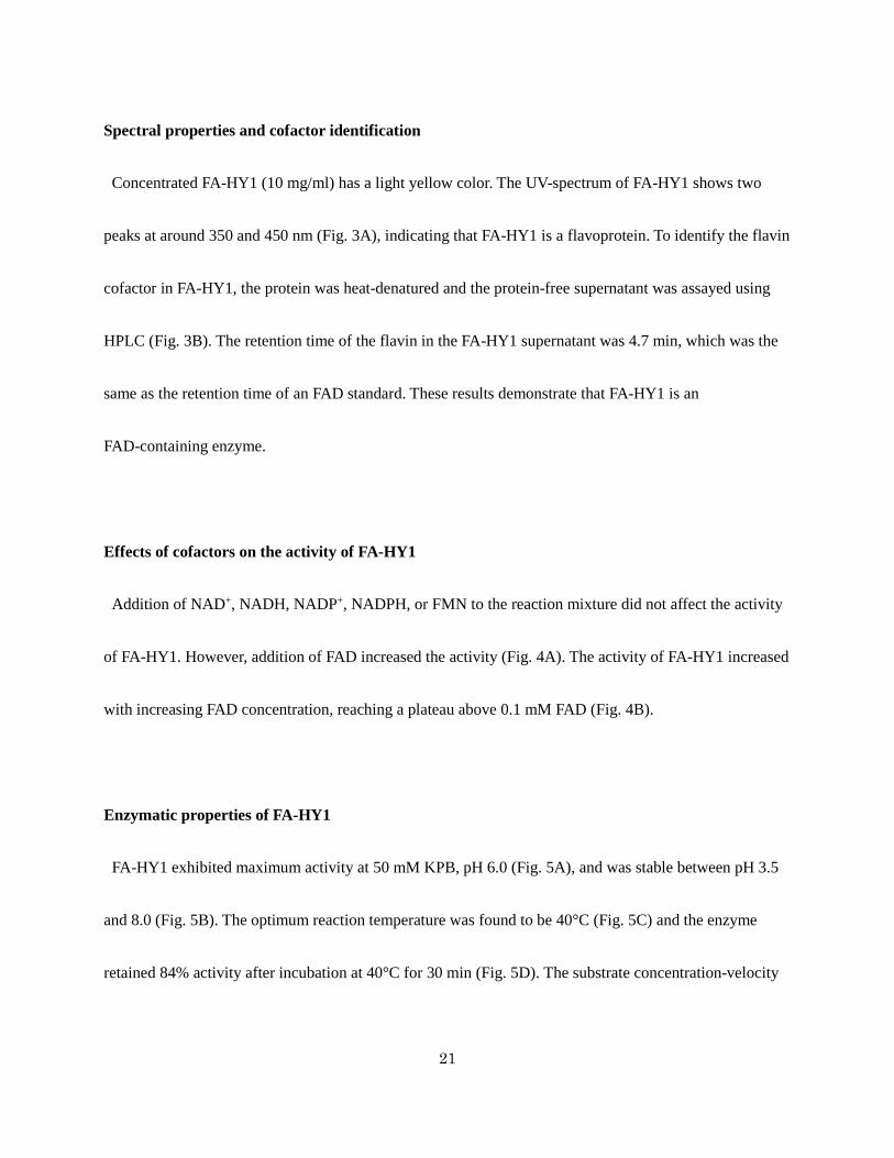
Spectral properties and cofactor identification
Concentrated FA-HY1 (10 mg/ml) has a light yellow color. The UV-spectrum of FA-HY1 shows two
peaks at around 350 and 450 nm (Fig. 3A), indicating that FA-HY1 is a flavoprotein. To identify the flavin
cofactor in FA-HY1, the protein was heat-denatured and the protein-free supernatant was assayed using
HPLC (Fig. 3B). The retention time of the flavin in the FA-HY1 supernatant was 4.7 min, which was the
same as the retention time of an FAD standard. These results demonstrate that FA-HY1 is an
FAD-containing enzyme.
Effects of cofactors on the activity of FA-HY1
Addition of NAD+, NADH, NADP+, NADPH, or FMN to the reaction mixture did not affect the activity
of FA-HY1. However, addition of FAD increased the activity (Fig. 4A). The activity of FA-HY1 increased
with increasing FAD concentration, reaching a plateau above 0.1 mM FAD (Fig. 4B).
Enzymatic properties of FA-HY1
FA-HY1 exhibited maximum activity at 50 mM KPB, pH 6.0 (Fig. 5A), and was stable between pH 3.5
and 8.0 (Fig. 5B). The optimum reaction temperature was found to be 40°C (Fig. 5C) and the enzyme
retained 84% activity after incubation at 40°C for 30 min (Fig. 5D). The substrate concentration-velocity

22
curve for LA hydration showed a sigmoid shape (Fig. 6). Fitting the data to the Hill equation resulted in an
apparent Km value for LA of 24 μM, a kcat value of 2.4 s-1, and a Hill coefficient of 3.6.
Substrate specificity of FA-HY1
To examine the substrate specificity of FA-HY1, various FAs were incubated with purified FA-HY1.
Hydroxy FA products generated by FA-HY1 are listed in Table 1. C18 FAs with a cis carbon-carbon
double bond at the Δ12 position, such as LA, pinolenic acid (cis-5,cis-9,cis-12-octadecatrienoic acid),
columbinic acid (trans-5,cis-9,cis-12-octadecatrienoic acid), γ-linolenic acid
(cis-6,cis-9,cis-12-octadecatrienoic acid), α-linolenic acid (cis-9,cis-12,cis-15-octadecatrienoic acid), and
stearidonic acid (cis-6,cis-9,cis-12,cis-15-octadecatetraenoic acid) were converted into the corresponding
13-hydroxy FAs. In contrast, FAs with a trans carbon-carbon double bond at the Δ12 position (linoelaidic
acid, trans-9,trans-12-octadecadienoic acid) and FA esters (methyl linoleate and ethyl linoleate) were not
hydrated. C20 FAs with a cis carbon-carbon double bond at the Δ14 position, such as
cis-11,cis-14-eicosadienoic acid, sciadonic acid (cis-5,cis-11,cis-14-eicosatrienoic acid),
dihomo-γ-linolenic acid (cis-8,cis-11,cis-14-eicosatrienoic acid), cis-11,cis-14,cis-17-eicosatrienoic acid,
and arachidonic acid (cis-5,cis-8,cis-11,cis-14-eicosatetraenoic acid), were converted into the
corresponding 15-hydroxy FAs. In particular, sciadonic acid and arachidonic acid were converted

23
efficiently to the corresponding 15-hydroxy FAs, giving yields of 51.7% and 45.7% against the added
PUFAs, respectively. However, eicosatetraenoic acid (cis-8,cis-11,cis-14,cis-17-eicosatetraenoic acid) was
converted into the corresponding 12-hydroxy FA and EPA was not hydrated. Mead acid
(cis-5,cis-8,cis-11-eicosatrienoic acid) was converted into the corresponding 12-hydroxy FA. DHA was
converted into the corresponding 14-hydroxy FA; however, the yield was low. FA-HY1 showed broad
substrate specificity and could convert C16-22 FAs into the corresponding hydroxy FAs.
The chemical structure of the product from α-linolenic acid, 13-hydroxy-cis-9,cis-15-octadecadienoic
acid, was determined by GC-MS analysis of its pyrrolidide derivative, as well as NMR analysis (Fig. 7).
The chemical structure of the product from arachidonic acid, 15-hydroxy-cis-5,cis-8,cis-11-eicosatrienoic
acid, was determined by GC-MS analysis of its TMS and pyrrolidide derivatives (Fig. 8). In the same way,
the chemical structures of other products were also determined through a combination of GC-MS and
NMR analyses.
The absolute stereochemistry of 13-hydroxy-cis-9-octadecenoic acid and
13-hydroxy-cis-9,cis-15-octadecadienoic acid produced by FA-HY1 were determined to be (R) and (S),
respectively, based on analyses of their NMR spectra. The enantiomeric purity (ee) of each of the products
was greater than 99% by HPCL analysis (data not shown).

24
Dehydration reaction catalyzed by FA-HY1
To examine the dehydration reaction catalyzed by FA-HY1, the substrate 13-hydroxy-cis-9-octadecenoic
acid was incubated with purified FA-HY1. Fig. 9A shows a GC chromatogram of the methylated FAs
produced by FA-HY1 from 13-hydroxy-cis-9-octadecenoic acid. The dehydration reaction catalyzed by
FA-HY1 generated cis-9,trans-13-octadecadienoic acid and LA with the yields of 20.3% and 19.3%,
respectively. The chemical structure of the cis-9,trans-13-octadecadienoic acid produced in the reaction
was determined by GC-MS and MNR analyses. Mass spectra of pyrrolidide derivatives of the isolated FA
methyl ester identified the isolated FA as 9,13-octadecadienoic acid (Fig 9B). 1H-NMR and DQF-COSY
also suggested one partial structure as -CH2-CH=CH-CH2-CH2-CH=CH-CH2-. The coupling constants
were J = 10.8 Hz and J = 12.5 Hz, respectively, indicating that one double bond was in the cis
configuration and the other was in the trans configuration. Based on the results of these spectral analyses,
one of the dehydrated products was identified as cis-9,trans-13-octadecadienoic acid.
Discussion
The ability to hydrate unsaturated FAs is present in wide range of bacteria, because unsaturated FAs may
be toxic to many bacteria (25-27). The MCRA protein family is highly conserved among different

25
bacterial species, including gram-positive and gram-negative bacteria, and some of them have FA
hydratase activity (12, 13, 16-19, 28).
In this study, we revealed that a gut bacterium L. acidophilus NTV001 has a high ability to produce
13-hydroxy-cis-9-octadecenoic acid from LA. L. acidophilus NTV001 was shown to have two hydratases.
One of them, FA-HY1, was responsible for the production of 13-hydroxy-cis-9-octadecenoic acid from
LA, and the other, FA-HY2, was responsible for the production of 10-hydroxy-cis-12-octadecenoic acid
from LA. FA-HY1 and FA-HY2 belong to the MCRA family of proteins and have FAD-binding motifs at
their N termini. UV-visible spectra of purified FA-HY1 contain two peaks at approximately 350 and 450
nm; FA-HY1 was found to be an FAD-containing enzyme (see Fig. 3). The carbon-chain lengths of the FA
substrates of previously reported MCRA protein family hydratases have been limited to C16 and C18.
However, FA-HY1 showed broad substrate specificity and could convert not only C16 and C18 FFAs, but
also C20 and C22 FFAs into the corresponding hydroxy FAs (see Table 1). To our knowledge, this is the
first report of the production of 15-hydroxy-cis-5,cis-11-eicosadienoic acid,
12-hydroxy-cis-14,cis-17-eicosadienoic acid, 15-hydroxy-cis-11,cis-17-eicosadienoic acid, and
14-hydroxy-cis-4,cis-7,cis-10,cis-16,cis-19-docosapentanoic acid from sciadonic acid
(cis-5,cis-11,cis-14-eicosatrienoic acid), cis-11,cis-14,cis-17-eicosatrienoic acid, and DHA, respectively,
by a bacterial enzyme. The products 13-Hydroxy-cis-5,cis-9-octadecadienoic acid,

26
13-hydroxy-trans-5,cis-9-octadecadienoic acid, 13-hydroxy-cis-9,cis-15-octadecadienoci acid, and
13-hydroxy-cis-6,cis-9,cis-15-octadecatrienoic acid were also newly produced from the C18 FAs
pinolenic acid (cis-5,cis-9,cis-12-octadecatrienoic acid), columbinic acid
(trans-5,cis-9,cis-12-octadecatrienoic acid), α-linolenic acid (cis-9,cis-12,cis-15-octadecatrienoic acid),
and stearidonic acid (cis-6,cis-9,cis-12,cis-15-octadecatetraenoic acid), respectively, by FA-HY1.
The 13-hydroxy-cis-9-octadecenoic acid produced by FA-HY1 from LA was of the (R)-configuration. It
was distinct from that of the 13-hydroxy-cis-9-octadecenoic acid produced by L. acidophilus IFO 13951,
which had the (S)-configuration (14). The 10-hydroxy-cis-12-octadecenoic acid produced by FA-HY2 has
the (S)-configuration (data not shown), which is the same as that of the 10-hydroxy-cis-12-octadecenoic
acid produced by CLA-HY (12).
FA-HY1 catalyzed a dehydration reaction that produced LA and cis-9,trans-13-octadecadienoic acid
from 13-hydroxy-cis-9-octadecenoic acid (see Fig. 9 and 10). The same reaction was observed when
13-hydroxy-cis-6,cis-9-octadecadienoic acid was used as a substrate (data not shown). The dehydration
products from 13-hydroxy-cis-6,cis-9-octadecadienoic acid were γ-linolenic acid and cis-6,cis-9,
trans-13-octadecatrienoic acid.
Hydroxy FAs are widely used as starting materials in the chemical, medical, and cosmetic industries
because of their increased reactivity and viscosity compared with non-hydroxy unsaturated FAs (29). The

27
bioactivities of hydroxy FAs have recently attracted considerable attention and have been intensively
studied (1-4). In our previous study, 13-hydroxy-cis-9-octadecenoic acid was detected at higher levels in
SPF mice than in germ-free mice (6). Further investigation of the bioactivities of
13-hydroxy-cis-9-octadecenoic acid is needed. Various 13-hydroxy FAs produced by FA-HY1 will be very
useful to elucidate the physiological functions of endogenous 13-hydroxy-cis-9-octadecenoic acid.
10-Hydroxy-cis-9-octadecenoic acid, a derivative of LA, suppresses intestinal inflammation via GPR40.
In contrast, 10-hydroxyoctadecanoic acid, a derivative of oleic acid and lacks a carbon-carbon double
bond, did not exert a colitis-suppressive activity (3). These results suggest that the presence of
carbon-carbon double bonds in hydroxy FAs is related with their physiological functions. In this study, we
succeeded in production of various hydroxy FAs by using FA-HY1, and these are varying in the position
of the hydroxyl group and the number of carbon-carbon double bond. These products will contribute in
pushing forward studies on bioactivities of hydroxy FAs. Evaluation of the biological properties of
13-hydroxy FAs and 15-hydroxy FAs generated by FA-HY1 will open new industrial and medicinal
applications.
Acknowledgments

28
This study was supported by the Bio-Oriented Technology Research Advancement Institution of Japan
(to J.O.), the Advanced Low Carbon Technology Research and Development Program of Japan (to S.K.),
the Science and Technology Research Promotion Program for Agriculture, Forestry, Fisheries and Food
Industry from the Ministry of Agriculture, Forestry and Fisheries of Japan (to J.O.), and the NEDO
Innovation Commercialization Venture Support Project (to collaboration of NITTO PHARMA and J.O.).

29
References
1. Kim, K. R., and D. K. Oh. 2013. Production of hydroxy fatty acids by microbial fatty
acid-hydroxylation enzymes. Biotechnol. Adv. 31: 1473–1485.
2. Bergamo, P., D. Luongo, J. Miyamoto, E. Cocca, S. Kishino, J. Ogawa, S. Tanabe, and M. Rossi.
2014. Immunomodulatory activitiy of a gut microbial metabolite of dietary linoleic acid,
10-hydroxy-cis-12-octadecenoic acid, associated with improved antioxidant/detoxifying defences.
J. Funct. Foods. 11: 192–202.
3. Miyamoto, J., T. Mizukure, S. B. Park, S. Kishino, I. Kimura, K. Hirano, P. Bergamo, M. Rossi, T.
Suzuki, M. Arita, J. Ogawa, and S. Tanabe. 2015. A Gut Microbial Metabolite of Linoleic Acid,
10-Hydroxy-cis-12-octadecenoic Acid, Ameliorates Intestinal Epithelial Barrier Impairment
Partially via GPR40-MEK-ERK Pathway. J. Biol. Chem. 290: 2902–2918.
4. Yore, M. M., I. Syed, P. M. Moraes-Vieira, T. Zhang, M. A. Herman, E. A. Homan, R. T. Patel, J.
Lee, S. Chen, O. D. Peroni, A. S. Dhanedhwar, A. Hammarstedt, U. Smith, T. E. McGraw, A.
Saghatelian, and B. B. Kahn. 2014. Discovery of a class of endogenous mammalian lipids with
anti-diabetic and anti-inflammatory effects. Cell. 159: 318–332.
5. Goto, T., Y. I. Kim, T. Furuzono, N. Takahashi, K. Yamakuni, H. E. Yang, Y. Li, R. Ohue, W.
Nomura, T. Sugawara, R. Yu, N. Kitamura, S. B. Park, S. Kishino, J. Ogawa, and T. Kawada. 2015.

30
10-oxo-12(Z)-octadecenoic acid, a linoleic acid metabolite produced by gut lactic acid bacteria,
potently activates PPARγ and stimulates adipogenesis. Biochem. Biophys. Res. Commun. 459:
597-603.
6. Kishino, S., M. Takeuchi, S. B. Park, A. Hirata, N. Kitamura, J. Kunisawa, H. Kiyono, R.
Iwamoto, Y. Isobe, M. Arita, H. Arai, K. Ueda, J. Shima. S. Takahashi. K. Yokezeki, S. Shimizu,
and J. Ogawa. 2013. Polyunsaturated fatty acid saturation by gut lactic acid bacteria affecting host
lipid composition. Proc. Natl. Acad. Sci. U S A. 110: 17808–17813.
7. Ogawa, J., K. Matsumura, S. Kishino, Y. Omura, and S. Shimizu. 2001. Conjugated linoleic acid
accumulation via 10-hydroxy-12-octadecaenoic acid during microaerobic transformation of
linoleic acid by Lactobacillus acidophilus. Appl. Environ. Microbiol. 67: 1246–1252.
8. Yamada, Y., H. Uemura, H. Nakaya, K. Sakata, T. Takatori, M. Nagao, H. Iwase, and K. Iwadate.
1996. Production of hydroxy fatty acid (10-hydroxy-12(Z)-octadecenoic acid) by Lactobacillus
plantarum from linoleic acid and its cardiac effects to guinea pig papillary muscles. Biochem.
Biophys. Res. Commun. 226: 391–395.
9 Kishino, S., J. Ogawa, Y. Omura, K. Matsumura, and S. Shimizu. 2002. Conjugated linoleic acid
production from linoleic acid by lactic acid bacteria. J. Am. Oil Chem. Soc. 79: 159–163.
31
10. Hudson, J. A., B. Morvan, and K. N. Joblin. 1998. Hydration of linoleic acid by bacteria isolated
from ruminants. FEMS Microbiol. Lett. 169: 277–282.
11. Yu, I. S., S. J. Yeom, H. J. Kim, J. K. Lee, Y. H. Kim, and D. K. Oh. 2008. Substrate specificity of
Stenotrophomonas nitritireducens in the hydroxylation of unsaturated fatty acid. Appl. Microbiol.
Biotechnol. 78: 157–163.
12. Takeuchi, M., S. Kishino, A. Hirata, S. B. Park, N. Kitamura, and J. Ogawa. 2014.
Characterization of the linoleic acid Δ9 hydratase catalyzing the first step of polyunsaturated fatty
acid saturation metabolism in Lactobacillus plantarum AKU 1009a. J. Biosci. Bioeng. In press.
13. Volkov, A., A. Liavonchanka, O. Kamneva, T. Fiedler, C. Goebel, B. Kreikemeyer, and I. Feussner.
2010. Myosin cross-reactive antigen of Streptococcus pyogenes M49 encodes a fatty acid double
bond hydratase that plays a role in oleic acid detoxification and bacterial virulence. J. Biol. Chem.
285: 10353–10361.
14. Kishimoto, N., I. Yamamoto, K. Toraishi, S. Yoshioka, K. Saito, H. Masuda, and T. Fujita. 2003.
Two distinct pathways for the formation of hydroxy FA from linoleic acid by lactic acid bacteria.
Lipids. 38: 1269–1274.
15. Takeuchi, M., S. Kishino, K. Tanabe, A. Hirata, S. B. Park, S. Shimizu, and J. Ogawa. 2013.
Hydroxy fatty acid production by Pediococcus sp. Eur. J. Lipid. Sci. Technol. 115: 386–393.

32
16. O'Connell, K. J., M. O. Motherway, A. A. Hennessey, F. Brodhun, R. P. Ross, I. Feussner, C.
Stanton, G. F. Fitzgerald, and D. van Sinderen. 2013. Identification and characterization of an
oleate hydratase-encoding gene from Bifidobacterium breve. Bioengineered. 4: 313–321.
17. Volkov, A., S. Khoshnevis, P. Neumann, C. Herrfurth, D. Wohlwend, R. Ficner, and I. Feussner.
2013. Crystal structure analysis of a fatty acid double-bond hydratase from Lactobacillus
acidophilus. Acta. Crystallogr. D. Biol. Crystallogr. 69: 648–657.
18. Bevers, L. E., M. W. Pinkse, P. D. Verhaert, and W. R. Hagen. 2009. Oleate hydratase catalyzes
the hydration of a nonactivated carbon-carbon bond. J. Bacteriol. 191: 5010–5012.
19. Joo, Y. C., K. W. Jeong, S. J. Yeom, Y. S. Kim, Y. Kim, and D. K. Oh. 2012. Biochemical
characterization and FAD-binding analysis of oleate hydratase from Macrococcus caseolyticus.
Biochimie. 94: 907–915.
20 Okuda, T., A. Ando, H. Negoro, T. Muratsubaki, H. Kikukawa, T. Sakamoto, E. Sakuradani, S.
Shimizu, and J. Ogawa. J. Biosci. Bioeng. In press.
21. Kishino, S., J. Ogawa, A. Ando, K. Yokozeki, and S. Shimizu. 2010. Microbial production of
conjugated gamma-linolenic acid from gamma-linolenic acid by Lactobacillus plantarum AKU
1009a. J. Appl. Microbiol. 108: 2012–2018.

33
22. Ohtani, I., T. Kusumi, Y. Kashman, and H. Kakisawa. 1991. High-field FT NMR application of
Mosher's method. The absolute configurations of marine terpenoids. J. Am. Chem. Soc. 113:
4092–4096.
23. Bowden, K., I. M. Heilbron, E. R. H. Jones, and B.C. L. Weedon. 1946. 13. Researches on
acetylenic compounds. Part I. The preparation of acetylenic ketones by oxidation of acetylenic
carbinols and glycols. J. Chem. Soc. (Resumed). 39–45.
24. Dalla, V., J. P. Catteau, and P. Pale. 1999. Mechanistic rationale for the NaBH4 reduction of α-keto
esters. Tetrahedron Letters. 40: 5193–5196.
25. Greenway, D. L., and K. G. Dyke. 1979. Mechanism of the inhibitory action of linoleic acid on
the growth of Staphylococcus aureus. J. Gen. Microbiol. 115: 233–245.
26. Raychowdhury, M. K., R. Goswami, and P. Chakrabarti. 1985. Effect of unsaturated fatty acids in
growth inhibition of some penicillin-resistant and sensitive bacteria. J. Appl. Bacteriol. 59: 183–
188.
27. Zheng, C. J., J. S. Yoo, T. G. Lee, H. Y. Cho, Y. H. Kim, and W. G. Kim. 2005. Fatty acid
synthesis is a target for antibacterial activity of unsaturated fatty acids. FEBS Lett. 579: 5157–
5162.

34
28. Yang, B., H. Chen, Y. Song, Y. Q. Chen, H. Zhang, and W. Chen. 2013. Myosin-cross-reactive
antigens from four different lactic acid bacteria are fatty acid hydratases. Biotechnol. Lett. 35:
75-81.
29. Metzger, J. O., and U. Bornscheuer. 2006. Lipids as renewable resources: current state of
chemical and biotechnological conversion and diversification. Appl. Microbiol. Biotechnol. 71:
13–22.

35
Figure Legends
Figure 1. Analysis of LA hydration by E. coli transformants expressing FA-HY1 and FA-HY2
(A) Representative GC chromatogram of a reaction mixture containing E. coli Rosetta2
(DE3)/pET21b-fa-hy1 and LA. The internal standard (I.S.) is n-heptadecanoic acid. (B) GC-MS spectrum
of the TMS derivative of the methyl ester of the newly generated product from the hydration of LA by E.
coli Rosetta2 (DE3)/pET21b-fa-hy1. (C) Representative GC chromatogram of the reaction mixture
containing E. coli Rosetta2 (DE3)/pET21b-fa-hy2 and LA. (D) GC-MS spectrum of the TMS derivative of
the methyl ester of the newly generated product from the hydration of LA by E. coli Rosetta2
(DE3)/pET21b-fa-hy2.
Figure 2. SDS-PAGE analysis of purified FA-HY1
M: Protein molecular weight marker; lane 1: Cell-free extracts of E. coli expressing FA-HY1; lane 2:
Mono Q 10/100 GL column eluate; lane 3: Superdex 200 10/300 GL column eluate. The 12.5%
SDS-PAGE gels were stained with Coomassie blue and destained by subsequent washing in distillated
water.
Figure 3. Spectral properties of FA-HY1 and cofactor identification

36
(A) FA-HY1 spectra were acquired at an enzyme concentration of 10 mg/ml. (B) The HPLC
chromatograms of protein-free FA-HY1 supernatant, 40 μM FAD, and 40 μM FMN are shown. The
retention times of the peaks from the protein-free FA-HY1 supernatant and the FAD standard were both
4.7 min; the retention time of FMN was 6.0 min.
Figure 4. Effects of added cofactors on the activity of FA-HY1
(A) Each cofactor was added to a reaction mixture containing 50 mM KPB (pH 6.0), 10 mM LA, 0.3
mg/ml BSA, and 50 µg/ml enzyme. Reactions were conducted at 37°C for 15 min with shaking (130
strokes/min). Control reaction was carried out without the addition of cofactors. (B) Reactions were
carried out by varying the concentration of FAD from 0 to 0.3 mM.
Figure 5. Enzymatic properties of FA-HY1
(A) Reactions were carried out in the indicated buffer containing 10 mM LA, 0.3 mg/ml BSA, 0.1 mM
FAD, and 0.5 µg/ml enzyme at 37°C for 15 min with shaking. The value obtained with 50 mM KPB (pH
6.0) was defined as 100%. (B) The enzyme was pre-incubated at 4°C for 24 h in the indicated buffer. The
reactions were carried out at 37°C for 15 min, with shaking, in 50 mM KPB (pH 6.0) containing 10 mM
LA, 0.3 mg/ml BSA, 0.1 mM FAD, and 0.5 µg/ml pre-incubated enzyme. The value obtained with 50 mM
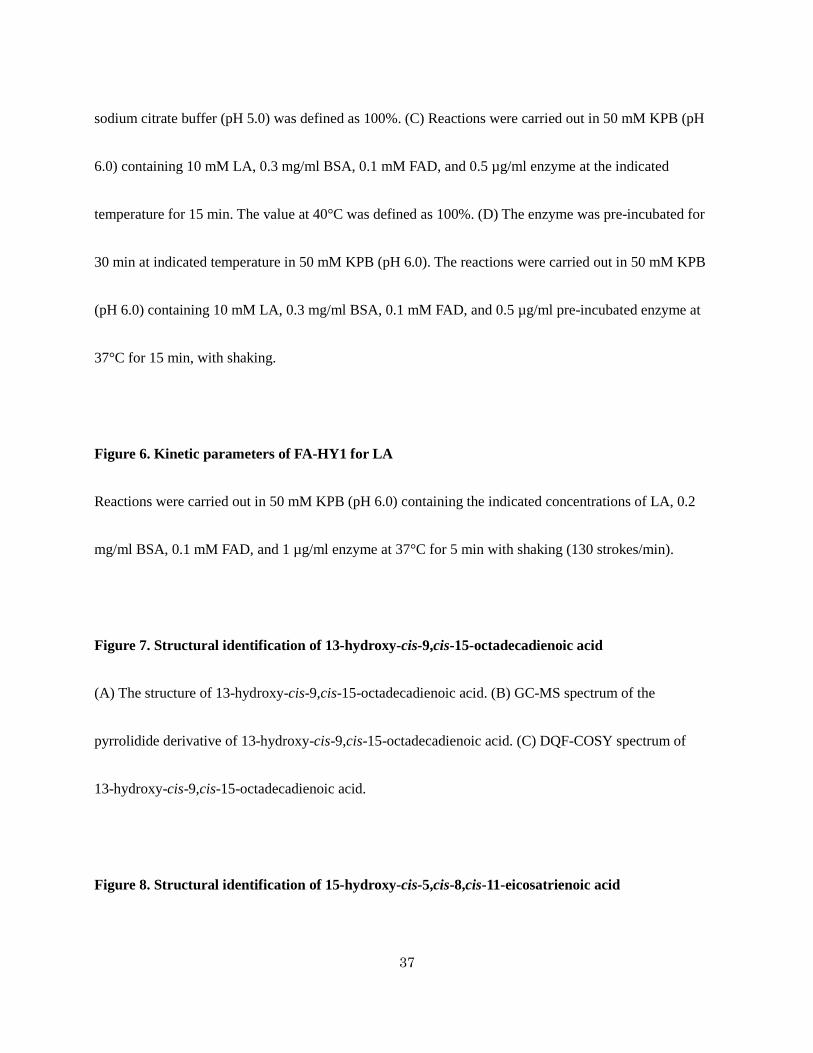
37
sodium citrate buffer (pH 5.0) was defined as 100%. (C) Reactions were carried out in 50 mM KPB (pH
6.0) containing 10 mM LA, 0.3 mg/ml BSA, 0.1 mM FAD, and 0.5 µg/ml enzyme at the indicated
temperature for 15 min. The value at 40°C was defined as 100%. (D) The enzyme was pre-incubated for
30 min at indicated temperature in 50 mM KPB (pH 6.0). The reactions were carried out in 50 mM KPB
(pH 6.0) containing 10 mM LA, 0.3 mg/ml BSA, 0.1 mM FAD, and 0.5 µg/ml pre-incubated enzyme at
37°C for 15 min, with shaking.
Figure 6. Kinetic parameters of FA-HY1 for LA
Reactions were carried out in 50 mM KPB (pH 6.0) containing the indicated concentrations of LA, 0.2
mg/ml BSA, 0.1 mM FAD, and 1 µg/ml enzyme at 37°C for 5 min with shaking (130 strokes/min).
Figure 7. Structural identification of 13-hydroxy-cis-9,cis-15-octadecadienoic acid
(A) The structure of 13-hydroxy-cis-9,cis-15-octadecadienoic acid. (B) GC-MS spectrum of the
pyrrolidide derivative of 13-hydroxy-cis-9,cis-15-octadecadienoic acid. (C) DQF-COSY spectrum of
13-hydroxy-cis-9,cis-15-octadecadienoic acid.
Figure 8. Structural identification of 15-hydroxy-cis-5,cis-8,cis-11-eicosatrienoic acid

38
(A) The structure of 15-hydroxy-cis-5,cis-8,cis-11-eicosatrienoic acid. (B) GC-MS spectrum of the TMS
derivative of 15-hydroxy-cis-5,cis-8,cis-11-eicosatrienoic acid methyl ester. (C) GC-MS spectrum of the
pyrrolidide derivative of 15-hydroxy-cis-5,cis-8,cis-11-eicosatrienoic acid.
Figure 9. Analysis of the dehydration reaction catalyzed by FA-HY1
(A) GC chromatogram of the methylated FAs produced by FA-HY1 from 13-hydroxy-cis-9-octadecanoic
acid. Dehydration reactions were carried out in 50 mM KPB (pH 6.0) containing 10 mM purified
13-hydroxy-cis-9-octadecenoic acid, 0.3 mg/ml BSA, 0.1 mM FAD, and 0.1 mg/ml FA-HY1 at 37°C for
16 h with shaking (130 strokes/min) under anaerobic conditions. (B) GC-MS spectrum of the pyrrolidide
derivative of cis-9,trans-13-octadecadienoic acid.
Figure 10. Hydration/dehydration reaction catalyzed by FA-HY1
FA-HY1 hydrates LA at its cis-12-double bond, yielding 13-hydroxy-cis-9-octadecenoic acid. FA-HY1
dehydrates 13-hydroxy-cis-9-octadecenoic acid, yielding LA and cis-9,trans-13-octadecadienoic acid.

39
TABLE 1.Substrate specificity of FA-HY1
Substrate Product(s) Yield (%)
16:1Δ9cis 10-hydroxy-16:0 2.2
18:1Δ9cis 10-hydroxy-18:0 0.6
18:1Δ11cis 12-hydroxy-18:0 58.8
18:1Δ11trans No products detected −
18:2Δ9cis, Δ12cis 13-hydroxy-cis-9-18:1 48.1
18:2Δ9trans, Δ12trans No products detected −
18:3Δ5cis, Δ9cis, Δ12cis 13-hydroxy-cis-5,cis-9-18:2 57.0
18:3Δ5trans, Δ9cis, Δ12cis 13-hydroxy-trans-5,cis-9-18:2 45.5
18:3Δ6cis, Δ9cis, Δ12cis 13-hydroxy-cis-6,cis-9-18:2 56.6
10-hydroxy-cis-6,cis-12-18:2 0.1
18:3Δ9cis, Δ12cis, Δ15cis 13-hydroxy-cis-9,cis-15-18:2 53.8
18:4Δ6cis, Δ9cis, Δ12cis, 15cis 13-hydroxy-cis-6, cis-9,cis-15-18:3 12.1
20:2Δ11cis, Δ14cis 15-hydroxy-cis-11-20:1 9.2
20:3Δ5cis, Δ8cis, Δ11cis 12-hydroxy-cis-5,cis-8-20:2 18.7
20:3Δ5cis, Δ11cis, Δ14cis 15-hydroxy-cis-5,cis-11-20:2 51.7

40
20:3Δ8cis, Δ11cis, Δ14cis 15-hydroxy-cis-8,cis-11-20:2 14.1
12-hydroxy-cis-8,cis-14-20:2 4.7
20:3Δ11cis, Δ14cis, Δ17cis 12-hydroxy-cis-14,cis-17-20:2 0.4
15-hydroxy-cis-11,cis-17-20:2 0.2
20:4Δ5cis, Δ8cis, Δ11cis, Δ14cis 15-hydroxy-cis-5,cis-8,cis-11-20:3 45.7
20:4Δ8cis, Δ11cis, Δ14cis, Δ17cis 12-hydroxy-cis-8,cis-14,cis-17-20:3 2.1
EPA No products detected −
DHA 14-hydroxy-cis-4,cis-7,cis-10,cis-16,cis-19-22:5 0.6
Methyl linoleate No products detected −
Ethyl linoleate No products detected −
16:0 (hexadecanoic acid), 16:1 (hexadecenoic acid), 18:0 (octadecanoic acid),
18:1 (octadecenoic acid), 18:2 (octadecadienoic acid), 18:3 (octadecatrienoic acid),
18:4 (octadecatetraenoic acid), 20:1 (eicosadecenoic acid), 20:2 (eicosadienoic acid),
20:3 (eicosatrienoic acid), 20:4 (eicosatetraenoic acid), 22:5 (docosapentaenoic acid)

41
Figure 1. Analysis of LA hydration by E. coli transformants expressing FA-HY1 and FA-HY2

42
Figure 2. SDS-PAGE analysis of purified FA-HY1

43
Figure 3. Spectral properties of FA-HY1 and cofactor identification

44
Figure 4. Effects of added cofactors on the activity of FA-HY1

45
Figure 5. Enzymatic properties of FA-HY1

46
Figure 6. Kinetic parameters of FA-HY1 for LA

47
Figure 7. Structural identification of 13-hydroxy-cis-9,cis-15-octadecadienoic acid

48
Figure 8. Structural identification of 15-hydroxy-cis-5,cis-8,cis-11-eicosatrienoic acid

49
Figure 9. Analysis of the dehydration reaction catalyzed by FA-HY1

50
Figure 10. Hydration/dehydration reaction catalyzed by FA-HY1