tissues of the body - bowen university
TRANSCRIPT

Tissues of the bodyANA 209

Histology: The Study of Tissues
2

Tissues and Histology
Tissue Level of Organization
Epithelial
Connective
Muscle
Nervous
Histology: Microscopic Study of Tissues
3

Epithelial Tissue
Protective covering of surfaces, both
outside and inside the body.
Consist of cells with little extracellular
material between them.
Cover surfaces of the body and form
glands that are derived
developmentally from body surfaces.
4

Epithelial TissueOne free surface not attached to other cells
Lateral surface, attached to other epithelial
cells
Basal surface attached to basement
membraneBasement membrane- specialized type of extracellular material
secreted by epithelial cells (glycoproteins) and connective tissue
cells which help to attach epithelial cells to the underlying tissue
5

Epithelium Characteristics
Consists almost entirely
of cells
Covers body surfaces
and forms glands
Has free and basal
surface
Specialized cell
contacts
Avascular
Undergoes mitosis
6

Functions of Epithelia
Protecting underlying structures
Acting as barriers
Permitting the passage of substances
Secreting substances
Absorbing substances
7

Classification of Epithelium
Simple
Squamous, cuboidal, columnar
Stratified
Squamous, cuboidal, columnar
Pseudostratified
Columnar
Transitional
Cuboidal to columnar when not stretched and
squamouslike when stretched
8

Classification of Epithelium
Simple- consists of a single layer of cells, with each layer extending from basement membrane to free surface
Squamous, cuboidal, columnar (w/ or w/o microvilli)
Squamous- flat or scale-like
Cuboidal- cube shaped, as wide as tall
Columnar- tall and thin, taller than wide
9

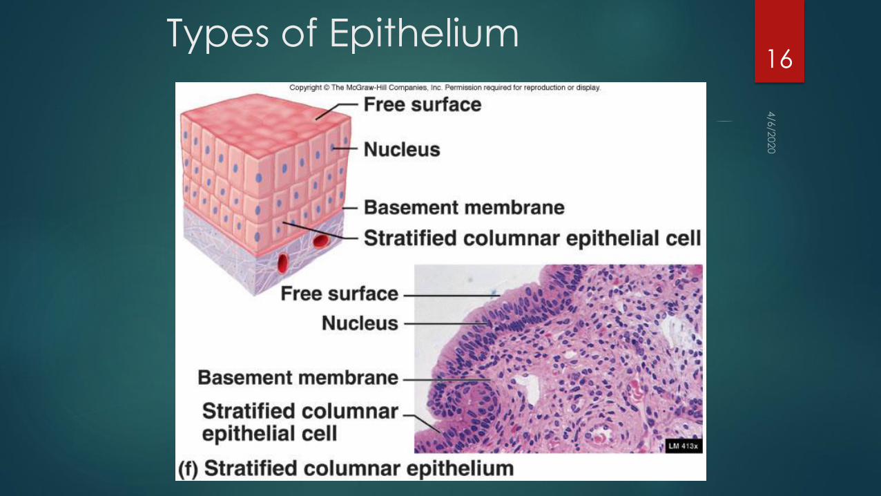
Types of Epithelium10

Types of Epithelium11

Types of Epithelium12

Classification of Epithelium
Stratified- consists of more than one layer
of cells, only one of which is attached to
the basement membrane
Squamous (wet and dry), Cuboidal,
Columnar
13

Types of Epithelium14

Types of Epithelium15
Types of Epithelium16

Classification of Epithelium
Pseudostratified- single layer of cells;
some tall and thin and reach the free
surface while others do not
Nucleii of these cells are at different
levels and appear stratified
Cells are almost always ciliated and
are associated with goblet cells that
secrete mucus onto the free surface
17

Types of Epithelium18

Classification of Epithelium
Transitional- stratified cells that appear
cuboidal when the organ or tube is
not stretched, and squamous when
the organ or tube is stretched by fluid
19

Types of Epithelium20

Epithelial Tissue
Epithelial cells retain the ability to undergo mitosis
and therefore can replace damaged cells with
new ones
Undifferentiated cells (stem cells) continuously
divide and produce new cells. In some types
of epithelia, such as skin and digestive tract,
cells that are lost or die are continuously
replaced by new ones.
21
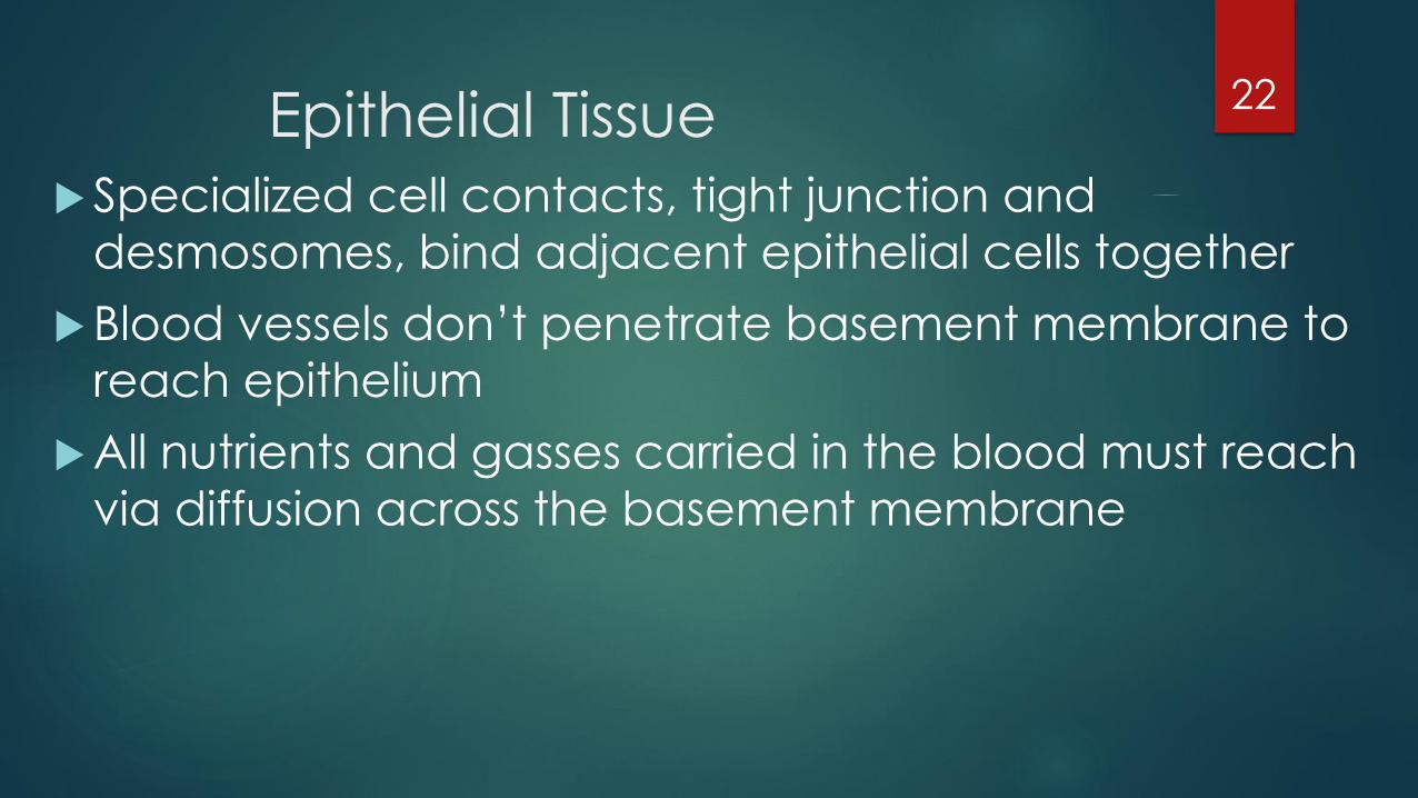
Epithelial Tissue
Specialized cell contacts, tight junction and
desmosomes, bind adjacent epithelial cells together
Blood vessels don’t penetrate basement membrane to
reach epithelium
All nutrients and gasses carried in the blood must reach
via diffusion across the basement membrane
22

Cell Connections
Functions
Bind cells together
Form permeability
layer
Intercellular
communication
Types
Desmosomes
Tight
Gap
23
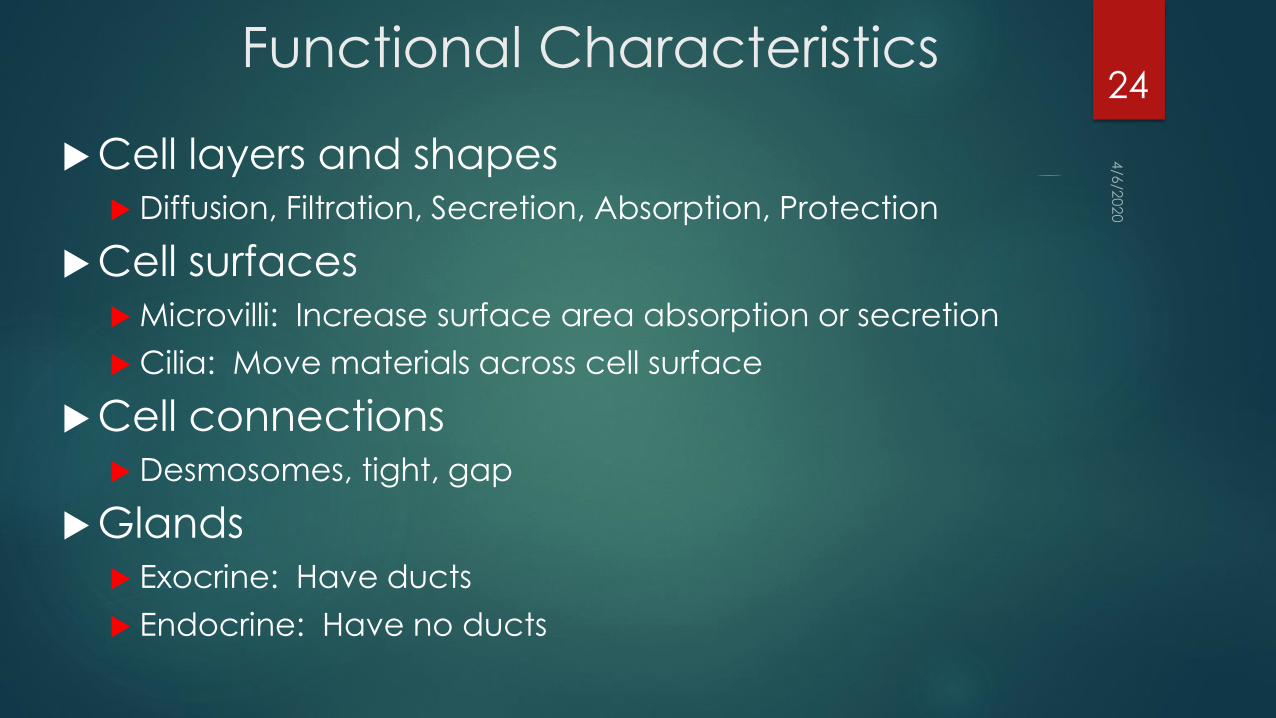
Functional Characteristics
Cell layers and shapes Diffusion, Filtration, Secretion, Absorption, Protection
Cell surfaces Microvilli: Increase surface area absorption or secretion
Cilia: Move materials across cell surface
Cell connections Desmosomes, tight, gap
Glands Exocrine: Have ducts
Endocrine: Have no ducts
24

Glands
Secretory organs
Composed primarily of epithelium with a supporting network of connective tissue
Glands with ducts are termed exocrine
Glands without ducts are termed endocrine
Cellular products of endocrine glands are hormones
25

Exocrine Glands
Unicellular
Goblet
cells
26

Multicellular Exocrine Glands27

Exocrine Glands and Secretion
Types
Merocrine
Sweat glands
Apocrine
Mammary
glands
Holocrine
Sebaceous
glands
28

Connective Tissue
Abundant
Consists of cell separated by extracellular
matrix
Diverse
Performs variety of important functions
29

Functions of Connective
TissueEnclosing and separating as capsules around
organs
Connecting tissues to one another as tendons
and ligaments
Supporting and moving as bones
Storing as fat
Cushioning and insulating as fat
Transporting as blood
Protecting as cells of the immune system
30
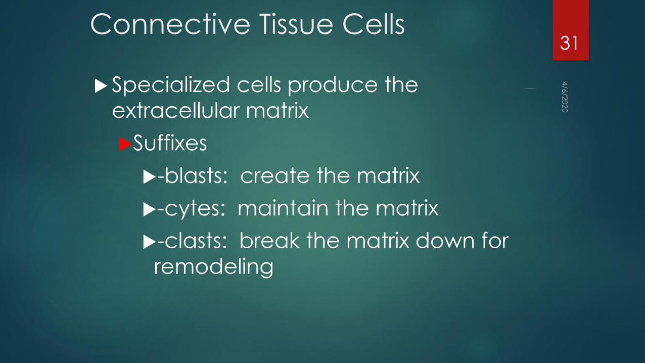
Connective Tissue Cells
Specialized cells produce the
extracellular matrix
Suffixes
-blasts: create the matrix
-cytes: maintain the matrix
-clasts: break the matrix down for
remodeling
31
Extracellular Matrix
Components
Protein fibersCollagen which is most common protein in body
Reticular fill spaces between tissues and organs
Elastic returns to its original shape after distension or compression
Ground substanceShapeless background
Fluid
32

Collagen Fibers
Strong and flexible but inelastic
Many types (at least 20)
33

Reticular Fibers
Very fine collagen fibers
Very short and thin
Branch to form networks
Not as strong as most collagen fibers
34
Elastic Fibers
Contain a protein called elastin
Has the ability to return to original shape
35
Other Matrix
MoleculesGround substance
Consists of hyaluronic acid, gives a slippery
quality to fluid
Good lubricant for joint cavities
Proteoglycans trap large quantities of H2O
which give them the ability to return to
original shape
Adhesive molecules
36

The structure of the matrix gives connective tissue types most of
their functional characteristics.
Bone and cartilage
wt bearing
Tendons and ligaments
withstand stress
Dermis of skin
Withstand damage
37

Loose Connective Tissue
Also known as areolar tissue
Loose packing material of most organs and tissues
Attaches skin to underlying tissues
Contains collagen, reticular, elastic fibers and variety of
cells 38

Dense Connective Tissue
Form thick bundles and fill nearly all of the extracellular space
Most of the cells of developing dense connective tissue are spindle
shaped fibroblasts
Dense connective tissue can be subdivided into 2 major groups
Regular
Irregular
39

Regular Dense Connective
Tissue Has protein fibers in the extracellular matrix that are oriented
predominantly in one direction
Has abundant collagen fibers which give it a white appearance
These tissues form structures such as tendons and ligaments
Resist stretching and give strength in direction of fibers
Examples are tendons and ligaments
40

Dense Regular Connective Tissue41

Irregular Dense
Contains protein fibers arranged as a mesh work of randomly oriented fibers
Alternately the fibers within a given layer can be oriented in one direction whereas the fibers of adjacent areas are oriented at nearly right angles of that layer
Sheets of connective tissue are formed that have strength in many directions
Examples are the dermis of the skin and the connective tissue capsules surrounding organs
42

Irregular Dense Elastic
In addition to collagen fibers
oriented in many directions,
there are abundant elastic fibers
in the layers of this tissue
Examples are found in the walls
of elastic arteries
43

Dense Irregular Collagenous, and
Elastic44

Connective Tissue with Special
Properties Adipose tissue
Consists of adipocytes
Types
Yellow (white)
most abundant, white at birth and yellows with
age
Brown
found only in specific areas of body as axillae,
neck and near kidneys
Reticular tissue
Forms framework of lymphatic tissue
Characterized by network of fibers and cells
45

Adipose Tissue Consists of adipocytes or fat cells, which contain large amounts of
lipids
Unlike other connective tissue types, adipose is composed of large cells and a small amount of extracellular matrix that consists of loosely arranged collagen and reticular fibers with some scattered elastic fibers
This tissue is found in the subcutaneous areas, renal pelvis, around kidneys, attached to surface of colon, mammary glands, and in loose connective tissue.
It serves to insulate and protect, as well as to provide for energy storage
46

Adipose Tissue47

Hemopoietic tissue/ blood
Abundant extracellular matrix
Free moving cells in a liquid matrix
This allows for rapid movement throughout the body
Most of the matrix is produced in cells contained in other tissues rather than blood cells
Found mostly in bone marrow
Two types of bone marrow
Yellow and red
48

Blood
Matrix between the
cells is liquid
Hemopoietic tissue
Forms blood cells
Found in bone marrow
Yellow
Red
49

Bone Marrow50

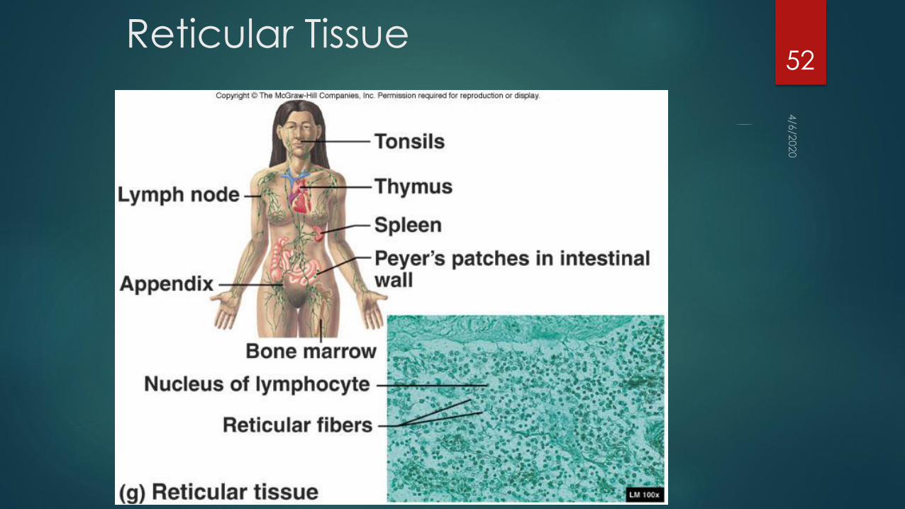
Reticular Tissue
Forms the framework of lymphatic tissue such as in the spleen and
lymph nodes, as well as bone marrow and liver
Characterized by a network of reticular fibers and reticular cells
Reticular cells produce the reticular fibers and remain closely attached
to them.
51
Reticular Tissue52

Cartilage
Composed of cartilage cells, or chondrocytes located in spaces
called lacunae
The surface of nearly all cartilage is surrounded by a layer of dense
irregular connective tissue called the perichondrium
Cartilage cells arise from the perichondrium to secrete cartilage
matrix
Cartilage has no blood vessels or nerve except those of the perichondrium.
Hyaline cartilage has large amounts of both collagen fibers and
proteoglycans
53

Hyaline Cartilage
Large amounts of both collagen fibers and protoglycans
Collagen fibers are evenly dispersed throughout the ground
substance
In joints, quite smooth
Found in areas where strong support and flexibility are needed, such
as in the rib cage, trachea, and bronchi. Also covers areas of bones
54

Hyaline Cartilage
55

Fibrocartilage
Has more collagen fibers than proteoglycans and much thicker
bundles of collagen fibers than hyaline
Slightly compressible, very tough
Found between vertebrae and areas of body where a great deal of
pressure is applied to joints (knee, jaw, etc.)
56

Fibrocartilage
57
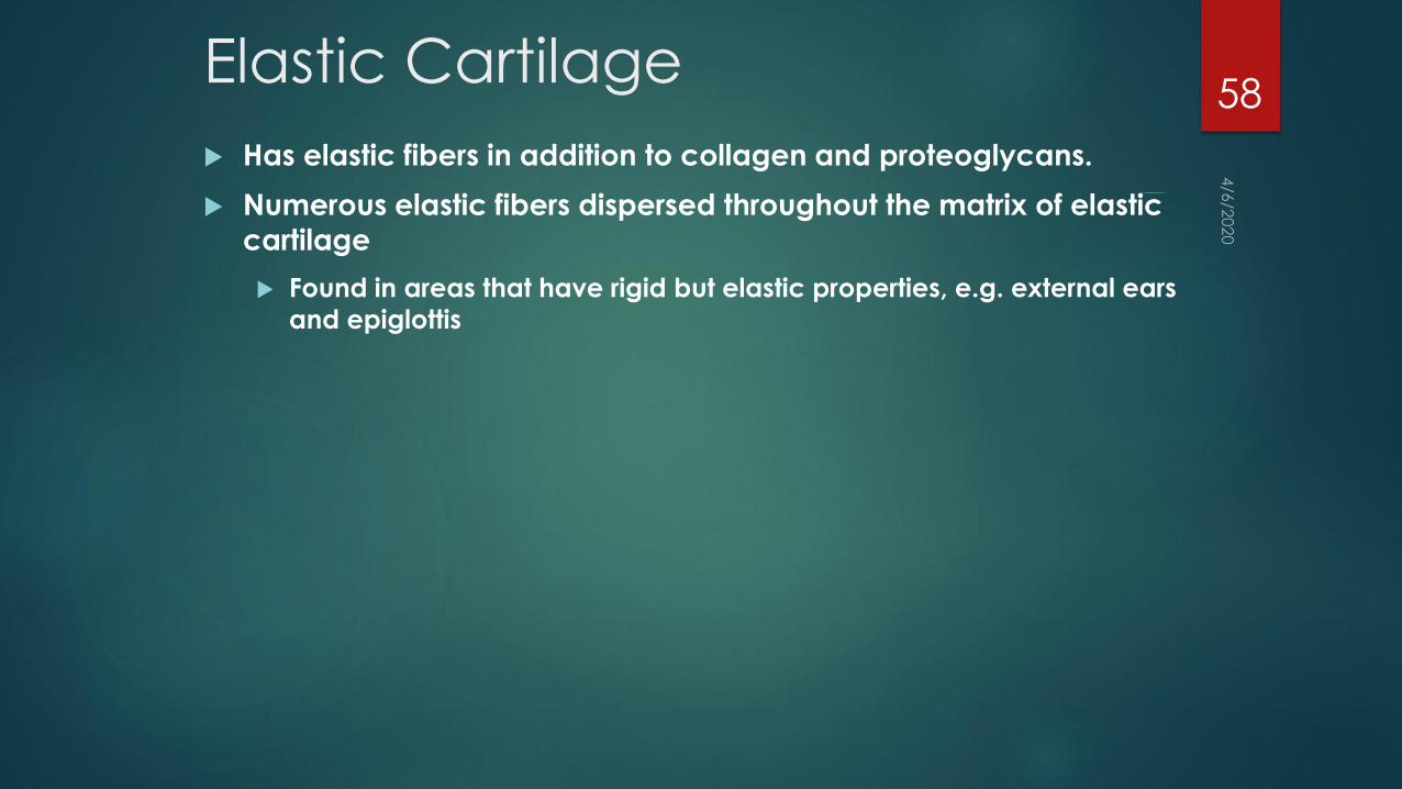
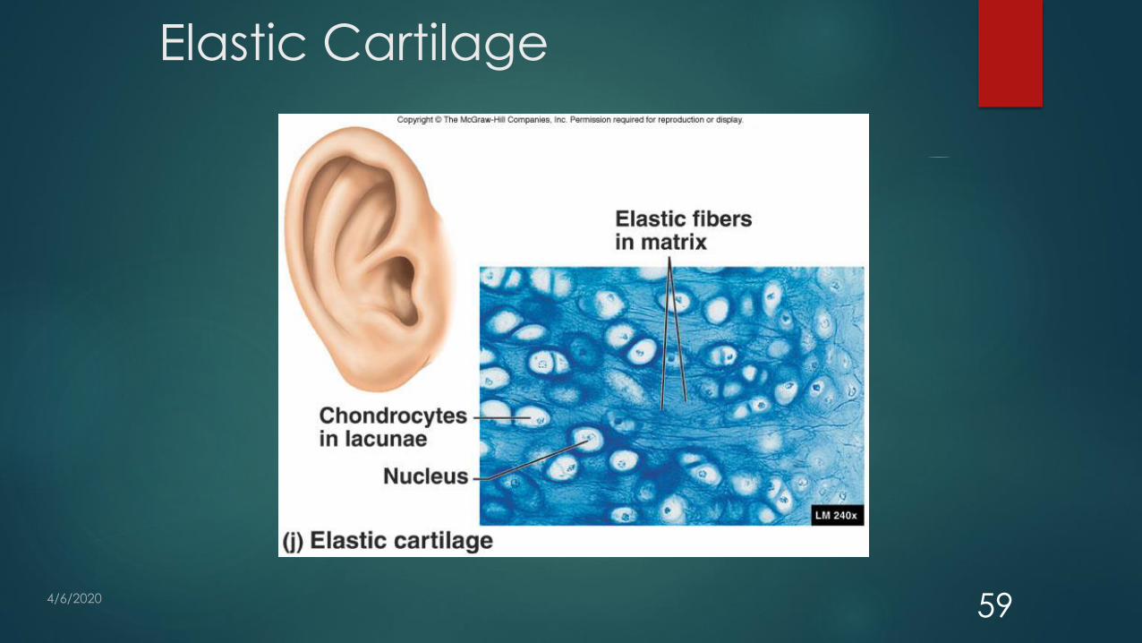
Elastic Cartilage
Has elastic fibers in addition to collagen and proteoglycans.
Numerous elastic fibers dispersed throughout the matrix of elastic
cartilage
Found in areas that have rigid but elastic properties, e.g. external ears
and epiglottis
58
Elastic Cartilage
59

Bone
A hard connective tissue that consists of living cells and mineralized matrix with-
Organic- protein fibers,
primarily collagen and other organic materials
Inorganic- hydroxyapatite (specialized crystals)
contain calcium and phosphate
The strength and rigidity of bone allow it to support and protect other structures within the body
Osteocytes (bone cells) located within lacunae of the matrix (similar to cartilage)
60

Two types of Bone
Cancellous or spongy
Has spaces between trabeculae beams or plates of bone and therefore
resemble a sponge
Located in the interior of the bones of the skull, vert, sternum, and pelvis, as
well on ends of long bones.
Compact bone
More solid with almost no space between many thin layers of lamellae of
bone.
Located on outer bones and shafts of long bones
61

Bone62

Muscle Shortens with force
3 main types
Skeletal
Cardiac
Smooth
Grouped according to structure and function.
Structure
Can be either striated, or non striated.
Function
Can be voluntary or involuntary
63

Skeletal muscle
attach to bone and assist with movement of the body
Cardiac muscle
Located in the heart and pumps blood under involuntary control
Smooth muscle
Located within the hollow organs such as the stomach ad intestines and
regulates the size of the organs, forces fluids through tubes , controls the
amount of light entering the eye.
64
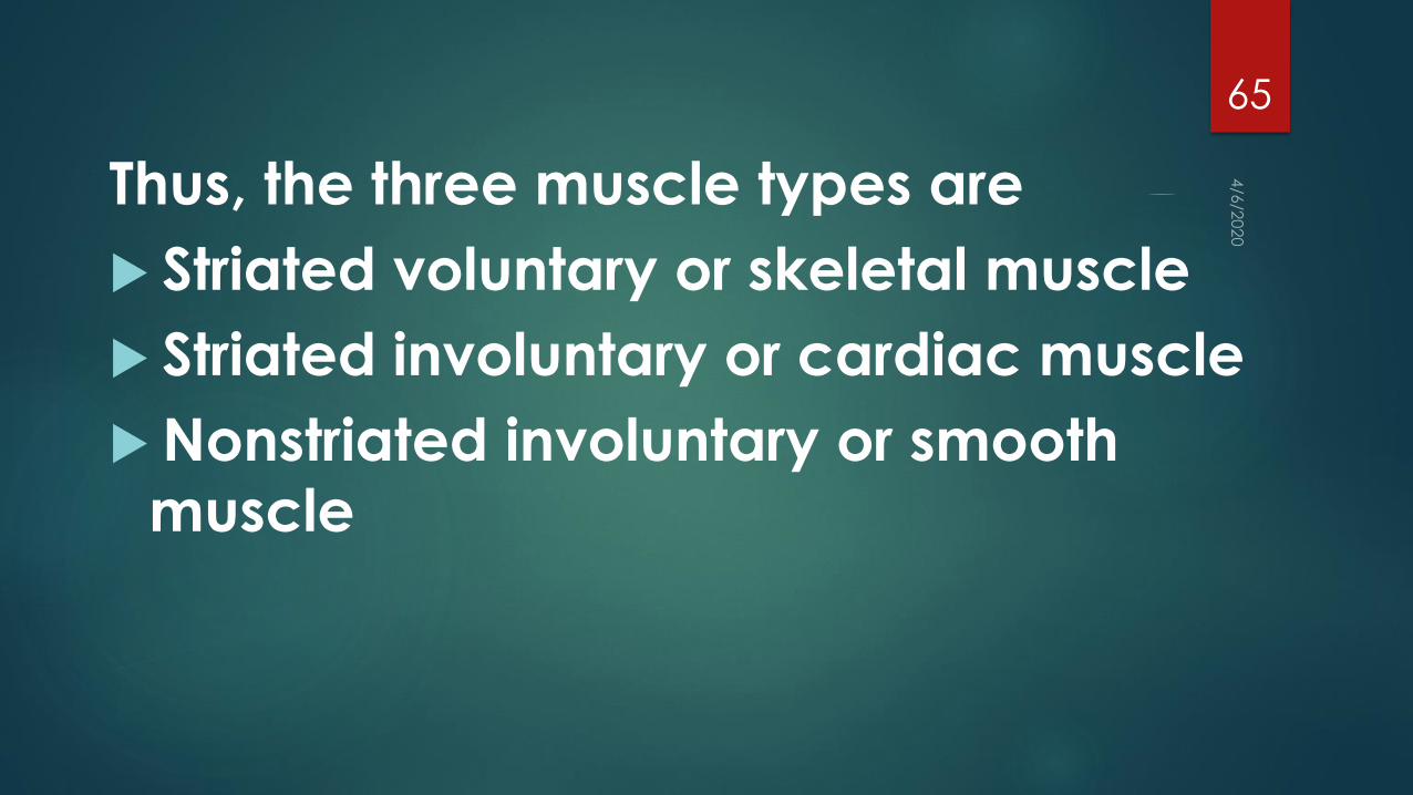
Thus, the three muscle types are
Striated voluntary or skeletal muscle
Striated involuntary or cardiac muscle
Nonstriated involuntary or smooth
muscle
65

Muscle Tissue
Characteristics
Contracts or shortens with force
Moves entire body and pumps blood
Types
Skeletal
Striated and voluntary
Cardiac
Striated and involuntary
Smooth
Nonstriated and involuntary
66
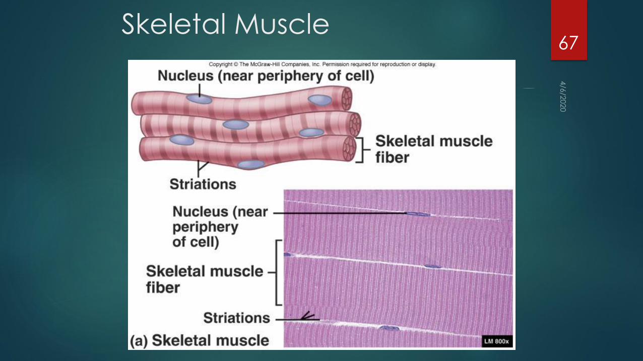
Skeletal Muscle67

Cardiac Muscle68

Smooth Muscle69

Nervous
Found in the brain spinal cord and nerves
Able to conduct electric impulses called action potentials
Consists of neurons responsible for conductive ability and support
cells called neuroglia.
70

Neurons or nerve cells Actual conducting cells of nervous tissue
Composed of three parts
Cell body, contains nucleus,
Dendrites receive action potential and send them to the cell body
Shorter than axons, taper to a fine tip
Axon usually conduct action potential away from the cell body
Much longer than dendrites, constant diameter
71

Multipolar Neurons.
Neurons with several dendrites and one axon
Bipolar Neurons
Neurons that have a single dendrite and an axon
Unipolar Neurons.
Neurons with one axon and no dendrites
72

Neurons73

Neuroglia74

Functions of Blood
Distribution - nutrients, wastes, hormones, gases, etc.
Self-sealing – hemostasis
Disease/ infection fighting

Blood = connective tissue
extracellular
matrix:
Plasma
specialized cells:
(= Formed elements)
RBCs
WBCs
Plateletscolor ?
volume ?
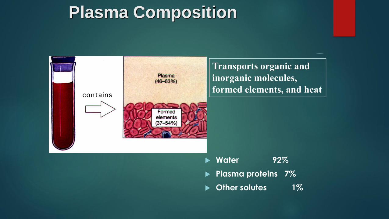
Plasma Composition
Water 92%
Plasma proteins 7%
Other solutes 1%
Transports organic and
inorganic molecules,
formed elements, and heat
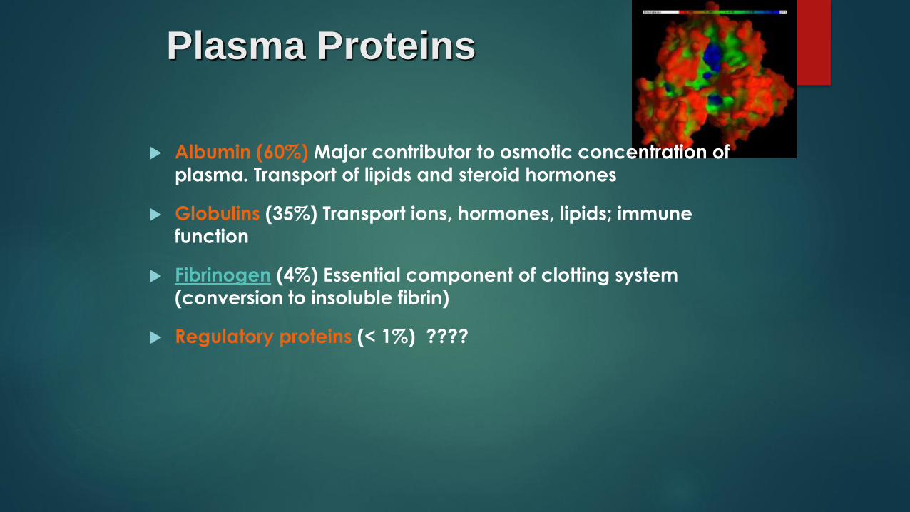
Plasma Proteins
Albumin (60%) Major contributor to osmotic concentration of
plasma. Transport of lipids and steroid hormones
Globulins (35%) Transport ions, hormones, lipids; immune
function
Fibrinogen (4%) Essential component of clotting system
(conversion to insoluble fibrin)
Regulatory proteins (< 1%) ????
Other Solutes
Electrolytes: Normal extracellular
fluid ion composition (????)
Organic nutrients: glucose, FA,
AA
Organic wastes: urea, bilirubin

Difference between Plasma and Interstitial Fluid :
Plasma has more:
Dissolved O2 O2 diffuses out into tissue
Dissolved proteins (too big to cross caps.)
Albumins
Globulins
globulins
and globulins
Fibrinogen
Similar concentration: Salts & small molecules
serum = plasma -
Difference between
plasma and serum?

. . . . 2 more things:
Most plasma proteins are made in liver. Exception: ?
Lipoproteins = particles containing lipids (cholesterol &
triglycerids) and proteins (albumins & globulins)

Formed Elements
Red and White Blood
Cells
Platelets
Platelets
WBCs
RBCs.1%
99.9%

Formed Elements cont.Why white blood cells???
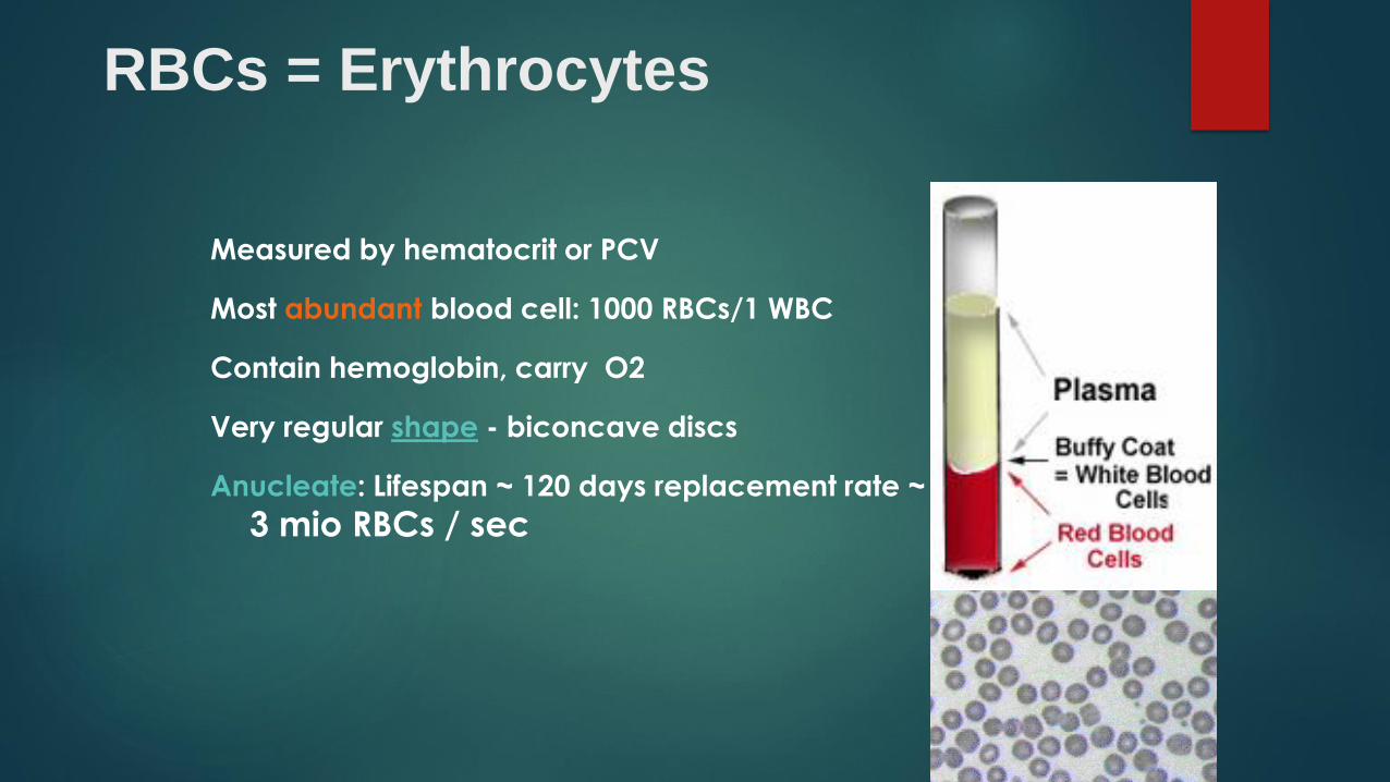
RBCs = Erythrocytes
Measured by hematocrit or PCV
Most abundant blood cell: 1000 RBCs/1 WBC
Contain hemoglobin, carry O2
Very regular shape - biconcave discs
Anucleate: Lifespan ~ 120 days replacement rate ~
3 mio RBCs / sec

Structure of Hemoglobin (Hb)
Fe ion in heme
group
reversibly binds
O2
How many oxygen
molecules can 1
Hb molecule
carry?

ABO & Rh Blood Types
Blood groups (types) based on specific RBC surface
antigens (= proteins)
> 30 common varieties of antigens known. Most important
ABO & Rh blood type ?

ABO Blood typing:
4 combinations possible
A surface antigen = blood type A
B surface antigen = blood type B
both surface antigens = type AB
neither surface antigen = type O
Rh surface antigen = + blood type
no Rh antigen = negative blood type

. . . 2 - 8 months after birth:
Anti-A and anti-B antibodies can be formed in plasma !
normally NO
anti Rh present

Transfusion ReactionTransfusion of incompatible blood can be fatal!
Universal Donor vs.
Universal RecipientOnly for emergencies - must be
given slowly !
Clinical Brief
Anemia: p. 536
Reduced oxygen carrying ability of blood. Causes??
Polycythemia: Erythrocytosis: excessive increase in RBCs
Polycythemia vera:
Blood Doping: p. 545Via direct transfusion, or
EPO use
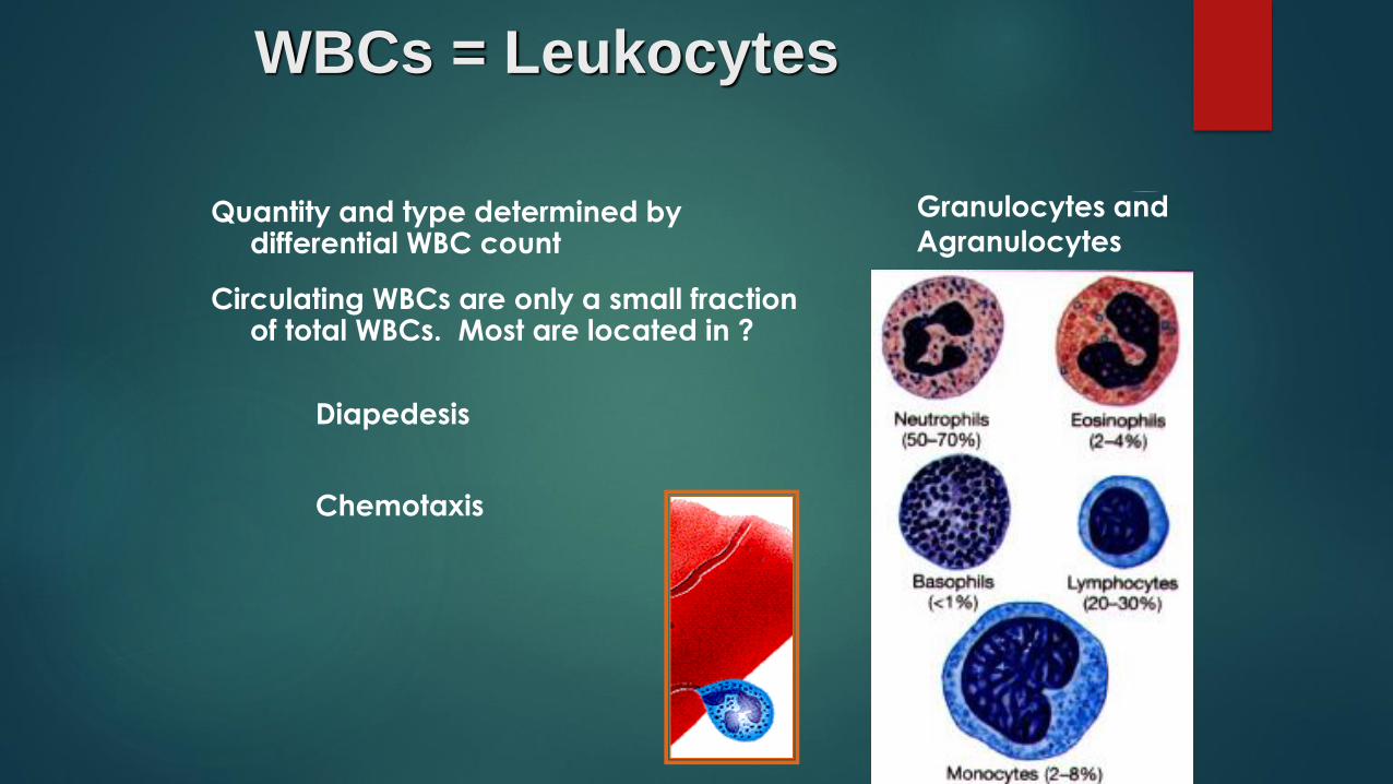
WBCs = Leukocytes
Quantity and type determined by differential WBC count
Circulating WBCs are only a small fraction of total WBCs. Most are located in ?
Diapedesis
Chemotaxis
Granulocytes and
Agranulocytes
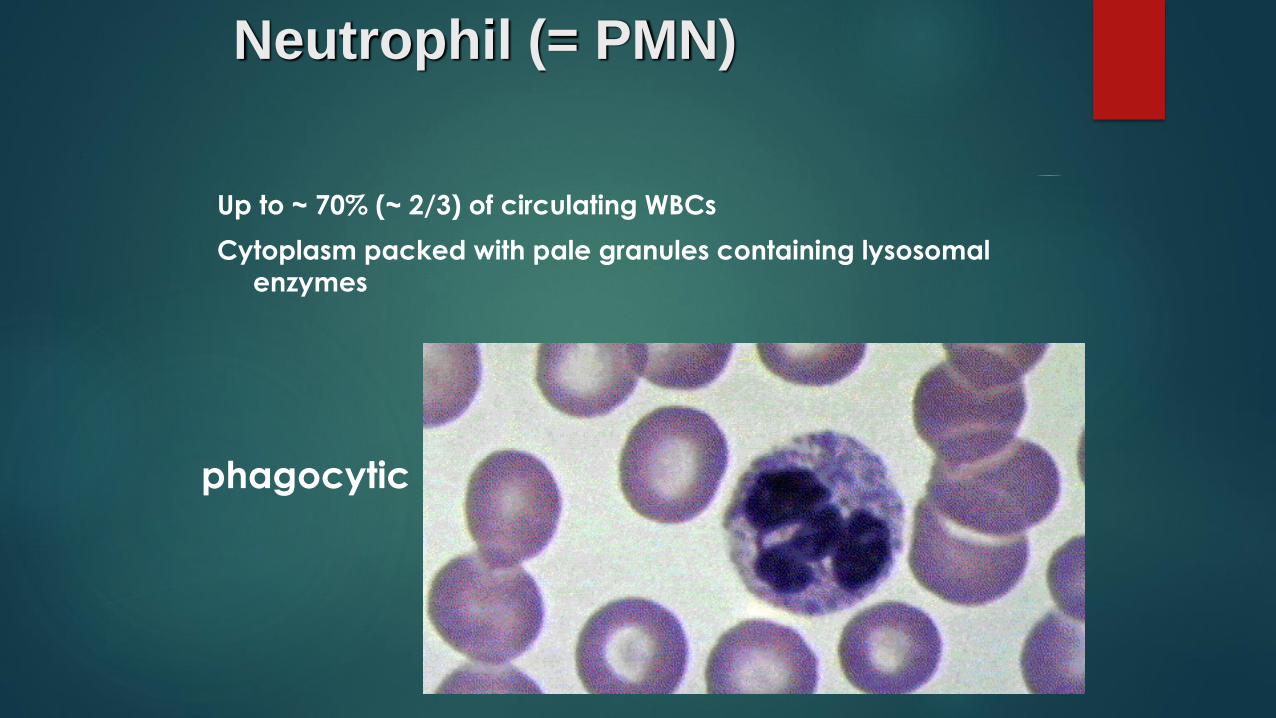
Neutrophil (= PMN)
Up to ~ 70% (~ 2/3) of circulating WBCs
Cytoplasm packed with pale granules containing lysosomal
enzymes
phagocytic

Eosinophil
~ 2% - 4% of circulating WBCs
Granules stain with eosin
Increased in allergies and parasitic infections

Basophil
< 1% of circulating WBCs
Granules stain with basic dyes and contain histamine
Discharge of histamine promotes inflammation at site
of injury (Similar to mast cells)

Monocyte
~ 2% - 8% of circulating WBCs
Large kidney shaped nucleus
In tissue called Macrophage

Lymphocytes
~ 20% - 30% of circulating WBCs
Relatively small (slightly larger than RBCs)
Large round nucleus
B, T, NK

Platelets = Thrombocytes
Cell fragments of Megakaryocytes
(~ 4,000 thrombocytes per Megakaryocyte)
~ 160 m
Lifespan ~ 12 days
involved in blood clotting

Abnormal Blood Cell Counts
Leukopenia < 2,500/ L (normal 6000 – 9000)
Leukocytosis > 30,000/ L
Thrombocytopenia: < 80,000/ L (normal ~ 350,000)
Thrombocytosis: > 1,000,000/ L
Also
Lymphopenia vs. _____________
_________vs. Neutrophilia
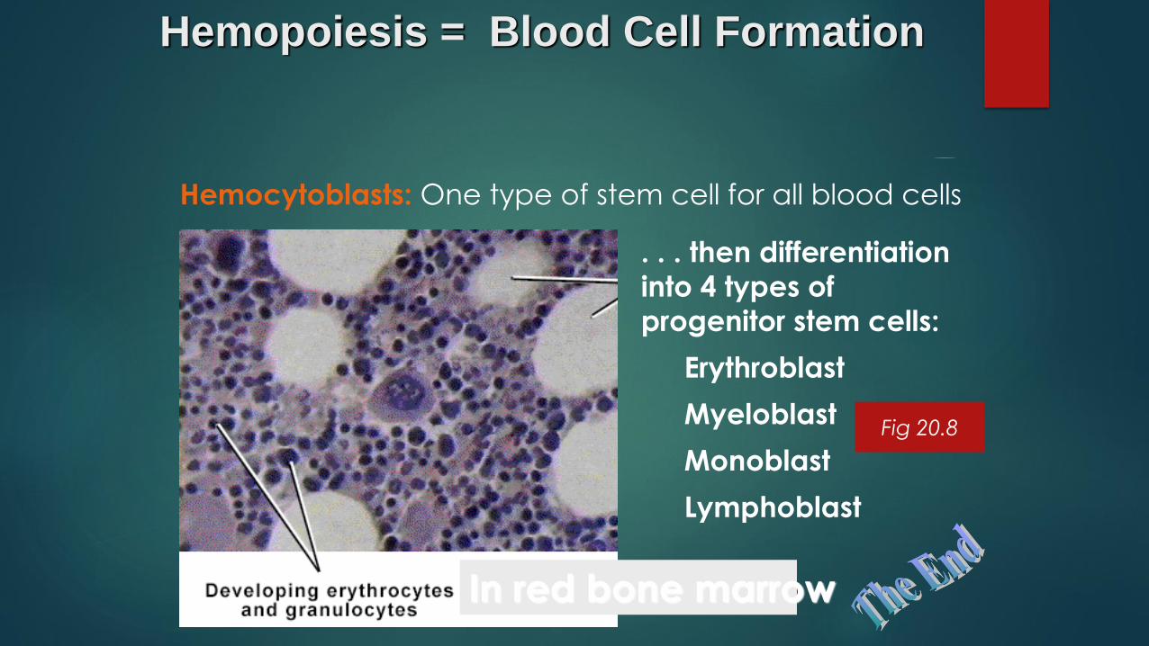
Hemopoiesis = Blood Cell Formation
Hemocytoblasts: One type of stem cell for all blood cells
In red bone marrow
. . . then differentiation
into 4 types of
progenitor stem cells:
Erythroblast
Myeloblast
Monoblast
Lymphoblast
Fig 20.8