tissue-specific expression and developmental regulation of
TRANSCRIPT

Tissue-specific expression and developmental regulation of cytochrome
b561 genes in Arabidopsis thaliana and Raphanus sativus
Wim Verelsta,1,*, Jyoti Kapilaa, Janice de Almeida Englerb, Julie M. Stonec, Roland Caubergsa and Han Asardc
aLaboratory of Plant Physiology, University of Antwerp, Groenenborgerlaan 171, B-2020 Antwerp, BelgiumbLaboratory of Plant Genetics, University of Gent–VIB, K.L. Ledeganckstraat 35, B-9000 Gent, BelgiumcDepartment of Biochemistry, Plant Science Initiative, Beadle Center, University of Nebraska—Lincoln, Lincoln, NE 68588–0664, USA1Present address: Max Planck Institute for Plant Breeding Research, Department of Molecular Plant Genetics, Carl-von-Linne-Weg 10,D-50829 Cologne, Germany*Corresponding author, e-mail: [email protected]
Received 20 February 2003; revised 4 June 2003
Ascorbate (Asc) is an essential molecule in many aspects ofdevelopment and stress responses in plants and animals. Cyto-
chromes b561 (cyts b561) are tightly coupled to Asc homeo-
stasis. These proteins are found in mammalian tissues, where
they are involved in the regeneration of Asc, serving thesynthesis of catecholamine neurotransmitters, and in intestinal
iron reduction. Plant genomes encode homologous membrane-
associated, Asc-reducible cyts b561. The expression of these
proteins in plants, however, has so far not been studied. Wehave now examined the expression of two Arabidopsis thalianacyt b561-encoding genes—Artb561-1 and Artb561-2—using
relative-quantitative RT-PCR and in situ hybridization (ISH)
techniques. The genes show overlapping and distinct tissue-and organ-specific expression patterns. Transcripts of both
genes are found in leaf epidermal cells, and expression seems
to correlate with leaf maturation and cessation of cell elonga-
tion. Both genes are also expressed in the epidermal cell layerof stems and roots in the L1 layer of the shoot apex, in the
vascular system of leaves, stems and roots, and in the root
pericycle. In addition, Artb561-1 is expressed in the root cap,
whereas Artb561-2 mRNA is found in the epidermis of lateralroots, in the root meristem, and in unfertilized ovules. These
observations provide important information for the elucidation
of the physiological function of cyts b561 in plants.
Introduction
Ascorbate (Asc) plays a prominent role in a large numberof cellular processes, as a major component in maintainingredox homeostasis. Apart from its function as an antioxi-dant, it acts as a cofactor for several metabolic enzymes(Arrigoni and De Tullio 2000). As a consequence of theseprocesses, the pool of reduced Asc molecules becomesdepleted. The hydrophilic nature of Asc prevents it fromdiffusing freely through biological membranes. Therefore,subcellular compartments that cannot synthesize Ascrequire mechanisms for replenishing reduced Asc mol-ecules. This can be achieved by either trans-membranetransport of the molecule itself (Horemans et al. 2000a,2000b), by trans-membrane transport of electrons, usingoxidized Asc (dehydro ascorbate, DHA) or monodehydroascorbate (MDHA) as electron acceptors (Asard et al.
1992, Horemans et al. 1994), or by a combination ofboth mechanisms. One protein known to replenishreduced Asc in a mammalian subcellular compartment isthe chromaffin granule cytochrome b561 (cyt b561). Thisprotein is responsible for the transfer of electrons intospecialized chromaffin granule vesicles, where catechol-amine neurotransmitters are synthesized by an Asc-dependent enzyme (Wakefield et al. 1986, Njus et al. 1987).
A family of membrane proteins with sequence hom-ology to the chromaffin granule cyt b561, likely to playan important role in plant cellular Asc metabolism, hasonly recently been identified (Asard et al. 2001). The con-servation of structural features between the putative plantand animal cyt b561 protein sequences is striking (Asardet al. 2001). The overall amino acid identity between
PHYSIOLOGIA PLANTARUM120: 312–318. 2004 Copyright# Physiologia Plantarum 2004
Printed in Denmark – all rights reserved
Abbreviations – Asc, ascorbate; cyt(s) b561, cytochrome(s) b561; DHA, dehydro ascorbate; ISH, in situ hybridization; MDHA, monodehydroascorbate.
312 Physiol. Plant. 120, 2004
human chromaffin granule cyt b561 and the proteinsencoded by the A. thaliana Artb561-1 and Artb561-2genes is 36% and 35%, respectively. Conserved featuresinclude six predicted transmembrane helices, four Hisresidues likely to be involved in heme ligation, and apredicted binding site for MDHA (Asard et al. 2001).A putative binding site for Asc (Okuyama et al. 1998) isnot as well conserved among plant cyts b561 (Verelst andAsard 2003). At least one member of this family of plantproteins is plasma membrane localized and has beenextensively characterized at the biochemical level(Asard et al. 1998, 2000, 2001).
The bovine adrenal chromaffin cyt b561 has beenpurified (Perin et al. 1988, Tsubaki et al. 1997), andrecent attempts to purify the corresponding proteinfrom plants have been met with some success (Trostet al. 2000, Asard et al. 2001, Berczi et al. 2001). Allavailable information on plant cyt b561 has beenobtained through biochemical studies and sequence com-parisons with the animal system. Although most bio-chemical work has been performed on purified plasmamembranes of bean hypocotyls (Phaseolus vulgaris L.),the presence of cyt b561 has been reported in a variety oforgans from a large number of vascular plants (Asardet al. 1998, 2001). However, the physiological function ofthe protein in plants remains unknown. The mammalianchromaffin cyt b561 shuttles electrons across the chro-maffin granule membrane, from cytosolic Asc to intra-vesicular MDHA, thereby replenishing the pool ofreduced Asc molecules (Wakefield et al. 1986, Njuset al. 1987, 2001). In vitro studies on bean hypocotylplasma membrane vesicles have shown that the plantcyt b561 is also capable of transmembrane electron trans-port from Asc to MDHA and ferricyanide (Asard et al.1992, Horemans et al. 1994). These experiments indicatethat plant plasma membrane cyt b561 could shuttle elec-trons between intracellular Asc and an extracellularacceptor, similar to the mode of action of the chromaffincyt b561 (Asard et al. 1998). Therefore, the plasma mem-brane cyt b561 in plants may be capable of regeneratingAsc in the apoplast using electrons from cytosolic Asc. Itmay thus be involved in the physiological actions of Ascin the extracellular space (Asard et al. 2001).
Cyt b561 research has advanced by the recent identifi-cation of additional cyt b561 homologues in animals. Aferric reductase in the plasma membrane of duodenalbrush border cells was found to be highly homologousto the chromaffin granule cyt b561 (McKie et al. 2001).The presence of multiple cyts b561 supports the hypothesisthat cyts b561 may occur in different subcellular locationsin different tissues, carrying out specialized functions.
Based on similarities to the animal cyt b561 proteinsequences, a number of putative cyt b561-encoding geneshave been identified in dicots and monocots (Asard et al.2001, Verelst and Asard 2003). As in mammals, more thanone cyt b561 homologue is present in most plant specieswhere cyt b561 genes have been identified. The occurrenceof different isoforms of the cyt b561 protein in plants alsopoints to potentially different functions for these proteins
in various tissues. Moreover, there is evidence for plantcyts b561 to reside in multiple subcellular compartments,as indicated by sucrose gradient centrifugation analyses ofcauliflower, bean and mung bean membranes (Asard et al.1987, Scagliarini et al. 1998).
Using relative-quantitative RT-PCR, we studied theexpression patterns of two putative Arabidopsis thalianacyt b561 encoding genes (Artb561-1 and Artb561-2,TAIR accession numbers At4g25570 and At5g38630,respectively) at the organ level in A. thaliana. In addition,in situ hybridization analyses (ISH) were used toexamine the expression patterns of both genes in moredetail, at the tissue level in A. thaliana and Raphanussativus (radish) sections. While all previous studies oncyt b561 in plants yielded information on biochemicalproperties at the membrane level, these experiments arethe first to provide information at the whole plant level.These results may contribute to our understanding of thephysiological functions of cyt b561 in plants, by promot-ing the formulation of working hypotheses that can bethe basis for future studies.
Materials and methods
Plant material and growth conditions
A. thaliana L. cv. Columbia Col-0 and R. sativus L. seeds(bought from a local market) were surface sterilized andsown on K1 germination medium (Valvekens et al. 1988).Petri dishes were placed under growth chamber conditions(60%humidity, 22�C, 80mEm�2 s�1) with a light regime of16h of light and 8h of darkness. A. thaliana seedlings werecollected 7 days after germination, and radish roots andshoot apices 12 days after germination. A. thaliana plantswere also grown in soil, under the same growth conditionsas sterile-grown plants, and after 4–6 weeks flowers andsiliques were collected from these plants for ISH.
A. thaliana roots used for RNA isolation were grownin liquid cultures following the procedure of theMeyerowitzLaboratory (http://www.caltech.edu/�meyerowitz), andcollected 20 days after germination.
Probe preparation
Using EST clones 170N15T7 (accession number R65413)and ATTS1779 (accession number Z26702) as templates,the ESTs of Artb561-1 and Artb561-2, respectively, wereamplified by PCR, using SP6 and T7 primers. The PCRproducts of, respectively, 927 and 678 bp were purifiedby gel extraction and used as templates for in vitrotranscription, using an SP6/T7 Transcription Kit(Roche Diagnostics, Mannheim, Germany) and [a-35S]-UTP (Amersham, Roosendaal, the Netherlands). Senseand antisense radiolabelled riboprobes of both geneswere synthesized and hydrolysed to an average lengthof 300 nucleotides as described by de Almeida Engleret al. (2001), and column-purified using ‘Bio-Spin 30Chromatography Columns’ (Bio-Rad, Hercules, CA,USA). The quality and length of the transcripts was
Physiol. Plant. 120, 2004 313
checked on a denaturing agarose gel (de Almeida Engleret al. 2001).
In situ hybridization (ISH)
ISH was essentially performed as described by deAlmeida Engler et al. (2001). Plant material was fixedin 2.5% glutaraldehyde in 0.1M cacodylate buffer,pH7.0, and subsequently embedded in paraffin asdescribed by de Almeida Engler et al. (2001). A. thalianaand R. sativus tissue sections were hybridized with senseor antisense 35S-UTP labelled RNA probes of theArtb561-1 and Artb561-2 genes. For each slide 15�106
cpm of [a-35S]-UTP-labelled RNA-probe were used forhybridization. Exposure times varied. Images were takenwith a digital Axiocam (Zeiss) under standard dark andbright field optics.
Relative-quantitative RT-PCR
Total RNA was isolated from 100 to 300mg frozenA. thaliana tissues (seedlings, leaves, flowers, roots)using TriPure Isolation Reagent (Roche Diagnostics,Mannheim, Germany), following the manufacturer’sinstructions. The concentration of total RNA was quan-tified spectrophotometrically at 260 nm. About 3mg ofthis RNA was used for cDNA synthesis with a ‘FirstStrand cDNA Synthesis Kit’ (Fermentas, Vilnius,Lithuania). RT-PCR conditions were optimized forboth Artb561-1 and Artb561-2 genes, using 150 ng ofcDNA and gene-specific primers overlapping intronregions. The manufacturer’s instructions were followed,and care was taken that the PCR amplification reactionswere stopped in the linear range, to allow reliable quan-tification of the amplicons. For quantification, Universal18S rRNA primers and competimers (QuantumRNA18S Internal Standards; Ambion, Austin, TX, USA)were used in a ratio of one to nine, as determined intrial experiments (according to the kit’s manual).RT-PCR bands were visualized in agarose gels usingethidium bromide. After scanning, band intensities werequantified using OptiQuant Image Analysis Software(Packard, Meriden, CT, USA).
Results
Relative-quantitative RT-PCR analyses
Relative-quantitative RT-PCR was used to examine theexpression of Artb561-1 and Artb561-2mRNA in differentA. thaliana organs. We tested the expression of both genesin young seedlings (11 days after germination (DAG)), inroots (from liquid cultures, 20 DAG), young and mature(fully expanded) leaves (both from plants at 25, 30, 47 and65 DAG), flower buds, unfertilized flowers and fertilizedflowers (from plants 65 DAG). At least three independentsamples were tested for each organ type. Levels of 18SrRNA were used as an internal standard. The amplifica-
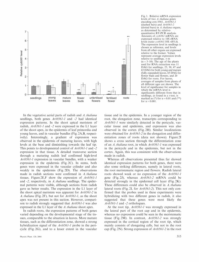
tion of this abundant transcript was suppressed usingAmbion’s competimer technology (Ambion), so that theamount of 18S amplicon in RT-PCR was of the sameorder of magnitude as that of the Artb561-1 and Artb561-2 amplicons. Expression levels were expressed relativeto that of 18S rRNA. To compare the mRNA levels ofthe cytb561 genes in different organs, the averageamount of cytb561 mRNA in seedlings was arbitrarilyset to 1, and the levels in other organs were expressedrelative to this value (Fig. 1). It was not possible tomake an absolute quantitative comparison between thetwo genes studied in these experiments, since the RT-PCR method is only relative-quantitative, and differentamounts of PCR cycles were used for optimal amplifica-tion of both transcripts.
For both genes the mRNA levels in young leaves werecomparable to those in seedlings (1.09� 0.48 and0.82� 0.30, respectively). In mature leaves the levelswere slightly higher than in seedlings, but statisticallynot significantly different (1.54� 0.27 and 1.32� 0.39,respectively). Plant age (DAG) did not affect theresults for leaves, because similar values were obtained forleaves of comparable developmental stages collected fromplants 25, 30, 47 and 65 DAG (data not shown).
In flower buds and unfertilized flowers, the Artb561-1transcript levels (0.28� 0.06 and 0.27� 0.05) were sig-nificantly lower than in seedlings, whereas the levels ofArtb561-2 mRNA in these samples were comparable tothose in seedlings (1.19� 0.21 in flower buds and1.32� 0.16 in unfertilized flowers). In fertilized flowersthat were starting to form seeds, levels of both geneswere very low: the Artb561-1 and Artb561-2 transcriptlevels were 0.08� 0.02 and 0.43� 0.13, respectively. Forboth genes the reduction in expression in fertilized flowerswas about 70%, when compared to flower buds orunfertilized flowers. The expression in flowers thus sug-gested developmental regulation of both genes. Therewas also a pronounced organ-specificity, especiallyapparent in roots as compared to other organs. In roots,the expression levels of both genes were significantlyhigher than in seedlings (3.50� 0.36 and 5.78� 0.76).However, we can not fully exclude an effect of growthconditions (liquid culture versus soil-grown plants) onthe Artb561-1 and -2 expression levels in roots. Ourdata suggest a differential expression of these two genes.
In situ hybridization
ISH was used to investigate the expression patterns ofthe two putative cyt b561 encoding genes in tissue sec-tions of A. thaliana and its closely related species R. sativus(radish). The A. thaliana and R. sativus genomes arevery similar (Martinez et al. 1992), and radish tissueshave the advantage of being considerably larger thanA. thaliana, which allows more refined analysis of theexpression patterns. Control hybridization with senseprobes gave no significant background signal, and there-fore only results obtained with antisense probes arepresented here.
314 Physiol. Plant. 120, 2004
In the vegetative aerial parts of radish and A. thalianaseedlings, both genes Artb561-1 and -2 had identicalexpression patterns. In the shoot apical meristem ofradish, Artb561-1 and -2 were expressed in the L1 layerof the shoot apex, in the epidermis of leaf primordia andyoung leaves, and in vascular bundles (Fig. 2A,B, respect-ively). Interestingly, a gradient of expression wasobserved in the epidermis of maturing leaves, with highlevels at the base and diminishing towards the leaf tip.This points to developmental control of Artb561-1 and -2expression in that tissue. A detailed transverse sectionthrough a maturing radish leaf confirmed high-levelArtb561-1 expression in vascular bundles, with a weakerexpression in the epidermis (Fig. 2C). In stems, bothgenes were expressed in the vascular cylinder and alsoweakly in the epidermis (Fig. 2D). The observationsmade in radish sections were confirmed in A. thalianatissues. Figure 2E,F show the expression of Artb561-1and -2, respectively, in A. thaliana seedlings. The epider-mal patterns were visible, although sections from radishgave us better results. The expression in the L1 layer ofthe shoot apical meristem was apparent for Artb561-2 inA. thaliana (Fig. 2F), but not for Artb561-1, as the shootapex was not present in this section. However, compari-son to radish strongly suggested that Artb561-1 was alsoexpressed in the L1 layer of the A. thaliana shoot apex.
In radish roots, the expression patterns of both genesvaried depending on the developmental stage of the tis-sues, comparable to the situation in leaves. More maturetissues, such as the differentiation zone, showed a stronghybridization signal of the Artb561-1 probe in the peri-cycle (Fig. 2G), and to a lesser extent in the vascular
tissue and in the epidermis. In a younger region of theroot, the elongation zone, transcripts corresponding toArtb561-1 were similarly detected in the pericycle, vas-cular tissue and epidermis, and expression was alsoobserved in the cortex (Fig. 2H). Similar localizationswere obtained for Artb561-2 in the elongation and differ-entiation zones of roots (data not shown). Figure 2Ishows a cross section through the differentiation zoneof an A. thaliana root, in which Artb561-1 was expressedin the pericycle and in the epidermis, but not in thecortex. Again, this was consistent with the observationsmade in radish.
Whereas all observations presented thus far showedidentical expression patterns for both genes, there werealso some striking differences, namely in lateral roots,the root meristematic region and flowers. Radish lateralroots showed weak or no expression of the Artb561-1gene (Fig. 2J), whereas Artb561-2 mRNA could bedetected strongly in the epidermal cell layer (Fig. 2K).These differences could also be observed in A. thalianalateral roots (Fig. 2L for Artb561-2). This not only con-firmed that the probes used in these experiments werehybridizing with two different genes in radish, it alsosuggested that these genes were most likely theArtb561-1 and -2 orthologues.
At the root tip, Artb561-1 was strongly expressed inthe lateral part of the root cap and in the epidermis,whereas no expression could be seen in the meristematictissue (Fig. 2M). In contrast, Artb561-2 was stronglyexpressed in the cortical region of the root tip, whichmainly consists of elongating cells, but not in the rootcap (Fig. 2N). Strong expression of Artb561-2 in the root
0
1
2
3
4
5
6
7
unfertilisedflowers
fertilisedflower
roots
Rel
ativ
e cy
tb56
1 le
vel
seedlings youngleaves
matureleaves
flowerbuds
** **
**
**
***
Fig. 1. Relative mRNA expressionlevels of two A. thaliana genesencoding cyts b561, Artb561-1(dashed bars) and Artb561-2(dotted bars) in A. thaliana organs,as determined by relative-quantitative RT-PCR analysis.Amounts of cytb561 mRNA areexpressed relative to 18S rRNAlevels (ratio cytb561/18S rRNA).The expression level in seedlings ischosen as reference, and levelsfrom all other organs are expressedrelative to the former. Valuesrepresent average expression levelsrelative to seedlings, � SE
(n¼ 3–10). The age of the plantsused for RNA extraction was 11DAG for seedlings, 25, 30, 47 and65 DAG for both young and mature(fully expanded) leaves, 65 DAG forflower buds and flowers, and 20DAG for roots. For leaves,averages of samples from plants ofall different ages are shown. Thelevel of significance for samples inwhich the mRNA level issignificantly different from that inseedlings, as found in a t-test, ismarked as (*) for a¼ 0.01 and (**)for a¼ 0.001.
Physiol. Plant. 120, 2004 315

Fig. 2. In situ hybridization with Artb561-1 and Artb561-2 antisense probes in R. sativus (A, B, C, D, G, H, J, K, M, N, O) and A. thaliana (E,F, I, L, P, Q) tissue sections, detected by autoradiography. For most pictures both dark and bright field images are shown (the latter withprime). The hybridization signal is seen as white dots in dark field and as black dots in bright field optics. (A) Artb561-1 expression in a radishshoot apex showing hybridization signal in the epidermis of leaf primordia (LP), young leaves (YL), maturing leaves (ML), and in the L1 layerof the shoot apical meristem (SAM); (B) Artb561-2 expression in a radish shoot apex showing a similar but weaker signal in the epidermis as in(A); (C) Transverse section of a radish leaf, showing Artb561-1 transcripts in the epidermal cell layer (Ep) and in the vascular bundles (VB), butnot in the mesophyll (Me); (D) Transverse section through a radish stem, showing Artb561-2 expression in the vascular cylinder; (E) and (F)Expression of Artb561-1 and Artb561-2, respectively, in the leaf epidermis and L1 layer of A. thaliana seedlings; (G) Transverse section throughthe differentiation zone of a radish root hybridized with the Artb561-1 antisense probe, showing signal in the pericycle cell layer (Pe);X¼ xylem, Ep¼ epidermis; (H) Transverse section through the elongation zone of a radish root, showing Artb561-1 expression in pericycle,vascular cylinder, epidermis and cortex; a similar pattern was observed for Artb561-2 (data not shown); (I) Artb561-1 expression in thepericycle of an A. thaliana root, confirming the pattern seen in radish; (J) Longitudinal section of an emerging radish lateral root showingArtb561-1 expression in the pericycle, but not in the lateral root; (K) Similar view for Artb561-2, showing expression in the epidermis of theemerging lateral root; (L) A. thaliana root with emerging lateral roots, expressing Artb561-2 (LRM: lateral root meristem); (M) Transversesection through a radish root meristem hybridized with Artb561-1, showing expression in the lateral part of the root cap, and in the epidermis;(N) Transverse section through the root elongation zone, where Artb561-2 is expressed in all expanding tissues, but primarily in the cortex;(O) Longitudinal section through a radish root meristem, showing expression of Artb561-2 in the meristem; (P) Ovary of an A. thaliana flower,expressing Artb561-2 in unfertilized ovules. Arrows point to examples of unfertilized ovules; (Q) Mature embryo of A. thaliana with Artb561-2expression in the root tip and mainly in the tips of the cotyledons. Bars in A and B¼ 200mm; in C, D, E, F, G, H, J, K, N, O, P and Q¼ 100mm;in I, L and M¼ 50mm.
316 Physiol. Plant. 120, 2004
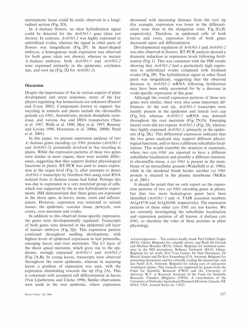
meristematic tissue could be easily observed in a longi-tudinal section (Fig. 2O).
In A. thaliana flowers, no clear hybridization signalcould be detected for the Artb561-1 gene (data notshown). In contrast, Artb561-2 was highly expressed inunfertilized ovules, whereas the signal in other parts offlowers was insignificant (Fig. 2P). In heart-shapedembryos, a homogenous weak expression was observedfor both genes (data not shown), whereas in matureA. thaliana embryos, both Artb561-1 and Artb561-2were expressed primarily in the epidermis, cotyledontips, and root tip (Fig. 2Q for Artb561-2).
Discussion
Despite the importance of Asc in various aspects of plantdevelopment and stress responses, many of the keyplayers regulating Asc homeostasis are unknown (Pastoriand Foyer 2002). Components known to support Ascrecycling in animals and plants include the chromaffingranule cyt b561, thioredoxins, protein disulphide isom-erase, and various Asc and DHA transporters (Njuset al. 1987, Wells et al. 1990, May et al. 1997, Rumseyand Levine 1998, Horemans et al. 2000a, 2000b, Patelet al. 2001).
In this paper, we present expression analyses of twoA. thaliana genes encoding cyt b561 proteins (Artb561-1and Artb561-2), potentially involved in Asc recycling inplants. While the expression patterns of these two geneswere similar in most organs, there were notable differ-ences, suggesting that they support distinct physiologicalfunctions in plants. RT-PCR was used to assess expres-sion at the organ level (Fig. 1), after attempts to detectArtb561-1 transcripts by Northern blot using total RNAisolated from A. thaliana tissues had failed. Perhaps thiswas due to expression in a very restricted group of cells,which was supported by the in situ hybridization experi-ments. ISH demonstrated that these genes are expressedin the shoot apex, in leaves, stems, roots and inflores-cences. However, expression was restricted to certaintissues: the epidermis, vascular tissue, pericycle, rootcortex, root meristem and ovules.
In addition to this observed tissue-specific expression,the genes were developmentally regulated. Transcriptsof both genes were detected in the epidermal cell layerof mature embryos (Fig. 2Q). This expression patterncontinued throughout seedling development, withhighest levels of epidermal expression in leaf primordia,emerging leaves and root meristems. The L1 layer ofthe shoot apical meristem, which gives rise to the epi-dermis, strongly expressed Artb561-1 and Artb561-2(Fig. 2A,B). In young leaves, transcripts were observedthroughout the entire epidermis, whereas in maturingleaves a gradient of expression was seen, with theexpression diminishing towards the tip (Fig. 2A). Thisis consistent with acropetal cell differentiation in leaves(Van Lijsebettens and Clarke 1998). Similar observationswere made in the root epidermis, where expression
decreased with increasing distance from the root tip(for example, expression was lower in the differenti-ation zone than in the elongation zone; Fig. 2G,H,respectively). Therefore, in epidermal cells of bothleaves and roots, expression levels of both genesdecreased upon cell differentiation.
Developmental regulation of Artb561-1 and Artb561-2was also observed in flowers. RT-PCR analysis showed adramatic reduction in expression levels following fertil-ization (Fig. 1). This was consistent with the ISH resultsshowing that Artb561-2 had a particularly high expres-sion in unfertilized ovules compared with fertilizedovules (Fig. 2P). The hybridization signal in other floralparts was insignificant, suggesting that the observeddecrease in Artb561-2 mRNA following fertilizationmay have been solely accounted for by a decrease inovule-specific expression of this gene.
Although the overall expression patterns of these twogenes were similar, there were also some important dif-ferences. At the root tip, Artb561-1 transcripts weremainly present in the epidermis and lateral root cap(Fig. 2M), whereas Artb561-2 mRNA was detectedthroughout the root meristem (Fig. 2N,O). Emerginglateral roots did not express Artb561-1 (Fig. 2J), whereasthey highly expressed Artb561-2, primarily in the epider-mis (Fig. 2K). This differential expression indicates thatthe two genes analysed may support different physio-logical functions, and/or have a different subcellular local-ization. This would resemble the situation in mammals,where two cyts b561 are reported to have a differentsubcellular localization and possibly a different function:in chromaffin tissue, a cyt b561 is present in the mem-brane of an intracellular granule (Wakefield et al. 1986),while in the duodenal brush border another cyt b561protein is situated in the plasma membrane (McKieet al. 2001).
It should be noted that we only report on the expres-sion patterns of two cyt b561 encoding genes in plants,but that two more homologous genes have beenidentified (Artb561–3 and -4, TAIR accession numbersAt1g14730 and At1g26100, respectively). The expressionpatterns of these other cyts b561 are not known. Weare currently investigating the subcellular localizationand expression patterns of all known A. thaliana cytsb561, to elucidate the roles of these proteins in plantphysiology.
Acknowledgements – The authors kindly thank Prof Gilbert Engler(RUG, Ghent, Belgium) for valuable advice, and Ruth De Groodtand Marleen Brunain (RUG, Ghent, Belgium) for technical assist-ance in the ISH procedures, Rebecca Verbanck (RUG, Ghent,Belgium) for art work, Prof Yves Guisez, Dr Nele Horemans, DrMarcel Jansen and Dr Kris Vissenberg (UA, Antwerp, Belgium) forinteresting discussions and for critically reading the manuscript, andJan Neefs (UA, Antwerp, Belgium) for taking care of soil-grownArabidopsis plants. This research was supported by grants from theFund for Scientific Research (FWO) and the University ofAntwerp; W.V. is Research Assistant of the Fund for ScientificResearch, Flanders (Belgium) (FWO). A contribution of theUniversity ofNebraskaAgricultural ResearchDivision, Lincoln, NE68583, USA. Journal Series no. 13822.
Physiol. Plant. 120, 2004 317
References
de Almeida Engler J, De Groodt R, Van Montagu M, Engler G(2001) In situ hybridization to mRNA of Arabidopsis tissuesections. Meth Enzymol 23: 325–334
Arrigoni O, De Tullio MC (2000) The role of ascorbic acid in cellmetabolism: between gene-directed functions and unpredictablechemical reactions. Plant Physiol 157: 481–488
AsardH,Caubergs RJ, RendersD,DeGreef JA (1987)Duroquinone-stimulated NADH oxidase and b-type cytochromes in the plasmamembrane of cauliflower and mung beans. Plant Sci 53: 109–119
Asard H, Horemans N, Caubergs RJ (1992) Transmembrane elec-tron transport in ascorbate-loaded plasma membrane vesiclesfrom higher plants involves a b-type cytochrome. FEBS Lett306: 143–146
Asard H, Horemans N, Preger V, Trost P (1998) Plasma membraneb-type cytochromes. In: Asard H, Berczi A, Caubergs RJ (eds)Plasma Membrane Redox Systems and Their Role in BiologicalStress and Disease. Kluwer, Dordrecht, The Netherlands, pp 1–31
Asard H, Kapila J, Verelst W, Berczi A (2001) Higher-plant plasmamembrane cytochrome b561: a protein in search of a function.Protoplasma 217: 77–93
Asard H, Terol-Alcayde J, Preger V, Del-Favero J, Verelst W,Sparla F, Perez-. Alonso M, Trost P (2000) Arabidopsis thalianasequence analysis confirms the presence of cyt b-561 in plants:Evidence for a novel protein family. Plant Physiol Biochem 38:905–912
Berczi A, Luthje S, Asard H (2001) b-Type cytochromes in plasmamembranes of Phaseolus vulgaris hypocotyls, Arabidopsis thalianaand Zea mays roots. Protoplasma 217: 50–55
Horemans N, Asard H, Caubergs RJ (1994) The role of ascorbatefree radical as an electron acceptor to cytochrome b-mediatedtrans-plasma membrane electron transport in higher plants.Plant Physiol 104: 1455–1458
Horemans N, Foyer C, Asard H (2000a) Transport and action ofascorbate at the plant plasma membrane. Trends Plant Sci 5:263–267
Horemans N, Foyer C, Potters G, Asard H (2000b) Ascorbatefunction and associated transport systems. Plant Physiol Bio-chem 38: 531–540
Martinez MC, Jørgensen J-E, Lawton MA, Lamb CJ, Doerner PW(1992) Spatial pattern of cdc2 expression in relation to meristemactivity and cell proliferation during plant development. ProcNatl Acad Sci USA 89: 7360–7364
May J, Mendiretta S, Hill KE, Burk RF (1997) Reduction ofdehydroascorbate to ascorbate by the selenoenzyme thioredoxinreductase. J Biol Chem 272: 22607–22610
McKie AT, Barrow D, Latunde-Dada GO, Rolfs A, Sager G,Mudaly E, Mudaly M, Richardson C, Barlow D, Bomford A,Peters TJ, Raja KB, Shirali S, Hediger MA, Farzaneh F,Simpson RJ (2001) An iron-regulated ferric reductase associatedwith the absorption of dietary iron. Science 291: 1755–1759
Njus D, Kelley PM, Harnadek GJ, Pacquing YV (1987) Mechanismof ascorbic acid regeneration mediated by cytochrome b561. AnnNY Acad Sci 493: 108–119
Njus D, Wigle M, Kelley PM, Kipp BH, Schlegel HB (2001)Mechanism of ascorbic acid oxidation by cytochrome b561.Biochemistry 40: 11905–11911
Okuyama E, Yamamoto R, Ichikawa Y, Tsubaki M (1998) Struc-tural basis for the electron transfer across the chromaffin vesiclemembranes catalyzed by cytochrome b561: analyses of DNAnucleotide sequences and visible absorption spectra. BiochimBiophys Acta 1383: 269–278
Pastori GM, Foyer CH (2002) Common components, networks,and pathways of cross-tolerance to stress. The central role of‘Redox’ and abscisic acid-mediated controls. Plant Physiol 129:460–468
Patel M, McIntosh L, Bliss T, Ho D, Sapolsky R (2001) Inter-actions among ascorbate, dehydroascorbate and glucose transportin cultured hippocampal neurons and glia. Brain Res 916: 127–135
Perın MS, Fried VA, Slaughter CA, Sudhof TC (1988) The struc-ture of cytochrome b561, a secretory vesicle-specific electrontransport protein. EMBO J 7: 2697–2703
Rumsey SC, Levine M (1998) Absorption, transport, and dispos-ition of ascorbic acid in humans. J Nutr Biochem 9: 116–130
Scagliarini S, Rotino L, Baurle I, Pupıllo P, Asard H, Trost P (1998)Purification of the ascorbate reducible b-type cytochrome of theplasma membrane of etiolated bean seedlings. Protoplasma 205:66–73
Trost P, Berczi A, Sparla F, Sponza G, Marzadori B, Asard H,Pupillo P (2000) Purification of cytochrome b-561 from beanhypocotyl plasma membrane: evidence for the presence of twoheme centres. Biochim Biophys Acta 1468: 1–5
Tsubaki M, Nakayama M, Okuyama E, Ichikawa Y, Hori H (1997)Existence of two heme B centers in cytochrome b561 frombovine adrenal chromaffin vesicles as revealed by a new purifi-cation procedure and EPR spectroscopy. J Biol Chem 272:23206–23210
Valvekens D, Van Montagu M, Van Lijsebettens M (1988) Agro-bacterium tumefaciens mediated transformation of Arabidopsisthaliana root explants by using kanamycin selection. Proc NatlAcad Sci USA 85: 5536–5540
Van Lijsebettens M, Clarke J (1998) Leaf development in Arabidop-sis. Plant Physiol Biochem 36: 47–60
Verelst W, Asard H (2003) A phylogenetic study of cytochromeb561 proteins. Genome Biol 4: R38
Wakefield LM, Cass AEG, Radda GK (1986) Functional couplingbetween enzymes of the chromaffin granule membrane. J BiolChem 261: 9739–9745
Wells WW, Xu DP, Yang YF, Rocque PA (1990) Mammalianthioltransferase (glutaredoxin) and protein disulfide isomerasehave dehydroascorbate reductase activity. J Biol Chem 265:15361–15364
Edited by C. H. Foyer
318 Physiol. Plant. 120, 2004