tinnitus - university of texas medical branch · –pulsatile tinnitus which may decrease with...
TRANSCRIPT

Tinnitus
Gordon Shields, MD
Faculty Advisor: Francis B. Quinn, Jr., MD
The University of Texas Medical Branch
Department of Otolaryngology
Grand Rounds Presentation
January 22, 2003

“…only my ears whistle and buzz
continuously day and night. I can say
I am living a wretched life.”
Ludwig Von Beethoven - 1801
Tinnitus
• Definition
• Classification
• Objective tinnitus – pulsatile
• Subjective tinnitus
• Theories
• Evaluation
• Treatment

Introduction
• Tinnitus -“The perception of sound in the
absence of external stimuli.”
• Tinnere – means “ringing” in Latin
• Includes Buzzing, roaring, clicking, pulsatile
sounds

Tinnitus
• May be perceived as unilateral or bilateral
• Originating in the ears or around the head
• First or only symptom of a disease process or
auditory/psychological annoyance

Tinnitus
• 40 million affected in the United States
• 10 million severely affected
• Most common in 40-70 year-olds
• More common in men than women

Classification
• Objective tinnitus – sound produced by
paraauditory structures which may be heard by
an examiner
• Subjective tinnitus – sound is only perceived by
the patient (most common)
Tinnitus
• Pulsatile tinnitus – matches pulse or a rushing
sound
– Possible vascular etiology
– Either objective or subjective
– Increased or turbulent bloodflow through
paraauditory structures

Objective -Pulsatile tinnitus
• Arteriovenous malformations
• Vascular tumors
• Venous hum
• Atherosclerosis
• Ectopic carotid artery
• Persistent stapedial artery
• Dehiscent jugular bulb
• Vascular loops
• Cardiac murmurs
• Pregnancy
• Anemia
• Thyrotoxicosis
• Paget’s disease
• Benign intracranial
hypertension
Arteriovenous malformations
• Congenital lesions
• Occipital artery and transverse sinus, internal
carotid and vertebral arteries, middle meningeal
and greater superficial petrosal arteries
• Mandible
• Brain parenchyma
• Dura

Arteriovenous malformations
• Pulsatile tinnitus
• Headache
• Papilledema
• Discoloration of skin or mucosa
Vascular tumors
• Glomus tympanicum
– Paraganglioma of middle ear
– Pulsatile tinnitus which may decrease with ipsilateral
carotid artery compression
– Reddish mass behind tympanic membrane which
blanches with positive pressure
– Conductive hearing loss

Vascular tumors
• Glomus jugulare
– Paraganlioma of jugular fossa
– Pulsatile tinnitus
– Conductive hearing loss if into middle ear
– Cranial neuropathies

Venous hum
• Benign intracranial hypertension
• Dehiscent jugular bulb
• Transverse sinus partial obstruction
• Increased cardiac output from
– Pregnancy
– Thyrotoxicosis
– Anemia

Benign Intracranial Hypertension
• Young, obese, female patients
• Hearing loss
• Aural fullness
• Dizziness
• Headaches
• Visual disturbance
• Papilledema, pressure >200mm H20 on LP

Benign Intracranial Hypertension
• Sismanis and Smoker 1994
– 100 patients with pulsatile tinnitus
– 42 found to have BIH syndrome
– 16 glomus tumors
– 15 atherosclerotic carotid artery disease
BIH Syndrome
• Treatment
– Weight loss
– Diuretics
– Subarachnoid-peritoneal shunt
– Gastric bypass for weight reduction
Muscular Causes of Tinnitus
• Palatal myoclonus
– Clicking sound
– Rapid (60-200 beats/min), intermittent
– Contracture of tensor palantini, levator palatini,
levator veli palatini, tensor tympani,
salpingopharyngeal, superior constrictors
– Muscle spasm seen orally or transnasally
– Rhythmic compliance change on tympanogram

Myoclonus
• Palatal myoclonus associations:
– Multiple Sclerosis and other degenerative
neurological disorders
– Small vessel disease
– Tumors
• treatments: muscle relaxants, botulinum toxin
injection
Stapedius Muscle Spasm
• Idiopathic stapedial muscle spasm
– Rough, rumbling, crackling sound
– Exacerbated by outside sounds
– Brief and intermittent
– May be able to see tympanic membrane movement
– Treatments: avoidance of stimulants, muscle
relaxants, sometimes surgical division of tensor
tympani and stapedius muscles

Patulous Eustachian Tube
• Eustachian tube remains open abnormally
• Ocean roar sound
• Changes with respiration
• Lying down or head in dependent position provides
relief

Patulous Eustachian Tube
• Tympanogram will show changes in compliance with
respiration
• Significant weight loss, radiation to the nasopharynx
• Previous treatments: caustics, mucosal irritants,
saturated solution of potassium iodide, Teflon or
gelfoam injection around torus tubarius

Subjective Tinnitus
• Much more common than objective
• Usually nonpulsatile
• Presbycusis
• Noise exposure
• Meniere’s disease
• Otosclerosis
• Head trauma
• Acoustic neuroma
• Drugs
• Middle ear effusion
• TMJ problems
• Depression
• Hyperlipidemia
• Meningitis
• Syphilis
Conductive hearing loss
• Conductive hearing loss decreases level of
background noise
• Normal paraauditory sounds seem amplified
• Cerumen impaction, otosclerosis, middle ear
effusion are examples
• Treating the cause of conductive hearing loss
may alleviate the tinnitus

Other subjective tinnitus
• Poorly understood mechanisms of tinnitus
production
• Abnormal conditions in the cochlea, cochlear
nerve, ascending auditory pathways, auditory
cortex
• Hyperactive hair cells
• Chemical imbalance
CNS Mechanisms
• Reorganization of central pathways with hearing
loss (similar to phantom limb pain)
• Disinhibition of dorsal cochlear nucleus with
increase in spontaneous activity of central
auditory system

Neurophysiologic Model
• Proposed by Jastreboff
• Result of interaction of subsystems in the nervous system
• Auditory pathways playing a role in development and appearance of tinnitus
• Limbic system responsible for tinnitus annoyance
• Negative reinforcement enhances perception of tinnitus and increases time it is perceived

Role of Depression
• Depression is more prevalent in patients with
chronic tinnitus than in those without tinnitus
• Folmer et al (1999) reported patients with
depression rated the severity of their tinnitus
higher although loudness scores were the same
• Which comes first, depression or tinnitus?

Drugs that cause tinnitus
• Antinflammatories
• Antibiotics
(aminoglycosides)
• Antidepressants
(heterocyclines)
• Aspirin
• Quinine
• Loop diuretics
• Chemotherapeutic agents
(cisplatin, vincristine)

Evaluation - History
• Careful history
• Quality
• Pitch
• Loudness
• Constant/intermittent
• Onset
• Alleviating/aggravating factors

Evaluation - History
• Infection
• Trauma
• Noise exposure
• Medication usage
• Medical history
• Hearing loss
• Vertigo
• Pain
• Family history
• Impact on patient

Evaluation – Physical Exam
• Complete head & neck exam
• General physical exam
• Otoscopy (glomus tympanicum, dehiscent
jugular bulb)
• Search for audible bruit in pulsatile tinnitus
– Auscultate over orbit, mastoid process, skull, neck,
heart using bell and diaphragm of stethoscope
– Toynbee tube to auscultate EAC
Evaluation – Physical Exam
• Light exercise to increase pulsatile tinnitus
• Light pressure on the neck (decreases venous
hum)
• Valsalva maneuver (decrease venous hum)
• Turning the head (decrease venous hum)

Evaluation - Audiometry
• PTA, speech descrimination scores,
tympanometry, acoustic reflexes
• Pitch matching
• Loudness matching
• Masking level

Evaluation - Audiometry
• Vascular or palatomyoclonus induced tinnitus –
graph of compliance vs. time
• Patulous Eustachian tube – changes in
compliance with respiration
• Asymmetric sensorineural hearing loss or speech
discrimination, unilateral tinnitus suggests
possible acoustic neuroma - MRI

From: Tyler RS, Babin RW. Tinnitus. In: Cummings CW, ed. Otolaryngology-Head and Neck Surgery, second
edition. St. Louis, Mosby-Year Book, 1993:3032.
Laboratory studies
• As indicated by history and physical exam
• Possibilities include:
– Hematocrit
– FTA absorption test
– Blood chemistries
– Thyroid studies
– Lipid battery

Imaging
• Pulsatile tinnitus
• Reviewed by Weissman and Hirsch (2000)
• Contrast enhanced CT of temporal bones, skull
base, brain, calvaria as first-line study
• Sismanis and Smoker (1994) recommended CT
for retrotympanic mass, MRI/MRA if normal
otoscopy

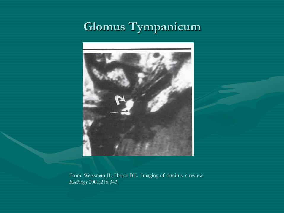
• Glomus tympanicum – bone algorithm CT scan
best shows extent of mass
• May not be able to see enhancement of small
tumor
• Tumor enhances on T1-weighted images with
gadolinium or on T2-weighted images
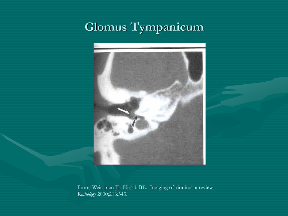
Glomus Tympanicum
From: Weissman JL, Hirsch BE. Imaging of tinnitus: a review.
Radiology 2000;216:343.
Glomus Tympanicum
From: Weissman JL, Hirsch BE. Imaging of tinnitus: a review.
Radiology 2000;216:343.
Imaging
• Glomus jugulare
– Erosion of osseous jugular fossa
– Enhance with contrast, may not be able to
differentiate jugular vein and tumor
– Enhance with T1-weighted MRI with gadolinium
and on T2-weighted images
– Characteristic “salt and pepper” appearance on MRI

Glomus jugulare
From: Weissman JL, Hirsch BE. Imaging of tinnitus: a review.
Radiology 2000;216:344.
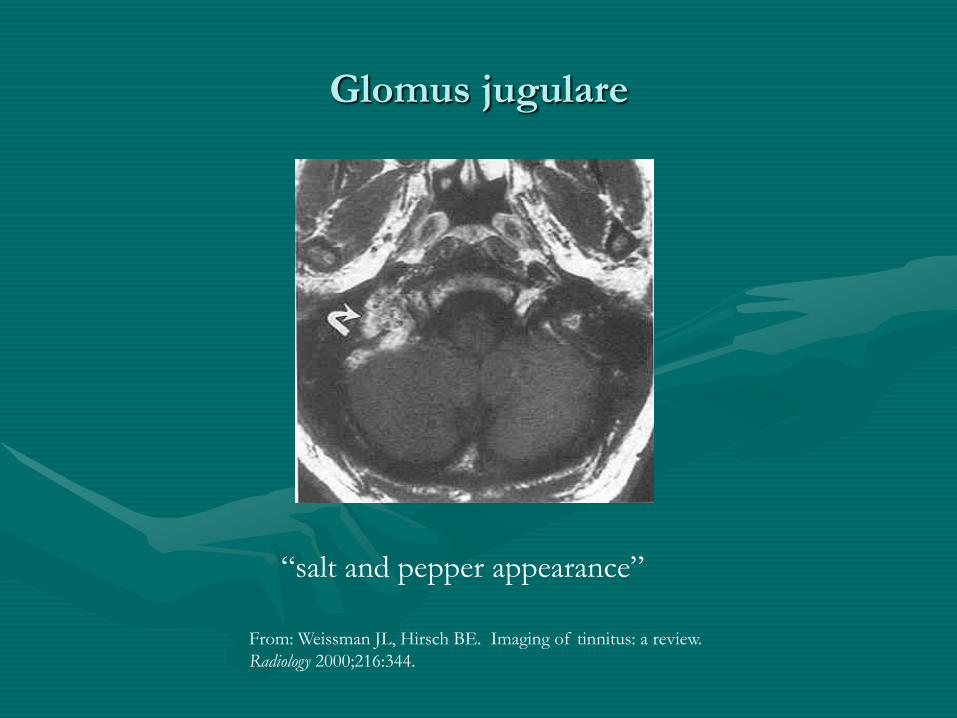
Glomus jugulare
“salt and pepper appearance”
From: Weissman JL, Hirsch BE. Imaging of tinnitus: a review.
Radiology 2000;216:344.
Imaging
• Arteriovenous malformations – readily apparent
on contrasted CT and MRI
• Normal otoscopic exam and pulsatile tinnitus
may be dural arteriovenous fistula
– Often invisible on contrasted CT and MRI/MRA
– Angiography may be only diagnostic test

Imagining
• Shin et al (2000)
– MRI/MRA initially if subjective pulsatile tinnitus
– Angiography if objective with audible bruit in order
to identify dural arteriovenous fistula

Imaging
• Other contrast enhanced CT diagnoses
• Aberrant carotid artery
• Dehiscent carotid artery
• Dehiscent jugular bulb
• Persistent stapedial artery
– Soft tissue on promontory
– Enlargement of facial nerve canal
– Absence of foramen spinosum

Persistent Stapedial Artery
From: Araujo MF et al. Radiology quiz case I: persistent stapedial artery. Arch
Otolaryngol Head Neck Surg 2002;128:456.

Imaging
• Acoustic Neuroma
– Unilateral tinnitus, asymmetric sensorineural hearing
loss or speech descrimination scores
– T1-weighted MRI with gadolinium enhancement of
CP angle is study of choice
– Thin section T2-weighted MRI of temporal bones
and IACs may be acceptable screening test

Acoustic Neuroma
From: Weissman JL, Hirsch BE. Imaging of tinnitus: a
review. Radiology 2000;216:348.

Acoustic Neuroma
From: Weissman JL, Hirsch BE. Imaging of tinnitus: a review.
Radiology 2000;216:348.

Imaging
• Benign intracranial hypertension
– MRI
– Small ventricles
– Empty sella

BIH – Empty Sella
Sismanis A, Smoker W. Pulsatile tinnitus: recent advances in diagnosis.
Laryngoscope 1994;104:685.

Treatments
• Multiple treatments
• Avoidance of dietary
stimulants: coffee, tea,
cola, etc.
• Smoking cessation
• Avoid medications
known to cause tinnitus
• Reassurance
• White noise from radio
or home masking
machine
Treatments - Medicines
• Many medications have been researched for the
treatment of tinnitus:
– Intravenous lidocaine suppresses tinnitus but is
impractical to use clinically
– Tocainide is oral analog which is ineffective
– Carbamazepine ineffective and may cause bone
marrow suppression

Treatments - Medicines
• Alprazolam (Xanax)
– Johnson et al (1993) found 76% of 17 patients had
reduction in the loudness of their tinnitus using both
a tinnitus synthesizer and VAS (dose 0.5mg-1.5
mg/day)
– Dependence problem, long-term use is not
recommended

Treatments - Medicines
• Nortriptyline and amitriptyline
– May have some benefit
– Dobie et al reported on 92 patients
– 67% nortriptlyine benefit, 40%placebo
• Ginko biloba
– Extract at doses of 120-160mg per day
– Shown to be effective in some trials and not in others
– Needs further study

Treatments
• Hearing aids – amplification of background
noise can decrease tinnitus
• Maskers – produce sound to mask tinnitus
• Tinnitus instrument – combination of hearing
aid and masker

Treatments
• Tinnitus Retraining Therapy
– Based on neurophysiologic model
– Combination of masking with low level broadband
noise for several hours per day and counseling to
achieve habituation of the reaction to tinnitus and
perception of the tinnitus itself

Treatments
• Electrical stimulation of the cochlea
– Transcutaneous, round window, promontory
stimulation have all been tried
– Direct current can cause permanent damage
– Steenersen and Cronin have used transcutaneous
stimulation of the auricle and tragus decreasing
tinnitus in 53% of 500 patients

Treatments
• Cochlear implants
– Have shown some promise in relief of tinnitus
– Ito and Sakakihara (1994) reported that in 26
patients implanted who had tinnitus 77% reported
either tinnitus was abolished or suppressed, 8%
reported worsening
Treatments
• Surgery
– Used for treatment of arteriovenous malformations,
glomus tumors, otosclerosis, acoustic neuroma
– Some authors have reported success with cochlear
nerve section in patients who have intractable
tinnitus and have failed all other treatments, this is
not widely accepted
Treatments
• Biofeedback
• Hypnosis
• Magnetic stimulation
• Acupuncture
• Conflicting reports of benefit

Conclusions
• Tinnitus is a common problem with an
extensive differential
• Need to identify medical process if involved
• Pulsatile/Nonpulsatile is important distinction
• Will only become more common with aging of
our population
• Research into mechanism and treatments is
needed to better help our patients