tiie infectivity and virulence of …ejtafs.mardi.gov.my/jtafs/13-3/trypanosoma eyansin.pdfdi...
TRANSCRIPT

MARDI Res. Bul l . , (1985) 13, 3: (291-301)
TIIE INFECTIVITY AND VIRULENCE OF TRYPANOSOMA.EYANS/INLOCAL INDIAN DAIRY X KEDAH.KELANTAN CATTLE
O. ABAS MAZNI* and A.H. ZAINAL-ABIDIN**
Keywords: Infectivity, Intermittent fever, Local Indian Dairy x Kedah-Kelantan cattle (LID xKK), Parasitaemia, Trypanosoma evansi, Virulence.
RINGKASAN
Satu strain Trypanosonu evansi (TE) telah diasingkan baru-baru ini dari kerbau sawah di Pusat
Ternakan Kerbau MARDI, Bukit Ridan, Pahang, Malaysia Barat. Beberapa kajian telah dilakukan
untuk menentukan infektiviti dan kevirulenan parasit ini pada lembu-lembu tempatan. Enam ekor
lembu tempatan baka Local Indian Dairy x Kedah-Kelantan (LID x KK) telah disuntik secara subkutin
dengan 5.0 x 10⁷ TE, setiap lembu. Didapati lembu-lembu ini rentan kepada infeksi TE. Parasitemiayang parah berpanjangan selama 34 hari selepas infeksi bermula. Kenaikan suhu tubuh berlaku pada
semua haiwan berinfeksi dan kejadian ini dicerap hanya pada peringkat awal infeksi. Walau
bagaimanapun didapati kenaikan parasitemia ini tidak berlaku secara serentak dengan kenaikan suhu
tubuh haiwan. Di samping itu perubahan dalam nilainilai hematologi (RBC, PCV dan Hb) telah juga
ditentukan.
6mbu-lembu yang pulih dan menjadi imun dari infeksi awal telah dicabar sekali lagi dengan dos
gandaan (iaitu 1.0 x 10E TE),290 hari selepas infeksi awal. Lembu-lembu ini menunjukkanpembentukan parasitemia yang lewat (kelewatan selama tiga hari dari infeksi biasa). Demam yang
berulang-ulang telah juga dicerap.
INTRODUCTION
Surra or trypanosomiasis is known tobe prevalent amongst horses, camels, cattleand buffaloes in various parts of Africa,India, other Asian countries and many otherparts of the world. The disease is invariablyfatal in camels and horses. Cattle andbuffaloes are usually resistant to the disease,but under stress and in the young, thedisease may prove fatal (LINcnno, 1899;MurHeR.l t , 1925; LvEn and SnnwRn, 1935;BRen and SHenMA, 1962; VenuA,7973).
For buffaloes, especially in South EastAsia, the agent of the disease, thetrypanosomes, become one of the mostimportant protozoan parasites althoughresults obtained from experimentalinfections have been somewhat variable. InMalaysia, the prevalence of trypanosomiasisin cattle and buffaloes is practically un-known and the disease has not received the
same attention as in the equines. Surra inhorses and cattle has been recorded in thiscountry for the first time at the turn of thecentury (FnnzEn and SnvuoNos, 1909). Anoutbreak of the disease in horses hadoccurred in the Equine Unit of the UniversitiPertanian Malaysia, Serdang, Selangor (Ncand VnNsslow, 1978). Local cattle andbuffaloes act as the reservoir hosts of theparasite (Nc and VnNSeLow, 1978) and theparasite is considered as non-pathogenicand of no economic importance. These twotypes of animals have also been reported notto be clinically affected by the parasite (Ncand VeNser-ow, 1978).
Until recently, buffaloes, calves andweaners at the MARDI Buffalo BreedingFarm, in Bukit Ridan, Pahang werereported to show signs of chronic emacia-tion, anaemia and even deaths (AnesMazNt, ZnlNnL-AslolN and RaunrnlsHNRN,1984). Those animals with emaciation also
*Livestock Research Division, MARDI, Serdang, Selangor, Malaysia..*Department of Tnology, Faculty of Life Sciences, Universiti Kebangsaan Malaysia, Bangi, Selangor, Malaysia.
291

showed enlarged prescapular lymph nodesand examination of the blood indicated thepresence of Trypanosoms evansi. Theisolate was found to be very pathogenic tomice, rats and Mongolian gerbils (ZntNel--AsrnrN. SwRlseL, SRSRntNRn, KwoNc andAnes MnzNr. 1982).
This paper reports some of the resultsof the observations on the infectivity andvirulence of this parasite in our local cattle.
MATERIAI,S AND METHODS
Experimental Animals
Eleven healthy Local Indian Dairy xKedah-Kelantan (LID x KK) calves of 8- 12months old were used in the study. Micro-scopic examinat ions of wet b lood f i lms,blood smears and subinoculations of freshblood into ICR strain mice indicated thatthey were free from trypanosome infection.-fhe
calves were kept in groups of two in apen under r enclosed bui ld ing. Each cal freceived 2 kg commercial concentrate andNapier grass (Pennisetum purpureum) adlibitum daily.
Parasite and the Preparation of Inoculum
The strain of Trypanosoma evansi(TE) used in this study was obtained froman infected swamp buffalo from MARDIBuffalo Breeding Farm in Bukit Ridan,Pahang. It was maintained in mice byweekly passage of infected blood.
For the preparation of inoculum,blood from several infected mice having thesame degree of parasitaemia was used. Theblood was normally drawn at the peak ofparasitaemia which occurred in the third orfourth day post infection. The blood wasdrawn by cardiac puncture and dilutedser ia l ly us ing cold normal sa l ine (0.85%) orcitrate solution. The final dilution wasadjusted to contain 5.0 x 10⁷ TE permi l l i l i t re .
Experimental Design
In the first experiment, 11 calves wererandomly divided into two groups. Group Iconsisted of f ive animals as the control andreceived 1.0 ml steri le normal saline givensubcutaneously. Group II consisted of sixanimals in fected wi th 5.0 x 10⁷ TE peranimal subcutaneously. A second experi-ment was carried out 290 days after the firstinfection whereby three calves in Group Iand all calves in Group II were given achallenge dose of 1.0 x 10" TE per animalsubcutaneously.
Examination of Wet Blood Films and Smears
Wet blood fi lms and thin blood smearswere prepared daily using blood obtainedfrom the ear vein of the infected animals.They were prepared early in the morningand used for observation and quantif icationof parasitaemias. The wet blood fi lms wereused for the estimation of degree of parasi-taemia using the following adaptations(Cnenenn and Lur , 1956):
+ : 1 - 2 trypanosomes per field++ :3 - 4 t rypanosomes per f ie ld
+ + + : manv trypanosomes per field++++ : teeming numbers per f ie ld
The fi lms were observed under microscopeusing X40 magnification.
The thin blood smears were fixed inabsolute alcohol and stained with theGiemsa's stain and observed under micro-scope (X100 magnification) to confirm thepresence of the parasites.
Observations on Symptoms, Rectal Tem-perature and Body Weight
Infected animals were examined dailyto observe their general body conditions,appetite and behaviour. Daily rectaltemperatures were recorded at 8 am and 12noon, 4 pm and 8 pm. Weekly body weightswere also recorded.
Haematological Examinations
Weekly haematological examinationswere done on blood samoles collected from
292
the jugular vein into vacuumtainers contain-ing EDTA and analysed for red blood cellcount (RBC) and haemoglobin (Hb) con-centration using the Coulter Countet ZF 6system. Packed cell volume (PCV) wasdetermined by the haematocrit method andthe plasma protein (PP) was read on therefractometer (Bellingham and Stanley Ltd.England).
Biological Tests
Monthly biological tests were carriedout to determine the sterility of the blood inthe infected and control animals. For this,blood was collected from the animals andthen subinoculated intraperitoneally intomice. Wet blood films were prepared fromthese mice every other day for a period ofup to 20 days and observed for the presenceof the parasites.
REST]LTS
Clinical Signs of the Infection
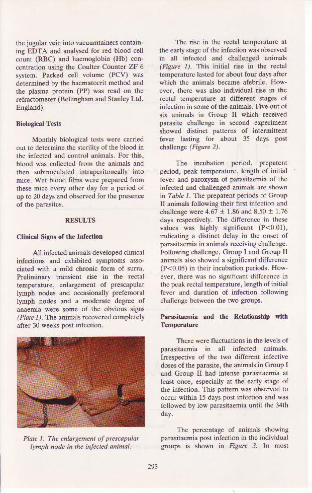
All infected animals developed clinicalinfections and exhibited symptoms asso-ciated with a mild chronic form of surra.Preliminary transient rise in the rectaltemperature, enlargement of prescapularlymph nodes and occasionally prefemorallymph nodes and a moderate degree ofanaemia were some of the obvious signs(Plate 1). The animals recovered completelyafter 30 weeks post infection.
Plate 1. The enlargement of prescapularlymph node in the infected animal.
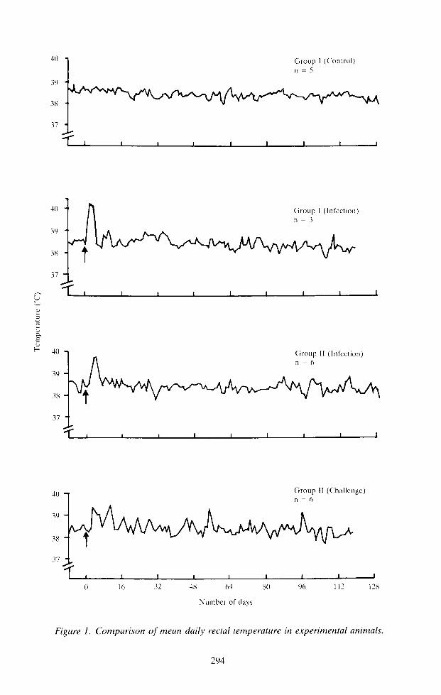
The rise in the rectal temperature atthe early stage of the infection was observedin all infected and challenged animals(Figure 1). This initial rise in the rectaltemperature lasted for about four days afterwhich the animals became afebrile. How-ever, there was also individual rise in therectal temperature at different stages ofinfection in some of the animals. Five out ofsix animals in Group II which receivedparasite challenge in second experimentshowed distinct patterns of intermittentfever lasting for about 35 days postchallenge (Figure 2).
The incubation period, prepatentperiod, peak temperature, length of initialfever and paroxysm of parasitaemia of theinfected and challenged animals are shownin Tuble 1. The prepatent periods of GroupII animals following their first infection andchallenge were 4.67 + 1.86 and 8.50 + 1..76days respectively. The difference in thesevalues was highly significant (P<0.01),indicating a distinct delay in the onset ofparasitaemia in animals receiving challenge.Following challenge, Group I and Group IIanimals also showed a significant difference(P<0.05) in their incubation periods. How-ever, there was no significant difference inthe peak rectal temperature, length of initialfever and duration of infection followingchallenge between the two groups.
Parasitaemia and the Relationship withTemperature
There were fluctuations in the levels ofparasitaemia in all infected animals.Irrespective of the two different infectivedoses of the parasite, the animals in Group Iand Group II had intense parasitaemia atleast once, especially at the early stage ofthe infection. This pattern was observed tooccur within 15 days post infection and wasfollowed by low parasitaemia until the 34thday.
The percentage of animals showingparasitaemia post infection in the individualgroups is shown in Figure 3. In most
293
:t0
39
38
37
.l( )
39
313
1 1
,'J
:-=t
f,F !+0
39
38
37
'll.)
39
38
37
tr;l
Number of day's
Figure I. Comparison of mean daily rectal temperature in experimental animals.

Code no. o f an ima ls
H 5(X)7
e - - - < 6 ( X ) 2
o . . . . . . . . . o 6 ( x ) 6
\,
-i:
a
1 l
.l( )
39
38
37
lI
E l l 16 l0 )1 2lJ 32 36 ,10 11 -18 52
Number o f days
Figure 2. The course of intermittent fever in 3 animals in Group II following challenge.
Table 1. Mean prepatent period, incubation period, peak temperature and length on initialfever of experimental animals
Treatment groupPrepatent period
No. of animals (days)lncubat ionper iod Peaktemperature
(days) of initial fever ('C)Length of initial
fever (days)
II (Infection)
II (Challenge)
I (Infection)
6 1.67 + 1 .86b* *
6 8 .50+7.76a
3 4.67 +3. '79ab
3.17+0.98ab*
3.67 +0.52a
2.33+0.58b
40.56+32.0
40.47+0.M
40.97+0.47
4 .0+ t .26+
3.5+0.84
4.0+ 1 .0
Presence of intermittent fever*Means in the same column followed by different
**Means in the same column followed bv different
animals, parasitaemia did not usually coin-cide with the rise in body temperature.However, intense parasitaemia whichoccurred at the early stage of infectionassociated with a marked increase in rectaltemperature. At the later stage of theinfection, parasites were also seen whenthere was no fever.
Body Weight and Average Daily Gain
All infected animals showed decreasein body weight during the first two weekspost infection at the time when the fever andparasitaemia were high. However, signi-ficant difference (P<0.05) in the averagedaily gain between Group I and Group IIanimals during the first experiment was onlyobserved in the third month post infection.Following challenge there was also signi-ficant difference (P<0.05) in the averagedaily gain during the second and fourth
letter differ at P<0.05.letter differ at P<0.01 .
months post infection (Table 2) betweenthese animals.
Biological Tests
The biological tests were alwayspositive while the peripheral blood smearswere negative on many occasions. Animalsin Group II were positive up to fourthmonth post infection after which only two ofthem still harboured the parasite up to thefifth month.
Haematological Changes
Infections have been observed to beassociated with the reduction in PCV. Hband RBC values. In Group II animals, thesevalues deteriorated gradually between thefirst and 14th weeks post infection. ThePCV value of this group droppedby 76.57o,from a preinfection mean of 36.37o to a
295

ss
u
-
E
100
80
6t)
.10
20
U
80
60
40
20
0
100
ti0
6(J
40
20
0
Post infect ion (da1-s)
Figure 3. The percentage of animals with parasitaemia for the first 38 days post infection.
Table 2. Average daily gain in all experimental animals
Group I I ( Infect ion)
Group I I (Cha l l enge )
Group I ( Infect ion)
Treatment group No. of animalsAverage daily gain (kg) months post infection
I
II
II (Challenge)
I (Infection)
o
0
3
0.04a* 0.18a 0.20a 0.20a 0.21a 0.17a-0 .26a -0 .01ab 0 .07b 0 .80a 0 .10a 0 .10a-0.01a -0.06b -0.05b -0.10b 0.02a 0.60a
0.10a -0.20b -0.01b 0.03b 0.05a 0.07a
*Means in the same column followed by different letter are significantly different at P<0.05
mean of 30.47o (Figure 4). The PCV valueof Group I following infection droppedrapidly by 233% from a preinfection meanof 43% to a mean of 35 per cent.
The PCV , Hb and RBC values ofGroup I and Group II after their firstinfection and challenge were different from
the preinfection values of Group I betweenthe first and sixth months post infection(Figure 5). The post infection values becamequite similar to the preinfection values onlyafter six months.
Plasma proteins (PP) in Group II werehigher as compared to Group I (Figure 6). A
296

t - . 4
a_{
- l turt. rr i t ! \
Mean of Group I
Mean of Group I ll l
g1
(,
5
o-
O
0 8 1 6 1 1 3 1
Number o f wceks
Figure 4. Red blood cell counts (RBC),packed cell volumes (PCV) and haemoglobin(Hb) concentrations of experimental animals.
>< Group I ( lnfect ion)
r< Group I I (Chal lenge)
Number o f weeks
Figure 5. Red blood cell counts (RBC),pqcked ceLl volumes (PCV) and haemoglobin(Hb) concentrations of experimental animals.
\a
O
^ 1 1
E
8d
- a l lr
l 5
l - 3
tr
3- t l
9
7
r r L\ i J a ,l l \.i i'i
t't\t \L
0
291

sudden increase in the PP value of Group IIwas observed during the period between thefirst and fourth months post challenge.There was also a significant increase in thePP value (P<0.05) of Group I before andafter infection during the third and fourthmonths post infection (Figure 6).
DISCUSSION
The course of the disease due to Z.evansi infections in LID x KK cattle was of amild type surra. The main symptomsobserved were preliminary rise in rectaltemperature, enlargement of prescapularlymph nodes and occasionally prefemorallymph nodes, decrease in body weight and amoderate degree of anaemia with eventualrecovery of the animals. Similar observa-tions have been recorded by other workersand the authors' finding may indicate thatcattle used in this study were relatively moreresistant to T. evansi (LINceno, 1899;HurcR.l, and Melsr, l9l2: MurHERrt,1925: MaHereN. 1934: Lven and SenweR.1935). Some of the animals were foundpositive with the parasite up to the fifthmonth post infection and this may alsoindicate the existence of a carrier stase inthe infection.
Incidence of parasitaemia in the
peripheral blood was intermittent. Fluctua-tions in the parasitaemia were indicated byperiodic peaks and the absence of thetrypanosomes from the circulation whichwas eventually followed by low parasitaemia.In the later stages of the infections, theparasite could only be detected by intraperi-toneal injection of the blood into cleanmice. Similar findings were also observed byVenve (1973) and RezzneuE, MTSHRA andSeHnt (1976). The variation in the parasi-taemia noted within the individuals can berelated to the capacity of the individual todelay or limit the level of parasitaemia. Inthe challenged animals (Group II), three ofthem still showed intense parasitaemiafollowing challenge. Furthermore, it wasalso found that the high (intense) parasi-taemia did coincide with the rise in the bodytemperature although Bnen and SHenue(1.962), BeNser- (1966), SRrvRsrRvn,MRr-uorne and Lyr,n (1969), CunNo andSrNcH (\97I) and Venue (1973) hadreported that no correlation between parasi-taemia and the rise in the body temperatureduring Z. evansi infections in buffaloes,dogs, rabbits and donkeys.
In the present study, slight tomoderate decrease in the haematologicalvalues were also observed in the infectedanimals durine the first to the sixth months
Weeks
experimental animals.
Inoculat ion c.-r Group I
e< Group II
E
=
^ t ' \
Weeks
Figure 6. Values of plasma proteins in all
>-o Group I ( Infect ion)
. - - Group I I (Chal lenge)
-./
-ltt I
f \v
298

post infection. During this period, thesevalues decreased when compared with thoseof the control. Then thereafter, the valueswere found to be within those of the control.Although this paiasite did not cause severe
anaemia, there was indication that it diddecrease the blood values. The anaemiaobserved was of a progressive type. It fell
into two distinct phases : an initial or 'acute'
phase, characterized by a rapidly developinganaemia and accompanied by high level ofparasitaemia and a 'chronic' phase duringwhich the low PCV levels remained staticfor the extended period. During this chronicphase there was little or absence of parasites
in the peripheral blood. Even whenparasites could no longer be detected in the
blood, PCV values showed little tendency torecover and it took up to six months for theblood pictures to return to the values of thecontrol. Similar patterns were also recordedin T. congolense infection (FteNNes, 1954;1970).
Anaemia is the most significant factorin the disease process in naturally occurringand experimentally induced bovinetrypanosomiasis (Honuuv, l92l; MunReY,1974). These mechanisms, acting singly or
in concert, have been implicated as thefactors underlying the anaemia. These are
haemodilution (FteNNrs, 1954; Nnvlon,1971; Hor-r',rES, 1976), increased red cellbreakdown (Mnuo and Holuns, 1975;Holues, 1976; PnEsron and Weloe, 1976)and reduced cell synthesis (FteNNes, 1954;1970). There have been reports suggestingthat anaemia accompanying ?" evansi inf.ec-tion is due to the inhibition of erythropoeiticactivity in the bone marrow (RtcuenosoN
and KENoen, 1962; SRIvastevR et al.,1969). There is also a possibility of an
immunologically mediated mechanismbeing responsible for the development ofthe accompanying anaemia (Assoxu, 1975).
Animals infected with a massive doseof 1.0 x 10⁸ TE showed a much higher PPvalue. Similarly there was a marked increasein this value in the challenged animals at theinitial stage of the infection. This is probablydue to an increase in the globulin level asreported by Venua (1973) in calves infectedwith L evansi.
The results from the study indicatedthat the local LID x KK cattle were suscep-tible to T. evansi with mild severity althoughit caused emaciation and anaemia with eventualrecovery. Earlier studies have indicated thatN' Dama breed was trypanotolerant andpartially resistant to trypanosomiasis ascompared with the Zebu breed, to the samedisease (FAO, 1976). From the results ofthe present study, it can be suggested thatLID x KK cattle were also trypano-tolerant to T. evansi.
ACKNOWLEDGEMENTS
The authors would like to express theirgrateful thanks to Mr. Syed Ali Syed AbuBakar, formerly Director of Animal Pro-duction Division, MARDI, for his supportand encouragement. They gratefullyacknowledge the assistance of Prof. A.G.Luckins, University of Edinburgh and Prof.M.J. Clarkson, University of Liverpool,United Kingdom for confirming the identifi-cation of T. evansi. Special thanks to Mr.Ramli Ahmad and Mr. Malek Abdullah fortheir assistance in carrying out the trial.Thanks are also due to Ms. Laila Zainal andMrs. Rokiah Uda for typing the manuscript.
ABSTRACT
A strain of Trypanosoma evansi (TE) was recently isolated from swamp buffalloes at Bukit Ridan
Station, MARDI, Pahang in West Malaysia. Some studies were carried out on the local cattle to
determine the infectivity and virulence of the parasite. Six local cattle, Local Indian Dairy x Kedah-
Kelantan (LID x KK), were infected subcutaneously with 5.0x 10⁷ TE per animal. It was found that the
local cattle were susceptible to this parasite. Intense parasitaemia in the blood persisted until 34 days
post infection. A rise in the body temperature was observed in all infected animals but only at the initial
299

stage of the infection. However, it was found that the rise in parasitaemia did not coincide with the risein the body temp€rature. Changes in the haematological pictures (RBC, PCV and Hb) were alsoobserved. Recovered animals were challenged with a double dose i.e. 1.0 x ld TE. 290 days after theinitial infection. It was found that all the animals showed a delayed parasitaemia (i.e. three days later) ascompared with the parasitaemia in the control. Intermittent fever also occurred in these animals.
REFERENCES
ABAs MezNr, O., ZeNeL-AsrorN, A.H. and Hwcnn, F. and Melrx, J. (1912). Special pathologyRnu*nIsrxrN, P. (1984). Observation on the and therapeutics of the disease of domesticprevalence of Trypanosoma evcnsi in swamp animals. Bailliera, Tindall and Cox, London, I :buffaloes in Bukit Ridan, Pahang (unpubl.). 814 pp.
Assoxu, R.K.G. (1975). Irnmunological studies of the Lncrno, A. (1899). Report on surra in equines.
mechanism of anaemia in experimental T. evansi bovines, buffaloes and canines. Bombay Govt.
infections in rats. Intemational loumal for Press II (l), 160pp.Parasitology 5, 137 -45.
LvER, P.R.K. and Senwen, S.M. (1935). Bovine surraBANSAL, S.R. (1%6). Some aspects of Trypanosomiasis in India with a description of recent outbreak.
(Suna). M.Sc. (Vet.) Thesis, Agra University, Indian l. Vet Sci and Anim. Hwb. 5,158-70.India.
Mlx,r,l,lN, M.R. (1934). A note on bovineBn,c'n, P.S. and Snenu.n, R.M. (1962). Bovine trypanosomiasis in Hyderabad State. Indian. l.
trypanosomiasis or surra in the Punjab. Puzjab Vet. Sci and Anim. Husb.4,242-6.Veterinarian 2,l-4.
M,c.r'4o, E. and Holvrs, P.H. (1975). The erythro-Cnrnene, D.J. and Lul, T.J. (1956). The nuclear shift kinetics of Zebu cattle chronically infected with
index and other haematological indices in Surra. Trypanosonta congolense. Research in Ve4in4ryAm. J. Vet. Res. 17,615-25. Siince tE. t05-6.
CueNo, K. and SrNcu, R.P. (1971). A study on theclinical course of trypanosomiasis in goats,donkeys, dogs and rabbits experimentallyinfected with Trypanosoma evansi. J. Res.(Ludh) 8,274-9.
FAO (1976). First FAO Expert consultation onresearch on trypanotolerance and breeding oftrypanotolerance animal. Food and AgriculruralOrganisation of the United Nations.
Ftrr.rNns, R.N.T-W (1954). Haematological studies ontrypanosomiasis of cattle. Veterinary Record 66,123-34.
MUKHERJT, J.N. (1925). Bovine surra and its treatment.Indian Vet. l. 2,28-33.
MuRRAy, M. (1974). The pathology of Africantrypanosomiasis. ln Progress in Immunology(ed. Bnavr, L. and HeLseRoM, J.), pp. 181-92. Amsterdam and Oxford: North HollandPublishing Company.
NAyLoR, D.C. (1971). The haematology and histo-pathology oI Trypanosoma congolense infectionin cattle. Tropical Animal Health and Produc-tion 3, 159-68.
(1970). Pathogenesis and Pathology of Nc, B.K. Y. and Vnxselow, B. (1978). Outbreak ofAnimal Trypanosomiasis (ed. Muucmt, H.E.), surra in horses and the pathogenesis of thepp.729-50.l,ondon: George Allen and Unwin anaemia. Kajian Veterinar 10, 88-9.Ltd.
PnesroN, J.M. and Weloe, B.T. (1976), Studies onFRAzER, H. and SevuoNDs, S.L. (19@). Suna in the African Trypanosomiasis. Final. Report to
Federated Malays States. J. Comp. Pth. Therap. Department of the Army Walter Reed Army22,185-92. (abstract). Institute of Research, DAMD 17-941.2.
HoLMEs, P.H. (1976). The use of radioisotopic tracer RAzzAeuE, A., Mtsune, S.S. and S,e.u,r,r, B.N. (1976).techniques in the study of the pathogenesis of Effects of cortisone and splenectomy on thethe trypanosomiasis. In Nuclear Techniques in symptoms and course of experimental T. evansiAnimal Production and Health pp. 463-74. infections in buffalo calves. M.Sc. (Vet.) Thesis,Vienna: International Atomic Energy Agency. Rajendra Agricultural University, Bihar, India.
HoRNBy, H.E. (1921). Trypanosome and Rrcxenosox, U.F. and KrNoer-1, S.B. (1962).trypanosomiasis of cattle. J. Comparaive Veterinary Protozoology 3rd ed., 32pp.Pathology Y,211-Q. Edinburgh, UK: Lover & Boyd.
300
Snryestavr, R.V., Melsorn,a., M.N. and Lven, P.R.(1969). Pathology of experimental T. evansi indogs. Indian l. Anim. Sci. 39,307-14.
Venvr, B.H. (1973). Studies on some aspects of thetrypanosomiasis (surra) in cattle and buffaloes.Ph.D. Thesis, Haryana Agricultural University,India.
Accepted for publication on 1Oth September, 1985.
Zennl-AuotN, A.H., Swerrel, S., SesenrNeH, I. ,Kwor.rc, P.G. and Aaes MnzNr, O. (1982).Infections of T. evansi in laboratory animals.Abstract 18th Annual Scientific Seminar ofMalaysian Society of Parasitology and TropicalMedicine, Penang, p.6.
3 0 1