tiglicamides a–c, cyclodepsipeptides from the marine cyanobacterium lyngbya confervoides
TRANSCRIPT
Phytochemistry 70 (2009) 2058–2063
Contents lists available at ScienceDirect
Phytochemistry
journal homepage: www.elsevier .com/locate /phytochem
Tiglicamides A–C, cyclodepsipeptides from the marine cyanobacteriumLyngbya confervoides
Susan Matthew a, Valerie J. Paul b, Hendrik Luesch a,*
a Department of Medicinal Chemistry, University of Florida, Gainesville, Florida 32610, USAb Smithsonian Marine Station, 701 Seaway Drive, Fort Pierce, Florida 34949, USA
a r t i c l e i n f o a b s t r a c t
Article history:Received 22 May 2009Received in revised form 13 August 2009Available online 6 October 2009
Keywords:Marine cyanobacteriaLyngbya confervoidesCyclodepsipeptidesNonribosomal peptide synthesisElastase inhibitors
0031-9422/$ - see front matter � 2009 Elsevier Ltd. Adoi:10.1016/j.phytochem.2009.09.010
* Corresponding author. Tel.: +1 (352) 273 7738; faE-mail address: [email protected] (H. Luesch).
The Floridian marine cyanobacterium Lyngbya confervoides afforded cyclodepsipeptides, termed tiglica-mides A–C (1–3), along with their previously reported analogues largamides A–C (4–6), all of which pos-sess an unusual tiglic acid moiety. Their structures were deduced by one- and two-dimensional NMRcombined with mass spectrometry and the absolute configurations established by chiral HPLC and Mar-fey’s analysis of the degradation products. Compounds 1–3 moderately inhibited porcine pancreatic elas-tase in vitro with IC50 values from 2.14 to 7.28 lM. Compounds 1–6 differ from each other by one aminoacid residue within the cyclic core structure, suggesting an unusually relaxed substrate specificity of thenonribosomal peptide synthetase that is the putative biosynthetic enzyme responsible for the corre-sponding amino acid incorporation.
� 2009 Elsevier Ltd. All rights reserved.
1. Introduction
Cyanobacteria are a group of microorganisms with the distinc-tion of being the most ancient known organisms on Earth (Schopf,1996). The secondary metabolites produced by cyanobacteria areextremely diverse in their structural motifs, with modified poly-peptides and peptide–polyketide hybrids being the most commonlyencountered. Most of these peptides are thought to be biosynthe-sized by either nonribosomal polypeptide synthetases (NRPS) ormixed polyketide synthase–NRPS pathways (Tan, 2007); however,several peptidic natural products were recently shown to be maderibosomally (McIntosh et al., 2009). The secondary metabolites pro-duced by cyanobacteria exhibit a broad spectrum of biological activ-ities affecting a variety of bacterial, viral, fungal and mammaliantargets. Among marine cyanobacteria, the genus Lyngbya is consid-ered to be the most prolific producer of natural products with over200 compounds reported (Blunt and Munro, 2008).
Here we describe the isolation, structure elucidation and bio-logical evaluation of three new analogues of largamides A–C (4–6) (Plaza and Bewley, 2006), which we named tiglicamides A–C(1–3), from a recollection of the Floridian marine cyanobacteriumLyngbya confervoides that also afforded compounds 4–6 (Matthewet al., 2009). Our previous chemical investigations of the same spe-cies already yielded several structurally unrelated secondarymetabolites, including serine protease inhibitors, namely lyngby-
ll rights reserved.
x: +1 (352) 273 7741.
astatins 4–6 (Matthew et al., 2007; Taori et al., 2007), pompano-peptin A (Matthew et al., 2008), along with largamides D–H(Plaza and Bewley, 2006). Due to the structural homology to larga-mides A–C (4–6), which are moderate inhibitors of porcine pancre-atic elastase (Matthew et al., 2009), we tested tiglicamides A–C (1–3) for activity against this enzyme.
Among the five main classes of proteolytic enzymes (aspartic,serine, cysteine, metallo- and threonine), the serine proteases con-stitute the most extensively studied enzyme family. Serine prote-ases are known to regulate important biological processes, whichmakes them attractive therapeutic targets (Ilies et al., 2002). Elas-tase is a serine protease implicated in adult respiratory distresssyndrome (ARDS), rheumatoid arthritis, pulmonary emphysema,cystic fibrosis and chronic bronchitis. Despite extensive researchefforts, there are relatively few elastase inhibitors in advancedstages of development; however, one of them, sivelestat (ONO-5046), has already been launched in Japan for the treatment ofacute lung injury associated with systemic inflammatory responsesyndrome (SIRS) (Abbenante and Fairlie, 2005). The investigationof natural products from marine cyanobacteria as a source of novelserine protease inhibitors may eventually aid the development ofmore promising therapeutic leads.
2. Results and discussion
The marine cyanobacterium L. confervoides collected near Ft.Lauderdale (Florida, USA) was extracted with organic solventsand the organic extract subjected to HP-20 chromatographic

Table 11H and 13C NMR spectroscopic assignments for tiglicamide A (1) (600 MHz, DMF-d7).
Unit C/H No. dH (J in Hz) dC, mult. HMBCa
Htyr 1 171.4, qC2 4.61, br m 50.6, CH 1, 33 2.12, m; 1.82, m 33.9, CH2 2, 34 2.47, m (2H) 30.8, CH2 2, 3, 5, 6/105 132.2, qC6/10 7.04, d (8.0) 129.8, CH 47/9 6.71, d (8.0) 115.1, CH 4, 58 156.3, qCOH 9.31, s 7/9NH 7.72, d (9.4) 1 (Glu)
Glu 1 171.2, qC2 4.55, br m 52.9, CH3 2.50, m; 2.19, m 26.7, CH2 2, 4, 54 2.56, m; 2.47, m 30.7, CH2 2, 3, 55 174.9, qCOH Not observedNH 7.55, d (8.6) 1 (Abu)
Abu 1 163.8, qC2 129.5, qC3 6.57, br q (6.8) 128.7, CH 1, 44 1.78, d (7.0) 12.4, CH3 1, 2, 3NH 10.21, s 1 (Ala)
Ala 1 175.5, qC2 4.36, br q (6.7) 50.3, CH 33 1.40, d (6.7) 16.4, CH3 1, 2
S. Matthew et al. / Phytochemistry 70 (2009) 2058–2063 2059
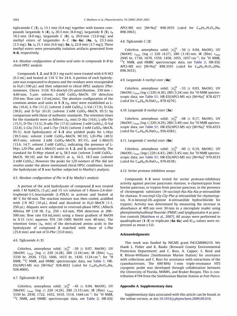
fractionation, and several HPLC purifications to yield compounds1–3 as colorless, amorphous solids. The structures of 1–3 (Fig. 1)were determined by a combination of NMR (1H, COSY, TOCSY, ROESY,HSQC and HMBC) spectroscopic analyses and mass spectrometry.
Compound 1 was isolated as a colorless amorphous solid. Apseudomolecular [M+Na]+ ion peak at m/z 928.4032 in the HR-ESI/APCI-MS suggested a molecular formula of C45H59N7O13, whichwas in agreement with the putative molecular composition basedon NMR spectroscopic data. A detailed 2D NMR analysis in DMF-d7
(Table 1) enabled identification of the five common amino acidsalanine (Ala), valine (Val), glutamic acid (Glu), tyrosine (Tyr), thre-onine (Thr), the two modified amino acid residues homotyrosine(Htyr) and 2-amino-2-butenoic acid (Abu) and a tiglic acid (Tig)unit in 1, thus indicating a striking resemblance to largamidesA�C (4–6) (Fig 1). The presence of the tiglic acid unit is corrobo-rated by the appearance of two deshielded methyl groups (dH
1.81 and 1.73) and one olefinic methine proton (dH-3 6.44, br q)in the 1H NMR spectrum; the latter further showed correlationsto a quaternary carbon at d 132.4 (C-2, Tig) and a carbonyl carbonat d 169.1 (C-1,Tig) in the HMBC spectrum. In addition, the olefinicproton d 6.44 (H-3) showed a ROESY cross peak to a methyl reso-nating at d 1.73 (H3-3) and lacked a correlation to the methyl atd 1.81 (H3-2), indicating E geometry of the double bond and con-firming a tigloyl group in 1 as in 4–6. The geometry of the Abu unit
Fig. 1. Structures of tiglicamides A–C (1–3), largamides A–C (4–6) and theircorresponding methyl esters 4a–6a.
NH 8.86, br s 1 (Thr)Thr 1 170.2, qC
2 4.78, br m 55.5, CH 13 5.39, br q 73.1, CH 14 1.19, d (5.9) 15.9, CH3 2, 3NH 7.89, d (8.2) 1 (Tyr)
Tyr 1 172.4, qC2 4.77, br m 55.4, CH 1, 1 (Val)3 3.08, dd (�13.2, 3.9) 37.8, CH2 2, 4, 5/9
2.84, dd (�13.2, 9.6)4 128.2, qC5/9 7.12, d (7.8) 130.5, CH 3, 6/8, 76/8 6.75, d (7.8) 115.1, CH 5/9, 77 156.7, qCOH 9.35, s 6/8, 7NH 8.08, br d (7.5)
Val 1 171.8, qC2 4.30, br dd 58.8, CH 1, 3, 4, 53 2.04, m 31.3, CH 1, 2, 4, 54 0.76, d (6.3) 19.3, CH3 2, 3, 55 0.73, d (6.3) 17.9, CH3 2, 3, 4NH 7.29, d (8.8) 1 (Tig)
Tig 1 169.1, qC2 132.4, qC3 6.44, br q (6.3) 130.1, CH 1, 44 1.73, br d (6.3) 13.4, CH3 2, 35 1.81, br s 12.2, CH3 1, 2, 3
a Protons showing long-range correlation with indicated carbon.
was deduced as Z based on a ROESY cross peak between the AbuNH (dH 10.21) and Abu methyl group (dH 1.78). HMBC analysis sup-ported by ROESY correlations unambiguously established the lin-ear sequence of the amino acid units and tiglic acid moiety(Table 1). The deshielded proton signal at dH 5.39 (Thr) was indic-ative of a lactone functionality which arises from ester linkage of 1from the carbonyl of Htyr and the hydroxyl group of Thr. The IRspectrum of 1, displaying absorptions at 1722 and 1652 cm�1 char-acteristic of amide and ester functionalities, respectively, sup-ported the proposed depsipeptide structure.
A [M+Na]+ peak at m/z 898.3935 in the HR-ESI/APCI-MS of 2 inconjunction with 2D NMR data suggested a molecular formula ofC44H57N7O12 for compound 2. The 1H NMR spectrum indicated thatcompound 2 is closely related to 1. 2D NMR analysis (1H, COSY,HSQC, HMBC, ROESY) provided further evidence for the presenceof Ala, Val, Thr, Glu, and two aromatic amino acid residues, only

2060 S. Matthew et al. / Phytochemistry 70 (2009) 2058–2063
one of which was para-substituted (Tyr) in 2. The Htyr unit in 1was replaced by a Phe residue in 2 (Table 2), which accountedfor the 30 mass unit difference (CH2O) compared with 1. The pres-ence of the Phe residue in the cyclic core was confirmed based onan HMBC correlation of the Phe NH proton to C-1 of the Glu residue(Table 2).
HR-ESI/APCI-MS analysis for compound 3 provided an [M+Na]+
peak at m/z 898.3591, suggesting a molecular formula ofC40H57N7O13S. The 1H NMR of 3 displayed signals characteristicfor 1 and 2 with the exception of the fewer aromatic signals and
Table 2NMR spectroscopic data for tiglicamides B (2) and C (3) in DMF-d7 (600 MHz).
Unit C/H No. Tiglicamide B (2)
dH (J in Hz) dC, mult. HMBCa
Pheb/Met(O)c 1 170.9, qC2 4.77, br m 52.9, CH 1, 33 3.29, dd (�13.6, 4.9) 37.6, CH2 2, 4, 5/8
2.76, dd (�13.6, 10.3)4 138.5, qC
5/9 7.29, m 129.7, CH 3, 6/9, 76/8 7.27, m 128.3, CH 3, 5/8, 77 7.19, m 126.4, CH 5/8, 6/9S-Me – – –NH 7.84, d (9.4) 2, 1 (Glu)
Glu 1 171.1, qC2 4.42, br m 52.4, CH 3, 43 2.44, m; 2.10, m 27.0, CH2 2, 4, 54 2.50, m; 2.41, m 30.6, CH2 2, 3, 55 174.9, qCOH Not observedNH 7.60, d (9.0) 2, 1 (Abu)
Abu 1 163.9, qC2 130.9, qC3 6.65, q (7.0) 129.0, CH 1, 4
4 1.80, d (7.0) 12.6, CH3 1, 2, 3NH 10.23, s 1, 1 (Ala)
Ala 1 175.7, qC2 4.39, br m 50.2, CH 1, 33 1.42, d (6.8) 16.4, CH3 1, 2NH 8.89, br s 1, 2, 3
Thr 1 170.4, qC2 4.77, br m 55.2, CH 13 5.45, br q 72.9, CH4 1.18, d (5.0) 15.7, CH3 2, 3NH 7.92, d (8.2) 2, 1 (Tyr)
Tyr 1 172.7, qC2 4.77, br m 55.3, CH 33 3.08, dd (�13.7, 4.5) 37.7, CH2 2, 4, 5/9
2.83, dd (�13.7, 10.1)4 128.5, qC5/9 7.13, d (7.8) 130.5, CH 3, 6/8, 76/8 6.73, d (7.8) 115.2, CH 5/9, 77 157.0, qCOH 9.37, br s 6/8, 7NH 8.09, d (8.1) 1 (Val)
Val 1 171.8, qC2 4.28, br t (7.3) 58.8, CH 1, 3, 4, 5, 1 (Tig)3 2.02, m 31.4, CH 1, 2, 4, 54 0.74, d (6.8) 19.4, CH3 2, 3, 55 0.72, d (6.8) 18.0, CH3 2, 3, 4NH 7.26, d (7.3) 2, 1 (Tig)
Tig 1 169.2, qC2 132.5, qC3 6.43, br q (6.8) 130.3, CH 1, 4, 54 1.70, br d (6.8) 13.5, CH3 2, 35 1.79, br s 12.3, CH3 1, 2, 3
a Protons showing long-range correlation with indicated carbon.b Refers to compound 2.c Refers to compound 3.d Signal doubling because of diastereomers at chiral S*.e Signal doubling observed.
two additional methyl singlets at dH 2.55/2.52 (1:1), which inte-grated together for one methyl group (S–Me). A detailed 2D NMRanalysis (COSY, TOCSY, HSQC, HMBC, ROESY) indicated that com-pound 3 contained only one aromatic amino acid residue and dif-fers from 1 or 2 only by the presence of a methionine sulfoxide(Met(O)) unit instead of Htyr or Phe, respectively (Table 2). TheMet(O) residue was verified by 2D NMR spectroscopic analysis,where the two methyl singlets at dH 2.55/2.52 showed HSQC corre-lations to the doubled signals at dC 38.7/37.6 and HMBC correla-tions to dC 50.56/50.60. This doubling of signals in the ratio of
Tiglicamide C (3)
dH (J in Hz) dC, mult. HMBCa
170.7, qC4.80, br m 50.0, CH 12.31/2.26d, m 24.85, 24.90d, CH2 2, 41.96/1.86d, m2.81/2.75d, m 50.56, 50.60d, CH2 32.71/2.63d, m– –– –– –2.55, 2.52d, s 38.7, 37.6d, CH3 47.71, d (9.4) 1(Glu)
171.7, qC4.48, br m 53.6, CH 3, 42.47, m; 2.18, m 26.9, CH2 1, 2, 4, 52.60, m; 2.48, m 30.9, CH2 2, 3, 5
175.0, qCNot observed7.49, d (8.6), 7.48, d (8.6)e 1 (Abu)
164.9, qC130.6, qC
6.54, qd (7.0, 1.3)/6.53 129.3, CH 1, 4qd (7.0, 1.3)e
1.77, dq (7.1, 1.3) 12.6, CH3 1, 2, 310.25, s 1, 1 (Ala)
176.1, qC4.34, br m 50.6, CH 1, 31.40, d (7.0) 16.7, CH3 1, 28.90, d (3.0)/8.89, d (3.2)e 1, 3
170.3, qC4.80, br m 55.5, CH 15.429, qd (6.4, 2.9)/5.427, qd (6.4, 2.9)e 73.9, CH 1, 1 (Met(O))1.20, d (6.4) 16.1, CH3 2, 37.95, d (8.1) 2, 1 (Tyr)
172.8, qC4.79, br m 55.8, CH 1, 3, 1 (Val)3.06, dd (�14.0, 4.8) 38.1, CH2 1, 2, 4, 5/92.83, dd (�14.0, 10.0)
128.4, qC7.12, d (8.5) 131.1, CH 3, 6/8, 76.71, d (8.5) 115.6, CH 5/9, 7
157.3, qC9.35, s 6/8, 78.12, d (8.5) 2, 1 (Val)
172.0, qC4.29, dd (6.8, 2.7)/4.28, dd (6.8, 2.7)e 59.2, CH 1, 3, 4, 5, 1 (Tig)2.02, m 31.5, CH 1, 2, 4, 50.75, d (6.8) 19.7, CH3 2, 3, 50.72, d (6.8) 18.3, CH3 2, 3, 47.27, d (8.6) 1, 2, 1 (Tig)
169.3, qC132.7, qC
6.43, qq (7.0, 1.5) 130.6, CH 1, 4, 51.72, dq (7.0, 1.5) 13.7, CH3 2, 31.80, br s 12.4, CH3 1, 2, 3

Table 3Inhibition of porcine pancreatic elastase.
IC50 in lMa IC50 in lMb IC50 in lMc
1 2.14 ± 0.19 4 1.41 ± 0.28 4a 2.94 ± 0.192 6.99 ± 0.74 5 0.53 ± 0.19 5a 2.23 ± 0.083 7.28 ± 0.95 6 1.15 ± 0.46 6a 2.16 ± 0.13
a n = 4.b n = 4 (taken from Matthew et al., 2009).c n = 3.
S. Matthew et al. / Phytochemistry 70 (2009) 2058–2063 2061
1:1 suggested the occurrence of epimeric R and S sulfoxides. The IRabsorption at 1037 cm�1 also supported the sulfoxide assignment.The sulfoxide is most likely an isolation artifact formed by oxida-tion of the methionine-containing natural product (Matthewet al., 2008; Gunasekera et al., 2008; Harrigan et al., 1999).
The absolute configuration of all amino acid units in compounds1–3 was deduced by chiral HPLC of the acid hydrolysis products,which indicated D configuration for glutamic acid and tyrosineand L configuration for all other amino acids. However, none ofthe chiral HPLC conditions employed were successful in resolvingthe D/L-isomeric peaks for Phe. Hence the acid hydrolyzate of 2was subjected to Marfey’s analysis (Fujii et al., 1997), establishingL configuration for Phe in 2.
Compounds 1–3 were tested for serine protease-inhibitoryactivity. They showed moderate activity against porcine pancreaticelastase in vitro with IC50 values ranging from 2.14 to 7.28 lM(Table 3). While the described compounds are two to three ordersof magnitudes less potent against the same enzyme or other mam-malian elastases than lyngbyastatins 4–7 (Matthew et al., 2007;Taori et al., 2007) or ONO-5046 (Kawabata et al., 1991), prelimin-ary data suggested some selectivity towards elastase. The activitiesof two other serine proteases tested (chymotrypsin, trypsin) werenot compromised by compounds 1–3 at concentrations up to50 lM. These results are consistent with those previously reportedfor their analogues (4–6) (Matthew et al., 2009). Since we also iso-lated the corresponding largamide methyl esters 4a–6a (presum-ably isolation artifacts), we were able to probe the effect ofmethylation at that position. Compounds 4a–6a retainedlow-micromolar inhibitory activity (Table 3), indicating that thecarboxylic acid residue is not a requisite element for elastase-inhibitory activity (Matthew et al., 2009).
3. Conclusion
The L. confervoides that yielded tiglicamides A–C (1–3) is a par-ticularly prolific source of secondary metabolites (Matthew et al.,2007, 2008, 2009; Taori et al., 2007), with already 13 previously re-ported structures belonging to six different structural families(Sharp et al., 2009). The similarity to the largamides A–C (4–6)and, specifically, the variability of the last amino acid position(N ? C) suggests relaxed substrate specificity of the correspondingputative biosynthetic NRPS enzyme, allowing the incorporation ofat least six different amino acids, viz. Htyr (1), Phe (2), Met (3), Leu(4), 2-amino-5-(40-hydroxy-phenyl)pentanoic acid (Ahppa) (5),and 2-amino-5-(40-hydroxy-phenyl)hexanoic acid (Ahpha) (6) atthat position. Largamides A–C (4–6) were major metabolites in thiscyanobacterium, while tiglicamides A–C (1–3) were only minormetabolites. It is unclear if the differing yields are reflective ofthe relative efficiencies of substrate activation or due to geneticheterogeneity of the cyanobacterial samples and consequently bio-synthetic enzymes, although the 16S rDNA sequence was identicalfor at least three distinct collections at different times (Sharp et al.,2009). Detailed genetic studies of this intriguing cyanobacteriumwill provide novel insights into the biosynthesis of compounds1–6 and other co-produced L. confervoides metabolites.
4. Experimental
4.1. General experimental procedures
Optical rotation was measured on a Perkin–Elmer 341 polarim-eter. UV spectra were recorded using a SpectraMax M5 (MolecularDevices). 1H and 2D NMR spectra for 1 and 2 were recorded inDMF-d7 on a Bruker 600 MHz spectrometer equipped with a 1-mm high-temperature superconducting cryogenic probe and 3was recorded in 5 mm cryogenic probe operating at 600 MHz and150 MHz using residual solvent signals as the internal standard(dH 8.02, dC 162.7). HSQC experiments were optimized for1JCH = 145 Hz, and HMBC experiments were optimized fornJC,H = 7 Hz for 1 and 2 and 10 Hz for 3. HRMS data were obtainedusing an Agilent LC-TOF mass spectrometer equipped with anAPCI/ESI multimode ion source detector (UCR Mass SpectrometryFacility, University of California at Riverside), and low resolutionmass spectra were obtained on a A3200 Q TRAP LC/MS/MS (hybridtriple quadrupole linear ion trap mass spectrometer, Applied Bio-systems, USA) with an electrospray ionization (ESI) interface oper-ated in positive mode. HPLC-based compound isolation wasperformed on a Shimadzu LC-20AT prominence LC with peakdetection by a Shimadzu SPD-M20A prominence diode arraydetector.
4.2. Marine cyanobacterial samples
Samples of L. confervoides were collected at approximately 15 mdepth from reefs near the Port Everglades Inlet, Fort Lauderdale,Florida, USA (26�05.99020N, 80�05.01840W) in August 2004 andMay and August 2005. S. Golubic identified the cyanobacterium(Paul et al., 2005) and its 16S rDNA gene sequence has been re-ported (Paul et al., 2005; Sharp et al., 2009).
4.3. Extraction and isolation
The freeze-dried organisms collected through 2004–2005(�2700 g dry weight) were extracted with EtOAc–MeOH (1:1) toafford a crude extract (�400 g) which was suspended in H2O(500 mL) then defatted with hexanes (500 mL � 3; �2 g). The con-centrated aqueous layer enriched with salt was further partitionedbetween n-BuOH (250 mL � 3) and H2O. The combined n-BuOH ex-tract (12 g) was applied on a Diaion HP-20 (Supelco) resin (120 g),and subsequently fractionated with H2O and increasing concentra-tions of MeOH, and then with MeCN and finally with CH2Cl2 toyield 8 fractions [Fr. 1: H2O (100%, 2 L, �6.8 g); Fr. 2: H2O:MeOH(80:20, 1 L, 854 mg); Fr. 3: H2O:MeOH (50:50, 1 L, 272 mg); Fr. 4:H2O:MeOH (50:50–25:75, 1 L, 400 mg); Fr. 5: H2O:MeOH (25:75–0:100, 1 L, 430 mg); Fr. 6: MeOH (100%, 1 L, 950 mg); Fr. 7: MeCN(100%, 1 L, 490 mg), Fr. 8: CH2Cl2 (100%, 1 L, 457 mg)]. Fractions 5and 6 were subjected to reversed-phase preparative HPLC (Phe-nomenex Luna-C18 10 l, 100 � 21.20 mm, 5.0 mL/min; PDA detec-tion at 200–400 nm) using a MeOH–0.05% aqueous TFA lineargradient (40–100% over 30 min and then MeOH for 15 min). Thelargamide- and tiglicamide-rich fractions eluting between tR 15–25 min were collected and subjected to repeated semipreparativereversed-phase HPLC (Phenomenex Synergi 4u Hydro-RP,250 � 10 mm, 2.0 mL/min; PDA detection at 200–400 nm) usingtwo sequential linear gradients of MeOH in 0.05% aqueous TFA(60–90% over 25 min, 90–100% over 10 min) to give semi-purecompounds 1–6 eluting between tR 16–21 min. The final purifica-tion of the compounds was achieved by means of a PhenomenexLuna Phenyl-hexyl column 10 � 250 mm, using the same HPLCconditions as described above to afford tiglicamide A (1), tR
16.0 min (1.2 mg), tiglicamide B (2), tR 21.8 min (0.8 mg), and
2062 S. Matthew et al. / Phytochemistry 70 (2009) 2058–2063
tiglicamide C (3), tR 13.1 min (0.4 mg) together with known com-pounds largamide A (4), tR 20.3 min (8.0 mg), largamide B (5), tR
18.2 min (8.0 mg), largamide C (6), tR 20.0 min (12.0 mg) andmethyl esters of largamides A�C (4a�6a) [4a, tR 23.3 min(2.5 mg); 5a, tR 21.1 min (6.6 mg); 6a, tR 22.8 min (3.7 mg)]. Thesemethyl esters were presumably isolation artifacts generated from4–6, respectively.
4.4. Absolute configuration of amino acid units in compounds 1–3 bychiral HPLC analysis
Compounds 1, 2, and 3 (0.1 mg each) were treated with 6 N HCl(0.3 mL) and heated at 116 �C for 24 h. A portion of each hydroly-zate was evaporated to dryness and the residues were resuspendedin H2O (100 lL) and then subjected to chiral HPLC analysis (Phe-nomenex, Chirex 3126 N,S-dioctyl-(D)-penicillamine, 250 mm �4.60 mm, 5 lm; solvent, 2 mM CuSO4–MeCN; UV detection254 nm; flow rate 1.0 mL/min). The absolute configuration of thecommon amino acid units in 1–3 (tR, min) were established as L-Ala (10.4), L-Thr (11.2) (solvent 2 mM CuSO4), L-Val (17.9), D-Glu(58.0), and D-Tyr (61.0) (solvent 2 mM CuSO4–MeCN, 95:5) bycomparison with those of authentic standards. The retention timesfor the standards were as follows (tR, min) D-Ala (14.6), L-allo-Thr(15.5), D-Thr (13.5), D-allo-Thr (17.4) (solvent 2 mM CuSO4), D-Val(22.8), L-Glu (53.5), and L-Tyr (54.0) (solvent 2 mM CuSO4–MeCN,95:5). Acid hydrolyzates of 1–3 also yielded peaks for L-Htyr(39.5 min; solvent 2 mM CuSO4–MeCN, 90:10), L/D-Phe (49.5/49.5 min; solvent 2 mM CuSO4–MeCN, 85:15), and L-Met(O)(13.6, 14.7; solvent 2 mM CuSO4), indicating the presence of L-Htyr, L/D-Phe, and L-Met(O) units in 1, 2, and 3, respectively. Thestandard for D-Htyr eluted at tR 58.5 min (solvent 2 mM CuSO4–MeCN, 90:10) and for D-Met(O) at tR 16.5, 18.5 min (solvent2 mM CuSO4). However the peaks for L/D-isomers of Phe did notresolve under the above mentioned chiral HPLC conditions; hencethe hydrolyzate of 3 was further subjected to Marfey’s analysis.
4.5. Absolute configuration of Phe in 2 by Marfey’s analysis
A portion of the acid hydrolyzate of compound 2 was treatedwith 1 M NaHCO3 (5 lL) and 1% v/v solution of 1-fluoro-2,4-dini-trophenyl-5-L-leucinamide (L-FDLA) in acetone and heated at40 �C for 60 min. The reaction mixture was then cooled, acidifiedwith 2 N HCl (10 lL), dried and dissolved in H2O–MeCN (1:1,250 lL). Aliquots were subjected to reversed-phase HPLC (AlltechAlltima HP C18 HL 5 l, 250 � 4.6 mm, PDA detection at 200–500 nm; flow rate 0.8 mL/min) using a linear gradient of MeOHin 0.1% (v/v) aqueous TFA (50–100% MeOH over 40 min). Theretention times (tR, min) of the derivatized amino acids in thehydrolyzate of compound 2 matched with those of L-Phe(25.8 min) and not of D-Phe (33.0 min).
4.6. Tiglicamide A (1)
Colorless, amorphous solid; ½a�20D �50 (c 0.07, MeOH); UV
(MeOH) kmax (log e) 220 (4.28), 280 (3.34) nm; IR (film) mmax
3330 br, 2926, 1722, 1666, 1615 br, 1430, 1124 cm-1; for 1HNMR, 13C NMR, and HMBC spectroscopic data, see Table 1; HR-ESI/APCI-MS m/z [M+Na]+ 928.4032 (calcd for C45H59N7O13Na,928.4069).
4.7. Tiglicamide B (2)
Colorless, amorphous solid; ½a�20D �45 (c 0.04, MeOH); UV
(MeOH) kmax (log e) 220 (4.26), 280 (3.15) nm; IR (film) mmax
3330 br, 2930, 1722, 1652, 1610, 1514, 1444 cm–1; for 1H NMR,13C NMR, and HMBC spectroscopic data, see Table 2; HR-ESI/
APCI-MS m/z [M+Na]+ 898.3935 (calcd for C44H57N7O12Na,898.3963).
4.8. Tiglicamide C (3)
Colorless, amorphous solid; ½a�20D �56 (c 0.04, MeOH); UV
(MeOH) kmax (log e) 220 (4.27), 280 (3.18) nm; IR (film) mmax
2945 br, 1730, 1670, 1559, 1458, 1055, 1037 cm–1; for 1H NMR,13C NMR, and HMBC spectroscopic data, see Table 2; HR-ESI/APCI-MS m/z [M+Na]+ 898.3591 (calcd for C40H57N7O13SNa,898.3633).
4.9. Largamide A methyl ester (4a)
Colorless, amorphous solid; ½a�20D �53 (c 0.03, MeOH); UV
(MeOH) kmax (log e) 220 (4.30), 280 (3.24) nm; for 1H NMR spectro-scopic data, see Table S1; HR-ESI/APCI-MS m/z [M+Na]+ 878.4277(calcd for C42H61N7NaO12, 878.4276).
4.10. Largamide B methyl ester (5a)
Colorless, amorphous solid; ½a�20D �68 (c 0.17, MeOH); UV
(MeOH) kmax (log e) 220 (4.29), 280 (3.49) nm; for 1H NMR spectro-scopic data, see Table S1; HR-ESI/APCI-MS m/z [M+Na]+ 956.4353(calcd for C47H63N7NaO13, 956.4382).
4.11. Largamide C methyl ester (6a)
Colorless, amorphous solid; ½a�20D �60 (c 0.10, MeOH); UV
(MeOH) kmax (loge) 220 (4.43), 280 (3.45) nm; for 1H NMR spectro-scopic data, see Table S1; HR-ESI/APCI-MS m/z [M+Na]+ 970.4533(calcd for C48H65N7NaO13, 970.4538).
4.12. Serine protease inhibition assays
Compounds 1–3 were tested for serine protease-inhibitoryactivity against porcine pancreatic elastase, a-chymotrypsin frombovine pancreas, or trypsin from porcine pancreas, in the presenceof chromogenic substrates (N-succinyl-Ala-Ala-Ala-p-nitroanilidefor elastase, N-succinyl-Gly-Gly-Phe-p-nitroanilide for chymotryp-sin, N-a-benzoyl-DL-arginine 4-nitroanilide hydrochloride fortrypsin). Activity was determined by measuring the increase inabsorbance at 405 nm over 30 min in a microplate reader usingphenylmethylsulfonyl fluoride (PMSF) and lyngbyastatin 4 as posi-tive controls [Matthew et al., 2007]. All assays were performed inquadruplicate (1–3) or triplicate (4a–6a) and IC50 values were ex-pressed as mean ± S.D.
Acknowledgments
This work was funded by NIGMS grant P41GM086210. Wethank L. Fisher and K. Banks (Broward County EnvironmentalProtection Department) and C. Ross, A. Capper, S. Reed andR. Ritson-Williams (Smithsonian Marine Station) for assistancewith collections and C. Ross for assistance with extractions of thecyanobacterium. The 600 MHz 1-mm triple-resonance HTScryogenic probe was developed through collaboration betweenthe University of Florida, NHMFL, and Bruker Biospin. This is con-tribution #794 from the Smithsonian Marine Station at Fort Pierce.
Appendix A. Supplementary data
Supplementary data associated with this article can be found, inthe online version, at doi:10.1016/j.phytochem.2009.09.010.
S. Matthew et al. / Phytochemistry 70 (2009) 2058–2063 2063
References
Abbenante, G., Fairlie, D.P., 2005. Protease inhibitors in the clinic. Med. Chem. 1, 71–104.
Blunt, J.W., Munro, M.H., 2008. Dictionary of Marine Natural Products with CD-ROM. Chapman and Hall, CRC, Boca Raton.
Fujii, K., Ikai, Y., Mayumi, T., Oka, H., Suzuki, M., Harada, K.I., 1997. A nonempiricalmethod using LC/MS for determination of the absolute configuration ofconstituent amino acids in a peptide: combination of Marfey’s method withmass spectrometry and its practical application. Anal. Chem. 69, 5146–5151.
Gunasekera, S.P., Williams, R.R., Paul, V.J., 2008. Carriebowmide, a newcyclodepsipeptide from the marine cyanobacterium Lyngbya polychroa. J. Nat.Prod. 71, 2060–2063.
Harrigan, G.G., Luesch, H., Yoshida, W.Y., Moore, R.E., Nagle, D.G., Paul, V.J., 1999.Symplostatin 2: a dolastatin 13 analogue from the marine cyanobacteriumSymploca hydnoides. J. Nat. Prod. 62, 655–658.
Ilies, M.A., Supuran, C.T., Scozzafava, A., 2002. Therapeutic applications ofserine protease inhibitors. Expert Opinion on Therapeutic Patents 12, 1181–1214.
Kawabata, K., Suzuki, M., Sugitani, M., Imaki, K., Toda, M., Miyamoto, T., 1991. ONO-5046, a novel inhibitor of human neutrophil elastase. Biochem. Biophys. Res.Commun. 177, 814–820.
Matthew, S., Ross, C., Rocca, J.R., Paul, V.J., Luesch, H., 2007. Lyngbyastatin 4, adolastatin 13 analogue with elastase and chymotrypsin inhibitory activityfrom the marine cyanobacterium Lyngbya confervoides. J. Nat. Prod. 70, 124–127.
Matthew, S., Ross, C., Paul, V.J., Luesch, H., 2008. Pompanopeptins A and B, newcyclic peptides from the marine cyanobacterium Lyngbya confervoides.Tetrahedron 64, 4081–4089.
Matthew, S., Paul, V.J., Luesch, H., 2009. Largamides A–C, tiglic acid-containingcyclodepsipeptides with elastase-inhibitory activity from the marinecyanobacterium Lyngbya confervoides. Planta Med. 75, 528–533.
McIntosh, J.A., Donia, M.S., Schmidt, E.W., 2009. Ribosomal peptide naturalproducts: bridging the ribosomal and nonribosomal worlds. Nat. Prod. Rep.26, 537–559.
Paul, V.J., Thacker, R.W., Banks, K., Golubic, S., 2005. Benthic cyanobacterial bloomimpacts the reefs of South Florida (Broward County, USA). Coral Reefs 24, 693–697.
Plaza, A., Bewley, C.A., 2006. Largamides A–H, unusual cyclic peptides from themarine cyanobacterium Oscillatoria sp. J. Org. Chem. 71, 6898–6907.
Schopf, J.W., 1996. Cyanobacteria: pioneers of early earth. Nova Hedwigia 112, 12–32.
Sharp, K., Arthur, K.E., Gu, L., Ross, C., Harrison, G., Gunasekera, S.P., Meickle, T.,Matthew, S., Luesch, H., Thacker, R.W., Sherman, D.H., Paul, V.J., 2009.Phylogenetic and chemical diversity of three chemotypes of bloom-formingLyngbya species (cyanobacteria: Oscillatoriales) from reefs of southeasternFlorida. Appl. Environ. Microbiol. 75, 2879–2888.
Tan, L.T., 2007. Bioactive natural products from marine cyanobacteria for drugdiscovery. Phytochemistry 68, 954–979.
Taori, K., Matthew, S., Rocca, J.R., Paul, V.J., Luesch, H., 2007. Lyngbyastatins 5–7,potent elastase inhibitors from Floridian marine cyanobacteria, Lyngbya spp. J.Nat. Prod. 70, 1593–1600.