tick-borne pathogens shape the native microbiome within
TRANSCRIPT

The University of Southern Mississippi The University of Southern Mississippi
The Aquila Digital Community The Aquila Digital Community
Faculty Publications
9-1-2020
Tick-Borne Pathogens Shape the Native Microbiome Within Tick Tick-Borne Pathogens Shape the Native Microbiome Within Tick
Vectors Vectors
Abdulsalam Adegoke University of Southern Mississippi, [email protected]
Deepak Kumar University of Southern Mississippi, [email protected]
Cailyn Bobo University of Southern Mississippi, [email protected]
Muhammad Imran Rashid University of Veterinary and Animal Sciences Lahore, [email protected]
Aneela Zameer Durrani University of Veterinary and Animal Sciences Lahore, [email protected]
See next page for additional authors
Follow this and additional works at: https://aquila.usm.edu/fac_pubs
Part of the Microbiology Commons
Recommended Citation Recommended Citation Adegoke, A., Kumar, D., Bobo, C., Rashid, M., Durrani, A., Sajid, M., Karim, S. (2020). Tick-Borne Pathogens Shape the Native Microbiome Within Tick Vectors. Microorganisms, 8(9), 1-16. Available at: https://aquila.usm.edu/fac_pubs/18236
This Article is brought to you for free and open access by The Aquila Digital Community. It has been accepted for inclusion in Faculty Publications by an authorized administrator of The Aquila Digital Community. For more information, please contact [email protected].

Authors Authors Abdulsalam Adegoke, Deepak Kumar, Cailyn Bobo, Muhammad Imran Rashid, Aneela Zameer Durrani, Muhammad Sohail Sajid, and Shahid Karim
This article is available at The Aquila Digital Community: https://aquila.usm.edu/fac_pubs/18236

microorganisms
Article
Tick-Borne Pathogens Shape the Native MicrobiomeWithin Tick Vectors
Abdulsalam Adegoke 1 , Deepak Kumar 1, Cailyn Bobo 1, Muhammad Imran Rashid 2 ,Aneela Zameer Durrani 3 , Muhammad Sohail Sajid 4 and Shahid Karim 1,*
1 Center for Molecular and Cellular Biosciences, School of Biological, Environmental and Earth Sciences,University of Southern Mississippi, Hattiesburg, MS 39406, USA; [email protected] (A.A.);[email protected] (D.K.); [email protected] (C.B.)
2 Department of Parasitology, Faculty of Veterinary Science, The University of Veterinary and Animal Sciences,Lahore 54000, Pakistan; [email protected]
3 Department of Clinical Medicine and Surgery, Faculty of Veterinary Science, The University of Veterinaryand Animal Sciences, Lahore 54000, Pakistan; [email protected]
4 Department of Parasitology, Faculty of Veterinary Science, University of Agriculture,Faisalabad 38000, Pakistan; [email protected]
* Correspondence: [email protected]; Tel.: +01-601-266-6232
Received: 13 July 2020; Accepted: 21 August 2020; Published: 25 August 2020�����������������
Abstract: Ticks are blood-feeding arthropods and transmit a variety of medically important viral,bacterial, protozoan pathogens to animals and humans. Ticks also harbor a diverse communityof microbes linked to their biological processes, such as hematophagy, and hence affect vectorcompetence. The interactions between bacterial and/or protozoan pathogens and the tick microbiomeis a black-box, and therefore we tested the hypothesis that the presence of a protozoan or bacterialpathogen will alter the microbial composition within a tick. Hence, this study was designed todefine the microbial composition of two tick species, Hyalomma (H.) anatolicum and Rhipicephalus (R.)microplus. We used a combination of PCR based pathogen (Anaplasma marginale and Theileria species)and symbiont (Wolbachia species) identification followed by metagenomic sequencing and comparisonof the microbial communities in PCR positive and negative ticks. A total of 1786 operational taxonomicunits was identified representing 25 phyla, 50 classes, and 342 genera. The phylum Proteobacteria,Firmicutes, Actinobacteriota, and Bacteroidota were the most represented bacteria group. Alpha andbeta diversity were not significantly affected in the presence or absence of Theileria sp. and A. marginaleas see with H. anatolicum ticks. Interestingly, bacterial communities were significantly reduced inTheileria sp. infected R. microplus ticks, while also exhibiting a significant reduction in microbialrichness and evenness. Putting these observations together, we referred to the effect the presence ofTheileria sp. has on R. microplus a “pathogen-induced dysbiosis”. We also identify the presence ofPlasmodium falciparum, the causative agent of human malaria from the microbiome of both H. anatolicumand R. microplus ticks. These findings support the presence of a “pathogen-induced dysbiosis” withinthe tick and further validation experiments are required to investigate how they are important in thevector competence of ticks. Understanding the mechanism of “pathogen-induced dysbiosis” on tickmicrobial composition may aid the discovery of intervention strategies for the control of emergingtick-borne infections.
Keywords: ticks; microbiome; Hyalomma anatolicum; Rhipicephalus microplus; Anaplasma marginale;Theileria sp.; Francisella; Wolbachia; Pakistan
Microorganisms 2020, 8, 1299; doi:10.3390/microorganisms8091299 www.mdpi.com/journal/microorganisms

Microorganisms 2020, 8, 1299 2 of 16
1. Introduction
Ticks are obligate, blood-feeding ectoparasites of vertebrate animals that depend on the host’sblood for nutrition and reproduction. They elicit significant blood loss and also transmit disease-causingbacteria, viruses, and protozoa from one host to another, which makes them significant to public health.In Pakistan, livestock farming and production serve as one of the major drivers of the macro-economy aswith the latest animal census showing an estimated population of 40 million, 47.8 million, 76.1 million,and 30.9 million buffalo, cattle, goats, and sheep, respectively, all contributing to an estimated 11.22%of the country’s gross domestic product (GDP) [1].
Previously reported tick species infesting livestock in Pakistan includes both hard and softtick species. In Pakistan, the most reported ticks found infesting livestock are hard ticks of thegenus Haemaphysalis, Hyalomma, and Rhipicephalus, and soft ticks of the genus Argas andOrnithodoros [2–5]. Hyalomma and Rhipicephalus ticks are responsible for transmitting majortick-borne pathogens that affect livestock animals in Pakistan. Anaplasma marginale, Anaplasma centrale,Babesia bovis, and Babesia bigemina are which are known to cause cattle fever are transmittedby Rhipicephalus microplus ticks [6]. Tropical theileriosis, a small ruminant disease caused byTheileria annulata and the Crimean Congo hemorrhagic fever virus have been reported to be transmittedby Hyalomma anatolicum [6,7].
Ticks also harbor several distinct microbial communities, members of which have been shownto play an important role in tick biology. Analysis of the genome of such microbes has revealedspecific regions coding for essential vitamins, most of which are lacking in the tick’s blood meal [8–10],emphasizing their possible role as nutritional mutualists. Recent evidence also suggests that somemembers of these microbial communities can potentially interact with tick-borne pathogens bothdirectly and indirectly. It was reported that removing the midgut bacteria of black-legged ticksIxodes scapularis by feeding on antibiotic-treated mice impairs infection by Borrelia burgdorferi [11].Another tick-borne pathogen, Anaplasma. phagocytophilum was shown to reduce the viability of themicrobial population, thus facilitating its colonization of the I. scapularis midgut [12].
Along with the development in tick microbiome studies, adequate information exists on howtick-borne human pathogens shape the tick microbial communities to facilitate their colonizationand subsequent transmission. While adequate research has been carried out on the microbiome oflivestock infesting ticks, there is very little scientific understanding of the interactions that occursbetween tick-transmitted pathogens and the tick microbial communities. Apart from Karim et al. [4],who reported important bacterial genera found in ticks from Pakistan, there is a general lack ofresearch in understanding how specific bacterial or protozoan pathogens of animal origin shapes themicrobiome of several tick vectors.
The main purpose of this study is to develop an understanding of changes that occurs in themicrobial composition within a tick vector when such a tick acquires a bacterial or protozoan pathogenof animal source. The key research question of this study was whether or not the overall abundanceand diversity of tick’s microbial communities are reduced in the presence of a protozoan or bacteriapathogen. This study provides an exciting opportunity to advance our knowledge of tick microbiomeand tick-borne pathogen interactions. A full discussion of the molecular mechanisms of reportedinteractions lies beyond the scope of this study. We will proceed to investigate specific microbialchanges and interactions as part of our ongoing study. It is also beyond the scope of this study toexamine whether host blood meal impacts microbial diversity.
2. Materials and Methods
2.1. Tick Collection and Identification
Fully engorged adult Hyalomma anatolicum and Rhipicephalus microplus ticks were randomlyand carefully removed from livestock animals from four livestock producing regions in Pakistan(Sialkot [32◦29′33.7”N, 74◦31′52.8”E], Gujrat [32◦34′22”N, 74◦04′44” E], Gujranwala [32◦9′24”N,

Microorganisms 2020, 8, 1299 3 of 16
74◦11′24”E], and Sheikhupura [31◦42 47” N, 73◦58′41” E]). This was done by careful removal offully engorged ticks using tweezers with care been taken to keep the mouthparts intact. All tickswere kept in separate vials containing 70% ethanol and details of the location, and the host wasrecorded. For this study, a total of 320 ticks were selected and shipped from Pakistan to the Universityof Southern Mississippi for further analysis using the U.S. Department of Agriculture’s Animaland Plant Health Inspection Service (permit # 11122050). Identification of ticks to the genus levelwas carried out by an expert taxonomist at the United States National Tick Collection (USNTC)according to the criteria used in previously published reports [2,13,14]. All stages were examined on anOlympus SZX16 stereoscopic microscope (Olympus Life Science, Center Valley, PA, USA). To furtherconfirm morphological identification, ticks homogenates were subjected to molecular identificationby amplifying the highly conserved 708 bp mitochondrial Cytochrome Oxidase I gene (COI) [15].The nucleotide accession numbers were MT876643, MT876644, and MT876645.
2.2. Genomic DNA Extraction
High-quality DNA was extracted from all the 320 ticks. Before DNA extraction, ticks wereremoved from the transport vials, cleaned using 100% ethanol, dried, and subsequently cleaned using a10% sodium hypochlorite solution. Ticks were finally cleaned using distilled water and allowed to dryon a kimwipe paper. Homogenization of individual ticks was done mechanically, first by cutting ticksinto smaller pieces, followed by complete disruption using an automated, hand-held homogenizer.The DNeasy Blood & Tissue Kit (Qiagen, Germantown, MD, USA) was used to extract DNA fromindividual ticks with minor modification in the volume eluted (30 µL). The DNA concentrationsand quality were quantified using a nanodrop machine (Nanodrop One, Thermo Fisher Scientific,Pittsburgh, PA, USA) and DNA stored in −20 ◦C till further needed.
2.3. Detection of Pathogen and Endosymbiont
To detect the presence of pathogens and endosymbiont of interest, we utilized a PCR basedapproach to amplify the 18S rRNA gene of Theileria sp. [4], 16S rRNA gene of Anaplasma marginale [16],and GroEL gene of Wolbachia [17]. PCR positive DNAs were amplicon sequenced using both theforward and reverse primers and the partial sequences were subjected to NCBI BLAST program forfurther confirmation. Details of the primers, conditions, and amplicon sizes can be found in Table 1.The Wolbachia sp. nucleotide accession numbers are MT881679, MT881680, MT881681, MT881682,MT881683, MT881684, MT881685, MT881686, MT881687, MT881688, MT881689, MT881690, MT881691,MT881692, MT881693, MT881694, MT881695, MT881696, MT881697, MT881698, MT881699, MT881700,MT881701 and MT881702.
Table 1. List of primers used in this study and their respective amplicon sizes.
Target Genes Primer Sequence (5’–3’) Amplicon Size (bp) References
Amar 16S-F GGC GGT GAT CTG TAG CTG GTC TGA 270 bp [16]Amar 16S-R GCC CAA TAA TTC CGA ACA ACG CTT
Theileria sp 18S-F GGT AAT TCC AGC TCCAAT AG 300 bp [4]Theileria sp 18S-R ACC AAC AAA ATA GAA CCA AAG TC
16S rRNA 27F AGR GTT TGA TCM TGG CTC AGV1–V3 [13]
16S rRNA 519R GTN TTA CNG CGG CKG CTGCOI-F GGT CAA CAA ATC ATA AAG ATA TTG G 708 bp [15]COI-R TAA ACT TCA GGG TGA CCA AAA AAT CA
Wolbachia sp GroEL-F TGT ATT AGA TGA TAA CGT GC 800 bp [17]Wolbachia sp GroEL-R CCA TTT GCA GAA ATT ATT GCA
2.4. 16S rRNA Library Preparation and Sequencing
A total of 40 ticks (20 H. anatolicum and 20 R. microplus) were used for microbiome analysis.From each tick species, 5 Theileria sp. positive, 5 A. marginale positive, and 10 negative ticks were selectedfor 16S rRNA library preparation and sequencing. The hypervariable V1–V3 region of the 16S rRNA

Microorganisms 2020, 8, 1299 4 of 16
gene was PCR amplified using the forward primer 27F (5’-AGR GTT TGA TCM TGG CTC AG-3’) andthe reverse primer 519R (5’-GTN TTA CNG CGG CKG CTG-3’) as outlined by the 16S Illumina’s MiSeqprotocol (www.mrdnalab.com, Shallowater, TX, USA. Accessed on 11 July 2020). Briefly, PCR wasperformed using the HotStarTaq Plus Master Mix Kit (Qiagen, Germantown, Maryland, USA) under thefollowing conditions: 94 ◦C for 3 min, followed by 30–35 cycles of 94 ◦C for 30 s, 53 ◦C for 40 s and 72 ◦Cfor 1 min, after which a final elongation step at 72 ◦C for 5 min was performed. After amplification,PCR products were electrophoresed in 2% agarose gel to determine the success of amplification andthe relative intensity of bands. Multiple samples were pooled together in equal proportions basedon their molecular weight and DNA concentrations. Pooled samples were purified using calibratedAmpure XP beads. Then the pooled and purified PCR product was used to prepare Illumina DNAlibrary. Sequencing was performed at MR DNA (www.mrdnalab.com, Shallowater, TX, USA. Accessedon 11 July 2020) on a MiSeq following the manufacturer’s guidelines.
2.5. Sequence Analysis
Sequence analysis was carried out using the Quantitative Insights into Microbial Ecology (QIIME 2)pipeline unless stated otherwise. Briefly, the processing of raw fastq files was demultiplexed.The Atacama soil microbiome pipeline was incorporated for quality control of demultiplexed paired-endreads using the DADA2 plugin as previously described [18]. Low-quality sequences were trimmed andfiltered out, and subsequent merging of paired-end-reads was done ensuring 20 nucleotide overhangbetween forward and reverse reads. Chimeric sequences were removed from the sequence table.
Sequence alignment and subsequent construction of phylogenetic tree from representativesequences were performed using the MAFFT v. 7 and FasTree v. 2.1 plugins [19] Operational taxonomicassignment was performed using the qiime2 feature-classifier plugin v. 7.0, which was previouslytrained against the SILVA 138 database preclustered at 99%. Tables representing operational taxonomicunits (OTUs) and representative taxonomy were exported from R and used for diversity metric analysisusing the Microbiome Analyst web-based interface [20,21]. Raw data from this analysis were submitteddeposited and assigned the GenBank BioProject number #PRJNA600935.
2.6. Alpha Diversity
To establish whether alpha diversity differs across tick samples, reads were transformed and lowabundance OTUs were filtered from the datasets. The Observed OTU metric was used to estimatespecies richness by identifying unique OTUs present across the tick groups, while the Shannon indexwas used to estimate both richness and evenness.
2.7. Beta Diversity
To compare the differences in the microbiome between tick groups, based on measures ofdistance or dissimilarity, dissimilarity matrix was generated from log-transformed sequence data andordination of the plots was visualized using both the Principal Coordinates Analysis (PCoA) and theNonmetric Multidimensional Scaling (NMDS). The Bray–Curtis distance matrix was used to visualizecompositional differences in the microbiome across all groups.
2.8. Statistical Analysis
Statistical significance was inferred using the Mann–Whitney/Kruskal–Wallis method foralpha diversity and classical univariate comparison analysis, while the Permutational MANOVA(PERMANOVA) was used to test for the statistical significance of the dissimilarity measures.

Microorganisms 2020, 8, 1299 5 of 16
3. Results
3.1. Pathogen and Symbiont Prevalence
PCR analysis supported by amplicon sequencing and blast analysis of sequenced PCR productshowed that 23 (7.2%), 90 (28.1%), and 3 (0.9%) of the 320 individually tested ticks were positive forA. marginale, Theileria sp., and Wolbachia sp. (Table S1). We further determined the genetic relationshipof the identified Wolbachia sp. and the tick species used with publicly available sequences from NCBI(Figures S1 and S2). The Wolbachia GroEL gene identified in ticks from this study shows high similarityto Wolbachia pipientis strain wAlbB-HN2016 and wAlbB-FL2016, with 99% query cover and 99.61%identity (Figure S3). The query cover and percentage identities of the COI sequences from this studywere also compared to those previously deposited in the NCBI database (Figure S4).
3.2. Bacteria 16S rRNA Abundance Profile
A total of 2,787,815 million reads paired-end reads were generated. Analysis of the demultiplexedpaired-end-reads generated 2,787,815 reads which ranged from 36,124 to 123,736 with an average of65,609 reads. After passing the sequences through quality filtering, 119,802 of the raw reads werenon-chimeric which were subsequently used for taxonomic classification (Tables S2 and S3). Taxonomicclassification using the SILVA reference base identified 472 OTUs generated from R. microplus ticksbelonging to 10 phyla, 17 classes, and 146 genera. H. anatolicum had a total of 1314 OTUs representing15 phyla, 33 classes, and 196 genera.
3.3. Bacteria Relative Abundance
The relative proportion of bacteria at different taxonomic classification was further analyzed inboth tick species. Figure 1 presents the results obtained from the taxonomic classification of identifiedbacteria OTU at phylum, family, and genus taxonomic levels. Additional figures showing relativeabundances of bacteria species in individual samples can be found (Figure S5A,B).
The phylum Proteobacteria, Firmicutes, Actinobacteriota, and Bacteroidota were all found to bepresent in H. anatolicum ticks. As shown in Figure 1A, the phylum Proteobacteria was found to bepresent at an abundance of 87.5%, 68%, and 49% in Theileria sp. positive, uninfected and A. marginalepositive H. anatolicum ticks, respectively, while Firmicutes (25%) was only present in A. marginaleinfected ticks. Phylum level abundance in R. microplus ticks (Figure 1B) contrasts that shown inH. anatolicum. The entirety of the bacteria identified in Theileria sp. positive A. marginale belongs to thephylum Firmicutes (100%), while both A. marginale positive and positive R. microplus shares similarbacteria phylum distribution representing Actinobacteria, Bacteroidota, and Proteobacteria (Figure 1B).
Francisellaceae (37.5%) and Rickettsiales_fa (50%) constituted to approximately 87.5% of thebacteria family identified within Theileria sp. positive H. anatolicum ticks. These bacteria families werealso identified in uninfected ticks albeit at a much-reduced abundance (Figure 1C). Staphylococcaceaewas identified at a relative abundance of ~30% in A. marginale infected H. anatolicum. The familyAnaplasmataceae (37.5%) were identified at similar abundance in both A. marginale infected anduninfected H. anatolicum (Figure 1C). Distribution of the bacteria family in R. microplus ticksidentified Bacillaceae at a 100% abundance in Theileria sp. positive R. microplus, while the familyCorynebacteriaceae, Coxiellaceae, Flavobacteriaceae, Staphylococcaceae, and Weeksellaceae weredetected in similar abundances in A. marginale positive and uninfected R. microplus (Figure 1D).
Similar differences in the bacteria abundances were further identified at the genus level whichreflects those seen in the family and phylum. As can be seen from Figure 1E, five major genera;Acinetobacter, Anaplasma, Devosia, Norcadiopsis, and Sphingomonas which represents 87.5% of thebacteria were identified from A. marginale infected H. anatolicum ticks. The only genus of bacteriaidentified in Theileria sp. positive H. anatolicum ticks was Candidatus_Midichloria (51.5%) andFrancisella (36%). The genus Candidatus_Midichloria and Francisella in addition to Ehrlichia, Hydrobacter,and Corynebacterium were identified in uninfected ticks (Figure 1E). Figure 1F shows similar bacterial

Microorganisms 2020, 8, 1299 6 of 16
composition at the genus level between uninfected and A. marginale positive R. microplus ticks, while theonly identified genus in Theileria sp. positive R. microplus is Bacillus. These results of the bacteriaabundance indicate that there is an association between the presences of Theileria sp. and how it shapesthe bacteria composition of the two different tick species.
Figure 1. 16S bacteria abundance profiles of H. anatolicum and R. microplus. (A,B) Bacteria abundanceat phylum. (C,D). Bacteria abundance at the family level. (E,F) Bacteria abundance at the genus level.Hyalomma anatolicum (left panel), R. microplus (right panel).

Microorganisms 2020, 8, 1299 7 of 16
3.4. Eukaryote 18S rRNA Abundance
We equally identified and compared eukaryote species in both ticks. Surprisingly, we detectedthe presence of Plasmodium falciparum in both tick groups, and Hepatozoon americanum in H. anatolicumticks, both of which have a higher abundance in the Theileria sp. positive ticks (Figure 2A,B).
Figure 2. 18S eukaryote abundance profiles of H. anatolicum and R. microplus. (A) The relativeabundance of identified eukaryote species from Theileria sp. positive and uninfected H. anatolicumticks. (B) The relative abundance of identified eukaryote species from Theileria sp. positive, A. marginalepositive, and uninfected R. microplus ticks.
3.5. Microbial Richness and Evenness
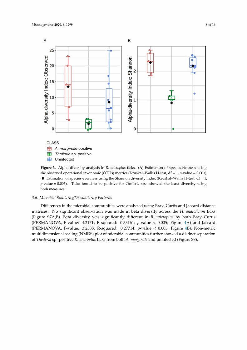
Microbial profile richness and evenness were estimated using the alpha diversity metrics observedOTUs and Shannon index. There was no significant difference between richness and evenness withinthe H. anatolicum ticks, irrespective of the PCR status (Figure S6A,B). Interestingly, R. microplusticks showed comparable significant differences between Theileria sp. positive and uninfected ticks(Figure 3A,B). R. microplus ticks positive for Theileria sp. exhibited significantly reduced species richnessand evenness index across all metrics used to analyze alpha diversity.
Microorganisms 2020, 8, 1299 8 of 16
Figure 3. Alpha diversity analysis in R. microplus ticks. (A) Estimation of species richness usingthe observed operational taxonomic (OTUs) metrics (Kruskal–Wallis H-test, df = 1, p-value = 0.003).(B) Estimation of species evenness using the Shannon diversity index (Kruskal–Wallis H-test, df = 1,p-value = 0.005). Ticks found to be positive for Theileria sp. showed the least diversity usingboth measures.
3.6. Microbial Similarity/Dissimilarity Patterns
Differences in the microbial communities were analyzed using Bray–Curtis and Jaccard distancematrices. No significant observation was made in beta diversity across the H. anatolicum ticks(Figure S7A,B). Beta diversity was significantly different in R. microplus by both Bray–Curtis(PERMANOVA, F-value: 4.2171; R-squared: 0.33161; p-value < 0.005; Figure 4A) and Jaccard(PERMANOVA, F-value: 3.2588; R-squared: 0.27714; p-value < 0.005; Figure 4B). Non-metricmultidimensional scaling (NMDS) plot of microbial communities further showed a distinct separationof Theileria sp. positive R. microplus ticks from both A. marginale and uninfected (Figure S8).

Microorganisms 2020, 8, 1299 9 of 16
Figure 4. Estimation of differences in the microbial communities as a measure of Beta diversity analysisin R. microplus ticks. (A) Principal coordinate analysis (PCoA) of the Bray–Curtis distance matrix(PERMANOVA, F-value: 4.2171; R: 0.33161; p-value < 0.005). (B) Principal coordinate analysis (PCoA)Jaccard distance matrix (PERMANOVA, F-value: 3.2588; R: 0.27714; p-value < 0.005).
3.7. Community Profiling and Correlation Analysis of R. microplus Ticks
To assess the extent to which highly abundant bacteria phylum and genus were represented inR. microplus ticks, we used a combination of pattern correlation and heat map analysis. A very strongpositive correlation was seen between the presence of Bacillus and Theileria sp. positive R. microplus

Microorganisms 2020, 8, 1299 10 of 16
ticks (Figure 5). A similar observation was also seen in the heat map where the genus Bacillus showsthe highest presence in Theileria sp. positive R. microplus ticks (Figure 6A).
Figure 5. Pattern correlation analysis of top 7 bacteria genera in R. microplus ticks. Ticks positive withTheileria sp. showed a positive correlation with the presence of Bacillus.
To explore how top taxa differ, classical univariate statistical comparisons analysis was appliedto identify bacterial genus that exhibits significant differences (t-test/ANOVA) in their composition.Significant differences were observed in the abundance of the genus Acinetobacter, Staphylococcus,and Bacillus. Heat map analysis of OTU abundance was also estimated for H. anatolicum ticks, none ofwhich was statistically significant (Figure S9).
In summary, the results of alpha and beta diversity as well as correlation analysis suggest thata strong association exists between Theileria sp., reduced alpha diversity metrics, and the distinctclustering separation exhibited by Theileria sp. positive R. microplus ticks.

Microorganisms 2020, 8, 1299 11 of 16
Figure 6. Heat map and classical univariate compositional analysis in R. microplus ticks. (A) Heatmap correlation analysis between A. marginale positive, Theileria sp. positive and uninfected ticks.Log-transformed count of (B) Acinetobacter (FDR = 0.19277, df = 1, p-value = 0.007), (C) Staphylococcus(FDR = 0.19277, df = 1, p-value = 0.007), and (D) Bacillus (FDR = 0.0002, df = 1, p-value = 3.94e-223) inA. marginale positive, Theileria sp. positive and uninfected R. microplus ticks.
4. Discussion
The present study was designed to determine the changes that occur to the microbiome compositionand diversity within the tick vectors when naturally infected with protozoan and bacterial tick-bornepathogens. Although many studies have detailed the plethora of interactions that occur betweentick-transmitted pathogens and the microbiome of ticks such as endosymbionts and pathogeninteractions [22], pathogen induction of antimicrobial production by the tick vector [12,23], only oneof such studies compared the microbiome of pathogen-infected and uninfected ticks [24]. In thisstudy, we tested field-collected H. anatolicum and R. microplus ticks for the presence of Theileria sp.,

Microorganisms 2020, 8, 1299 12 of 16
Wolbachia sp., and A. marginale, and further compared the overall microbial distribution, richness,and diversity between Theileria sp. and R. microplus positive ticks. In the current study, the estimatedpercentage of Theileria sp. positive ticks was higher compared to those reported in previous studies forH. anatolicum, while A. marginale prevalence was similar to previous reports [6,25,26].
The major bacteria phyla reported across all the tick groups irrespective of the PCR status wereProteobacteria, Bacteroidota, Firmicutes, and Actinobacteriota. These support observations fromearlier tick microbiome studies [4,11,27,28]. Surprisingly, we identified a much lower number ofbacterial reads and OTU from R. microplus ticks compared to H. anatolicum. This observation could bea function of the differences in the lifecycle of the two tick species. Rhipicephalus microplus is knownas a one-host tick, whereas H. anatolicum ticks could spend their life cycle using 2–3 hosts. Spendingdifferent life stages on different animal hosts will likely expose the H. anatolicum ticks to a plethora ofhost skin microbial communities as well as host-blood [29] associated microbial communities. Some ofthe identified bacterial genera from H. anatolicum ticks such as Staphylococcus, Corynebacterium,Sphingomonas, and Cutibacterium have been previously shown to be common constituents of the skinmicroflora [29].
We also observed the presence of Theileria sp. was associated with an increased abundanceof Candidatus_Midichloria and Francisella compared to the uninfected or A. marginale infected H.anatolicum. Candidatus_Midichloria and Francisella are obligate, vertically maintained endosymbiontsin the phylum Proteobacteria. Candidatus_Midichloria belongs to the Alphaproteobacteria group ofobligate intracellular bacteria first detected in Ixodes ricinus [30], while Francisella-like endosymbiontis a Gammaproteobacteria with widespread distribution in hard ticks [31]. It is interesting tocompare these findings to an elegant observation made by Budachetri and colleagues [22] whoproposed Candidatus_Midichloria mitochondrii as facilitating Rickettsia parkeri colonization of theAmblyomma maculatum tissues by protecting R. parkeri from the deleterious effect of reactive oxygenspecies. Our observations do require further experimental validation to understand the interactionbetween Theileria sp. and Candidatus_Midichloria within the tick vector.
The detection of Bacillus as the only bacteria genera identified from Theileria sp. positive R. microplusticks was an unexpected, albeit important finding (Figure 1F). This observation was further validated bya significantly strong association between the presence of Theileria sp. and the Bacillus group of bacteriaas seen in Figures 5 and 6A. We also found a higher abundance of the phylum Firmicutes (SupplementaryMaterials) and genus Bacillus was associated with Theileria sp. presence in R. microplus ticks (Figure 6).It is difficult to explain this result, but it might be related to the ability of Theileria sp. to interfere withthe mammalian host’s immune response by expressing proteins necessary for its transformation [32].However, its impact on the tick microbiota has yet to be shown. Inhibition of important microbialmetabolic pathways by Theileria sp.-associated proteins [33] could have led to a pathogen-associateddysbiosis. Bacillus ability to form spores when exposed to unfavorable physiological conditions wouldexplain their exclusive presence in Theileria sp. positive R. microplus ticks.
We also observed that R. microplus ticks exhibited a significantly lower microbial diversity andcomposition when compared to H. anatolicum ticks. Within the R. microplus, those positive withTheileria sp. showed a significantly lesser amount of identified bacteria OTUs and a significantlyreduced species richness and evenness (Figure 3A,B), while also displaying a different microbialcomposition as observed on the ordination plots (Figure 4A,B; Figure S5). These observations wereconsistent with those of Mann et al. [34], who reported that Trypanosoma cruzi infected kissing bugsexhibited a higher abundance of selected bacteria group, but not consistent with a study by Sweiand Kwan [29] who reported that Ixodes pacificus ticks positive with the Lyme disease spirochete,Borrelia burgdorferi, had no significant differences in the microbiome richness and composition whencompared to those not infected.
Another interesting finding from our study was the detection of the vertically transmittedWolbachia sp. bacteria from a small number of the ticks used in this study. Wolbachia is an endosymbiontthat has been identified in two-thirds of insects, including mosquitos [35], with known ability to

Microorganisms 2020, 8, 1299 13 of 16
interfere with their host’s reproduction [36] through a series of physiological alterations, one of whichis cytoplasmic incompatibility [37]. Studies showed the presence of Wolbachia in tick species hasassociated it with a form of hyperparasitism where Wolbachia was found infecting another parasitoidthat parasitizes the ticks [38,39].
The unexpected identification of Plasmodium falciparum from ticks used in this study is anunpredicted finding. Pakistan is a malaria-endemic country and we observed an increase in therelative abundance of Plasmodium falciparum in Theileria sp. positive H. anatolicum when comparedto the uninfected ticks (Figure 2A,B). It seems possible that this could have occurred from the ticksaccidentally feeding on a P. falciparum-infected human host. While this is a possibility in multi-hostticks as seen in Hyalomma species, this is highly an unlikely occurrence in R. microplus which is aone-host tick. These results, therefore, need to be interpreted with caution as ticks are not reservoirs orcompetent vectors of P. falciparum.
This finding, while preliminary, suggests that the presence of Theileria sp. within R. microplusticks reduces the overall microbial diversity which we proposed as “pathogen-induced dysbiosis”.The mechanism behind this phenomenon could be induced by Theileria sp. factors in an attempt tocolonize the tick vector, or it could be a result of the innate immune response mounted by the tick.These findings may help us to understand the intricate interplay of the pathogen–microbiome–vectorinteractions. However, more research on these observed interactions needs to be undertaken beforethe association between microbiome dysbiosis and the presence of a pathogen can be drawn.
5. Conclusions
The present study was designed to determine the effect of bacteria and protozoan pathogen on themicrobiome of field-collected ticks. Using a combination of PCR based assay and 16S rRNA sequencing,we investigated how the presence of Theileria sp. and A. marginale shapes the overall microbiomeof both H. anatolicum and R. microplus ticks. We reported a strong association between the presenceof Theileria sp. and a completely reduced microbial diversity and abundance in R. microplus ticks.This study established the extent of the diversity of the microbial community within two importanttick species from Pakistan and revealed the presence of Theileria sp., A. marginale, and additionalpathogenic bacteria that could be of public health significance. A limitation of this study was thedifficulty in obtaining tissue samples of ticks, as they were field collected. Future tick developmentaland tissue-specific studies will generate new insights into specific interactions between tick-bornepathogens and their associated microbiomes.
Supplementary Materials: The following are available online at http://www.mdpi.com/2076-2607/8/9/1299/s1.Figure S1: Phylogenetic analysis of amplified COI sequences of R. microplus and H. anatolicum ticks with otherarthropod’s COI genes from NCBI, Figure S2: Genetic relationship of identified Wolbachia GroEL genes comparedwith selected sequences from NCBI, Figure S3: Blast results of the percentage identity and query cover of WolbachiaGroEL sequence from the current study when compared to previously deposited GroEL sequences from NCBI,Figure S4: Blast results of the percentage identity and query cover of Hyalomma anatolicum and Rhipicephalusmicroplus COI sequence from the current study when compared to previously deposited COI sequences fromNCBI, Figure S5: Relative abundances of bacteria species from individual tick samples used in this study, Figure S6.Alpha diversity analysis in R. microplus ticks, Figure S7: Visualization of differences in the microbial communitiesusing Principal coordinate analysis (PCoA) of Bray_Curtis and Jaccard distance matrix, Figure S8: Non-metricmultidimensional scaling (NMDS) plot of Bray_Curtis and Jaccard distance matrix, Table S1: Total number of ticksused in this study and the percentage of those identified with A. marginale, Theileria, and Wolbachia based on PCRanalysis and amplicon sequencing, Table S2: Total number of reads that used in the analysis after filtering andremoval of chimeras in H. anatolicum ticks, Table S3: Total number of reads that used in the analysis after filteringand removal of chimeras in R. microplus ticks.
Author Contributions: Conceptualization: S.K., data curation: A.A., D.K. and S.K.; formal analysis: A.A., C.B.,D.K. and S.K.; funding acquisition: S.K., M.I.R., A.Z.D. and M.S.S.; investigation: A.A., D.K., M.I.R., A.Z.D., M.S.S.and S.K.; methodology: A.A. and S.K.; project administration: S.K., M.I.R., A.Z.D. and M.S.S.; Resources: S.K.,M.I.R., A.Z.D. and M.S.S.; supervision: S.K.; validation: A.A., D.K. and S.K.; visualization: A.A., D.K. and S.K.;writing—original draft: A.A. and S.K.; and writing—review and editing: S.K. All authors have read and agreed tothe published version of the manuscript.

Microorganisms 2020, 8, 1299 14 of 16
Funding: This research was funded by a Pakistan-US Science and Technology Cooperation Program award(US Department of States; the Mississippi INBRE (an institutional Award (IDeA) from the National Institute ofGeneral Medical Sciences of the National Institutes of Health under award P20GM103476). Cailyn Bobo wassupported by the Eagle SPUR award from the USM Drapeau Center for Undergraduate Research. The fundersplayed no role in the study design, data collection, and analysis, decision to publish, or preparation ofthe manuscript.
Conflicts of Interest: The authors declare that they have no competing interests.
Availability of data and material: The datasets supporting the conclusion of this article are included within thearticle and its additional files. Raw data are available from the corresponding author upon request.
References
1. Ministry of Finance, Government of Pakistan. Available online: http://www.finance.gov.pk/survey_1819.html(accessed on 11 July 2020).
2. Hoogstraal, H.; Varma, M.G.R. Haemaphysalis cornupunctata sp. n. and H. kashmirensis sp. n. from Kashmir,with Notes on H. sundrai Sharif and H. sewelli Sharif of India and Pakistan (Ixodoidea, Ixodidae). J. Parasitol.1962, 48, 185. [CrossRef] [PubMed]
3. Robertson, R.G.; Wisseman, C.L.; Traub, R. Tick-borne rickettsiae of the spotted fever group in West Pakistan:I. Isolation of strains from ticks in different habitats. Am. J. Epidemiol. 1970, 92, 382–394. [CrossRef] [PubMed]
4. Karim, S.; Budachetri, K.; Mukherjee, N.; Williams, J.; Kausar, A.; Hassan, M.J.; Adamson, S.; Dowd, S.E.;Apanskevich, D.; Arijo, A.; et al. A study of ticks and tick-borne livestock pathogens in Pakistan. PLoS Negl.Trop. Dis. 2017, 11, e0005681. [CrossRef]
5. Zeb, J.; Szekeres, S.; Takács, N.; Kontschán, J.; Shams, S.; Ayaz, S.; Hornok, S. Genetic diversity, piroplasmsand trypanosomes in Rhipicephalus microplus and Hyalomma anatolicum collected from cattle in northernPakistan. Exp. Appl. Acarol. 2019, 79, 233–243. [CrossRef]
6. Jabbar, A.; Abbas, T.; Sandhu, Z.U.D.; Saddiqi, H.A.; Qamar, M.F.; Gasser, R.B. Tick-borne diseases of bovinesin Pakistan: Major scope for future research and improved control. Parasites Vectors 2015, 8, 283. [CrossRef]
7. Atif, M.; Saqib, A.; Ikram, R.; Sarwar, M.R.; Scahill, S. The reasons why Pakistan might be at high riskof Crimean Congo hemorrhagic fever epidemic; a scoping review of the literature. Virol. J. 2017, 14, 63.[CrossRef]
8. Sjödin, A.; Svensson, K.; Ohrman, C.; Ahlinder, J.; Lindgren, P.; Duodu, S.; Johansson, A.; Colquhoun, D.J.;Larsson, P.; Forsman, M. Genome characterization of the genus Francisella reveals insight into similarevolutionary paths in pathogens of mammals and fish. BMC Genomics 2012, 13, 268. [CrossRef]
9. Gottlieb, Y.; Lalzar, I.; Klasson, L. Distinctive genome reduction rates revealed by genomic analyses of twoCoxiella-like endosymbionts in ticks. Genome Biol. Evol. 2015, 7, 1779–1796. [CrossRef]
10. Smith, T.A.; Driscoll, T.; Gillespie, J.J.; Raghavan, R. A Coxiella-like endosymbiont is a potential vitaminsource for the lone star tick. Genome Biol. Evol. 2015, 7, 831–838. [CrossRef] [PubMed]
11. Narasimhan, S.; Rajeevan, N.; Liu, L.; Zhao, Y.O.; Heisig, J.; Pan, J.; Eppler-Epstein, R.; Deponte, K.; Fish, D.;Fikrig, E. Gut microbiota of the tick vector Ixodes scapularis modulate colonization of the Lyme diseasespirochete. Cell Host Microbe 2014, 15, 58–71. [CrossRef] [PubMed]
12. Abraham, N.M.; Liu, L.; Jutras, B.L.; Yadav, A.K.; Narasimhan, S.; Gopalakrishnan, V.; Ansari, J.M.;Jefferson, K.K.; Cava, F.; Jacobs-Wagner, C.; et al. Pathogen-mediated manipulation of arthropod microbiotato promote infection. Proc. Natl. Acad. Sci. USA 2017, 114, E781–E790. [CrossRef] [PubMed]
13. Dowd, S.E.; Callaway, T.R.; Wolcott, R.D.; Sun, Y.; McKeehan, T.; Hagevoort, R.G.; Edrington, T.S. Evaluationof the bacterial diversity in the feces of cattle using 16S rDNA bacterial tag-encoded FLX ampliconpyrosequencing (bTEFAP). BMC Microbiol. 2008, 8, 125. [CrossRef] [PubMed]
14. Mukherjee, N.; Beati, L.; Sellers, M.; Burton, L.; Adamson, S.; Robbins, R.G.; Moore, F.; Karim, S. Importationof exotic ticks and tick-borne spotted fever group rickettsiae into the United States by migrating songbirds.Ticks Tick-borne Dis. 2014, 5, 127–134. [CrossRef] [PubMed]
15. Dougherty, L.; Li, J. Molecular phylogeny and morphological distinctions of two popular bivalves,Ctenoides scabei and Ctenoides mitis. J. Mar. Boil. 2017, 2017, 1–9. [CrossRef]
16. Kundave, V.; Ram, H.; Banerjee, P.S.; Garg, R.; Mahendran, K.; Ravikumar, G.; Tiwari, A.K. Development ofmultiplex PCR assay for concurrent detection of tick-borne haemoparasitic infections in bovines. Acta Parasitol.2018, 63, 759–765. [CrossRef] [PubMed]

Microorganisms 2020, 8, 1299 15 of 16
17. Masui, S.; Sasaki, T.; Ishikawa, H. groE-Homologous Operon of Wolbachia, an Intracellular Symbiont ofArthropods: A New Approach for Their Phylogeny. Zool. Sci. 1997, 14, 701–706. [CrossRef]
18. Callahan, B.J.; McMurdie, P.J.; Rosen, M.J.; Han, A.W.; Johnson, A.J.A.; Holmes, S.P. DADA2: High-resolutionsample inference from Illumina amplicon data. Nat. Methods 2016, 13, 581–583. [CrossRef]
19. Price, M.N.; Dehal, P.S.; Arkin, A.P. FastTree 2—Approximately maximum-likelihood trees for largealignments. PLoS ONE 2010, 5, e9490. [CrossRef]
20. Chong, J.; Liu, P.; Zhou, G.; Xia, J. Using Microbiome Analyst for comprehensive statistical, functional,and meta-analysis of microbiome data. Nat. Protoc. 2020, 15, 799–821. [CrossRef]
21. Dhariwal, A.; Chong, J.; Habib, S.; King, I.L.; Agellon, L.B.; Xia, J. Microbiome Analyst: A web-based toolfor comprehensive statistical, visual and meta-analysis of microbiome data. Nucleic Acids Res. 2017, 45,W180–W188. [CrossRef]
22. Budachetri, K.; Kumar, D.; Crispell, G.; Beck, C.; Dasch, G.; Karim, S. The tick endosymbiont CandidatusMidichloria mitochondrii and selenoproteins are essential for the growth of Rickettsia parkeri in the Gulf Coasttick vector. Microbiome 2018, 6, 141. [CrossRef] [PubMed]
23. Narasimhan, S.; Schuijt, T.J.; Abraham, N.M.; Rajeevan, N.; Coumou, J.; Graham, M.; Robson, A.; Wu, M.-J.;Daffre, S.; Hovius, J.W.; et al. Modulation of the tick gut milieu by a secreted tick protein favors Borreliaburgdorferi colonization. Nat. Commun. 2017, 8, 1–17. [CrossRef] [PubMed]
24. Fryxell, R.T.T.; DeBruyn, J.M. The Microbiome of Ehrlichia-infected and uninfected lone star ticks(Amblyomma americanum). PLoS ONE 2016, 11, e0146651.
25. Durrani, A.Z.; Kamal, N. Identification of ticks and detection of blood protozoa in Friesian cattle bypolymerase chain reaction test and estimation of blood parameters in district Kasur, Pakistan. Trop. Anim.Heal. Prod. 2008, 40, 441–447. [CrossRef]
26. Rehman, A.; Nijhof, A.M.; Sauter-Louis, C.; Schauer, B.; Staubach, C.; Conraths, F.J. Distribution of ticksinfesting ruminants and risk factors associated with high tick prevalence in livestock farms in the semi-aridand arid agro-ecological zones of Pakistan. Parasites Vectors 2017, 10, 1–15. [CrossRef]
27. Budachetri, K.; Browning, R.E.; Adamson, S.W.; Dowd, S.E.; Chao, C.-C.; Ching, W.-M.; Karim, S. An insightinto the microbiome of the Amblyomma maculatum (Acari: Ixodidae). J. Med. Entomol. 2014, 51, 119–129.[CrossRef]
28. Travanty, N.V.; Ponnusamy, L.; Kakumanu, M.L.; Nicholson, W.L.; Apperson, C.S. Diversity and structure ofthe bacterial microbiome of the American dog tick, Dermacentor variabilis, is dominated by the endosymbiontFrancisella. Symbiosis 2019, 79, 1–12. [CrossRef]
29. Swei, A.; Kwan, J.Y. Tick microbiome and pathogen acquisition altered by host blood meal. ISME J. 2017, 11,813–816. [CrossRef]
30. Epis, S.; Mandrioli, M.; Genchi, M.; Montagna, M.; Sacchi, L.; Pistone, D.; Sassera, D. Localization of thebacterial symbiont Candidatus Midichloria mitochondrii within the hard tick Ixodes ricinus by whole-mountFISH staining. Ticks Tick-borne Dis. 2013, 4, 39–45. [CrossRef]
31. Dergousoff, S.J.; Chilton, N.B. Association of different genetic types of Francisella-like organisms with therocky mountain wood tick (Dermacentor andersoni) and the American Dog Tick (Dermacentor variabilis) inLocalities Near Their Northern Distributional Limits. Appl. Environ. Microbiol. 2011, 78, 965–971. [CrossRef]
32. Chakraborty, S.; Roy, S.; Mistry, H.U.; Murthy, S.; George, N.; Bhandari, V.; Sharma, P. Potential sabotageof host cell physiology by apicomplexan parasites for their survival benefits. Front. Immunol. 2017, 8.[CrossRef] [PubMed]
33. Bahia, A.C.; Oliveira, J.H.M.; Kubota, M.S.; Araújo, H.R.C.; Lima, J.B.P.; Ríos-Velásquez, C.M.; Lacerda, M.V.G.;De Oliveira, P.L.; Traub-Csekö, Y.M.; Pimenta, P.F.P. The Role of Reactive Oxygen Species in Anopheles aquasalisResponse to Plasmodium vivax Infection. PLoS ONE 2013, 8, 1–10. [CrossRef] [PubMed]
34. Mann, A.E.; Mitchell, E.A.; Zhang, Y.; Curtis-Robles, R.; Thapa, S.; Hamer, S.A.; Allen, M.S. Comparisonof the bacterial gut microbiome of North American Triatoma spp. With and without Trypanosoma cruzi.Front. Microbiol. 2020, 11, 364. [CrossRef] [PubMed]
35. Hilgenboecker, K.; Hammerstein, P.; Schlattmann, P.; Telschow, A.; Werren, J.H. How many species areinfected with Wolbachia? A statistical analysis of current data. FEMS Microbiol. Lett. 2008, 281, 215–220.[CrossRef]
36. Sicard, M.; Bonneau, M.; Weill, M. Wolbachia prevalence, diversity, and ability to induce cytoplasmicincompatibility in mosquitoes. Curr. Opin. Insect Sci. 2019, 34, 12–20. [CrossRef] [PubMed]

Microorganisms 2020, 8, 1299 16 of 16
37. Stouthamer, R.; Breeuwer, J.A.J.; Hurst, G.D.D. Wolbachia Pipientis: Microbial manipulator of arthropodreproduction. Annu. Rev. Microbiol. 1999, 53, 71–102. [CrossRef]
38. Plantard, O.; Bouju-Albert, A.; Malard, M.-A.; Hermouet, A.; Capron, G.; Verheyden, H. Detection ofWolbachia in the tick Ixodes ricinus is due to the presence of the Hymenoptera endoparasitoid Ixodiphagus hookeri.PLoS ONE 2012, 7, e30692. [CrossRef]
39. Tijsse-Klasen, E.; Braks, M.; Scholte, E.J.; Sprong, H. Parasites of vectors—Ixodiphagus hookeri and its Wolbachiasymbionts in ticks in The Netherlands. Parasites Vectors 2011, 4, 228. [CrossRef]
© 2020 by the authors. Licensee MDPI, Basel, Switzerland. This article is an open accessarticle distributed under the terms and conditions of the Creative Commons Attribution(CC BY) license (http://creativecommons.org/licenses/by/4.0/).