thymic stromal lymphopoietin drives the development of il ...thymic stromal lymphopoietin drives the...
TRANSCRIPT

Thymic stromal lymphopoietin drives the developmentof IL-13+ Th2 cellsSotaro Ochiaia, Ferdinand Jagota, Ryan L. Kylea, Evelyn Hydea, Ruby F. Whitea, Melanie Prouta, Alfonso J. Schmidta,Hidehiro Yamaneb, Olivier Lamiablea, Graham Le Grosa,1, and Franca Ronchesea,1,2
aMalaghan Institute of Medical Research, 6242 Wellington, New Zealand; and bCellular Immunology Section & Cytokine Biology Unit, National Institute ofAllergy and Infectious Diseases, National Institutes of Health, Bethesda, MD 20892-1892
Edited by Kenneth M. Murphy, Washington University, St. Louis, MO, and approved December 15, 2017 (received for review August 27, 2017)
T helper 2 (Th2) cells are pivotal in the development of allergy.Allergen exposure primes IL-4+ Th2 cells in lymph node, but pro-duction of effector cytokines including IL-5 and IL-13 is thought torequire additional signals from antigen and the environment. Herewe report that a substantial proportion of naive CD4+ T cells inspleen and lymph node express receptors for the epithelium-derived inflammatory cytokine thymic stromal lymphopoietin(TSLP). Culture of naive CD4+ T cells in anti-(a)CD3, aCD28, andTSLP-supplemented Th2 conditions enabled the development of aunique population of IL-13-single positive (IL-13-SP) CD4+ T cells;TSLP and Th2 conditions were both required for their development.Sorting experiments revealed that IL-13-SP Th2 cells originated fromIL-4-negative precursors and coexpressed transcripts for theTh2 cytokines IL-5 and IL-9. In vivo, high TSLP levels acted directlyon CD4+ T cells to induce the development of IL-13-SP and IL-4+IL-13+ double-positive populations in lymph node. These cells werephenotypically similar to Th2 effector cells and were CXCR5lowPD1low
and expressed low levels of Bcl6 and Il21 transcripts and highlevels of Gata3, Il3, and Il5. Our findings suggest a role of TSLPin directly promoting Th2 cell effector function and support thenotion of TSLP as a key driver of Th2 inflammation.
TSLP | Th2 cells | effector T cells | IL-13 | IL-4
The cytokine-regulated differentiation of naive CD4+ T cellsinto type 2 helper T (Th2) cells expressing IL-4, IL-13, and
IL-5 represents a key event in the development of allergic re-sponses (1, 2). Initial investigations led to the concept that CD4+
T cells primed in Th2 conditions rapidly acquire the ability toproduce multiple Th2 cytokines, with stochastic mechanismsdriving heterogeneity in cytokine production at the effector stage(3). Contrary to that notion, recent work suggests a more complexregulation in which Th2 cytokine production is temporally andspatially compartmentalized (4–6). These studies suggest that theinitial priming of Th2 cells to IL-4 production in lymph node (LN)is followed by the gain of capacity to produce additional cytokineson secondary exposure to antigen in nonlymphoid tissue (7). Inline with those first observations, detailed studies using the inhaledallergen house dust mite (HDM) showed that naive CD4+ T cellsare primed in LN to a CXCR5+PD1+BCL6+ T follicular helper(Tfh) phenotype, producing IL-4 and IL-21 but no IL-5 or IL-13.On secondary exposure to HDM via the airway, primed Th2 cellsin lung are able to differentiate into CXCR5loPD1lo Th2 effectorcells that are GATA3+ and produce IL-13, but express lowerBCL6 and IL-21 than Tfh cells in LN (8–10).The cytokines IL-25, IL-33, and thymic stromal lymphopoietin
(TSLP) are produced by epithelia exposed to various insults (11)and are proposed to play a key role in the acquisition of effectorfunction by primed Th2 cells (7). However, how these tissue-derived factors mediate their effect on the evolving Th2 responseis not well understood. IL-33 was recently found to promoteTh2 effector activity by suppressing BCL6 expression in Tfh cells,in turn de-repressing IL-4 transcription (12). This mechanism ofaction is compatible with the known expression of the IL-33 re-ceptor ST2 on activated Th2 cells (13). Interestingly, unlike ST2,
expression of the TSLP receptor (TSLPR) does not require T cellactivation and is already observed on naive CD4+ T cells. Based onthis different pattern of receptor expression, it seems reasonable toassume that the effect of TSLP, IL-25, and IL-33 on the differ-entiation of CD4+ T cells to Th2 effectors may also differ.TSLP is an epithelial cytokine that plays a key role in a range
of conditions, including allergic responses, viral infections, andtumor progression (14–16). TSLP binds to a heterodimeric re-ceptor formed by a TSLP-specific TSLPR subunit and the IL7Rsignaling chain (14, 17) and acts on several immune cell typesincluding dendritic cells, type 2 innate lymphoid cells (ILC2),mast cells, basophils, and T cells (14, 16). The direct effects ofTSLP on T cells have been studied by several groups, and bothhuman and mouse CD4+ T cells stimulated in a TCR-dependentmanner in the presence of TSLP show enhanced proliferationand IL-4 secretion (18, 19). It remains unclear whether TSLPmay become available in LN during T cell priming, or whetherexposure is restricted to epithelia such as skin and mucosalsurfaces, which are only accessed by T cells after activation.We used the Il4 and Il13 transcriptional reporter 4C13R mice
(20) to specifically investigate the effect of TSLP on the differ-entiation of Th2 cells in vitro and in vivo. We find that, in ad-dition to enhancing IL-4 expression by CD4+ T cells (19), TSLPwas able to drive the development of a separate population ofIL-13-DsRed single-positive (IL-13DR SP) CD4+ T cells thatalso expressed Il5 and Il9 transcripts and originated from IL-4-AmCyan (IL-4AC)-negative T cell precursors in vitro. In vivo,IL-13DR SP were found in LN, but lacked expression of Tfhsurface markers and expressed the low Bcl6 and high Gata3characteristic of effector Th2 cells. Thus, our findings identify
Significance
T helper 2 (Th2) cells are defined by their ability to produce thehallmark cytokine IL-4. However, to mediate allergic inflammationin tissues, Th2 cells must secrete additional cytokines including IL-13 and IL-5. We used IL-4 and IL-13 dual-reporter mice to showthat naive CD4+ T cells cultured in the presence of IL-4 and thymicstromal lymphopoietin (TSLP) generate a population of IL-4negIL-13pos Th2 cells that develop from IL-4neg precursors and expressthe Th2 effector cytokines IL-5 and IL-9. In vivo, high TSLP levelspromote the development of a similar population of IL-4negIL-13pos
T cells that also express Gata3, Il5, and Il3 transcripts. Thus, TSLPdrives the early differentiation of a distinct population of effectorTh2 cells with pro-inflammatory properties.
Author contributions: S.O., R.L.K., H.Y., G.L.G., and F.R. designed research; S.O., F.J., R.L.K.,E.H., R.F.W., M.P., A.J.S., and O.L. performed research; S.O., F.J., R.L.K., E.H., R.F.W., M.P.,A.J.S., O.L., and F.R. analyzed data; and S.O., G.L.G., and F.R. wrote the paper.
The authors declare no conflict of interest.
This article is a PNAS Direct Submission.
This open access article is distributed under Creative Commons Attribution-NonCommercial-NoDerivatives License 4.0 (CC BY-NC-ND).1G.L.G. and F.R. contributed equally to this work.2To whom correspondence should be addressed. Email: [email protected].
This article contains supporting information online at www.pnas.org/lookup/suppl/doi:10.1073/pnas.1714348115/-/DCSupplemental.
www.pnas.org/cgi/doi/10.1073/pnas.1714348115 PNAS Early Edition | 1 of 6
IMMUNOLO
GYAND
INFLAMMATION
Dow
nloa
ded
by g
uest
on
Apr
il 12
, 202
0
TSLP as a key factor supporting the early differentiation of ef-fector Th2 cells both in vitro and in vivo.
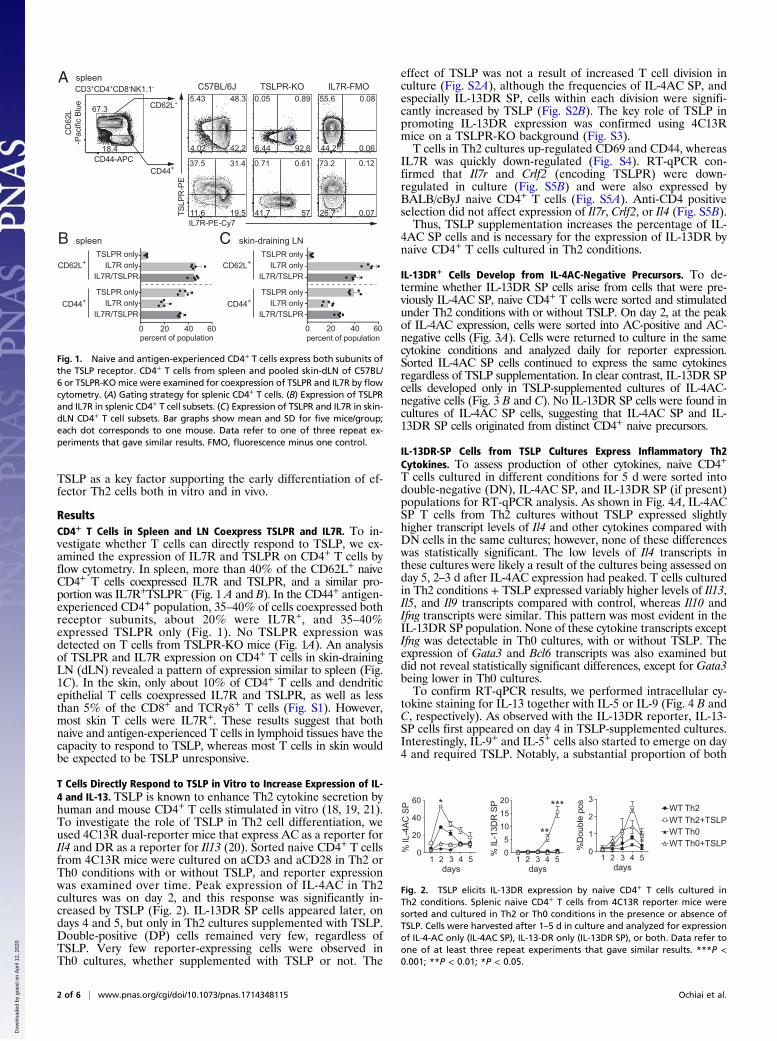
ResultsCD4+ T Cells in Spleen and LN Coexpress TSLPR and IL7R. To in-vestigate whether T cells can directly respond to TSLP, we ex-amined the expression of IL7R and TSLPR on CD4+ T cells byflow cytometry. In spleen, more than 40% of the CD62L+ naiveCD4+ T cells coexpressed IL7R and TSLPR, and a similar pro-portion was IL7R+TSLPR− (Fig. 1 A and B). In the CD44+ antigen-experienced CD4+ population, 35–40% of cells coexpressed bothreceptor subunits, about 20% were IL7R+, and 35–40%expressed TSLPR only (Fig. 1). No TSLPR expression wasdetected on T cells from TSLPR-KO mice (Fig. 1A). An analysisof TSLPR and IL7R expression on CD4+ T cells in skin-drainingLN (dLN) revealed a pattern of expression similar to spleen (Fig.1C). In the skin, only about 10% of CD4+ T cells and dendriticepithelial T cells coexpressed IL7R and TSLPR, as well as lessthan 5% of the CD8+ and TCRγδ+ T cells (Fig. S1). However,most skin T cells were IL7R+. These results suggest that bothnaive and antigen-experienced T cells in lymphoid tissues have thecapacity to respond to TSLP, whereas most T cells in skin wouldbe expected to be TSLP unresponsive.
T Cells Directly Respond to TSLP in Vitro to Increase Expression of IL-4 and IL-13. TSLP is known to enhance Th2 cytokine secretion byhuman and mouse CD4+ T cells stimulated in vitro (18, 19, 21).To investigate the role of TSLP in Th2 cell differentiation, weused 4C13R dual-reporter mice that express AC as a reporter forIl4 and DR as a reporter for Il13 (20). Sorted naive CD4+ T cellsfrom 4C13R mice were cultured on aCD3 and aCD28 in Th2 orTh0 conditions with or without TSLP, and reporter expressionwas examined over time. Peak expression of IL-4AC in Th2cultures was on day 2, and this response was significantly in-creased by TSLP (Fig. 2). IL-13DR SP cells appeared later, ondays 4 and 5, but only in Th2 cultures supplemented with TSLP.Double-positive (DP) cells remained very few, regardless ofTSLP. Very few reporter-expressing cells were observed inTh0 cultures, whether supplemented with TSLP or not. The
effect of TSLP was not a result of increased T cell division inculture (Fig. S2A), although the frequencies of IL-4AC SP, andespecially IL-13DR SP, cells within each division were signifi-cantly increased by TSLP (Fig. S2B). The key role of TSLP inpromoting IL-13DR expression was confirmed using 4C13Rmice on a TSLPR-KO background (Fig. S3).T cells in Th2 cultures up-regulated CD69 and CD44, whereas
IL7R was quickly down-regulated (Fig. S4). RT-qPCR con-firmed that Il7r and Crlf2 (encoding TSLPR) were down-regulated in culture (Fig. S5B) and were also expressed byBALB/cByJ naive CD4+ T cells (Fig. S5A). Anti-CD4 positiveselection did not affect expression of Il7r, Crlf2, or Il4 (Fig. S5B).Thus, TSLP supplementation increases the percentage of IL-
4AC SP cells and is necessary for the expression of IL-13DR bynaive CD4+ T cells cultured in Th2 conditions.
IL-13DR+ Cells Develop from IL-4AC-Negative Precursors. To de-termine whether IL-13DR SP cells arise from cells that were pre-viously IL-4AC SP, naive CD4+ T cells were sorted and stimulatedunder Th2 conditions with or without TSLP. On day 2, at the peakof IL-4AC expression, cells were sorted into AC-positive and AC-negative cells (Fig. 3A). Cells were returned to culture in the samecytokine conditions and analyzed daily for reporter expression.Sorted IL-4AC SP cells continued to express the same cytokinesregardless of TSLP supplementation. In clear contrast, IL-13DR SPcells developed only in TSLP-supplemented cultures of IL-4AC-negative cells (Fig. 3 B and C). No IL-13DR SP cells were found incultures of IL-4AC SP cells, suggesting that IL-4AC SP and IL-13DR SP cells originated from distinct CD4+ naive precursors.
IL-13DR-SP Cells from TSLP Cultures Express Inflammatory Th2Cytokines. To assess production of other cytokines, naive CD4+T cells cultured in different conditions for 5 d were sorted intodouble-negative (DN), IL-4AC SP, and IL-13DR SP (if present)populations for RT-qPCR analysis. As shown in Fig. 4A, IL-4ACSP T cells from Th2 cultures without TSLP expressed slightlyhigher transcript levels of Il4 and other cytokines compared withDN cells in the same cultures; however, none of these differenceswas statistically significant. The low levels of Il4 transcripts inthese cultures were likely a result of the cultures being assessed onday 5, 2–3 d after IL-4AC expression had peaked. T cells culturedin Th2 conditions + TSLP expressed variably higher levels of Il13,Il5, and Il9 transcripts compared with control, whereas Il10 andIfng transcripts were similar. This pattern was most evident in theIL-13DR SP population. None of these cytokine transcripts exceptIfng was detectable in Th0 cultures, with or without TSLP. Theexpression of Gata3 and Bcl6 transcripts was also examined butdid not reveal statistically significant differences, except for Gata3being lower in Th0 cultures.To confirm RT-qPCR results, we performed intracellular cy-
tokine staining for IL-13 together with IL-5 or IL-9 (Fig. 4 B andC, respectively). As observed with the IL-13DR reporter, IL-13-SP cells first appeared on day 4 in TSLP-supplemented cultures.Interestingly, IL-9+ and IL-5+ cells also started to emerge on day4 and required TSLP. Notably, a substantial proportion of both
spleen skin-draining LN
37.5 31.4
19.511.6
0.71 0.61
5741.7
0.05 0.89
92.66.44
73.2 0.12
0.0726.7
18.4
67.35.43 48.3
42.24.02
IL7R-PE-Cy7
TSLP
R-P
ECD44+
CD62L+
CD44-APC
CD
62L
-Pac
ific
Blue
CD3+CD4+CD8-NK1.1- C57BL/6J TSLPR-KO IL7R-FMOspleen
TSLPR onlyIL7R only
IL7R/TSLPRCD62L+
CD44+TSLPR only
IL7R onlyIL7R/TSLPR
TSLPR onlyIL7R only
IL7R/TSLPRCD62L+
CD44+TSLPR only
IL7R onlyIL7R/TSLPR
A
B C
55.6 0.08
0.0644.2
percent of population0 20 40 60
percent of population0 20 40 60
Fig. 1. Naive and antigen-experienced CD4+ T cells express both subunits ofthe TSLP receptor. CD4+ T cells from spleen and pooled skin-dLN of C57BL/6 or TSLPR-KOmice were examined for coexpression of TSLPR and IL7R by flowcytometry. (A) Gating strategy for splenic CD4+ T cells. (B) Expression of TSLPRand IL7R in splenic CD4+ T cell subsets. (C) Expression of TSLPR and IL7R in skin-dLN CD4+ T cell subsets. Bar graphs show mean and SD for five mice/group;each dot corresponds to one mouse. Data refer to one of three repeat ex-periments that gave similar results. FMO, fluorescence minus one control.
0
20
40
60
0
1
2
3
05
101520
WT Th2WT Th2+TSLPWT Th0WT Th0+TSLP
%D
oubl
e po
s* ***
**
days1 2 3 4 5
days1 2 3 4 5
days1 2 3 4 5
% IL
-4AC
SP
% IL
-13D
R S
P
Fig. 2. TSLP elicits IL-13DR expression by naive CD4+ T cells cultured inTh2 conditions. Splenic naive CD4+ T cells from 4C13R reporter mice weresorted and cultured in Th2 or Th0 conditions in the presence or absence ofTSLP. Cells were harvested after 1–5 d in culture and analyzed for expressionof IL-4-AC only (IL-4AC SP), IL-13-DR only (IL-13DR SP), or both. Data refer toone of at least three repeat experiments that gave similar results. ***P <0.001; **P < 0.01; *P < 0.05.
2 of 6 | www.pnas.org/cgi/doi/10.1073/pnas.1714348115 Ochiai et al.
Dow
nloa
ded
by g
uest
on
Apr
il 12
, 202
0

IL-9+ and IL-5+ cells also expressed IL-13. Thus, IL-13+ cellsgenerated in Th2+TSLP cultures are heterogeneous and includesubsets that also produce IL-9 and/or IL-5.
High Levels of TSLP in Vivo Drive Expression of IL-13DR and an EffectorPhenotype in dLN CD4+ T Cells. To investigate the effects of TSLPin vivo, C57BL/6 mice were either treated with MC903 on ear skinfor up to 4 consecutive days or injected intradermally with HDMonce into the ear pinna. The levels of Tslp transcripts in the epi-dermal layer were quantified by RT-qPCR at different times aftertreatment (Fig. 5A). MC903 induced strong Tslp transcription,which peaked on day 4. HDM also induced Tslp, but the peaklevels were 10- to 100-fold lower than for MC903. The serumlevels of TSLP, which are undetectable in untreated C57BL/6 mice, were strongly elevated for several days in MC903-treatedmice (Fig. 5B), but remained below the level of detection inHDM-treated mice at all times tested (12, 24, and 48 h).The phenotype of cytokine reporter-expressing CD4+CD44hi
T cells in vivo was examined at the peak of total LN cellularity onday 7. In HDM-sensitized mice, most of the IL-4AC SP cells inLN also expressed high levels of the Tfh markers PD-1 andCXCR5 (Fig. 5C). As previously reported (8–10), no DP or IL-13DR SP cells were detected in these mice. In contrast, IL-4ACSP, IL-4AC IL-13DR DP, and about 1% IL-13DR SP cells wereall detected in dLN after MC903 treatment (Fig. 5E). The ex-pression of Tfh markers on these T cell populations was variableand was highest on IL-4AC SP cells and lowest on IL-13DR SPcells. In each case, the larger proportion of cells expressed lowPD-1 and CXCR5, suggesting a trend to an inflammatory Th2effector (Th2eff) phenotype. Similar differences in pattern ofcytokine and Tfh marker expression between the HDM andMC903 models were also observed, although less prominently,on days 4 and 10 (Fig. 5 E and F). Loss of TSLP responsiveness
in TSLPR-KO mice did not affect the IL-4AC response toHDM, but reduced the percentages of reporter-positive cellsresponding to MC903 to background levels (Fig. 6A).To determine whether TSLP was acting directly on CD4+
T cells or via other cell populations including dendritic cells, wecarried out adoptive transfer experiments in which 4C13R WTand TSLPR-KO CD4+ T cells were coinjected into TSLPR-WThosts treated with MC903 (Fig. S6A). These experiments showedthat only WT T cells were able to express IL-13DR afterMC903 immunization (Fig. 6 B and C). In contrast, IL-4AC wasexpressed on both TSLPR-WT and TSLPR-KO CD4+ T cells.To further define the phenotype of cytokine reporter-expressing
CD4+ T cells in HDM and MC903 immunized mice, IL-4AC SP,IL-13DR SP, and IL-13DR DP cells were sorted from skin-dLNon day 7 and analyzed by RT-qPCR without further restimulation.IL-4AC SP cells from HDM orMC903-treated mice expressed thehigh Bcl6 and low Gata3 characteristic of Tfh cells (Fig. 7). Incontrast, IL-13DR SP and DP cells were Gata3 high and Bcl6 low,consistent with a Th2eff phenotype. Reporter-negative CD4+
T cells from naive or immunized mice expressed low levels ofboth transcripts.Cytokine transcripts were also examined: Il4 transcripts were
detected in IL-4AC SP cells from HDM and MC903 mice, andalso in IL-13DR-DP and, to a lesser extent, IL-13DR SP cellsfrom MC903-immunized mice (Fig. 7). Il21 transcripts showed asimilar pattern of expression (Fig. 7), which is consistent withthese populations including high proportions of Tfh cells. Ex-pression of Il13, Il5, and Il3 transcripts was also examined andwas highest in IL-13DR SP and IL-13DR DP cells fromMC903 mice, but low in IL-4AC SP cells (Fig. 7). This cytokine
IL-4ACposIL-4ACneg
IL-4ACposIL-4ACneg
Th2
Th2+TSLP0
20406080
100
% IL
-4A
C S
P
3 4 5days
02468
% D
oubl
e po
s
3 4 5days
A
C
02468
10
% IL
-13D
R S
P
3 4 5days
**
**
17.066.7 0100 91.66.93
IL-4ACposIL-4ACnegTh2 pre-sort
IL-4-AmCyan
DA
PI
Day 2 post-sort
4.1 0.79
7.03
39.4 4.05
1.86
5.24 0.77
0.98
32.1 4.63
1.84
IL-4ACpos IL-4ACnegTh2+TSLP
IL-4ACpos IL-4ACnegTh2
IL-13-DsRed
IL-4
-Am
Cya
n
Day 5B
Fig. 3. IL-13DR-expressing Th2 cells arise from an IL-4AC-negative populationin the presence of TSLP. Naive CD4+ T cells were purified and cultured as inFig. 2. On day 2, cells were harvested, sorted into IL-4AC+ or IL-4AC−, andcultured in the same media for up to 3 d before analysis of reporter ex-pression. (A) Expression of IL-4AC in representative presort and sortedpopulations on day 2. (B) Expression of IL-4AC and IL-13DR in reculturedpopulations on day 5. (C) Time-course analysis of IL-4AC and/or IL-13DR re-porter expression in culture. Graphs show mean ± SD for three independentcultures/group. Data refer to one of three repeat experiments that gavesimilar results. **P < 0.01.
Th2Th2+TSLP
A
B
C
% IL
-9 IL
-13
DP
0
1
2
3
days
*
3 4 5
% IL
-13
SP
0
5
10
15
days
**
**
3 4 5
Th2Th2+TSLP
0.51.01.52.02.5
% IL
-9 S
P
days
**
3 4 50.0
% IL
-5 S
P
0.00.10.20.30.4
days3 4 5 %
IL-5
IL-1
3 D
P
0
2
4
6
days
*
*
3 4 5
% IL
-13
SP
5
10
15
days
**
**
03 4 5
AC+Th2
DN DR+AC+DN DN DNTh2+TSLP Th0 Th0
+TSLP
Il4Il13Il5Il9
Gata3Bcl6
Il10Ifng
log2
, rel
exp
ress
ion4
20-2-4-6-8
**** **** ******** **** ******** **** ********
****
*** ****** ****** ***
***** **
**
*
*
Fig. 4. Culture in Th2 conditions and TSLP generates a population ofTh2 cells that express IL-13, IL-5, and IL-9. Naive CD4+ T cells were purifiedand cultured as in Fig. 2. (A) Cultured cells were harvested on day 5 andsorted into DN, IL-4AC SP (AC+), or where applicable, IL-13DR SP (DR+) forRT-qPCR analysis. Cytokine expression data are normalized to Gapdh andrelative to Th2 DN cells (left column). (B and C) On day 3–5, T cells wereharvested, restimulated with aCD3/aCD28, and stained for intracellular IL-13 and IL-9 (B), or IL-13 and IL-5 (C). Graphs show mean ± SD for three in-dependent cultures/group. Data refer to one of two repeat experiments thatgave similar results. **P < 0.01; *P < 0.05.
Ochiai et al. PNAS Early Edition | 3 of 6
IMMUNOLO
GYAND
INFLAMMATION
Dow
nloa
ded
by g
uest
on
Apr
il 12
, 202
0

expression pattern is consistent with the Th2eff phenotype of thesecells. Together, these results suggest that TSLP acts directly onCD4+ T cells to promote their development into Th2eff in LN.
DiscussionIn this study, we report that culture in TSLP and Th2 conditionspromotes the differentiation of naive CD4+ T cells into a pop-ulation of IL-13-SP Th2 cells. The development of these cells inculture does not involve an IL-4+ stage and is accompanied bythe up-regulation of Il5, Il9, and Il13 transcripts, but not Il10 orIfng. In vivo, high TSLP is associated with the acquisition of aTh2eff phenotype in dLN, with down-regulation of Bcl6 and Il21
transcripts and up-regulation of Gata3, Il3, Il5, and Il13. Thesedata suggest TSLP can act early during Th2 cell differentiation tosupport the development of a Th2eff phenotype.We used purified naive CD4+ T cells from 4C13R mice, which
express fluorescent reporters for IL-4 and IL-13, and a defined invitro culture system to show that expression of the Th2 cytokinesIL-4 and IL-13 in cultured naive CD4+ T cells follows distinctlydifferent kinetics. Expression of IL-4AC peaked on day 2 and thendeclined regardless of the presence of TSLP in culture, whereasexpression of IL-13DR peaked on day 5, but only in the presenceof TSLP. This was not the result of an effect of TSLP on the di-vision rate of cultured T cells (19), possibly because of the use ofoptimal culture conditions in our experiments. Instead, the dif-ferent kinetics of IL-4AC and IL-13DR expression appeared toreflect the development of two separate functional T cell subsets,as indicated by sorting experiments showing that IL-13DR SPT cells did not originate from the IL-4AC+ population. The cy-tokine profiles of cells cultured with or without TSLP also differedsubstantially, with T cells from TSLP cultures expressing higherlevels of Il13, Il5, and Il9 transcripts; similar Il10 and Ifng; andlower Il4 compared with T cells cultured without TSLP. The ex-pression of multiple Th2 cytokines including IL-5 has also beenreported for highly differentiated effector memory Th2 cells inhuman peripheral blood (22).An interesting observation was that IL-4AC expression was
not maintained in our cultured T cells, despite the continuedavailability of aCD3-aCD28 stimulation and cytokines in themedium. This decline in IL-4AC may be a result of limitations ofthe in vitro culture system; for example, it may suggest that IL-4+
T cells require additional stimuli that are not provided in thesecultures. The full differentiation of Tfh cells in vivo has been0
1
2
tot c
ells
/LN
(x10
7 )
4 7 100
10203040
4 7 10
% IL
-4A
C S
P
0.0
0.5
1.0
1.5
2.0
4 7 10
% IL
-13D
R S
P
% D
oubl
e po
sitiv
e
0
2
4
6
4 7 10
00.51.01.52.02.5
IL-4
AC
SP
cells
/LN
(x10
5 )
0
0.5
1.0
Dou
ble
posi
tive
cells
/LN
(x10
4 )
IL-1
3DR
SP
cells
/LN
(x10
3 )
4 7 104 7 10
HDM Tfh
MC903 TfhMC903 Teff
HDM Teff
HDMMC903
days days days
days days days days
1.8594.4
20.969.0
44.2
53.6
19.2 6.64
1.2
IL-4-AmCyan SP IL-13-DsRed SP Double PositiveMC903 (d7)CD4+ CD44+ T cells
IL-4
-Am
Cya
n
IL-13-DsRed
PD1-
PE-C
y7
CXCR5-BV605
85.811.2
40.7 0.10
0.07IL-4
-Am
Cya
n
IL-13-DsRed
PD1-
PE-C
y7
CXCR5-BV605
IL-4-AmCyan SPHDM (d7)CD4+ CD44+ T cells
Tslp
mR
NA
rela
tive
expr
essi
on (x
102 )
0 1 2 3 4 7 10
80
0
40
60
20
(d)
*******
MC903
*
A
12 24HDM
480
1
2 **
*
0 (h) 0 2 4 7012345
seru
m T
SLP
(ng/
ml)
*****
(d)MC903
B
C
D
E
F
******* ****
***
****
****
*** ****
*
****
**
*
****
******
012345
4 7 10
****
***
Fig. 5. High TSLP levels promote the development of CD4+ IL-13DR+ cellswith a PD1lowCXCR5low phenotype in vivo. C57BL/6 and 4C13R mice weretreated with HDM or MC903. Tissues were collected for analysis at the in-dicated points. (A) Expression of Tslp transcripts in the epidermal layers ofC57BL/6 mice. Expression is normalized to 18S RNA and relative to day 0.(B) Serum TSLP in MC903-treated C57BL/6 mice. Data are from one of tworepeat experiments that gave similar results; each dot corresponds to onemouse. (C and D) Phenotype of reporter-expressing CD4+CD44high T cells inthe dLN of HDM-treated (C) or MC903-treated (D) 4C13R mice on day 7. Noreporter expression was detected in the CD44-low population. (E) Total dLNcellularity and percentage of reporter-expressing CD4+CD44high T cells in4C13R mice treated with HDM or MC903. (F) Numbers of reporter-expressingCD4+CD44high T cells with a “Tfh” or “Th2eff” phenotype in the dLN of 4C13Rmice treated with HDM or MC903. Phenotype was determined as in C and D.Line graphs show mean ± SD for six mice/group from one of two repeat ex-periments that gave similar results. P values in F refer to the comparisons ofHDM to MC903. ****P < 0.0001; ***P < 0.001; **P < 0.01; *P < 0.05.
A
B
CD
45.1
AP
C
CD45.2 FITC
CD3+CD4+
CD45.1 APC
CD
45.2
FIT
C Transferred
IL-13DR0.7%
0.4%CD44hi
IL-4
AC
CD45.2+
CD44 BUV737
CD44hi
CD
4 B
V60
5
IL-4AC SP
WT
KO
Double Pos
WT
KO
IL-13DR SP
WT
KO
C
0
10
20
% IL
-4A
C S
P
+ -
NT+ -
HDM+ -
MC903
ns
TSLPR
*5
15
25
0.0
0.5
1.0
1.5%
IL-1
3DR
SP
+ + +- - -
ns
***
TSLPR
NTHDM
MC903
0.00.51.01.52.02.5
% D
oubl
e P
os
+ - + - + -
ns
***
TSLPR
NTHDM
MC903
Ratio TSLPR-KO / WT
EtOH (tot)MC903 (tot)
IL-4AC SPDouble PosIL-13DR SP
0 0.5 1
ns
ns
******
3.2%
Fig. 6. The development of CD4+ IL-13DR cells in vivo requires expression ofTSLPR. (A) 4C13R reporter mice bred onto a C57BL/6 or TSLPR-KO backgroundwere treated with HDM or MC903. The dLN were harvested on day 7, andreporter expression was evaluated by flow cytometry as in Fig. 5 C and D.(B) CD4-enriched cells from CD45.1+2+ WT mice and CD45.2+ TSLPR-KO mice,both expressing the 4C13R double reporter, were transferred into WT CD45.1+
recipient mice at a ∼1:1 ratio. Recipient mice were treatedwith EtOH or MC903,and dLN were harvested on day 7 for analysis of reporter expression. Dot plotsshow concatenated data from five mice. (C) Ratios of adoptively transferredTSLPR-KO to WT cells in the indicated populations; P values refer to thecomparison with the MC903-tot group. Bar graphs show mean and SD fromone of two to three repeat experiments that gave similar results; each dotrepresents one mouse. ***P < 0.001; **P < 0.01; *P < 0.05; ns: not significant.
4 of 6 | www.pnas.org/cgi/doi/10.1073/pnas.1714348115 Ochiai et al.
Dow
nloa
ded
by g
uest
on
Apr
il 12
, 202
0

shown to require a coordinated set of events that follow theinitial BCL6 up-regulation, including IL-6 and IL-21 signalingand contact with B cells (23, 24). These signals were not providedin our culture conditions.To complement our in vitro studies, we examined the role of
TSLP in the differentiation of Th2eff versus Tfh in vivo. Previousstudies have shown that the differentiation of Th2eff cells in vivorequires a “tissue checkpoint” phase in which Th2 cells acquire atranscriptional profile similar to ILC2 (7) and become able toproduce effector cytokines and mediate protective immune re-sponses. This “tissue checkpoint” was shown to require IL-25,IL-33, and TSLP signaling, indicating that these cytokines, eitherindividually or as a group, are necessary for the acquisition ofTh2eff function. We exploited the properties of MC903, apowerful inducer of keratinocyte TSLP production, to differen-tiate cytokine requirement from the requirement for entry intononlymphoid tissues. MC903 dramatically elevates serum TSLPlevels without inducing TSLP transcription in LN (25). We showthat exposure to systemic TSLP in LN was sufficient for Th2 cellsto acquire features of Th2eff, such as low expression of Bcl6,Il21, and Tfh surface markers, and gain of Gata3, Il3, Il5, andIl13. These changes were especially striking in the rare IL-13-DR-SP population, but were also observed, albeit with lowerintensity, in the more abundant DP cells. Neither of these pop-ulations was observed in HDM-immunized mice. Therefore, lowlevels of TSLP, such as in the HDM model, would presumablyremain restricted to the epithelia and affect only local cells such asdendritic cells, thus favoring the priming of Th2 cells with a Tfhphenotype (26–28). In contrast, high levels of TSLP acting sys-temically can directly support the differentiation of IL-13DR SPand IL-13DR DP cells in LN. This early effect of systemic TSLPon Th2eff differentiation is consistent with the exacerbated airwayinflammation observed in mice primed with allergen in combina-tion with TSLP orMC903 (29, 30) and the coexpression of TSLPRand IL7R on about 50% of peripheral naive CD4+ T cells.The IL-13DR SP cells from in vitro cultures or ex vivo
expressed characteristics of Th2eff cells such as high Il5 and Il13
and low Bcl6. BCL6 is known to prevent up-regulation of GATA-3 and the expression of GATA3-dependent cytokines, including IL-13 (31). The down-regulation of BCL6 can also be mechanisticallylinked to TSLP via the known ability of TSLPR signaling to activateSTAT5 (32). In CD4+ T cells, activated STAT5 can antagonizeBCL6 activity by competing for binding to the same genome targetsequences, thereby suppressing Tfh development (33, 34) and pro-moting the differentiation of Th2eff expressing high levels of GATA-3 (31). In addition, enforced expression of a constitutively activatedSTAT5 enables CD4+ T cells primed in neutral conditions to pro-duce multiple Th2 cytokines including IL-5, IL-9, and IL-13 (35). Inseveral of those studies, it was IL-2, not TSLP, that was proposed toact as the STAT5 activator for Th2eff differentiation. Together withthe data in this paper, those studies suggest that multiple T cell cy-tokine receptors that signal via STAT5, including the receptors forIL-2, TSLP, and IL-25 (36), may similarly affect Th2eff differentia-tion. Interestingly, two recent studies on Th9 cells have also pro-posed overlapping roles for IL-2 and TSLP in directing the choice ofCD4+ T cells to differentiate into Tfh versus Th9 cells (37, 38) byenabling STAT5 to compete for BCL6 transcriptional activity (37).Similarly, in Th1 cells, IL-2-dependent STAT5 activation ultimatelyleads to T-bet activation, repression of BCL6 function and Tfh de-velopment (39), and Th1 effector function in vivo (40). Thus, ourstudies reveal a previously undocumented role of TSLP in regulatingTh2eff function in vitro and in vivo via the potential activation ofsignaling events that are common to several different cytokines andT cell differentiation pathways.Treatment with MC903 is used as a mouse model of atopic
dermatitis and atopic march (25, 30, 41–43). Although inductionof skin inflammation by MC903 does not require T cells, Th2responses are generated in this model and can be demonstrated inskin-dLN and skin by assessing IL-4 production (25, 30, 44). In thisarticle, we show that the systemic levels of TSLP induced byMC903 drive the differentiation of inflammatory Th2 cells thathave already deviated away from the Tfh phenotype and haveacquired the capacity to produce multiple Th2 effector cytokinesin the LN. In clinical atopic dermatitis, increased TSLP serumlevels have been reported in children (45), as well as in a smallstudy in adults (46), suggesting that systemic levels of TSLP arenot a peculiarity of the MC903 mouse model but are likely to beimportant drivers of local inflammation and the allergic march inpatients. These data may provide an understanding of the complexpathogenesis of atopic dermatitis and the allergic march.
Materials and MethodsMice. C57BL/6J, BALB/cByJ, B6.SJ-OTII, TSLPR-KO (47), and 4C13R dual-reportermice (20) on a C57BL/6, B6.SJL-PtprcaPepcb/Boy, or TSLPR-KO backgroundwere bred on site and used when 6–10 wk old. Mice were age- and sex-matched within experiments. All experimental procedures were approvedby the Victoria University of Wellington Animal Ethics Committee and per-formed according to institutional guidelines.
Tissue Processing. Spleen and LN cell suspensions were prepared by pressingthrough 70-μm nylon strainers (BD Falcon). For skin T cell preparations, earswere split into dorsal and ventral layers and digested with collagenase IV(Sigma-Aldrich) and DNase I (Roche) for 30 min at 37 °C before passingthrough 70-μm nylon strainers.
T Cell Sorting and Flow Cytometry. Naive CD62L+CD44low CD4+ T cells wereenriched by positive selection (Dynabeads CD4 Positive Isolation Kit; Invitrogen);labeled with fluorescent antibodies specific for CD4, CD8, CD44, and CD62L; andpurified by sorting on a BD INFLUX (Becton Dickinson) with BDFACS software,version 1.2.0.142 (BD). Cells for flow cytometry were resuspended in PBS with1% FBS, 2 mM EDTA, 0.01% azide, and then incubated in anti-(a)CD16/CD32 followed by the appropriate fluorescent antibodies and DAPI stainingto exclude nonviable cells (Table S1). Data were acquired using a custom LSR-Fortessa SORP cytometer (Becton Dickinson) with BD FACSDiva software,version 6.1.1 (BD), and analyzed using FlowJo software (Tree Star).
In Vitro T Cell Cultures. Sorted naive CD4+ T cells were stimulated with plate-bound aCD3 and soluble aCD28 in Th2 (hrIL-2, mrIL-4) or Th0 (hrIL-2, aIL-4,aIFN-γ) conditions with or without mouse recombinant TSLP, as in Table S2.
0
2
4
6
log2
rel e
xpr
NT HDM MC903
********** ns
Bcl6
0.00.51.01.52.0
log2
rel e
xpr
NT HDM MC903
ns ns
ns***
Gata3
0
1
2
3
log2
rel e
xpr
NT HDM MC903
ns
** ****ns
Il4
0.00.51.01.52.02.5
log2
rel e
xpr
NT HDM MC903
ns**
nsIl3
log2
rel e
xpr
NT HDM MC9030
2
4
6
ns
****ns********
Il21
Double NegativeIL-4-AmCyan SPIL-13-DsRed SPDouble Positive
0.0
0.5
1.0
1.5lo
g2 re
l exp
r
NT HDM MC903
ns
*******
Il5
0.0
0.5
1.0
1.5
log2
rel e
xpr
NT HDM MC903
ns**** *
Il13
Fig. 7. CD4+ IL-13DR SP LN T cells express Th2 cytokine transcripts in vivo.4C13R mice were treated with HDM or MC903; untreated mice (NT) wereused as controls. dLN were harvested on day 7, and CD4+CD44high T cellswere sorted according to reporter expression as indicated, without in vitrorestimulation. Expression of the indicated transcripts is shown relative toGapdh and the IL-13DR SP population. Data are from one of three repeatexperiments that gave similar results; each dot refers to one mouse. ****P <0.0001; ***P < 0.001; **P < 0.01; *P < 0.05; ns: not significant.
Ochiai et al. PNAS Early Edition | 5 of 6
IMMUNOLO
GYAND
INFLAMMATION
Dow
nloa
ded
by g
uest
on
Apr
il 12
, 202
0

Cytokines were replenished every 48 h to compensate for consumptionduring culture. In some experiments, sorted cells were labeled with 1 μMCFSE (Invitrogen) to track cell division.
Quantitative RT-PCR. Total RNA was extracted from 1,000 (Taqman) or 50,000(SYBR green) sorted cells, using the Quick-RNAMicroPrep (Zymo Research), andreverse-transcribed into cDNA, using the High-Capacity RNA-to-cDNA Kit (Ap-plied Biosystems). Target genes were quantitatively amplified using the Real-Time Taqman PCR assays (Applied Biosystems) in duplex (VIC and FAM probedyes in Table S3) or PowerUp SYBR Green Master Mix (Applied Biosystems),using primers in Table S4. Each qPCR reaction was performed using theQuantStudio 7 Flex (Applied Biosytems). Cycle threshold values were converted totheoretical expressions (2CT value) and normalized by GAPDH or 18S expression.
Immunizations and in Vivo Treatments. Immunization conditions were de-termined on the basis of prior dose–response experiments. For MC903treatments, mice were anesthetized and 4 nM MC903 (Cayman ChemicalCompany) in 100% ethanol was topically applied to ear skin on up to 4consecutive days (48). For HDM immunizations, mice were anesthetized and200 μg HDM (Greer) in PBS was injected intradermally into the ear pinnae.Mice were killed on the indicated days for TSLP measurements or for char-acterization of the T cell response.
T Cell Adoptive Transfer. CD4+ T cells from CD45.1+2+ TSLPR-WT or CD45.2+
TSLPR-KO donors, both expressing the 4C13R double reporter, were enrichedby positive selection, using either CD4 Dynabeads (Invitrogen) or CD4microbeads with an autoMACS Pro Separator (Miltenyi Biotec), and in-jected i.v. into OTII mice back-crossed onto a CD45.1+ background. OTII-recipient mice were used as their response to MC903 is reduced comparedwith C57BL/6 mice, thus allowing a more sensitive detection of transferredT cells. Similar results but lower donor cell recoveries were obtained usingB6-SJ recipients. MC903 treatment was started 1 d after T cell transfer, andmice were killed on day 8.
Statistics. Statistical analyses were performed using Prism 7.0 (GraphPad).Mean and SD are shown in all graphs. Data were analyzed using ANOVAwithBonferroni posttest. P values lower than 0.05 were considered significant.
ACKNOWLEDGMENTS. We thank Dr. Warren Leonard (NIH), for giftingTSLPR-KO mice. This work was funded by Independent Research Organiza-tion funding from the Health Research Council of New Zealand, and theMarjorie Barclay Trust. F.J. was supported by a Scholarship by the FrenchMinistère de l’Enseignement Supérieur et de la Recherche. R.L.K. was sup-ported by the Betty Coker PhD scholarship. H.Y. was supported by the Intra-mural Research Program of the National Institute of Allergy and InfectiousDiseases, NIH.
1. Finkelman FD, Hogan SP, Hershey GK, Rothenberg ME, Wills-Karp M (2010) Impor-tance of cytokines in murine allergic airway disease and human asthma. J Immunol184:1663–1674.
2. Wynn TA (2015) Type 2 cytokines: Mechanisms and therapeutic strategies. Nat RevImmunol 15:271–282.
3. Guo L, Hu-Li J, Paul WE (2005) Probabilistic regulation in TH2 cells accounts formonoallelic expression of IL-4 and IL-13. Immunity 23:89–99.
4. Glatman Zaretsky A, et al. (2009) T follicular helper cells differentiate from Th2 cells inresponse to helminth antigens. J Exp Med 206:991–999.
5. King IL, Mohrs M (2009) IL-4-producing CD4+ T cells in reactive lymph nodes duringhelminth infection are T follicular helper cells. J Exp Med 206:1001–1007.
6. Liang HE, et al. (2011) Divergent expression patterns of IL-4 and IL-13 define uniquefunctions in allergic immunity. Nat Immunol 13:58–66.
7. Van Dyken SJ, et al. (2016) A tissue checkpoint regulates type 2 immunity. NatImmunol 17:1381–1387.
8. Ballesteros-Tato A, et al. (2016) T follicular helper cell plasticity shapes pathogenic Thelper 2 cell-mediated immunity to inhaled house dust mite. Immunity 44:259–273.
9. Coquet JM, et al. (2015) Interleukin-21-producing CD4(+) T cells promote type 2 im-munity to house dust mites. Immunity 43:318–330.
10. Hondowicz BD, et al. (2016) Interleukin-2-dependent allergen-specific tissue-residentmemory cells drive asthma. Immunity 44:155–166.
11. Hammad H, Lambrecht BN (2015) Barrier epithelial cells and the control of type2 immunity. Immunity 43:29–40.
12. Ogasawara T, et al. (2017) Development of chronic allergic responses by dampeningBcl6-mediated suppressor activity in memory T helper 2 cells. Proc Natl Acad Sci USA114:E741–E750.
13. Löhning M, et al. (1998) T1/ST2 is preferentially expressed on murine Th2 cells, in-dependent of interleukin 4, interleukin 5, and interleukin 10, and important forTh2 effector function. Proc Natl Acad Sci USA 95:6930–6935.
14. Liu YJ, et al. (2007) TSLP: An epithelial cell cytokine that regulates T cell differenti-ation by conditioning dendritic cell maturation. Annu Rev Immunol 25:193–219.
15. Lo Kuan E, Ziegler SF (2014) Thymic stromal lymphopoietin and cancer. J Immunol193:4283–4288.
16. Ziegler SF (2012) Thymic stromal lymphopoietin and allergic disease. J Allergy ClinImmunol 130:845–852.
17. Park LS, et al. (2000) Cloning of the murine thymic stromal lymphopoietin (TSLP)receptor: Formation of a functional heteromeric complex requires interleukin 7 re-ceptor. J Exp Med 192:659–670.
18. Rochman I, Watanabe N, Arima K, Liu YJ, Leonard WJ (2007) Cutting edge: Directaction of thymic stromal lymphopoietin on activated human CD4+ T cells. J Immunol178:6720–6724.
19. Kitajima M, Lee HC, Nakayama T, Ziegler SF (2011) TSLP enhances the function ofhelper type 2 cells. Eur J Immunol 41:1862–1871.
20. Roediger B, et al. (2013) Cutaneous immunosurveillance and regulation of in-flammation by group 2 innate lymphoid cells. Nat Immunol 14:564–573.
21. Omori M, Ziegler S (2007) Induction of IL-4 expression in CD4(+) T cells by thymicstromal lymphopoietin. J Immunol 178:1396–1404.
22. Upadhyaya B, Yin Y, Hill BJ, Douek DC, Prussin C (2011) Hierarchical IL-5 expression definesa subpopulation of highly differentiated human Th2 cells. J Immunol 187:3111–3120.
23. Choi YS, et al. (2011) ICOS receptor instructs T follicular helper cell versus effector celldifferentiation via induction of the transcriptional repressor Bcl6. Immunity 34:932–946.
24. Eto D, et al. (2011) IL-21 and IL-6 are critical for different aspects of B cell immunityand redundantly induce optimal follicular helper CD4 T cell (Tfh) differentiation. PLoSOne 6:e17739.
25. Li M, et al. (2009) Induction of thymic stromal lymphopoietin expression in kerati-nocytes is necessary for generating an atopic dermatitis upon application of the ac-tive vitamin D3 analogue MC903 on mouse skin. J Invest Dermatol 129:498–502.
26. Bell BD, et al. (2013) The transcription factor STAT5 is critical in dendritic cells for thedevelopment of TH2 but not TH1 responses. Nat Immunol 14:364–371.
27. Ochiai S, et al. (2014) CD326(lo)CD103(lo)CD11b(lo) dermal dendritic cells are acti-vated by thymic stromal lymphopoietin during contact sensitization in mice.J Immunol 193:2504–2511.
28. Pattarini L, et al. (2017) TSLP-activated dendritic cells induce human T follicular helpercell differentiation through OX40-ligand. J Exp Med 214:1529–1546.
29. Han H, et al. (2012) Thymic stromal lymphopoietin (TSLP)-mediated dermal in-flammation aggravates experimental asthma. Mucosal Immunol 5:342–351.
30. Leyva-Castillo JM, Hener P, Jiang H, Li M (2013) TSLP produced by keratinocytespromotes allergen sensitization through skin and thereby triggers atopic march inmice. J Invest Dermatol 133:154–163.
31. Kusam S, Toney LM, Sato H, Dent AL (2003) Inhibition of Th2 differentiation andGATA-3 expression by BCL-6. J Immunol 170:2435–2441.
32. Rochman Y, et al. (2010) Thymic stromal lymphopoietin-mediated STAT5 phosphor-ylation via kinases JAK1 and JAK2 reveals a key difference from IL-7-induced signal-ing. Proc Natl Acad Sci USA 107:19455–19460.
33. Liu X, et al. (2016) Genome-wide analysis identifies Bcl6-controlled regulatory net-works during T follicular helper cell differentiation. Cell Rep 14:1735–1747.
34. Nurieva RI, et al. (2012) STAT5 protein negatively regulates T follicular helper (Tfh)cell generation and function. J Biol Chem 287:11234–11239.
35. Zhu J, Cote-Sierra J, Guo L, Paul WE (2003) Stat5 activation plays a critical role inTh2 differentiation. Immunity 19:739–748.
36. Wu L, et al. (2015) A novel IL-25 signaling pathway through STAT5. J Immunol 194:4528–4534.
37. LiaoW, et al. (2014) Opposing actions of IL-2 and IL-21 on Th9 differentiation correlate withtheir differential regulation of BCL6 expression. Proc Natl Acad Sci USA 111:3508–3513.
38. Yao W, et al. (2013) Interleukin-9 is required for allergic airway inflammation me-diated by the cytokine TSLP. Immunity 38:360–372.
39. Nakayamada S, et al. (2011) Early Th1 cell differentiation is marked by a Tfh cell-liketransition. Immunity 35:919–931.
40. Pepper M, Pagán AJ, Igyártó BZ, Taylor JJ, Jenkins MK (2011) Opposing signals fromthe Bcl6 transcription factor and the interleukin-2 receptor generate T helper 1 cen-tral and effector memory cells. Immunity 35:583–595.
41. He R, et al. (2008) TSLP acts on infiltrating effector T cells to drive allergic skin in-flammation. Proc Natl Acad Sci USA 105:11875–11880.
42. Jiang H, Hener P, Li J, Li M (2012) Skin thymic stromal lymphopoietin promotes airwaysensitization to inhalant house dust mites leading to allergic asthma in mice. Allergy67:1078–1082.
43. Zhang Z, et al. (2009) Thymic stromal lymphopoietin overproduced by keratinocytes inmouse skin aggravates experimental asthma. Proc Natl Acad Sci USA 106:1536–1541.
44. Elentner A, et al. (2009) Langerhans cells are critical in the development of atopicdermatitis-like inflammation and symptoms in mice. J Cell Mol Med 13:2658–2672.
45. Lee EB, et al. (2010) Increased serum thymic stromal lymphopoietin in children withatopic dermatitis. Pediatr Allergy Immunol 21:e457–e460.
46. Alysandratos KD, et al. (2010) Increased affected skin gene expression and serumlevels of thymic stromal lymphopoietin in atopic dermatitis. Ann Allergy AsthmaImmunol 105:403–404.
47. Al-Shami A, et al. (2004) A role for thymic stromal lymphopoietin in CD4(+) T celldevelopment. J Exp Med 200:159–168.
48. Li M, et al. (2006) Topical vitamin D3 and low-calcemic analogs induce thymic stromallymphopoietin in mouse keratinocytes and trigger an atopic dermatitis. Proc NatlAcad Sci USA 103:11736–11741.
6 of 6 | www.pnas.org/cgi/doi/10.1073/pnas.1714348115 Ochiai et al.
Dow
nloa
ded
by g
uest
on
Apr
il 12
, 202
0