three new species of gall-forming psyllids (hemiptera...
TRANSCRIPT

Full Terms & Conditions of access and use can be found athttp://www.tandfonline.com/action/journalInformation?journalCode=tnah20
Download by: [200.46.49.81] Date: 02 December 2015, At: 13:07
Journal of Natural History
ISSN: 0022-2933 (Print) 1464-5262 (Online) Journal homepage: http://www.tandfonline.com/loi/tnah20
Three new species of gall-forming psyllids(Hemiptera: Psylloidea) from Papua New Guinea,with new records and notes on related species
Diana M. Percy, Philip T. Butterill & Igor Malenovský
To cite this article: Diana M. Percy, Philip T. Butterill & Igor Malenovský (2015): Threenew species of gall-forming psyllids (Hemiptera: Psylloidea) from Papua New Guinea,with new records and notes on related species, Journal of Natural History, DOI:10.1080/00222933.2015.1104394
To link to this article: http://dx.doi.org/10.1080/00222933.2015.1104394
Published online: 02 Dec 2015.
Submit your article to this journal
View related articles
View Crossmark data

Three new species of gall-forming psyllids (Hemiptera:Psylloidea) from Papua New Guinea, with new records andnotes on related speciesDiana M. Percya, Philip T. Butterillb,c and Igor Malenovskýd
aDepartment of Life Sciences, Natural History Museum, London, UK; bFaculty of Science, Biology Centre,Czech Academy of Sciences, University of South Bohemia, Ceske Budejovice, Czech Republic; cNew GuineaBinatang Research Centre, Madang, Papua New Guinea; dDepartment of Botany and Zoology, Faculty ofScience, Masaryk University, Brno, Czech Republic
ABSTRACTThree new species of gall-forming psyllids (Hemiptera: Psylloidea) inthe families Triozidae and Phacopteronidae are described fromPapua New Guinea: Trioza incrustata Percy, sp. nov. makes enclosedleaf margin galls on Celtis philippensis (Cannabaceae), Trioza grallataPercy, sp. nov. makes enclosed leaf surface galls on Elaeocarpusschlechterianus (Elaeocarpaceae), and Cornegenapsylla allophyliMalenovský and Percy, sp. nov. makes enclosed leaf margin gallson Allophylus cobbe (Sapindaceae). Descriptions of the adult andimmature morphology for these new species are provided, anddifferences in adult and immature morphology betweenCornegenapsylla allophyli and Cornegenapsylla sinica Yang and Li,1982 (the type species of Cornegenapsylla) are illustrated. We reportnew records for Papua New Guinea of Pseudophacopteron tubercula-tum (Crawford, 1912) on Alstonia sp. (Apocynaceae), Pauropsyllatriozoptera Crawford, 1913 on Ficus trachypison (Moraceae) andPauropsylla udei Rübsaamen, 1899 on Ficus variegata, with descrip-tions of the variation found in the latter two widespread Asianspecies. Pauropsylla reticulata Mathur, 1975 is synonymized withP. udei. Mitochondrial DNA barcodes for the new species, as well asfor P. triozoptera and P. udei are provided.
ARTICLE HISTORYReceived 10 May 2015Accepted 2 September 2015
KEYWORDSgalling biology;mitochondrial DNA barcode;morphology;Phacopteronidae; taxonomy;Triozidae
Introduction
The diversity, distribution and taxonomic placement of gall-inducing taxa amongpsyllid families has been reviewed by Hodkinson (1984, 2009), Burckhardt (2005),Yang et al. (2006), and Yang and Raman (2007). There is no well-established estimateof the number of galling psyllid species worldwide, although a higher percentage ofgalling species is known to occur in particular psyllid families, e.g. Triozidae,Phacopteronidae and Calophyidae (Burckhardt 2005; Malenovský et al. 2007; Yangand Raman 2007), and in geographic distribution, e.g. higher rates of galling in thetropics, and particularly in South East Asia and the Pacific (Hodkinson 1983;
CONTACT Diana M. Percy [email protected]://www.zoobank.org/urn:lsid:zoobank.org:pub:4B86581B-D99F-47D9-97AA-1C67C9335156
JOURNAL OF NATURAL HISTORY, 2015http://dx.doi.org/10.1080/00222933.2015.1104394
© 2015 Taylor & Francis
Dow
nloa
ded
by [
200.
46.4
9.81
] at
13:
07 0
2 D
ecem
ber
2015

Burckhardt 2005). Numbers of galling psyllids may be equally as high in Afrotropicaland Neoptropical regions but these faunas are poorly known (D. Burckhardt pers.comm.). Confounding factors affecting attempts to estimate the number of gallingspecies include poorly resolved taxonomies, large numbers of species still needing tobe described particularly from the tropics, and uncertain associations between galltypes and particular psyllid taxa, e.g. those listed as ‘undertermined psyllid galls’ inHodkinson (1983). In other cases, general leaf distortion through feeding has beenconsidered a gall by some but not others; while some psyllid taxa make one type ofgall, such as a pit gall, as an early instar and another type of gall, such as a leaf roll orenclosed leaf gall, as a later instar. Furthermore, although some taxa have beenestablished as root gallers, little is known about the extent of this type of gallinghabit (Lauterer and Baudyš 1968; Bird and Hodkinson 1999). The majority of psyllidgall types, however, are produced on the leaves of plants, e.g. ~75% of gallingspecies in Taiwan (Yang et al. 2006).
In other regional faunas where the native psyllid fauna has been relatively wellstudied, galling species may make up less than 15% (e.g. UK) of the total psyllidfauna in temperate zones, and more than 50% (e.g. Hawaiian Islands) in the tropics(Zimmerman 1948; Hodkinson 2009). In Taiwan, ~100 species of gallers represent~60% of the described psyllid fauna (Yang and Raman 2007), and in Japan ~45species of gallers represents ~50% of the described fauna (Yukawa and Masuda1996). Notably, both of these oriental galling faunas are dominated by taxa in thefamily Triozidae (Yukawa and Masuda 1996; Yang and Raman 2007; Yang et al.2013).
The psyllid fauna of Papua New Guinea (PNG) is poorly known, with only 15species recorded (Ouvrard 2015), but it may be reasonable to expect a ratio ofgalling to non-galling taxa similar to that found in other oriental faunas, as well asa similar dominance of Triozidae among the gallers. The species treated here weremostly collected by P.T.B. during ecological surveys of galling insects from lowlandand montane rainforests in Madang and Morobe Provinces, PNG, in association withthe New Guinea Binatang Research Centre (http://www.entu.cas.cz/png/parataxoweb.htm). Three new species are described, two in the family Triozidae and one inPhacopteronidae, and the taxonomy of Cornegenapsylla sinica Yang and Li, 1982(Phacopteronidae) is reviewed. Cornegenapsylla was a previously monotypic genuscontaining the longan psyllid (Cornegenapsylla sinica), which is widespread in Asia,including China, Malaysia and Thailand (Yang and Li 1982; Hodkinson 1986; Martinand Lau 2011), and new records from Singapore are reported here. We also reportnew records for PNG of Pauropsylla triozoptera Crawford, 1913 on Ficus trachypison(Moraceae) and of Pauropsylla udei Rübsaamen, 1899 on Ficus variegata with descrip-tion of the variation found in the PNG specimens and galls relative to specimensfrom elsewhere (Hong Kong, Bangladesh, Philippines) for these widespread Asianspecies; we synonymize Pauropsylla reticulata Mathur, 1975 with P. udei; and wereport new records for PNG of Pseudophacopteron tuberculatum (Crawford, 1912) onAlstonia sp. (Apocynaceae). We include mitochondrial DNA barcodes for the newspecies, as well as for P. triozoptera and P. udei to provide an estimation of mito-chondrial genetic distances between these taxa.
2 D. M. PERCY ET AL.
Dow
nloa
ded
by [
200.
46.4
9.81
] at
13:
07 0
2 D
ecem
ber
2015

Material and methods
Lowland field collections were made in the period August 2010 to March 2011 (with a fewsamples dating back to 1995 and 1996) near the villages of Baitabag, Mis and Ohu, MadangProvince, PNG, which are each surrounded by approximately 200 ha of mixed secondaryand primary rainforest. Further collections were made in montane rainforest in the periodAugust 2010 to November 2012 near Yawan village in the Finisterre Range of mountains,Morobe Province. Adults were reared from galls placed in clear plastic bags and pegged towashing lines in open-sided barns, and immatures were dissected from galls in the field onan ad hoc basis. Adults and immatures were preserved in 95% ethanol.
Ethanol-preserved material was macerated and cleared in 10% potassium hydroxideand clove oil and slide mounted in Canada balsam as described in Hodkinson and White(1979). Morphological terminology follows Hodkinson and White (1979), Hollis (1984),White and Hodkinson (1985), Percy (2003) and Malenovský and Burckhardt (2009). TheDNA barcodes provided here were sequenced from two mitochondrial gene regions,cytochrome c oxidase subunit I (COI), and cytochrome B (cytB). DNA was obtained frommaterial preserved in ethanol, and protocols for DNA extraction, polymerase chainreaction and sequencing follow those described in Percy (2003); polymerase chainreaction primers for COI and cytB, respectively, are given in Simon et al. (1994) andTimmermans et al. (2010). Genetic distances reported here were obtained using neigh-bour-joining analyses with uncorrected (p) distances in PAUP* (Swofford 2003). The DNAsequences are deposited in GenBank.
Type material is deposited in: Natural History Museum, London, UK (BMNH); NationalAgricultural Research Institute of PNG, Lae, PNG (NARI); Institut Royal des SciencesNaturelles, Bruxelles, Belgium (IRNB); Moravské zemské muzeum [Moravian Museum],Brno, Czech Republic (MMBC); and Naturhistorisches Museum, Basel, Switzerland(NHMB). Other institutional abbreviations cited: Muséum d’histoire naturelle, Geneva,Switzerland (MHNG); National Museum of Natural History, Smithsonian Institution, col-lections deposited in the Systematic Entomology Laboratory, United States Departmentof Agriculture, Beltsville, MD, USA (USNM); Zoological Institute, Russian Academy ofSciences, St Petersburg, Russia (ZISP).
Abbreviations used in the descriptions are as follows (all measurements are recordedin mm): Adults: WL, forewing length; HW, head width; AL, antennal length; GP, genalprocess length; PB, distal proboscis segment length; HVW, ratio head width : vertexwidth; ALHW, ratio antennal length : head width; VLGP, ratio vertex length : genalprocess length; VLW, ratio vertex length : width; WLW, ratio forewing length : width;CUR, ratio forewing cell cu1 width : height; MR, ratio forewing cell m2 width : height;TLFL, ratio hind leg tibia length : femur length. Adult male terminalia: MP, proctigerlength; PL, paramere length; AEL, distal aedeagus segment length; MSLH, ratio subge-nital plate length : height; AHS, ratio distal aedeagus segment length : aedeagus hooklength; PLSH, ratio paramere length : subgenital plate height. Adult female terminalia:FP, proctiger length; FSP, subgenital plate length; RL, circumanal ring length; OV,ovipositor valvulae dorsalis length; FPRL, ratio female proctiger : circumanal ring length;FPHW, ratio female proctiger : head width; FPFSP: ratio female proctiger : subgenitalplate length. Immatures: BL, body length; BW, body width; WL, forewing pad length;
JOURNAL OF NATURAL HISTORY 3
Dow
nloa
ded
by [
200.
46.4
9.81
] at
13:
07 0
2 D
ecem
ber
2015

CPL, caudal plate length; CPW, caudal plate width; RW, circumanal ring width; HW, headwidth; AL, antennal length.
Taxonomy
Family TRIOZIDAE Löw, 1879Trioza incrustata Percy, sp. nov.
(Figures 1A–L; 2A–F; 3A)
Adult colour (ethanol material)Forewing without pattern, but membrane generally fuscous, veins light brown. Bodygenerally mid to light brown, antennal segments 3–10 darker brown.
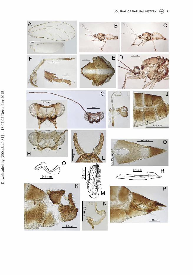
Adult structureForewings (Figure 1A) long and narrow with acutely pointed apex, cell cu1 distinctlylarger than cell m2, vein Rs short, curving to the wing margin; a group of marginalradular spines present in cells cu1, m1 and m2; surface spinules sparsely scattered in allcells; wing margins and veins sparsely covered with short to minute setae. Head notstrongly deflexed with genal processes short, divergent and conical, more or lesssymmetrical and broadly rounded at apex (Figure 1C). Vertex more or less flat dorsally,with lateral ocelli lying on small tubercles, medial epicranial suture distinct. Antennae(Figure 1B) long and slender, 3rd antennal segment 0.6 × head width, 10-segmentedwith rhinaria apically on segments 4, 6, 8 and 9, terminal segment with one apical setamedium long (~0.05 mm), paired with a short blunt tube-like seta (less than half thelength, ~0.02 mm). Clypeus (Figure 1D) triangular in lateral view, bearing two mediumlong setae apically. Distal segment of proboscis medium long. Dorsum of thorax withscattered short setae. Hind leg (Figure 1G) with meracanthus well developed andstraight; metatibia with a single large genual spine basally and 1+3 (typically) or 1+2shortly stalked sclerotized apical spurs; metabasitarsus constricted medially and longerthan apical tarsus. Male terminalia (Figure 1H) with proctiger lobed basally; paramere(Figure 1I) expanded basally into a distinct internal bulge and then tapering evenly toa darkly sclerotized apex; apical aedeagus segment (Figure 1J) short, hooked and withan extended, sharply pointed beak. Female terminalia (Figure 1K) short, proctigercurving downwards and slightly longer than the bluntly terminating subgenitalplate, circumanal ring relatively large, 0.36 × length of proctiger, and composed of adouble row of cells; ovipositor valvulae dorsales (Figure 1L) in profile with medialbulge dorsally.
Adult measurements (mm) and ratios (1♂, 1 ♀)WL: 3.12–3.55; HW: 0.68–0.73; AL: 1.85–2.06; GP: 0.12; PB: 0.14–0.15; HVW: 1.85–1.96;ALHW: 2.71-2.83; VLGP: 2.00–2.38; VLW: 0.62–0.83; WLW: 2.85–2.86; CUR: 1.37–1.60; MR:
4 D. M. PERCY ET AL.
Dow
nloa
ded
by [
200.
46.4
9.81
] at
13:
07 0
2 D
ecem
ber
2015

Figure 1. Adult Trioza incrustata Percy, sp. nov. (A) Forewing and hindwing (inset); (B, C) head andantenna; (D) clypeus; (E) proboscis; (F) thorax dorsum; (G) hind leg; (H) male terminalia; (I) paramere(inner surface); (J) distal aedeagus segment; (K) female terminalia; (L) ovipositor.
JOURNAL OF NATURAL HISTORY 5
Dow
nloa
ded
by [
200.
46.4
9.81
] at
13:
07 0
2 D
ecem
ber
2015

0.65–0.73; TLFL: 1.37–1.47. ♂: MP: 0.22; PL: 0.22; AEL: 0.18; MSLH: 1.00; AHS: 1.83; PLSH:1.00. ♀: FP: 0.49; FSP: 0.32; RL: 0.18; OV: 0,12; FPRL: 2.77; FPHW: 0.67; FPFSP: 1.53.
Immature structureBody outline elongate ovoid and more or less uninterrupted (Figure 2A, E, F). Forewingbuds with pronounced humeral lobe. Antennae of 5th instar (Figure 2C) with seven oreight segments bearing four rhinaria, one each apically on segments 3 and 5 and two onthe terminal segment; 4th instar antennae 3- or 4-segmented bearing one rhinarium on3rd segment, and two rhinaria on terminal 4th segment; 3rd instar antennae 3-segmen-ted bearing one rhinarium on 2nd segment and one on terminal 3rd segment; 2nd instarantennae 1-segmented bearing one rhinarium. Tarsi with well-developed claws andcrescent arolia (Figure 2C). Distinct ‘thoracic lobes’ visible in 5th instar (see insetFigure 2A). Anus situated ventrally; circumanal ring broad and shallowly V-shaped,with a single row of elongate cells.
Immature chaetotaxyThe 2nd–5th instars with stalked, fan-shaped setae around the margin (Figure 2B, F),3rd–5th instars with dorsal surface covered in distinct pattern created by thearrangement of simple setae on round tubercle-like annuli (Figure 2A, B; absent inthe 2nd instars with the pattern becoming progressively more intricate in olderinstars).
Immature measurements (mm), 5th instar (n = 2)BL: 2.09–2.15; BW: 1.39–1.42; WL: 1.24–1.27; CPL: 0.76–0.79; CPW: 1.24–1.27; RW:0.26–0.27; HW: 0.62–0.67; AL: 0.28.
Host plantCeltis philippensis (Cannabaceae)
DistributionPNG, Madang Province.
BiologyThe gall is a leaf fold at the margin of the leaf and consists of the upper adaxial surfacefolding down to make the gall on the lower abaxial leaf surface (Figure 3A). The foldbecomes sealed and appears to contain a single immature. There is sometimes a singlefold, but more often several discrete chambers, in this case, it is not clear whether eachchamber is completely sealed from the others. When mature, the gall seal opens along theintersection between adaxial and abaxial leaf surfaces. The density of galls and immaturesis high with ~70 immatures collected from three sample sites. Rearing from galls producedfive adult psyllids from two of these sites. Other insect associates found in the galls includececidomyid larvae, and chalcid pupae and larvae (Encyrtidae and Eulophidae).
EtymologyThe specific epithet refers to the distinct pattern on the dorsum of older immaturesappearing as an encrustation formed by the tubercle-like annuli at the base of the setae.
6 D. M. PERCY ET AL.
Dow
nloa
ded
by [
200.
46.4
9.81
] at
13:
07 0
2 D
ecem
ber
2015

Figure 2. Immatures, Trioza incrustata Percy, sp. nov. (A–F), and Trioza grallata Percy, sp. nov. (G–K).(A–D) 5th instar; (B) outline illustrating distinct pattern on dorsal surface; (C) antenna and tarsi; (D)circumanal ring; (E) 4th instar; (F) 2nd instar; (G–I) 5th instar; (H) antenna and tarsi; (I) circumanalring; (J) 3rd instar; (K) 1st instar.
JOURNAL OF NATURAL HISTORY 7
Dow
nloa
ded
by [
200.
46.4
9.81
] at
13:
07 0
2 D
ecem
ber
2015

8 D. M. PERCY ET AL.
Dow
nloa
ded
by [
200.
46.4
9.81
] at
13:
07 0
2 D
ecem
ber
2015

Perfect passive participle derived from the Latin verb incrustare, to have an ornamentalcover.
CommentsThe host plant is a common lowland rainforest tree in New Guinea and much of SouthEast Asia (George Weiblen, pers. comm.). Six additional triozid taxa are known from theplant genus Celtis. Yang (1984) described two of these species from Taiwan, Triozaceltisae Yang, 1984 and Trioza lineata Yang, 1984 on Celtis tetrandra and Celtis sinensis,respectively. Li (2011) described an additional species from Celtis sinensis, Trioza long-igenitus (Li 2011), and the immatures and biology for Trioza bifasciaticeltis Li and Yang,1991, which has free-living immatures on the lower abaxial surface of leaves. Althoughno biology is mentioned for the immatures of T. celtisae, this species is related toT. lineata, which is also described as having free-living immatures on the lower abaxialsurface of leaves (Yang 1984). Li (2011) placed the above species in a new genus,Metatriozidus, but this was considered artificial and synonymized with Trioza by Yanget al. (2013). A fifth Asian species, Trioza brevifrons Kuwayama, 1910, is recorded fromKorea, Japan and Taiwan. The host-plant in Korea and Japan is Celtis sinensis var.japonica (Kwon 1983), but the host association of the type material from Taiwan isunknown, and these specimens appear to differ from those in Korea and Japan (Kwon1983; Yang et al. 2013). Lastly, a South American species, Leuronota fuscata (Laing,1923) develops on Celtis iguanaea (Burckhardt and Queiroz 2012, and D. Burckhardtpers. comm.). None of these Celtis-feeding triozid species appears related to Triozaincrustata sp. nov., and no clear affiliations are apparent within the Triozidae; wetherefore place this taxon within the artificially large (polyphyletic) genus TriozaFoerster, 1848. Trioza incrustata is the first species of Trioza known to produce leafmargin galls on Celtis.
Type materialHolotype, ♂ (slide mounted), Mis village, Madang Province, PNG (5°11ʹS, 145°47ʹE, 50 m),21 March 2011, ex Celtis philippensis, (HE06) P. Butterill leg. (BMNH). Paratypes, 1 ♀
(HE07), immatures: 2 5th, 3 4th, 1 3rd, 2 2nd (GALL015) as for holotype (BMNH). Othermaterial: immatures and galls, Baitabag village (5°8ʹS, 145°46ʹE, 100 m), and Ohu village
Figure 3. Galls Papua New Guinea (A–D), Singapore (E), Taiwan (F). (A) Trioza incrustata Percy, sp.nov.: leaf margin galls on Celtis philippensis (Cannabaceae) (early and later gall development viewedfrom lower leaf surface); (B) Cornegenapsylla allophyli Malenovský and Percy, sp. nov.: leaf margingalls on Allophylus cobbe (Sapindaceae) (early and later gall development viewed from upper leafsurface); (C) Trioza grallata Percy, sp. nov.: leaf surface galls on Elaeocarpus schlechterianus(Elaeocarpaceae) (adaxial and abaxial leaf surfaces); (D) Pauropsylla udei Rübsaamen: conical leafsurface galls on Ficus variegata (Moraceae) produced on the abaxial leaf surface (inset above: galldetail on lower leaf surface; inset below: gall viewed from upper leaf surface); (E) Pauropsylla udeiRübsaamen: globular leaf surface galls on Ficus variegata (Moraceae) produced on the adaxial leafsurface, images on right show a dissected gall chamber and presence of the eclosed adult within; (F)Pauropsylla triozoptera Crawford: conical leaf surface galls produced on the adaxial leaf surface ofFicus cf. ampelas (Moraceae).
JOURNAL OF NATURAL HISTORY 9
Dow
nloa
ded
by [
200.
46.4
9.81
] at
13:
07 0
2 D
ecem
ber
2015

(5°13ʹS, 145°40ʹE, 200 m), near Madang, Madang Province, PNG, March 2011, ex Celtisphilippensis, P. Butterill leg. (NARI).
Gene sequencesGenBank: KT588301 (COI), KT588307 (cytB) (PNGHE06–11).
Trioza grallata Percy, sp. nov.(Figures 2G–K; 4A–R; 3C)
Adult colour (ethanol material)Forewings without pattern but membrane fuscous, veins brown. Body generally mid todark brown, with distinct pale longitudinal band on the dorsum of the thorax (Figure 4E)and sometimes extending forward through the vertex. Ventral part of the abdomen,particularly in females, may also be paler. Antennal segments 3–10 dark brown to black.Legs paler except apices of tibiae and tarsi, which are dark brown to black.
Adult structureForewings (Figure 4A) distinctly broader in the apical half with a bluntly acute apex;height of cells cu1 and m2 subequal; vein Rs short, curving evenly to the wing margin; agroup of marginal radular spines present in cells cu1, m1 and m2; surface spinules eitherabsent or very sparsely distributed; wing margins and veins with short to minute setae.Head (Figure 4G) with genal processes short, widely divergent, asymmetrical, andterminating in slightly swollen, blunt apices with two or three long stout setae; distinctswellings also present below the genae (Figure 4H). Vertex more or less flat dorsally, withlateral ocelli lying on small tubercles, medial epicranial suture distinct. Antennae(Figure 4G) long and slender, 3rd antennal segment 0.75–0.81 × head width, 10-seg-mented with rhinaria apically on segments 4, 6, 8 and 9, terminal segment with one longapical seta (0.09–0.12 mm), paired with a short stout tube-like seta (less than half thelength, ~0.02 mm). Clypeus (Figure 4I) well rounded and ventrally scaly, rough surfaced,and covered in short setae plus two long setae. Distal segment of proboscis mediumlong. Dorsum of thorax covered in short, stout setae. Hind leg (Figure 4F) with mer-acanthus well developed and straight; metatibia slender and elongate (almost twice thelength of the metafemur), with one, but more usually a pair of genual spines basally and1+3 (occasionally 1+4) large, distinctly stalked and sclerotized apical spurs; metabasi-tarsus constricted basally and subequal in length to apical tarsus. Sternites with numer-ous stout setae (Figure 4J). Male terminalia (Figure 4K) with proctiger strongly lobedmedially and constricted to a narrow tube dorsally. Paramere (Figure 4L, M) expandedinto a lobe in the basal half and then more or less parallel sided and slightly archedbackwards, the posterior margin with a comb of long stout setae, and terminating in asmall posteriorly directed sclerotized hook. Apical aedeagus segment (Figure 4O) short,base swollen, strongly curved basally and then angled apically, with a rounded andslightly inflated apex. Female terminalia (Figure 4P) with proctiger more or less straightdorsally, ventral margins lobed medially; apex acute, darker, and covered in short stoutsetae; proctiger markedly longer than (> 1.5 ×) the bluntly terminating subgenital plate,apex of subgenital plate notched and terminating in several long setae; circumanal ring
10 D. M. PERCY ET AL.
Dow
nloa
ded
by [
200.
46.4
9.81
] at
13:
07 0
2 D
ecem
ber
2015
JOURNAL OF NATURAL HISTORY 11
Dow
nloa
ded
by [
200.
46.4
9.81
] at
13:
07 0
2 D
ecem
ber
2015

(Figure 4Q) is long and narrow, 0.3 × the length of proctiger, and composed of a doublerow of cells; inner ovipositor valve (Figure 4R) small and in profile slightly bulgingdorsally.
Adult measurements (mm) and ratios (4 ♂ 3 ♀)WL: 3.30–3.58; HW: 0.67–0.71; AL: 2.27–2.51; GP: 0.14–0.17; PB: 0.15–0.18; HVW: 1.92–1.96; ALHW: 3.41-3.61; VLGP: 1.2–1.67; VLW: 0.50–0.63; WLW: 2.38–2.45; CUR: 1.19–1.33;MR: 0.68–0.74; TLFL: 1.77–1.86. ♂: MP: 0.19–0.20; PL: 0.24–0.25; AEL: 0.18–0.20; MSLH:1.17–1.29; AHS: 2.23–2.50; PLSH: 0.95–1.11. ♀: FP: 0.72–0.74; FSP: 0.44–0.49; RL: 0.22; OV:0.14–0.15; FPRL: 3.21–3.37; FPHW: 1.02–1.06; FPFSP: 1.51–1.65.
Immature structureBody outline of older instars elongate ovoid with protruding wing bud margins, butlacking an obvious humeral lobe (Figure 2G, J). Antennae of 5th instar (Figure 2H) 7- or8-segmented, bearing four rhinaria apically on segments 4, 5, 7 and 8; 3rd instarantennae 4-segmented bearing three rhinaria, one on 3rd and two on 4th segment;2nd instar antennae 3-segmented bearing two rhinaria on 2nd and 3rd segments; 1stinstar antenna with a single segment bearing a single rhinarium. Tarsi with weaklydeveloped claws and crescent arolia (Figure 2H). Small ‘thoracic lobes’ visible in 5thinstars. Anus situated ventrally; circumanal ring composed of single, sometimes inter-rupted, row of rounded to elongate cells (Figure 2I).
Immature chaetotaxyThe 1st–5th instars have scattered medium to long simple setae on margins and surfacesof head, thorax, and abdomen; in addition, the 5th instar also has irregular patches ofshort, stout, thorn-like cuticular projections on the dorsal surface of the abdomen(Figure 2G).
Immature measurements (mm), 5th instar (n = 2)BL: 1.39–1.73; BW: 1.00–1.21; WL: 0.64–0.79; CPL: 0.73; CPW: 0.82; RW: 0.16; HW: 0.52–0.54; AL: 0.33.
Host plantsElaeocarpus schlechterianus (Elaeocarpaceae).
DistributionPNG, Morobe Province.
Figure 4. Adult Trioza grallata Percy, sp. nov. (A) Forewing and hindwing (inset); (B) male whole; (C)female whole; (D) head and thorax; (E) thorax dorsum; (F) hind leg; (G) head and antenna; (H) headtilted back to show sub-genal swellings; (I) clypeus and proboscis; (J) abdominal segments showingsetae on ventral sclerites; (K) male terminalia; (L) parameres (posterior); (M) paramere (inner surface);(N) aedeagus; (O) distal aedeagus segment; (P) female terminalia; (Q) female proctiger dorsum; (R)ovipositor.
12 D. M. PERCY ET AL.
Dow
nloa
ded
by [
200.
46.4
9.81
] at
13:
07 0
2 D
ecem
ber
2015

BiologyThis species produces small, round to ovoid, enclosed blister-like galls on the leaf blade(Figure 3C), which are exited mostly on the upper adaxial leaf surface. In all gallsdissected (n = 13), there was a single unilocular gall chamber, and when present, asingle immature psyllid. Incubating galls for rearing produced ~90 adult psyllids.Associated insects included cecidomyid and sciarid midges, and chalcid parasitoids.
EtymologyThe specific epithet refers to the long, slender tibiae, hence from the Latin gralla, a stilt,grallata = bearing stilts (adjective).
CommentsThe host plant is a New Guinea endemic known from rather few collections, at eleva-tions of 850–1850 m, from Jayapura (Papua) through the highlands to Morobe Province.(George Weiblen pers. comm., with reference to Coode 1981). Five species of Trioza areknown from the plant genus Elaeocarpus. Matsumoto (1999) treated three of these,together with two additional species that may also be Elaeocarpus-feeders, as the ‘Triozamaculata group’. The maculata group is found in Japan, Taiwan, Vietnam and Malaysia(Sabah). Trioza grallata sp. nov., on Elaeocarpus schlechterianus, does not appear to berelated to this group. Neither does it appear to be related to either of the two remainingspecies known from Elaeocarpus that both produce small galls on the leaves. One ofthese species, from Taiwan, Trioza elaeocarpi Yang, 1984, produces small round galls onleaves of Elaeocarpus sylvestris (Yang 1984), and the other, Trioza indigena Tuthill, 1951,from the Caroline Islands forms small leaf galls on Elaeocarpus kusanoi, which is endemicto the island of Pohnpei, Senyavin Islands (Tuthill 1951). Li (2011) transferred Triozamaculata and Trioza elaeocarpi to Triozopsis Li, 2005, but Yang et al. (2013) subsequentlysynonymized Triozopsis with Trioza; as with Trioza incrustata sp. nov., no clear affiliationsare apparent within Triozidae.
Type materialHolotype, ♂ (slide mounted), close to Yawan village, Morobe Province, PNG (06°08ʹS,146°52ʹE, 1700 m), 7 February 2011, (HE08) P. Butterill leg. (BMNH). Paratypes, 5 ♂, 5 ♀,(HE08, HE09), immatures: 1 5th, 3 4th, 2 3rd, 1 2nd instar (GALL363 ex Elaeocarpusschlechterianus) as for holotype (BMNH). Other material: immatures, 1st–5th instars(HEY1, HEY3, GALL359, GALL360 ex Elaeocarpus schlechterianus) as for holotype, P.Butterill leg. (NARI).
Gene sequencesGenBank: KT588302 (COI), KT588308 (cytB) (PNGHE08–11).
Pauropsylla triozoptera Crawford, 1913(Figures 3F, 5G–H)
Pauropsylla triozoptera Crawford, 1913: 296; Yang et al. (2013): 46.Sympauropsylla triozoptera (Crawford), Enderlein (1921): 116.Neotrioza triozoptera (Crawford), Li (2011): 1315.
JOURNAL OF NATURAL HISTORY 13
Dow
nloa
ded
by [
200.
46.4
9.81
] at
13:
07 0
2 D
ecem
ber
2015

Material examined9 ♂, 4 ♀, 2 5th instars, Ohu village, Madang Province, PNG, 11 April 1995, (handcollecting/beating) hatched from leaf galls ex Ficus trachypison, Y. Basset leg. (MMBC);4 5th instars (+ 4 larvae of Lepidoptera ‘commensal’), same data as previous but 11January 1995, from galls on mature leaves; 4 ♂, 2 ♀, Baitabag village, Madang Province,PNG, March 1996, ex Ficus trachypison (TRA 3), V. Novotný leg. (MMBC). 2 ♂, 1 ♀, LagunaCollege, Laguna, Philippines, 10 June 1976, leaf galls ex Ficus ulmifolia, R. Braza leg.(BMNH); 2 ♂, 3 ♀, Davao Experiment Station, Philippines, March 1964, traps air level, M.Gavarra leg. (BMNH); 2 ♂, 4 ♀, same data as previous but January 1964 (BMNH); 1 ♂, 4 ♀,same data as previous but traps ground level, October–December 1961 (BMNH). 1 5thinstar, 5 1st–2nd instars, Taiwan (22.0499°N, 120.8576°E, 150 m), 30 January 2010,dissected from galls ex Ficus cf. ampelas, D. Percy leg. (BMNH).
CommentsThis species is widespread in eastern Asia. We report the first records for PNG, MadangProvince, on Ficus trachypison (Moraceae). All known Pauropsylla are gall inducing. Thisspecies shares with Pauropsylla udei a galling habit on Ficus in PNG, but there arenotable morphological differences between the two species, including the male termi-nalia illustrated in Figure 5(G, I); the head structure (illustrated by Uichanco 1921);antennae with three rhinaria on segment 3 and a single rhinarium on segments 4, 6, 8and 9 (the latter associated with a very long seta as long as segment 10) in P. triozoptera,versus only four on segments 4, 6, 8 and 9 (associated with a short seta) in P. udei;metatibia with three apical spurs in P. triozoptera versus four (2 + 2) in P. udei, and thefemale terminalia. The immatures can also be differentiated primarily by the largedistinctly shaped circumanal pore area in P. triozoptera (Figure 6H, and illustrated byYang 1984) versus much reduced anus lacking circumanal pore area in P. udei. Geneticdivergence between the two species is high (mitochondrial DNA divergence > 20%).
Host plantsFicus tinctoria (= F. gibbosa), F. ulmifolia and possibly F. ampelas in Taiwan (Yang 1984;Hodkinson 1986; Yang et al. 2013), F. trachypison (new record) in PNG.
BiologyDissected galls from PNG (on F. trachypison) and from Taiwan (on F. cf. ampelas)contained a single immature per gall. As described below for P. udei, some noticeablevariation in gall phenotype within this species was found. All galls produced byP. triozoptera are enclosed galls on the leaf blade, but in PNG the gall exterior is coveredin spine-like trichomes, whereas in Taiwan the galls are smooth, without trichomes(Figure 3F). Furthermore, the shape of the galls in Taiwan is narrowly conical, and inPNG it is globular or broadly conical (bell-shaped), produced on the upper adaxial leafsurface; on some leaves from PNG, mature galls appear to fuse, forming an irregularbumpy mass in which individual galls are difficult to distinguish. In PNG, the galls onF. trachypison may be parasitized by a species of Braconidae (an adult braconid waspresent in the sample together with the gall). Parasitization of gall-inducing psyllids byBraconidae has been reported for Pauropsylla braconae Li, 2000 in Li et al., 2000 on Ficus
14 D. M. PERCY ET AL.
Dow
nloa
ded
by [
200.
46.4
9.81
] at
13:
07 0
2 D
ecem
ber
2015

Figure 5. Adult comparison of Cornegenapsylla allophyli Malenovský and Percy, sp. nov. (A, D, E) andCornegenapsylla sinica Yang and Li (B, C, F). (A, B) Forewings: (A) C. allophyli, (B) C. sinica; (C, D)heads: (C) C. sinica, (D) C. allophyli; (E, F) male terminalia: (E) C. allophyli, (F) C. sinica. Comparison ofadult male terminalia in specimens of Pauropsylla triozoptera Crawford (G, left to right): from PapuaNew Guinea, Philippines, with inset schematic of gall phenotypes; (H) 5th instar circumanal porearea of P. triozoptera from Taiwan. Comparison of adult male terminalia in specimens Pauropsyllaudei Rübsaamen (I, left to right): from Papua New Guinea, Hong Kong, Bangladesh, Philippines, withinset schematic of gall phenotypes.
JOURNAL OF NATURAL HISTORY 15
Dow
nloa
ded
by [
200.
46.4
9.81
] at
13:
07 0
2 D
ecem
ber
2015

Figure 6. Adult Cornegenapsylla allophyli Malenovský and Percy, sp. nov. (A) Forewings and hindwing,upper forewing imaged with reflected light and lower with transmitted light; (B) hind leg; (C) thoraxdorsum; (D) clypeus and proboscis; (E) antenna, terminal two segments (inset); (F) antennal rhinaria on4th (lower), 5th (middle) and 6th (upper) segments; (G) head, lateral (left), ventral (middle) and lateralshowing reduced but distinct genal tubercles (right); (H) male terminalia; (I) paramere (inner surface); (J)distal aedeagus segment; (K) female terminalia, circumanal ring (inset); (L) ovipositor.
16 D. M. PERCY ET AL.
Dow
nloa
ded
by [
200.
46.4
9.81
] at
13:
07 0
2 D
ecem
ber
2015

hainanensis in China (Yunnan) (Li et al. 2000). In Taiwan, a 5th instar was foundparasitized by Psyllaephagus sp. (Encyrtidae, det. J. Noyes, BMNH).
DistributionTaiwan (Yang 1984; Li 2011; Yang et al. 2013), Japan: mainland, Ryukyu Islands(Hodkinson 1983, 1986), Indonesia: Java (Hodkinson 1986), Philippines (Crawford 1913;Hodkinson 1983, 1986), PNG (new record), Fiji (Hodkinson 1983, 1986).
Gene sequencesGenBank: KT588303 (COI), KT588309 (cytB) (TAI83–10).
Pauropsylla udei Rübsaamen, 1899(Figures 3D,E; 5I)
Pauropsylla udei Rübsaamen, 1899: 264.Pauropsylla montana Uichanco, 1919: 546, nomen nudum; Uichanco (1921): 265.Pauropsylla bakeri Crawford, 1915: 258; Crawford (1919): 145.Pauropsylla reticulata Mathur, 1975: 102 syn. nov.
Material examined2 ♂, 11 2nd–5th instars, Mis village (5°11ʹS, 145°47ʹE, 50 m) and Ohu village (5°13’S, 145°40ʹE, 200 m), Madang Province, PNG, September–November 2010 and February–March2011, reared from conical galls ex Ficus variegata, P. Butterill leg. (BMNH); 1 ♀, dissectedfrom conical galls in alcohol, Baitabag village, Madang Province, PNG, January 1996, exFicus variegata, V. Novotný leg. (MMBC); 1 ♂, 2 ♀, numerous circular galls and imma-tures, Singapore (1.353°N, 103.778°E, 75 m), 6 November 2012, ex Ficus variegata,D. Percy leg. (BMNH); 9 ♂, Sylhet, Bangladesh, 1 November 1930, ex circular galls, M.Bose leg. (BMNH), same as type series of Pauropsylla reticulata Mathur, 1975 syn. nov.;2 ♂, 1 ♀, Pokfulam, Hong Kong, 14 March 1973, ex Ficus variegata (BMNH); 1 ♂, 3 ♀, HaKwai Chung Tsuen, Hong Kong, 7 November 2005, C. Lau leg. (BMNH); 12 4th–5thinstars, Hang Mei Village, Tai O, Lantau Island, Hong Kong, 12 April 2010, ex Ficusvariegata circular galls, J. Martin leg. (BMNH); 1 ♀, 4 4th–5th instars, Gunung MuluNational Park, Borneo, Sarawak, Malaysia, 27 June 1978, ex Ficus, V. Eastop leg.(BMNH); 2 ♂, 2 ♀, Laguna College, Laguna, Philippines, 30 December 1975, ex Ficusvariegata leaf galls, R. Braza leg. (BMNH).
CommentsThis species is widespread in Asia. We report new records for PNG, Madang Province, onFicus variegata (Moraceae), and illustrate some of the variation found in the structure ofthe male terminalia from different parts of the Oriental Region (Figure 5I). The PNGspecimens have a narrower male paramere and proctiger, and the female terminalia areshorter, but otherwise specimens are similar to those examined from Hong Kong andBangladesh; specimens from the Philippines show the most structural difference in maleterminalia and further investigation of regional patterns of intra-specific variation isneeded to assess whether there is sufficient divergence to warrant recognizing separatespecies. The 2nd and 5th instar immatures from PNG are covered in medium to long
JOURNAL OF NATURAL HISTORY 17
Dow
nloa
ded
by [
200.
46.4
9.81
] at
13:
07 0
2 D
ecem
ber
2015

simple setae as illustrated in Rübsaamen (1899). Immatures examined from Sarawak andHong Kong did not reveal noticeable differences. Genetic divergence between PNG andSingapore specimens supports the need for further intra-specific investigation (mito-chondrial DNA divergence was relatively high, ~10%), as does the striking gall typedifferences between PNG and other areas.
Comments on synonymizationNine adult specimens of P. reticulata Mathur, 1975 collected together with the typeseries at the type locality, together with two dried galls, were sent to BMNH in 1931(Mathur 1975). The suspected synonymy of this material was noted by Hollis (1984, p.28), and after examining this material, DMP concurs with his assessment and thesynonymy is formalized here. The two galls are spherical and globular, and appearsimilar to those found on Ficus variegata (Moraceae) from Singapore (Figure 5E) andthe Philippines (illustrated by Uichanco 1919), but they appear to lack exterior tri-chomes. In addition, there is some uncertainty regarding the host-plant range, as theholotype and paratype series (including BMNH material) was collected ‘ex galls onunknown plant’, but Mathur (1975) also cites other material collected from WestBengal, India (13 July 1935) ex galls on leaves of Breonia chinensis (= Anthocephalusindicus; Rubiaceae) in his description of P. reticulata. Furthermore, the type series ofP. udei was originally described with the host as an unknown species of Rubiaceae inSumatra, which was subsequently considered a host record error by Uichanco (1921),and certainly the original illustration and description of the galls by Rübsaamen (1899)for P. udei resemble closely those illustrated here on Ficus variegata leaves (Figure 5E).
Host plantVarious Ficus (Moraceae) species, including F. fulva (=F. chlorocarpa) and F. variegata(Uichanco 1919; Hodkinson 1983, 1986). In PNG it was collected on F. variegata.
BiologyAs noted for P. triozoptera above, we report notable variation in the gall phenotype. Inall cases, the galls of P. udei are enclosed and produced on the leaf blade. In PNG thegalls are conical and smooth, without trichomes, and produced on the lower, abaxial leafsurface (Figure 3D). Those found in the Philippines, Singapore and Hong Kong arespherical or oval, usually produced on the upper, adaxial leaf surface, and the outersurface of each gall is covered in trichomes that are cream to red; gall coverage can bedense on younger leaves (Figure 3E).
DistributionBangladesh (Assam in Hodkinson 1983, 1986); China (Hong Kong; Hodkinson 1983, 1986;Martin and Lau 2011); Indonesia (Hodkinson 1983, 1986); Malaysia (Hodkinson 1983);PNG (new record); Philippines (Uichanco 1919; Hodkinson 1983, 1986); Singapore(Hodkinson 1983). Possibly India (Mathur 1975, as P. reticulata; see comments onsynonymization above).
18 D. M. PERCY ET AL.
Dow
nloa
ded
by [
200.
46.4
9.81
] at
13:
07 0
2 D
ecem
ber
2015

Gene sequencesGenBank: KT588305 (COI), KT588311 (cytB) (PNGHE05-10); and KT588304 (COI), KT588310(cytB) (SING01–12).
Family PHACOPTERONIDAE Heslop-Harrison, 1958Cornegenapsylla allophyli Malenovský and Percy, sp. nov.
(Figures 3B; 5A,D,E; 6A–L; 7A–D)
Adult colour (ethanol material)Forewing membrane (Figure 6A) clear except for brown infuscations along the veins;veins brown in basal wing half, light ochreous in apical half. Hindwing membraneinfuscate basally (Figure 6A). Body almost uniformly brown, legs paler. Antenna(Figure 6E) with segments 1–3 brown, segment 4 uniformly off-white, segments 5–8off-white basally and dark brown to black apically, segments 9–10 entirely dark brown toblack.
Adult structureForewing (Figure 6A) with apex broadly truncate; cell cu1 small; membrane lackingsurface spinules, but with small patches of radular spines close to veins at the margin.Head distinctly wider than thorax. Vertex (Figure 6G) almost flat dorsally, with lateralocelli lying on small tubercles, rounded down to frons; medial epicranial suture distinctanteriorly and posteriorly, but indistinct medially. Genae very small, weakly swollen, witha small tubercle below insertion of antennae (Figure 6G). Antenna (Figure 6E) relativelylong; the 3rd segment strikingly enlarged, approximately as long as segments 4 and 5together; segments 4–10 slender; segments 4–9 each with widely elliptic rhinariumlargely bordered with wreath of small, acute cuticular spines, segments 4–8 each withan additional sensilla lying in a transverse groove closely adjacent to rhinarium(Figure 6F); terminal setae subequal, slightly shorter than segments 9 and 10 together(Figure 6E). Clypeus (Figure 6D) somewhat flattened, rounded at apex, lacking longsetae. Apical segment of proboscis short. Metatibia with a small genual spine basally,and 13–15 evenly sized unsclerotized spurs apically; metabasitarsus slightly longer thanapical tarsal segment and bearing two sclerotized lateral spurs (Figure 6B). Male sub-genital plate elongate, strongly produced into a posterior hump (Figure 6H). Maleproctiger (Figure 6H) narrowly conical. Paramere (Figure 6I) curved posteriorly, withapex broadly rounded; inner side covered with fine setae. Distal segment of aedeagus(Figure 6J) with a rounded, unhooked apex. Female terminalia (Figure 6K) long; proctigerwith dorsal margin nearly straight; circumanal ring relatively small, 0.2 × length ofproctiger, and composed of a double row of cells; subgenital plate gradually narrowingto a pointed apex; inner ovipositor valve dorsally broadly triangular in profile (Figure 4L).
Adult measurements (mm) and ratios (3 ♂, 2 ♀)WL: 1.72–2.16; HW: 0.51–0.61; AL: 1.13–1.30; GP: 0.03–0.06; PB: 0.09; HVW: 1.82–20;ALHW: 2.05–2.17; VLGP: 3.50–70; VLW: 0.64–0.70; WLW: 2.20–2.29; CUR: 2.83–30; MR:0.55–0.63; TLFL: 1.11–1.18. ♂: MP: 0.16–0.21; PL: 0.16–0.18; AEL: 0.19–0.21; MSLH: 1.58–
JOURNAL OF NATURAL HISTORY 19
Dow
nloa
ded
by [
200.
46.4
9.81
] at
13:
07 0
2 D
ecem
ber
2015

1.77; AHS: 2.36–2.40; PLSH: 0.91–0.92. ♀: FP: 0.62–0.74; FSP: 0.47–0.54; RL: 0.14; OV: 0.19;FPRL: 4.53; FPHW: 1.02–1.25; FPFSP: 1.31.
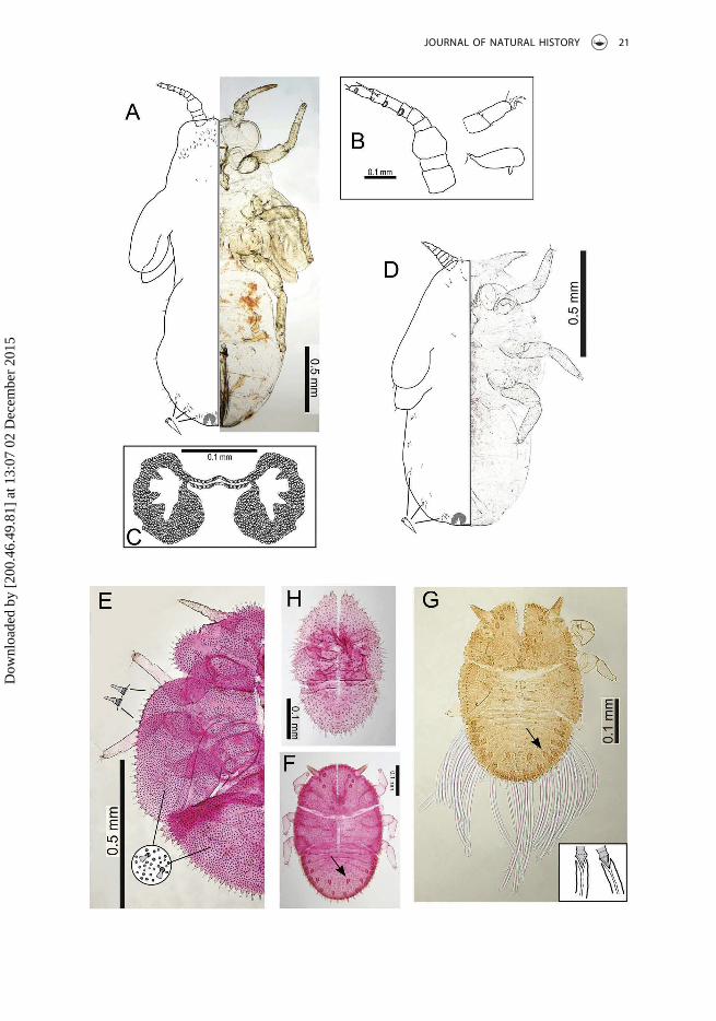
Immature structureBody outline elongate, with protruding wing buds lacking humeral lobes (Figure 7A, D).Antennae of 5th instar (Figure 7B) with 8 or 9 segments bearing four rhinaria, one eachapically on segments 6–9; 3rd instar antennae 5- to 6-segmented bearing three rhinaria,one each on terminal 3 segments. Tarsi with well-developed claws, arolia very small(Figure 7B). Anus situated terminally, circumanal pore area with large and irregularlateral fields of multiple pores extending to both ventral and dorsal surfaces; circumanalring medially narrow with a single row of elongate cells (Figure 7C).
Immature chaetotaxyImmatures with sparse coverage of short simple setae on body dorsal surface andmargins. Dorsal surface of caudal plate/abdominal tergites with small groups of pointedlanceolate setae close to the body margin (Figure 7A, D).
Immature measurements (mm), 5th instar (n = 2)BL: 2.09–2.24; BW: 1.21–1.30; WL: 0.67; CPL: 1.06–1.24; CPW: 0.94–0.97; RW: 0.32–0.37;HW: 0.62–0.63; AL: 0.42–0.46.
EggStout laterally positioned pedicel at base, apical tail present (Figure 7B: dissected from5th instar immatures).
Host plantAllophylus cobbe (= Pometia pinnata) (Sapindaceae).
DistributionPNG, Madang Province.
BiologyThe gall is a leaf fold on the margin of the leaf and consists of the lower abaxial surfacefolding up over the upper adaxial surface to make the gall on the upper leaf surface(Figure 3B).
EtymologyA noun in the genitive case, named in reference to the host plant genus Allophylus.
CommentsThis species is placed in the hitherto monotypic genus, Cornegenapsylla Yang and Li,1982. It shares with the type species, C. sinica, the forewing pattern with bands of darkpigmentation around the veins, the forewing shape (the forewing being elongate withan almost rectangular apex), and the head shape, though it lacks the well-developedgenal processes of C. sinica (comparison of forewing, head and male terminalia areprovided in Figure 5A–F). It differs most distinctly from C. sinica in the larger body size,
20 D. M. PERCY ET AL.
Dow
nloa
ded
by [
200.
46.4
9.81
] at
13:
07 0
2 D
ecem
ber
2015
JOURNAL OF NATURAL HISTORY 21
Dow
nloa
ded
by [
200.
46.4
9.81
] at
13:
07 0
2 D
ecem
ber
2015

the very short genal processes, the swollen 3rd antennal segment, the male subgenitalplate produced into a posterior bulge, the elongate shape of the female terminalia, theimmature morphology, and the host plant species and galling biology. Both speciesproduce galls on host plants in the family Sapindaceae, but C. sinica immatures producedeep pit galls on young leaves of Dimocarpus longan (Yang 1984), whereas C. allophyliimmatures produce enclosed galls on the leaf margins of mostly mature leaves ofAllophylus cobbe. These different biologies are reflected in the different immaturestructure and chaetotaxy, with C. sinica immatures showing the ventro-dorsally flattenedbody form, and marginal placement of setae typical of pit gall formers; whereasC. allophyli immatures have irregularly dispersed simple setae and non-flattened, broadlyinflated body form typical of enclosed gall formers.
Ongoing work by I.M. suggests that Cornegenapsylla is affiliated with the Neotropicalgenus Phacosemoides Lima and Guitton, 1962, as well as a number of species from theAfrotropical Pseudophacopteron caffrariense-group as defined in Malenovský andBurckhardt (2009).
Type materialHolotype, ♂ (slide mounted), Mis village near Madang, Madang Province, PNG (5°11ʹS,145°47ʹE, 50 m), 13 October 2011, (HE01) P. Butterill leg. (BMNH). Paratypes, 1 ♂, 1 ♀
(HE02, HE03), immatures: 1 5th, 1 3rd (GALL087 ex Allophylus cobbe), as for holotype(BMNH); 2 ♂ 3 ♀, Baiteta, Madang Province, PNG, 16 May 1995 (AR 16), 27 June 1995,canopy fogging, Sloanea forbesii (Elaeocarpaceae) (AR 19), 28 May 1996 (at light, AR 53),24 June 1996 (at light, T2), O. Missa leg. (IRNB, MMBC, NHMB, dry and slide mounted andpreserved in ethanol).
Gene sequencesGenBank: KT588306 (cytB) (PNGHE02-11).
Cornegenapsylla sinica Yang and Li, 1982(Figures 5B,C,F; 7E–H)
Cornegenapsylla sinica Yang and Li, 1982: 124; Burckhardt and Ouvrard (2012): 17.Neophacopteron euphoriae Yang, 1984: 165; Li (2011): 1223.Phacopteron sinicum (Yang and Li), Li (2011): 1223.
Material examined3 ♂, 2 ♀, 2nd–5th instars, Singapore Botanic Gardens, Singapore, 2 January 1997, exDimocarpus longan, J. Martin leg. (BMNH); 4 ♂, 1 ♀, 2nd–4th instars, Bangkok, Thailand,
Figure 7. Immatures, Cornegenapsylla allophyli Malenovský and Percy, sp. nov. (A–D), andCornegenapsylla sinica Yang and Li (E–H). (A–C) 5th instar; (B) antenna, tarsi, and egg; (C) circumanalpore area and ring; (D) 3rd instar; (E) 5th instar (stained); (F, G) 3rd instars (F stained), with (G)showing production of long waxy filaments from large pointed setae; (H) 2nd instar (stained).
22 D. M. PERCY ET AL.
Dow
nloa
ded
by [
200.
46.4
9.81
] at
13:
07 0
2 D
ecem
ber
2015

1961, ex Dimocarpus longan, A. Manjikul leg. (BMNH); 1 ♂, 3 ♀, Johor, Malaysia, 13 April1970, ex Nephelium malaiense, Dept. Agric. leg. (BMNH).
CommentsWith the synonymization of Neophacopteron euphoriae and Cornegenapsylla sinica by Li(2011), it is confirmed that there is a single species on longan that is widely distributedin South East Asia. Burckhardt and Ouvrard (2012) mistakenly mention the combinationCornegenapsylla euphoriae (Yang, 1984) when reinstating Cornegenapsylla as a validgenus. Cornegenapsylla sinica remains unusual within the Phacopteronidae for thelong genal processes and the male terminalia covered with stout lanceolate setae, aswell as a characteristic near vertical feeding posture (Yang et al. 2009). Without anexplanation, but probably based on the relatively well-developed genae in the typespecies of Phacopteron Buckton, 1896, Li (2011) placed C. sinica in Phacopteron, whichwas subsequently reversed by Burckhardt and Ouvrard (2012). The type species ofPhacopteron, Phacopteron lentiginosum Buckton, 1896 is a widespread species fromPakistan to South East Asia. The inclusion of the only other species in Phacopteron,Phacopteron gabrieli Navasero and Calilung, 2000, from the Philippines is considereddoubtful. Phacopteron lentiginosum produces a completely enclosed round gall on theleaves of Garuga spp. (Burseraceae) (Hodkinson 1986; Li 2011), which dehisces afteradult eclosion inside the gall (Raman 1987). A similar biology with emergence of theadult from a sac-like gall is illustrated here for Pauropsylla udei (Singapore specimens) onFicus variegata (Figure 3E).
The 1st–5th instar immatures of C. sinica were described and illustrated by Yang(1984). The 5th instar body margin is bordered with bluntly pointed setae situated oncuticular tubercles and the dorsum of 5th instars has sparsely distributed minute club-shaped setae (Figure 7E). Here we provide illustrations of 2nd, 3rd and 5th instars(Singapore specimens, Figure 7E, F, H), and a 3rd instar immature (Thailand specimens)showing the production of waxy filaments from the large pointed dorsal setae(Figure 7G). In contrast to C. allophyli sp. nov., the antenna of 5th instar immatures ofC. sinica is 6- to 7-segmented, bearing only two rhinaria on the apical segments, and theanus is situated ventrally, surrounded by a small transverse circumanal ring composed ofa single row of cells.
Host plantDimocarpus longan (Sapindaceae).
DistributionChina: Fujian, Hainan, Guangdong, Guangxi, Hong Kong (Yang and Li 1982; Martin andLau 2011; Li 2011; and BMNH data); Malaysia (Hodkinson 1986); Taiwan (Yang 1984; Yanget al. 2009); Thailand (Hodkinson 1986), and Singapore (new record, BMNH data).
BiologyThe immatures produce deep pit galls on young leaves (Yang 1984). Cornegenapsyllasinica is a pest of longan, an important fruit crop in South East Asia. It causes economicdamage by direct effects of feeding, and possibly as well by the transmission of a
JOURNAL OF NATURAL HISTORY 23
Dow
nloa
ded
by [
200.
46.4
9.81
] at
13:
07 0
2 D
ecem
ber
2015

‛filamentous virus‘ (a suspected phytoplasma, cf. Nguyen et al. 2012) that is the agent oflongan witches’ broom disease (Chen et al. 1992, 2001; Xu et al. 2001; Yen et al. 2005).
Pseudophacopteron tuberculatum (Crawford, 1912)
Pauropsylla tuberculata Crawford, 1912: 430.Pseudophacopteron tuberculatum (Crawford), Enderlein (1921).
Material examined (only specimens from PNG listed)1 ♂, 1 ♀, Varirata National Park, Central Province, 13–15 October 1987, (ex Alstonia) D.Hollis leg. (BMNH); 2 ♂, 8 ♀, Baiteta, Madang Province, 30 June 1995, (canopy foggingFicus sp., AR 22) O. Missa leg. (IRNB, MMBC).
CommentsBoth the host plant and the psyllid are widespread in Asia. We report new records onAlstonia sp. (probably Alstonia scholaris or A. brassii) for mainland PNG, Central andMadang Provinces, while psyllid galls on Alstonia scholaris probably induced by thisspecies have been recorded from Ralum, Bismarck Archipelago by Rübsaamen (1905).The host is used in agroforestry for timber as well as other uses and is also planted asan ornamental (Orwa et al. 2009). Pseudophacopteron tuberculatum is considered aserious pest of plantations of Alstonia scholaris in the Philippines (Braza and Calilung1981). Its taxonomy, however, is in need of revision because there are likely to beundescribed species on the same host in the Oriental and Australasian Regions (BMNHdata) and Pseudophacopteron alstonium Yang and Li, 1983, which is similar toPseudophacopteron tuberculatum, was described from Alstonia scholaris in China(Li 2011). The specimens cited above from PNG correspond well in their morphologywith the original description (Crawford 1912) and type specimens of Pauropsyllatuberculata in USNM examined by I.M.
Host plantAlstonia scholaris (Apocynaceae) (Crawford 1912; Mathur 1935, 1975).
DistributionBangladesh (BMNH data), Myanmar (Mathur 1975), India (Assam, Bihar, Bombay, TamilNadu, West Bengal; Mani 1948; Mathur 1975; ZISP data), Indonesia (Java, Salajar, Sebesi,Sulawesi, Sumatra, Tanimbar; Docters van Leeuwen-Reijnvaan and Docters vanLeeuwen-Reijnvaan 1910, 1916; Hodkinson 1983, 1986), Laos (MMBC data), Malaysia(Mathur 1975), PNG (mainland; new record, and Bismarck Archipelago; Rübsaamen1905), Philippines (Luzon and Mindanao; Uichanco 1919; 1921; Braza and Calilung1981; BMNH data), Thailand (Hodkinson 1983; 1986; MHNG data) and Vietnam (MHNGdata).
BiologyThis species makes enclosed conical or barrel-shaped galls usually on the leaf blade, andoften along the mid-vein. Mathur (1935) noted about 20 scattered galls on a single leaf.More rarely galls can be found on young fruits or branches (Houard 1923; Mani 1948,
24 D. M. PERCY ET AL.
Dow
nloa
ded
by [
200.
46.4
9.81
] at
13:
07 0
2 D
ecem
ber
2015

1964). The galls have been described and illustrated by several authors (e.g. Rübsaamen1905; Docters van Leeuwen-Reijnvaan and Docters van Leeuwen-Reijnvaan 1910, 1916;Uichanco 1919; Houard 1923; Mani 1964; Yang and Li 1983).
Discussion
Many more psyllid species undoubtedly remain to be discovered and described fromPNG and adjacent regions. Determining the taxonomic affinities of the Trioza speciesdescribed here is partly hindered by the current poor knowledge of the regional faunaand the fluctuating state of revisionary systematics in the family Triozidae. It is unlikelythat either of the Trioza species described here will be of economic importance.
In contrast, the fruit of Allophylus cobbe is edible and locally popular, sometimesknown commonly as the ‘Fijian Longan’, and therefore Cornegenapsylla allophyli has thepotential to have an economic impact, but, despite the widespread distribution of thehost-plant from India, China, Thailand, out to Pacific Islands such as Fiji and Samoa(Orwa et al. 2009), the psyllid may not be sufficiently abundant or widespread to beconsidered a potential pest. Several other phacopteronid species are known as pests ofeconomically important fruit or timber trees, especially in South East Asia, including thecongeneric Cornegenapsylla sinica, which injures shoots and leaves of longan(Dimocarpus longan) and may be a viral or phytoplasma vector. In addition,Pseudophacopteron album (Yang and Tsay 1980) (= P. canarium Yang and Li, 1983) is aserious pest of Chinese olive (Canarium album) in South China, necessitating controlmeasures (Lu and Liu 2001; Li 2011); heavy infestations of Pseudophacopteron calilungaeNavasero, 1998 damage the inflorescences of pili nut (Canarium ovatum) in thePhilippines (Navasero 1998); and Pseudophacopteron tuberculatum (Crawford 1912)causes economic loss in plantations of white cheese-wood (Alstonia scholaris) in thePhilippines (Braza and Calilung 1981).
The intraspecific variation, and seeming interspecific convergence, in gall typesproduced by P. triozoptera and P. udei from different regions is striking and needs tobe investigated further. Gall phenotypes are usually conserved within species (e.g.Lonchocarpus-feeding psyllids from Central America; Hollis and Martin 1997), but thegall variation reported here does not correlate well with adult morphological variation orhost variation. Together, genetic divergence and the different gall types suggest thatthere are grounds to more closely examine morphological divergence. The presence ofmultiple species would not be surprising over such large geographic distributions, but itremains surprising that there is not more morphological divergence evident. Only morecomprehensive sampling from across the Oriental Region for both species is likely toyield a better understanding of these patterns of variation.
Acknowledgements
D.M.P. is grateful to Adrian Loo and Jeffrey Lee for assistance in the field in Singapore. I.M. thanksDaniel Burckhardt (Naturhistorisches Museum Basel, Switzerland) for his support and inspiringdiscussions during the revision of the Phacopteronidae systematics and Jérôme Constant (IRNB)and Vojtěch Novotný (Institute of Entomology, Biology Centre of the Czech Academy of Sciences,
JOURNAL OF NATURAL HISTORY 25
Dow
nloa
ded
by [
200.
46.4
9.81
] at
13:
07 0
2 D
ecem
ber
2015

České Budějovice) for loans or gifts of material. We are grateful to Daniel Burckhardt, DavidOuvrard, and one anonymous reviewer for helpful comments that improved the manuscript.
Disclosure statement
No potential conflict of interest was reported by the authors.
Funding
P.T.B. is grateful for funding from the European Union Centre of Excellence for the global study ofthe function and biodiversity of forest ecosystems (CZ.1.07/2.3.00/20.0064), the Grant Agency of theCzech Republic (13-10486S), the Grant Agency of the University of South Bohemia (136/20101/P),the US National Science Foundation (Division of Environmental Biology 0515678), the ChristensenFund (USA), and the UK Darwin Initiative for the Survival of Species (14/054), which supported thePNG fieldwork. Funding for the molecular work was provided by the Natural History Museum LifeSciences Departmental Investment Fund (SDF13001) to D.M.P.
Geolocation information
Trioza incrustata (point): 5°11ʹS, 145°47ʹE; 5°8ʹS, 145°46ʹE; 5°13ʹS, 145°40ʹE.Trioza grallata (point): 06°08ʹS, 146°52ʹE.Pauropsylla triozoptera (point): 22.0499°N, 120.8576°E.Pauropsylla udei (point): 5°11ʹS, 145°47ʹE; 5°13ʹS, 145°40ʹE; 1.353°N, 103.778°E.Cornegenapsylla allophyli (point): 5°11ʹS, 145°47ʹE.
References
Bird JM, Hodkinson ID. 1999. Species at the edge of their range: the significance of the thermalenvironment for the distribution of congeneric Craspedolepta species (Sternorrhyncha:Psylloidea) living on Chamerion angustifolium (Onagraceae). Eur J Entomol. 96:103–109.
Braza RD, Calilung VJ. 1981. Some Philippine Psyllidae (Psyllidae: Homoptera). Philipp Entomol.4:319–360.
Buckton GB. 1896. Notes on a new psyllid. Indian Mus Notes. 3:18–19.Burckhardt D. 2005. Biology, ecology, and evolution of gall-inducing psyllids (Hemiptera:
Psylloidea). In: Raman A, Schaefer CW, Withers TM, editors. Biology, ecology, and evolution ofgall-inducing arthropods. Enfield, Plymouth: Science Publishers; p. 143–157.
Burckhardt D, Ouvrard D. 2012. A revised classification of the jumping plant-lice (Hemiptera:Psylloidea). Zootaxa. 3509:1–34.
Burckhardt D, Queiroz DL. 2012. Checklist and comments on the jumping plant-lice (Hemiptera:Psylloidea) from Brazil. Zootaxa. 3571:26–48.
Chen JY, Chen JY, Xu XD. 2001. Advances in research of longan witches’ broom disease. ActaHortic (ISHS). 558:413–416.
Chen JY, Xu CF, Li KB, Xia YH. 1992. On transmission of longan witches’ broom disease by insectvectors. Acta Phytopathol Sin. 22:245–249.
Coode MJE. 1981. Elaeocarpaceae. In: Henty EE, editor. Handbooks of the flora of Papua NewGuinea. Melbourne: Melbourne University Press; p. 38–185.
Crawford DL. 1912. Indian Psyllidae. Rec Indian Mus. 7:419–435 + plates 33–35.Crawford DL. 1913. New genera and species of Psyllidae from the Philippine Islands. Philipp J Sci.
8:293–301.Crawford DL. 1915. Ceylonese and Philippine Psyllidae (Homoptera). Philipp J Sci. 10:257–269.
26 D. M. PERCY ET AL.
Dow
nloa
ded
by [
200.
46.4
9.81
] at
13:
07 0
2 D
ecem
ber
2015

Crawford DL. 1919. The jumping plant lice of the Palaeotropics and the South Pacific Islands -Family Psyllidae, or Chermidae, Homoptera. Philipp J Sci. 15:139–207.
Docters van Leeuwen-Reijnvaan J, Docters van Leeuwen-Reijnvaan W. 1910. Einige Gallen aus Java.Dritter Beitrag. Marcellia. 9:37–61.
Docters van Leeuwen-Reijnvaan W, Docters van Leeuwen-Reijnvaan J. 1916. Niederlandisch-Ostindische Gallen. No. 8. Beschreibungen von Gallen aus Sumatra und Simaloer. Bulletin duJardin Botanique de Buitenzorg, Ser. 2. 21:3–19.
Enderlein G. 1921. Psyllidologica VI. Zoologischer Anzeiger. 52:115–123.Foerster A. 1848. Uebersicht der Gattungen und Arten in der Familie der Psylloden.
Verhandlungen des Naturhistorischen Vereins der Preussischen Rheinlande. 5:65–98.Heslop-Harrison G. 1958. Subfamily separation in the homopterous Psyllidae-III. Ann Mag Nat Hist.
13:561–579.Hodkinson ID. 1983. The psyllids (Homoptera: Psylloidea) of the Austro-Oriental, Pacific and
Hawaiian zoogeographical realms: an annotated check list. J Nat Hist. 17:341–377.Hodkinson ID. 1984. The biology and ecology of the gall-forming Psylloidea. In: Ananthakrishnan R,
editor. The biology of gall forming insects. London: Edward Arnold; p. 59–77.Hodkinson ID. 1986. The psyllids (Homoptera: Psylloidea) of the Oriental Zoogeographical Region:
an annotated check list. J Nat Hist. 20:299–357.Hodkinson ID. 2009. Life cycle variation and adaptation in jumping plant lice (Insecta: Hemiptera:
Psylloidea): a global synthesis. J Nat Hist. 43:65–179.Hodkinson ID, White IM. 1979. Homoptera: Psylloidea. Handb Ident Br Insects. 2:1–98.Hollis D. 1984. Afrotropical jumping plant lice of the family Triozidae (Homoptera: Psylloidea). Bull
Br Mus Nat Hist Entomol. 49:1–102.Hollis D, Martin JH. 1997. Jumping plantlice (Insecta: Hemiptera) attacking Lonchocarpus species
(Leguminosae), including ‘Black Cabbage Bark’, in Belize. J Nat Hist. 31:237–267.Houard C. 1923. Les Zoocécidies des Plantes d’Afrique, d’Asie et d’Océanie. Tome second,
Dicotylédones (2e Partie), Index bibliographique, Nos 1807 à 3293. Paris: Librairie scientifiqueJules Hermann; p. 503–1056.
Kuwayama S. 1910. Die Psylliden Japans. II. Trans Sapporo Nat Hist Soc. 3:53–66.Kwon YJ. 1983. Psylloidea of Korea. 181 pp. Series 2. Seoul: Insecta Koreana.Laing F. 1923. On some Psyllidae (Hem.-Hom.) from the New World. Ann Mag Nat Hist. 11:696–705.Lauterer P, Baudyš E. 1968. Description of a new gall on Chamaenerion angustifolium (L.) Scop.
produced by the larva of Craspedolepta subpunctata (Först.), with notes on the bionomics of thispsyllid. Casopsis Moravského Musea. 53:243–248.
Li F. 2005. Homoptera: Psylloidea. In: Yang X-K, editor. Insects fauna of Middle-West Qinling Rangeand South Mountains of Gansu Province. Beijing: Science Press; p. 142–213.
Li F. 2011. Psyllidomorpha of China (Insecta: Hemiptera). Beijing: Science Press; i–xli, 1976 pp.Li F, van Achterberg C, He J. 2000. New species of the family Triozidae (Homoptera: Psylloidea)
from China, and the first record of Psylloidea as host of Braconidae (Hymenoptera). ZoologischeMedelingen. 74:359–366.
Li F, Yang CK. 1991. Six new species of Trioza (Homoptera: Psylloidea: Triozidae) from China.Entomotaxonomia. 13:263–274.
Lima AMDC, Guitton N. 1962. Nôvo inseto galicola, Phacosemoides sicki, G, N., sp. n. (Homoptera,Psyllidae, Ciriacreminae). Memórias do Instituto Oswaldo Cruz. 60:219–224.
Löw F. 1879. Zur systematik der Psylloden. Verh Zool-Bot Ges Wien. 28:586–610.Lu JH, Liu X. 2001. Control effect of pesticides on Pseudophacopteron canarium. Wuyi Sci J.
17:60–63.Malenovský I, Burckhardt D. 2009. A review of the Afrotropical jumping plant-lice of the
Phacopteronidae (Hemiptera: Psylloidea). Zootaxa. 2086:1–74.Malenovský I, Burckhardt D, Tamesse JL. 2007. Jumping plant-lice of the family Phacopteronidae
(Hemiptera: Psylloidea) from Cameroon. J Nat Hist. 41:1875–1927.Mani MS. 1948. Cécidozoa and zoocécidia from India. J R Asiatic Soc Bengal, Sci. 14:27–195.Mani MS. 1964. Ecology of plant galls. The Hague: Dr. W. Junk Publishers.
JOURNAL OF NATURAL HISTORY 27
Dow
nloa
ded
by [
200.
46.4
9.81
] at
13:
07 0
2 D
ecem
ber
2015

Martin JH, Lau CSK. 2011. The Hemiptera-Sternorrhyncha (Insecta) of Hong Kong, China - anannotated inventory citing voucher specimens and published records. Zootaxa. 2847:1–122.
Mathur RN. 1935. On the biology of the Psyllidae (Homopt.). Indian For Rec. 1:35–71.Mathur RN. 1975. Psyllidae of the Indian subcontinent. New Delhi (India): Indian Council of
Agricultural Research; 429 pp.Matsumoto K. 1999. A taxonomic study of the Trioza maculata group, the jumping plant-lice
feeding on the plants of Elaeocarpus (Homoptera: Psylloidea, Triozidae). Entomol Sci. 2:439–445.Navasero MV. 1998. Pseudophacopteron calilungae, a new psylloid (Hemiptera: Psylloidea:
Phacopteronidae) injurious to pili nut, Canarium ovatum Engl. Philipp Entomol. 12:7–11.Navasero MV, Calilung VJ. 2000. Phacopteron gabrieli, a new psylloid (Hemiptera: Psylloidea:
Phacopteronidae) from Mount Makiling, Luzon Island, Philippines. Philipp Entomol. 14:49–52.Nguyen TD, Paltrinieri S, Mejia JF, Trinh HX, Bertaccini A. 2012. Detection and identification of
phytoplasmas associated with longan witches’ broom in Vietnam. Phytopathogenic Mollicutes.2:23–27.
Orwa C, Mutua A, Kindt R, Jamnadass R, Anthony S. 2009. Agroforestree database: a tree referenceand selection guide, version 4.0. [cited 2015 Feb 20] Available from: http://www.worldagrofor-estry.org/sites/treedbs/treedatabases.asp
Ouvrard D. 2015. Psyl’list: the world psylloidea database. [cited 2015 Mar 31] Available from: http://www.hemiptera-databases.com/psyllist/
Percy DM. 2003. Radiation, diversity and host plant interactions among island and continentallegume-feeding psyllids. Evolution. 57:2540–2556.
Raman A. 1987. On the cecidogenesis and nutritive tissues of the leaf galls of Garruga pinnataRoxburgh (Burseraceae) induced by Phacopteron lentiginosum Buckton (Pauropsyllinae:Psyllinae: Homoptera). Phytophaga. 1:141–159.
Rübsaamen EH. 1899. Mitteilungen über neue und bekannte Gallen aus Europa, Asien, Afrika undAmerika. Entomologische Nachrichten. 25:225–282.
Rübsaamen EH. 1905. Beiträge zur Kenntnis aussereuropäischer Zoocecidien. I. Gallen vonBismarck Archipel. Marcellia. 4:5–25.
Simon C, Frati F, Beckenbach A, Crespi B, Liu H, Flook P. 1994. Evolution, weighting, andphylogenetic utility of mitochondrial gene sequences and a compilation of conserved polymer-ase chain reaction primers. Ann Entomol Soc Am. 87:651–701.
Swofford DL. 2003. PAUP*: phylogenetic analysis using parsimony (*and other methods), version 4.Sunderland (MA): Sinauer.
Timmermans MJ, Dodsworth S, Culverwell CL, Bocak L, Ahrens D, Littlewood DTJ, Pons J, VoglerAP. 2010. Why barcode? High-throughput multiplex sequencing of mitochondrial genomes formolecular systematics. Nucleic Acids Res. 38:e197.
Tuthill LD. 1951. Records and descriptions of some Micronesian Psyllidae (Homoptera). Pac Sci.5:273–278.
Uichanco LB. 1919. A biological and systematic study of Philippine plant galls. Philipp J Sci.14:527–554.
Uichanco LB. 1921. New records and species of Psyllidae from the Philippine islands, withdescriptions of some preadult stages and habits. Philipp J Sci. 18:259–288.
White IM, Hodkinson ID. 1985. Nymphal taxonomy and systematics of the Psylloidea (Homoptera).Bull Br Mus Nat Hist Entomol. 50:123–301.
Xu XD, Zheng SQ, Huang JS, Xu JH, Chen QY, Liu HY. 2001. Effects of Cornegenapsylla sinica on themetabolism of active oxygen in longan leaves. Acta Hortic (ISHS). 558:417–419.
Yang CK, Li F. 1983. A preliminary study on Chinese Pseudophacopteron with descriptions of threenew species (Homoptera: Psyllidae). Wuyi Sci J. 3:120–128.
Yang CK, Li FS. 1982. A new genus and species of Ciriacreminae (Homoptera: Psyllidae) injuring thelongan tree. Wuyi Sci J. 2:124–127.
Yang CT. 1984. Psyllidae of Taiwan. Taiwan Mus Spec Publ Ser. 3:1–305.Yang CT, Tsay CI. 1980. A new species of Chineura (Homoptera, Psyllidae) from Taiwan. Proc Natl
Sci Counc Taiwan. 4:65–68.
28 D. M. PERCY ET AL.
Dow
nloa
ded
by [
200.
46.4
9.81
] at
13:
07 0
2 D
ecem
ber
2015

Yang MM, Burckhardt D, Fang SJ. 2013. Psylloidea of Taiwan. Vol. II. Taiwan: National Chung HsingUniversity; 160 pp.
Yang MM, Burckhardt D, Fang S-J. 2009. Psylloidea of Taiwan. Vol. I. Taiwan: National Chung HsingUniversity; 96 pp.
Yang MM, Liao LH, Lou MF, Chen WC, Huang SS, Tung GS, Weng YC, Shen CC. 2006. Diversity,biology, and nutritional adaptation of psyllids and their galls in Taiwan. In: Ozaki K, Yukawa J,Ohgushi T, Price PW, editors. Galling arthropods and their associates. Japan: Springer; p. 33–42.
Yang M-M, Raman A. 2007. Diversity, richness, and patterns of radiation among gall-inducingpsyllids (Hemiptera: Psylloidea) in the orient and eastern Palearctic. Orient Insects. 41:55–65.
Yen CR, Chau CN, Chang JW, Tzeng JC. 2005. Longan production in Taiwan. Acta Hortic (ISHS).665:61–66.
Yukawa J, Masuda H. 1996. Insect and mite galls of Japan in color (in Japanese). Tokyo: ZenkokuNôson Kyôiku Kyôkai; 826 pp.
Zimmerman EC. 1948. Insects of Hawaii. Volume 5. Homoptera: Sternorhyncha. SuperfamilyPsylloidea. Honolulu: University of Hawaii Press; p. 11–38.
JOURNAL OF NATURAL HISTORY 29
Dow
nloa
ded
by [
200.
46.4
9.81
] at
13:
07 0
2 D
ecem
ber
2015