this is your brain. this is how it works.. parts of the brain: keep in mind there are two distinct...
TRANSCRIPT
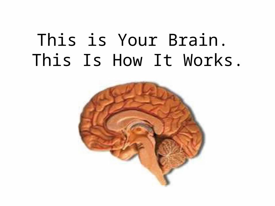
This is Your Brain. This Is How It Works.
•

Parts of the brain: Keep in mind there are two distinct sides with different
functions

The Brainstem(Pathway to the Body)
• Base of brain• Unconscious work• Autonomic functions
(survival)

The Cerebellum(Balance)
• “little brain”• Large in size• 11% of brain’s
weight• Center of balance

The brain has 4 areas called lobes (Predict the functions)
• Frontal
• Parietal
• Temporal
• Occiptal

The Frontal Lobes(Problem Solving)
• Largest part• Move your body• Highly developed• Forms your
personality

Frontal Lobe
• The frontal lobes are responsible for allowing you to think of the past, plan for the future, focus your attention, solve problems, make decisions, and have conversation with others. This region is also responsible for thinking creatively and analytically in a problem-solving mode.

The Parietal Lobes(Touching)
• Two major divisions• Anterior and
posterior• Senses hot and
cold, hard and soft, and pain.
• Taste and smell• Helps integrate the
senses

The Parietal Lobes
• The brain must always know where each part of the body is located and its relation to it’s surroundings. The anterior part (front) is responsible for receiving incoming sensory stimuli. The posterior part (rear) is continuously analyzing to give a person a sense of spatial awareness.

The Temporal Lobes(Hearing)
• Process auditory stimuli
• Subdivisions• Wernicke’s Area
(Speech) (left in circle)
• Broca’s Area (Language) (right of circle)

The Temporal Lobes
• Subdivisions cope with hearing, language, and some aspects to memory. Wernicke’s Area is critical for speech including reading. It allows us to comprehend or interpret speech and to words together correctly so they make sense. Broca’s area is behind the frontal lobes. This area is the center of our speech. It also relates to other language areas such as writing and reading.

The Occipital Lobes (Seeing)
• Located at lower central back of brain
• Processes visual stimuli
• This area gives a person the ability to see and observe.

Taking sides….two sides that is!
• Two sides or hemispheres of the brain: LEFT and RIGHT
• We have two cerebral hemispheres connected by the corpus callosum. This is a bundle of nerves that allows each side of the brain to communicate with each other.
• Each side of the brain processes things differently.
• It is an outdated assumption that “artsy” type people are right-brained.

Taking sides….how the two sides process information that is!
Left Brain
• Logical• Sequential• Rational• Analytical• Objective• Looks at parts
Right Brain
• Random• Intuitive• Holistic• Synthesizing• Subjective• Looks at wholes

Left Hemisphere• processes things more in parts and
sequentially • recognizes positive emotions• Identified with practicality and rationality• Understands symbols and representations• Processes rapid auditory information
faster than the right (crucial for separating the sounds of speech into distinct units for comprehension)
• is responsible for language development. It develops slower in boys, that is why males usually develop more language problems than females.

Right Hemisphere
• Recognizes negative emotions
• High level mathematicians, problem solvers, and chess players use
• The “non-verbal” side
• Responds to touch and music (sensory)
• Intuitive
• Responsive to color and shape
• Emotional and originative

Taking sides….what information the two sides recognize!
Left Brain
• Letters
• Numbers
• Words
Right Brain
• Faces
• Places
• Objects
based on Sousa (1995, p. 88)

Taking sides….take the test!
Hemispheric Dominance Inventory Test
athttp://brain.web-us.com/brain/braindominance.htm
Then learn more at:http://brain.web-us.com/brain/LRBrain.html

Cerebrum -The largest division of the brain. It is divided into two hemispheres, each of which is divided into four lobes.
CerebrumCerebrum
Cerebellum
http://williamcalvin.com/BrainForAllSeasons/img/bonoboLH-humanLH-viaTWD.gif

Cerebral Cortex
Cerebral Cortex
Cerebral Cortex - The outermost layer of gray matter making up the superficial aspect of the cerebrum.
http://www.bioon.com/book/biology/whole/image/1/1-6.tif.jpg

Cerebral Features:
• Sulci – Small grooves dividing the gyri
– Central Sulcus – Divides the Frontal Lobe from the Parietal Lobe
• Fissures – Deep grooves, generally dividing large regions/lobes of the brain
– Longitudinal Fissure – Divides the two Cerebral Hemispheres
– Transverse Fissure – Separates the Cerebrum from the Cerebellum
– Sylvian/Lateral Fissure – Divides the Temporal Lobe from the Frontal and Parietal Lobes
• Gyri – Elevated ridges “winding” around the brain.

Gyri (ridge)
Fissure
(deep groove)
Sulci (groove)
http://williamcalvin.com/BrainForAllSeasons/img/bonoboLH-humanLH-viaTWD.gif

Longitudinal Fissure
Transverse Fissure
Sylvian/Lateral Fissure
Central Sulcus
http://www.bioon.com/book/biology/whole/image/1/1-8.tif.jpg http://www.dalbsoutss.eq.edu.au/Sheepbrains_Me/human_brain.gif
Specific Sulci/Fissures:

Further Investigation
Phineas Gage: Phineas Gage was a railroad worker in the 19th century living in Cavendish, Vermont. One of his jobs was to set off explosive charges in large rock in order to break them into smaller pieces. On one of these instances, the detonation occurred prior to his expectations, resulting in a 42 inch long, 1.2 inch wide, metal rod to be blown right up through his skull and out the top. The rod entered his skull below his left cheek bone and exited after passing through the anterior frontal lobe of his brain.
Frontal

Remarkably, Gage never lost consciousness, or quickly regained it (there is still some debate), suffered little to no pain, and was awake and alert when he reached a doctor approximately 45 minutes later. He had a normal pulse and normal vision, and following a short period of rest, returned to work several days later. However, he was not unaffected by this accident.
Learn more about Phineas Gage: http://en.wikipedia.org/wiki/Phineas_GageFrontal
http://www.sruweb.com/~walsh/gage5.jpg

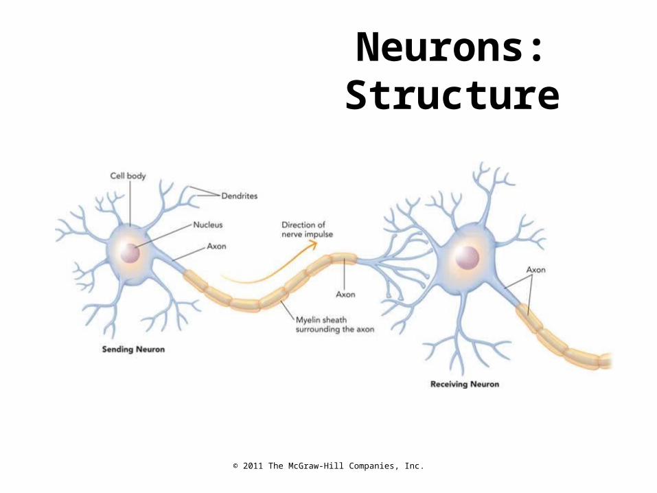
NeuronsThree main parts:
– Dendrites– Receive messages
from other neurons – Cell body
– Contains the genetic information determining cell function
– Axons– Conducts electrical
impulses
© 2011 The McGraw-Hill Companies, Inc.
Neurons: Structure

© 2011 The McGraw-Hill Companies, Inc.
Synapses and Neurotransmitters

Synapse
• The synapse is the junction between an axon terminal and an adjacent dendrite or cell body.
• Neurotransmitter (NT) molecules are released from the axon terminal into the synapse when the action potential arrives at the axon terminal.
© 2004 John Wiley & Sons, Inc.Huffman: PSYCHOLOGY IN ACTION, 7E

© 2011 The McGraw-Hill Companies, Inc.
Neurotransmitters
Neurotransmitters carry information
across the synaptic gap to next neuron.

© 2011 The McGraw-Hill Companies, Inc.
Neurotransmitters
Glutamate– excitatory – learning & memory– involved in many psychological disorders
Norepinephrine– stress and mania: ↑ norepinephrine levels– depression: ↓ norepinephrine levels– regulates sleep states in conjunction with ACh

© 2011 The McGraw-Hill Companies, Inc.
Neurotransmitters
Dopamine
– voluntary movement– reward anticipation– stimulant drugs: activate dopamine
receptors– Parkinson’s disease: ↓ dopamine levels– schizophrenia: ↑ dopamine levels

© 2011 The McGraw-Hill Companies, Inc.
Neurotransmitters
Serotonin– regulation of sleep, mood, attention, learning– depression: ↓ serotonin levels– prozac: ↑ serotonin levels
Endorphins– natural opiates– mediate feelings of pleasure and pain

Glial Cells
Surround neurons and hold them in placeManufacture nutrient chemicals neurons
needAbsorb toxins and waste materials

Nerve Conduction: The Myelin Sheath
Insulation layer covers axons in the brain and spinal cord.
Allows for high-speed conduction.
Multiple sclerosis occurs when immune system attacks the sheath

The Nervous System
Three types of neurons:– Sensory: Carry input messages from the
sense organs to the spinal cord and brain– Motor: Transmit impulses from the brain and
spinal cord to the muscles and organs– Interneurons: Perform connective or
associative functions in the nervous system

The Nervous System
Central Nervous System (CNS)– Brain and Spinal Cord
Peripheral Nervous System (PNS)– Connects the CNS with the muscles, glands,
and sensory receptors

The Peripheral Nervous System
Subdivided into:– Somatic nervous system: Consists of sensory and
motor neurons that bind together to create nerves to transmit messages to sensory receptors
– Autonomic nervous system: Controls glands and smooth muscles in bodily organs
• Sympathetic nervous system: arouses the body• Parasympathetic nervous system: slows down
body processes

Brain Structures: Thalamus and Hypothalamus
Thalamus: Routes sensory information to higher brain structures
Hypothalamus:– Major role in motivation
and emotions– Connects with the
endocrine system– Involved in pain/pleasure
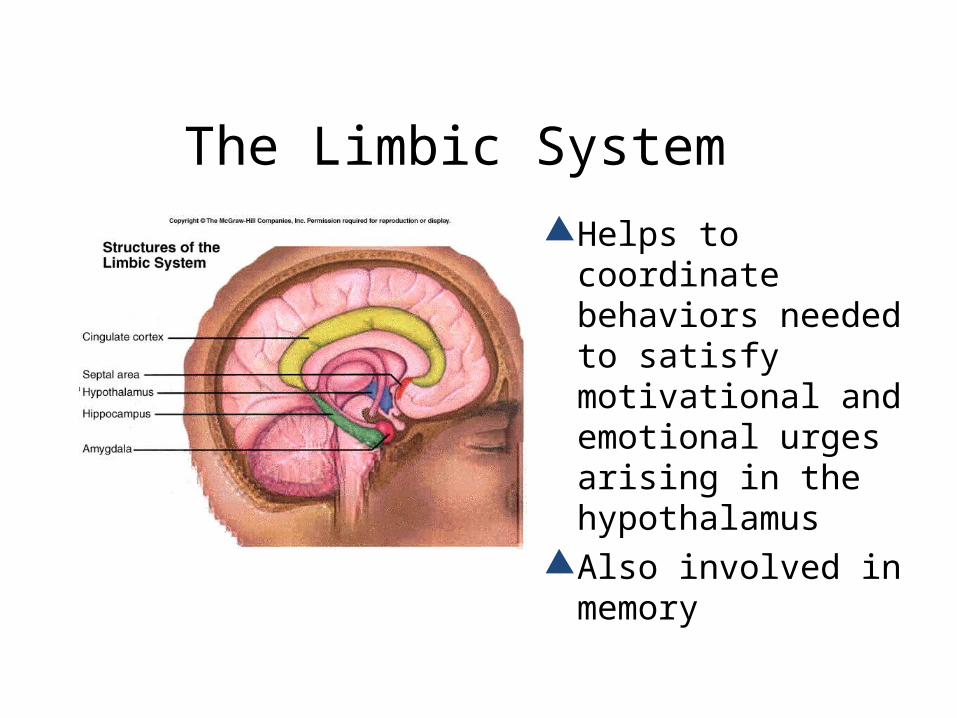
The Limbic System
Helps to coordinate behaviors needed to satisfy motivational and emotional urges arising in the hypothalamus
Also involved in memory

Studying the Brain: Brain Imaging
CT Scans: Beam of X-rays takes pictures of narrow slices of the brain
PET Scans: Measures brain activity, including metabolism, blood flow, and neurotransmitter activity
MRI: Used to study brain structure and activity– FMRI allows for studying brain function as
people perform various tasks

Genetic InfluencesTwin Studies
Compare:
– Monozygotic (MZ) twins are genetically identical
– Dizygotic (DZ) twins share 50% of genetic endowment
Adoption Studies: Twins separated at birth
– Compare twin with both adoptive and biological parent
– Helps determine heritability of traits

The Endocrine System

Endocrine System• One of the body’s
two communication systems
• A set of glands that produce hormones—chemical messengers that circulate in the blood

Hormone• Chemical messengers produced by the
endocrine glands and circulated in the blood
• Similar to neurotransmitters in that they are also messengers
• Slower communication system, but with longer lasting effects

Pituitary Gland• The endocrine system’s gland that, in
conjunction with the brain, controls the other endocrine glands
• Called the “master gland”
• Located at the base of the brain and connects to the hypothalamus
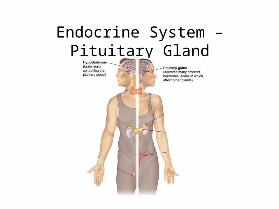
Endocrine System – Pituitary Gland
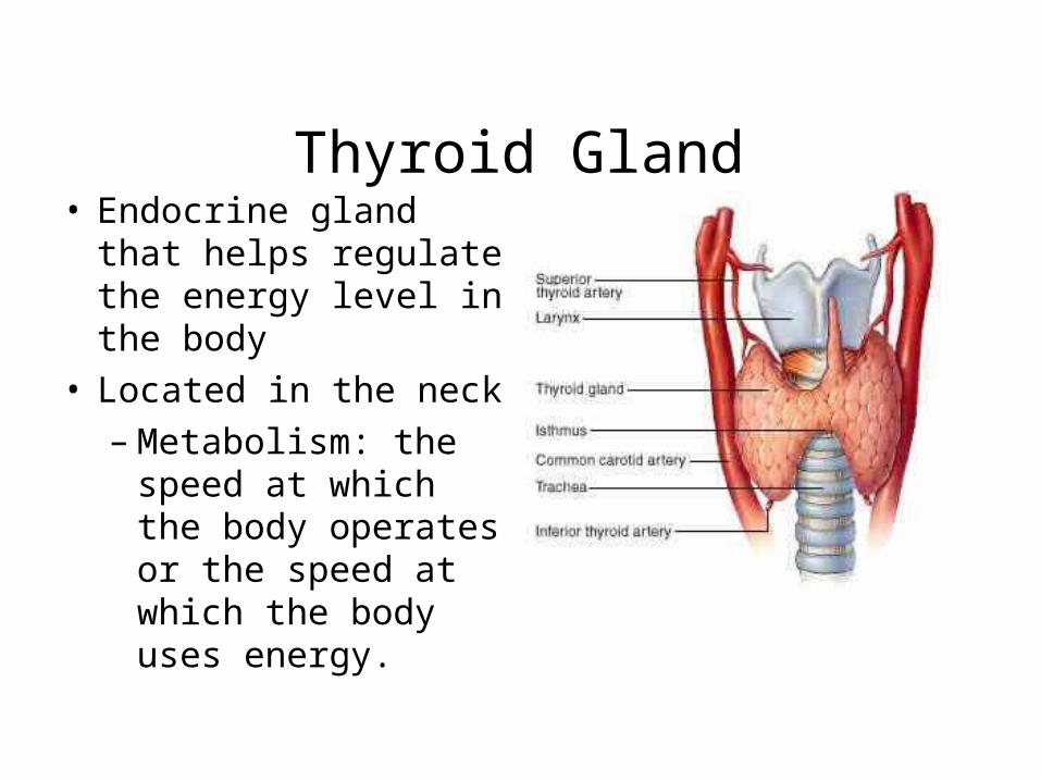
Thyroid Gland• Endocrine gland that
helps regulate the energy level in the body
• Located in the neck
– Metabolism: the speed at which the body operates or the speed at which the body uses energy.

Adrenal Gland• Endocrine glands that help to arouse the body
in times of stress• Located just above the kidneys• Release epinephrine (adrenaline) and
norepinephrine (noradrenaline)– Adrenaline: chemicals that prepares the
body for emergency activity by increasing blood pressure, breathing rat, and energy level

Endocrine System – Adrenal Gland

Pancreatic Gland• Regulates the
level of blood sugar in the blood

Sex Glands (Gonads)
• Ovaries (females) and testes (males) are the glands that influence emotion and physical development.
• Testosterone – primary males hormone• Estrogen – primary female hormone• Males and females have both estrogen
and testosterone in their systems.

Endocrine System – Sex Glands

Sensation and
Perception

Sensation a process by which our sensory receptors
and nervous system receive and represent stimulus energy
Perception a process of organizing and interpreting
sensory information, enabling us to recognize meaningful objects and events

Sensation Our sensory and perceptual processes work
together to help us sort out complex processes

Sensation- Thresholds Absolute Threshold
The level of sensory stimulation necessary for sensation to occur.
minimum stimulation needed to detect a particular stimulus 50% of the time
Difference Threshold minimum difference between two stimuli
required for detection 50% of the time just noticeable difference (JND)

Sensation- Thresholds
Subliminal When stimuli
are below one’s absolute threshold for conscious awareness
0
25
50
75
100
Low Absolutethreshold
Medium
Intensity of stimulus
Percentageof correctdetections
Subliminal stimuli

Sensation- Thresholds
Weber’s Law- to perceive as different, two stimuli must differ by a constant minimum percentage light intensity- 8% weight- 2% tone frequency- 0.3%
Sensory adaptation- diminished sensitivity as a consequence of constant stimulation

Vision- Stabilized Images on the Retina

Light
• White light: light as it originates from the sun or a bulb before in is broken into different frequencies.

Vision Transduction
conversion of one form of energy to another
in sensation, transforming of stimulus energies into neural impulses
Wavelength the distance from the peak of one wave to
the peak of the next

Vision Hue
dimension of color determined by wavelength of light
Intensity amount of energy in a wave determined
by amplitude brightness loudness
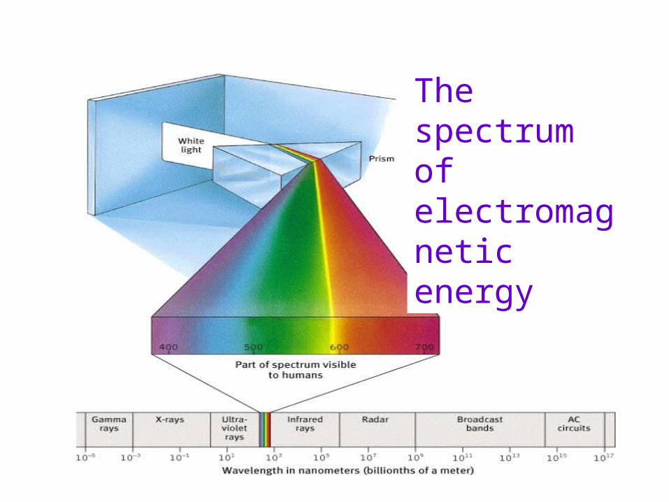
The spectrum of electromagnetic energy

Vision- Physical Properties of Waves
Short wavelength=high frequency(bluish colors, high-pitched sounds)
Long wavelength=low frequency(reddish colors, low-pitched sounds)
Great amplitude(bright colors, loud sounds)
Small amplitude(dull colors, soft sounds)

Vision Pupil- adjustable opening in the center of the eye Iris- a ring of muscle that forms the colored portion
of the eye around the pupil and controls the size of the pupil opening
Lens- transparent structure behind pupil that changes shape to focus images on the retina

Vision

Vision Accommodation- the process by which the
eye’s lens changes shape to help focus near or far objects on the retina
Retina- the light-sensitive inner surface of the eye, containing receptor rods and cones plus layers of neurons that begin the processing of visual information

Retina’s Reaction to Light- Receptors
Rods peripheral retina detect black, white and gray twilight or low light
Cones near center of retina fine detail and color vision daylight or well-lit conditions

Retina’s Reaction to Light
Optic nerve- nerve that carries neural impulses from the eye to the brain
Blind Spot- point at which the optic nerve leaves the eye, creating a “blind spot” because there are no receptor cells located there
Fovea- central point in the retina, around which the eye’s cones cluster


Pathways from the Eyes to the Visual Cortex


Color-Deficient Vision
People who suffer red-green blindness have trouble perceiving the number within the design

Opponent Process- Afterimage Effect
Image that remains after stimulation of the retina has ended. Cones not used fire to bring the visual systems back in balance

Audition (Hearing) Audition
the sense of hearing Frequency
the number of complete wavelengths that pass a point in a given time
Pitch a tone’s highness or lowness depends on frequency

Audition (Hearing) Timbre
The complexity of sound Intensity
How loud a sound is Decibels
a measure of how loud the sound is

The Intensity of Some Common Sounds


Audition- The Ear Middle Ear
chamber between eardrum and cochlea containing three tiny bones (hammer, anvil, stirrup) that concentrate the vibrations of the eardrum on the cochlea’s oval window
Inner Ear innermost part of the ear, contining the cochlea,
semicurcular canals, and vestibular sacs Cochlea
coiled, bony, fluid-filled tube in the inner ear through which

Touch
Skin Sensations pressure
only skin sensation with identifiable receptors
warmth cold pain

Taste Taste Sensations
sweet sour salty bitter
Sensory Interaction the principle that one sense may influence
another as when the smell of food influences its taste

Smell (Olfaction)
Receptor cells inolfactory membrane
Nasal passage
Olfactorybulb
Olfactorynerve

Perception
z\\\\\\

Perception
– The set of processes by which we recognize, organize, and make sense of the sensations we receive from environmental stimuli
• Percept– Complex mental representation integrating
particular sensational aspects of a figure

Perceptual experience involves four elements:
– Distal (far) stimulus• The object in the external world
– Informational medium• Reflected light, sound waves, chemical molecules, or
tactile information coming from the environment– Proximal (near) stimulus
• Representation of the distal stimulus in sensory receptors (e.g. picture on the retina)
– Perceptual object• Mental representation of the distal stimulus

Perceptual Constancies

Size constancy
– The perception that an object maintains the same size despite changes in the size of the proximal stimulation
• The same object at two different distances projects different-sized images on the retina
• Size constancy can be used to elicit illusions (e.g. Ponzo illusion or Müller-Lyer Illusion)

Color Constancy
• The ability to perceive an object as the same color regardless of the environment

Brightness Constancy
• The ability to keep an objects brightness constant as the object is moved to various enviroments.

Shape constancy
– The perception that an object maintains the same shape despite changes in the size of the proximal stimulus
• Involves the perceived distance of different parts of the object from the observer

Space Constancy
• The ability to keep objects in the environment steady by perceiving either ourselves or outside objects as moving

Depth Perception
• Importance of depth perception– When you drive, you use depth to assess the
distance of an approaching automobile– When you decide to call out to a friend walking
down the street, you determine how loudly to call, based on how far away you perceive your friend to be

Depth Cues• Eleanor Gibson and her
Visual Cliff Experiment.• If you are old enough to
crawl, you are old enough to see depth perception.
• We see depth by using two cues that researchers have put in two categories:
• Monocular Cues• Binocular Cues

Perceptual Organization• When we are given a cluster of sensations, we
organize them into a “gestalt” or a “whole”
• “The whole is greater than the sum of the parts.”
• We take in sensory information and infer a perception that makes sense to us based on our past experiences.

Proximity• A perceptual cue that involves grouping
together things that are near each other • Our mind has
“Rules” for Grouping

Similarity
• A perceptual cue that involves grouping like things
together

Closure
• The process of filling in the missing details of what is viewed

Illusions
• Inaccurate perceptions

Müller-Lyer Lines• Eye-movement theory: Arrowheads
influence extent of eye movements• Equal lines however one looks longer
then the other.

Looks like President Clinton and Vice President Gore, right?Wrong... It's Clinton's face twice, with two different haircuts.
What do you see?

There's a face... and the word liar
What do
What do you see?

No, they're both the same size
Is the left center circle bigger?

It's a spiral, right?
No, these are a bunch of independent circles

Keep staring at the black dot. After a whilethe gray haze around it will appear to shrink.

Can you find the dog?

Stare at the black lightbulb for at least 30 seconds.Then immediately stare at a white area on the screenor at a sheet of paper. You should see a glowing light bulb!

Do you see a couple or a skull?
Sleep

Rhythms of Waking and Sleep
• All animals produce endogenous circadian rhythms, internal mechanisms that operate on an approximately 24 hour cycle.– Regulates the sleep/ wake cycle.– Also regulates the frequency of eating and
drinking, body temperature, secretion of hormones, volume of urination, and sensitivity to drugs.

Fig. 9-2, p. 267

Circadian rhythms:• Remains consistent despite lack of
environmental cues indicating the time of day
• Can differ between people and lead to different patterns of wakefulness and alertness.
• Change as a function of age.– Example: sleep patterns from childhood to late
adulthood.

Rhythms of Waking and Sleep
• Human circadian clock generates a rhythm slightly longer than 24 hours when it has no external cue to set it.
• Most people can adjust to 23- or 25- hour day but not to a 22- or 28- hour day.
• Bright light late in the day can lengthen the circadian rhythm.

Stages of Sleep And Brain Mechanisms
• Rapid eye movement sleep (REM) are periods characterized by rapid eye movements during sleep.
• Also known as “paradoxical sleep” because it is deep sleep in some ways, but light sleep in other ways.
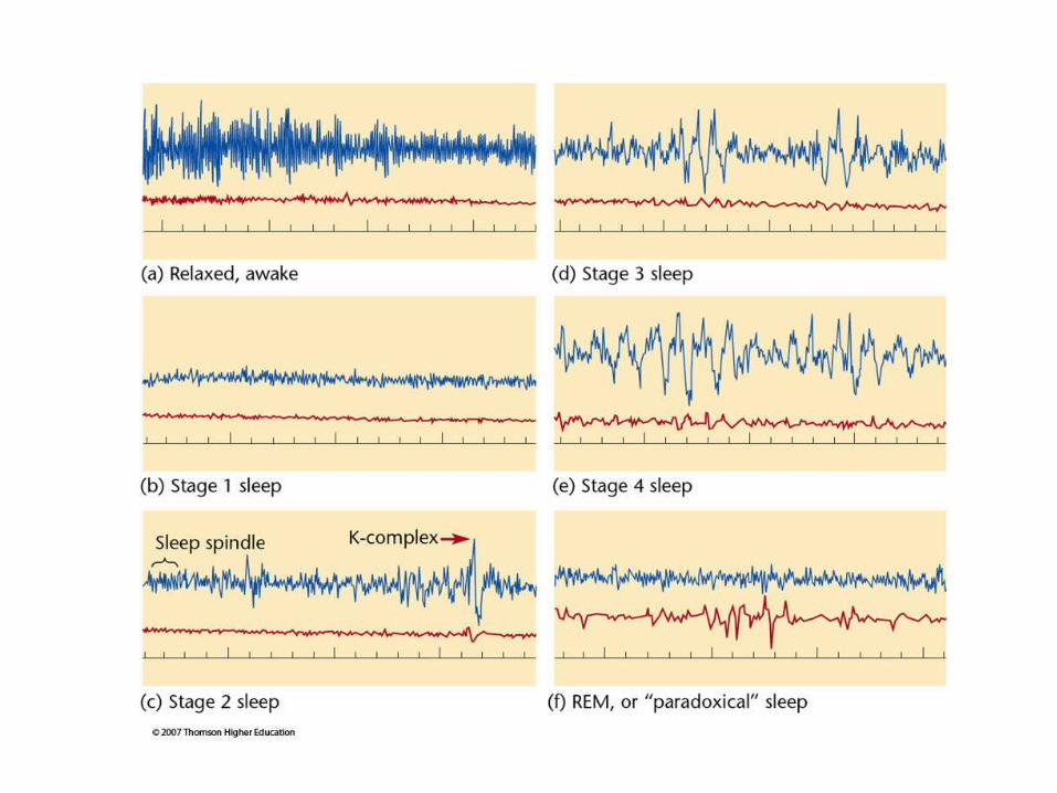

Stages of Sleep And Brain Mechanisms
• Stages other than REM are referred to as non-REM sleep (NREM).
• When one falls asleep, they progress through stages 1, 2, 3, and 4 in sequential order.
• After about an hour, the person begins to cycle back through the stages from stage 4 to stages 3 and 2 and than REM.
• The sequence repeats with each cycle lasting approximately 90 minutes.

Stages of Sleep And Brain Mechanisms
• Stage 3 and 4 sleep predominate early in the night. – The length of stages 3 and 4 decrease as the night
progresses. • REM sleep is predominant later in the night.
– Length of the REM stages increases as the night progresses.
• REM is strongly associated with dreaming, but people also report dreaming in other stages of sleep.
Stages of Sleep And Brain Mechanisms
• During REM sleep: – Activity increases in the pons (triggers the
onset of REM sleep), limbic system, parietal cortex and temporal cortex.
– Activity decreases in the primary visual cortex, the motor cortex, and the dorsolateral prefrontal cortex.

Insomnia• is a sleep disorder associated with inability
to fall asleep or stay asleep.– Results in inadequate sleep.– Caused by a number of factors including noise,
stress, pain medication.– Can also be the result of disorders such as
epilepsy, Parkinson’s disease, depression, anxiety or other psychiatric conditions.
– Dependence on sleeping pills and shifts in the circadian rhythms can also result in insomnia.

Sleep apnea• is a sleep disorder characterized by the
inability to breathe while sleeping for a prolonged period of time.
• Consequences include sleepiness during the day, impaired attention, depression, and sometimes heart problems.
• Cognitive impairment can result from loss of neurons due to insufficient oxygen levels.
• Causes include, genetics, hormones, old age, and deterioration of the brain mechanisms that control breathing and obesity.

Narcolepsy• is a sleep disorder characterized by
frequent periods of sleepiness.• Four main symptoms include:
– Gradual or sudden attack of sleepiness.– Occasional cataplexy - muscle weakness
triggered by strong emotions.– Sleep paralysis- inability to move while asleep
or waking up.– Hypnagogic hallucinations- dreamlike
experiences the person has difficulty distinguishing from reality.

Periodic Limb Movement Disorder
• is the repeated involuntary movement of the legs and arms while sleeping.– Legs kick once every 20 to 30 seconds for
periods of minutes to hours.– Usually occurs during NREM sleep.

REM Behavior Disorder• is associated with vigorous movement
during REM sleep.– Usually associated with acting out dreams.– Occurs mostly in the elderly and in older men
with brain diseases such as Parkinson’s.– Associated with damage to the pons (inhibit
the spinal neurons that control large muscle movements).

• “Night terrors” are experiences of intense anxiety from which a person awakens screaming in terror.– Usually occurs in NREM sleep.
• “Sleep talking” occurs during both REM and NREM sleep.
• “Sleepwalking” runs in families, mostly occurs in young children, and occurs mostly in stage 3 or 4 sleep.

Why Sleep? Why REM? Why Dreams?
• The original function of sleep was to probably conserve energy.
• Conservation of energy is accomplished via:– Decrease in body temperature of about 1-2
Celsius degrees in mammals.– Decrease in muscle activity.

Why Sleep? Why REM? Why Dreams?
• Animals also increase their sleep time during food shortages.– sleep is analogous to the hibernation of
animals.• Animals sleep habits and are influenced by
particular aspects of their life including:– how many hours they spend each day devoted
to looking for food.– Safety from predators while they sleep
• Examples: Sleep patterns of dolphins, migratory birds, and swifts.

Why Sleep? Why REM? Why Dreams?
• Sleep enables restorative processes in the brain to occur.– Proteins are rebuilt.– Energy supplies are replenished.
• Moderate sleep deprivation results in impaired concentration, irritability, hallucinations, tremors, unpleasent mood, and decreased responses of the immune system.

Why Sleep? Why REM? Why Dreams?
• People vary in their need for sleep.– Most sleep about 8 hours.
• Prolonged sleep deprivation in laboratory animals results in:– Increased metabolic rate, appetite and body
temperature.– Immune system failure and decrease in brain
activity.

Why Sleep? Why REM? Why Dreams?
• Humans spend one-third of their life asleep.• One-fifth of sleep time is spent in REM.• Species vary in amount of sleep time spent in
REM.– Percentage of REM sleep is positively correlated
with the total amount of sleep in most animals.
• Among humans, those who get the most sleep have the highest percentage of REM.

Fig. 9-18, p. 289

Why Sleep? Why REM? Why Dreams?
• REM deprivation results in the following:– Increased attempts of the brain/ body for REM
sleep throughout the night.– Increased time spent in REM when no longer
REM deprived.• Subjects deprived of REM for 4 to 7 nights
increased REM by 50% when no longer REM deprived.