this is the peer reviewed version of the following article ... et al... · "this is the peer...
TRANSCRIPT

"This is the peer reviewed version of the following article: [Nielsen DA, Pernice M, Schliep M, Sablok G,Jeffries TC, Kühl M, Wangpraseurt D, Ralph PJ, Larkum AW Environmental microbiology Jul 2015], which has been published in final form at [http://dx.doi.org/10.1111/1462-2920.12983]. This article may be used for non-commercial purposes in accordance with Wiley Terms and Conditions for Self-Archiving."
1
Microenvironment and Phylogenetic Diversity of Prochloron Inhabiting the Surface of 1
Crustose Didemnid Ascidians 2
3
Daniel Aagren Nielsen1*, Mathieu Pernice1, Martin Schliep1, Gaurav Sablok1, Thomas 4
Jeffries1, Michael Kühl1,2, Peter J. Ralph1 and Anthony W.D. Larkum1 5
6
1Plant Functional Biology and Climate Change Cluster, University of Technology Sydney, 7
Ultimo, New South Wales 2007, Australia 8
2Marine Biology Section, Department of Biology, University of Copenhagen, 9
Strandpromenaden 5, DK-3000 Helsingør, Denmark 10
11
12
*Corresponding author, e-mail: [email protected], Phone: +61 02 9514 8406 13
14
Running title: Prochloron microenvironment 15
16
Keywords: Surface Prochloron, didemnid ascidian, microenvironment, phylogeny, 17
microsensor 18
19
20
2
ABSTRACT 21
The cyanobacterium Prochloron didemni is primarily found in symbiotic relationships with 22
various marine hosts such as ascidians and sponges. Prochloron remains to be successfully 23
cultivated outside of its host, which reflects a lack of knowledge of its unique 24
ecophysiological requirements. We investigated the microenvironment and diversity of 25
Prochloron inhabiting the upper, exposed surface of didemnid ascidians, providing the first 26
insights into this microhabitat. The pH and O2 concentration in this Prochloron biofilm 27
changes dynamically with irradiance, where photosynthetic activity measurements showed 28
low light adaptation (Ek ∼80±7 µmol photons m-2 s-1) but high light tolerance. Surface 29
Prochloron cells exhibited a different fine structure to Prochloron cells from cloacal cavities 30
in other ascidians, the principle difference being a central area of many vacuoles dissected by 31
single thylakoids in the surface Prochloron. Cyanobacterial 16S rDNA pyro-sequencing of 32
the biofilm community on four ascidians resulted in 433 operational taxonomic units (OTUs) 33
where and average of ~85% (65-99%) of all sequence reads, represented by 136 OTUs, were 34
identified as Prochloron via BLAST search. All of the major Prochloron-OTUs clustered 35
into independent, highly supported phylotypes separate from sequences reported for internal 36
Prochloron, suggesting a hitherto unexplored genetic variability among Prochloron 37
colonizing the outer surface of didemnids. 38
39

3
INTRODUCTION 40
Prochloron didemni (Lewin, 1977) is a large (7-25 μm in diameter), spherical 41
cyanobacterium (Cox, 1986) commonly found as an obligate extracellular symbiont 42
associated with mainly didemnid ascidians (Lewin and Cheng, 1975; Newcomb and Pugh, 43
1975), but also holothurians and sponges (Cheng and Lewin, 1984; Parry, 1986). The 44
photosystems of Prochloron are typical of a cyanobacterium (Hiller and Larkum, 1985; 45
Christen et al., 1999); however, its major pigment protein complex that binds both Chl a and 46
b (Hiller and Larkum, 1985) belongs to a special group shared with Prochlorococcus, 47
Prochlorothix and the Chl d-containing cyanobacterium, Acaryochloris marina (Chen et al., 48
2005). Prochloron is typically located inside the cloacal compartment of didemnid ascidians, 49
but may also occur in other parts such as on the outer surface of asymbiotic ascidians (e.g. 50
Didemnum candidum) (Cox, 1986; Lewin and Cheng, 1989) and even intra-cellularly (Hirose 51
et al., 1996; Hirose, 2014). Prochloron from the external and internal parts of ascidians have 52
been shown to exhibit significantly different morphological characteristics (Cox, 1986), 53
leading to the suggestion that at least two and potentially more species of Prochloron exist. 54
However, phylogenetic studies based on sequencing of the 16S rRNA gene have so far 55
indicated that Prochloron are conspecific, with Prochloron didemni being the only species in 56
the Prochloron genus (Stackebrandt et al., 1982; Stam et al., 1985; Holton et al., 1990; 57
Münchhoff et al., 2007; Donia et al., 2011b). 58
The symbiotic relationship between Prochloron and its host is still poorly understood. 59
None of the ascidians with external Prochloron are known to harbour morphological 60
adaptations to accommodate the symbionts, and often these ascidians can be found entirely 61
without Prochloron cover, suggesting that this particular association is non-obligatory to the 62
host (McCourt et al., 1984; Cox, 1986). In other cases, the photosynthetic activity of 63
Prochloron has been shown to enhance host respiration (Pardy, 1984; Koike et al., 1993) and 64

4
growth (Olson, 1986), with Prochloron photosynthates contributing up to ~60% of the hosts’ 65
carbon demand (Alberte et al., 1987). The disjunct positioning of the symbionts in the hosts 66
cloacal cavity, in the test or on the outer test surface, seems counter intuitive to their function 67
in providing a source of carbon, and suggests the presence of an as yet undiscovered nutrient 68
uptake mechanism in the ascidian, facilitating translocation of low molecular weight 69
compounds from the symbionts into the host (Pardy and Lewin, 1981; Griffiths and Thinh, 70
1983; Hirose and Maruyama, 2004). 71
There have been many attempts to unravel the physiology and symbiotic nature of 72
Prochloron (Lewin and Cheng, 1989; Kühl and Larkum, 2002) (and references therein) but 73
progress has been limited as Prochloron has never been successfully cultivated outside its 74
host, and only a single non-confirmed report of short-term culture success with the use of the 75
amino acid tryptophan has been published (Patterson and Withers, 1982). The genome of 76
Prochloron didemni, extracted from the cloacal cavity of the ascidian Lissoclinum patella, 77
was recently sequenced, revealing a complex set of primary metabolic genes, gene encoding 78
for secondary metabolites and absence of genome reduction (Donia et al., 2011b). This 79
suggests that Prochloron didemni may be able to thrive independently of the ascidian host 80
and that this putative symbiotic interaction might in fact not be obligatory for the symbiont, 81
although reports of free-living Prochloron are scarce (Cox, 1986; Münchhoff et al., 2007). 82
A recent study of the in-hospite microenvironment of L. patella (Kühl et al., 2012) 83
showed an extremely dynamic system with steep spatial and temporal O2 and pH gradients 84
resulting from photosynthetic activity of the symbiont, as modulated by ambient irradiance 85
levels, and a high holobiont respiration rate. Prochloron therefore seems to thrive in 86
environments with strong diurnal fluctuations of the chemical environment, much akin to 87
other cyanobacteria found in highly productive biofilm ecosystems (Kühl et al., 1996). 88
Knowledge of the phylogenetic diversity of Prochloron and the environmental conditions of 89
5
its microhabitat may provide fundamental new insight into its putative symbiotic interaction 90
with its ascidian hosts. In this respect, the primary outstanding questions regarding ascidian-91
Prochloron symbioses are: (i) whether the different locations inside or on the ascidians host 92
tissue result in different microenvironmental conditions surrounding the Prochloron, and (ii) 93
to what extent the Prochloron associated with these different tissue specific locations are 94
different. Here we present the first study describing the microhabitat and phylogeny of 95
Prochloron located on the outer surface of didemnid ascidians. 96
97
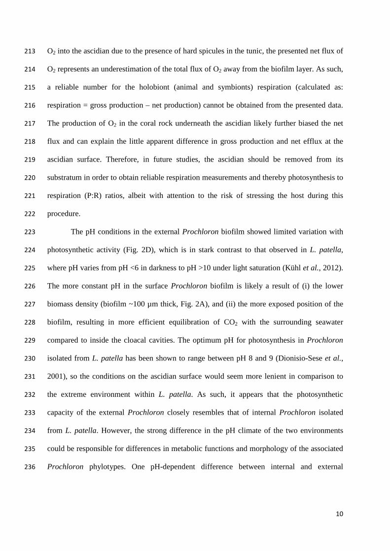
RESULTS 98
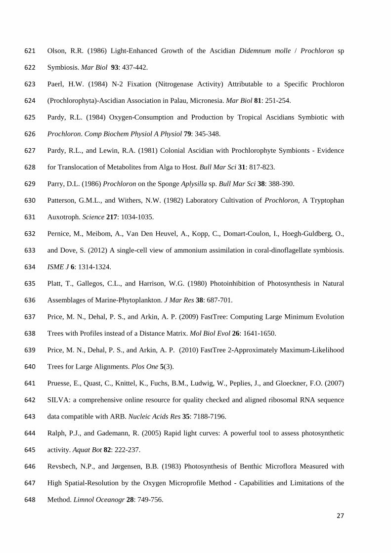
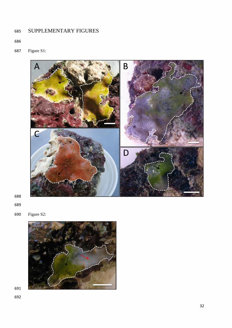
Microscopy imaging: Green Prochloron biofilms were observed on a fraction of the 99
ascidians found in this study, and in most cases the biofilm only covered part of the test 100
surface (Fig. S1). Prochloron biofilms were not observed inside or below the investigated 101
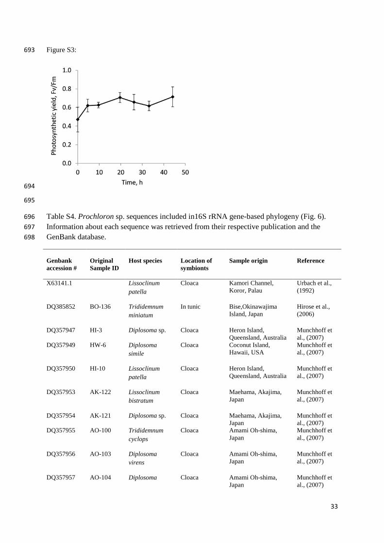
ascidians (data not shown). One ascidian sample was examined in further detail (Fig. S2): 102
The Prochloron biofilm patch was homogenous and only a few cell layers thick (Fig. 1A). 103
Microscopy investigations of the biofilm revealed only cell morphotypes resembling that of 104
Prochloron. Fluorescence imaging of ascidian cross-sections revealed the presence of a 105
transparent exo-polymeric, mucoid layer (m), extruding from the ascidian surface and 106
engulfing the Prochloron cells (Fig. 1B; seen as dark structures in the image on a background 107
of fluorescing resin). At high magnification transmission electron microscopy (TEM), the 108
mucus appeared as a fibrous substance, containing some bacteria-like inclusions (Fig. 1C and 109
F). The relatively small Prochloron cells (7-10 µm in diameter) exhibited a typical peripheral 110
band of loosely stacked thylakoid (t) membranes (Fig. 1D and E), which expanded into 111
numerous small “vacuoles” (v) in the centre of the cell (Fig. 1D). Further observations 112
indicated that up to 10 thylakoid membranes were stacked in peripheral bands, some of them 113

6
running among the many central vacuoles. The surface of all examined Prochloron cells was 114
covered with virus-like structures approximately 100 nm in length (Fig. 1D and E, arrow). 115
116
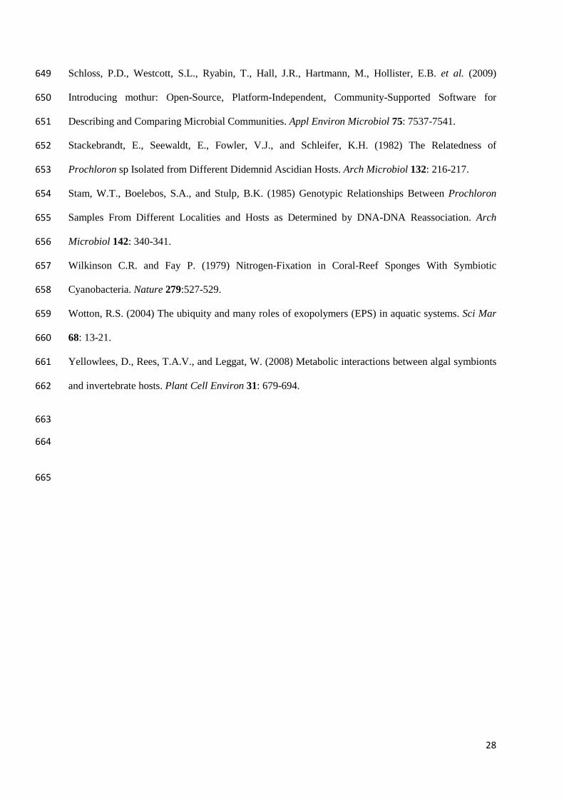
Oxygen and pH dynamics: Oxygen and pH dynamics was investigated on a white ascidian 117
with approximately 50% Prochloron coverage (depicted in figure S2). The O2 concentration 118
at the Prochloron covered surface of the ascidian rapidly increased upon illumination; from 119
hypoxic in the dark to hyper-oxic at higher light levels (Fig. 2A). For each irradiance step, a 120
new steady state O2 concentration and pH was reached within 5 minutes of illumination. An 121
increase in O2 concentration was also observed at Prochloron-free areas although to a lesser 122
extent (Fig. 2A). Profiles of O2 concentration towards the Prochloron-covered surface 123
showed a net efflux of O2 reaching a maximum of 0.15 ± 0.01 nmol O2 cm-2 s-1 at a photon 124
irradiance (PAR, 400-700 nm) of 155 µmol photons m-2 s-1, which remained fairly constant at 125
higher light levels (Fig. 2C). Locally, pH increased up to 8.33 ± 0.02 during illumination and 126
decreased to a minimum of 7.90 ± 0.09 in the dark (Fig. 2D). The Prochloron-free ascidian 127
surfaces consumed O2 from the water column at low light, but exhibited net O2 efflux at high 128
irradiance, reaching a maximum value of 0.06 nmol O2 cm-2 s-1 at 380 µmol photons m-2 s-1 129
(Fig. 2C). In the absence of symbionts, pH decreased towards the surface in both dark and 130
light to 8.00 ± 0.03 and 8.11, respectively (Fig. 2E). Only one pH profile was obtained in the 131
light due to damage to the fragile pH electrode. 132
Gross photosynthesis (Fig. 2B) measured at the Prochloron-covered surface revealed 133
a light utilisation efficiency of α = 0.013 ± 0.002 mol O2 per mol photons, with a rapid 134
increase in O2 production at low light levels and a minimum saturating irradiance (Ek) of ∼80 135
(±7) µmol photons m-2 s-1 according to the model fit. The volume-specific gross O2 136
production rate reached a maximum of 7.6 (±1.3) nmol O2 cm-3 s-1 (model fit: Pmax = 8.1 137
(±0.3) nmol O2 cm-3 s-1), corresponding to an areal gross photosynthesis rate of 0.16 (±0.01) 138

7
nmol O2 cm-2 s-1 at 380 µmol photons m-2 s-1 (assuming that the measured change in O2 139
concentration is averaged over a volume corresponding to a sphere with a diameter of 0.2 140
mm), followed by a slight decrease in photosynthesis at the highest irradiance (800 µmol 141
photons m-2 s-1). 142
143
Cyanobacterial phylogeny: Pyrosequencing of the 16S rDNA gene from biofilm of four 144
individual ascidian samples with surface Prochloron (Fig. S1), yielded a total of 8,285 high 145
quality sequences grouping into 433 operational taxonomic units (OTUs) with >3% raw 146
sequence divergence. Of these, 136 OTUs were most similar to Prochloron sp. sequences. 147
297 OTUs, representing ~15% of all obtained sequences, were of non-Prochloron origin and 148
dominated by nitrogen fixing marine cyanobacteria such as Pleurocapsa sp. and Symploca sp. 149
(Fig. 3). The four biofilm samples grouped two and two with respect to sequence diversity, 150
with the two red ascidians showing much lower sequence diversity (total of 29 and 34 OTUs) 151
and higher percentage Prochloron cover (>98%) than the two gray samples (total of 151 and 152
358 OTUs and >65% Prochloron cover) (Fig. 3). The 12 Prochloron OTUs included in 153
figure 4 represent >92% of all the Prochloron-like sequences obtained and were selected for 154
inclusion based on their read length (>437bp) and their relative abundance (≥25 highly 155
similar sequences per OTU). Phylogenetic mapping of the representative sequence of each of 156
the 12 OTUs supported the presence of separate phylotypes of Prochloron (bootstrap values 157
>80%) (Fig. 4), with all de novo sequences clustering away from all but two previously 158
published sequences (GenBank accession number DQ357967 and DQ385852), which were 159
both isolated from within the tunic of the colonial ascidian Trididemnum miniatum (Hirose et 160
al., 2006; Münchhoff et al., 2007). A strong separation (bootstrap = 100%) was found 161
between sequences representing Prochloron from the cloacal cavity of ascidians and the 162

8
presented surface sequences including the only other published surface Prochloron sequence 163
(JX099360, Hirose et al., 2012). 164
165
DISCUSSION 166
External, surface-associated Prochloron has generally been considered of less importance to 167
their host than their internal counterpart (Cox, 1986), mainly because of the apparent lack of 168
morphological adaptations of the host to support the symbiosis and the lack of vertical 169
transfer of symbionts from parent to progeny in these ascidians (Hirose, 2014). However, the 170
exo-polymeric matrix observed on the surface of the investigated ascidian, clearly extending 171
from the host surface, encapsulated the surface Prochloron cells (Fig. 1B, C) and may 172
represent an important means of attachment; forming a stabilised environment for growth in 173
an otherwise flow-exposed habitat, much in line with the function of exo-polymeric 174
substances in other microbial biofilms (Wotton, 2004). A similar fibrous, exo-polymeric 175
matrix at the host/symbiont interface has also been observed in the more protected 176
environment of the cloacal cavity of L. patella, although at a much smaller scale (Kühl et al., 177
2012). While more ultra-structural work is necessary to confirm the general presence of such 178
a matrix on ascidians with surface-associated Prochloron, this could represent a previously 179
unrecognised morphological adaptation of the ascidian to accommodate Prochloron 180
specifically, and as such may embody an important structural link in the establishment and/or 181
maintenance of this symbiosis. 182
The surface-associated Prochloron responded rapidly to changing light conditions 183
(Fig. 2A, B), and exhibited efficient photosynthesis at low irradiance levels with saturation at 184
~200 µmol photons m-2 s-1 (Fig. 2B). This is in agreement with the conditions under which 185
the host ascidian is typically found, i.e. on the underside of stable coral rubble, where only 186
diffuse, reflected light may be available for photosynthesis (Brakel, 1979; Kühl et al., 2007). 187
9
A similar photosynthetic response was found previously with Prochloron located in the 188
cloaca of the didemnids Diplosoma virens (Kühl et al., 2005) and L. patella (Alberte et al., 189
1986; Kühl et al., 2012). While D. virens inhabits low light, cryptic reef habitats, L. patella is 190
commonly found exposed to full sunlight. However, the spiculous tunic of L. patella has been 191
shown to strongly attenuate the light reaching the cloacal cavity, so that the symbionts 192
residing there mostly experience relatively low light conditions (Kühl et al., 2012). The 193
present study indicates some level of photoinhibition at 800 µmol photons m-2 s-1 (Fig. 2B) 194
which is comparable to what has been observed previously for Prochloron cells extracted 195
from the cloacal cavity of low-light acclimated L. patella colonies (Alberte et al., 1986), 196
although Kühl et al. (2012) observed no such inhibition below 1000 µmol photons m-2 s-1. 197
Thus, despite being adapted to low light habitats, the Prochloron cells are able to cope with 198
much higher light levels than normally encountered and can adapt to full sunlight exposure 199
(Alberte et al., 1986). This flexibility could be of benefit during a free living stage with 200
exposure to more variable irradiance levels. 201
While tempting to suggest, production of O2 by the surface Prochloron biofilm is 202
unlikely to be a major driver in the symbiosis. Prochloron is the only phototroph reported to 203
form coherent biofilm patches on ascidians, and didemnid ascidians are the only colonial 204
ascidians on which these patches occur; and even then the occurrence of patches is sporadic. 205
Ascidian zooids are efficient filter feeders and can presumably ventilate their immediate 206
microenvironment by advective O2 transport. The net O2 efflux detected on areas of the 207
ascidians with no Prochloron cover (Fig. 2E), was found to be a result of O2 production by 208
the substratum below the thin (~1.5 mm) ascidian colony. This was evident from the 209
cessation of O2 evolution when the ascidian was removed from its substratum, as well as 210
from a high O2 production by algae in the substratum itself (data not shown). The identity of 211
the algae in the coral rock was not investigated. As it was not possible to measure the flux of 212
10
O2 into the ascidian due to the presence of hard spicules in the tunic, the presented net flux of 213
O2 represents an underestimation of the total flux of O2 away from the biofilm layer. As such, 214
a reliable number for the holobiont (animal and symbionts) respiration (calculated as: 215
respiration = gross production – net production) cannot be obtained from the presented data. 216
The production of O2 in the coral rock underneath the ascidian likely further biased the net 217
flux and can explain the little apparent difference in gross production and net efflux at the 218
ascidian surface. Therefore, in future studies, the ascidian should be removed from its 219
substratum in order to obtain reliable respiration measurements and thereby photosynthesis to 220
respiration (P:R) ratios, albeit with attention to the risk of stressing the host during this 221
procedure. 222
The pH conditions in the external Prochloron biofilm showed limited variation with 223
photosynthetic activity (Fig. 2D), which is in stark contrast to that observed in L. patella, 224
where pH varies from pH <6 in darkness to pH >10 under light saturation (Kühl et al., 2012). 225
The more constant pH in the surface Prochloron biofilm is likely a result of (i) the lower 226
biomass density (biofilm ~100 µm thick, Fig. 2A), and (ii) the more exposed position of the 227
biofilm, resulting in more efficient equilibration of CO2 with the surrounding seawater 228
compared to inside the cloacal cavities. The optimum pH for photosynthesis in Prochloron 229
isolated from L. patella has been shown to range between pH 8 and 9 (Dionisio-Sese et al., 230
2001), so the conditions on the ascidian surface would seem more lenient in comparison to 231
the extreme environment within L. patella. As such, it appears that the photosynthetic 232
capacity of the external Prochloron closely resembles that of internal Prochloron isolated 233
from L. patella. However, the strong difference in the pH climate of the two environments 234
could be responsible for differences in metabolic functions and morphology of the associated 235
Prochloron phylotypes. One pH-dependent difference between internal and external 236

11
Prochloron habitat could e.g. be related to the extent of inorganic carbon limitation for 237
photosynthesis under high irradiance but this remains to be further explored. 238
The lack of vertical transfer of symbionts in ascidians with surface associated 239
Prochloron (Hirose, 2014) indicates that colonisation of the ascidian is a result of seeding 240
from the water column (Cox, 1986), which evidently could allow for colonisation by 241
cyanobacteria other than Prochloron. Despite this, in two of the four biofilm samples 242
investigated, only less than 3% of all sequences were related to non-Prochloron 243
cyanobacteria (Fig. 3). In the two other samples, a larger diversity was observed while 244
Prochloron sequences still dominated (65 to 79% of cyanobacterial 16s rDNA sequences). Of 245
sequences belonging to non-Prochloron cyanobacteria, the nitrogen fixing (diazotrophic) 246
Pleurocapsa sp. and Symploca sp. dominated. The presence of diazotrophic organisms has 247
also been observed in the microbial communities of L. patella (Behrendt et al., 2012). 248
Association with nitrogen fixing organisms could be of benefit to the ascidian host in the 249
generally nitrogen limited waters of coral reefs, and similar associations are also known from 250
other organisms living on the reef such as corals (Lema et al., 2012) and sponges (Wilkinson 251
and Fay, 1979). Considering the reported lack of nitrogenase genes in the Prochloron 252
genome (Donia et al., 2011b), the presence of diazotrophs in Prochloron biofilms gives 253
weight to previous reports of nitrogen fixation by Prochloron communities (Paerl, H. W., 254
1984; Kline et al., 1999). The dichotomous community structure observed between the 255
ascidian samples investigated here highlights a new and potentially important study area 256
investigating the host-specificity of cyanobacterial biofilms on crustose ascidians. In a recent 257
study , it was found that the microbial diversity on the surfaces of two colonial ascidians was 258
lower than in the surrounding seawater (da Silva Oliveira et al., 2013), demonstrating 259
selective pressure for or against certain prokaryotes. Similarly, in a survey of the microbial 260
diversity in L. patella, Behrendt et al. (2012) found lower cyanobacterial diversity in the 261

12
Prochloron-inhabited cloaca, with a Shannon index of diversity of just 1.8 ± 0.7 compared to 262
4.4-5.4 reported in marine planktonic habitats (Schloss et al., 2009). Thus, some unidentified 263
mechanisms seem to ensure primary colonisation by Prochloron, emphasising the connection 264
between didemnid ascidians and this particular taxon of cyanobacteria. The frequent 265
formation of bioactive secondary metabolites such as patellamide and other cyanobactins in 266
didemnid-Prochloron associations may play an important role in such allelopathy against 267
other cyanobacteria (Donia et al., 2011b; Donia et al., 2011a). 268
Until now, significant genetic divergence in the Prochloron genus has not been 269
reported even amongst geographically widely separated samples (Münchhoff et al., 2007; 270
Donia et al., 2011b). In present study, one OTU (denovo385, Fig. 4) dominated all four 271
biofilm samples, containing 56-92% of all Prochloron sequences. This phylotype was closely 272
related to Prochloron inhabiting the tunic of the ascidian Trididemnum miniatum (Hirose et 273
al., 2006; Münchhoff et al., 2007). However, a rare biosphere of Prochloron with notable 274
sequence diversity was also detected, all clustering separately from previously reported 275
internal (cloaca) Prochloron sequences obtained from diverse geographical and host origin 276
(see supplementary table S4). Importantly, these results suggest a hitherto undiscovered 277
speciation of the Prochloron genus into several separate phylotypes. This potential speciation 278
of the Prochloron genus might have been hidden till now due to the almost exclusive focus 279
on Prochloron living inside ascidians as opposed to the potentially less specialised externally 280
associated types. The phylogenetic separation of the internal and external Prochloron shown 281
here finally provides a phylogenetic basis for the long known morphological differences 282
between Prochloron from these two environments (Cox, 1986; Hirose et al., 2012): External 283
Prochloron exhibit a reticulate type of vacuolation, i.e., a central region filled with vacuoles 284
from expanded portions of the thylakoids (see figure 1A), which differs from the single large 285
central thylakoid vacuole observed in internal Prochloron (Cox, 1986, see figure 4). 286
13
Interestingly, the intra-tunic dwelling Prochloron (represented by the sequences DQ357967 287
and DQ385852 in figure 4) from the ascidian Trididemnum miniatum (Hirose et al., 2006), 288
which clusters with the surface Prochloron sequences presented in this study, exhibit a 289
reticulate vacuolation similar to that of the surface Prochloron. While the structural 290
differences between external, surface-associated and internal Prochloron provide support for 291
phylogenetic divergence, the only database sequence that is reported to have originated from 292
an external surface-associated Prochloron (JX099360, Hirose et al., 2012) is 293
phylogenetically affiliated with the sequences reported here and the sequences from internal 294
Prochloron (Fig. 4). However, the lower bootstrap values (77-79%) separating this sequence 295
from the sequences obtained in this study suggests that these are more closely related than to 296
the internal Prochloron sequences. 297
The lack of success in cultivating Prochloron has been taken to indicate that these 298
cyanobacteria rely on as yet unidentified compounds excreted from the ascidian (Yellowlees 299
et al., 2008). Specifically, a significant decrease in photosynthesis of internal Prochloron 300
over just a few hours after isolation from the host has led to the suggestion that the carbon 301
uptake mechanism is directly affected (Critchley and Andrews, 1984; Alberte et al., 1987; 302
Christen et al., 1999). Interestingly, however, we found that external Prochloron cells 303
retained their photosynthetic potential, as measured by variable chlorophyll fluorescence, for 304
45 h after isolation with no indication of a decline (see supplementary information Fig. S2). 305
This indicates that external Prochloron may be less dependent on its host than its internal 306
counterpart, which is also in line with their comparatively looser association. 307
In conclusion, we provide the very first insights into the phylogenetic diversity and 308
photosynthetic performance of Prochloron inhabiting the exposed surface of crustose 309
didemnid ascidians. Our results indicate that external surface-associated Prochloron have a 310
similar photosynthetic capacity as reported for Prochloron thriving inside the cloacal cavity 311

14
of other didemnid ascidians, and they can retain their photosynthetic capacity for much 312
longer when isolated form the ascidian surface. However, the microenvironmental conditions 313
experienced by the surface Prochloron differ substantially from that experienced by internal 314
Prochloron, suggesting a significant plasticity in the environmental requirements of these 315
cyanobacteria. The phylogenetic divergence between the surface Prochloron investigated in 316
the present study and previously published sequences from internal Prochloron strongly 317
suggests an evolutionary adaptation to these different environmental conditions and the 318
existence of multiple Prochloron phylotypes or species. Given the fact that Prochloron lives 319
in association with numerous other marine hosts such as holothurians and sponges, this work 320
further emphasizes the need to explore the diversity of Prochloron from an evolutionary 321
point of view in order to better understand where and when this symbiosis evolved. 322
323
EXPERIMENTAL PROCEDURES 324
Sampling and sample conservation: Individuals of encrusting ascidians covered with green 325
patches of Prochloron cells were sampled during low tide at the border of the reef flat and the 326
inner reef crest of the Heron Island lagoon, Great Barrier Reef, Australia (151° 55’ 00 E, 23° 327
26’ 07 S during 2013 and 2014 field trips. The ascidians (Red2013, Red2014, Gray2014a, 328
Gray2014b; see supplementary figure S1) were all found in cryptic habitats on the underside 329
of dead coral rubble patches, which were partly air-exposed during low tide. Coral substrate 330
with adherent specimens of the particular ascidian was transferred to a container with 331
seawater and immediately transported to Heron Island Research Station, where the samples 332
were maintained in shaded, outdoor aquaria under a continuous flow of fresh seawater. Prior 333
to sampling of the Prochloron biofilm for phylogenetic analysis, each individual ascidian was 334
placed in 0.2 µm filtered seawater for at least 1 hour in order to reduce contamination by non-335
associated cells. A soft paint brush was then used to gently brush off the biofilm from the 336

15
surface of the ascidian into fresh 0.2 µm filtered seawater. The collected cells were spun 337
down at 500g for 1 minute and the supernatant replaced with RNAlater™ (Ambion, Inc., 338
US). Fixed cells were stored at 4°C until further analysis. For transmission electron 339
microscopy (TEM), a small piece of ascidian with a Prochloron surface biofilm (~1 cm2) was 340
cut out with a scalpel and transferred to an Eppendorf tube containing 1.5 mL TEM fixative 341
(1X phosphate buffered saline [PBS, pH 7.2], 0.8% paraformaldehyde solution, 2.5% 342
glutaraldehyde, 0.65 M sucrose) for 24 h at 4°C before being transferred to a clean container 343
and washed several times in 1X PBS containing 0.65 M sucrose. 344
345
Microscopy: For bright field imaging, a thin (<1 mm) ascidian cross section was excised 346
with a scalpel, positioned on a microscope slide and covered with a coverslip. Imaging was 347
done on a compound microscope equipped with a camera (Olympus BX51 with DP71 CCD). 348
For TEM, a piece of pre-fixed ascidian (~4 mm2) (see above) was dehydrated step wise in 349
ethanol at increasing concentrations (50%, 70%, 90% and 100%) and embedded in Spürr 350
resin after pre-staining with Osmium as described previously (Pernice et al., 2012). Briefly, 351
resin blocks were cut into 100-120 nm sections using a microtome (Ultracut E Leica 352
Microsystems, Australia) with a diamond knife, mounted onto TEM grids and counterstained 353
with 2% uranyl acetate (10 min) and Reynold’s lead citrate (10 min). Tissue sections were 354
imaged at the Centre for Microscopy and Microanalysis (University of Sydney, Australia) 355
using a transmission electron microscope (JEOL JEM1400, Korea LTD) operated at 80 kV 356
accelerating voltage. In order to visualise faint structures, fluorescence imaging of thin 357
sections (2 µm), prepared as described for TEM but with no counter stain, was carried out on 358
an inverted fluorescence microscope (Eclipse-Ti, Nikon, US), using blue light (FITC filter 359
settings) for excitation of the resin. 360
361

16
Flow cell and microsensor setup: Measurements were conducted on an intact ascidian (3 362
cm2) attached to a pieces of substrate (coral rubble). The ascidian was left on its substrate in 363
order to avoid disturbing the fragile tissue and surface biofilm. The sample was placed in an 364
acrylic flow chamber supplied with seawater (salinity 35 psu) at a flow velocity of 365
approximately 3 cm s-1, and maintained at a constant temperature of 26°C (± 0.5°C) with an 366
aquarium heater (Sonpar Aquarium Equipment, China). Illumination was provided by a fibre 367
optic tungsten-halogen light source (Schott KL 2500, Germany) equipped with a collimating 368
lens. Incident irradiance levels were measured in µmol photons m-2 s-1 (PAR, 400-700 nm) 369
with a miniature scalar irradiance quantum sensor (Walz GmbH, Germany) positioned over a 370
black, non-reflecting surface at a distance from the light source equal to the distance of the 371
sample during measurements. A microsensor (optode or electrode, see details below) was 372
mounted above the flow chamber on a motorised micromanipulator for automated profiling 373
(MMS, Unisense A/S, Denmark). 374
375
O2 and pH measurements: A fast responding O2 optode (tip size 50 µm, 90% response time 376
<0.5 s; Pyroscience GmbH, Germany) was connected to a logger (Firesting, Pyroscience 377
GmbH, Germany) and the O2 signal was monitored and saved on to a PC via the Firesting 378
logger software (v. 2.30). A two-point calibration of the optode was done at atmospheric O2 379
concentration and at zero % O2 in a solution of Na2SO3 (20% w/v) at experimental 380
temperature. For gross photosynthesis measurements and profiling of O2 concentration, the 381
following Firesting software settings were employed: 0.25 seconds per measurement, 10 ms 382
flash time, 100% flash intensity, 200% signal amplification. 383
pH measurements were made with pH glass microelectrodes (50 µm tip size, Unisense A/S, 384
Denmark), connected to a high impedance millivolt meter (Unisense A/S, Denmark) with an 385
external standard 2 mm reference electrode (Ionode LLC, Australia). The pH electrodes were 386

17
calibrated via a linear fit to the millivolt output measured in pH 4, 7 and 10 buffers, resulting 387
in a slope of ~51 mv/pH units, and the signal was logged during measurements (SensorTrace 388
PRO Unisense A/S, Denmark). 389
Before each profile measurement, the microsensor was manually positioned at the 390
surface of the ascidian/Prochloron-layer, as observed through a dissection microscope 391
mounted on an articulating arm (American Scope, United States). This position was set as 392
zero depth and the microsensor was then retracted vertically a set distance to the starting 393
point of the profile. Each of the O2 and pH profiles was commenced 1.0 and 2.0 mm from the 394
ascidian surface, respectively, and performed towards the surface in vertical steps of 100 µm. 395
At each position, the sensor signal was allowed to stabilise for 5 seconds (95% signal 396
response <5s) before logging the O2 concentration. Four replicate profiles were recorded at 397
each light level (down-welling photon irradiance: 19, 46, 83, 155, 380 and 800 µmol photons 398
m-2 s-1) in areas with and without Prochloron biofilm (see figure S1), respectively, . Between 399
each light level, an acclimation period of ~10 minutes was included, which was observed to 400
be sufficient for establishing a steady state O2 concentration and pH after a change in light 401
(see Fig. 2). The net O2 efflux, i.e., the net photosynthesis, was calculated from measured 402
steady-state concentration profiles using Fick’s first law of diffusion with a diffusion 403
coefficient of 2.30 x 10⁻⁵ cm2 s-1 (calculated for an experimental temperature of 26°C and 404
salinity of 35 psu; (Garcia and Gordon, 1992). Rapid light-dark shifts were conducted with 405
the O2 optode tip placed at the surface of the ascidian following the procedure of Revsbech 406
and Jørgensen (1983). 407
In order to take into account any photosynthesis resulting from the pulsed red LED 408
light (wavelength 620 nm) emitted by the optode during the operation, a set of O2 409
measurements were carried out at steady state in the dark with identical optode settings as 410
used for measuring gross photosynthesis. To minimise O2 production from light emitted by 411
18
the optode, the measuring rate was reduced to once every 2 seconds. As a result of the 412
optode’s flashing LED measurement light, O2 was produced in the dark at a rate of 0.8 ± 0.27 413
nmol cm-3 s-1. This would result in a proportional under-estimation of the gross 414
photosynthesis in the light, and therefore this value was added to the measured gross 415
photosynthetic rate of the Prochloron layer at each light level. Curve fitting of experimental 416
P vs. E curves were performed according to Platt et al. (1980) and Ralph and Gademann 417
(2005) and allowed estimation of light utilisation efficiency (alpha), maximum 418
photosynthetic rate (Pmax), and the minimum saturating irradiance (Ek =Pmax/alpha) and. To 419
measure longer term O2 and pH dynamics, the sensor was positioned and left at the surface of 420
the ascidian for the duration of the experiment (45 minutes), while the irradiance level was 421
increased every 5 minutes. 422
423
DNA extraction, sequencing and data analysis: Biofilm material from four individual 424
ascidians was analysed as follows: Prior to DNA extraction, cells were pelleted at 1500 g for 425
5 min after which the DNA was extracted according to the manufacturer’s instructions with 426
the Powersoil DNA isolation kit (MO BIO, USA). The extracted DNA was resuspended in 427
100 µL of sterile water. The DNA was sequenced at Molecular Research LP (Shallowater, 428
Texas, USA) using standard protocols (Dowd et al., 2008). Briefly, the sample was amplified 429
using the cyanobacterial-specific primer pair 357F (CCTACGGGAGGCAGCAG) / 809R 430
(GCTTCGGCACAGCTCGGGTCGATA) (modified from (Jungblut et al., 2005)). A single-431
step 30 cycle PCR using HotStarTaq Plus Master Mix Kit (Qiagen, Valencia, CA) was used 432
under the following conditions: 94oC for 3 minutes, followed by 28 cycles of 94°C for 30 433
seconds; 50°C for 40 seconds and 72oC for 1 minute; after which a final elongation step at 434
72°C for 5 minutes was performed. Following PCR, the amplicon product was purified using 435
Agencourt Ampure beads (Agencourt Bioscience Corporation, MA, USA). The sample was 436

19
sequenced utilizing Roche 454 FLX titanium instruments and reagents and following 437
manufacturer’s guidelines. DNA sequences were processed using the Quantitative Insights 438
Into Microbial Ecology (QIIME) pipeline (Caporaso et al., 2010). Sequences were de-439
multiplexed and reads shorter than 200 bp as well as reads with a quality score <25 or 440
containing homopolymers exceeding 6 bp were discarded. ACACIA (Bragg et al., 2012) was 441
used to de-noise and error correct the dataset. Following chimera removal using UCHIME 442
(Edgar et al., 2011), Operational Taxonomic Units (OTUs) were defined at 97% sequence 443
identity using UCLUST (Edgar, 2010) and assigned taxonomy against the SILVA database 444
(version 111), (Pruesse et al., 2007) using BLAST (Altschul et al., 1990). 445
Phylogenetic analysis: DNA sequences were aligned using the multiple sequence alignment 446
program MAFFT (Katoh et al., 2002) employing the L-INS-I algorithm. In following 447
assessment regions with gaps were eliminated, and alignments were refined using trimAL 448
(Capella-Gutierrez et al., 2009). After re-assessment of alignments, sequences were trimmed 449
to same sequence length (437bp) length and phylogenetic trees were constructed using 450
Maximum Likelihood (ML) and the Generalised Time-Reversible (GTR) model in FastTree 451
(Price et al., 2009; Price et al., 2010) with a bootstrap resampling number of 1000. Finally, 452
the topology of the trees was assessed using PhyML (Guidon et al., 2010) and IQTree 453
(Nguyen et al., 2015). Prochloron reference sequences plus the outgroup sequence were 454
obtained from GenBank via BLAST searches and the NCBI Taxonomy Browser. The reads 455
corresponding to this study are available from NCBI archive. 456
457
ACKNOWLEDGEMENTS 458
We thank Dr. Katherina Petrou (University of Technology Sydney) for help with sample 459
collection and parameterisation of photosynthetic light curve. This project was supported by 460
the Danish Council for Independent Research | Natural Science (MK), the Villum-Foundation 461

20
(MK) and the Plant Functional Biology and Climate Change Cluster (C3, UTS). Ascidian 462
samples were collected under the Great Barrier Reef Marine Parks Permit G12/35118.1 and 463
G14/36977.1. Authors declare no conflict of interest. 464
465
466
21
FIGURE LEGENDS 467
Figure 1. Microscopy and transmission electron micrographs (TEM) showing: (A) a cross 468
section of an ascidian (as) with Prochloron biofilm on the exposed, upper surface (arrow) and 469
a magnified section of the surface layer with individual Prochloron cells visible; (B) 470
fluorescence image of the interphase between the ascidian/Prochloron layer, showing matrix 471
protrusions (m) from the ascidian, encasing the Prochloron cells (p); (C) Prochloron cell (p) 472
encased in ascidian exo-polymeric matrix (m); (D) close up of Prochloron cell showing 473
internal vacuoles (v) and stacked thylakoid membranes (t); (E) high magnification image of 474
section of Prochloron cell, showing 10 thylakoid membranes (t) stacked in peripheral band 475
and virus-like particles (arrow) attached to the outer membrane; (F) Edge (arrow) of ascidian 476
surface matrix showing its fibrous structure and bacteria-like inclusions (b). 477
478
Figure 2. A: Oxygen and pH dynamics at the ascidian surface with and without a Prochloron 479
biofilm (black and grey line, respectively). Vertical, dashed lines indicate changes in surface 480
irradiance noted as number in the upper part of the graph (0-350 µmol photons m-2 s-1). 481
Shaded areas indicate periods of darkness. The horizontal, dashed line indicates O2 saturation 482
point at experimental temperature and salinity. Numbers in brackets indicate steady state pH 483
at the surface of the Prochloron layer under the different irradiance regimes. No change in 484
pH could be detected at the bare surface (pH 8.05). B: Gross photosynthesis vs. irradiance (P 485
vs. E) curve measured in the Prochloron layer (closed diamonds) and a bare surface of the 486
ascidian (open circles). Solid line shows a non-linear curve fit according to (Harrison and 487
Platt, 1986). Dashed lines represent 95% confidence interval. (C) Net O2 flux measured over 488
the Prochloron layer (closed diamonds) and bare surface (open circles). Note: for technical 489
reasons no flux rate measurements were obtained in the dark (0 PAR). D: Steady state 490
profiles of pH measured from the overlaying water towards the Prochloron layer and bare 491
22
ascidian surface (E) in light (closed symbols, 155 µmol photons m-2 s-1) and darkness (open 492
symbols). Only one measurement (n=1) was obtained of pH on the bare surface (D) in light. 493
In all graphs error bars indicate standard deviation of replicate profiles (n=4). 494
495
Figure 3: Percentage distribution of the dominant cyanobacterial 16s rDNA sequences from 496
the individual ascidian biofilms investigated in this study. Numbers in parenthesis indicate 497
number of high quality sequences obtained from each sample. 498
499
Figure 4. The phylogenetic relationships of the Prochloron inhabiting the surface of 500
didemnid ascidians inferred from 16S rDNA gene analysis using Maximum Likelihood. 501
Numbers in parentheses after each “denovo” sequence indicate number of sequences 502
represented by that branch from each ascidian. Database sequences are represented by 503
GenBank accession numbers. Bootstrap values for 1000 trees calculated by the Maximum 504
Likelihood method are indicated with open (>80%), closed (>90%) and red (100%) circles at 505
branch points. Synechocystis sp. PCC 6803 (NR_074311.1), Prochlorococcus marinus subsp. 506
Pastoris (AF180967) and denovo62 which group closest to Synechococcus sp. were used to 507
form an outgroup to root the tree. Right: Published TEM images of Prochloron sp. cells 508
representing specific sequences indicated by arrows (included with permission). 509
510
REFERENCES 511
Alberte, R.S., Cheng, L., and Lewin, R.A. (1986) Photosynthetic Characteristics of Prochloron sp 512
Ascidian Symbioses .1. Light and Temperature Responses of the Algal Symbiont of 513
Lissoclinumpatella. Mar Biol 90: 575-587. 514

23
Alberte, R.S., Cheng, L., and Lewin, R.A. (1987) Characteristics of Prochloron Ascidian Symbioses 515
.2. Photosynthesis-Irradiance Relationships and Carbon Balance of Associations from Palau, 516
Micronesia. Symbiosis 4: 147-170. 517
Altschul, S.F., Gish, W., Miller, W., Myers, E.W., and Lipman, D.J. (1990) Basic Local Alignment 518
Search Tool. J Mol Biol 215: 403-410. 519
Behrendt, L., Larkum, A.W.D., Trampe, E., Norman, A., Sørensen, S.J., and Kühl, M. (2012) 520
Microbial diversity of biofilm communities in microniches associated with the didemnid ascidian 521
Lissoclinum patella. ISME J 6: 1222-1237. 522
Bragg, L., Stone, G., Imelfort, M., Hugenholtz, P. and Tyson, G.W. (2012). Fast, accurate 523
error-correction of amplicon pyrosequences using Acacia. Nature Methods 9: 425-426. 524
Brakel, W.H. (1979) Small-Scale Spatial Variation in Light Available to Coral Reef Benthos: 525
Quantum Irradiance Measurements from a Jamaican Reef. Bull Mar Sci 29: 406-413. 526
Capella-Gutierrez, S., Silla-Martinez, J. M., and Gabaldon, T. (2009) trimAl: a tool for automated 527
alignment trimming in large-scale phylogenetic analyses. Bioinformatics 25: 1972-1973. 528
Caporaso, J.G., Kuczynski, J., Stombaugh, J., Bittinger, K., Bushman, F.D., Costello, E.K. et al. 529
(2010) QIIME allows analysis of high-throughput community sequencing data. Nature Methods 7: 530
335-336. 531
Chen, M., Hiller, R.G., Howe, C.J., and Larkum, A.W.D. (2005) Unique origin and lateral transfer of 532
prokaryotic chlorophyll-b and chlorophyll-d light-harvesting systems. Mol Bio Evol 22: 21-28. 533
Cheng, L., and Lewin, R.A. (1984) Prochloron on Synaptula. Bull Mar Sci 35: 95-98. 534
Christen, G., Stevens, G., Lukins, P.B., Renger, G., and Larkum, A.W.D. (1999) Isolation and 535
characterisation of oxygen evolving thylakoids from the marine prokaryote Prochloron didemni. 536
FEBS Lett 449: 264-268. 537
Cox, G. (1986) Comparison of Prochloron from Different Hosts .1. Structural and Ultrastructural 538
Characteristics. New Phytol 104: 429-&. 539
Critchley, C., and Andrews, T.J. (1984) Photosynthesis and Plasmamembrane Permeability Properties 540
of Prochloron. Arch Microbiol 138: 247-250. 541
24
da Silva Oliveira, F.A., Colares, G.B., Hissa, D.C., Angelim, A.L., Melo, V.M.M., and Lotufo, 542
T.M.C. (2013) Microbial epibionts of the colonial ascidians Didemnum galacteum and Cystodytes sp. 543
Symbiosis 59: 57-63. 544
Dionisio-Sese, M.L., Maruyama, T., and Miyachi, S. (2001) Photosynthesis of Prochloron as affected 545
by environmental factors. Mar Biotechnol 3: 74-79. 546
Donia, M.S., Fricke, W.F., Ravel, J., and Schmidt, E.W. (2011a) Variation in Tropical Reef Symbiont 547
Metagenomes Defined by Secondary Metabolism. Plos One 6. 548
Donia, M.S., Fricke, W.F., Partensky, F., Cox, J., Elshahawi, S.I., White, J.R. et al. (2011b) Complex 549
microbiome underlying secondary and primary metabolism in the tunicate-Prochloron symbiosis. 550
Proc Natl Acad Sci U S A 108: E1423-E1432. 551
Dowd, S.E., Callaway, T.R., Wolcott, R.D., Sun, Y., McKeehan, T., Hagevoort, R.G., and Edrington, 552
T.S. (2008) Evaluation of the bacterial diversity in the feces of cattle using 16S rDNA bacterial tag-553
encoded FLX amplicon pyrosequencing (bTEFAP). Bmc Microbiol 8. 554
Edgar, R.C. (2010) Search and clustering orders of magnitude faster than BLAST. Bioinformatics 26: 555
2460-2461. 556
Edgar, R.C., Haas, B.J., Clemente, J.C., Quince, C. and Knight R. (2011) UCHIME improves 557
sensitivity and speed of chimera detection. Bioinformatics 27: 2194-2200. 558
Garcia, H.E., and Gordon, L.I. (1992) Oxygen Solubility in Seawater - Better Fitting Equations. 559
Limnol Oceanogr 37: 1307-1312. 560
Griffiths, D.J., and Thinh, L.V. (1983) Transfer of Photosynthetically Fixed Carbon Between the 561
Prokaryotic Green-Alga Prochloron and its Ascidian Host. Aust J Mar Fresh Res 34: 431-440. 562
Guindon, S., Dufayard JF., Lefort, V., Anisimova, M., Hordijk W., and Gascuel O. (2010) New 563
Algorithms and Methods to Estimate Maximum-Likelihood Phylogenies: Assessing the Performance 564
of PhyML 3.0. Syst Biol 59: 307-321. 565
Harrison, W.G., and Platt, T. (1986) Photosynthesis-Irradiance Relationships in Polar and Temperate 566
Phytoplankton Populations. Pol Biol 5: 153-164. 567
25
Hiller, R.G., and Larkum, A.W.D. (1985) The Chlorophyll-Protein Complexes of Prochloron sp 568
(Prochlorophyta). Biochim Biophys Acta 806: 107-115. 569
Hirose, E. (2014) Ascidian Photosymbiosis: Diversity of Cyanobacterial Transmission During 570
Embryogenesis. Genesis: 1-11. 571
Hirose, E., and Maruyama, T. (2004) What are the benefits in the ascidian-Prochloron symbiosis? 572
Endocyt Cell Res 15: 51-62. 573
Hirose, E., Hirose, M., and Neilan, B.A. (2006) Localization of symbiotic cyanobacteria in the 574
colonial ascidian Trididemnum miniatum (Didemnidae, Ascidiacea). Zool Sci 23: 435-442. 575
Hirose, E., Maruyama, T., Cheng, L., and Lewin, R.A. (1996) Intracellular symbiosis of a 576
photosynthetic prokaryote, Prochloron sp, in a colonial ascidian. Invertebr Biol 115: 343-348. 577
Hirose, E., Turon, X., Lopez-Legentil, S., Erwin, P.M., and Hirose, M. (2012) First records of 578
didemnid ascidians harbouring Prochloron from Caribbean Panama: genetic relationships between 579
Caribbean and Pacific photosymbionts and host ascidians. Syst Biodivers 10: 435-445. 580
Holton, R.W., Stam, W.T., and Boelebos, S.A. (1990) DNA-DNA Reassociation Studies With DNA 581
From Prochloron (Prochlorophyta) Samples of Indo-West Pacific Origin. J Phycol 26: 358-361. 582
Jungblut, A.D., Hawes, I., Mountfort, D., Hitzfeld, B., Dietrich, D.R., Burns, B.P., and Neilan, B.A. 583
(2005) Diversity within cyanobacterial mat communities in variable salinity meltwater ponds of 584
McMurdo Ice Shelf, Antarctica. Environ Microbiol 7: 519-529. 585
Katoh, K., Misawa, K., Kuma, K. and Miyata, T. (2002). MAFFT: a novel method for rapid multiple 586
sequence alignment based on fast Fourier transform. Nucleic Acids Res 30: 3059-3066. 587
Kline, T.C. and Lewin R.A. (1999) Natural N-15/N-14 abundance as evidence for N-2 fixation by 588
Prochloron (Prochlorophyta) endosymbiotic with didemnid ascidians. Symbiosis 26: 193-198. 589
Koike, I., Yamamuro, M., and Pollard, P.C. (1993) Carbon and Nitrogen Budgets of 2 Ascidians and 590
their Symbiont, Prochloron, in a Tropical Seagrass Meadow. Aust J Mar Fresh Res 44: 173-182. 591
Kühl, M., and Larkum, A.W.D. (2002) The microenvironment and photosynthetic performance of 592
Prochloron sp in symbiosis with didemnid ascidians. Symb: Mech Mod Syst 4: 273-290. 593
26
Kühl, M., Chen, M., and Larkum, A.D. (2007) Biology of the Chlorophyll d-Containing 594
Cyanobacterium Acaryochloris marina. In Algae and Cyanobacteria in Extreme Environments. 595
Seckbach, J. (ed): Springer Netherlands, pp. 101-123. 596
Kühl, M., Glud, R.N., Ploug, H., and Ramsing, N.B. (1996) Microenvironmental control of 597
photosynthesis and photosynthesis-coupled respiration in an epilithic cyanobacterial biofilm. J Phycol 598
32: 799-812. 599
Kühl, M., Chen, M., Ralph, P.J., Schreiber, U., and Larkum, A.W.D. (2005) A niche for 600
cyanobacteria containing chlorophyll d. Nature 433: 820-820. 601
Kühl, M., Behrendt, L., Trampe, E., Qvortrup, K., Schreiber, U., Borisov, S.M. et al. (2012) 602
Microenvironmental Ecology of the Chlorophyll b-Containing Symbiotic Cyanobacterium 603
Prochloron in the Didemnid Ascidian Lissoclinum patella. Front microbiol 3: 402. 604
Lema, K.A., Willis, B.L. and Bourne, D.G. (2012) Corals Form Characteristic Associations 605
with Symbiotic Nitrogen-Fixing Bacteria. Appl Environ Microbiol 78: 3136-3144. 606
Lewin, R.A. (1977) Prochloron, type genus of the Prochlorophyta. Phycologia 16: 217-217. 607
Lewin, R.A., and Cheng, L. (1975) Associations of microscopic algae with didemnid ascidians. 608
Phycologia 14: 149-152. 609
Lewin, R.A., and Cheng, L. (1989) Prochloron A Microbial Enigma: Chapman and Hall. 610
McCourt, R.M., Michaels, A.F., and Hoshaw, R.W. (1984) Seasonality of Symbiotic Prochloron 611
(Prochlorophyta) and its Didemnid Host in the Northern Gulf of California. Phycologia 23: 95-101. 612
Münchhoff, J., Hirose, E., Maruyama, T., Sunairi, M., Burns, B.P., and Neilan, B.A. (2007) Host 613
specificity and phylogeography of the prochlorophyte Prochloron sp., an obligate symbiont in 614
didemnid ascidians. Environ Microbiol 9: 890-899. 615
Newcomb, E.H., and Pugh, T.D. (1975) Blue-Green-Algae Associated with Ascidians of Great 616
Barrier Reef. Nature 253: 533-534. 617
Nguyen, L. T., Schmidt, H. A., von Haeseler, A., and Minh, B. Q. (2015) IQ-TREE: A Fast and 618
Effective Stochastic Algorithm for Estimating Maximum-Likelihood Phylogenies. Mol Biol Evol 32: 619
268-274. 620
27
Olson, R.R. (1986) Light-Enhanced Growth of the Ascidian Didemnum molle / Prochloron sp 621
Symbiosis. Mar Biol 93: 437-442. 622
Paerl, H.W. (1984) N-2 Fixation (Nitrogenase Activity) Attributable to a Specific Prochloron 623
(Prochlorophyta)-Ascidian Association in Palau, Micronesia. Mar Biol 81: 251-254. 624
Pardy, R.L. (1984) Oxygen-Consumption and Production by Tropical Ascidians Symbiotic with 625
Prochloron. Comp Biochem Physiol A Physiol 79: 345-348. 626
Pardy, R.L., and Lewin, R.A. (1981) Colonial Ascidian with Prochlorophyte Symbionts - Evidence 627
for Translocation of Metabolites from Alga to Host. Bull Mar Sci 31: 817-823. 628
Parry, D.L. (1986) Prochloron on the Sponge Aplysilla sp. Bull Mar Sci 38: 388-390. 629
Patterson, G.M.L., and Withers, N.W. (1982) Laboratory Cultivation of Prochloron, A Tryptophan 630
Auxotroph. Science 217: 1034-1035. 631
Pernice, M., Meibom, A., Van Den Heuvel, A., Kopp, C., Domart-Coulon, I., Hoegh-Guldberg, O., 632
and Dove, S. (2012) A single-cell view of ammonium assimilation in coral-dinoflagellate symbiosis. 633
ISME J 6: 1314-1324. 634
Platt, T., Gallegos, C.L., and Harrison, W.G. (1980) Photoinhibition of Photosynthesis in Natural 635
Assemblages of Marine-Phytoplankton. J Mar Res 38: 687-701. 636
Price, M. N., Dehal, P. S., and Arkin, A. P. (2009) FastTree: Computing Large Minimum Evolution 637
Trees with Profiles instead of a Distance Matrix. Mol Biol Evol 26: 1641-1650. 638
Price, M. N., Dehal, P. S., and Arkin, A. P. (2010) FastTree 2-Approximately Maximum-Likelihood 639
Trees for Large Alignments. Plos One 5(3). 640
Pruesse, E., Quast, C., Knittel, K., Fuchs, B.M., Ludwig, W., Peplies, J., and Gloeckner, F.O. (2007) 641
SILVA: a comprehensive online resource for quality checked and aligned ribosomal RNA sequence 642
data compatible with ARB. Nucleic Acids Res 35: 7188-7196. 643
Ralph, P.J., and Gademann, R. (2005) Rapid light curves: A powerful tool to assess photosynthetic 644
activity. Aquat Bot 82: 222-237. 645
Revsbech, N.P., and Jørgensen, B.B. (1983) Photosynthesis of Benthic Microflora Measured with 646
High Spatial-Resolution by the Oxygen Microprofile Method - Capabilities and Limitations of the 647
Method. Limnol Oceanogr 28: 749-756. 648
28
Schloss, P.D., Westcott, S.L., Ryabin, T., Hall, J.R., Hartmann, M., Hollister, E.B. et al. (2009) 649
Introducing mothur: Open-Source, Platform-Independent, Community-Supported Software for 650
Describing and Comparing Microbial Communities. Appl Environ Microbiol 75: 7537-7541. 651
Stackebrandt, E., Seewaldt, E., Fowler, V.J., and Schleifer, K.H. (1982) The Relatedness of 652
Prochloron sp Isolated from Different Didemnid Ascidian Hosts. Arch Microbiol 132: 216-217. 653
Stam, W.T., Boelebos, S.A., and Stulp, B.K. (1985) Genotypic Relationships Between Prochloron 654
Samples From Different Localities and Hosts as Determined by DNA-DNA Reassociation. Arch 655
Microbiol 142: 340-341. 656
Wilkinson C.R. and Fay P. (1979) Nitrogen-Fixation in Coral-Reef Sponges With Symbiotic 657
Cyanobacteria. Nature 279:527-529. 658
Wotton, R.S. (2004) The ubiquity and many roles of exopolymers (EPS) in aquatic systems. Sci Mar 659
68: 13-21. 660
Yellowlees, D., Rees, T.A.V., and Leggat, W. (2008) Metabolic interactions between algal symbionts 661
and invertebrate hosts. Plant Cell Environ 31: 679-694. 662
663
664
665
29
FIGURES 666
Figure 1: 667
668
669
670
30
Figure 2: 671
672
673
674

31
Figure 3: 675
676
677
678
Figure 4: 679
680
681
682
683
684
32
SUPPLEMENTARY FIGURES 685
686
Figure S1: 687
688
689
Figure S2: 690
691
692
33
Figure S3: 693
694
695
Table S4. Prochloron sp. sequences included in16S rRNA gene-based phylogeny (Fig. 6). 696 Information about each sequence was retrieved from their respective publication and the 697 GenBank database. 698
Genbank accession #
Original Sample ID
Host species
Location of symbionts
Sample origin
Reference
X63141.1 Lissoclinum patella
Cloaca Kamori Channel, Koror, Palau
Urbach et al., (1992)
DQ385852 BO-136 Trididemnum miniatum
In tunic Bise,Okinawajima Island, Japan
Hirose et al., (2006)
DQ357947 HI-3 Diplosoma sp. Cloaca Heron Island, Queensland, Australia
Munchhoff et al., (2007)
DQ357949 HW-6 Diplosoma simile
Cloaca Coconut Island, Hawaii, USA
Munchhoff et al., (2007)
DQ357950 HI-10 Lissoclinum patella
Cloaca Heron Island, Queensland, Australia
Munchhoff et al., (2007)
DQ357953
AK-122 Lissoclinum bistratum
Cloaca Maehama, Akajima, Japan
Munchhoff et al., (2007)
DQ357954 AK-121 Diplosoma sp. Cloaca Maehama, Akajima, Japan
Munchhoff et al., (2007)
DQ357955 AO-100 Trididemnum cyclops
Cloaca Amami Oh-shima, Japan
Munchhoff et al., (2007)
DQ357956 AO-103 Diplosoma virens
Cloaca Amami Oh-shima, Japan
Munchhoff et al., (2007)
DQ357957
AO-104 Diplosoma Cloaca Amami Oh-shima, Japan
Munchhoff et al., (2007)

34
simileguwa
DQ357962 TA-113 Diplosoma virens
Cloaca Oh-gomori, Takarajima, Japan
Munchhoff et al., (2007)
DQ357964 LI-86 Diplosoma simile
Cloaca Blue Lagoon, Lizard Island, Queensland, Australia
Munchhoff et al., (2007)
DQ357966 NA-107 Diplosoma simile
Cloaca Yoriki, Nakanoshima, Japan
Munchhoff et al., (2007)
DQ357967
HA-96 Trididemnum miniatum
In tunic Haterumajima, Japan Munchhoff et al., (2007)
DQ357968 AO-101 Lissoclinum sp. (cf. Lissoclinum bistratum)
Cloaca Ayamaru, Amami Ohshima, Japan
Munchhoff et al., (2007)
DQ357969 LI-88 Trididemnum paracyclops
Cloaca Blue Lagoon, Lizard Island, Australia
Munchhoff et al., (2007)
AB723719 A127 Diplosoma simile
Cloaca Okinawa, Okinawajima Is., Teniya, Japan
Hirose et al., (2012)
AB723720 Diplosoma simile
Cloaca Bocas del Toro, Crawl Key, Panama
Hirose et al., (2012)
AB723721 Diplosoma simile
Cloaca Bocas del Toro, Isla Cristobal, Panama
Hirose et al., (2012)
JX099360 PLV2 Lissoclinum verrilli
Test surface Panama, Bocas del Toro, Isla Cristobal
Hirose et al., (2012)
699
700