third edition dermatopathology · wiley also publishes its books in a variety of electronic...
TRANSCRIPT

THIRD EDITION
Christine J. Ko and Ronald J. Barr
DIAGNOSIS BY FIRST IMPRESSION
Dermatopathology


DermatopathologyDiagnosis by First Impression

To Ulla, Anna, Jessica, and Sara who let me pursuemy career while they took care of everything else. (RJB)
To Peter, Dylan, and Owen. (CJK)

DermatopathologyDiagnosis by First Impression
Third Edition
Christine J. KoProfessor of Dermatology and PathologyYale University School of MedicineNew Haven, CT, USA
Ronald J. BarrDermatopathologistLaguna Pathology Medical GroupLaguna Beach, CAandProfessor Emeritus, Dermatology and PathologyUniversity of CaliforniaIrvine, CA, USA

This edition fi rst published 2017 © 2017 by John Wiley & Sons, LtdFirst edition 2008 © 2008 Christine J. Ko and Ronald J. BarrSecond edition 2011 © 2011 John Wiley & Sons, Ltd
Registered offi ce: John Wiley & Sons, Ltd, The Atrium, Southern Gate, Chichester, West Sussex, PO19 8SQ, UKEditorial offi ces: 9600 Garsington Road, Oxford, OX4 2DQ, UK The Atrium, Southern Gate, Chichester, West Sussex, PO19 8SQ, UK 1606 Golden Aspen Drive, Suites 103 and 104, Ames, Iowa 50010, USA
For details of our global editorial offi ces, for customer services and for information about how to apply for permission to reuse the copyright material in this book please see our website at www.wiley.com/wiley-blackwell
The right of the author to be identifi ed as the author of this work has been asserted in accordance with the UK Copyright, Designsand Patents Act 1988.
All rights reserved. No part of this publication may be reproduced, stored in a retrieval system, or transmitted, in any form or by anymeans, electronic, mechanical, photocopying, recording or otherwise, except as permitted by the UK Copyright, Designs and PatentsAct 1988, without the prior permission of the publisher.
Designations used by companies to distinguish their products are often claimed as trademarks. All brand names and product namesused in this book are trade names, service marks, trademarks or registered trademarks of their respective owners. The publisheris not associated with any product or vendor mentioned in this book. It is sold on the understanding that the publisher is notengaged in rendering professional services. If professional advice or other expert assistance is required, the services of a competentprofessional should be sought.
The contents of this work are intended to further general scientifi c research, understanding, and discussion only and are notintended and should not be relied upon as recommending or promoting a specifi c method, diagnosis, or treatment by healthscience practitioners for any particular patient. The publisher and the author make no representations or warranties with respectto the accuracy or completeness of the contents of this work and specifi cally disclaim all warranties, including without limitationany implied warranties of fi tness for a particular purpose. In view of ongoing research, equipment modifi cations, changes ingovernmental regulations, and the constant fl ow of information relating to the use of medicines, equipment, and devices, thereader is urged to review and evaluate the information provided in the package insert or instructions for each medicine, equipment,or device for, among other things, any changes in the instructions or indication of usage and for added warnings and precautions.Readers should consult with a specialist where appropriate. The fact that an organization or Website is referred to in this work as acitation and/or a potential source of further information does not mean that the author or the publisher endorses the informationthe organization or Website may provide or recommendations it may make. Further, readers should be aware that Internet Websiteslisted in this work may have changed or disappeared between when this work was written and when it is read. No warranty maybe created or extended by any promotional statements for this work. Neither the publisher nor the author shall be liable for anydamages arising herefrom.
Library of Congress Cataloging-in-Publication Data
Names: Ko, Christine J., author. | Barr, Ronald J. (Dermatopathologist), author.Title: Dermatopathology : diagnosis by fi rst impression / Christine J. Ko, Ronald J. Barr.Description: Third edition. | Oxford, UK ; Ames, Iowa : John Wiley & Sons, Inc., 2017. | Includes indexes.Identifi ers: LCCN 2016016543 (print) | LCCN 2016017183 (ebook) | ISBN 9781119149453 (pbk.) |ISBN 9781119149460 (pdf) | ISBN 9781119149477 (epub)Subjects: | MESH: Skin Diseases—diagnosis | Skin Diseases—pathology | Skin—pathology | Microscopy | AtlasesClassifi cation: LCC RL96 (print) | LCC RL96 (ebook) | NLM WR 17 | DDC 616.5/075—dc23LC record available at https://lccn.loc.gov/2016016543
ISBN 9781119149453
A catalogue record for this book is available from the British Library.
Wiley also publishes its books in a variety of electronic formats. Some content that appears in print may not be available inelectronic books.
Set in 9/12pt Frutiger LT Std by Aptar a Inc., New Delhi, India
1 2017

v
Contents
Preface, vi
Acknowledgments, vii
About the Companion Website, viii
Introduction, 1
Chapter 1 Shape on Low Power, 23Epidermis
Regular acanthosis, 25
Lobular proliferation, 29
Reticulated proliferation, 35
Central pore, 42
Epidermal perforation, 46
Dermis
Circular islands, 49
Cords/tubules and comma shapes, 53
Space with a lining, 59
Papillations, 70
Polypoid (dome-shaped), 77
Square/rectangular, 82
Palisading reactions, 88
Pseudoepitheliomatous hyperplasia above abscesses, 93
Pink ball, (see Chapter 6)
Chapter 2 Gestalt: Rash/infl ammatory, 97Epidermal changes
Parakeratosis, 99
Spongiosis, 102
Papulosquamous (psoriasiform), 106
Interface (vacuolar), 112
Interface (lichenoid), 117
Infl ammation: Specifi c patterns and Cell Type
Epidermal eosinophils, 123
Perivascular, 127
Band-like dermal/papillary dermal infi ltrate, 131
Diffuse/nodular, 137
Subcutaneous, 144
Chapter 3 Cell Type, 153Melanocytic, 155
Spindle cells, 164
Endothelial, 178
Giant, 192
Clear, 202
Chapter 4 “Top-Down”, 219Hyperkeratosis/parakeratosis, 221
Upper epidermal change, 228
Acantholysis, 238
Subepidermal space/cleft, 248
Granular “material” in cells, 255
“Busy” dermis, 260
Dermal material, 263
Fat necrosis, 276
Chapter 5 Color – Blue, 279Blue tumor, 281
Mucin and glands or ducts, 291
Mucin, 295
Chapter 6 Color – Pink, 303Pink ball of spindle cells, 305
Pink material, 308
Pink dermis, 315
Epidermal necrosis, 317
Index (Pattern), 323
Index (Histological Category), 329
Index (Alphabetical), 333

vi
Preface
The purpose of this book is to focus on a selection of commonly tested entities, showing low to high power views. Major differences among diagnoses that are sometimes confused are emphasized on Key Differences pages to help train the eye to rapidly notice distinctive features. As a picture is worth a thousand words, text is kept to a minimum. This book should be used as a companion to dermatopathology textbooks and as a pictorial reference/study tool, given that this approach is utilized by the experienced dermatopathologist
when constructing examination questions. Often the major distractors are based on gestalt rather than etiology or conventional classifi cations. It is often the lookalikes that are the most deceptive even though they have no obvious relationship to the correct diagnosis. This book will also be helpful to the dermatopathology novice as it introduces a simple and effective way to approach a slide, and to that end, common diagnoses have been specifi cally included (i.e. actinickeratosis, basal cell carcinoma).
vii
Acknowledgments
Dr. James H. Graham, MD, master of dermatopathology anddermatology, who taught me most of what I know.
Ronald J. Barr
Dr. Ronald Barr, Dr. Scott Binder, my dermatopathologycolleagues at Yale (Dr. Jennifer McNiff, Dr. Earl Glusac,Dr. Rossitza Lazova, Dr. Shawn Cowper, Dr. Antonio Subtil,Dr. Anjela Galan, Dr. Marcus Bosenberg, and Dr. PeggyMyung), Dr. Jean Bolognia – their insights over the yearshave been invaluable. We also acknowledge the residents atYale, those in Thailand, and Hadas Skupsky, who rotated withDr. Barr; all of whom gave constructive feedback on how toimprove the atlas for this edition. Thanks are also due to theteam at Wiley Blackwell for all their efforts to improve the atlas.
Christine J. Ko

viii
About the Companion Website
This book is accompanied by a companion website:
www.wiley.com/go/ko/dermatopathology3e
The website includes:
• Interactive multiple choice questions • PowerPoints of all fi gures from the book for downloading

Dermatopathology: Diagnosis by First Impression, Third Edition. By Christine J. Ko and Ronald J. Barr. © 2017 John Wiley & Sons, Ltd. Published 2017 by John Wiley & Sons, Ltd. Companion website: www.wiley.com/go/ko/dermatopathology3e
Key concepts in cognitive psychology come into play during visual recognition, and having some understanding of how the brain processes visual information can be helpful in training the eye to see (Table 1). In figure-ground separation, the brain focuses on a perceived figure and tends to ignore the background. Thus, an important initial step in diagnosing disease when viewing microscopic slides is to train the brain to accurately identify the most important features (“figure”). In order to make sense of visual stimuli, the brain also automatically groups information. With all else being equal, similar objects will be grouped together, closer objects will be grouped together, and objects perceived as having a similar color/texture or common enclosure (“common region”) will be grouped together. Clues such as body site (Figure 4) and absence of obvious pathology (Figure 5 and Table 2) can also be useful.
Recognizing a disease process on a histopathologic slide becomes instantaneous, with increasing familiarity. Breaking this process down into the “how” is difficult, especially given that the steps may not be the same for each individual. Nonetheless, on a basic level, it is important to separate a solitary growth (“tumor” or “lesion”) from a rash (“inflammatory” process; Figures 1–3), focus on the most obvious pathologic finding, and run through a differential diagnosis. With experience, that “obvious” pathologic finding (i.e. where to start) becomes second nature. The diseases in this atlas are grouped, arbitrarily, by such findings (see the Index by Pattern). Notably, basic algorithms are ultimately overly simplistic, and there is overlap of the two major divisions in Figure 1 (tumor versus rash). For example, clear cell acanthoma can architecturally mimic psoriasis, mycosis fungoides can appear to be a dermatitis, and epithelioid sarcoma can be confused with a palisading granulomatous process.
Introduction
Table 1 Visual recognition in dermatopathology as related to cognitive psychology.
Dermatopathology Cognitive psychology concept
Overview (2×/4× ocular) • “Tumor” versus “Rash”– Architecture– Body site– Cell type
• Gestalt– Figure-ground separation– Grouping
Higher magnification (10×/20×/40× ocular)
• Confirm cell type/morphology• Finer details of architecture
• Grouping using finer details– Similarity– Proximity– Common region

Figure 1 Gestalt impression of a slide• A major initial breakpoint in evaluating a specimen on a
slide is the determination of the type of process: tumor/growth versus rash/infl ammatory
Note In some cases, it is not readily apparent if the process is a tumor or an infl ammatory process (examples include mycosisfungoides, a form of cutaneous T-cell lymphoma, as well asdeep fungal infections, which can induce fl orid epidermalhyperplasia mimicking a squamous cell carcinoma).
2 Introduction | Tumor versus rash
Tumor/Growth• Location (Figure 2A)• Architecture (Figure 2B; see Chapter 1)• Cell type (Figure 2C and D; see Chapter 3)• Other clues, including color (see Chapters 4−6)
Rash/inflammatory (see Chapter 2)• Epidermal changes (Figure 3A)• Distribution of inflammation (Figure 3B)• Cell type (Figure 3C)• Other clues (see Chapters 3–6)

Figure 2(A) Location of the tumor• Important characteristics to consider for a tumor/growth
include location (A), architecture (B), cell type (C), andbenignancy versus malignancy (D). The eye can be trainedto focus in on the blue areas (fi gure-ground separation;grouping)
Introduction | Tumor location 3
Epidermal
Dermal
Subcutaneous
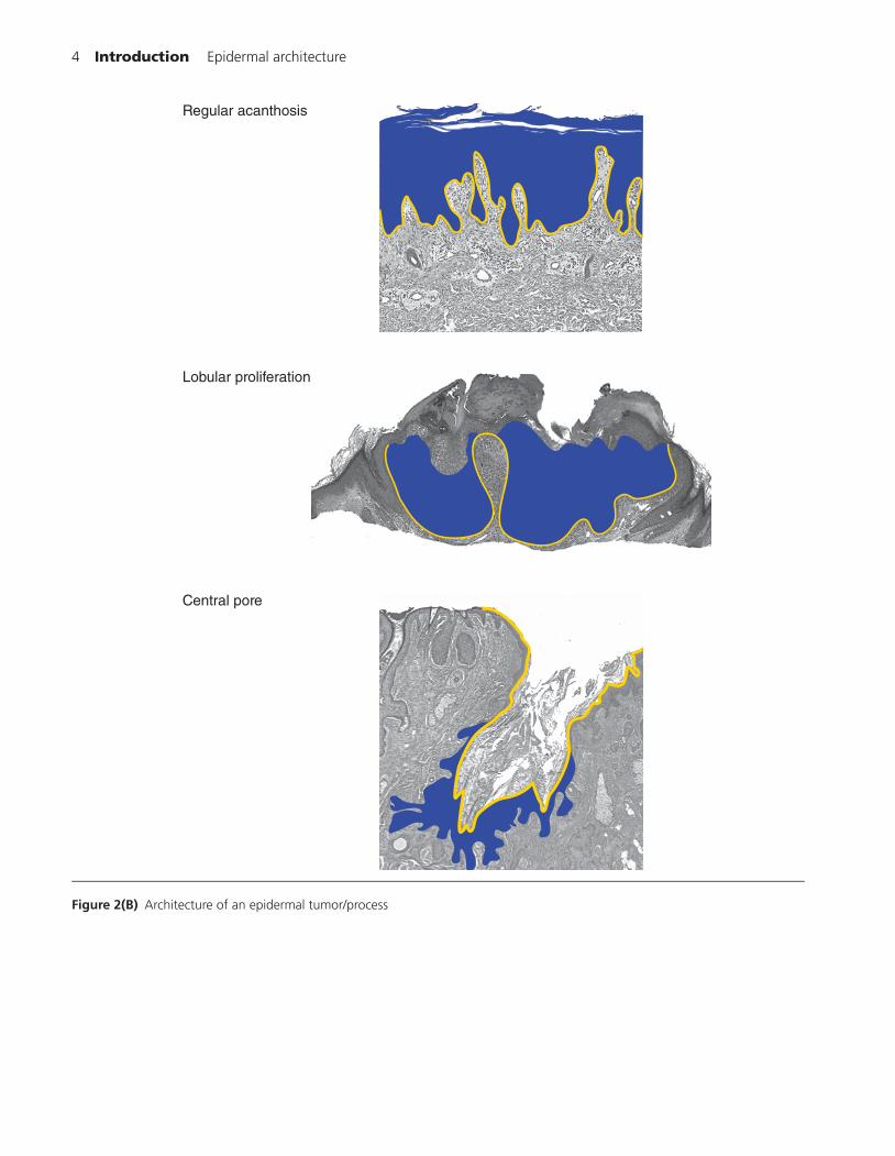
Figure 2(B) Architecture of an epidermal tumor/process
4 Introduction | Epidermal architecture
Regular acanthosis
Lobular proliferation
Central pore
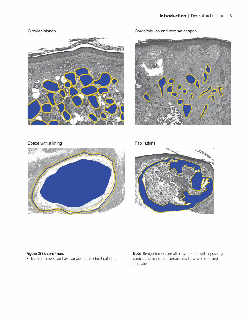
Figure 2(B), continued• Dermal tumors can have various architectural patterns
Note Benign tumors are often symmetric with a pushing border, and malignant tumors may be asymmetric andinfi ltrative.
Introduction | Dermal architecture 5
Circular islands Cords/tubules and comma shapes
Space with a lining Papillations

Figure 2(B), continued
6 Introduction | Dermal architecture
Polypoid (dome-shaped)
Square/rectangular
Palisading reactions
Pseudoepitheliomatoushyperplasia above abscesses
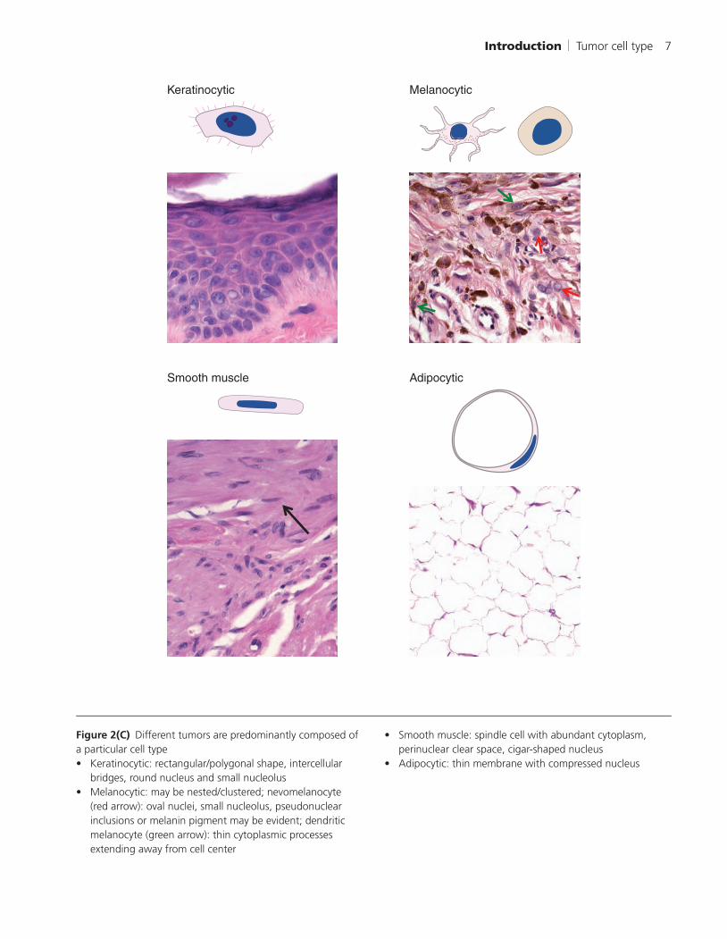
Figure 2(C) Different tumors are predominantly composed of a particular cell type• Keratinocytic: rectangular/polygonal shape, intercellular
bridges, round nucleus and small nucleolus• Melanocytic: may be nested/clustered; nevomelanocyte
(red arrow): oval nuclei, small nucleolus, pseudonuclear inclusions or melanin pigment may be evident; dendriticmelanocyte (green arrow): thin cytoplasmic processesextending away from cell center
• Smooth muscle: spindle cell with abundant cytoplasm,perinuclear clear space, cigar-shaped nucleus
• Adipocytic: thin membrane with compressed nucleus
Introduction | Tumor cell type 7
Keratinocytic Melanocytic
Smooth muscle Adipocytic

Figure 2(C), continued• Neural: spindle cell with tapered nucleus, pink cytoplasm
(green arrows)• Fibroblast: spindle cell with oval nucleus (yellow arrows)• Endothelial: blue nuclei surrounding vascular spaces (red
arrows)
8 Introduction | Tumor cell type
Neural Fibroblastic Endothelial

Figure 2(C), continued• Hair follicle: matrical cells are round to oval and dark blue
(red arrow); outer root sheath cells are pale pink (green arrow)
• Sebocytes: bubbly cytoplasm (yellow arrow) and centralnucleus that may be star-shaped (scalloped)
• Eccrine gland and duct: the gland has clear cells (bluearrow); the duct has an eosinophilic pink cuticle
• Apocrine gland and duct: the gland often showsdecapitation secretion (black arrow)
Introduction | Tumor cell type 9

Figure 2(D) Cytologic features are important in pointing toward a benign versus malignant tumor• Malignant cells have high nuclear: cytoplasmic ratio, irregular chromatin pattern, irregular nuclear contours, irregular nucleolar
shape and size• Primarily nuclear details suggest cytological malignancy• Cytoplasmic features point to differentiation: keratinocytes – eosinophilic, hyalinized cytoplasm, melanocytes – fi ne brown pigment
10 Introduction | Benign versus malignant
Malignant cell
Benign nevomelanocytes (left) versus Melanoma cells (right)Small nucleus, abundant cytoplasm Large nucleus, relatively little cytoplasmSmooth nuclear border Irregular nuclear borderChromatin pattern nondescript Irregular, chunky nuclear contents (chromatin)Inconspicuous nucleolus 1 or more large, purple nucleoli
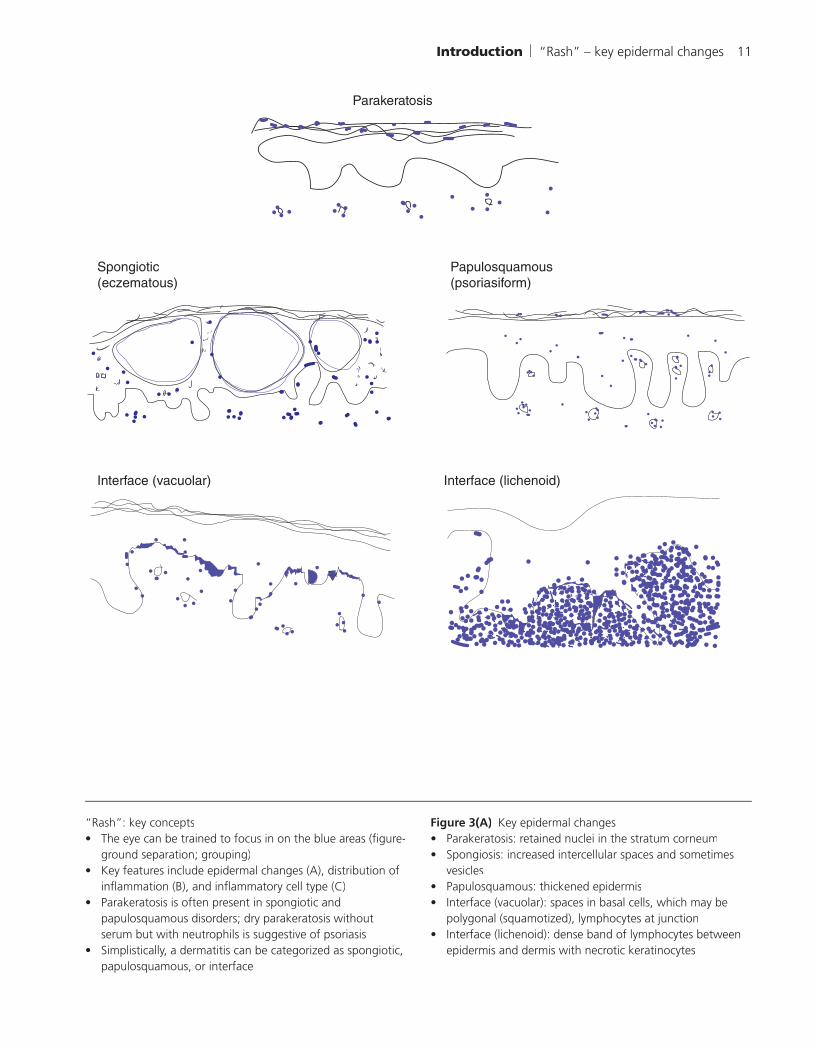
“Rash”: key concepts• The eye can be trained to focus in on the blue areas (fi gure-
ground separation; grouping)• Key features include epidermal changes (A), distribution of
infl ammation (B), and infl ammatory cell type (C)• Parakeratosis is often present in spongiotic and
papulosquamous disorders; dry parakeratosis withoutserum but with neutrophils is suggestive of psoriasis
• Simplistically, a dermatitis can be categorized as spongiotic,papulosquamous, or interface
Figure 3(A) Key epidermal changes• Parakeratosis: retained nuclei in the stratum corneum• Spongiosis: increased intercellular spaces and sometimes
vesicles• Papulosquamous: thickened epidermis• Interface (vacuolar): spaces in basal cells, which may be
polygonal (squamotized), lymphocytes at junction• Interface (lichenoid): dense band of lymphocytes between
epidermis and dermis with necrotic keratinocytes
Introduction | “Rash” – key epidermal changes 11
Parakeratosis
Spongiotic (eczematous)
Papulosquamous(psoriasiform)
Interface (vacuolar) Interface (lichenoid)

Figure 3(B) Distribution of infl ammation – major patterns. See Figure 3(A) for lichenoid
12 Introduction | “Rash” – distribution of infl ammation
Perivascular Interstitial
Nodular Perifollicular
Subcutaneous (septal) Subcutaneous (lobular)

Figure 3(C) The morphology of key infl ammatory cells• Lymphocyte: round blue nucleus, little cytoplasm• Neutrophil: multilobed nucleus• Eosinophil: bilobed nucleus with bright pink-red
cytoplasmic granules
• Histiocyte: oval nucleus• Giant cell: multiple nuclei in one cell• Plasma cell: clock-faced nucleus on one side of cell,
perinuclear clear space
Introduction | “Rash” – cell type 13
Lymphocytes Neutrophils
Eosinophils Histiocytes
Plasma cellsGiant cells

Characteristic body sites• The location on the body (body site) can often be
determined by training the eye/brain to perceive certainfeatures
• Figure 4: Acral (A), mucosal (B), eyelid (C), axilla (D)
Figure 4(A) Acral skin Note Meissner’s corpuscles (black arrow), Pacinian corpuscles (red arrow), and thick stratum corneum with a stratumlucidum (green arrow).
14 Introduction | Acral skin
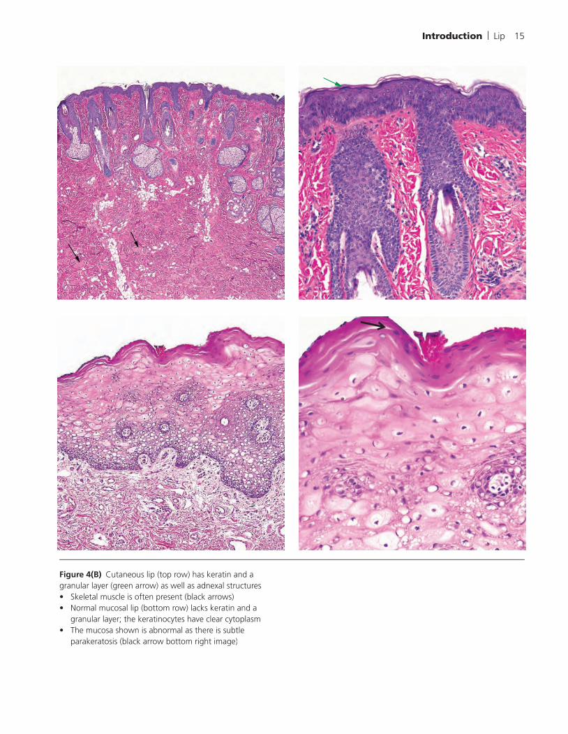
Figure 4(B) Cutaneous lip (top row) has keratin and a granular layer (green arrow) as well as adnexal structures• Skeletal muscle is often present (black arrows)• Normal mucosal lip (bottom row) lacks keratin and a
granular layer; the keratinocytes have clear cytoplasm• The mucosa shown is abnormal as there is subtle
parakeratosis (black arrow bottom right image)
Introduction | Lip 15

Figure 4(C) Eyelid skin has a thin epidermis with dermal vellus hairs (red arrow) and skeletal muscle (black arrow)
16 Introduction | Eyelid

Figure 4(D) Axilla• The epidermis is undulating, often with basilar melanin
pigment. There are apocrine glands in the deep dermis
Introduction | Axilla 17
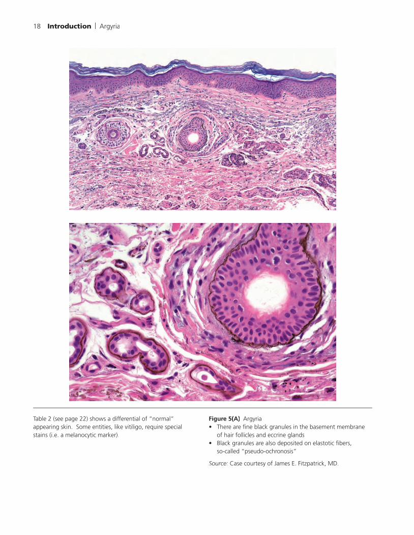
Table 2 (see page 22) shows a differential of “normal” appearing skin. Some entities, like vitiligo, require specialstains (i.e. a melanocytic marker).
Figure 5(A) Argyria• There are fi ne black granules in the basement membrane
of hair follicles and eccrine glands• Black granules are also deposited on elastotic fi bers,
so-called “pseudo-ochronosis”
Source: Case courtesy of James E. Fitzpatrick, MD.
18 Introduction | Argyria

Figure 5(B) Ichthyosis vulgaris• This example from an older patient has solar elastosis in the
dermis• There is hyperekeratosis above an attenuated granular layer
Source: Case courtesy of Jeff D. Harvell, MD.
Introduction | Ichthyosis vulgaris 19

Figure 5(C) Tinea versicolor• Yeast and hyphal forms in the stratum corneum
20 Introduction | Tinea versicolor