therapeutics bnc105: a novel tubulin polymerization...
TRANSCRIPT
1562
Published OnlineFirst June 1, 2010; DOI: 10.1158/1535-7163.MCT-09-0815
Research Article Molecular
CancerTherapeutics
BNC105: A Novel Tubulin Polymerization Inhibitor ThatSelectively Disrupts Tumor Vasculature and DisplaysSingle-Agent Antitumor Efficacy
Gabriel Kremmidiotis, Annabell F. Leske, Tina C. Lavranos, Donna Beaumont, Jelena Gasic,Allison Hall, Michael O'Callaghan, Clayton A. Matthews, and Bernard Flynn
Abstract
uthors'ustralia
ote: Supancer The
orresponalgleish S1-8-8354-om.au
oi: 10.115
©2010 Am
Mol Canc
Dow
Vascular disruption agents (VDA) cause occlusion of tumor vasculature, resulting in hypoxia-driventumor cell necrosis. Tumor vascular disruption is a therapeutic strategy of great potential; however, VDAscurrently under development display a narrow therapeutic margin, with cardiovascular toxicity posing adose-limiting obstacle. Discovery of new VDAs, which display a wider therapeutic margin, may allowattainment of improved clinical outcomes. To identify such compounds, we used an in vitro selectivityscreening approach that exploits the fact that tumor endothelial cells are in a constant state of activationand angiogenesis and do not undergo senescence. Our effort yielded the compound BNC105. This com-pound acts as a tubulin polymerization inhibitor and displays 80-fold higher potency against endothelialcells that are actively proliferating or are engaged in the formation of in vitro capillaries compared withnonproliferating endothelial cells or endothelium found in stable capillaries. This selectivity was not ob-served with CA4, a VDA currently under evaluation in phase III clinical trials. BNC105 is more potentand offers a wider therapeutic window. CA4 produces 90% vascular disruption at its no observed adverseevent level (NOAEL), whereas BNC105 causes 95% vascular disruption at 1/8th of its NOAEL. Tissuedistribution analysis of BNC105 in tumor-bearing mice showed that while the drug is cleared from alltissues 24 hours after administration, it is still present at high concentrations within the solid tumor mass.Furthermore, BNC105 treatment causes tumor regressions with complete tumor clearance in 20% of trea-ted animals. Mol Cancer Ther; 9(6); 1562–73. ©2010 AACR.
Introduction
Molecules that bind to different sites within the tubulinpolymer can produce varied molecular outcomes andconfer anticancer activity in diverse tumor indications.Tubulin-binding agents can be classified as tubulin stabi-lizers or tubulin destabilizers. A tubulin-binding site thataffects stabilization has been defined by the interaction oftaxanes. A large number of natural products and small-molecule compounds have been shown to have theireffect via the taxane binding site, promoting tubulin po-lymerization and stabilization of microtubules (1). Dispa-rate to the taxane site, the binding of Vinca alkaloids tothe tubulin polymer occurs at a site that mediates tubulindestabilization. Colchicine interactions with tubulin de-
Affiliation: Bionomics Ltd., Thebarton, South Australia,
plementary material for this article is available at Molecularrapeutics Online (http://mct.aacrjournals.org/).
ding Author: Gabriel Kremmidiotis, Bionomics Limited, 31treet, Thebarton, 5031 South Australia, Australia. Phone:6123; Fax: 61-8-8354-6199. E-mail: gkremmid@bionomics.
8/1535-7163.MCT-09-0815
AA
NC
CD6c
d
erican Association for Cancer Research.
er Ther; 9(6) June 2010
on June 3, 2018. ©mct.aacrjournals.org nloaded from
fine an additional binding site that also causes tubulindestabilization (2).Although agents binding to the taxane and Vinca sites
have been successfully used as anticancer chemothera-peutics, by comparison the colchicine-site binders havebeen relatively neglected, possibly due to the poor thera-peutic margin exhibited by colchicine. Recently, severalagents have been described that bind at the colchicine siteand are capable of causing blood vessel disruption withinsolid tumors. This class of compounds is defined as vas-cular disruption agents (VDA). Many of the colchicine-site binding agents that exhibit VDA capability werebased on natural products such as combretastatins(CA4P, OXi-4503, and AVE-8062), colchicines (ZD6126),and phenylahistin (NPI-2358), whereas others were dis-covered independently (MN-029 and EPC2407; refs. 3, 4).The preclinical evaluation of tubulin-targeting VDAs
has shown that these compounds are able to disrupttumor blood flow, causing tumor hypoxia and necrosis(5). However, this vascular disruption spares the outercellular rim of the tumor mass, which seems to besupported by normal blood vessels that surround thetumor capsule and remain unaffected by VDA action(6). It is believed that this viable rim mediates recoveryfrom the vascular damage caused by VDA treatment (6).
2010 American Association for Cancer Research.

BNC105: A Vascular Disruption Agent
Published OnlineFirst June 1, 2010; DOI: 10.1158/1535-7163.MCT-09-0815
Preclinical evidence suggests that VDAs are not effectiveat stopping tumor growth when used as single agentsbut may synergize with conventional chemotherapeuticsor antiangiogenic agents (7).Experience with colchicine-site binders such as CA4P
and ZD6126 in a clinical setting has shown their abilityto disrupt blood flow within human tumors (8, 9). How-ever, negative side effects, such as cardiotoxicity, have re-sulted in the discontinuation of the clinical developmentof ZD6126 (9). Nevertheless, CA4P remains in develop-ment and is currently undergoing evaluation in phaseIII clinical trials for anaplastic thyroid carcinoma (FACTtrial #NCT00507429) and for nonsquamous non–smallcell lung cancer (FALCON trial #NCT00653939).Further improvement in the therapeutic window dis-
played by current VDAs may be achieved by exploitingthe fact that tumor endothelial cells are in a constantstate of activation and angiogenesis and do not undergosenescence (10). We constructed a chemical library ofmore than 100 compounds based on diversification ofthe combretastatin pharmacophore. We used two in vitrocorrelates of endothelial cell function to screen thislibrary for compounds that exhibit selectivity forendothelial cells that are in a state of activation or an-giogenesis. This effort led to the discovery of a noveltubulin-targeting agent, BNC105. The data presentedin this report show that BNC105 exhibits a wider ther-apeutic window than CA4P. This increased therapeuticwindow enables administration of BNC105 at higherdose levels, yielding single-agent efficacy in human xe-nograft tumor models.
Materials and Methods
Cell culture and cell linesAll in vitro assays were carried out using endothelial
cells derived from human umbilical vein (HUVEC;Clonetics, Lonza) or human abdominal aorta (HAAE-1;American Type Culture Collection). HUVEC were rou-tinely cultured in EGM-2 medium (Clonetics) andHAAE-1 cells were cultured in F12K medium (Gibco,Invitrogen) containing 10% FCS, 0.1 mg/mL heparin(Sigma-Aldrich), 0.03 mg/mL endothelial cell growthsupplement (Sigma-Aldrich), 2 mmol/L penicillin-strep-tomycin-glutamine (Gibco, Invitrogen), and 10 mmol/LHEPES buffered solution (Gibco, Invitrogen). Cell culturesbetween passages 2 and 6 were used for all assays. Cancercell lines included Calu 6, DU145, Colo 205 (AmericanType Culture Collection), and MDA-MB-231 (kind giftfrom the Women's and Children's Hospital, Adelaide,Australia). Calu 6 and DU145 cells were cultured inMEM (Gibco) with 10% FCS, 2 mmol/L penicillin-strepto-mycin-glutamine, 10 mmol/L HEPES buffered solution,1 mmol/L sodium pyruvate solution (Gibco), and0.1 mmol/L nonessential amino acids solution (Gibco).Colo 205 cells were cultured in RPMI 1640 (Gibco) with10%FCS and2mmol/Lpenicillin-streptomycin-glutamine.MDA-MB-231 cells were cultured in RPMI 1640 (Gibco)
www.aacrjournals.org
on June 3, 2018. ©mct.aacrjournals.org Downloaded from
with 10% FCS, 2 mmol/L penicillin-streptomycin-glutamine, and 10 mmol/L HEPES buffered solution. Allcells were cultured in a humidified incubator at 37°C with5% CO2.
Endothelial cell proliferation assayHUVEC or HAAE-1 cell cultures were exposed to a
concentration range of 0.1 to 1,000 nmol/L for each testcompound tested. Proliferation assays were carried out intriplicate in 96-well plates. For activated growth condi-tions, HUVEC and HAAE-1 cells were seeded at 2,500and 5,000 per well, respectively, and cultured in EGM-2or medium containing 0.03 mg/mL endothelial cellgrowth supplement (Sigma-Aldrich) as described above.For quiescent growth conditions, HUVEC and HAAE-1cells were seeded at 15,000 and 5,000 per well, respective-ly, in basal medium (EBM-2 or F12K) containing 0.5%FCS and antibiotics. Cells were allowed to adhere over-night followed by incubation with compounds underevaluation for 48 hours. Metabolically active cells weremeasured using CellTiter 96 Aqueous One Solution(Promega Corp.) according to the manufacturer's instruc-tions, and absorbance readings taken at 492 nm. Controlabsorbance readings for quiescent endothelial cells re-mained relatively unchanged over the 48-hour cultureperiod. Absorbance readings for each compound concen-tration were normalized to corresponding vehicle controlcultures. A sigmoidal dose-response curve was fitted tothe data, and the concentration at which proliferationdecreased by 50% was calculated using GraphPad Prism4 software (San Diego).
Capillary formation and disruption assaysCapillary formation assays were conducted in 96-well
plates using HUVEC and HAAE-1 cells plated on a Ma-trigel layer (BD Biosciences) with 25,000 and 20,000 cellsper well, respectively, and incubated for 22 hours. Cap-illary disruption assays were carried out in a 96-wellplate format using HUVEC cells plated at 25,000 perwell in EGM-2 medium on a Matrigel layer (BD Bio-sciences). Capillaries were allowed to form over a 16-hour period before the addition of test compound orcontrol. Cells were stained using calcein AM (Calbio-chem) at room temperature for 40 minutes after a briefwashing step. Images were acquired immediately fol-lowing compound addition and 5 hours after exposureto test compound. Tube formation was quantified bymeasuring the length of capillary structures using thesoftware NIH ImageJ.
Tubulin polymerization assaysIn vitro tubulin polymerization assays were done
using a Tubulin Polymerization Assay Kit (Cytoskeleton,Inc.) in a 384-well format, which used the polymerizationof bovine neuronal tubulin in vitro in the presenceof varying concentrations of BNC105 or CA4. Fluores-cence measurements were obtained at 1-minute intervalsover a 42-minute period. Fluorescent measurements were
Mol Cancer Ther; 9(6) June 2010 1563
2010 American Association for Cancer Research.

Kremmidiotis et al.
1564
Published OnlineFirst June 1, 2010; DOI: 10.1158/1535-7163.MCT-09-0815
taken following excitation at 360 nm, and emissionwasmea-sured at 420 nm using a BMG Flurostar fluorescent platereader. Data were analyzed as relative fluorescent units.
F-Actin staining and immunohistochemical analysisHUVEC were plated on glass coverslips in a six-well
plate at 7.5 × 10⁴ cells per well in EGM-2 medium andincubated overnight. On the following day, cells were50% to 60% confluent and the cell medium was replacedwith EGM-2 medium containing 10 nmol/L BNC105 or0.1% DMSO vehicle control. Cells were incubated for20 minutes at 37°C before being fixed in 3.7% formalde-hyde/PBS solution for 10 minutes at room temperatureand then permeabilized in 0.5% Triton X-100/PBS solu-tion for 5 minutes at 4°C. Following permeabilization,FITC-phalloidin (Sigma-Aldrich) was added to each wellat 1 μg/mL in PBS and incubated in darkness for 1 hour.Coverslips were mounted on glass slides using Vecta-shield containing 4′,6-diamidino-2-phenylindole (VectorLaboratories) and stored in darkness at 4°C. Images ofthe cells showing F-actin and 4′,6-diamidino-2-phenylin-dole stainingwere digitally acquiredusing the 100×objectivelens of an Olympus BX51 microscope and photographedusing a charge-coupled device camera (Optronics).
Endothelial monolayer permeability assayThe permeability of an endothelial cell monolayer was
assessed using an In Vitro Vascular Permeability AssayKit (Chemicon, Millipore) as previously described (11).Briefly, HUVEC were plated at 5 × 10⁴ cells per insertin EGM-2 medium and incubated for 48 hours to reach100% confluency. BNC105 or CA4 were diluted inEGM-2 medium and added to the upper chamber ofthe apparatus. Following 15 minutes of incubation, thecompounds were removed and FITC-conjugated dextranwas added for 5 minutes. Fluorescent measurements ofthe lower chamber were taken after excitation at 520 nm,and emission was measured at 485 nm using a BMGFluorstar fluorescent plate reader. Data were analyzedas relative fluorescent units.
Pharmacokinetics and metabolism assaysFor in vivo studies, all the animals were housed
under pathogen-free conditions and cared for in accor-dance with the NHMRC guidelines and the AustralianCode of Practice for the Care and Use of Animals forScientific Purposes. To facilitate i.v. administration ofBNC105, the phosphate ester of this compound wassynthesized (BNC105P) and dissolved in isotonic saline(Table 1B). Pharmacokinetic studies were conducted bybolus tail-vein i.v. administration of BNC105P to non-fasting male Swiss mice. Postadministration blood sam-ples were collected into tubes containing anticoagulantand a protease inhibitor cocktail via cardiac punctureup to 6 hours post-dose. Blood plasma was separatedby centrifugation and frozen until analysis. Extractswere prepared and analyzed by high-performanceliquid chromatography-mass spectrometry (HPLC-MS)
Mol Cancer Ther; 9(6) June 2010
on June 3, 2018. ©mct.aacrjournals.org Downloaded from
against a standard curve. Pharmacokinetic variableswere determined using a noncompartmental method(WinNonLin). Metabolism assays were conducted usingrecombinant CYP450 Supersomes. BNC105 and
2
Table 1. BNC105 is a CA4 analogue thatselectively inhibits the proliferation of activatedendothelial cells
A
Compound
010 Ameri
ActivatedEC50
M
can Associa
QuiescentEC50
olecular C
tion for Can
Selectivity ratio,quiescent/activated
HUVEC
BNC105 0.31 nmol/L 25 nmol/L 80.6 0075 0.36 nmol/L 2.7 nmol/L 7.5 0079 3.28 nmol/L 186 nmol/L 56.7 CA4 3.6 nmol/L 3.9 nmol/L 1.1 Paclitaxel 2.63 2.73 1 Vinblastine 0.86 1.03 1.2 Vincristine 2.16 1.76 0.8HAAEC
BNC105 0.1 nmol/L 12 nmol/L 120 CA4 2.2 nmol/L 1.3 nmol/L 0.6B
Compound
Structure C ompound StructureBNC105
CA4BNC105P
CA4PNOTE: A, measurement of the activity of BNC105, twochemically related compounds (0075 and 0079), CA4, pac-litaxel, vinblastine, and vincristine on the proliferation of ac-tivated and quiescent HUVEC and HAAEC. EC50 values arebased on three replicates obtained following a 48-h expo-sure to increasing doses of compound. Cell proliferationwas determined using CellTitre 96 Aqueous One Reagent(Promega) and calculated relative to vehicle control. B,structure of BNC105, the prodrug BNC105P, and the com-parator CA4 with its prodrug CA4P.
ancer Therapeutics
cer Research.
BNC105: A Vascular Disruption Agent
Published OnlineFirst June 1, 2010; DOI: 10.1158/1535-7163.MCT-09-0815
BNC105P were added to Supersomes incubation solu-tions to a final concentration of 1 μmol/L and incuba-ted at 37°C with required cofactors for 10 minutesbefore the addition of the NADPH-regenerating system.Samples were taken throughout the 45-minute incuba-tion period and quenched with 100% acetonitrile con-taining diazepam as an analytic internal standard.Analysis of samples was done by mass spectrometryon Micromass Q-TOF Micro and Micromass QuattroUltima Pt triple quadrupole instruments for BNC105and BNC105P, respectively, coupled to a Waters 2795HPLC device. The in vitro clearance values were calcula-ted and expressed relative to the CYP450 content duringincubation.
Vascular disruption assayFemale BALB/c nu/nu mice at 6 to 8 weeks [Animal
Resources Centre (ARC), Perth, Australia] were injecteds.c. with human cancer cell lines representing breast(MDA-MB-231), lung (Calu 6), colon (Colo 205), or pros-tate (DU145) cancer. Cells were resuspended in Dulbecco'sPBS (Sigma-Aldrich) and 2 × 10⁶ cells in 50 μL of PBS weres.c. injected in the hind flank (Calu 6, Colo 205, andDU145) or near themammary fat pad (MDA-MB-231). Tu-mors were grown to an average size of 300 mm3 beforetreatment. BNC105P treatment consisted of a single i.v. in-jection at dose levels ranging from 1 to 150 mg/kg. Salinetreatment was included as a vehicle control. Twenty-fourhours after the injection of the compound, animals werei.v. injected with 10 mg/kg of H33342 Hoechst fluorescentdye (Invitrogen), which acts as a marker of blood perfu-sion in tumors and normal tissues (12). After 1 minute, an-imals were euthanized and tumors were excised forhistologic examination in optimum cutting temperaturecompound (Tissue-Tek, Sakura Finetek). Images of histo-logic sections were captured and assembled using MWSnap software version 3.0.0.74 (Mirek Wojtowicz) andMotic Images Assembly 1.0 Pro (Motic China Group Co.Ltd.). Quantification of H33342 Hoechst and terminaldeoxyribonucleotidyl transferase–mediated dUTP nick endlabeling (TUNEL) stainingwas done using the softwareNIHImageJ.
BNC105 tissue distribution assayFemale BALB/c nu/nu mice at 6 weeks (ARC) were
s.c. inoculated with human MDA-MB-231 breast cancercells. Tumors were grown to an average size of 200 mm3
before a single bolus i.v. injection of 10 mg/kg BNC105Pwas administered. Tumors and tissues (liver, kidney,heart, brain, and spleen) were harvested and snap-frozenat 15 minutes and 2, 6, and 24 hours after administration(n = 3 per time point). Tissues were homogenized andprepared using protein precipitation with acetonitrile.Supernatants containing BNC105 were subsequentlyanalyzed by HPLC-MS on a Quattro Premier triplequadrupole mass spectrometer. Sample concentrationswere determined against calibration standards preparedin acetonitrile-water.
www.aacrjournals.org
on June 3, 2018. ©mct.aacrjournals.org Downloaded from
Tumor growth inhibition assayFemale BALB/c nu/nu mice at 6 to 8 weeks (ARC) were
s.c. inoculated with human cancer cell lines derived fromlung (Calu 6) and breast (MDA-MB-231) cancers. Tumorswere grown to anaverage size of 200mm3before commenc-ing treatment. BNC105P at dose levels ranging from10 to 80mg/kg and saline vehicle control were administered by i.v.injection as described. Tumor volume (in cubic millimeters)was measured two to three times per week. Animals wereeuthanized if tumor volume exceeded 2,000 mm3.
Statistical analysesResults from tumor growth inhibition assays were an-
alyzed for statistical significance using two-tailed t tests(P < 0.05) and GraphPad Prism version 4 software.Results from VDA analysis were analyzed for statisticalsignificance using one-way ANOVA with post hoc test(Dunnett's).
Results
Identification of BNC105, a compound that exhibitshigh activity against proliferating endothelial cellsand comparatively low activity against quiescentendothelial cellsA library of CA4 chemical analogues was evaluated for
cytotoxic activity against growth factor–supplementedendothelial cells and growth factor–deprived endothelialcell cultures. Our analyses identified three compoundsthat exhibit selectivity for activated endothelial cells.The compound BNC105 exhibited the highest degree ofselectivity, being 80 times more active against activatedendothelial cells than against quiescent endothelial cells(Table 1A). This degree of selectivity was not observedwith CA4, paclitaxel, vinblastine, or vincristine. Further-more, BNC105 exhibited significantly higher potencythan CA4, paclitaxel, vinblastine, or vincristine againstactively proliferating endothelial cells (Table 1A). Inaddition to BNC105, two other compounds in our librarywith similar structures displayed selectivity for activelyproliferating endothelial cells, but to a lesser extent thanBNC105 (Table 1A).To confirm that the selectivity displayed for activated
endothelial cells is not restricted to HUVEC, we in-vestigated the activity of BNC105 against activatedand quiescent HAAEC HAAE-1 cells. As observed inHUVEC, BNC105 exhibited selectivity for activatedHAAEC HAAE-1 cells (Table 1A). This selectivity foractivated cells over quiescent cells was not observedwith CA4.
BNC105 disrupts the formation of endothelialcapillaries but does not disrupt preformed capillariesWe investigated the activity of BNC105 on endothelial
cells engaged in capillary tube formation in vitro. Endo-thelial cells were placed on a Matrigel matrix andallowed to form capillary tubes in the presence orabsence of compound. Each compound was applied at
Mol Cancer Ther; 9(6) June 2010 1565
2010 American Association for Cancer Research.

Kremmidiotis et al.
1566
Published OnlineFirst June 1, 2010; DOI: 10.1158/1535-7163.MCT-09-0815
concentrations that were shown to display activity in theproliferation assay. Based on our results, BNC105inhibited the formation of capillary tubes at 1 nmol/L(Fig. 1A). This compound was more potent than CA4,which inhibited capillary formation at 10 nmol/L(Fig. 1B). These results show that BNC105 is highly po-tent against activated endothelial cells that are engagedin the formation of new capillaries.Subsequently, we investigated the activity of BNC105
on preformed capillaries. Endothelial cells were platedon Matrigel and allowed to form capillary tubes for 22hours, followed by exposure to each of the compounds un-der investigation. Quantification of total capillary lengthat the 0-hour and the 5-hour time points showed that atthe 5-hour time point, there was a 22% decrease in theDMSO control cultures, an 11% decrease in the BNC105cultures, and a 65% decrease in the CA4 cultures. Theseresults indicate that although BNC105 is capable of inhi-biting the formation of capillaries at 1 nmol/L (Fig. 1A),
Mol Cancer Ther; 9(6) June 2010
on June 3, 2018. ©mct.aacrjournals.org Downloaded from
it does not disrupt preformed capillaries at that concen-tration (Fig. 1C). This result is in agreement with ourproliferation assay results, providing further evidencethat BNC105 exhibits selectivity for activated endotheli-al cells. In contrast, CA4 disrupted preformed capillarytubes at 10 nmol/L (Fig. 1D), which is the same concen-tration required to block the formation of new capillarytubes (Fig. 1B). Staining with calcein AM showed theviability of the cells involved in the in vitro capillariesin this assay.
BNC105 is a tubulin polymerization inhibitorTo confirm that BNC105 acts as a tubulin polymer-
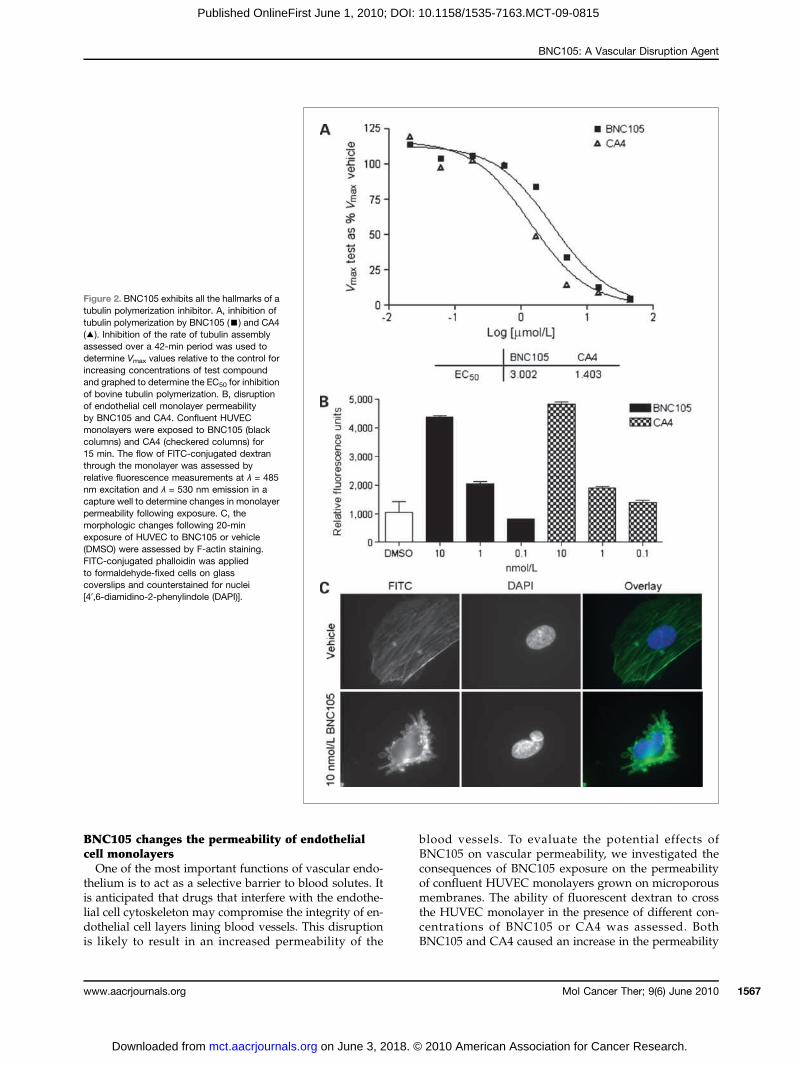
ization inhibitor, we tested its ability to alter the dynam-ics of tubulin polymerization in vitro. The change influorescence that occurs when tubulin monomers formelongated fibers was used as a measure of tubulin poly-merization. Using CA4 as a positive control, BNC105 wasshown to inhibit tubulin polymerization (Fig. 2A).
Mole
2010 American Association
Figure 1. BNC105 selectivelyinhibits the formation of endothelialcell capillaries but does not affectpreformed capillaries. A and B,disruption of capillary formation byBNC105 and CA4. HUVEC andtest compound were concurrentlyadded to 96-well plates precoatedwith Matrigel and incubated for22 h. C and D, effects of BNC105and CA4 on stable, preformedcapillaries. Preformed capillaries(16 h after HUVEC culture in96-well Matrigel-coated plates)were treated with varyingconcentrations of BNC105 andCA4 and monitored for 5 h.Capillaries were stained withcalcein AM to verify viable cellstructures after 5 h.
cular Cancer Therapeutics
for Cancer Research.
BNC105: A Vascular Disruption Agent
Published OnlineFirst June 1, 2010; DOI: 10.1158/1535-7163.MCT-09-0815
BNC105 changes the permeability of endothelialcell monolayersOne of the most important functions of vascular endo-
thelium is to act as a selective barrier to blood solutes. Itis anticipated that drugs that interfere with the endothe-lial cell cytoskeleton may compromise the integrity of en-dothelial cell layers lining blood vessels. This disruptionis likely to result in an increased permeability of the
www.aacrjournals.org
on June 3, 2018. ©mct.aacrjournals.org Downloaded from
blood vessels. To evaluate the potential effects ofBNC105 on vascular permeability, we investigated theconsequences of BNC105 exposure on the permeabilityof confluent HUVEC monolayers grown on microporousmembranes. The ability of fluorescent dextran to crossthe HUVEC monolayer in the presence of different con-centrations of BNC105 or CA4 was assessed. BothBNC105 and CA4 caused an increase in the permeability
Figure 2. BNC105 exhibits all the hallmarks of atubulin polymerization inhibitor. A, inhibition oftubulin polymerization by BNC105 (▪) and CA4(▴). Inhibition of the rate of tubulin assemblyassessed over a 42-min period was used todetermine Vmax values relative to the control forincreasing concentrations of test compoundand graphed to determine the EC50 for inhibitionof bovine tubulin polymerization. B, disruptionof endothelial cell monolayer permeabilityby BNC105 and CA4. Confluent HUVECmonolayers were exposed to BNC105 (blackcolumns) and CA4 (checkered columns) for15 min. The flow of FITC-conjugated dextranthrough the monolayer was assessed byrelative fluorescence measurements at λ = 485nm excitation and λ = 530 nm emission in acapture well to determine changes in monolayerpermeability following exposure. C, themorphologic changes following 20-minexposure of HUVEC to BNC105 or vehicle(DMSO) were assessed by F-actin staining.FITC-conjugated phalloidin was appliedto formaldehyde-fixed cells on glasscoverslips and counterstained for nuclei[4′,6-diamidino-2-phenylindole (DAPI)].
Mol Cancer Ther; 9(6) June 2010 1567
2010 American Association for Cancer Research.
Kremmidiotis et al.
1568
Published OnlineFirst June 1, 2010; DOI: 10.1158/1535-7163.MCT-09-0815
of HUVEC monolayers (Fig. 2B). Unlike in the prolifera-tion assay, where the activity of BNC105 is considerablyhigher than that of CA4 in activated endothelial cell cul-tures, both compounds seem to be of equal potency in themonolayer permeability assay. This is probably a reflec-tion of the fact that the cells in these confluent mono-layers are in a nonproliferating (quiescent) state. This isconsistent with the selectivity exhibited by BNC105 foractivated/proliferating endothelial cells.
BNC105 induces endothelial cell blebbingIt has previously been shown that CA4 induces cyto-
skeletal alterations that manifest in a cell blebbing mor-phology, where actin accumulates around cell membranemicelle-like protrusions (13). To addresswhether the tubu-lin polymerization inhibition activity of BNC105 operatesvia the same mechanism as CA4, HUVEC were exposedto different concentrations (0.1, 1, and 10 nmol/L) ofBNC105 and CA4 for 20 minutes. The application of eachagent at 10 nmol/L caused cell blebbing (Fig. 2C).
Pharmacokinetics and metabolism of BNC105To facilitate i.v. administration of BNC105, the phos-
phate ester of this compound was synthesized (BNC105P;Table 1B) and dissolved in isotonic saline. Plasma pharma-cokinetic analysis of BNC105 following i.v. administrationof BNC105Pwas undertaken. Following administration ofBNC105P at 6, 18, and 60 mg/kg, BNC105 displayed aCmax of 3, 34, and 93 μmol/L and an AUClast of 30, 268,and 814 min·μmol/L, respectively. Tmax was at 2 minutes,suggesting rapid conversion of BNC105P to BNC105.Metabolism of BNC105 was found to be predominatelyby the cytochrome P450 CYP2C19 isoform.
BNC105 disrupts the solid tumor vasculatureThe efficacy of BNC105 in disrupting tumor vascula-
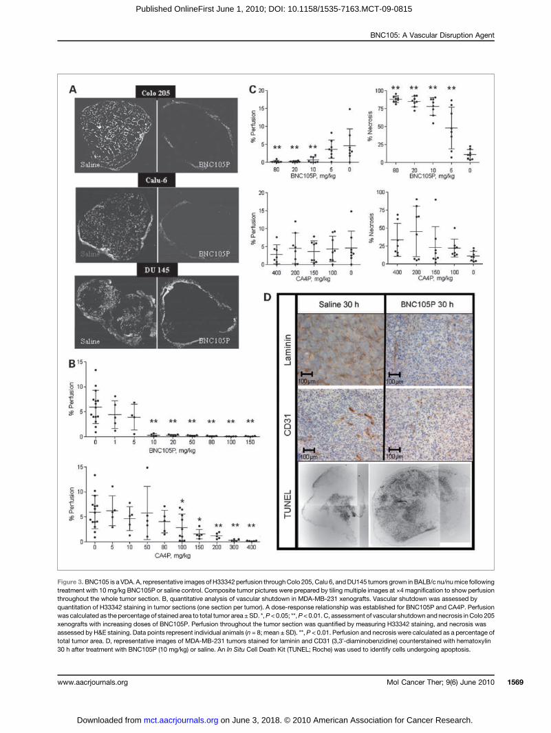
ture in animals bearing solid tumors derived from hu-man breast (MDA-MB-231), lung (Calu 6), colon (Colo205), and prostate (DU145) was evaluated. BNC105 wasformulated as a disodium phosphate (BNC105P) for i.v.administration. Animals were treated with a single ad-ministration of BNC105P. Dose response of tumor vascu-lature disruption to BNC105P treatment was assessedhistologically using H33342 perfusion 24 hours after ad-ministration. Complete tumor vascular disruption wasobserved in all tumor types evaluated at dose levels≥10 mg/kg (Fig. 3A).Quantitative analysis of tumor vascular disruption
showed that BNC105P achieves statistically significantdisruption at dose levels ≥10 mg/kg in all tumor typesinvestigated. The maximum effect was seen in MDA-MB-231 tumors,with efficacy evident at doses as low as 1mg/kg(Fig. 3B). Staining of tumor sections with H&E enabledquantitative measurement of tumor necrosis resultingfrom the effects of BNC105. Treatment with BNC105Pat 20 mg/kg gave rise to >75% necrosis in MDA-MB-231tumors. A similar level of necrosis was achieved withCA4P at a considerably higher dose (300 mg/kg; Supple-
Mol Cancer Ther; 9(6) June 2010
on June 3, 2018. ©mct.aacrjournals.org Downloaded from
mentary Figure 1). Furthermore, in the Colo 205 xenograftmodel, BNC105P treatment resulted in high degree oftumor vascular disruption with a corresponding level ofnecrosis that was directly proportional to the dose levelof BNC105P. In comparison, CA4P was ineffective in thismodel (Fig. 3C).
BNC105 alters the solid tumor microenvironmentWe undertook a number of histologic analyses to as-
sess the damage caused by BNC105 on the integrity ofcomponents within the tumormicroenvironment (Fig. 3D).Our analyses included evaluation of the level of apoptosis,the integrity of endothelial cells, and the basement mem-brane protein laminin. Our observations show that 30hours after treatment with BNC105, there is a decreasein staining with the CD31 endothelial cell marker, whichattests to the destruction of tumor endothelial cells, and aconsiderable reduction in laminin staining, which is evi-dence of basement membrane degradation. Furthermore,TUNEL analysis shows a significant increase in the num-ber of apoptotic cells within the tumor. Quantification ofTUNEL staining showed that apoptosis in BNC105P-treated tumor sections was approximately eight timeshigher comparedwith control (BNC105P, 22.35% of the tu-mor section area; control, 2.91% of the tumor section area).
BNC105 has a wider therapeutic window than CA4We investigated the therapeutic window for BNC105
and CA4 as defined by the range between the lowestdose level causing 50% reduction in tumor vascular per-fusion and the maximum no observed adverse eventlevel (NOAEL). Acute maximum tolerated dose studiesin mice showed a maximum NOAEL of 80 mg/kgBNC105P and 300mg/kgCA4P. Solid tumorswere grownin BALB/c nu/nu mice following injection of the humanbreast cancer cell lineMDA-MB-231 into the mammary fatpad.Mice carrying solid breast tumorswere treatedwith asingle i.v. bolus injection of BNC105P at dose levels rang-ing from 1 to 150 mg/kg or a single dose of CA4P at doselevels ranging from 5 to 400 mg/kg. Twenty-four hoursafter the injection of the compounds, the animals were in-jected with the fluorescent dye H33342 before euthanasia.The tumors were excised for histologic examination.Quantitative analyses of H33342 staining in tumorsections revealed that BNC105P treatment caused ∼50%vascular shutdown at 1 mg/kg and almost completevascular shutdown at 10 mg/kg. CA4P required a doseof 100 mg/kg to reach 50% shutdown and a dose of300 mg/kg to cause 90% vascular shutdown (Fig. 3B).
BNC105 is preferentially retained withinsolid tumorsThe distribution of BNC105 following i.v. administra-
tion of 10 mg/kg BNC105P was evaluated in BALB/cnu/nu mice bearing MDA-MB-231 tumors. It washypothesized that the selective vascular collapse causedby BNC105 in the tumor would result in preferential re-tention of BNC105 inside the tumor mass. Experimentally,
Molecular Cancer Therapeutics
2010 American Association for Cancer Research.
BNC105: A Vascular Disruption Agent
Published OnlineFirst June 1, 2010; DOI: 10.1158/1535-7163.MCT-09-0815
Figure 3. BNC105 is a VDA. A, representative images of H33342 perfusion throughColo 205, Calu 6, andDU145 tumors grown in BALB/c nu/numice followingtreatment with 10 mg/kg BNC105P or saline control. Composite tumor pictures were prepared by tiling multiple images at ×4 magnification to show perfusionthroughout the whole tumor section. B, quantitative analysis of vascular shutdown in MDA-MB-231 xenografts. Vascular shutdown was assessed byquantitation of H33342 staining in tumor sections (one section per tumor). A dose-response relationship was established for BNC105P and CA4P. Perfusionwas calculated as the percentage of stained area to total tumor area ±SD. *,P <0.05; **,P<0.01. C, assessment of vascular shutdown and necrosis inColo 205xenografts with increasing doses of BNC105P. Perfusion throughout the tumor section was quantified by measuring H33342 staining, and necrosis wasassessed by H&E staining. Data points represent individual animals (n = 8; mean ± SD). **, P < 0.01. Perfusion and necrosis were calculated as a percentage oftotal tumor area. D, representative images of MDA-MB-231 tumors stained for laminin and CD31 (3,3′-diaminobenzidine) counterstained with hematoxylin30 h after treatment with BNC105P (10 mg/kg) or saline. An In Situ Cell Death Kit (TUNEL; Roche) was used to identify cells undergoing apoptosis.
Mol Cancer Ther; 9(6) June 2010www.aacrjournals.org 1569
on June 3, 2018. © 2010 American Association for Cancer Research. mct.aacrjournals.org Downloaded from

Kremmidiotis et al.
1570
Published OnlineFirst June 1, 2010; DOI: 10.1158/1535-7163.MCT-09-0815
the levels of BNC105 were found to be significantlyreduced in all healthy tissues 24 hours after dosing (<5%),yet they remained high in the tumor (64%; Table 2). This is aresult of trapping the drug in the tumor following vascularshutdown and may cause a longer exposure time of tumorcells to BNC105.
BNC105 causes solid tumor regressionand clearanceWe investigated the effect of BNC105P treatment on
tumor growth inhibition using a weekly dosing schedule.The logic behind this schedule was to expose the tumor toconsecutive cycles of vascular shutdown events. Animalswere dosed i.v. with a total of eight doses of BNC105P at10mg/kg per dose per week. The data obtained show thatBNC105P causes significant suppression of tumor growth(Fig. 4A).The activity of BNC105P treatment in suppressing
tumor growth was compared with that of CA4P in micebearing MDA-MB-231 tumors. The animals were i.v.treated with BNC105P or CA4P on days 1 and 5 andtumor growth was monitored over a period of 26 days.The suppression of tumor growth achieved with CA4Pat 150 mg/kg (one half of the maximum NOAEL) wasachieved with BNC105P at 10 mg/kg (one eighth of themaximum NOAEL; Supplementary Figure 2).Based on the wide therapeutic window captured by
BNC105, we evaluated the tumor growth inhibitory activ-ity of BNC105 at dose levels higher than 10 mg/kg. It washypothesized that at higher dose levels, BNC105 mayachieve tumor regression by enlisting both the antivascularand the antimitotic activities inherent to its mechanism ofaction. Repeat administrations at doses ranging from 10 to80 mg/kg showed that BNC105P is not tolerated at doselevels ≥60 mg/kg. At dose levels ≤40 mg/kg, BNC105Pwas well tolerated. Animals bearing Calu 6 (lung cancer)human tumors were treated with two administrations ofBNC105P 1 week apart at 40 mg/kg. BNC105 displayed
Mol Cancer Ther; 9(6) June 2010
on June 3, 2018. ©mct.aacrjournals.org Downloaded from
significant tumor growth inhibition in this tumor model(Fig. 4B).Mice bearing MDA-MB-231 solid tumors were treated
with two weekly doses of BNC105P at the start of each oftwo 37-day cycles of dosing. The dose level of 40 mg/kgproduced an average 52% regression in tumor size in thefirst 2weeks posttreatment and a prolongation in suppres-sion of tumor growth up to day 95 (mean of 544.19 mm3,SD ± 921.37 mm3) versus saline control (mean of 1745.68mm3, SD ± 1,444.97 mm3 at day 51 when these animalswere euthanized). A subsequent study investigated theeffect of a 28-day dosing cycle with BNC105P administra-tion at 40 mg/kg on day 1 and day 8 of each cycle. Micebearing MDA-MB-231 solid tumors were dosed for a totalof three cycles. This treatment regimen resulted in signifi-cant tumor growth suppression, with a number of treatedanimals experiencing tumor regression or being cleared oftheir tumor burden (Fig. 4C).To confirm and further substantiate these findings, we
evaluated this dosing regimen using a larger number ofanimals bearing tumors that varied in size between 200and 600 mm3. Animals were treated with two 28-day cy-cles of BNC105P at 40 mg/kg. Significant differences intumor growth between BNC105P-treated (n = 64) and sa-line-treated (n = 20) animals were observed as early asday 4 (P < 0.001, unpaired t test; Prism analysis) throughto day 70. In the BNC105P-treated group, 34% of tumorsshowed significantly delayed growth, 4.7% exhibited nogrowth, 6.3% regressed, and 14% of tumors were cleared.Figure 4D depicts a BALB/c nu/nu mouse in which tu-mor growth was arrested and the tumor disappeared byday 60 of BNC105P two-cycle treatment. Cleared tumorsdid not reappear for the duration of the study.
Discussion
The dependency of solid tumors on blood supply forgrowth and metastasis has provided the impetus for
Table 2. BNC105 is selectively retained within tumors following administration of BNC105P
Organ
BNC105 concentration (ng/mL) after i.v.administration of BNC105P2010 American
% BNC105 remaining 24 hafter administration of BNC105P
[BNC105 (24 h) / BNC105 (0.25 h) × 100]
0.25 h 2 h 6 h 24 hTumor
416 327 349 267 64 Liver 345 135 84 <LLQ 0 Heart 474 234 77 24 5 Spleen 1866 835 355 28 2 Kidney 733 361 159 30 4 Brain 1762 263 782 77 4 1NOTE: Organs and tumors from animals treated with 10 mg/kg BNC105P were removed and the BNC105P derivative compoundBNC105 was extracted. The concentration of BNC105 was determined using HPLC-MS and is reported as nanograms per milliliter.The percentages of BNC105 remaining in the tumor and normal organs after 24 h were calculated.Abbreviation: LLQ, Lower Limit of Quantification.
Molecular Cancer Therapeutics
Association for Cancer Research.

BNC105: A Vascular Disruption Agent
Published OnlineFirst June 1, 2010; DOI: 10.1158/1535-7163.MCT-09-0815
targeting the vasculature that supports tumor growth.Vascular targeting strategies include the suppression ofnew blood vessel formation using inhibitors of angiogen-esis and the disruption of established blood vessels usingVDAs. Antiangiogenic agents require chronic administra-tion and operate through prevention of neovasculariza-tion. VDAs are more suited to acute administration andachieve immediate vascular damage effects. VDAs exerttheir effects by increasing permeability and interstitialfluid pressure, leading to plasma leakage, blood vesseldiameter reduction, decreased blood flow, and vascularshutdown. There are currently a number of tubulin poly-merization inhibitors in clinical development as VDAs.CA4P has progressed through to phase III clinical trials,and AVE8062 (Sanofi-Aventis) has been reported to be inphase III evaluation for advanced-stage soft tissue sarco-ma after failure of anthracycline and ifosfamide che-motherapies (trial NCT00699517). Other compounds
www.aacrjournals.org
on June 3, 2018. ©mct.aacrjournals.org Downloaded from
include OXi4503 (14), NPI-2358 (15), CYT-997 (16),MPC-6827 (17), and EPC2407 (18, 19). These latter agentshave completed phase I evaluation and are under evalu-ation in phase II trials. The number of compounds beingevaluated in this arena attests to the potential utility ofthe VDA approach. Many efforts are aimed at identifyingagents that improve on the therapeutic capacity of CA4Pby attaining a wider therapeutic window. The most fre-quently reported DLT events reported for this class ofcompounds (acute coronary and/or thrombophleboticevents, blood pressure, and heart rate changes) are con-sistent with their mechanism of action. In an attempt toidentify compounds that offer a wider therapeutic win-dow, we used in vitro screening assays that exploit thedifferentiating features in activated endothelial cells com-pared with endothelial cells that are in a quiescent state.The discovery of BNC105was based on its selectivity for
endothelial cells that are in a state of active proliferation or
Figure 4. BNC105 causes significant tumor growth inhibition. A, in vivo tumor growth inhibition activity of BNC105 in the MDA-MB-231 breast carcinomamodel. Animals (n = 10) were treated weekly with 10 mg/kg BNC105P (▴) or saline vehicle control (▪; mean ± SEM). Treatment was administered ondays 1, 8, 15, 22, 29, 36, 43, and 50. Significant differences in tumor volume of treated group as determined by two-tailed t test are indicated (*, P < 0.05; **,P < 0.01; ***, P < 0.001). B, in vivo tumor growth inhibition activity of BNC105 in the Calu 6 (lung anaplastic carcinoma) xenograft model. Animals (n = 10)were treated with 40 mg/kg BNC105P (▴) or saline vehicle control (▪) on days 1 and 8 and tumor volume was measured (mean ± SEM). Significantdifferences in tumor volume of treated group as determined by two-tailed t test are indicated by (*, P < 0.05; **, P < 0.01; ***, P < 0.001). C, tumor growthsuppression in MDA-MB-231 breast tumors following treatment with BNC105P. Animals (n = 10) were treated with 40 mg/kg BNC105P (▴) or salinevehicle control (▪) on days 1, 8, 29, 36, 64, and 71 (mean ± SEM). Significant differences in tumor volume of treated group as determined by two-tailed t testare indicated (*, P < 0.05; **, P < 0.01; ***, P < 0.001). D, representative images obtained from a MDA-MB-231 tumor–bearing mouse that experiencedtumor clearance following four doses of BNC105P treatment at 40 mg/kg.
Mol Cancer Ther; 9(6) June 2010 1571
2010 American Association for Cancer Research.
Kremmidiotis et al.
1572
Published OnlineFirst June 1, 2010; DOI: 10.1158/1535-7163.MCT-09-0815
angiogenesis. BNC105 is far less potent against cells in aquiescent state, as shown in two independent in vitroassay paradigms. Endothelial cells actively proliferatingin culture were found to be highly susceptible to theaction of BNC105, whereas nonproliferating endothelialcells were 80 times less sensitive. Interestingly, CA4 didnot exhibit selectivity for the actively proliferating endo-thelial cell cultures. This is in contrast to previous reportsshowing that CA4 exhibits selectivity for proliferatingover confluent nonproliferating endothelial cells. This islikely to reflect differences in culture conditions used inthe assay. The cell culture described here compares expo-nentially growing endothelial cell cultures maintained instandard medium with all necessary growth factors toviable nonproliferating growth factor–deprived endo-thelial cell cultures. In comparison, prior reports ofCA4 selectivity were based on culture systems wherenonproliferating cultures were obtained through contactinhibition and the use of media derived from cancer cellcultures under hypoxic conditions (20).To further substantiate the selectivity seen with
BNC105, we used a different in vitro assay that is depen-dent on cytoskeletal differences in endothelial cells en-gaged in forming capillary tubes compared with thosein preformed stable capillaries. Consistent with the selec-tivity displayed for actively proliferating endothelialcells, BNC105 displayed selective action against cells en-gaged in capillary tube formation. This selectivity wasnot observed with CA4.The activity of BNC105 in a number of in vitro assays
showed the hallmarks of a tubulin-targeting compound(13, 21). BNC105 inhibits in vitro tubulin polymerization,induces increases in permeability in endothelial cellmonolayers, and causes endothelial cell blebbing. The ef-fects of BNC105 on cell blebbing and endothelial cellmonolayer permeability suggest that the ability ofBNC105 to act as a VDA is through its effects on the en-dothelial cell cytoskeleton. In subcutaneous solid tumoranimal models, BNC105 behaved as a classic tubulin po-lymerization inhibitor causing reduction in tumor vascu-lar perfusion, leading to destruction of tumor vessels.This was evidenced by a decrease in laminin and CD31staining as well as considerable tumor cell necrosis.Consistent with its in vitro efficacy against activated
endothelial cells, BNC105 was a potent VDAwithin solidtumors of human cancer origin. To facilitate i.v. adminis-tration, BNC105 was formulated as a disodium phos-phate prodrug (BNC105P), an approach previouslyreported for CA4 (CA4P; ref. 20). Plasma pharmacokineticanalysis of BNC105 following i.v. administration ofBNC105P showed that BNC105P is rapidly converted toBNC105. Partial VDA activity was seen at dose levels aslow as 1mg/kg BNC105P, with complete vascular disrup-tion seen in all cell lines evaluated at dose levels ≥10 mg/kg. Compared with CA4P, BNC105P is more effective andoffers a wider therapeutic window as defined by the min-imum single administration dose level that causes >95%vascular shutdown and the maximum NOAEL dose.
Mol Cancer Ther; 9(6) June 2010
on June 3, 2018. ©mct.aacrjournals.org Downloaded from
CA4P produced 90% vascular disruption at its NOAEL,whereas BNC105P produced 95% vascular disruption ata dose level eight times lower than its NOAEL.Evaluation of the effects of BNC105 on tumor growth in-
hibition have shown that this agent has significant efficacyin a breast cancermodelwhen administered at a 10mg/kg(minimum dose level causing >95% vascular disruption)repeat dose schedule. Furthermore, significantly enhancedtumor growth inhibition was seen when dosing was in-creased to 40 mg/kg per dose. Two 40 mg/kg doses ofBNC105P administered 1 week apart yielded significanttumorgrowth inhibition in amodel of human lung (Calu 6)cancer. Furthermore, tumor regressions and tumor burdenclearance were observed in animals bearing MDA-MB-231human breast cancer tumors following treatment with four40 mg/kg BNC105P doses over two 28-day cycles. Thisresult is consistent with a dramatic ischemic/necrotic effecton the tumors that surmounts any counter-response fromthe tumor cells. Based on its large therapeutic window, it isreasonable to believe that BNC105 can be administered atdose levels that combine a VDA effect with direct cytotoxicactivity due to inhibition of tubulin dynamics. Analysis ofBNC105 tissue distribution following administration ofBNC105P showed that BNC105 is preferentially retainedwithin the solid tumor mass compared with other normaltissues. The longer retention of BNC105 in the tumor massrelative to normal organs shows trapping of the compoundinside the tumor as a consequence of vascular shutdown,and provides for longer exposure of cancer cells toBNC105, yielding significant single-agent efficacy.It is reasonable to suggest that the strong tumor-
suppressive activity exhibited by BNC105 is a product ofseveral antitumor effects exerted on growing solid tumors.The data presented in this report show that BNC105 exhi-bits antiproliferative activity and also seems to be a stronginhibitor of capillary formation by endothelial cells inculture, suggesting antiangiogenic activity. Furthermore,through induction of cytoskeletal changes, BNC105 actsas a potent disruptor of tumor vasculature. All these ef-fects are potentially further enhanced through prolongedtumor-site drug exposure arising from the demonstratedpreferential retention of BNC105 within solid tumors.In summary, by using two independent in vitro assays
that differentiate between activated and quiescent endo-thelial cells, we identified BNC105, a novel tubulin-targeting agent that exhibits selectivity for activatedendothelium. BNC105 causes strong tumor vasculardisruption and exhibits single-agent efficacy in a numberof xenograft tumor models. BNC105 is currently underevaluation in a phase I clinical trial.
Disclosure of Potential Conflicts of Interest
All authors are employees of Bionomics Ltd.The costs of publication of this articleweredefrayed inpart by thepayment
of page charges. This article must therefore be hereby marked advertisementin accordance with 18 U.S.C. Section 1734 solely to indicate this fact.
Received 09/01/2009; revised 02/23/2010; accepted 03/23/2010;published OnlineFirst 06/01/2010.
Molecular Cancer Therapeutics
2010 American Association for Cancer Research.

BNC105: A Vascular Disruption Agent
Published OnlineFirst June 1, 2010; DOI: 10.1158/1535-7163.MCT-09-0815
References
1. Pellegrini F, Budman DR. Review: tubulin function, action of anti-tubulin drugs, and new drug development. Cancer Invest 2005;23:264–73.
2. Han Y, Malak H, Chaudhary AG, Chordia MD, Kingston DG, Bane S.Distances between the paclitaxel, colchicine, and exchangeable GTPbinding sites on tubulin. Biochemistry 1998;37:6636–44.
3. Pasquier E, André N, Braguer D. Targeting microtubules to inhibitangiogenesis and disrupt tumour vasculature: implications for cancertreatment. Curr Cancer Drug Targets 2007;7:566–81.
4. Hait WN, Rubin E, Goodin S. Tubulin-targeting agents. Cancer Che-mother Biol Response Modif 2005;22:35–59.
5. Tozer GM, Kanthou C, Baguley BC. Disrupting tumor blood vessels:tubulin-binding agents and the combretastatins. Nat Rev Cancer2005;5:423–35.
6. Salmon B, Siemann D. Characterizing the tumor response to treat-ment with combretastatin A4 phosphate. Int J Radiat Oncol Biol Phys2007;68:211–17.
7. Kanthou C, Tozer MT. Microtubule depolymerizing vascular disrupt-ing agents: novel therapeutic agents for oncology and other pathol-ogies. Int J Path 2009;90:284–94.
8. Dowlati A, Robertson K, Cooney M, et al. A phase I pharmacokineticand translational study of the novel vascular targeting agent combre-tastatin A-4 phosphate on a single-dose intravenous schedule in pa-tients with advanced cancer. Cancer Res 2002;62:3408–16.
9. Lo Russo PM, Gadgeel SM, Wozniak A, et al. Phase I clinical evalu-ation of ZD6126, a novel vascular-targeting agent, in patients withsolid tumors. Invest New Drugs 2008;26:159–67.
10. Bussolati B, Deambrosis I, Russo S, Deregibus MC, Camussi G.Altered angiogenesis and survival in human tumor-derived endothe-lial cells. FASEB J 2003;17:1159–61.
11. Watts ME, Woodcock M, Arnold S, Chaplin DJ. Effects of novel andconventional anti-cancer agents on human endothelial permeability:influence of tumor secreted factors. Anticancer Res 1997;17:71–75.
12. Trotter MJ, Olive PL, Chaplin DJ. Effect of vascular marker hoechst
www.aacrjournals.org
on June 3, 2018. ©mct.aacrjournals.org Downloaded from
33342 on tumor perfusion and cardiovascular function in the mouse.Br J Cancer 1990;62:903–8.
13. Kanthou C, Tozer GM. The tumor vascular targeting agent combre-tastatin A-4-phosphate induces reorganization of the actin cytoskel-eton and early membrane blebbing in human endothelial cells. Blood2002;99:2060–9.
14. Hua J, Sheng Y, Pinney KG, et al. Oxi4503, a novel vascular tar-geting agent: effects on blood flow and antitumor activity in com-parison to combretastatin A-4 phosphate. Anticancer Res 2003;23:1433–40.
15. Nicholson B, Lloyd GK, Miller BR, et al. NPI-2358 is a tubulin-depolymerizing agent: in vitro evidence for activity as a tumorvascular-disrupting agent. Anticancer Drugs 2006;17:25–31.
16. Burns CJ, Harte MF, Bu X, et al. Discovery of CYT997: a structurallynovel orally active microtubule targeting agent. Bioorg Med ChemLett 2009;19:4639–42.
17. Kasibhatla S, Baichwal V, Cai SX, et al. MPC-6827: a small-moleculeinhibitor of microtubule formation that is not a substrate for multidrugresistance pumps. Cancer Res 2007;67:5865–71.
18. Cai SX. Small molecule vascular disrupting agents: potential newdrugs for cancer treatment. Recent Pat Anticancer Drug Discov2007;2:79–101.
19. Cai SX, Drewe J, Kemnitzer W. Discovery of 4-aryl-4H-chromenes aspotent apoptosis inducers using a cell- and caspase-based anti-can-cer screening apoptosis program (ASAP): SAR studies and the iden-tification of novel vascular disrupting agents. Anticancer Agents MedChem 2009;9:437–56.
20. Dark GG, Hill SA, Prise VE, Tozer GM, Pettit GR, Chaplin DJ.Combretastatin A-4, an agent that displays potent and selec-tive toxicity towards tumor vasculature. Cancer Res 1997;57:1829–34.
21. El-Emir E, El-Emir E, Boxer GM, et al. Tumour parameters affected bycombretastatin A-4 phosphate therapy in a human colorectal xeno-graft model in nude mice. Eur J Cancer 2005;41:799–806.
Mol Cancer Ther; 9(6) June 2010 1573
2010 American Association for Cancer Research.

2010;9:1562-1573. Published OnlineFirst June 1, 2010.Mol Cancer Ther Gabriel Kremmidiotis, Annabell F. Leske, Tina C. Lavranos, et al. Single-Agent Antitumor EfficacySelectively Disrupts Tumor Vasculature and Displays BNC105: A Novel Tubulin Polymerization Inhibitor That
Updated version
10.1158/1535-7163.MCT-09-0815doi:
Access the most recent version of this article at:
Material
Supplementary
http://mct.aacrjournals.org/content/suppl/2010/05/28/1535-7163.MCT-09-0815.DC1
Access the most recent supplemental material at:
Cited articles
http://mct.aacrjournals.org/content/9/6/1562.full#ref-list-1
This article cites 21 articles, 5 of which you can access for free at:
Citing articles
http://mct.aacrjournals.org/content/9/6/1562.full#related-urls
This article has been cited by 3 HighWire-hosted articles. Access the articles at:
E-mail alerts related to this article or journal.Sign up to receive free email-alerts
Subscriptions
Reprints and
To order reprints of this article or to subscribe to the journal, contact the AACR Publications
Permissions
Rightslink site. Click on "Request Permissions" which will take you to the Copyright Clearance Center's (CCC)
.http://mct.aacrjournals.org/content/9/6/1562To request permission to re-use all or part of this article, use this link
on June 3, 2018. © 2010 American Association for Cancer Research. mct.aacrjournals.org Downloaded from
Published OnlineFirst June 1, 2010; DOI: 10.1158/1535-7163.MCT-09-0815