theoretical immunology 2020-2021
TRANSCRIPT

Ministry of Higher Education and Scientific Research
University of Baghdad
College of Science
Department of Biology
Theoretical Immunology
2020-2021
الدراستان الصباحية والمسائية -المرحلة الثالثة الفصل الدراسي الأول
المادة مدرسو
مهدي .د. رغد حربيأ
سماعيل.م.د. مي خليل إأ
م.د. ياسر باسم عبدالوهاب

Contents • Introduction and historical aspects • Natural resistance and acquired immunity: definitions,
- Natural resistance - Examples of natural resistance( species, race, sex, nutrition,
hormones, other factors) - Mechanisms of natural resistance
• External defense factors • Internal defense factors • Acquired immunity: passive and active, natural, artificial. • Components of immune system: cells- tissues- organs: -Primary and second lymphoid organ - Cell markers: immunoglobulin super family - Antigen presenting cells: macrophages- B cells- dendritic cells
- T cells group - Nk cells
- Polymorphnuclear cells & platelets • Phagocytosis process
- Mononuclear phagocytes, neutrophil, monocytes, receptors, - Mechanism of phagocytosis process - Mechanism of destruction: oxygen dependent- independent
• Effector system to eradicate the antigen. • Inflammation • The complement system • Immunoglobulins • Isotypes, allotypes and idiotypes antibodies • Hypersensitivity reactions • Major histocompatibility complex
References 1- Male D, Brostoff .J, Roth. D.B, Roitt.I. 2012. Immunology. 8th
edition (International edition). ELSEVIER. 2- Levinson, W. 2012. Review of Medical Microbiology and
Immunology. 12th edition (International edition). LANGE. 3- Male D, Brostoff .J, Roth. D.B, Roitt.I. 2008. Immunology. Seventh
edition (International edition.). ELSEVIER. 4- Roitt.I, Brostoff. J, Male.D. 1998. Immunology. Fourth edition.
Mosby.

0
Lecture One

Lec 1 Immunology/ undergraduate students 2020-2021
1
Introduction The entire history of immunology has been recorded in the past 200 years (it
was not until the late eighteenth century that a rational approach to the origin of disease developed.) Nevertheless, there were intimations as early as 430 B.C. that if one survived a disease, the person thereafter became "immune" to any subsequent exposures. However, this was never recognized as evidence of some type of internal defense system until the later part of the eighteenth century:
* The first subdivision of immunology was *immunity which means: (a condition of being able to resist a particular disease especially through preventing development of a pathogenic microorganism or by counteracting the effects of its products) or protection or prevention of infectious disease by vaccination and immunization. Now there are new subjects of study as autoimmunity, tumor immunology, interferon induction, transplantation, mechanisms of phagocytosis, role of complement, this health related component of immunology are closely bonded to the subjects of Virology, genetics, immunochemistry and serology. * Serology: is subdivision of immunity; it has its practical application for human and medicine, as a diagnostic aid. *Immunochemistry: concerns about the immunological phenomena as expression of complex chemical reactions between antigens and haptens with immunoglobulin, complement and cell markers. Historical aspects
1- Ancient Chinese and Arabic writings referred to the transmission of smallpox to healthy persons by inhalation of powdered smallpox crusts from diseased persons, immunization against smallpox was not practiced in the western world until 1721. 2- The scientific approach to smallpox immunization was by Edward Jenner (1749-1823), he transferred matter from cowpox lesion from infected animal to
Lec 1 Immunology/ undergraduate students 2020-2021
2
an uninfected person, then he subjected that person to an inoculation of pus taken directly from smallpox pustule, no infection occur. 3- The next great discoveries in immunity were made by Pasteur (1822-1895), a French chemist; he found that aged culture of the chicken cholera would not cause disease in chickens; Subsequently injections of young cultures which ordinarily killed normal chickens had no effect on the birds previously inoculated with the impotent culture. Aged culture of chicken cholera organisms thus became the first attenuated vaccine. Pasteur noticed that a similar type of protection against anthrax resulted when sheep were inoculated with cultures of Bacillus anthracis that had been cultivated at 42C. Such experiments were the key to the development of vaccines. Pasture most famous immunization was developed against rabies, the causative agent was not identified as a virus, he found that extracts prepared from infected cords that had been dried for several days at room temp.were less infectious than those not dried and infected cords that were dried for longer periods were not infectious at all. 4- The most important landmarks in immunity include:
• In 1798 Edward Jenner discovered smallpox immunization. • In 1880 Louis Pasteur who put the principles for attenuation of vaccines
and in 1881 he used heat for attenuation of vaccines. • In 1884 Elie Metchnikoff discovered phagocytosis • In 1890 Robert Koch put the principles for delayed type hypersensitivity • In 1901 Karl Landsteiner discovered human blood groups • In 1960 the immunogenetics and histocompatibility principles were put
by several scientists.
Definitions: • Natural Resistance (Innate immunity) or nonspecific immunity: is the natural resistances with which a person is born. It provides resistances through several physical, chemical and cellular approaches. Sometimes called inherited immunity which is the immunity conferred by ones genetic constitution, not acquired by exposure to infectious agents. • Acquired immunity is the immunity developed after birth.

Lec 1 Immunology/ undergraduate students 2020-2021
3
• Cell mediated immunity (CMI) is the immunity that depends on T lymphocytes and phagocytic cells.
• Humeral immunity is the immunity that depends on immunoglobulins. • Immunity is the condition of being resistant. • Passive immunity is the immunity transferred from one individual to
another. • Active immunity is the immunity which acquired by infection or
immunization.
Natural Resistance The natural resistance or natural immunity is known also as genetic immunity, inherent immunity, native immunity, nonspecific immunity. The innate immunity refers to type of resistance which each individual has by belonging to species, race, sex, or other factors associated with genetically controlled resistance. Natural immunity is unlike acquired immunity, it is thought as a non specific barrier that is effective against many different kinds of infectious agents, Because of this it is preferred to use the term of natural resistance to natural immunity, the word immunity often relates to specificity. Also do not form memory to body cells. Innate immune response range from external barriers (skin, mucous membrane, cilia, secretions, tissue fluids containing antimicrobial agents), to sophisticated receptors capable of recognizing broad classes of pathogenic organisms such as:
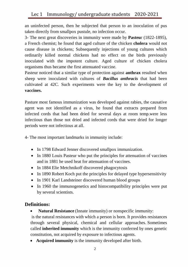
• Toll-like receptors (TLR,s): Innate immune receptors on certain leukocytes recognize pathogen- associated molecular patterns (PAMPs), which are common to many foreign invaders and are not normally present in the host.
Lec 1 Immunology/ undergraduate students 2020-2021
4
Fig 1: binding of a pathogen via its PAMP to a Toll-like receptors (TLR,s) PRR domain. the complement system : it is a set of over 20 different protein molecules always found in the blood. It can be activated by bacterial surface molecules; With an infection, this system of molecules is activated, leading to a sequence of events on the surface of the pathogen that helps destroy the pathogen and eliminate the infection. Complement is activated by innate immune response as well as by adaptive immune response. (In later lectures).
Natural resistance restriction factors: 1-Species Fowl malaria, avian tuberculosis, mouse pox, and many similar terms indicate the host range or species specificity certain pathogens possess. This also means that other species of animals are resistant to the infectious agent mentioned, e.g. human gets mumps but dogs and cats do not. Why one species contracts a certain disease when other species are resistant is not always explainable. In certain cases the body temps. of the animal will not permit growth of the

Lec 1 Immunology/ undergraduate students 2020-2021
5
pathogen, e.g. if body temp. of chicken is lowered from 39c to 37c, they become susceptible to anthrax. 2- Race or strain It is always difficult to evaluate differences between human races in there susceptibility or resistance to certain diseases, only in limited segments of population in which all persons share the same environment for example, military installations, prisons, and mental hospitals, do those populations show same resistance to infection for all individuals ??,Under these conditions racial differences can be detected. Blacks are more susceptible than whites to tuberculosis, and blacks are more resistant than whites to diphtheria, 3- Sex: Differences in susceptibility of 2 human sexes has been studied, such differences have been demonstrated among mice, for example: BALB/c female mice are more resistant to Listeria monocytogenes infection than males; however there was no difference among sexes in other mice strain like C57B/6. 4- Nutrition: Protein malnutrition lowers the level of C3 and factor B of the complement system, decreases the interferon response, and effect on the activity of T &B cells as well as neutrophils. Vitamin deficiencies exhibit significant effects on host defense. Vitamin A deficiency effects epithelial cell integrity and allows more skin infections to develop. Folic acid deficiencies lower T cell no. whereas vitamin B2, B1 deficiencies lower B cell activities .Vitamin C deficiencies have been related to increase or more severe bacterial and viral infections. Over nutrition may also harmful ,many bacteria needs iron for their growth, when transferrin and lactoferrin the body's natural iron transport compounds are saturated by an excess of this metal, free iron is available for bacterial growth, when iron is in short supply ,bacterial growth is restricted . 5- Hormone related resistance: Hormone imbalance (such as insulin diabetes mellitus) has a direct effect on susceptibility to a number of infections. Staphylococcal, streptococcal and certain fungal disease definitely occur more frequently in diabetics. Pregnancy is associated with marked hormonal alternations and an increase in urinary tract infections for example.

Lec 1 Immunology/ undergraduate students 2020-2021
6
6-Other factors: The age of an individual has a marked effect on immunity .The very young and very aged always have more infectious diseases than the middle aged groups. Mechanisms of natural resistance A- External defense factors: The first barrier that most microbes encounter against their successful invasion is the intact skin or mucus membranes. Skin represents a formidable mechanical barrier that only few organisms that can reach ,while being held outside the body ,the microorganisms are exposed to drying ,which alone may be enough to cause their death. In addition lactic acid, caproic acid and caprylic acid of the sebaceous glands have proven antibacterial effect. Saturated fatty acids are present in the skin secretions. These are fungistatic and are important in elimination of fungi that cause superficial skin and hair infections. Fungal infections are often being on areas of the body that devoid of sebaceous glands, for example between the toes. The uppermost portion of the respiratory tract is lined with visible hairs to exclude particles of great size, it is obvious that these will not remove particles the size of ordinary microbes, droplets in the range of 0.5 to 2um in diameter are inhaled somewhat more deeply than larger particles, the entire respiratory tract is bathed with mucus secretions that acts as a glue and holds particles coming in to contact with it directly on the membranes surface. The mucus is swept upward at a rate of 10-20 mm/ minute by the action of the ciliated epithelial cells .however the mucous membranes do not trap all droplet nuclei ,those less than 0.5 um in diameter maybe inhaled deeply into the pulmonary alveoli and initiate infection there ,some microorganisms may escape alveolar macrophages. In the digestive system the microorganisms encounter the tremendous acidity of the stomach, bordering on pH 1 because of the hydrochloric acid (HCL) secreted from the cells. As the microbe pass in to small intestine an alternation in pH to the slightly alkaline occurs because of the pH of the entering pancreatic and biliary fluids. A remarkable increase of microbe take place in the large intestines there are a tremendous no of nonpathogenic microorganisms

Lec 1 Immunology/ undergraduate students 2020-2021
7
there , if their no. is altered by a broad spectrum of antibiotic therapy ,intestinal yeast infections may occur .this is the role of natural flora in maintain body health. In the urinary system there is slight acidity, lysozyme content and the flushing action of the urine, all of which help cleans the urinary system. The exterior portions of the female reproductive system also are held at low pH because of the lactic acid in the secretions. The human eye is not protected by a third lid like that in lower animals but it is blessed with a defensive mechanism many lower animals' lack, the ability to cry; Crying has a simple mechanical benefit; it washes both small and large particles from the eye. A selective and very active antimicrobial substance, lysozyme, in tears is of much greater value. Lysozyme is a bactericidal enzyme, the cell walls of both grame +ve and to less extent grame –ve bacteria are susceptible to this enzyme. Lysozyme is a muraminidase that cleaves the ß-1, 4-glycoside bond that unites N-acetylglucosamine and muramic acid the back bone of the bacterial cell wall. Lipids in grame –ve cell wall tend to mask the substrate from lysozyme.
Fig2: Natural resistance factors that function on external surfaces of the body.

0
Lecture Two

Lec 2 Immunology/ undergraduate students 2020-2021
1
B- Internal defense factors Phagocytosis is the most vital of the internal defense factors of the immune defense system in animal. Natural internal defense system Phagocytes Neutrophils, Macrophages, Monocytes Opsonins C3b, Immunoglobulins Complement activation Alternative pathway, enzymatic activation, others Macromolecules Glycoprotein's, Lysozyme, Polyamines, others Complement is known to comprise 20 plasma proteins forming one of the major immune system components; it has different functions as attracting leucocytes, direct microbial killing, play important role in antibody development and other functions. Immunoglobulin's Igs (antibodies) they are soluble glycoproteins that recognize and bind to Antigens (Ags) present in the serum, tissue fluids, or on cell membranes. They are secreted by plasma cells. Soluble mediators of immunity A wide variety of molecules are involved in the development of immune response including;
- Antibodies (Abs) and cytokines produced by B lymphocytes and other cells. - Other molecules that are normally present in serum like acute phase proteins: The serum contains of many proteins that increase rapidly during infection and therefore called acute phase proteins such as C reactive protein (CRP).
2- Acquired Immunity(Adaptive immunity) Acquired immunity refers to that immunity which a person develops during a life time, it is antigen specific and may be based on humoral or circulating substances such as interferon (IFN) or antibodies, or may be cellular in origin which is more associated with the activities of macrophages and T lymphocytes.

Lec 2 Immunology/ undergraduate students 2020-2021
2
Antibody mediated immunity Immunity based on antibodies is most efficient; this form of immunity is divided in to active and passive immune response: І- Innate or natural resistance П-Acquired (adaptive) immunity: A- Natural acquired 1- Passive 2- Active B- Artificial acquired 1- Passive 2- Active In active immunity the individual synthesizes his own antibodies, whereas in passive immunity the individual receives these antibodies from some other individual, either a human or lower animal. Look at fig3.
Fig3: The main division and subdivision of immune response Actively acquired immunity (adaptive could be natural or artificial) A degree of naturally acquired active immunity results from any infection from which a person recovers, whether the illness is serious or sub clinical. Vaccines are developed that can be used in immunization, the immunity which is developed from giving an individual a vaccine of toxoid (Ag) is said to be artificially acquired type. Killed or attenuated strains of bacteria and viruses

Lec 2 Immunology/ undergraduate students 2020-2021
3
now are used widely for immunization against many diseases e.g. typhoid fever, smallpox, poliomyelitis. Toxoids that are detoxified but still antigenically active poisons excreted by certain bacteria are excellent antigens. Antibodies against toxoids are fully reactive with the native toxin and provide an excellent immunity against diseases caused by toxigenic bacteria (tetanus and diphtheria). Passively acquired immunity (adaptive could be natural or artificial) Passive immunity may be acquired by natural means, or by artificial means. Naturally acquired passive immunity in human beings usually refers to the transplacental passage of antibodies from the mother to her fetus, during the later part of her pregnancy. This is caused by IgG, since IgM, IgA, and IgD; do not pass the placental barrier. Colostrum contains secretory IgA and IgM but very little IgG. This nature protection is not found in bottle-fed babies. Artificial passive acquired immunity refers to the original production of antibodies in some other individual (could be human or animal) and giving these antibodies through a needle and syringe.
Fig 4: The adaptive immune response divisions.

Lec 2 Immunology/ undergraduate students 2020-2021
4
It must be remembered that foreign serum proteins are antigenic to the human being; consequently the human recipient will make antibodies against the administered foreign antibodies, which results in their speedy elimination. On the other hand, the reactivation of active immunity by booster injections of the vaccine or toxoid is comparatively risk free method which is commonly used Specificity and memory are key features of the adaptive immune response The high specificity of the adaptive immune response allows the generation of immunological memory – related to its use of highly individual antigen receptors. (Idea of vaccination).
Comparison of active and passive antibody mediated immunity is shown in this table:
Table 1: Comparison between active and passive immunization Active Passive Source
self
some other human or lower animal
Effectiveness High Moderate –low
Method 1- Disease itself 2- immunization:
Killed, attenuated or toxoids vaccine
- Maternal transplacental transfer of Abs
Time develop
5-14 days Immediate on injection
Duration Relatively long, years short few days t to several weeks

Lec 2 Immunology/ undergraduate students 2020-2021
5
Ease reactivation
Easy (booster) dangerous, possible anaphylaxis
Use Prophylactic prophylactic &therapeutic
Components of the immune system: cells, tissues& organs The lymphoid organs where lymphocytes differentiate and mature from stem cells are the primary lymphoid organs including: the thymus (site of T cells development), the fetal liver and postnatal bone marrow (the site of B cells development), then these cells (T &B) lineages migrate from the primary lymphoid organs to function in the secondary lymphoid organs- namely : the spleen, lymph nodes, mucosa –associated lymphoid tissues (MALT) , Table 2.

Lec 2 Immunology/ undergraduate students 2020-2021
6
Table 2: Primary and secondary lymphoid organs
In the adult animal, all immune cells originate from hematopoietic stem cells located in the bone marrow. Stems cells constantly divide and differentiate into various types of immune cells under the influence of cytokines. Cytokines: are small signaling proteins that help to regulate the behavior of the cells of the body. The bone marrow is ultimately responsible for the synthesis of eight types of cells: red blood cells, platelets, neutrophils, basophils, eosinophils, mast cells, monocytes/macrophages, T lymphocytes and B lymphocytes. Some of these cell types mature in the bone marrow itself, while others migrate through the circulatory system and undergo final maturation in other tissues.

Lec 2 Immunology/ undergraduate students 2020-2021
7
Fig 5: Production of blood cells from stem cells in the bone marrow. Lymphoid stem cells develop and mature within primary lymphoid organs: In mammals T cells mature in the thymus and B cells mature in the fetal liver and postnatal bone marrow. Birds have specialized site of B cell generation, the bursa of Fabricius.

Lec 2 Immunology/ undergraduate students 2020-2021
8
Lymphocytes migrate to and function in secondary lymphoid organs and tissues, Once they reach secondary tissues, they don’t remain there, they many move from one lymphoid organ to another via blood and lymph.
Fig 6: Origin of cells of the immune system

0
Lecture Three

Lec 3 Immunology/ undergraduate students 2020-2021
1
Cell markers As the cells of the immune system develop, they acquire molecules that are important for their function. These specific functional molecules are referred to as lineage markers because they identify the cell lineage e.g. lymphoid cells- T and B cells. Other marker molecules include those involved in regulating cell proliferation and function and those involved in regulating the number of cells participating in the immune response.
Fig 7: The immunoglobulin superfamily is the most important cell surface markers.
Antigen presenting cells A specialized cells termed antigen presenting cells (APC) that they digest the antigen and present it to other specialized cells .These professional APC include: macrophages, dendritic cells, and some B cells. 1-Macrophages

Lec 3 Immunology/ undergraduate students 2020-2021
2
A macrophage is a large white blood cell that is an important part of our immune system. The word macrophage literally means 'big eater.' It is an amoeba-like organism, and its job is to clean our body of microscopic debris and invaders. A macrophage has the ability to locate and 'eat' particles such as bacteria, viruses, fungi, and parasites.
They are born from white blood cells called monocytes, which are produced by stem cells in our bone marrow. Monocytes move through the blood stream, and when they leave the blood, they mature into macrophages; they increase inflammation stimulating the immune system.
They live for months, patrolling our cells and organs, Macrophages that encourage inflammation are called M1 macrophages, whereas those that decrease inflammation and encourage tissue repair are M2 macrophages. This difference is reflected in their metabolism, where macrophages have the unique ability to metabolize one amino acid, arginine, to either a "killer" molecule (nitric oxide) or a "repair" molecule (ornithine).
The Human macrophages are about 21 micrometers in diameter, they play very important role in immune response in phagocytosis, activation of immune cells wound healing, and play a role in iron homeostasis.
2-Dendritic cell
Fig 8: Dendritic cell with its projections.
Dendritic cells are found in tissue that has contact with the outside environment such as the over the skin (present as Langerhans cells) and in the linings of the nose, lungs, stomach and intestines. During development, they develop branched projections called “dendrites”, which is why the cells are so named, Fig 8 .

Lec 3 Immunology/ undergraduate students 2020-2021
3
3- B cells
B cells or B lymphocytes are a type of lymphocyte in the humoral immunity of the adaptive immune system. B cells can be distinguished from other lymphocytes, by the presence of a protein on the B cell's surface known as a B cell receptor (BCR). This specialized receptor protein allows a B cell to bind to a specific antigen.
Types of B cell:
• Plasma B cells are large B cells that have been exposed to antigen to produce and secrete large amounts of antibodies,.
Fig 9: B cell recognizes the antigen by specific Ig receptor.
• Memory B cells differentiate from activated B cells, specific to the antigen encountered during the primary immune response. These cells are able to live for a long time, and can respond quickly following a second exposure to the same antigen.
4-T Cells: T cells or T lymphocytes are a type of lymphocyte (itself a type of white blood cell) that play a central role in cell-mediated immunity (CMI). They can be distinguished from other lymphocytes, by the presence of a T-cell receptor (TCR) on the cell surface. There are several subsets of T cells, each with a

Lec 3 Immunology/ undergraduate students 2020-2021
4
distinct function. They recognize Ag via surface receptors T cell receptor (TCR). 1- They are called the T helper cells (Th), their cellular marker is called CD4.
Fig 11: CD4 cells interact with APC 2- Other type of T cell is responsible for destruction of host cells that are infected by intracellular pathogen (virus) and play important role in response to tumor, this kind of action is called cytotoxicity, these cells are called cytotoxic Tcells( Tc ),their cellular marker is called CD8. Look at the fig.
Fig 12: CD8 T cells interact with infected cells.

Lec 3 Immunology/ undergraduate students 2020-2021
5
3- Other T cells include memory cells, and regulatory cells.
5-NK Cells:
They are type of cytotoxic lymphocyte critical to the innate immune system. The role NK cells play is analogous to that of cytotoxic T cells, in vertebrate adaptive immune response. NK cells provide rapid responses to virally infected cells and respond to tumor formation, acting at around 3 days after infection. They account for 15% of blood lymphocytes.
Fig 13: Role of NK cells in eliminating tumor cells.

Lec 3 Immunology/ undergraduate students 2020-2021
6
Ploymorphonuclear granulocytes
Polymorph nuclear leukocytes (PMN), or granulocytes, are a type of white blood cell that have lobed nucleus. They arise from the myeloid cell line in the bone marrow and help the body to fight infection. PMNs include neutrophils, basophils and eosinophils, which circulate in the bloodstream, as well as mast cells, which reside in the tissues. Their primary role is to digest any foreign invaders. PMNs are referred to as granulocytes because of their appearance under the microscope. When they are stained for examination, the cytoplasm of the cell takes on a grainy appearance.
Eosinophiles These cells comprise 2-5% of blood leukocytes in healthy non allergic individuals. Human blood eosinophiles usually have bilobed nucleus and many cytoplasmic granules which stain with acidic dye as eosin. They are capable of phagocytosing and killing ingested microorganisms., they are important in allergic reaction Basophil & mast cell
These cells are found in very few numbers in the circulation and account for less than 0.2% of leukocytes. They mature in bone marrow while mast cells not found in the circulation and they mature in the connective tissue. These two types of cells are stimulated by an allergen (molecule that causes allergic reaction) thus they play important role in hypersensitivity reactions.

Lec 3 Immunology/ undergraduate students 2020-2021
7
Fig 14 : Basophil and mast cell functions Platelets Platelets are not cells, but cell fragments derived from megakaryocytes in the bone marrow. They contain granules, microtubules and actin /myosin f ilaments which are involved in clot contraction.
Lec 3 Immunology/ undergraduate students 2020-2021
8
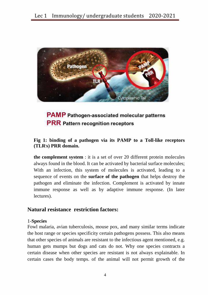
Phagocytosis: Neutrophils & Macrophages The most effective dynamic and complex defense strategies are carried out by specialized cells that travel through the body to search out and destroy microorganisms and other foreign substances. These include: Neutrophils, monocytes, and lymphocytes each spend a portion of their lives circulating in the blood. For differentiate between these cells look at the table. Table 3: properties of the 3 major human cell lineages involved in host defense.
Lymphocytes Monocyte- macrophages
Neutrophils
varies Phagocytosis Phagocytosis Primary function
No
Moderate
Many
Cytoplasmic granules
Lymphoid tissues
All tissues Blood, bone marrow
normal location
Yes Yes No Immunoregulatory cytokine production
Yes Yes No Antigen presentation
Neutrophils They are known as polymorph nuclear (PMN) leukocytes, they can be easily identified under light microscope by their characteristic multilobed nucleus and by abundant storage granules in their cytoplasm .The granules are specialized lysosome-membrane bounded vesicles contain digestive enzymes and other proteins that can be used to break down particles that have been engulfed by the cells. Most neutrophil granules are of 2 types called azurophilic granules and specific granules.

0
Lecture Four
Lec 4 Immunology/ undergraduate students 2020-2021
1
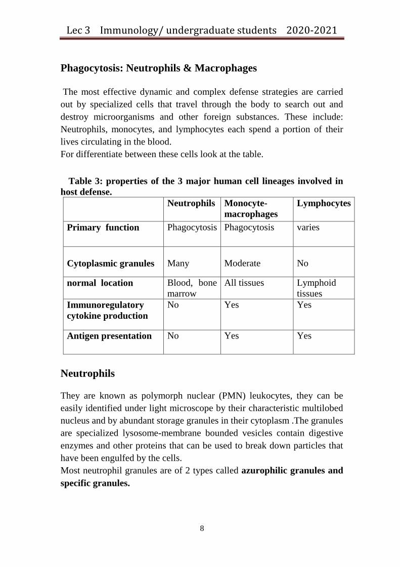
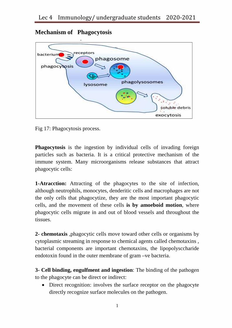
Mechanism of Phagocytosis .
Fig 17: Phagocytosis process. Phagocytosis is the ingestion by individual cells of invading foreign particles such as bacteria. It is a critical protective mechanism of the immune system. Many microorganisms release substances that attract phagocytic cells: 1-Atracction: Attracting of the phagocytes to the site of infection, although neutrophils, monocytes, denderitic cells and macrophages are not the only cells that phagocytize, they are the most important phagocytic cells, and the movement of these cells is by amoeboid motion, where phagocytic cells migrate in and out of blood vessels and throughout the tissues. 2- chemotaxis ,phagocytic cells move toward other cells or organisms by cytoplasmic streaming in response to chemical agents called chemotaxins , bacterial components are important chemotaxins, the lipopolysccharide endotoxin found in the outer membrane of gram –ve bacteria. 3- Cell binding, engulfment and ingestion: The binding of the pathogen to the phagocyte can be direct or indirect:
• Direct recognition: involves the surface receptor on the phagocyte directly recognize surface molecules on the pathogen.

Lec 4 Immunology/ undergraduate students 2020-2021
2
• Indirect recognition involves the deposition of serum derived molecules on to pathogen surface, and their subsequent binding to receptors on phagocyte (process of opsonization by antibodies or C3b). fig 18.
Fig 18: Opsonization of bacteria by antibody to be ingested by phagocyte. Phagocytes have receptors that recognize pathogens directly -without opsonization:
In absence of opsonins ,macrophages have a no. of receptors that allow them to recognize the pathogen associated molecules(PAMs),these are the biological macromolecules on the surface of the pathogen can be recognized by the elements of the innate immune system .For example the receptors on surface of macrophage cells that recognize these PAMs is called Toll like receptors(TLR s) bind to gram +ve , -ve bacteria, mycobarium, viruses, fungi.
Fig 19: Phagosome formation

Lec 4 Immunology/ undergraduate students 2020-2021
3
. Fig 20: formation of the phagolysosome by fusion of phagosome with lysosome... 4-destruction: Phagocytes can also damage invading pathogens through the generation of toxic products in a process known as the respiratory burst( oxidative burst). Production of these toxic metabolites is induced during phagocytosis of pathogens such as bacteria and catalyzed by a set of interrelated enzyme pathways. 2 types of granules exist in neutrophils: a- azurophilic granules (primary granules): like elastase, defensing, Myeloperoxidase (MPO)). b- Specific granules (secondary granules) which comprise 67% of all lysosomal granules, they contain alkaline phosphatase, lactoferrin and lysozyme The lysosomal granules of macrophages contain a similar array of enzymes and antimicrobial products. Mechanisms of Destruction The granule contents destroy foreign particles by 2main mechanisms, one is oxygen independent mechanism, and the other is oxygen dependent: 1- Oxygen independent intracellular killing this is done by the activity of various enzymes such as hydrolytic enzymes that are able to digest cell wall components of some bacteria ,or by defensins which are cationic proteins they are not enzymes but basic peptides contain large amount of arginine that kill microbes by interacting with the microbial cell

Lec 4 Immunology/ undergraduate students 2020-2021
4
membrane to form channels through which essential metabolites escape ,lysozyme which attacks bacterial cell walls and render the cell osmotically sensitive ,lactoferrine that binds iron, nitric oxide synthetase this enzyme is toxic to fungi ,tumor cells and some bacteria because the activity of this enzyme yields nitric oxide which is toxic . 2- Oxygen dependent intracellular killing:, It depends on the byproduct of the respiratory burst that accompanies phagocytosis and produce several other microbial oxygen metabolites:
A-Enzymatic generation of super oxide anion. glucose +NADP →NADPH + pentose phosphate NADPH + oxygen + NADPH oxidase → NADP + O 2- (toxic)
B-Spontaneous generation of single oxygen, hydrogen peroxide, and hydroxyl radicals O 2- + 2 H - → H2O2 + O- (toxic to bacteria) O2- + H2O2 → 2 OH - + 2 O-
C-Enzymatic generation of halogenating compounds: H2O2 + halides (Chloride ion) + MPO → hypochlorite (this product is antimicrobial more than H2O2, halides, and MPO), those activated halide could damage microorganisms by halogenation of bacterial cell wall or by decarboxylation of amino acids with the resultant production of toxic aldehydes.
Fig 21: The mechanism of oxidative burst.

0
Lecture Five

Lec 5 Immunology/ undergraduate students 2020-2021
1
Inflammation
1- Inflammation is a process that brings leucocytes and plasma molecules to sites of infection or tissue damage. The principal effects are an increase in blood supply, an increase in vascular permeability to large serum molecules and enhanced migration of leucocytes across the local vascular endothelium and in the direction of the site of inflammation. 2- The migration of cells is a complex process that depends on which populations of cells are involved, their state of activation and how they interact with endothelium in different vascular beds through the body. 3- Chemotactic molecules are important both in directing cell migration and in triggering leucocytes at the endothelial surface to initiate their migration. 4- Inflammatory mediators are released by mast cells, platelets and leucocytes in immune reactions, or following tissue damage act in concert with molecules released by the plasma enzyme systems to control vascular permeability and blood supply. Inflammation is the body's reaction to invasion by an infectious agent, antigen challenge, or a physical damage. The main symptoms include redness, swelling, heat, pain and loss of function of the inflamed area, it has 3 major components:
a- Increase in blood supply to infected area, bringing leucocytes and serum molecules to the affected sites. b- Increase in capillary permeability allowing exudation of the serum proteins (Abs, complement, etc.) required to control the infection. an increase in capillary permeability is caused by increase vascular transport across the endothelium. This permits larger molecules to traverse the endothelium thus allow antibody, complement and molecules of plasma enzymes to reach the site. c- Leucocytes migration in to damaged tissue increases.

Lec 5 Immunology/ undergraduate students 2020-2021
2
Migration of leucocytes
The migration of these cells from the blood stream to infected site depends on the expression of the adhesion molecules on the endothelium, in the inflammatory site the endothelium express adhesion molecules which are recognized by receptors on activated lymphocytes, or phagocytes. Neutrophils are the first cells to arrive at sites of inflammation caused by infection; they represent the major cell type for several days. If the antigen is not cleared the acute reaction will change to chronic inflammatory reaction, in this case few neutrophils are seen but large no. of CD4 Tcells (Th cells) and mononuclear phagocytes accumulate as well as B cells. There are 2 main stages in leucocytes migration: 1- The attachment of circulating cells to the vascular endothelium followed by movement between and through the endothelium cells. 2- Then cells migrate toward sites of infection under guidance of chemotactic stimuli.
Fig 23 : Main steps in acute inflammation. Inflammation is controlled by chemokines, plasma enzyme systems, cytokines and the products of mast cells, platelets and leucocytes. There are different types of mediators which control inflammatory reactions:

Lec 5 Immunology/ undergraduate students 2020-2021
3
Fast – acting mediators such as vasoactive amines, - Histamine released by mast cells and basophils increase the vascular permeability ,smooth muscle contraction -C3a and C5a causes mast cells degranulation, smooth muscle contraction, increase capillary permeability -binding of IgE to mast cells causes degranulation of the cell and trigger other cells.
Fig 24: Cytokines and Cell mediators play important role in cell migration in inflammation.
Fig 25: The inflammation may be acute or chronic depending on the stimulus.

Lec 5 Immunology/ undergraduate students 2020-2021
4
Effector systems to eradicate the antigen
1- Phagocytosis 2- Neutralization of viruses, toxins and venoms): antibodies can combat certain pathogens just by binding to them, for example antibody to the outer coat proteins of some rhinoviruses (which cause common cold) can prevent viral particles from binding to the cell receptors and prevent the infection. (fig 26)
Fig 26: Antibody neutralization.
3-Cytotoxic reactions are directed against whole cell that is too large to be phagocytized. In cytotoxic reactions the attacking cells direct their granules towards the target cell; as a result the granules are discharged into extracellular space close to the target cell.
The target cell may be recognized by: Specific antibody bound to the cell surface = antibody –
dependent cell mediated cytotoxicity (ADCC) T cell using their specific T cell receptors (TCR) = cell mediated
cytotoxicity which does not depend on Ab.
Cell mediated cytotoxicity (CMC) describes the way in which leucocytes recognize and destroy other cells; it is an essential defense against
• Intracellular pathogens, including viruses. • Some bacteria and some parasites. • Tumor cells.

Lec 5 Immunology/ undergraduate students 2020-2021
5
Several types of cells have cytotoxic activity including Tc, natural killer cells (NK). Tc has a marker CD8, this molecule can recognize specific surface protein that is present on the nucleated cells (MHC class 1), when the host cell is infected it will present the antigen bounded to MHC class 1,at the same time Tc has a TCR that can interact the complex (Ag+ MHC1) .
Fig 27: Recognition of target cell by Tc cells (e.g. for CMC)
Fig 28: The action of Tc cell on infected cell.

Lec 5 Immunology/ undergraduate students 2020-2021
6
Nk cells identify their target in a different way, they have a receptor to antibody (Ig), thus when an antibody binds to the target cell ,then NK cells interact with this complex and releases its cytotoxic enzymes to lies the infected cell This is called the ADCC.
Fig: Destruction of infected cell by NK cells through ADCC

0
Lecture Six

Lec 6 Immunology/ undergraduate students 2020-2021
1
The Complement System
• The complement system represents one of the main arms of innate immunity that bridges innate and acquired immune response, it supplies the body with a very important guard against pathogenic invasion, as well as a crucial participation in the pathogenesis of many autoimmune and inflammatory diseases and a novel non-inflammatory role in tissue regeneration and repair.
• Initially, complement was considered as a heat-labile source in serum which “complements” antibodies in bactericidal function. In terms of number of components, the complement system is composed of over than 50 proteins in plasma and on the surfaces of cells, these proteins amount to around 15% of the globulins.
• Although the complement factors in circulation are generally produced in the liver, it is now understood that most tissues and inflammatory cells like neutrophils and macrophages, are effective origins of complement factors.
• In addition to the main proteins of the complement cascade (C1-C9), the whole complement network consist of fluid-phase, cell-surface-associated and intracellular proteins that acts as pattern-recognition molecules (PRMs), proteases and convertases, regulators and signaling receptors that collectively mediate immune surveillance and tissue homeostasis.
• Several complement proteins are zymogens which are disseminated safely through the body and activated at the site of inflammation in a triggered enzyme cascade pattern where a proteolytic cleavage removes an inhibitory fragment and exposes the active site.
• Complement proteins is arranged in a hierarchical structure of the proteolytic cascade which is initiated with danger molecule surface identification, thereafter the subsequent event which is the release of potent proinflammatory mediators (Anaphylatoxins), opsonization and ultimately the analysis of the target by membrane attack complex (MAC) formation.

Lec 6 Immunology/ undergraduate students 2020-2021
2
• Basically, the complement system can be activated via three
pathways, the classical pathway (CP) that mainly tackles antibody-antigen complex. The second pathway is the lectin, which is directed to kill pathogens that expose mannose in their structure. Finally, the alternative pathway (AP) destroys different sorts of contagious factors such as viruses, bacteria and fungi, as well as having a role in immune surveillance against tumors.
• These pathways are linked into a common termination lane, leading to the formation of the membrane attack complex (MAC), this complex is composed of C5b, C6, C7, C8 and several C9 molecules and causes lysis of the pathogen, fig30.
• Different from many other enzymatic cascades, the complement system represents a more sophisticated pattern as it is based on the formation of consecutive activated protein fragments, which subsequently turn into convertases that cleave other complement proteins to form the terminal complex, these complexes maintain their stability and activity for a very short time.

Lec 6 Immunology/ undergraduate students 2020-2021
3
• Complement activation steps are monitored by many inhibitors, in order to achieve a balance between active detection and destruction of pathogens and reduction of bystander tissue damage. This regulation occurs mainly at the convertase level, and through the assembly of MAC.
• Functionally, this array of complement proteins in blood and
interstitial fluids together supply the immune system with multi-defense strategies upon activation, fig31. These include:
1. Direct lysis of targeted surfaces through MAC assembly (C5b-C9) which damages some bacteria and enveloped viruses, fig32.
2. Opsonization and removal of targeted surfaces via the opsonic complement fragments C4b, C3b, iC3b which bind to their corresponding complement receptors (CRs) on phagocytes like macrophages and neutrophils, fig33.
3. Stimulation of an inflammatory response by potent pro-inflammatory chemoattractants and anaphylatoxins. C5a is chemotactic for macrophages and polymorphs, C3a and C5a activate mast cells and basophils to trigger massive release of histamine and cytokines, fig34.
4. Priming of the adaptive immune response. Immune complexes with bound C3b are very efficient in priming B cells.
5. C3b disaggregates immune complexes and promotes their clearance.
6. The complement system has anti-inflammatory functions too, as it binds to immune complexes and apoptotic cells and helps in their clearance from the circulation and damaged tissues.

Lec 6 Immunology/ undergraduate students 2020-2021
4

Lec 6 Immunology/ undergraduate students 2020-2021
5
Complement Activation Pathways
(Three pathways, different starts but same end)
The potent activation products of full complement cascade: 1. C3 & C5 convertases (Enzymatic activity). 2. Opsonins: C3b & C4b (CP&MBL only). 3. Anaphylatoxins: C5a, C3a & C4a (CP&MBL only). 4. MAC (C5b-C9).
The classical Pathway • Main target (ligand) : antibody–antigen complexes. • Complement Recognition molecules: C1q –
C1r2C1s2 • Initiation Cleavage enzyme (Serine protease):
Activated C1s

Lec 6 Immunology/ undergraduate students 2020-2021
6
• Initiation and complete activation steps:
1. C1q –C1r2C1s2 binds to a ligand surface.
2. Consequently, C1s is activated and cleaves C4 into C4a &C4b and C2 into C2a&C2b.
3. C4b&C2a bind to compose the C3 convertase C4b2a
4. C4b2a cleaves C3 into C3a (anaphlyatoxin) &C3b (opsonin).
5. Another C3b molecule binds to C4b2a to form the C5 convertase C4b2aC3b.
6. C4b2aC3b splits C5 into C5a (Powerful anaphylatoxin) and C5b.
C5b fragment fuses with C6, C7, C8 and several C9 molecules to form the MAC and causes lysis to the pathogen via insertion and functional pore construction on the targeted cellular membranes.
The Lectin Pathway • Main target (ligand) : The surface microbial mannose molecules. • Recognition molecules: Mannose binding lectin MBL & ficolins. • Initiation cleavage enzyme (Serine protease): MBL-associated
serine proteases MASP2.
Initiation and complete activation steps:
1. Lectin recognition molecules (MBL or ficolins) bind to a microbial surface.
2. Consequently, MASP2 is activated and cleave C4 into C4a &C4b and C2 into C2a and C2b. From this point on, the CP and lectin complement pathways share the same events.

Lec 6 Immunology/ undergraduate students 2020-2021
7
Lec 6 Immunology/ undergraduate students 2020-2021
8
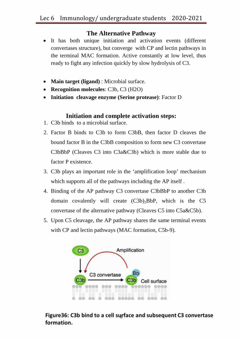
The Alternative Pathway • It has both unique initiation and activation events (different
convertases structure), but converge with CP and lectin pathways in the terminal MAC formation. Active constantly at low level, thus ready to fight any infection quickly by slow hydrolysis of C3.
• Main target (ligand) : Microbial surface. • Recognition molecules: C3b, C3 (H2O) • Initiation cleavage enzyme (Serine protease): Factor D
Initiation and complete activation steps:
1. C3b binds to a microbial surface.
2. Factor B binds to C3b to form C3bB, then factor D cleaves the
bound factor B in the C3bB composition to form new C3 convertase
C3bBbP (Cleaves C3 into C3a&C3b) which is more stable due to
factor P existence.
3. C3b plays an important role in the ‘amplification loop’ mechanism
which supports all of the pathways including the AP itself .
4. Binding of the AP pathway C3 convertase C3bBbP to another C3b
domain covalently will create (C3b)2BbP, which is the C5
convertase of the alternative pathway (Cleaves C5 into C5a&C5b).
5. Upon C5 cleavage, the AP pathway shares the same terminal events
with CP and lectin pathways (MAC formation, C5b-9).
Lec 6 Immunology/ undergraduate students 2020-2021
9
Complement deficiency
• Any disturbance in the complement system balance may lead to
serious harmful effects. Disorganized complement activity may be
due to several factors including excessive activation, insufficient
regulation or/and complement component deficiency.
• Less complement activity can result primarily from genetic
deficiency of complement proteins or may occur temporarily due to
complement consumption via certain conditions like infection,
injury or surgery.
• Deficiency of the early component of the CP in addition to C2 and
C4 is often connected with systemic lupus erythematous SLE.
• Lack of MBL is linked to respiratory tract infections, while MASP2
deficiency results in recurrent infections.
• Factor P deficiency was found to increase Neisseria meningitides
mediated mortality , while factrs B&D deficiency causes severe
recurrent bacterial infections.

Lec 6 Immunology/ undergraduate students 2020-2021
10

Lec 6 Immunology/ undergraduate students 2020-2021
11
Lecture Seven
Lec 7 Immunology/ under graduate students 2020-2021
1
Immunoglobulins (Igs)
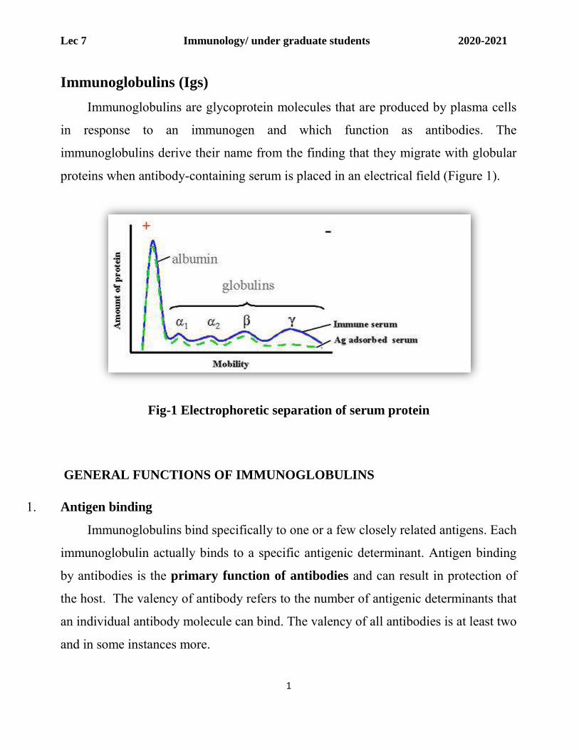
Immunoglobulins are glycoprotein molecules that are produced by plasma cells
in response to an immunogen and which function as antibodies. The
immunoglobulins derive their name from the finding that they migrate with globular
proteins when antibody-containing serum is placed in an electrical field (Figure 1).
Fig-1 Electrophoretic separation of serum protein
GENERAL FUNCTIONS OF IMMUNOGLOBULINS
1. Antigen binding
Immunoglobulins bind specifically to one or a few closely related antigens. Each
immunoglobulin actually binds to a specific antigenic determinant. Antigen binding
by antibodies is the primary function of antibodies and can result in protection of
the host. The valency of antibody refers to the number of antigenic determinants that
an individual antibody molecule can bind. The valency of all antibodies is at least two
and in some instances more.

Lec 7 Immunology/ under graduate students 2020-2021
2
Effector Functions
Frequently the binding of an antibody to an antigen has no direct biological
effect. Rather, the significant biological effects are a consequence of secondary
"effector functions" of antibodies. The immunoglobulins mediate a variety of these
effector functions. Usually the ability to carry out a particular effector function
requires that the antibody bind to its antigen. Not every immunoglobulin will mediate
all effector functions. Such effector functions include:
1. Fixation of complement - This result in lysis of cells and release of biologically
active molecules
2. Binding to various cell types - Phagocytic cells, lymphocytes, platelets, mast cells,
and basophils have receptors that bind immunoglobulins. This binding can activate
the cells to perform some function. Some immunoglobulins also bind to receptors on
placental trophoblasts, which results in transfer of the immunoglobulin across the
placenta. As a result, the transferred maternal antibodies provide immunity to the
fetus and newborn
BASIC STRUCTURE OF IMMUNOGLOBULINS
The basic structure of the immunoglobulins is illustrated in figure 2. Although
different immunoglobulins can differ structurally, they all are built from the same
basic units.
1. Heavy and Light Chains
All immunoglobulins have a four chain structure as their basic unit. They are
composed of two identical light chains (23kD) and two identical heavy chains (50-
70kD).
2. Disulfide bonds
Lec 7 Immunology/ under graduate students 2020-2021
3
1. Inter-chain disulfide bonds - The heavy and light chains and the two heavy chains
are held together by inter-chain disulfide bonds and by non-covalent interactions. The
number of inter-chain disulfide bonds varies among different immunoglobulin
molecules.
2. Intra-chain disulfide binds - Within each of the polypeptide chains there are also
intra-chain disulfide bonds.
3. Variable (V) and Constant (C) Regions
When the amino acid sequences of many different heavy chains and light chains were
compared, it became clear that both the heavy and light chain could be divided into
two regions based on variability in the amino acid sequences. These are the:
1. Light Chain - VL (110 amino acids) and CL (110 amino acids)
2. Heavy Chain - VH (110 amino acids) and CH (330-440 amino acids)
4. Hinge Region
This is the region at which the arms of the antibody molecule forms a Y. It is
called the hinge region because there is some flexibility in the molecule at this
point.
5. Domains
Three dimensional images of the immunoglobulin molecule show that it is not
straight as depicted in figure 2. Rather, it is folded into globular regions each of
which contains an intra-chain disulfide bond. These regions are called domains.
1. Light Chain Domains - VL and CL
2. Heavy Chain Domains - VH, CH1 - CH3 (or CH4)

Lec 7 Immunology/ under graduate students 2020-2021
4
Fig -2-The basic structure of immunoglobulin
6. Oligosaccharides
Carbohydrates are attached to the CH2 domain in most immunoglobulins.
However, in some cases carbohydrates may also be attached at other locations.
IMMUNOGLOBULIN FRAGMENTS:
Immunoglobulin fragments produced by proteolytic digestion have proven very
useful in elucidating structure/function relationships in immunoglobulins.
A. Fab
Digestion with papain breaks the immunoglobulin molecule in the hinge region before the H-H inter-chain disulfide bond Figure 3. This results in the formation of two identical fragments that contain the light chain and the VH and CH1 domains of the heavy chain.
Antigen binding - These fragments were called the Fab fragments because they
contained the antigen binding sites of the antibody. Each Fab fragment is monovalent
whereas the original molecule was divalent. The combining site of the antibody is
created by both VH and VL. An antibody is able to bind a particular antigenic
determinant because it has a particular combination of VH and VL. Different

Lec 7 Immunology/ under graduate students 2020-2021
5
combinations of a VH and VL result in antibodies that can bind a antigenic
determinants.
Fig-3
Fc Digestion with papain also produces a fragment that contains the remainder of
the two heavy chains each containing a CH2 and CH3 domain. This fragment was called Fc because it was easily crystallized.
Effector functions:
The effector functions of immunoglobulins are mediated by this part of the
molecule. Different functions are mediated by the different domains in this fragment
(figure 4). Normally the ability of an antibody to carry out an effector function
requires the prior binding of an antigen; however, there are exceptions to this rule.

Lec 7 Immunology/ under graduate students 2020-2021
6
Fig-4:immunoglobulin fragments: structure function relationships
F(ab')2
Treatment of immunoglobulins with pepsin results in cleavage of the heavy
chain after the H-H inter-chain disulfide bonds resulting in a fragment that contains
both antigen binding sites (figure 5). This fragment was called F(ab')2because it is
divalent. The Fc region of the molecule is digested into small peptides by pepsin. The
F(ab')2binds antigen but it does not mediate the effector functions of antibodies.
Fig-5
Immunoglobulin classes The immunoglobulins can be divided into five different classes, based on
differences in the amino acid sequences in the constant region of the heavy
chains. All immunoglobulins within a given class will have very similar heavy
chain constant regions. These differences can be detected by sequence studies or

Lec 7 Immunology/ under graduate students 2020-2021
7
more commonly by serological means (i.e. by the use of antibodies directed to
these differences).
1. IgG - Gamma heavy chains
2. IgM - Mu heavy chains
3. IgA - Alpha heavy chains
4. IgD - Delta heavy chains
5. IgE - Epsilon heavy chains
Immunoglobulin Subclasses The classes of immunoglobulins can be divided into subclasses based on small
differences in the amino acid sequences in the constant region of the heavy
chains. All immunoglobulins within a subclass will have very similar heavy chain
constant region amino acid sequences. Again these differences are most commonly
detected by serological means.
1. IgG Subclasses
a) IgG1 - Gamma 1 heavy chains
b) IgG2 - Gamma 2 heavy chains
c) IgG3 - Gamma 3 heavy chains
d) IgG4 - Gamma 4 heavy chains
2. IgA Subclasses
a) IgA1 - Alpha 1 heavy chains
b) IgA2 - Alpha 2 heavy chains
Immunoglobulin Types

Lec 7 Immunology/ under graduate students 2020-2021
8
Immunoglobulins can also be classified by the type of light chain that they have.
Light chain types are based on differences in the amino acid sequence in the constant
region of the light chain. These differences are detected by serological means.
1. Kappa light chains
2. Lambda light chains
Immunoglobulin Subtypes
The light chains can also be divided into subtypes based on differences in the
amino acid sequences in the constant region of the light chain.
1. Lambda subtypes
a) Lambda 1 b) Lambda 2 c) Lambda 3 d) Lambda 4
Nomenclature
Immunoglobulins are named based on the class, or subclass of the heavy chain
and type or subtype of light chain. Unless it is stated precisely, you should assume
that all subclass, types and subtypes are present. IgG means that all subclasses and
types are present.
STRUCTURE AND SOME PROPERTIES OF IG CLASSES AND
SUBCLASSES
1- IgG
Structure

Lec 7 Immunology/ under graduate students 2020-2021
9
The structures of the IgG subclasses are presented in figure 6. All IgG's are monomers (7S immunoglobulin). The subclasses differ in the number of disulfide bonds and length of the hinge region.
Properties
IgG is the most versatile immunoglobulin because it is capable of carrying
out all of the functions of immunoglobulin molecules.
a) IgG is the major Ig in serum - 75% of serum Ig is IgG
b) IgG is the major Ig in extra vascular spaces
c) Placental transfer - IgG is the only class of Ig that crosses the placenta. Transfer is
mediated by a receptor on placental cells for the Fc region of IgG. Not all subclasses
cross equally well; IgG2 does not cross well.
d) Fixes complement - Not all subclasses fix equally well; IgG4 does not fix
complement
e) Binding to cells - Macrophages, monocytes, PMNs and some lymphocytes have Fc
receptors for the Fc region of IgG. Not all subclasses bind equally well; IgG2 and
IgG4 do not bind to Fc receptors. A consequence of binding to the Fc receptors on
PMNs, monocytes and macrophages is that the cell can now internalize the antigen
better. The antibody has prepared the antigen for eating by the phagocytic cells. The
term opsonin is used to describe substances that enhance phagocytosis. IgG is a good
opsonin. Binding of IgG to Fc receptors on other types of cells results in the
activation of other functions.

Lec 7 Immunology/ under graduate students 2020-2021
10
Fig -6
2- IgM
Structure
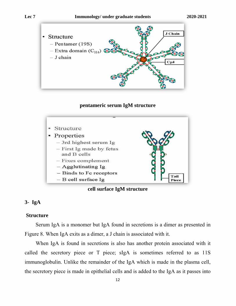
The structure of IgM is presented in figure 7. IgM normally exists as a pentamer
(19S immunoglobulin) but it can also exist as a monomer. In the pentameric form all
heavy chains are identical and all light chains are identical. Thus, the valence is
theoretically 10. IgM has an extra domain on the mu chain (CH4) and it has another
protein covalently bound via a S-S bond called the J chain. This chain functions in
polymerization of the molecule into a pentamer.
Properties
a) IgM is the third most common serum Ig.
b) IgM is the first Ig to be made by the fetus and the first Ig to be made by a virgin B
cells when it is stimulated by antigen.
c) As a consequence of its pentameric structure, IgM is a good complement fixing Ig.
Thus, IgM antibodies are very efficient in leading to the lysis of microorganisms.

Lec 7 Immunology/ under graduate students 2020-2021
11
d) As a consequence of its structure, IgM is also a good agglutinating Ig . Thus, IgM antibodies are very good in clumping microorganisms for eventual elimination from the body.
e) IgM binds to some cells via Fc receptors.
f) B cell surface Ig
Surface IgM exists as a monomer and lacks J chain but it has an extra 20 amino
acids at the C-terminus to anchor it into the membrane (figure 7). Cell surface IgM
functions as a receptor for antigen on B cells. Surface IgM is noncovalently
associated with two additional proteins in the membrane of the B cell called Ig-alpha
and Ig-beta as indicated in figure 10. These additional proteins act as signal
transducing molecules since the cytoplasmic tail of the Ig molecule itself is too short
to transduce a signal. Contact between surface immunoglobulin and an antigen is
required before a signal can be transduced by the Ig-alpha and Ig-beta chains. In the
case of T-independent antigens, contact between the antigen and surface
immunoglobulin is sufficient to activate B cells to differentiate into antibody
secreting plasma cells. However, for T-dependent antigens, a second signal provided
by helper T cells is required before B cells are activated.
Lec 7 Immunology/ under graduate students 2020-2021
12
pentameric serum IgM structure
cell surface IgM structure
3- IgA
Structure
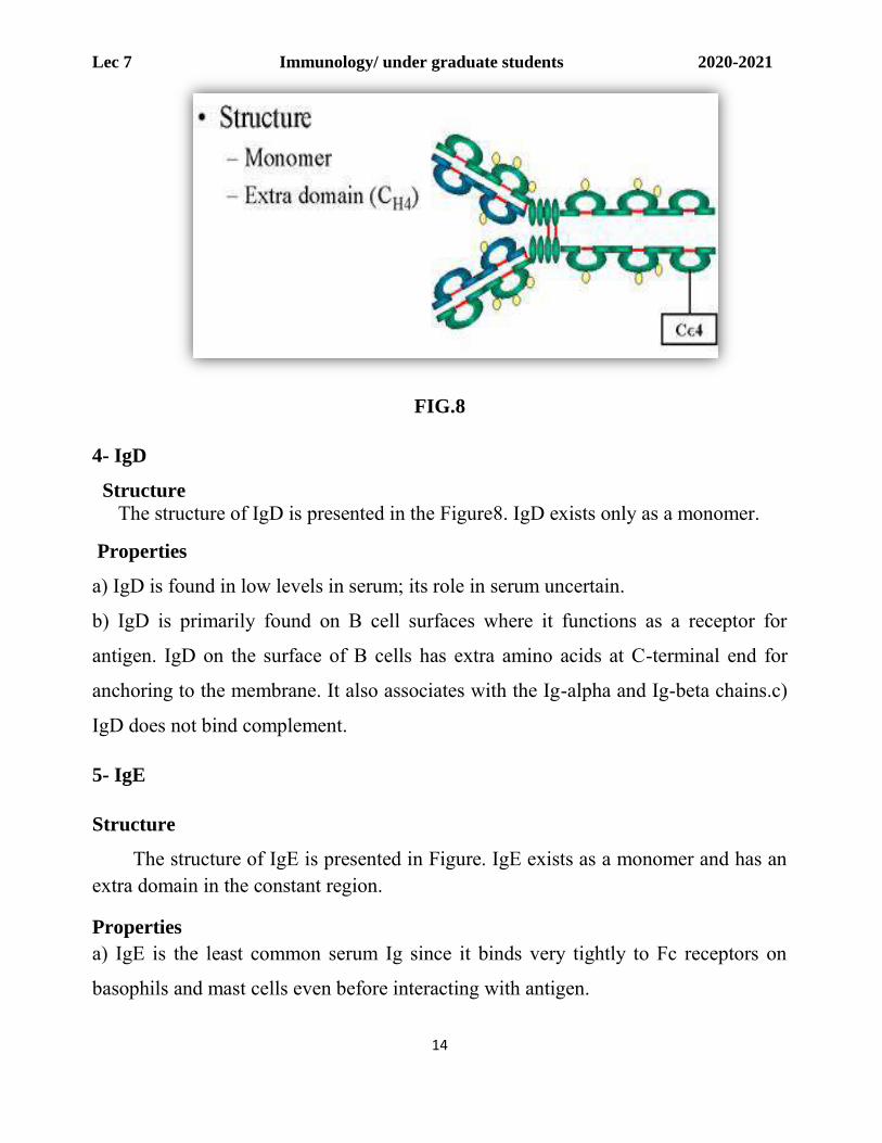
Serum IgA is a monomer but IgA found in secretions is a dimer as presented in
Figure 8. When IgA exits as a dimer, a J chain is associated with it.
When IgA is found in secretions is also has another protein associated with it
called the secretory piece or T piece; sIgA is sometimes referred to as 11S
immunoglobulin. Unlike the remainder of the IgA which is made in the plasma cell,
the secretory piece is made in epithelial cells and is added to the IgA as it passes into

Lec 7 Immunology/ under graduate students 2020-2021
13
the secretions (Figure 8). The secretory piece helps IgA to be transported across
mucosa and also protects it from degradation in the secretions.
Properties
a- IgA is the 2nd most common serum Ig.
b) IgA is the major class of Ig in secretions - tears, saliva, colostrum, mucus. Since it
is found in secretions secretory IgA is important in local (mucosal) immunity
c) Normally IgA does not fix complement, unless aggregated. d- IgA can binding
to some cells PMN & some lymphocyte
.
Lec 7 Immunology/ under graduate students 2020-2021
14
FIG.8
4- IgD
Structure The structure of IgD is presented in the Figure8. IgD exists only as a monomer.
Properties
a) IgD is found in low levels in serum; its role in serum uncertain.
b) IgD is primarily found on B cell surfaces where it functions as a receptor for
antigen. IgD on the surface of B cells has extra amino acids at C-terminal end for
anchoring to the membrane. It also associates with the Ig-alpha and Ig-beta chains.c)
IgD does not bind complement.
5- IgE
Structure
The structure of IgE is presented in Figure. IgE exists as a monomer and has an extra domain in the constant region.
Properties a) IgE is the least common serum Ig since it binds very tightly to Fc receptors on
basophils and mast cells even before interacting with antigen.

Lec 7 Immunology/ under graduate students 2020-2021
15
b) Involved in allergic reactions - As a consequence of its binding to basophils an
mast cells, IgE is involved in allergic reactions. Binding of the allergen to the IgE on
the cells results in the release of various pharmacological mediators that result in
allergic symptoms.
c) IgE also plays a role in parasitic helminth diseases. Since serum IgE levels rise in
parasitic diseases, measuring IgE levels is helpful in diagnosing parasitic infections.
Eosinophils have Fc receptors for IgE and binding of eosinophils to IgE-coated
helminths results in killing of the parasite.
Humoral immunity
Primarily involves bursa- or bone marrow-derived (B) lymphocytes, (B cells). The B
cell expresses specific immunoglobulin on its surface. When this surface
immunoglobulin interacts (meet) with its matching (homologous) antigen, the B-cell
is triggered to “proliferate” and “differentiate” into plasma cell [antibody producing
cell (APC)] which excrete vast quantities of immunoglobulins.
These produced immunoglobulins are specific for the same antigen (non-self) that
originally triggered the B lymphocyte.
Immunoglobulins, as proteins in the plasma fraction of the blood, comprise the
humoral (soluble) components of the specific immune system.
The primary and secondary immune responses
a. The primary immune response occurs following the first exposure to antigen and
produces a relatively small amount of antibody.
b. If a sufficient length of time elapses after the primary antigenic stimulation, the
antibody level will decrease markedly.

Lec 7 Immunology/ under graduate students 2020-2021
16
c. However, subsequent exposure to even a small amount of antigen will evoke an
anamnestic response (also called booster response, memory response, or secondary
immune response).
Fig. 9
Primary
Response
the body recognizes, remembers, and responds to specific
antigens and happens when the body is first introduced to a
foreign antigen and will produce a memory response
First
Exposure
IgM, low titer, and short-lived antibody response
Latent
period
5-7 days
Secondary
Response
there is memory response and subsequent contact with the same
antigen by memory cells results in a much stronger response with
primary IgG, high titer, long-lived antbody response, and a latent
period of hours to days

Lec 7 Immunology/ under graduate students 2020-2021
17
AFFINITY AND AVIDITY
Affinity Antibody affinity is the strength of the reaction between a single antigenic
determinant and a single combining site on the antibody. It is the sum of the attractive
and repulsive forces operating between the antigenic determinant and the combining
site of the antibody as illustrated in Figure 13.
Affinity is the equilibrium constant that describes the antigen-antibody reaction as
illustrated in Figure 3. Most antibodies have a high affinity for their antigens.
Avidity
Avidity is a measure of the overall strength of binding of an antigen with many
antigenic determinants and multivalent antibodies. Avidity is influenced by both the
valence of the antibody and the valence of the antigen. Avidity is more than the sum
of the individual affinities. This is illustrated in Figure 13.
To repeat, affinity refers to the strength of binding between a single antigenic
determinant and an individual antibody combining site whereas avidity refers to the
overall strength of binding between multivalent antigens and antibodies.
SPECIFICITY AND CROSS REACTIVITY
Specificity
It refers to the ability of an individual antibody combining site to react with only one
antigenic determinant or the ability of a population of antibody molecules to react
with only one antigen. In general, there is a high degree of specificity in antigen-
antibody reactions. Antibodies can distinguish differences in:
1. The primary structure of an antigen
2. Isomeric forms of an antigen
3. Secondary and tertiary structure of an antigen

Lec 7 Immunology/ under graduate students 2020-2021
18
Cross reactivity It refers to the ability of an individual antibody combining site to react with more
than one antigenic determinant or the ability of a population of antibody molecules to
react with more than one antigen. Figure 13 illustrates how cross reactions can arise.
Cross reactions arise because the cross reacting antigen shares anepitope in common
with the immunizing antigen or because it has an epitope which is structurally similar
to one on the immunizing antigen (multispecificity).
Fig. 13

Lec 7 Immunology/ under graduate students 2020-2021
19
TESTS FOR ANTIGEN-ANTIBODY REACTIONS
Agglutination Tests
When the antigen is particulate, the reaction of an antibody with the antigen can be
detected by agglutination (clumping) of the antigen. The general term agglutinin is
used to describe antibodies that agglutinate particulate antigens. When the antigen is
an erythrocyte the term hemagglutination is used. All antibodies can theoretically
agglutinate particulate antigens but IgM, due to its high valence, is particularly good
agglutininAgglutination tests can be used in a qualitative manner to assay for the
presence of an antigen or an antibody. The antibody is mixed with the particulate
antigen and a positive test is indicated by the agglutination of the particulate antigen.
(Figure 14). For example, a patient's red blood cells can be mixed with antibody to a
blood group antigen to determine a person's blood type
Fig-14
Applications of agglutination tests i. Determination of blood types or antibodies to blood group antigens.
ii. To assess bacterial infections.
Coomb's Test (Antiglobulin Test)

Lec 7 Immunology/ under graduate students 2020-2021
20
Direct Coomb's Test
When antibodies bind to erythrocytes, they do not always result in agglutination.
This can result from the antigen/antibody ratio being in antigen excess or antibody
excess or in some cases electrical charges on the red blood cells preventing the
effective cross linking of the cells. These antibodies that bind to but do not cause
agglutination of red blood cells are sometimes referred to as incomplete antibodies. In
no way is this meant to indicate that the antibodies are different in their structure,
although this was once thought to be the case. Rather, it is a functional definition
only. In order to detect the presence of non-agglutinating antibodies on red blood
cells, one simply adds a second antibody directed against the immunoglobulin
(antibody) coating the red cells. This anti-immunoglobulin can now cross link the red
blood cells and result in agglutination. This test is illustrated in Figure 15 and is
known as the Direct Coomb's test.
Indirect Coomb's Test
If it is necessary to know whether a serum sample has antibodies directed
against a particular red blood cell and you want to be sure that you also detect
potential non- agglutinating antibodies in the sample, an Indirect Coomb's test is
performed (Figure 16). This test is done by incubating the red blood cells with the
serum sample, washing out any unbound antibodies and then adding a second anti-
immunoglobulin reagent to cross link the cells.

Lec 7 Immunology/ under graduate students 2020-2021
21
Fig-16
Precipitation tests
Radial Immunodiffusion (Mancini)
In radial immunodiffusion antibody is incorporated into the agar gel as it is
poured and different dilutions of the antigen are placed in holes punched into the
agar. As the antigen diffuses into the gel, it reacts with the antibody and when the
equivalence point is reached a ring of precipitation is formed.
The diameter of the ring is proportional to the log of the concentration of antigen
since the amount of antibody is constant. Thus, by running different concentrations of
a standard antigen one can generate a standard cure from which one can quantitate
the amount of an antigen in an unknown sample. Thus, this is a quantitative test. If
more than one ring appears in the test, more than one antigen/antibody reaction has
occurred. This could be due to a mixture of antigens or antibodies. This test is
commonly used in the clinical laboratory for the determination of immunoglobulin
levels in patient samples.
Immunoelectrophoresis
In immunoelectrophoresis, a complex mixture of antigens is placed in a well punched
out of an agar gel and the antigens are electrophoresed so that the antigens are

Lec 7 Immunology/ under graduate students 2020-2021
22
separated according to their charge. After electrophoresis, a trough is cut in the gel
and antibodies are added. As the antibodies diffuse into the agar, precipitin lines are
produced in the equivalence zone when an antigen/antibody reaction occurs as
illustrated in Figure 17.
Fig-17
Radioimmunoassay (RIA)
Radioimmunoassays (RIA) are assays that are based on the measurement of radioactivity associated with immune complexes. In any particular test, the label may be on either the antigen or the antibody.
Fig-18
Enzyme Linked Immunosorbent Assay (ELISA)

Lec 7 Immunology/ under graduate students 2020-2021
23
Enzyme Linked Immunosorbent Assays (ELISA) are those that are based on the
measurement of an enzymatic reaction associated with immune complexes. In any
particular assay, the enzyme may be linked to either the antigen or the antibody.
Fig-19
Flow Cytometry
Flow cytometry is commonly used in the clinical laboratory to identify and
enumerate cells bearing a particular antigen. Cells in suspension are labeled with a
fluorescent tag by either direct or indirect immunofluorescence. The cells are then
analyzed on the flow cytometer.
Complement Fixation
When antigen and antibodies of the IgM or the IgG classes are mixed, complement is
“fixed” to the antigen-antibody aggregate. If this occurs on the surface of a red blood
cell, the complement cascade will be activated and hemolysis will occur.
Fig-20

Lec 7 Immunology/ under graduate students 2020-2021
24

Lec 8 Immunology/ undergraduate students 2020-2021
1
Lecture Eight

Lec 8 Immunology/ undergraduate students 2020-2021
2
Isotypes, Allotypes and Idiotypes Antibodies
ISOTYPES
Definition
Isotypes are antigenic determinants that characterize classes and subclasses of heavy chains and types and subtypes of light chains.
If human IgM is injected into a rabbit, the rabbit will recognize antigenic determinants on the heavy chain and light chain and make antibodies to them. If that antiserum is absorbed with human IgG the antibodies to the light chain determinants and any determinants in common between human IgM and IgG will be removed and the resulting antiserum will be reacting only with human IgM. Indeed, the antibodies will only react with the constant region of the μ chain. Antibodies to the variable region are rare perhaps because only a few copies of each different variable region are represented in the IgM and thus effective immunization does not occur. The determinants that are recognized by such antibodies are called isotypic determinants and the antibodies to those determinants are called anti-isotypic antibodies. Each class, subclass, type and subtype of immunoglobulin has its unique set of isotypic determinants.
Location
Heavy chain isotypes are found on the Fc portion of the constant region of the molecule while light chain isotypes are found in the constant region. The location of isotypic determinants is illustrated in Figure 1.
Occurrence
Isotypes are found in ALL NORMAL individuals in the species. The prefix Iso means same in all members of the species. Some individuals
Lec 8 Immunology/ undergraduate students 2020-2021
3
with immunodeficiencies may lack one or more isotypes but normal individuals have all isotypes.
Importance
Antibodies to isotypes are used for the quantitation of immunoglobulin classes and subclasses in various diseases, in the characterization of B cell leukemia and in the diagnosis of various immunodeficiency diseases.
ALLOTYPES
Definition
Allotypes are antigenic determinants specified by allelic forms of the immunoglobulin genes.Allotypes represent slight differences in the amino acid sequences of heavy or light chains of different individuals. Even a single amino acid difference can give rise to an allotypic determinant, although in many cases there are several amino acid substitutions that have occurred.
Allotypic differences are detected by using antibodies directed against allotypic determinants. These antibodies can be prepared by injecting the immunoglobulin from one person into another. In practice, however, we obtain anti-allotype antisera from women who have had multiple pregnancies or from people who have received blood transfusions or from some patients with rheumatoid arthritis.
Location
In humans, the allotypic differences are localized to the constant region of the heavy and light chains as illustrated in the Figure 2.
Occurrence
Individual allotypes are found in individual members of a species. All allotypes are not found in all members of the species. The prefix Allo means different in individuals of a species

Lec 8 Immunology/ undergraduate students 2020-2021
4
Importance
1. Monitoring bone marrow grafts Bone marrow grafts that produce a different allotype from the recipient can be used to monitor the graft.
2. Forensic medicine Km and Gm allotypes are detectable in blood stains and semen and are useful in forensic medicine.
3. Paternity testing The immunoglobulin allotypes are one of the characteristics used in legal cases involving paternity.
IDIOTYPES
Definition Unique antigenic determinants present on individual antibody molecules or on molecules of identical specificity.

Lec 8 Immunology/ undergraduate students 2020-2021
5
Identical specificity means that all antibodies molecules have the exact same hypervariable regions.
Antigenic determinants created by the combining site of an antibody are called idiotypes and the antibodies elicited to the idiotypes are called anti-Id antibodies. Idiotypes are the antigenic determinants created by the hypervariable regions of an antibody and the anti-idiotypic antibodies are those directed against the hypervariable regions of an antibody.
To understand what idiotypes are, it is helpful to understand how they are detected.
DNP-BSA
Strain A
anti-DNP Ab
Antibody against the combining site of anti-DNP Ab
Strain A
purified anti-DNP Ab
An antigen, in this case the hapten dinitrophenol, is injected into a mouse and antibodies (against DNP) are elicited. This antibody can be purified to homogeneity and injected into another mouse of the same strain. Most epitopes on the antibody will be seen by the second mouse's immune system as "self"; however, the epitopes that form the binding site to DNP (idiotopes - this is a term that is not often used and frequently is used interchangeably with idiotype) will be seen as foreign since the second mouse has not been injected with DNP-BSA. The second mouse will raise antibodies only against the idiotopes of the purified anti-DNP antibody. These are therefore anti-idiotypic antibodies

Lec 8 Immunology/ undergraduate students 2020-2021
6
Location Idiotypes are localized on the Fab fragment of the Ig molecules as illustrated in Figure 3. Specifically, they are localized at or near the hypervariable regions of the heavy and light chains. In many instances, the actual antigenic determinant (i.e. idiotype) may include some of the framework residues near the hypervariable region. Idiotypes are usually determinants created by both heavy and light chain HVR's although sometimes isolated heavy and light chains will express the idiotype.
importance
1. V region marker - Idiotypes are a useful marker for a particular variable region.
2. Regulation of immune responses
There is evidence that immune responses may be regulated by anti-Id antibodies directed against our own Id's.
3. Vaccines In some cases, anti-idiotypic antibodies actually stimulate B cells to make antibody and thus they can be used as a
Lec 8 Immunology/ undergraduate students 2020-2021
7
vaccine. This approach is being tried to immunize against highly dangerous pathogens that cannot be safely used as a vaccine
4. Treatment of B cell tumors
Anti-idiotypic antibodies directed against an idiotype on malignant B cells can be used to kill the cells. Killing occurs because of complement fixation or because toxic molecules are attached to the antibodies.
IMMUNOGLOBULINS-
ANTIGEN-ANTIBODY REACTIONS AND SELECTED TESTS
NATURE OF ANTIGEN-ANTIBODY REACTIONS
Lock and Key Concept
The combining site of an antibody is located in the Fab portion of the molecule and is constructed from the hypervariable regions of the heavy and light chains. X-Ray crystallography studies of antigen-antibody interactions show that the antigenic determinant nestles in a cleft formed by the combining site of the antibody as illustrated in Figure 1. Thus, our concept of antigen-antibody reactions is one of a key (i.e. the antigen) which fits into a lock (i.e. the antibody).
Non-covalent Bonds
The bonds that hold the antigen to the antibody combining site are all non-covalent in nature. These include hydrogen bonds, electrostatic bonds, Van der Waals forces and hydrophobic bonds. Multiple bonding between the antigen and the antibody ensures that the antigen will be bound tightly to the antibody.
Reversibility
Since antigen-antibody reactions occur via non-covalent bonds, they are by their nature reversible.

Lec 5 Immunology/ undergraduate students 2020-2021
4- Roitt.I, Brostoff. J, Male.D. 1998. Immunology. Fourth edition. Mosby.
Lecture Nine

Lec 9 Immunology/ under graduate students 2020-2021
1
HYPERSENSITIVITY REACTIONS:
Definitions
• Hypersensitivity reaction: a condition in which the normally protective immune
system has a harmful effect on the body • Allergy: an abnormal immunological response to an otherwise harmless
environmental stimulus (e.g., food, pollen, animal dander) • Autoimmune disease: an abnormal immunological response directed against an
antigen that is actually part of the body itself
Hypersensitivity refers to excessive, undesirable (damaging, discomfort-producing and sometimes fatal) reactions produced by the normal immune system. Hypersensitivity reactions require a pre-sensitized (immune) state of the host. Occasionally, the immune system responds inappropriately to the presence of antigen. These responses are referred to as hypersensitivities. There are four different types of hypersensitivities that result from different alterations of the immune system. These types are classified as:
• Type I: Immediate Hypersensitivity (Anaphylactic Reaction) • Type II: Cytotoxic Hypersensitivity (Antibody-dependent) • Type III: Immune Complex Hypersensitivity • Type IV: Delayed Hypersensitivity (Cell-Mediated)
TYPE I HYPERSENSITIVITY:
Type I hypersensitivity is also known as immediate or anaphylactic hypersensitivity. The reaction may involve skin, eyes (conjunctivitis), nasopharynx, bronchopulmonary tissues and gastrointestinal tract (gastroenteritis). The reaction may cause a range of symptoms from minor inconvenience to death. The reaction usually takes 15 - 30 minutes from the time of exposure to the antigen, although sometimes it may have a delayed onset (10 - 12 hours).
Immediate hypersensitivity is mediated by IgE. The primary cellular component in this hypersensitivity is the mast cell or basophil. The reaction is amplified and/or

Lec 9 Immunology/ under graduate students 2020-2021
2
modified by platelets, neutrophils and eosinophils. A biopsy of the reaction site demonstrates mainly mast cells and eosinophils.
The mechanism of reaction involves preferential production of IgE, in response to certain antigens (often called allergens). The precise mechanism as to why some individuals are more prone to type-I hypersensitivity is not clear. However, it has been shown that such individuals preferentially produce more of TH2 cells that secrete IL-4, IL-5 and IL-13 which in turn favor IgE class switch. IgE has very high affinity for its receptor (Fcε; CD23) on mast cells and basophils ( fig. 1).
Figure 1:(type 1hypersensitivity)
Examples of Type I hypersensitivities include allergies to penicillin, insect bites, molds, etc. A person's sensitivity to these allergens can be tested by a cutaneous reaction. If the specific antigen in question is injected intradermally and the patient is sensitive, a specific reaction can be observed within 15 minutes. Individuals who are hypersensitive to such allergens must avoid contact with large inoculate to prevent anaphylactic shock.
TYPE II HYPERSENSITIVITY
Type II or Cytotoxic Hypersensitivity also involves antibody-mediated reactions. However, the immunoglobulin class (isotype) is generally IgG. In addition, this process involves NK-cells rather than mast cells. NK-cells are, of course, involved
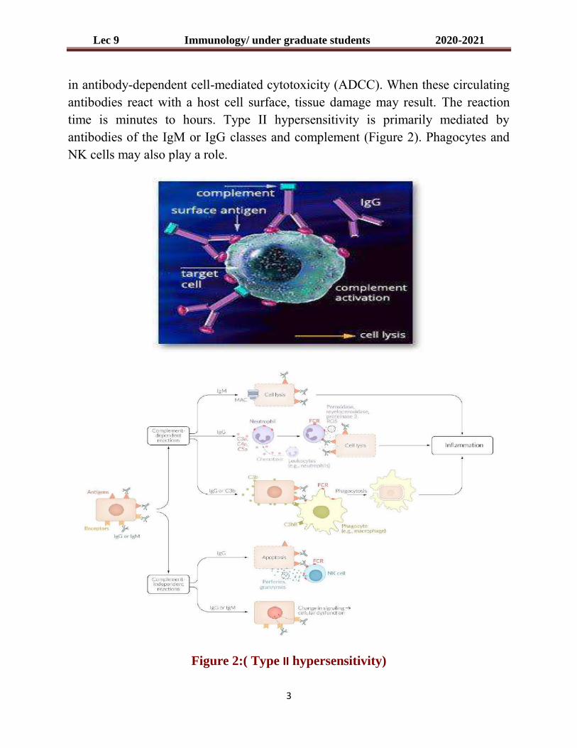
Lec 9 Immunology/ under graduate students 2020-2021
3
in antibody-dependent cell-mediated cytotoxicity (ADCC). When these circulating antibodies react with a host cell surface, tissue damage may result. The reaction time is minutes to hours. Type II hypersensitivity is primarily mediated by antibodies of the IgM or IgG classes and complement (Figure 2). Phagocytes and NK cells may also play a role.
Figure 2:( Type II hypersensitivity)

Lec 9 Immunology/ under graduate students 2020-2021
4
There are many examples of Type II hypersensitivity. These include:
• Autoimmune hemolytic anemia (AHA): This disease is generally inspired by a drug such as penicillin that becomes attached to the surface of red blood cells (RBC) and acts as hapten for the production of antibody which then binds the RBC surface leading to lysis of RBCs.
• Goodpasture's syndrome: Generally manifested as a glomerulonephritis, IgG antibodies that react against glomerular basement membrane surfaces can lead to kidney destruction.
TYPE III HYPERSENSITIVITY
Type III hypersensitivity is also known as immune complex hypersensitivity. The reaction may be general (e.g., serum sickness) or may involve individual organs including skin, kidneys, lungs (e.g., aspergillosis), blood vessels (e.g., polyarteritis), joints (e.g., rheumatoid arthritis) or other organs. This reaction may be the pathogenic mechanism of diseases caused by many microorganisms.
The reaction may take 3 - 10 hours after exposure to the antigen. It is mediated by soluble immune complexes. They are mostly of the IgG class, although IgM may also be involved. The antigen may be exogenous (chronic bacterial, viral or parasitic infections), or endogenous (non-organ specific autoimmunity: e.g., systemic lupus erythematosus, SLE). The antigen is soluble and not attached to the organ involved. Primary components are soluble immune complexes and complement (C3a, 4a and 5a). The damage is caused by platelets and neutrophils (Figure 3). The lesion contains primarily neutrophils and deposits of immune complexes and complement. Macrophages infiltrating in later stages may be involved in the healing process.
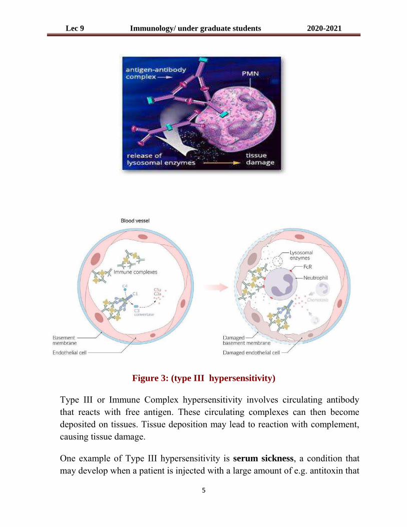
Lec 9 Immunology/ under graduate students 2020-2021
5
Figure 3: (type III hypersensitivity)
Type III or Immune Complex hypersensitivity involves circulating antibody that reacts with free antigen. These circulating complexes can then become deposited on tissues. Tissue deposition may lead to reaction with complement, causing tissue damage.
One example of Type III hypersensitivity is serum sickness, a condition that may develop when a patient is injected with a large amount of e.g. antitoxin that

Lec 9 Immunology/ under graduate students 2020-2021
6
was produced in an animal. After about 10 days, anti-antitoxin antibodies react with the antitoxin forming immune complexes that deposit in tissues. Type III hypersensitivities can be ascertained by intradermal injection of the antigen, followed by the observance of an "Arthus" reaction (swelling and redness at site of injection) after a few hours.
TYPE IV HYPERSENSITIVITY
Type IV hypersensitivity is also known as cell mediated or delayed type hypersensitivity. The classical example of this hypersensitivity is tuberculin reaction which peaks 48 hours after the injection of antigen (tuberculin). The lesion is characterized by induration and erythema.
Type IV hypersensitivity is involved in the pathogenesis of many autoimmune and infectious diseases (tuberculosis, leprosy, blastomycosis, histoplasmosis, toxoplasmosis, leishmaniasis, etc.) and granulomas due to infections and foreign antigens. Another form of delayed hypersensitivity is contact dermatitis (poison ivy, chemicals, heavy metals, etc.) in which the lesions are more papular. Type IV hypersensitivity can be classified into three categories depending on the time of onset and clinical and histological presentation.
Mechanisms of damage in delayed hypersensitivity include T lymphocytes and monocytes and/or macrophages. Cytotoxic T cells (Tc) cause direct damage whereas helper T (TH1) cells secrete cytokines which activate cytotoxic T cells and recruit and activate monocytes and macrophages, which cause the bulk of the damage (figure 4). The delayed hypersensitivity lesions mainly contain monocytes and a few T cells.
Corticosteroids and other immunosuppressive agents are used in treatment.

Lec 9 Immunology/ under graduate students 2020-2021
7
Figure 4: (Type IV HYPERSENSITIVITY)
Table 1 - Comparison of Different Types of hypersensitivity
characteristics type-I (anaphylactic)
type-II (cytotoxic)
type-III (immune complex)
type-IV (delayed type)
antibody IgE IgG, IgM IgG, IgM None
antigen exogenous cell surface soluble tissues & organs
response time 15-30 minutes minutes-hours 3-8 hours 48-72 hours
appearance weal & flare lysis and necrosis erythema and edema, necrosis
erythema and induration
histology basophils and eosinophil
antibody and complement
complement and neutrophils
monocytes and lymphocytes
transferred with antibody antibody antibody T-cells
examples allergic asthma, hay fever
erythroblastosis
fetalis, Goodpasture's nephritis
SLE, farmer's lung disease
tuberculin test, poison ivy, granuloma

Lec 10 Immunology/ undergraduate students 2020-2021
1
Lecture Ten

Lec 10 Immunology/ undergraduate students 2020-2021
2
MAJOR HISTOCOMPATIBILITY COMPLEX
The major histocompatibility complex (MHC) is a large genomic region or gene
family found in most vertebrates that encodes MHC molecules. MHC molecules
play an important role in the immune system and autoimmunity.
The Major Histocompatibility Complex (MHC) is a set of molecules displayed on
cell surfaces that are responsible for lymphocyte recognition and "antigen
presentation". The MHC molecules control the immune response through
recognition of "self" and "non-self" and, consequently, serve as targets in
transplantation rejection. The Class I and Class II MHC molecules belong to a
group of molecules known as the Immunoglobulin Supergene Family, which
includes immunoglobulins, T-cell receptors, CD4, CD8, and others.
The major histocompatibility complex is encoded by several genes located on
human chromosome 6. Class I molecules are encoded by the BCA region while
class II molecules are encoded by the D region. A region between these two on
chromosome 6 encodes class III molecules, including some complement
components.

Lec 10 Immunology/ undergraduate students 2020-2021
3
STRUCTURE OF CLASS I MHC MOLECULES
The molecule
Class I MHC molecules are composed of two polypeptide chains, a long α
chain and a short β chain called β2-microglobulin (figure 1). The α chain has
four regions.
• A cytoplasmic region, containing sites for phosphoylation and binding
to cytoskeletal elements.
• A transmembrane region containing hydrophilic amino acids by which
the molecule is anchored in the cell membrane.
• A highly conserved α3 immunoglubilin-like domain to which CD8
binds.
• A highly polymorphic peptide binding region formed from the α1 and
α2 domains. The β2- microglobulin associates with the α chain and
helps maintain the proper conformation of the molecule.
Class I MHC

Lec 10 Immunology/ undergraduate students 2020-2021
4
The most important function of the gene products for the Class-I genes is the
presentation of intracellular antigenic peptides to the cytotoxic T lymphocytes
(CD8+). The antigenic peptide is located in a cleft existing between the α1 and α2
regions in the heavy chain.
Within the MHC there are 6 genes that encode class I molecules HLA-A,
HLA –B, HLA-C, HLA-E, HLA-F and HLA-G. Among these HLA-A, HLA –B,
and HLA-C are the most important and are most polymorphic.
STRUCTURE OF CLASS II MHC MOLECULES
The molecule
Class II MHC molecules are composed of two polypeptide chains an α and a β
chain of approximately equal length (Figure 2). Both chains have four regions:
• A cytoplasmic region containing sites for phosphoylation and binding
to cytoskeletal elements
• A transmembrane region containing hydrophilic amino acids by which
the molecule is anchored in the cell membrane
• A highly conserved α2 domain and a highly conserved β2 domain to
which CD4 binds
• A highly polymorphic peptide binding region formed from the α1 and
β1 domains.
These molecules are present mostly in the membrane of the antigen presenting
cells (dendritic and phagocytic cells), where they present processed extracellular
antigenic peptides to the helper T lymphocytes (CD4+). The antigenic peptide is
located in a cleft formed by α1 and β1 peptides.

Lec 10 Immunology/ undergraduate students 2020-2021
5
Class II MHC
Within the MHC there are 5 loci that encode class II molecules, each of which
contains a gene for an α chain and at least one gene for a β chain. The loci are
designated as HLA-DP, HLA –DQ, HLA-DR, HLA-DM, and HLA-DO. Among
these, HLA-DP, HLA –DQ, and HLA-DR are the most important and are most
polymorphic.
Class I MHC and Class II MHC
MHC Class I MHC Class II
Nomenclature HLA-A,HLA-B,HLA-C HLA-DP, HLA-DQ, HLA-DR
Found on All nucleated somatic cells Macrophages, B-cells, Dendretic
cells, langerhans cells of skin, and
activated T- cells
Recognized by CD8 TC cells CD4 TH cells
Functions Presentation of Ag to TC cells leading
to elimination of tumor or infected
host cells
Presentation of Ag to TH cells
which secrete cytokines