theoretical determination of one-electron oxidation ... · ... the oxidation potentials for...
TRANSCRIPT
Theoretical Determination of One-Electron Oxidation Potentialsfor Nucleic Acid BasesBrian T. Psciuk, Richard L. Lord, Barbara H. Munk, and H. Bernhard Schlegel*
Department of Chemistry, Wayne State University, Detroit, Michigan 48202, United States
*S Supporting Information
ABSTRACT: The oxidation potentials for N-methyl substituted nucleic acid bases guanine, adenine, cytosine, thymine, uracil,xanthine, and 8-oxoguanine were computed using B3LYP and CBS-QB3 with the SMD solvation model. Acid−base andtautomeric equilibria present in aqueous solution were accounted for by combining standard redox potentials with calculated pKaand tautomerization energies to produce an ensemble averaged pH dependent potential. Gas phase free energies were computedusing B3LYP/aug-cc-pVTZ//B3LYP/6-31+G(d,p) and CBS-QB3. Solvation free energies were computed at the SMD/B3LYP/6-31+G(d,p) level of theory. Compared to experimental results, calculations with the CBS-QB3 level of theory have a meanabsolute error (MAE) of ca. 1 kcal/mol for the gas phase proton affinity/gas phase basicity and an MAE of ca. 0.04 eV for theadiabatic/vertical ionization potentials. The B3LYP calculations have a MAE of ∼2 kcal/mol for the proton affinity/gas phasebasicity data but systematically underestimated ionization potentials by 0.14−0.21 eV. Solvent cavities for charged solute specieswere rescaled uniformly by fitting computed pKa data to experimentally measured pKa values. After solvent cavity scaling, theMAEs for computed pKa's compared to experimental results are 0.7 for B3LYP and 0.9 for CBS-QB3. In acetonitrile, thecomputed E°(XH+•/XH) redox potentials are systematically lower than experimentally measured potentials by 0.21 V for CBS-QB3 and 0.33 V for B3LYP. However, the redox potentials relative to adenine are in very good agreement with experimentalresults, with MAEs of 0.10 V for CBS-QB3 and 0.07 V for B3LYP. In aqueous solution, B3LYP and CBS-QB3 have MAEs of 0.21and 0.19 V for E7(X
•,H+/XH). Replacing the methyl substituent with ribose changes the calculated E7 potentials by 0.1−0.2 V.The calculated difference between the guanine and adenine oxidation potentials is too large compared to experimental results,but the calculated difference between guanine and 8-oxoguanine is in good agreement with the measured values.
■ INTRODUCTION
In biological systems, DNA is persistently exposed to harmfuloxidizing agents. Most biological systems possess mechanismsfor repairing oxidative damage to DNA. Although these repairmechanisms tend to be highly successful, they cannot repair alldamaged DNA. Unrepaired oxidative damage to DNA can leadto mutations that are associated with carcinogenesis, cellularaging, and cellular death.1−6 Determining the most probablesites for DNA damage is very important for understanding themechanisms and reaction pathways leading to permanentmutations. It is known that guanine has the lowest oxidationpotential among the common nucleobases. There is also aconsensus concerning the qualitative trend in oxidationpotentials of the common nucleobases.7 Due to electrontransfer processes in DNA, the specific site where oxidativedamage takes place may be different from the site of chemicalmutation. The electrochemical properties of individual nucleo-sides are important for the understanding of oxidative damageto DNA. In the present paper, we have used electronic structurecalculations and a polarizable continuum solvation model tocalculate the electrochemical potentials of some nucleosides.The one-electron oxidation potentials of nucleosides have
been measured by a number of experimental groups in differentsolvents and at various pH levels in aqueous solution.8−12
Unfortunately, there is not yet a consensus set of redoxpotentials for the standard nucleic acids guanine, adenine,cytosine, thymine, and uracil. The various measured values havebeen discussed extensively, and concerns have been raised such
as cyclic voltammetry measurements that involve irreversibleredox reactions, solubility issues with specific nucleobases, andinaccurate reference compound potentials.10,12,13
The experimental studies by Steenken and co-workers8,9 andby Seidel et al.13 have attracted the most attention concerningnucleoside oxidation potentials. The measurements bySteenken and Jovanovic were made in aqueous solution atnear physiological pH using chemical oxidation and kinetic ratemeasurements of reference compounds reacting with thenucleobases. They determined half-cell potentials for guanineat pH 7 (E7 = 1.29 V) and adenine at pH 3 and pH 5 (E3 = 1.64 Vand E5 = 1.56 V). Standard potentials (E°) were also derivedby Steenken and Jovanovic. However, numerous tautomericand acid−base equilibria control the composition of thereactants and products for redox reactions in aqueous solution.For a redox reaction in aqueous solution at a given pH, the aciddissociation constants (Ka) are needed to relate the measuredpotentials to E°, and the derived E° becomes very sensitive tothe Ka values of the oxidized and reduced species. Themeasurements made by Seidel et al. were performed usingcyclic voltammetry in acetonitrile solution. By measuring theone-electron oxidation potentials in an aprotic solvent, theyeliminated the complications of acid−base equilibria. However,the cyclic voltammetry measurements were made with a single
Special Issue: Berny Schlegel Festschrift
Received: June 29, 2012Published: August 29, 2012
Article
pubs.acs.org/JCTC
© 2012 American Chemical Society 5107 dx.doi.org/10.1021/ct300550x | J. Chem. Theory Comput. 2012, 8, 5107−5123
voltammetric sweep due to the irreversibility of the redoxreactions.As computational power has increased over the years, sophis-
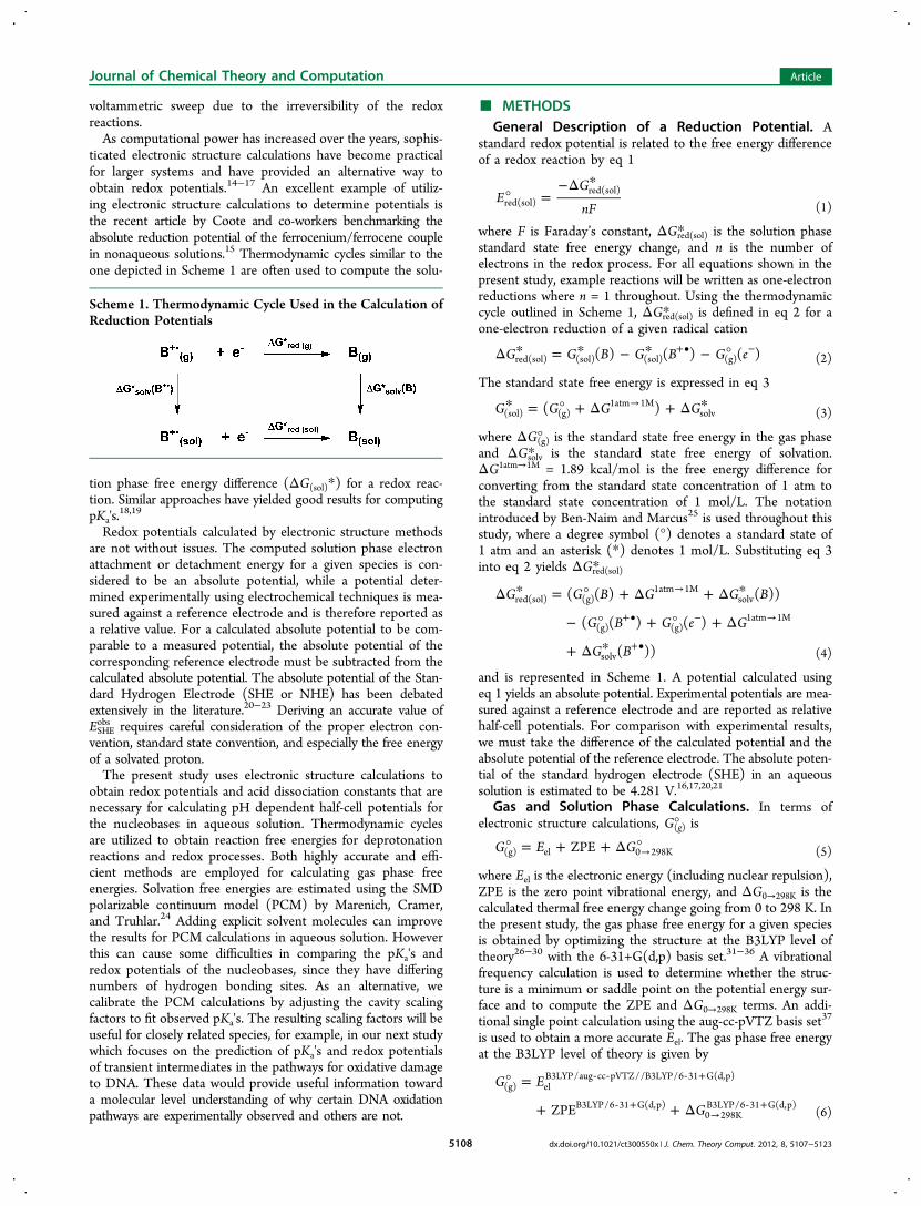
ticated electronic structure calculations have become practicalfor larger systems and have provided an alternative way toobtain redox potentials.14−17 An excellent example of utiliz-ing electronic structure calculations to determine potentials isthe recent article by Coote and co-workers benchmarking theabsolute reduction potential of the ferrocenium/ferrocene couplein nonaqueous solutions.15 Thermodynamic cycles similar to theone depicted in Scheme 1 are often used to compute the solu-
tion phase free energy difference (ΔG(sol)*) for a redox reac-tion. Similar approaches have yielded good results for computingpKa's.
18,19
Redox potentials calculated by electronic structure methodsare not without issues. The computed solution phase electronattachment or detachment energy for a given species is con-sidered to be an absolute potential, while a potential deter-mined experimentally using electrochemical techniques is mea-sured against a reference electrode and is therefore reported asa relative value. For a calculated absolute potential to be com-parable to a measured potential, the absolute potential of thecorresponding reference electrode must be subtracted from thecalculated absolute potential. The absolute potential of the Stan-dard Hydrogen Electrode (SHE or NHE) has been debatedextensively in the literature.20−23 Deriving an accurate value ofESHEobs requires careful consideration of the proper electron con-
vention, standard state convention, and especially the free energyof a solvated proton.The present study uses electronic structure calculations to
obtain redox potentials and acid dissociation constants that arenecessary for calculating pH dependent half-cell potentials forthe nucleobases in aqueous solution. Thermodynamic cyclesare utilized to obtain reaction free energies for deprotonationreactions and redox processes. Both highly accurate and effi-cient methods are employed for calculating gas phase freeenergies. Solvation free energies are estimated using the SMDpolarizable continuum model (PCM) by Marenich, Cramer,and Truhlar.24 Adding explicit solvent molecules can improvethe results for PCM calculations in aqueous solution. Howeverthis can cause some difficulties in comparing the pKa's andredox potentials of the nucleobases, since they have differingnumbers of hydrogen bonding sites. As an alternative, wecalibrate the PCM calculations by adjusting the cavity scalingfactors to fit observed pKa's. The resulting scaling factors will beuseful for closely related species, for example, in our next studywhich focuses on the prediction of pKa's and redox potentialsof transient intermediates in the pathways for oxidative damageto DNA. These data would provide useful information towarda molecular level understanding of why certain DNA oxidationpathways are experimentally observed and others are not.
■ METHODSGeneral Description of a Reduction Potential. A
standard redox potential is related to the free energy differenceof a redox reaction by eq 1
° =−Δ *
EG
nFred(sol)red(sol)
(1)
where F is Faraday’s constant, ΔGred(sol)* is the solution phasestandard state free energy change, and n is the number ofelectrons in the redox process. For all equations shown in thepresent study, example reactions will be written as one-electronreductions where n = 1 throughout. Using the thermodynamiccycle outlined in Scheme 1, ΔGred(sol)* is defined in eq 2 for aone-electron reduction of a given radical cation
Δ * = * − * − °+• −G G B G B G e( ) ( ) ( )red(sol) (sol) (sol) (g) (2)
The standard state free energy is expressed in eq 3
* = ° + Δ + Δ *→G G G G( )(sol) (g)1atm 1M
solv (3)
where ΔG(g)° is the standard state free energy in the gas phaseand ΔGsolv* is the standard state free energy of solvation.ΔG1atm→1M = 1.89 kcal/mol is the free energy difference forconverting from the standard state concentration of 1 atm tothe standard state concentration of 1 mol/L. The notationintroduced by Ben-Naim and Marcus25 is used throughout thisstudy, where a degree symbol (°) denotes a standard state of1 atm and an asterisk (*) denotes 1 mol/L. Substituting eq 3into eq 2 yields ΔGred(sol)*
Δ * = ° + Δ + Δ *
− ° + ° + Δ
+ Δ *
→
+• − →
+•
G G B G G B
G B G e G
G B
( ( ) ( ))
( ( ) ( )
( ))
red(sol) (g)1atm 1M
solv
(g) (g)1atm 1M
solv (4)
and is represented in Scheme 1. A potential calculated usingeq 1 yields an absolute potential. Experimental potentials are mea-sured against a reference electrode and are reported as relativehalf-cell potentials. For comparison with experimental results,we must take the difference of the calculated potential and theabsolute potential of the reference electrode. The absolute poten-tial of the standard hydrogen electrode (SHE) in an aqueoussolution is estimated to be 4.281 V.16,17,20,21
Gas and Solution Phase Calculations. In terms ofelectronic structure calculations, G(g)° is
° = + + Δ °→G E GZPE(g) el 0 298K (5)
where Eel is the electronic energy (including nuclear repulsion),ZPE is the zero point vibrational energy, and ΔG0→298K is thecalculated thermal free energy change going from 0 to 298 K. Inthe present study, the gas phase free energy for a given speciesis obtained by optimizing the structure at the B3LYP level oftheory26−30 with the 6-31+G(d,p) basis set.31−36 A vibrationalfrequency calculation is used to determine whether the struc-ture is a minimum or saddle point on the potential energy sur-face and to compute the ZPE and ΔG0→298K terms. An addi-tional single point calculation using the aug-cc-pVTZ basis set37
is used to obtain a more accurate Eel. The gas phase free energyat the B3LYP level of theory is given by
° =
+ + Δ
‐ ‐ ‐ +
‐ +→
‐ +
G E
GZPE
(g) elB3LYP/aug cc pVTZ//B3LYP/6 31 G(d,p)
B3LYP/6 31 G(d,p)0 298KB3LYP/6 31 G(d,p)
(6)
Scheme 1. Thermodynamic Cycle Used in the Calculation ofReduction Potentials
Journal of Chemical Theory and Computation Article
dx.doi.org/10.1021/ct300550x | J. Chem. Theory Comput. 2012, 8, 5107−51235108
An even more accurate method for computing gas phasefree energies is the CBS-QB3 compound model chemistry38,39
which has been shown to predict gas phase thermodynamic prop-erties at near chemical accuracy (MAE of 1.1 kcal/mol).For a given molecule, Ben-Naim and Marcus25 described the
free energy of solvation (ΔGsolv* ) shown in eq 7
Δ * = * ′ − *G G R G R( ) ( )solv (sol) (g) (7)
as the difference between the solution phase free energy of thesolution phase optimized molecule, R′, and the gas phase freeenergy of the gas phase optimized molecule, R. The solvationfree energy is computed using the SMD implicit solvationmodel24 at the B3LYP/6-31+G(d,p) level of theory and includesthe electrostatic, cavitation, and dispersion terms. The solutionphase free energies are computed by combining the solvation freeenergies calculated at the B3LYP level of theory with the gasphase free energies calculated with the B3LYP method
* =
+ + Δ
+ Δ + Δ *
‐ ‐ ‐ +
‐ +→
‐ +
→ ‐ +
G E
G
G G
ZPE
(sol) elB3LYP/aug cc pVTZ//B3LYP/6 31 G(d,p)
B3LYP/6 31 G(d,p)0K 298KB3LYP/6 31 G(d,p)
1atm 1Msolv
SMD/B3LYP/6 31 G(d,p)(8)
and the CBS-QB3 method
* = + + Δ *‐ → ‐ +G G G G(sol) (g)CBS QB3 1atm 1M
solvSMD/B3LYP/6 31 G(d,p)
(9)
All calculations in this study were carried out with a develop-ment version of Gaussian.40 Solution phase calculations wereperformed using the SMD implicit solvation model.24 TheSMD solvation model uses the integral equation formalism ofthe polarizable continuum model (IEF-PCM)41−44 with a param-etrized set of atomic radii to calculate the bulk electrostaticenergy contribution. The model calculates short-range inter-action energies between solvent and solute by using a modifiedsolvent-accessible surface area which incorporates parametersfor atomic and molecular surface tensions and hydrogen-bondacidity and basicity. The tessellated solute−solvent boundaryuses an average tesserae area of 0.2 Å2.The molecules studied here are canonical nucleobases and
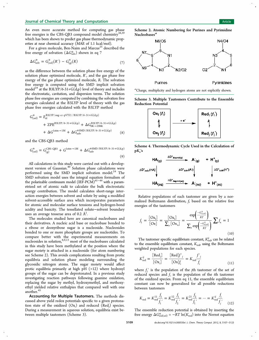
their derivatives. A nucleic acid base or nucleobase bonded toa ribose or deoxyribose sugar is a nucleoside. Nucleosidesbonded to one or more phosphate groups are nucleotides. Tocompare better with the experimental measurements onnucleosides in solution,8,9,13 most of the nucleobases calculatedin this study have been methylated at the position where thesugar moiety is attached in a nucleoside (for atom numberingsee Scheme 2). This avoids complications resulting from proticequilibria and solution phase modeling surrounding theglycosidic nitrogen atoms. The sugar moiety would affectprotic equilibria primarily at high pH (>12) where hydroxylgroups of the sugar can be deprotonated. In a previous studyinvestigating reaction pathways following guanine oxidation,replacing the sugar by methyl, hydroxymethyl, and methoxy-ethyl yielded relative enthalpies that compared well with oneanother.45
Accounting for Multiple Tautomers. The methods dis-cussed above yield redox potentials specific to a given protona-tion state of the oxidized (Oxi) and reduced (Redj) species.During a measurement in aqueous solution, equilibria exist be-tween multiple tautomers (Scheme 3).
Relative populations of each tautomer are given by a nor-malized Boltzmann distribution, f, based on the relative freeenergies of the tautomers
∑= =∑
=∑
=
− *
− *
⎜ ⎟
⎜ ⎟
⎛⎝
⎞⎠
⎛⎝
⎞⎠
f f[Ox ][Ox]
[Ox ][Ox ]
exp
exp1
n n
G
RT
nG
RT
n11 1
n
1(sol)
(sol)
(10)
The tautomer specific equilibrium constant, Kredij , can be related
to the ensemble equilibrium constant, Kred, using the Boltzmannweighted populations for each species.
= =′
=′
Kf
fK
f
f
[Red ]
[Ox ]
[Red]
[Ox]ij j
i
j
i
j
ired red
(11)
where f j′ is the population of the jth tautomer of the set ofreduced species and f i is the population of the ith tautomerof the oxidized species. From eq 11, the ensemble equilibriumconstant can now be generalized for all possible reductionsbetween tautomers
=′
=′
=′
= ··· =′
K Kf
fK
f
fK
f
fK
f
fij i
jred red
11 1
1red12 1
2red21 2
1red
(12)
The ensemble reduction potential is obtained by inserting thefree energy ΔGred(sol)* = −RT ln(Kred) into the Nernst equation
Scheme 3. Multiple Tautomers Contribute to the EnsembleReduction Potential
Scheme 4. Thermodynamic Cycle Used in the Calculation ofpKa's
Scheme 2. Atomic Numbering for Purines and PyrimidineNucleobasesa
aCharge, multiplicity and hydrogen atoms are not explicitly shown.
Journal of Chemical Theory and Computation Article
dx.doi.org/10.1021/ct300550x | J. Chem. Theory Comput. 2012, 8, 5107−51235109
° =ERTF
Kln( )red(sol) red (13)
Substituting eq 12 into eq 13 yields the ensemble reductionpotential expressed in terms of tautomer specific potentials
° = ° + − ′E ERTF
fRTF
fln( ) ln( )iji jred(sol) red(sol) (14)
Calculating pKa and E7. Experimental standard redox poten-tials are usually derived from potentials measured at a specific pH inaqueous solutions using the Nernst half-cell equation shown ineq 15
= ° −⎛⎝⎜
⎞⎠⎟E E
RTF
ln[Red][Ox]1/2
(15)
Conversely, a pH dependent potential can be derived from acalculated standard redox potential using the equilibriumconcentrations of the reduced and oxidized species. In proticsolvents, the concentrations of alternate protonation states exis-ting in solution within the applicable pH range need to beincluded for both the reduced and oxidized species. Assumingdilute concentrations (low ionic strength), a functional formcan be derived46,47 for calculating the pH dependent potentials
Table 2. Experimental and Calculated Gas Phase Adiabatic(AIE) and Vertical Ionization Energies (VIE) for NucleicAcid Bases (in eV)
adiabatic
exptl.a,b CBS-QB3 G3-B3 B3LYPc BP86c PMP2d
guanine 7.77 7.85 7.88 7.66 7.65 7.90adenine 8.26 8.28 8.30 8.07 8.04 8.23cytosine 8.68 8.71 8.83 8.56 8.50 8.78thymine 8.87 8.91 8.93 8.72 8.64 8.74uracil 9.32 9.32 9.35 9.21 9.14 9.36MAE to exptl.e 0.03 0.08 0.14 0.19 0.08
CBS-QB3 B3LYPc
9-methylguanine 7.65 7.479-methyladenine 8.10 7.891-methylcytosine 8.35 8.271-methylthymine 8.54 8.381-methyluracil 8.92 8.81
B3LYPc
guanosine 7.24adenosine 7.84cytidine 8.05thymidine 7.96uridine 8.28
vertical
exptl. CBS-QB3 G3-B3 B3LYPj BP86j PMP2d
guanine 8.28e 8.33 8.29 8.10 8.41 8.33adenine 8.48f 8.51 8.47 8.25 8.21 8.62cytosine 8.89g 8.86 8.91 8.69 8.64 8.69thymine 9.20h 9.18 9.21 9.00 8.88 9.07uracil 9.59i 9.58 9.56 9.45 9.29 9.43MAE to exptl. 0.03 0.02 0.18 0.23 0.13
exptl. CBS-QB3 G3-B3 B3LYPj BP86j PMP2
9-methylguanine 8.02e 8.03 8.08 7.81 7.76 8.179-methyladenine 8.39f 8.37 8.30 8.09 8.03 8.451-methylcytosine 8.65g 8.54 8.70 8.43 8.36 8.491-methylthymine 8.79 8.83 8.85 8.64 8.52 8.691-methyluracil 9.2 9.15 9.21 9.04 8.93 9.01MAE to exptl. 0.05 0.05 0.21 0.29 0.13
aRef 84. Estimated accuracy of ±0.05 V. bA separate set of valuesis given by NIST: guanine, 7.85 eV; adenine, 8.3 ± 0.1 eV; cytosine,8.45 eV; thymine, 9.0 ± 0.1 eV; uracil, 9.2 ± 0.1 eV.78 cFor the givenlevel of theory, the calculated AIE is the difference in energybetween the radical cation and neutral species where the energy ofeach species is the sum of the ΔG0→298K correction using the6-31+G(d,p) basis set and E0K using the aug-cc-pVTZ basis set onthe geometry optimized at the 6-31+G(d,p) basis set. dCrespo-Hernandez et al. study calculated at PMP2/6-31++G(d,p) level oftheory where adiabatic energies are for ZPE at HF/6-31++G(d,p) levelof theory.60 eRef 62. fRef 63. gRef 64. hRef 65. iRef 66. jFor the givenlevel of theory, the calculated VIE is the difference in E0K using theaug-cc-pVTZ basis set between the radical cation and neutral species atthe geometry of the neutral species optimized using the 6-31+G(d,p)basis set.
Table 1. Experimental and Calculated Gas Phase Basicitiesand Proton Affinities for Nucleic Acid Bases in kcal/mol
A + H+ → AH+
H+ site exptl.a CBS-QB3 G3B3 B3LYPc B3LYPd
proton affinityguanine N7 229.3 227.6b 228.2b 230.8 230.5b
adenine N1 225.3 223.6b 224.9b 227.1 226.7b
cytosine N3 227.0 226.9b 227.6b 229.2 228.9b
1-methylcytosine
N3 230e 230.1c 230.9c 232.7 232.4c
thymine O4 210.5 209.6c 210.6c 211.1 210.8c
uracil O4 208.6 206.8c 207.7c 208.2 207.8c
MAE to exptl. 1.1 0.7 1.5 1.3gas phase basicity
guanine N7 221.7 220.2b 220.9b 223.3 223.0b
adenine N1 218.1 216.3b 217.1b 218.9 218.3b
cytosine N3 219.0 218.9b 219.8b 221.0 219.9b
1-methylcytosine
N3 223e 222.4c 223.3c 224.8 224.5c
thymine O4 203.2 201.7c 202.6c 203.2 202.8c
uracil O4 201.2 198.9c 199.8c 200.2 199.9c
MAE to exptl. 1.3 0.8 1.2 0.9A− + H+ → AH
H+ site exptl.e CBS-QB3 G3B3 B3LYPc B3LYPd
proton affinityguanine N1 337.7b 338.2b 340.1 339.9b
guanine N9 335 335.2c 335.9c 338.1 337.8c
adenine N9 335 335.3c 335.9c 338.5 338.2c
cytosine N1 342 344.8c 345.3c 347.1 346.8c
thymine N3 346 346.3b 347.0b 347.1 346.9b
thymine N1 335 335.4c 336.2c 335.9 336.6c
uracil N3 347 346.3b 347.0b 346.6 346.9b
MAE to exptl. 0.8 1.2 2.4 2.2gas phase basicity
guanine N1 330.2b 330.6b 332.6 332.3b
guanine N9 328 327.7c 328.4c 330.6 330.3c
adenine N9 329 328.8c 328.7c 331.7 331.4c
cytosine N1 335 337.5c 337.8c 339.9 339.7c
thymine N3 339 338.5b 339.1b 339.3 339.0b
thymine N1 328 327.9c 328.7c 328.4 329.1c
uracil N3 338.1b 338.6b 338.7 338.4b
MAE to exptl. 0.7 0.9 2.2 2.1aRefs 78−81. experimental error of ±2 kcal/mol. bRef 54. cPresentstudy. dCalculated using the B3LYP/6-311++(3df,2p)//B3LYP/6-31++G(d,p) level of theory.54 eExperimental error of ±3 kcal/mol.56,82,83
Journal of Chemical Theory and Computation Article
dx.doi.org/10.1021/ct300550x | J. Chem. Theory Comput. 2012, 8, 5107−51235110
for a nucleobase that has three Ka's for the reduced form andtwo Ka's for the oxidized form
= ° +
++ + +
+ +
• +
− − −
− −
⎛⎝⎜
⎞⎠⎟
⎛⎝⎜
⎞⎠⎟
E E RTF
KK
RTF
K K K K K KK K K
(X , H /XH) ln
ln10 10 10
10 10
pHa1o
a1r
a1r a2r a3r a1r a2rpH
a1r2pH 3pH
a1o a2o a1opH 2pH
(16)
Similar expressions can be obtained for cases with differentnumbers of Ka's, e.g.
= ° +
++ +
+
• +
− −
−
⎛⎝⎜
⎞⎠⎟
⎛⎝⎜
⎞⎠⎟
E ERTF
KK
RTF
K K KK
(X , H /XH) ln
ln10 10
10
pHa1o
a1r
a1r a2r a1rpH 2pH
a1opH
(17)
“XH” signifies a specific nucleic acid species in the reducedor closed shell neutral state, and “X•” is the correspondingoxidized species that has been deprotonated following the one-electron transfer comprising the predicted redox couple for
processes occurring in aqueous solution at pH 7. “o” stands forthe oxidized species Ka, and “r” stands for the reduced speciesKa. For a redox potential in aqueous solution, acid dissociationconstants of the oxidized species (Kao) and reduced species(Kar) account for the concentrations of the relevant protonationstates. For example, the experimental values needed for the pHdependent potential of guanosine are pKa1r = 1.9, pKa2r = 9.3,pKa3r = 12.5, pKa1o = 3.9, pKa2o = 10.9, and E° = 1.58 V.9,48
Further discussions regarding the pH dependence of reductionpotentials can be found in the literature.46,47
Acid dissociation constants for short-lived radical species maybe difficult to measure experimentally, and very few measuredpKa's are available for radical nucleobase species. Additionally,discrepancies between experimentally measured pKa's of closedshell species are often larger than the reported measurementerror. Therefore, we have decided to also calculate the pKa'sneeded for pH dependent potentials. Previous computationalstudies by Verdolino et al.49 and Jang et al.50,51 have calculatedtautomer specific and ensemble pKa's for standard and modifiednucleic acid bases using a protocol very similar to the onedescribed here. To be consistent, we use calculated pKa's to
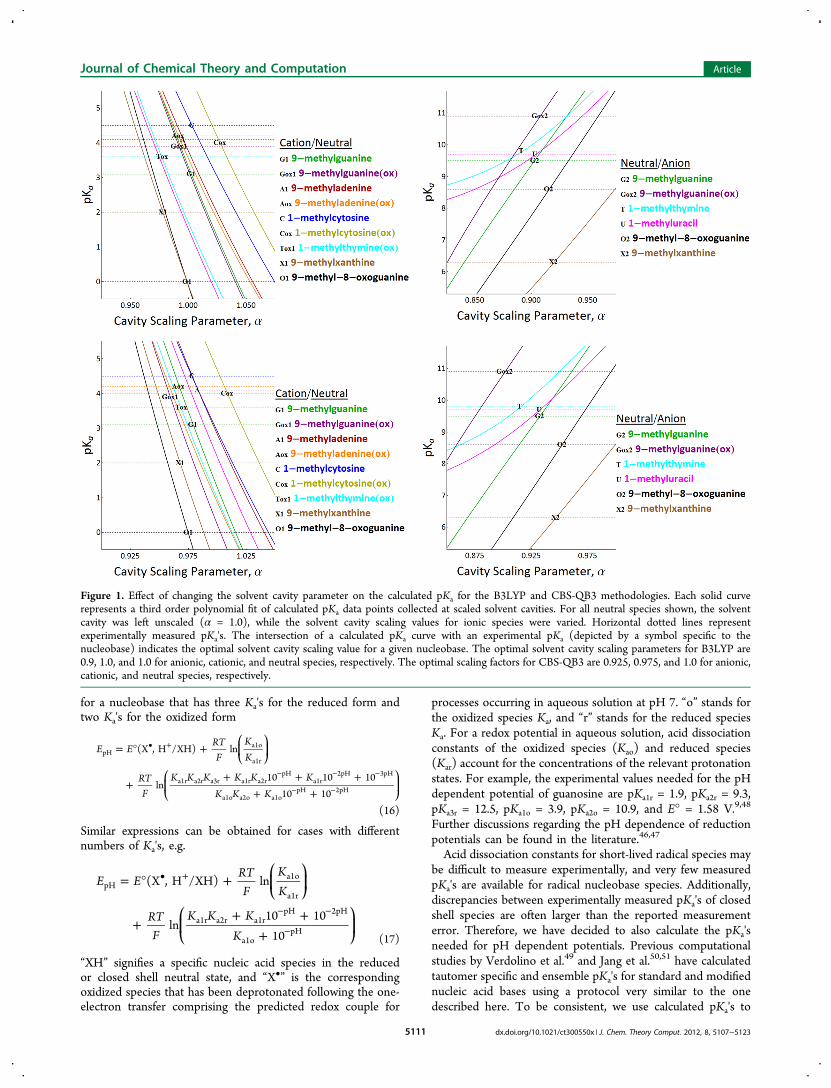
Figure 1. Effect of changing the solvent cavity parameter on the calculated pKa for the B3LYP and CBS-QB3 methodologies. Each solid curverepresents a third order polynomial fit of calculated pKa data points collected at scaled solvent cavities. For all neutral species shown, the solventcavity was left unscaled (α = 1.0), while the solvent cavity scaling values for ionic species were varied. Horizontal dotted lines representexperimentally measured pKa's. The intersection of a calculated pKa curve with an experimental pKa (depicted by a symbol specific to thenucleobase) indicates the optimal solvent cavity scaling value for a given nucleobase. The optimal solvent cavity scaling parameters for B3LYP are0.9, 1.0, and 1.0 for anionic, cationic, and neutral species, respectively. The optimal scaling factors for CBS-QB3 are 0.925, 0.975, and 1.0 for anionic,cationic, and neutral species, respectively.
Journal of Chemical Theory and Computation Article
dx.doi.org/10.1021/ct300550x | J. Chem. Theory Comput. 2012, 8, 5107−51235111
obtain the pH dependence of the calculated redox potentials.The ensemble pKa tautomers can be assigned Boltzmannweighted populations
= − + ′K K f fp p log( ) log( )iji ja a (18)
where f j′ is the population of the jth tautomer of the set ofdeprotonated species and f i is the population of the corres-ponding ith tautomer of the protonated species. The tautomerspecific pKa is
=Δ *
KG
RTp
2.303ij
ij
adeprot(aq)
(19)
and ΔGdeprot(aq)* is calculated using the thermodynamic cycle inScheme 4.The aqueous free energy for the deprotonation reaction is
given by
Δ * = ° + Δ * + °
+ Δ * − ° − Δ *
+ Δ
− − +
+
→
G G G G
G G G
G
(A ) (A ) (H )
(H ) (HA) (HA)
deprot(aq) (g) solv (g)
solv (g) solv
1atm 1M (20)
In this study, the literature value ΔGsolv* (H+) = −265.9 kcal/mol22 is used for the aqueous solvation free energy of H+.
The gas phase energies for the proton and the electron at0 K are zero. The gas phase free energy for H+ at 298 K isΔG(g)° (H+) = −6.287 kcal/mol and is derived from G(g)° = H(g)°− T·S(g)° where E0K = 0 au, H(g)° = 5/2RT = 1.48 kcal/mol, andS(g)° = 26.05 cal/mol·K. Similarly, the gas phase free energy ofthe electron at 298 K is G(g)° = −0.867 kcal/mol using H(g)° =0.752 kcal/mol and S(g)° = 5.434 cal/mol·K based on Fermi-Diracstatistics.52,53
■ RESULTS AND DISCUSSION
Redox potentials in solution include the intrinsic energy ofadding or removing an electron along with the solvation energyof the oxidized and reduced species. For the nucleobasesconsidered here, these are neutral, cationic, and anionic speciesdepending on the experimental pH conditions and whetheror not a proton transfer accompanies the electron transfer. Tocompare with experimentally observed values, the relativeabundance of the various tautomers at each redox level must betaken into account. In aqueous solution, acid−base equilibriamust also be included in the treatment. To assess the accuracyof the various levels of theory, we first compared the calculatedionization potential and gas phase basicity with experimentalvalues. Then, we examined the suitability of the SMD solvationmodel by calculating the pKa's for the nucleobases. To obtainensemble averaged pKa's, tautomeric equilibria are included for
Table 3. Experimental and Calculated pKa Values
exptl. CBS-QB3 B3LYP B3LYPj other calcd.
methyl subst. ribose subst. methyl subst. methyl subst. ribose subst. unsubst.
guaninepKa1 3.1a 1.9d 3.50 3.20 1.47 3.4,k 3.15m
pKa2 9.5a 9.2d 9.10 9.36 9.6,k 9.60m
pKa1ox 3.9e 2.53 3.34 2.61 4.01l
pKa2ox 10.9e 11.88 10.32adenine
pKa1 4.1b 3.6f 4.89 3.79 4.43 4.2k
pKaox 4.2g 2.82 3.90 3.18 2.01l
cytosinepKa1 4.5 4.2f 4.80 4.71 4.54 4.2k
pKaox ∼4e 6.72 5.69 6.46 3.37l
thyminepKa 9.8f 9.96 9.98 10.5k
pKaox 3.6e 3.04 1.69 2.88 6.40l
uracilpKa 9.7 9.2f 9.33 9.59pKaox 4.06 1.52 1.61
xanthinepKa1 2.0c 1.1c 1.31 0.44 −1.46pKa2 6.3c 5.7c 5.29 4.76pKa1ox −4.03 −5.52 −5.98pKa2ox 7.54 6.98
8-oxoguaninepKa1 0.1h −0.04 −0.12 −1.96 0.22m
pKa2 8.6h 7.09 8.13 8.0k, 8.69m
pKa1ox 0.06 −0.28 −0.54 6.83l
pKa2ox 6.6i 4.83 5.50MAE to exptl. 0.88 0.66 1.31
aRef 85. bRef 86. cRef 87. dRef 88. eRef 11. fRef 48. gValue is for deoxyribose substituted species.89 hRef 90. iRef 8. jComputed using B3LYP gasphase free energies for nucleosides. Solvent cavities were not scaled for calculations involving nucleosides. kVerdolino et al. calculated ensemble pKa'sfor nonmethylated nucleobases using Boltzmann-weighting for alternate tautomers at the B3LYP level of theory.49 lBaik et al. calculated tautomerspecific pKa's for nucleobases at PW91 level of theory.59 mGoddard and co-workers calculated ensemble pKa's for nonmethylated nucleobases usingBoltzmann-weighting for alternate tautomers at the B3LYP level of theory.50,51
Journal of Chemical Theory and Computation Article
dx.doi.org/10.1021/ct300550x | J. Chem. Theory Comput. 2012, 8, 5107−51235112
the different protonation states. Solvent cavity scaling parametersare adjusted to improve the agreement with experimental results.Next, redox potentials are calculated in acetonitrile, where acid−base equilibria are absent. Finally, redox potentials are calculatedin aqueous solution as a function of the pH, taking into accountboth the tautomeric and acid−base equilibria.Gas Phase Energies. For the gas phase reaction A + H+ →
AH+ (the upper part of Scheme 4), the proton affinity (PA) isthe reaction enthalpy and the gas phase basicity (GB) is thereaction free energy. Table 1 compares computed and mea-sured PA and GB energies for unsubstituted nucleobases andtheir anions. The results from a theoretical study by Moseret al.54 are also listed for comparison. The CBS-QB338,39 andthe G3B355 compound model chemistries are the most accuratecalculations in the table. Compared to experimental results, themean absolute errors (MAE) for CBS-QB3 and G3B3 are 0.7−1.3 kcal/mol and are well within the estimated experimentalerror of 2−3 kcal/mol. Both York and co-workers54 and Leeand co-workers56 noted that their calculated values for protonaffinities of neutral thymine and uracil disagreed with experi-mental results by as much as 4 kcal/mol. Calculations byWolken and Turecek57 showed that the most stable protonatedisomer of uracil is the N-1 deprotonated 2,4-dihydroxy form,rather than the expected N-1 protonated 2-oxo,4-hydroxy form(Scheme 5). The present B3LYP, G3B3, and CBS-QB3 calcula-
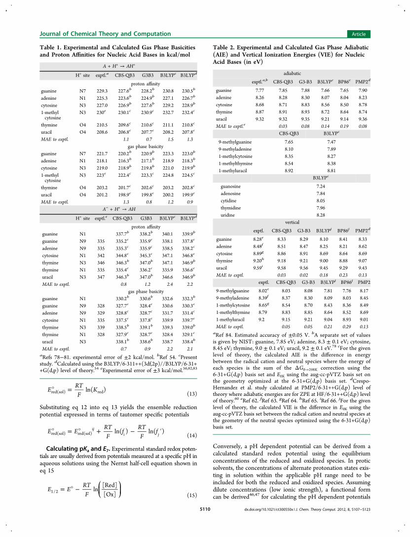
tions confirm that the N-1 deprotonated 2,4-dihydroxy form isthe most stable isomer for both thymine and uracil. The PA andGB for the neutrals and anions calculated with B3LYP/aug-cc-pVTZ are on average 1.7 and 2.1 kcal/mol higher than theexperimental and CBS-QB3 values, respectively.Table 2 compares the calculated vertical and adiabatic ioniza-
tion energies with experimental values. The adiabatic ionizationenergy is the gas phase analogue of the one-electron oxidationpotentials we are interested in, and systematic errors in theionization energies should be reflected in the redox potentials.The best agreement with experimental results is found forCBS-QB3 (MAE = 0.03−0.05 eV) and G3B3 (MAE = 0.05−0.08 eV). The MAEs are comparable to the estimated experi-mental error of ±0.05 eV. The B3LYP/aug-cc-pVTZ values aresystematically too low when compared to experimental results(MAE = 0.14−0.21 eV) and CBS-QB3 (MAE = 0.17−0.19 eV).The BP86/aug-cc-pVTZ calculations yield a slightly larger un-derestimation than B3LYP (MAE 0.19−0.29 eV vs experi-ment). This underestimation of ionization energies is known tobe characteristic of the B3LYP and other DFT levels of theoryas other studies have shown.54,58,59 The ionization energies cal-culated by Crespo-Hernandez et al.60 at the PMP2 level oftheory61 with a 6-31++G(d,p) basis have smaller MAEs thanthe DFT results, but the deviations are less systematic.
Experimental data show that methyl substitution of thenucleobases lowers the vertical ionization energy by 0.26 eVfor 9-methylguanine,62 0.09 eV for 9-methyladenine,63 0.24 eVfor 1-methylcytosine,64 0.40 eV for 1-methylthymine,65 and0.39 eV for 1-methyluracil.66 The calculated methyl substitutioneffects on the vertical ionization energy are in good agreementwith experimental results (MAE = 0.05 eV for CBS-QB3 andG3B3). Although there are no experimental data, calculationsshow that replacing the methyl substituent with ribose lowersthe ionization energy by an additional 0.1−0.4 eV. Calculationsby Crespo-Hernandez et al.67 find similar effects for methyl and2′-deoxyribose substitution of the bases. As recommended byCrespo-Hernandez et al., the present calculations use the antiorientation for the nucleosides since this conformation is morerelevant for the geometries in solution and in DNA.
Solvent Scaling Parameters. Calculated solvation freeenergies for ionic species generally have larger errors than forneutral species. A relatively simple way of addressing this erroris either to scale the individual atomic radii used to create thesolvent cavity or scale the entire solvent cavity. In addition tocompensating for different errors in solvation free energies forcharged species and for specific hydrogen bonding betweensolvent and solute, this may also adjust for systematic errors inthe calculated gas phase free energies of the molecular species.Other studies have incorporated cavity scaling techniques withsuccess.14,49,68−72 Figure 1 shows the pKa's as a function of thecavity scaling parameter α for the cations and the anions (thesolvent cavities for the neutrals were left unscaled, α = 1.00).The scale factors were chosen by comparison with experimentalresults, minimizing the average error and rounding to thenearest 0.025. The optimal scale factors at the CBS-QB3 level
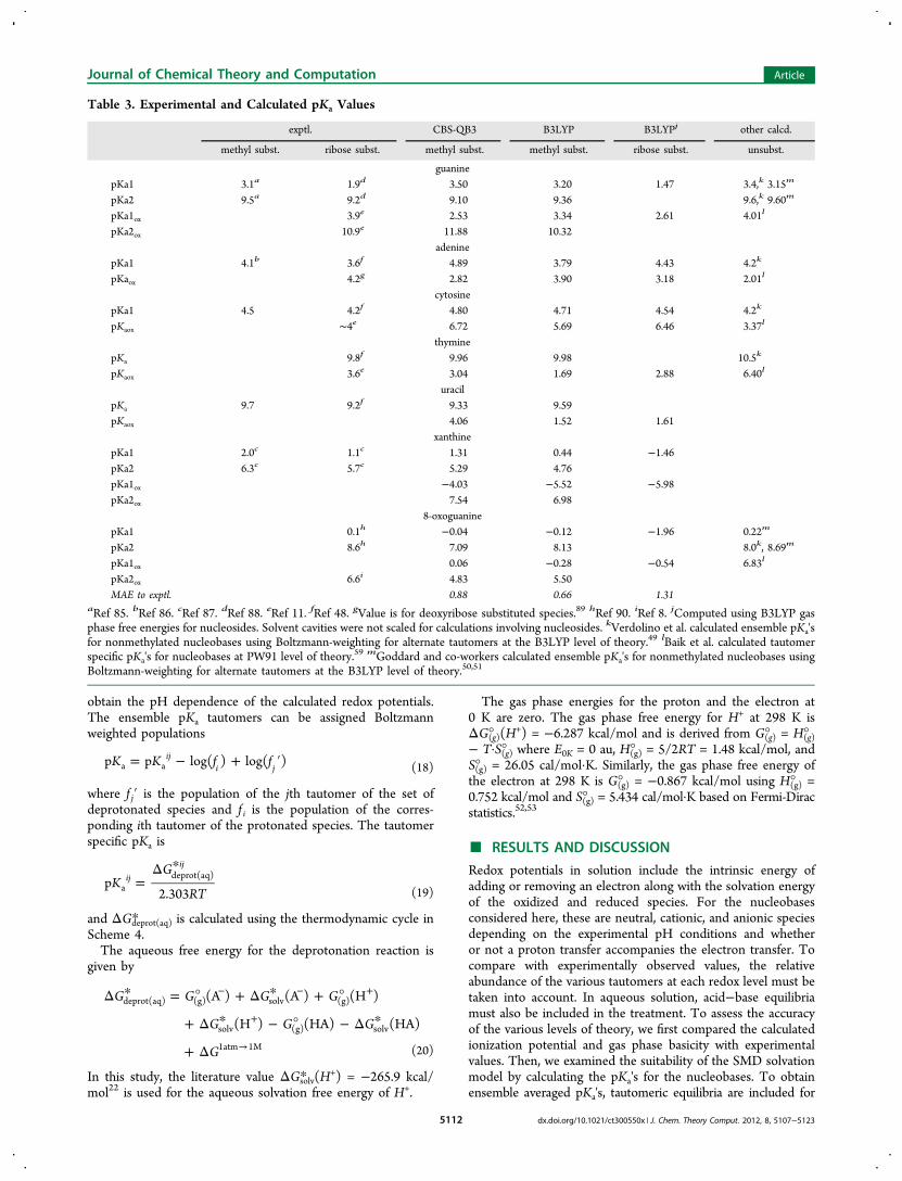
Figure 2. Linear correlation plots of calculated pKa's versusexperimental pKa's. Cation deprotonations are signified by a cross,and neutral deprotonations are signified by a dot.
Scheme 5. Lowest Energy Gas Phase Tautomers ofProtonated Uracil/Thymine
Journal of Chemical Theory and Computation Article
dx.doi.org/10.1021/ct300550x | J. Chem. Theory Comput. 2012, 8, 5107−51235113
of theory were found to be α = 0.975 for the cations and α =0.925 for the anions. The corresponding values for the B3LYPcalculations are α = 1.00 for the cations and α = 0.90 for theanions. A previous study by Verdolino et al.49 found α = 0.91for the cations and α = 0.83 for the anions for B3LYP cal-culations on a smaller set of comparisons for unoxidizednucleobases. Their study used an earlier version of the IEF-PCMsolvation model with UFF atomic radii and a solvent excludingsurface. The fact that the present scale factors are closer to unityreflects improvements in the solvation model.
Calculated pKa Values. Table 3 summarizes the pKa's cal-culated for the methyl substituted nucleobases at the CBS-QB3and B3LYP levels of theory with the optimal solvent cavityscaling factors. These values are compared with calculations byBaik et al.59 and Verdolino et al.49 Some pKa's have been mea-sured for the methyl substituted bases, but more experimentaldata are available for the nucleosides (sugar substituted nucleo-bases). Where available, the experimental value for the methyl sub-stituted base is used for comparison with the calculations. Thedifference between the experimental pKa's for the methyl- and
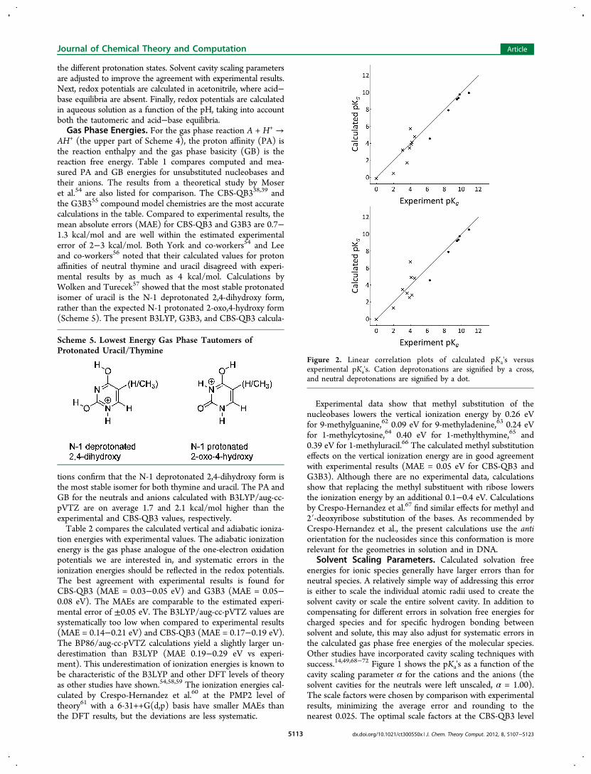
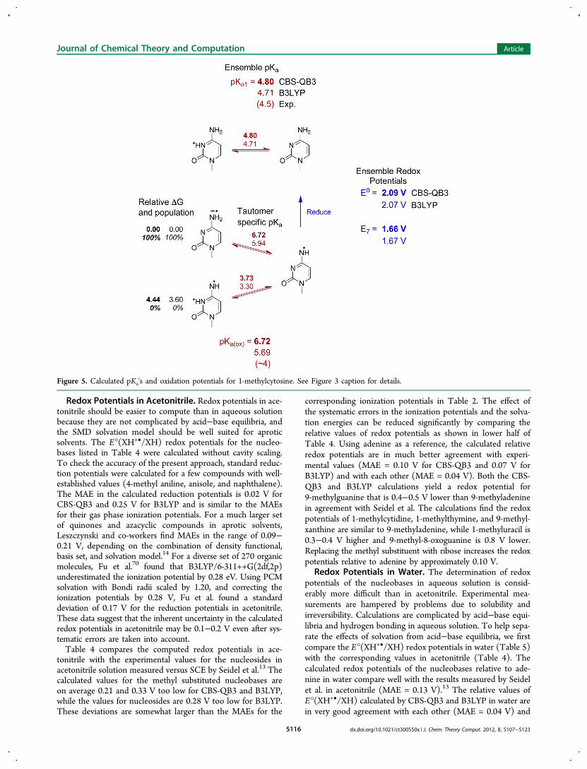
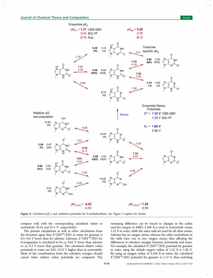
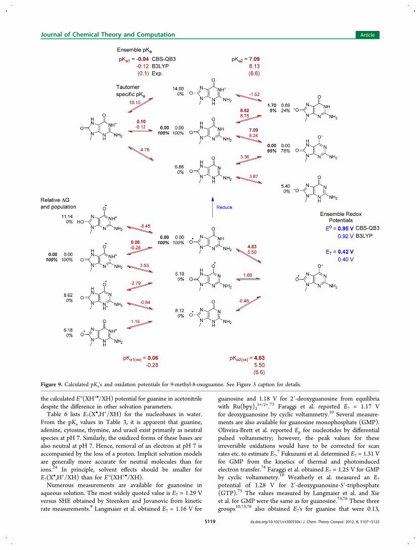
Figure 3. Calculated pKa's and oxidation potentials for 9-methylguanine. Experimentally measured values are shown in parentheses. CBS-QB3calculated values are shown in bold face text, while B3LYP calculated values are shown in regular text. Black numbers to the left of each isomerindicate the relative free energy in kcal/mol (regular font) and the population in percentage (italics). Red numbers shown in between isomersindicate calculated pKa's for the specific isomers, while red numbers on the top and bottom of the figure indicate the ensemble averaged pKa for eachacid/base equilibrium. Blue numbers shown between the reduced (top) and oxidized species (bottom) indicate calculated one-electron oxidationpotentials E° and E7.
Journal of Chemical Theory and Computation Article
dx.doi.org/10.1021/ct300550x | J. Chem. Theory Comput. 2012, 8, 5107−51235114
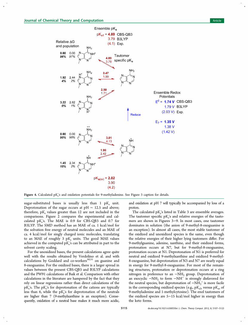
sugar-substituted bases is usually less than 1 pKa unit.Deprotonation of the sugar occurs at pH ∼ 12.5 and above;therefore, pKa values greater than 12 are not included in thecomparisons. Figure 2 compares the experimental and cal-culated pKa's. The MAE is 0.9 for CBS-QB3 and 0.7 forB3LYP. The SMD method has an MAE of ca. 1 kcal/mol forthe solvation free energy of neutral molecules and an MAE ofca. 4 kcal/mol for singly charged ionic molecules, translatingto an MAE of roughly 3 pKa units. The good MAE valuesachieved in the computed pKa's can be attributed in part to thesolvent cavity scaling.For the unoxidized bases, the present calculations agree quite
well with the results obtained by Verdolino et al. and withcalculations by Goddard and co-workers50,51 on guanine and8-oxoguanine. For the oxidized bases, there is a larger spread invalues between the present CBS-QB3 and B3LYP calculationsand the PW91 calculations of Baik et al. Comparison with othercalculations in the literature are hampered by the fact that theyrely on linear regressions rather than direct calculations of thepKa's. The pKa's for deprotonation of the cations are typicallyless than 6, while the pKa's for deprotonation of the neutralsare higher than 7 (9-methylxanthine is an exception). Conse-quently, oxidation of a neutral base makes it much more acidic,
and oxidation at pH 7 will typically be accompanied by loss of aproton.The calculated pKa's listed in Table 3 are ensemble averages.
The tautomer specific pKa's and relative energies of the tauto-mers are shown in Figures 3−9. In most cases, one tautomerdominates in solution (the anion of 9-methyl-8-oxoguanine isan exception). In almost all cases, the most stable tautomer ofthe oxidized and unoxidized species is the same, even thoughthe relative energies of their higher lying tautomers differ. For9-methylguanine, adenine, xanthine, and their oxidized forms,protonation occurs at N7, but for 9-methyl-8-oxoguanine,protonation occurs at N1. Deprotonation of N1 is preferred forneutral and oxidized 9-methylxanthine and oxidized 9-methyl-8-oxoguanine, but deprotonation of N3 and N7 are nearly equalin energy for 9-methyl-8-oxoguanine. For most of the remain-ing structures, protonation or deprotonation occurs at a ringnitrogen in preference to an −NH2 group. Deprotonation ofan exocyclic −NH2 to form −NH− is strongly disfavored forthe neutral species, but deprotonation of −NH2
+ is more facilein the corresponding oxidized species (e.g., pKa2r versus pKao of9-methyladenine and 1-methylcytosine). The enol tautomers ofthe oxidized species are 5−15 kcal/mol higher in energy thanthe keto forms.
Figure 4. Calculated pKa's and oxidation potentials for 9-methyladenine. See Figure 3 caption for details.
Journal of Chemical Theory and Computation Article
dx.doi.org/10.1021/ct300550x | J. Chem. Theory Comput. 2012, 8, 5107−51235115
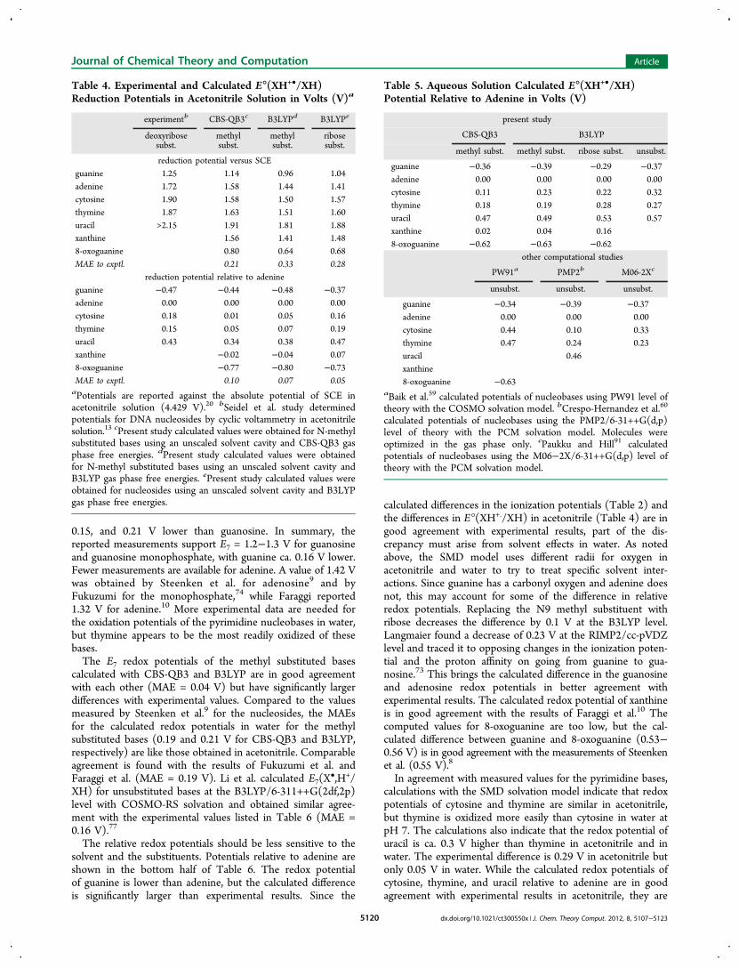
Redox Potentials in Acetonitrile. Redox potentials in ace-tonitrile should be easier to compute than in aqueous solutionbecause they are not complicated by acid−base equilibria, andthe SMD solvation model should be well suited for aproticsolvents. The E°(XH+•/XH) redox potentials for the nucleo-bases listed in Table 4 were calculated without cavity scaling.To check the accuracy of the present approach, standard reduc-tion potentials were calculated for a few compounds with well-established values (4-methyl aniline, anisole, and naphthalene).The MAE in the calculated reduction potentials is 0.02 V forCBS-QB3 and 0.25 V for B3LYP and is similar to the MAEsfor their gas phase ionization potentials. For a much larger setof quinones and azacyclic compounds in aprotic solvents,Leszczynski and co-workers find MAEs in the range of 0.09−0.21 V, depending on the combination of density functional,basis set, and solvation model.14 For a diverse set of 270 organicmolecules, Fu et al.70 found that B3LYP/6-311++G(2df,2p)underestimated the ionization potential by 0.28 eV. Using PCMsolvation with Bondi radii scaled by 1.20, and correcting theionization potentials by 0.28 V, Fu et al. found a standarddeviation of 0.17 V for the reduction potentials in acetonitrile.These data suggest that the inherent uncertainty in the calculatedredox potentials in acetonitrile may be 0.1−0.2 V even after sys-tematic errors are taken into account.Table 4 compares the computed redox potentials in ace-
tonitrile with the experimental values for the nucleosides inacetonitrile solution measured versus SCE by Seidel et al.13 Thecalculated values for the methyl substituted nucleobases areon average 0.21 and 0.33 V too low for CBS-QB3 and B3LYP,while the values for nucleosides are 0.28 V too low for B3LYP.These deviations are somewhat larger than the MAEs for the
corresponding ionization potentials in Table 2. The effect ofthe systematic errors in the ionization potentials and the solva-tion energies can be reduced significantly by comparing therelative values of redox potentials as shown in lower half ofTable 4. Using adenine as a reference, the calculated relativeredox potentials are in much better agreement with experi-mental values (MAE = 0.10 V for CBS-QB3 and 0.07 V forB3LYP) and with each other (MAE = 0.04 V). Both the CBS-QB3 and B3LYP calculations yield a redox potential for9-methylguanine that is 0.4−0.5 V lower than 9-methyladeninein agreement with Seidel et al. The calculations find the redoxpotentials of 1-methylcytidine, 1-methylthymine, and 9-methyl-xanthine are similar to 9-methyladenine, while 1-methyluracil is0.3−0.4 V higher and 9-methyl-8-oxoguanine is 0.8 V lower.Replacing the methyl substituent with ribose increases the redoxpotentials relative to adenine by approximately 0.10 V.
Redox Potentials in Water. The determination of redoxpotentials of the nucleobases in aqueous solution is consid-erably more difficult than in acetonitrile. Experimental mea-surements are hampered by problems due to solubility andirreversibility. Calculations are complicated by acid−base equi-libria and hydrogen bonding in aqueous solution. To help sepa-rate the effects of solvation from acid−base equilibria, we firstcompare the E°(XH+•/XH) redox potentials in water (Table 5)with the corresponding values in acetonitrile (Table 4). Thecalculated redox potentials of the nucleobases relative to ade-nine in water compare well with the results measured by Seidelet al. in acetonitrile (MAE = 0.13 V).13 The relative values ofE°(XH+•/XH) calculated by CBS-QB3 and B3LYP in water arein very good agreement with each other (MAE = 0.04 V) and
Figure 5. Calculated pKa's and oxidation potentials for 1-methylcytosine. See Figure 3 caption for details.
Journal of Chemical Theory and Computation Article
dx.doi.org/10.1021/ct300550x | J. Chem. Theory Comput. 2012, 8, 5107−51235116
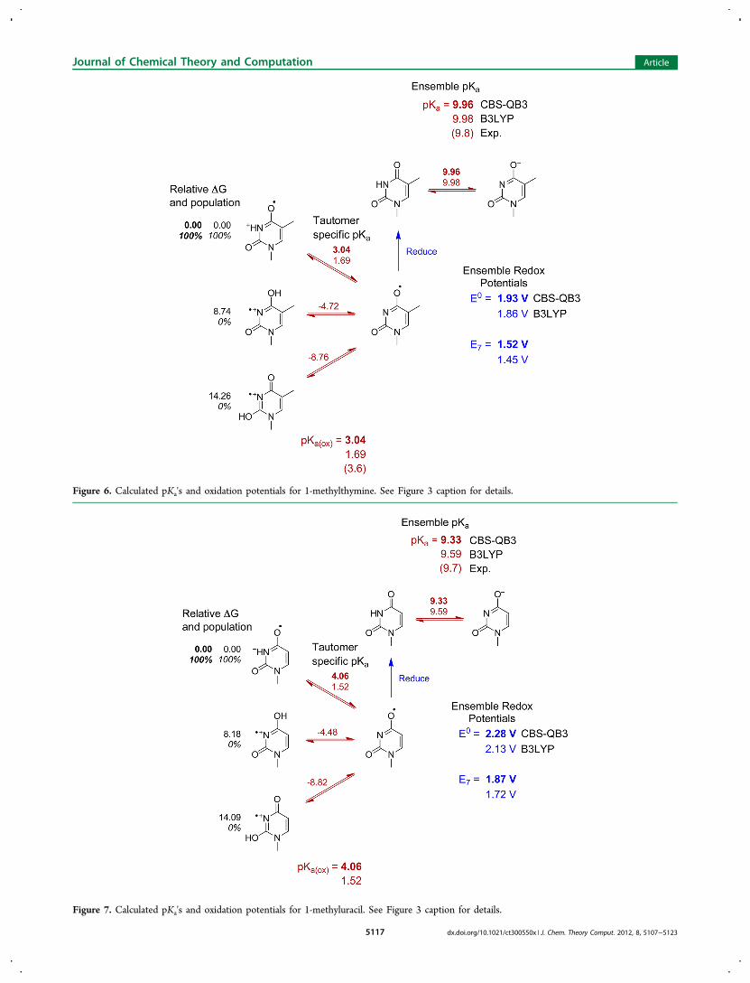
Figure 7. Calculated pKa's and oxidation potentials for 1-methyluracil. See Figure 3 caption for details.
Figure 6. Calculated pKa's and oxidation potentials for 1-methylthymine. See Figure 3 caption for details.
Journal of Chemical Theory and Computation Article
dx.doi.org/10.1021/ct300550x | J. Chem. Theory Comput. 2012, 8, 5107−51235117
compare well with the corresponding calculated values inacetonitrile (0.14 and 0.11 V, respectively).The present calculations as well as other calculations from
the literature agree that E°(XH+•/XH) in water for guanine is0.3−0.4 V lower than for adenine. Likewise, E°(XH+•/XH) for8-oxoguanine is calculated to be ca. 0.62 V lower than adenineor ca. 0.3 V lower than guanine. The calculated relative redoxpotentials in water are 0.01−0.12 V higher than in acetonitrile.Most of the contributions from the solvation energies shouldcancel when relative redox potentials are compared. The
remaining difference can be traced to changes in the radiusused for oxygen in SMD; 2.168 Å is used in acetonitrile versus1.52 Å in water, while the same radii are used for all other atoms.Adenine has no oxygen atoms, whereas the other nucleobases inthe table have one or two oxygen atoms, thus affecting thedifferences in solvation energies between acetonitrile and water.For example, the calculated E°(XH+•/XH) potential for guaninein water using the default oxygen radius of 1.52 Å is 1.20 V.By using an oxygen radius of 2.168 Å in water, the calculatedE°(XH+•/XH) potential for guanine is 1.13 V, thus matching
Figure 8. Calculated pKa's and oxidation potentials for 9-methylxanthine. See Figure 3 caption for details.
Journal of Chemical Theory and Computation Article
dx.doi.org/10.1021/ct300550x | J. Chem. Theory Comput. 2012, 8, 5107−51235118
the calculated E°(XH+•/XH) potential for guanine in acetonitriledespite the difference in other solvation parameters.Table 6 lists E7(X
•,H+/XH) for the nucleobases in water.From the pKa values in Table 3, it is apparent that guanine,adenine, cytosine, thymine, and uracil exist primarily as neutralspecies at pH 7. Similarly, the oxidized forms of these bases arealso neutral at pH 7. Hence, removal of an electron at pH 7 isaccompanied by the loss of a proton. Implicit solvation modelsare generally more accurate for neutral molecules than forions.24 In principle, solvent effects should be smaller forE7(X
•,H+/XH) than for E°(XH+•/XH).Numerous measurements are available for guanosine in
aqueous solution. The most widely quoted value is E7 = 1.29 Vversus SHE obtained by Steenken and Jovanovic from kineticrate measurements.9 Langmaier et al. obtained E7 = 1.16 V for
guanosine and 1.18 V for 2′-deoxyguanosine from equilibriawith Ru(bpy)3
3+/2+.73 Faraggi et al. reported E7 = 1.17 Vfor deoxyguanosine by cyclic voltammetry.10 Several measure-ments are also available for guanosine monophosphate (GMP).Oliveira-Brett et al. reported Ep for nucleotides by differentialpulsed voltammetry; however, the peak values for theseirreversible oxidations would have to be corrected for scanrates etc. to estimate E7.
7 Fukuzumi et al. determined E7 = 1.31 Vfor GMP from the kinetics of thermal and photoinducedelectron transfer.74 Faraggi et al. obtained E7 = 1.25 V for GMPby cyclic voltammetry.10 Weatherly et al. measured an E7
potential of 1.28 V for 2′-deoxyguanosine-5′-triphosphate(GTP).75 The values measured by Langmaier et al. and Xieet al. for GMP were the same as for guanosine.73,76 These threegroups10,73,76 also obtained E7's for guanine that were 0.13,
Figure 9. Calculated pKa's and oxidation potentials for 9-methyl-8-oxoguanine. See Figure 3 caption for details.
Journal of Chemical Theory and Computation Article
dx.doi.org/10.1021/ct300550x | J. Chem. Theory Comput. 2012, 8, 5107−51235119
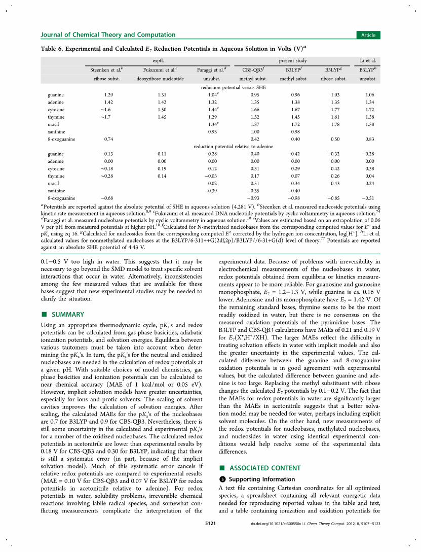
0.15, and 0.21 V lower than guanosine. In summary, thereported measurements support E7 = 1.2−1.3 V for guanosineand guanosine monophosphate, with guanine ca. 0.16 V lower.Fewer measurements are available for adenine. A value of 1.42 Vwas obtained by Steenken et al. for adenosine9 and byFukuzumi for the monophosphate,74 while Faraggi reported1.32 V for adenine.10 More experimental data are needed forthe oxidation potentials of the pyrimidine nucleobases in water,but thymine appears to be the most readily oxidized of thesebases.The E7 redox potentials of the methyl substituted bases
calculated with CBS-QB3 and B3LYP are in good agreementwith each other (MAE = 0.04 V) but have significantly largerdifferences with experimental values. Compared to the valuesmeasured by Steenken et al.9 for the nucleosides, the MAEsfor the calculated redox potentials in water for the methylsubstituted bases (0.19 and 0.21 V for CBS-QB3 and B3LYP,respectively) are like those obtained in acetonitrile. Comparableagreement is found with the results of Fukuzumi et al. andFaraggi et al. (MAE = 0.19 V). Li et al. calculated E7(X
•,H+/XH) for unsubstituted bases at the B3LYP/6-311++G(2df,2p)level with COSMO-RS solvation and obtained similar agree-ment with the experimental values listed in Table 6 (MAE =0.16 V).77
The relative redox potentials should be less sensitive to thesolvent and the substituents. Potentials relative to adenine areshown in the bottom half of Table 6. The redox potentialof guanine is lower than adenine, but the calculated differenceis significantly larger than experimental results. Since the
calculated differences in the ionization potentials (Table 2) andthe differences in E°(XH+./XH) in acetonitrile (Table 4) are ingood agreement with experimental results, part of the dis-crepancy must arise from solvent effects in water. As notedabove, the SMD model uses different radii for oxygen inacetonitrile and water to try to treat specific solvent inter-actions. Since guanine has a carbonyl oxygen and adenine doesnot, this may account for some of the difference in relativeredox potentials. Replacing the N9 methyl substituent withribose decreases the difference by 0.1 V at the B3LYP level.Langmaier found a decrease of 0.23 V at the RIMP2/cc-pVDZlevel and traced it to opposing changes in the ionization poten-tial and the proton affinity on going from guanine to gua-nosine.73 This brings the calculated difference in the guanosineand adenosine redox potentials in better agreement withexperimental results. The calculated redox potential of xanthineis in good agreement with the results of Faraggi et al.10 Thecomputed values for 8-oxoguanine are too low, but the cal-culated difference between guanine and 8-oxoguanine (0.53−0.56 V) is in good agreement with the measurements of Steenkenet al. (0.55 V).8
In agreement with measured values for the pyrimidine bases,calculations with the SMD solvation model indicate that redoxpotentials of cytosine and thymine are similar in acetonitrile,but thymine is oxidized more easily than cytosine in water atpH 7. The calculations also indicate that the redox potential ofuracil is ca. 0.3 V higher than thymine in acetonitrile and inwater. The experimental difference is 0.29 V in acetonitrile butonly 0.05 V in water. While the calculated redox potentials ofcytosine, thymine, and uracil relative to adenine are in goodagreement with experimental results in acetonitrile, they are
Table 4. Experimental and Calculated E°(XH+•/XH)Reduction Potentials in Acetonitrile Solution in Volts (V)a
experimentb CBS-QB3c B3LYPd B3LYPe
deoxyribosesubst.
methylsubst.
methylsubst.
ribosesubst.
reduction potential versus SCEguanine 1.25 1.14 0.96 1.04adenine 1.72 1.58 1.44 1.41cytosine 1.90 1.58 1.50 1.57thymine 1.87 1.63 1.51 1.60uracil >2.15 1.91 1.81 1.88xanthine 1.56 1.41 1.488-oxoguanine 0.80 0.64 0.68MAE to exptl. 0.21 0.33 0.28
reduction potential relative to adenineguanine −0.47 −0.44 −0.48 −0.37adenine 0.00 0.00 0.00 0.00cytosine 0.18 0.01 0.05 0.16thymine 0.15 0.05 0.07 0.19uracil 0.43 0.34 0.38 0.47xanthine −0.02 −0.04 0.078-oxoguanine −0.77 −0.80 −0.73MAE to exptl. 0.10 0.07 0.05aPotentials are reported against the absolute potential of SCE inacetonitrile solution (4.429 V).20 bSeidel et al. study determinedpotentials for DNA nucleosides by cyclic voltammetry in acetonitrilesolution.13 cPresent study calculated values were obtained for N-methylsubstituted bases using an unscaled solvent cavity and CBS-QB3 gasphase free energies. dPresent study calculated values were obtainedfor N-methyl substituted bases using an unscaled solvent cavity andB3LYP gas phase free energies. ePresent study calculated values wereobtained for nucleosides using an unscaled solvent cavity and B3LYPgas phase free energies.
Table 5. Aqueous Solution Calculated E°(XH+•/XH)Potential Relative to Adenine in Volts (V)
present study
CBS-QB3 B3LYP
methyl subst. methyl subst. ribose subst. unsubst.
guanine −0.36 −0.39 −0.29 −0.37adenine 0.00 0.00 0.00 0.00cytosine 0.11 0.23 0.22 0.32thymine 0.18 0.19 0.28 0.27uracil 0.47 0.49 0.53 0.57xanthine 0.02 0.04 0.168-oxoguanine −0.62 −0.63 −0.62
other computational studies
PW91a PMP2b M06-2Xc
unsubst. unsubst. unsubst.
guanine −0.34 −0.39 −0.37adenine 0.00 0.00 0.00cytosine 0.44 0.10 0.33thymine 0.47 0.24 0.23uracil 0.46xanthine8-oxoguanine −0.63
aBaik et al.59 calculated potentials of nucleobases using PW91 level oftheory with the COSMO solvation model. bCrespo-Hernandez et al.60
calculated potentials of nucleobases using the PMP2/6-31++G(d,p)level of theory with the PCM solvation model. Molecules wereoptimized in the gas phase only. cPaukku and Hill91 calculatedpotentials of nucleobases using the M06−2X/6-31++G(d,p) level oftheory with the PCM solvation model.
Journal of Chemical Theory and Computation Article
dx.doi.org/10.1021/ct300550x | J. Chem. Theory Comput. 2012, 8, 5107−51235120
0.1−0.5 V too high in water. This suggests that it may benecessary to go beyond the SMD model to treat specific solventinteractions that occur in water. Alternatively, inconsistenciesamong the few measured values that are available for thesebases suggest that new experimental studies may be needed toclarify the situation.
■ SUMMARY
Using an appropriate thermodynamic cycle, pKa's and redoxpotentials can be calculated from gas phase basicities, adiabaticionization potentials, and solvation energies. Equilibria betweenvarious tautomers must be taken into account when deter-mining the pKa's. In turn, the pKa's for the neutral and oxidizednucleobases are needed in the calculation of redox potentials ata given pH. With suitable choices of model chemistries, gasphase basicities and ionization potentials can be calculated tonear chemical accuracy (MAE of 1 kcal/mol or 0.05 eV).However, implicit solvation models have greater uncertainties,especially for ions and protic solvents. The scaling of solventcavities improves the calculation of solvation energies. Afterscaling, the calculated MAEs for the pKa's of the nucleobasesare 0.7 for B3LYP and 0.9 for CBS-QB3. Nevertheless, there isstill some uncertainty in the calculated and experimental pKa'sfor a number of the oxidized nucleobases. The calculated redoxpotentials in acetonitrile are lower than experimental results by0.18 V for CBS-QB3 and 0.30 for B3LYP, indicating that thereis still a systematic error (in part, because of the implicitsolvation model). Much of this systematic error cancels ifrelative redox potentials are compared to experimental results(MAE = 0.10 V for CBS-QB3 and 0.07 V for B3LYP for redoxpotentials in acetonitrile relative to adenine). For redoxpotentials in water, solubility problems, irreversible chemicalreactions involving labile radical species, and somewhat con-flicting measurements complicate the interpretation of the
experimental data. Because of problems with irreversibility inelectrochemical measurements of the nucleobases in water,redox potentials obtained from equilibria or kinetics measure-ments appear to be more reliable. For guanosine and guanosinemonophosphate, E7 = 1.2−1.3 V, while guanine is ca. 0.16 Vlower. Adenosine and its monophosphate have E7 = 1.42 V. Ofthe remaining standard bases, thymine seems to be the mostreadily oxidized in water, but there is no consensus on themeasured oxidation potentials of the pyrimidine bases. TheB3LYP and CBS-QB3 calculations have MAEs of 0.21 and 0.19 Vfor E7(X
•,H+/XH). The larger MAEs reflect the difficulty intreating solvation effects in water with implicit models and alsothe greater uncertainty in the experimental values. The cal-culated difference between the guanine and 8-oxoguanineoxidation potentials is in good agreement with experimentalvalues, but the calculated difference between guanine and ade-nine is too large. Replacing the methyl substituent with ribosechanges the calculated E7 potentials by 0.1−0.2 V. The fact thatthe MAEs for redox potentials in water are significantly largerthan the MAEs in acetonitrile suggests that a better solva-tion model may be needed for water, perhaps including explicitsolvent molecules. On the other hand, new measurements ofthe redox potentials for nucleobases, methylated nucleobases,and nucleosides in water using identical experimental con-ditions would help resolve some of the experimental datadifferences.
■ ASSOCIATED CONTENT
*S Supporting InformationA text file containing Cartesian coordinates for all optimizedspecies, a spreadsheet containing all relevant energetic dataneeded for reproducing reported values in the table and text,and a table containing ionization and oxidation potentials for
Table 6. Experimental and Calculated E7 Reduction Potentials in Aqueous Solution in Volts (V)a
exptl. present study Li et al.
Steenken et al.b Fukuzumi et al.c Faraggi et al.d CBS-QB3f B3LYPf B3LYPg B3LYPh
ribose subst. deoxyribose nucleotide unsubst. methyl subst. methyl subst. ribose subst. unsubst.
reduction potential versus SHEguanine 1.29 1.31 1.04e 0.95 0.96 1.03 1.06adenine 1.42 1.42 1.32 1.35 1.38 1.35 1.34cytosine ∼1.6 1.50 1.44e 1.66 1.67 1.77 1.72thymine ∼1.7 1.45 1.29 1.52 1.45 1.61 1.38uracil 1.34e 1.87 1.72 1.78 1.58xanthine 0.93 1.00 0.988-oxoguanine 0.74 0.42 0.40 0.50 0.83
reduction potential relative to adenineguanine −0.13 −0.11 −0.28 −0.40 −0.42 −0.32 −0.28adenine 0.00 0.00 0.00 0.00 0.00 0.00 0.00cytosine ∼0.18 0.19 0.12 0.31 0.29 0.42 0.38thymine ∼0.28 0.14 −0.03 0.17 0.07 0.26 0.04uracil 0.02 0.51 0.34 0.43 0.24xanthine −0.39 −0.35 −0.408-oxoguanine −0.68 −0.93 −0.98 −0.85 −0.51
aPotentials are reported against the absolute potential of SHE in aqueous solution (4.281 V). bSteenken et al. measured nucleoside potentials usingkinetic rate measurement in aqueous solution.8,9 cFukuzumi et al. measured DNA nucleotide potentials by cyclic voltammetry in aqueous solution.74dFaraggi et al. measured nucleobase potentials by cyclic voltammetry in aqueous solution.10 eValues are estimated based on an extrapolation of 0.06V per pH from measured potentials at higher pH.10 fCalculated for N-methylated nucleobases from the corresponding computed values for E° andpKa using eq 16. gCalculated for nucleosides from the corresponding computed E° corrected by the hydrogen ion concentration, log[H+]. hLi et al.calculated values for nonmethylated nucleobases at the B3LYP/6-311++G(2df,2p)/B3LYP//6-31+G(d) level of theory.77 Potentials are reportedagainst an absolute SHE potential of 4.43 V.
Journal of Chemical Theory and Computation Article
dx.doi.org/10.1021/ct300550x | J. Chem. Theory Comput. 2012, 8, 5107−51235121
reference compound species. This information is available freeof charge via the Internet at http://pubs.acs.org
■ AUTHOR INFORMATIONCorresponding Author*E-mail: [email protected] authors declare no competing financial interest.
■ ACKNOWLEDGMENTSThis work was supported by a grant from the National ScienceFoundation (CHE0910858 and CHE1212281). Wayne StateUniversity’s computing grid provided computational support.
■ REFERENCES(1) Pratviel, G.; Meunier, B. Chem.Eur. J. 2006, 12, 6018−6030.(2) Gimisis, T.; Cismas, C. Eur. J. Org. Chem. 2006, 1351−1378.(3) Burrows, C. J.; Muller, J. G. Chem. Rev. 1998, 98, 1109−1151.(4) Cadet, J.; Berger, M.; Douki, T.; Morin, B.; Raoul, S.; Ravanat, J.L.; Spinelli, S. Biol. Chem. 1997, 378, 1275−1286.(5) Breen, A. P.; Murphy, J. A. Free Radical Biol. Med. 1995, 18,1033−1077.(6) Ames, B. N.; Shigenaga, M. K.; Hagen, T. M. Proc. Natl. Acad. Sci.U. S. A. 1993, 90, 7915−7922.(7) Oliveira-Brett, A. M.; Piedade, J. A. P.; Silva, L. A.; Diculescu, V.C. Anal. Biochem. 2004, 332, 321−329.(8) Steenken, S.; Jovanovic, S. V.; Bietti, M.; Bernhard, K. J. Am.Chem. Soc. 2000, 122, 2373−2374.(9) Steenken, S.; Jovanovic, S. V. J. Am. Chem. Soc. 1997, 119, 617−618.(10) Faraggi, M.; Broitman, F.; Trent, J. B.; Klapper, M. H. J. Phys.Chem. 1996, 100, 14751−14761.(11) Steenken, S. Chem. Rev. 1989, 89, 503−520.(12) Jovanovic, S. V.; Simic, M. G. J. Phys. Chem. 1986, 90, 974−978.(13) Seidel, C. A. M.; Schulz, A.; Sauer, M. H. M. J. Phys. Chem.1996, 100, 5541−5553.(14) Sviatenko, L.; Isayev, O.; Gorb, L.; Hill, F.; Leszczynski, J. J.Comput. Chem. 2011, 32, 2195−2203.(15) Namazian, M.; Lin, C. Y.; Coote, M. L. J. Chem. Theory Comput.2010, 6, 2721−2725.(16) Lewis, A.; Bumpus, J. A.; Truhlar, D. G.; Cramer, C. J. J. Chem.Educ. 2004, 81, 596−604.(17) Lewis, A.; Bumpus, J. A.; Truhlar, D. G.; Cramer, C. J. J. Chem.Educ. 2007, 84, 934−934.(18) Ho, J. M.; Coote, M. L. Theor. Chem. Acc. 2010, 125, 3−21.(19) Ho, J. M.; Coote, M. L. Wiley Interdiscip. Rev.: Comput. Mol. Sci.2011, 1, 649−660.(20) Isse, A. A.; Gennaro, A. J. Phys. Chem. B 2010, 114, 7894−7899.(21) Kelly, C. P.; Cramer, C. J.; Truhlar, D. G. J. Phys. Chem. B 2006,110, 16066−16081.(22) Camaioni, D. M.; Schwerdtfeger, C. A. J. Phys. Chem. A 2005,109, 10795−10797.(23) Roy, L. E.; Jakubikova, E.; Guthrie, M. G.; Batista, E. R. J. Phys.Chem. A 2009, 113, 6745−6750.(24) Marenich, A. V.; Cramer, C. J.; Truhlar, D. G. J. Phys. Chem. B2009, 113, 6378−6396.(25) Ben-Naim, A.; Marcus, Y. J. Chem. Phys. 1984, 81, 2016−2027.(26) Stephens, P. J.; Devlin, F. J.; Chabalowski, C. F.; Frisch, M. J. J.Phys. Chem. 1994, 98, 11623−11627.(27) Becke, A. D. J. Chem. Phys. 1993, 98, 5648−5652.(28) Lee, C. T.; Yang, W. T.; Parr, R. G. Phys. Rev. B 1988, 37, 785−789.(29) Becke, A. D. Phys. Rev. A 1988, 38, 3098−3100.(30) Vosko, S. H.; Wilk, L.; Nusair, M. Can. J. Phys. 1980, 58, 1200−1211.(31) Francl, M. M.; Pietro, W. J.; Hehre, W. J.; Binkley, J. S.; Gordon,M. S.; Defrees, D. J.; Pople, J. A. J. Chem. Phys. 1982, 77, 3654−3665.
(32) Gordon, M. S. Chem. Phys. Lett. 1980, 76, 163−168.(33) Hariharan, P. C.; Pople, J. A. Mol. Phys. 1974, 27, 209−214.(34) Hariharan, P. C.; Pople, J. A. Theor. Chim. Acta 1973, 28, 213−222.(35) Hehre, W. J.; Ditchfield, R.; Pople, J. A. J. Chem. Phys. 1972, 56,2257.(36) Ditchfield, R.; Hehre, W. J.; Pople, J. A. J. Chem. Phys. 1971, 54,724−728.(37) Kendall, R. A.; Dunning, T. H.; Harrison, R. J. J. Chem. Phys.1992, 96, 6796−6806.(38) Montgomery, J. A.; Frisch, M. J.; Ochterski, J. W.; Petersson, G.A. J. Chem. Phys. 2000, 112, 6532−6542.(39) Montgomery, J. A.; Frisch, M. J.; Ochterski, J. W.; Petersson, G.A. J. Chem. Phys. 1999, 110, 2822−2827.(40) Frisch, M. J.; Trucks, G. W.; Schlegel, H. B.; Scuseria, G. E.;Robb, M. A.; et al. Gaussian Development Version, GDVRev. H20+;Gaussian, Inc.: Wallingford, CT, 2010.(41) Scalmani, G.; Frisch, M. J. J. Chem. Phys. 2010, 132, 114110.(42) Cossi, M.; Barone, V.; Mennucci, B.; Tomasi, J. Chem. Phys. Lett.1998, 286, 253−260.(43) Mennucci, B.; Tomasi, J. J. Chem. Phys. 1997, 106, 5151−5158.(44) Cances, E.; Mennucci, B.; Tomasi, J. J. Chem. Phys. 1997, 107,3032−3041.(45) Munk, B. H.; Burrows, C. J.; Schlegel, H. B. Chem. Res. Toxicol.2007, 20, 432−444.(46) Wardman, P. J. Phys. Chem. Ref. Data 1989, 18, 1637−1755.(47) Clark, W. M. Oxidation-Reduction Potentials of Organic Systems;Williams & Wilkins: Baltimore, MD, 1960; pp 107−148.(48) Dawson, R. M. C. Data for Biochemical Research, 3rd ed.;Clarendon Press: Oxford, U. K., 1986; pp 103−114.(49) Verdolino, V.; Cammi, R.; Munk, B. H.; Schlegel, H. B. J. Phys.Chem. B 2008, 112, 16860−16873.(50) Jang, Y. H.; Goddard, W. A.; Noyes, K. T.; Sowers, L. C.;Hwang, S.; Chung, D. S. J. Phys. Chem. B 2003, 107, 344−357.(51) Jang, Y. H.; Goddard, W. A.; Noyes, K. T.; Sowers, L. C.;Hwang, S.; Chung, D. S. Chem. Res. Toxicol. 2002, 15, 1023−1035.(52) Bartmess, J. E. J. Phys. Chem. 1994, 98, 6420−6424.(53) Bartmess, J. E. J. Phys. Chem. 1995, 99, 6755−6755.(54) Moser, A.; Range, K.; York, D. M. J. Phys. Chem. B 2010, 114,13911−13921.(55) Baboul, A. G.; Curtiss, L. A.; Redfern, P. C.; Raghavachari, K. J.Chem. Phys. 1999, 110, 7650−7657.(56) Liu, M.; Li, T. T.; Amegayibor, F. S.; Cardoso, D. S.; Fu, Y. L.;Lee, J. K. J. Org. Chem. 2008, 73, 9283−9291.(57) Wolken, J. K.; Turecek, F. J. Am. Soc. Mass Spectrom. 2000, 11,1065−1071.(58) Close, D. M. J. Phys. Chem. A 2004, 108, 10376−10379.(59) Baik, M. H.; Silverman, J. S.; Yang, I. V.; Ropp, P. A.; Szalai, V.A.; Yang, W. T.; Thorp, H. H. J. Phys. Chem. B 2001, 105, 6437−6444.(60) Crespo-Hernandez, C. E.; Arce, R.; Ishikawa, Y.; Gorb, L.;Leszczynski, J.; Close, D. M. J. Phys. Chem. A 2004, 108, 6373−6377.(61) Schlegel, H. B. J. Chem. Phys. 1986, 84, 4530−4534.(62) Lin, J.; Yu, C.; Peng, S.; Akiyama, I.; Li, K.; Lee, L. K.; Lebreton,P. R. J. Phys. Chem. 1980, 84, 1006−1012.(63) Lin, J.; Yu, C.; Peng, S.; Akiyama, I.; Li, K.; Lee, L. K.; Lebreton,P. R. J. Am. Chem. Soc. 1980, 102, 4627−4631.(64) Yu, C.; Peng, S.; Akiyama, I.; Lin, J.; Lebreton, P. R. J. Am.Chem. Soc. 1978, 100, 2303−2307.(65) Dougherty, D.; Wittel, K.; Meeks, J.; McGlynn, S. P. J. Am.Chem. Soc. 1976, 98, 3815−3820.(66) Denifl, S.; Sonnweber, B.; Hanel, G.; Scheier, P.; Mark, T. D.Int. J. Mass Spectrom. 2004, 238, 47−53.(67) Crespo-Hernandez, C. E.; Close, D. M.; Gorb, L.; Leszczynski, J.J. Phys. Chem. B 2007, 111, 5386−5395.(68) Ginovska, B.; Camaioni, D. M.; Dupuis, M. J. Chem. Phys. 2008,129, 014506.(69) Caricato, M.; Mennucci, B.; Tomasi, J. Mol. Phys. 2006, 104,875−887.
Journal of Chemical Theory and Computation Article
dx.doi.org/10.1021/ct300550x | J. Chem. Theory Comput. 2012, 8, 5107−51235122
(70) Fu, Y.; Liu, L.; Yu, H. Z.; Wang, Y. M.; Guo, Q. X. J. Am. Chem.Soc. 2005, 127, 7227−7234.(71) Orozco, M.; Luque, F. J. Chem. Phys. 1994, 182, 237−248.(72) Bachs, M.; Luque, F. J.; Orozco, M. J. Comput. Chem. 1994, 15,446−454.(73) Langmaier, J.; Samec, Z.; Samcova, E.; Hobza, P.; Reha, D. J.Phys. Chem. B 2004, 108, 15896−15899.(74) Fukuzumi, S.; Miyao, H.; Ohkubo, K.; Suenobu, T. J. Phys.Chem. A 2005, 109, 3285−3294.(75) Weatherly, S. C.; Yang, I. V.; Thorp, H. H. J. Am. Chem. Soc.2001, 123, 1236−1237.(76) Xie, H.; Yang, D. W.; Heller, A.; Gao, Z. Q. Biophys. J. 2007, 92,L70−L72.(77) Li, M. J.; Liu, W. X.; Peng, C. R.; Lu, W. C. Acta Phys.Chim.Sin. 2011, 27, 595−603.(78) Hunter, E. P. L.; Lias, S. G. J. Phys. Chem. Ref. Data 1998, 27,413−656.(79) Greco, F.; Liguori, A.; Sindona, G.; Uccella, N. J. Am. Chem. Soc.1990, 112, 9092−9096.(80) Meotner, M. J. Am. Chem. Soc. 1979, 101, 2396−2403.(81) Wilson, M. S.; McCloskey, J. A. J. Am. Chem. Soc. 1975, 97,3436−3444.(82) Zhachkina, A.; Liu, M.; Sun, X. J.; Amegayibor, F. S.; Lee, J. K. J.Org. Chem. 2009, 74, 7429−7440.(83) Chen, E. C. M.; Herder, C.; Chen, E. S. J. Mol. Struct. 2006, 798,126−133.(84) Orlov, V. M.; Smirnov, A. N.; Varshavsky, Y. M. TetrahedronLett. 1976, 4377−4378.(85) Song, B.; Zhao, J.; Griesser, R.; Meiser, C.; Sigel, H.; Lippert, B.Chem.Eur. J. 1999, 5, 2374−2387.(86) Kampf, G.; Kapinos, L. E.; Griesser, R.; Lippert, B.; Sigel, H. J.Chem. Soc., Perkin Trans. 2 2002, 1320−1327.(87) Kulikowska, E.; Kierdaszuk, B.; Shugar, D. Acta Biochim. Pol.2004, 51, 493−531.(88) Christensen, J. J.; Rytting, J. H.; Izatt, R. M. Biochemistry 1970,9, 4907−4913.(89) Kobayashi, K. J. Phys. Chem. B 2010, 114, 5600−5604.(90) Cho, B. P. Magn. Reson. Chem. 1993, 31, 1048−1053.(91) Paukku, Y.; Hill, G. J. Phys. Chem. A 2011, 115, 6738−6738.
Journal of Chemical Theory and Computation Article
dx.doi.org/10.1021/ct300550x | J. Chem. Theory Comput. 2012, 8, 5107−51235123