thengalreportermousedetects! the!response!ofthe!! … · supplementaryinformation!guide! ......
TRANSCRIPT

The Ngal Reporter Mouse Detects the Response of the Kidney to Injury in Real Time
Neal Paragas*1, Andong Qiu1*, Qingyin Zhang1, Benjamin Samstein1, Shi-Xian Deng1,
Kai M. Schmidt-Ott2, Melanie Viltard1, Wenqiang Yu1, Catherine S. Forster1, Gangli
Gong1, Yidong Liu1, Ritwij Kulkarni1, Kiyoshi Mori3, Avtandil Kalandadze1, Adam J.
Ratner1, Prasad Devarajan, Donald W. Landry1, Vivette D’Agati1, Chyuan-Sheng Lin1
and Jonathan Barasch†1.
1College of Physicians and Surgeons of Columbia University New York, N.Y. 10032 2Max-Delbruck Center for Molecular Medicine Berlin, Germany 3Kyoto University Graduate School of Medicine, Kyoto, Japan.
Nature Medicine: doi:10.1038/nm.2290

Supplementary Information Guide
A. Supplementary Methods
1. Western Blot
2. In situ hybridization and immunohistochemistry.
3. Kidney ischemia and cross transplantation
B. Supplementary Table 1: Sequence and primer information.
C. Supplementary Figures
1. Supplementary Fig. 1: Generation of the Ngal-Luc2/mC difusion reporter murine
model.
2. Supplementary Fig. 2: Functional verification of the Luc2/mC di-fusion reporter
gene.
3. Supplementary Fig. 3: PCR screening of the Ngal-targeted ES cell clones.
4. Supplementary Fig. 4: Ngal-Luc2/mC reported kidney damage in vivo after
ischemia reperfusion injury.
5. Supplementary Fig. 5: (a) Kidney Ngal-luc2 expression (left panel and outlined)
originates from injured kidney
6. Supplementary Fig. 6: uNgal derives from the kidney.
7. Supplementary Fig. 7: Neutrophils were not required for the expression of Ngal
in the kidney.
8. Supplementary Fig. 8: uNgal originates from the nephron.
9. Supplementary Fig. 9: Apoptotic S3 proximal tubule.
10. Supplementary Fig. 10: (a) Ngal-mCherry fluorescence was visible in the TAL
and CD.
11. Supplementary Fig. 11: Experimental NF-κB inhibitors
12. Supplementary Fig. 12: Model of sNgal reabsorption and uNgal excretion.
Nature Medicine: doi:10.1038/nm.2290
Supplementary Methods
Western Blot Urine and recombinant mouse Ngal standards were immunobloted using
polyclonal antibody to Ngal (R&D Systems, Minneapolis) and donkey anti-rabbit HRP-
labelled IgG antibodies (Jackson Immunoresearch).
In situ hybridization and immunohistochemistry. The paraffin sections were
dewaxed and then rehydrated by using Histoclear (Fisher Scientific) and a gradient of
ethanol, respectively, before in situ hybridization. Specific digoxigenin-labeled antisense
riboprobes were generated from mouse Ngal cDNA (Genbank accession number:
NM_008491) by using a Dig-labelling kit (Roche Applied Biosystems) and hybridized and
detected as previously described [(Li, J.Y., et al. Scara5 is a ferritin receptor mediating
non-transferrin iron delivery. Developmental cell 16, 35-46 (2009)). The hybridized
sections were counterstained with methyl green, dehydrated and mounted in Permount
(Fisher Scientific). Frozen and paraffin-embedded sections were used for
immunohistochemical analysis. Anti-mCherry (Clontech) and anti-v-ATPase B1/2 (Santa
Cruz Biotechnology) were used at a 1:50 dilution and antigen was localized by HRP-
DAB chromogen (R&D Systems) staining.
Kidney ischemia and cross transplantation The inferior vena cava (IVC) was
cannulated, and ice-cold heparinized saline (1mL, 10units/mL) administered via the IVC.
The kidneys were removed from the retroperitoneum with the aorta and the vena cava
en bloc, and the ureters were removed with a large patch of bladder. The recipient’s left
kidney was then removed following ligation of the vessels and cauterization of the ureter,
and its abdominal IVC and aorta exposed from the renal vessels to the iliac bifurcation.
The donor aorta and IVC were anastomosed to the recipient’s aorta and IVC using 10-0
suture, and the kidney graft reperfused. The donor ureter was inserted into the recipient
bladder and fixed by suturing and the abdominal incision was then closed. Four days
later, the previous incision was reopened and the recipient’s right kidney was removed,
and the abdominal incision was closed again. Both closure sutures are absorbable so
that suture removal was not necessary. Each mouse was given a sub-cutaneous
injection of Lactated Ringers (1-2 cc) that was pre-warmed to body temperature, and
was placed on warming water-blanket. Supplementary oxygen was administered during
and immediately post-operatively to minimize hypoxia.
Nature Medicine: doi:10.1038/nm.2290

Supplementary Table 1: Sequence and primer information.
Nature Medicine: doi:10.1038/nm.2290

Supplementary Figures
Supplementary Fig. 1: Generation of the Ngal-Luc2/mC difusion reporter murine model.
(a) The Ngal targeting BAC DNA was constructed by knockin of the Luc2/mC gene with
an LNL cassette (light blue box) at the translational start codon of the Ngal gene (Ngal
exon represented by grey box and UTR represented by dark blue). A DTA-Ampr
cassette, a negative selection marker, was placed 2kb downstream of the LNL cassette
in place of a 1kb DNA fragment. (b) The Ngal-targeting construct was electroporated into
KV1 ES cells, and homologous recombination led to the Ngal-targeted ES cells. (c) The
Ngal-targeted ES cells responded to 1mM cyanide and the TLR agonist, lipid A (4µg/ml)
with Luciferase luminescence. (d) PCR genotying of germline-transmitted F1 pups with
primers F1, R1 and R2 identified both the targeted and the wild type alleles at 476bp and
345bp, respectively. (e) The correct integration of the Luc2/mC gene in Ngal-targeted F1
mice was verified by long distance PCR (6819bp) with primers F2 and R3.
!"#$%%&%&
#'####(####)###*####+####,###-
!
"
!"#$ %& =>=
!"#$ %& =>= ?@$A$BC0
' ( ) * + ,
' ( ) * + ,
' ( ) *
' ( ) * + ,D;%EA9F6&#'()*#/%%&%&
'()*#9/0<&9;8<#2G8190529
@/0<&9&E#'()*#/%%&%&
' ( ) *
' ( ) * + ,
HG7G%G<G51##0&2G7I;8/9;G8
J!"#$ %& =>=
!"#$ %& =>=
?@$A$BC0
# K3;7&0/#L M#J#K+-.=N,#L M
O&8G9F6;8<L:'PQ'PQ(M L:(PQ)M
$!"#$%&%'()*(+%,,-(
KF/8;E& =;6;E$
@/0<&9&ER4AK&%%1
KG890G%R4AK&%%1
ST+UIST)UI
'UI
+UI
'SUIVUI,UI
##'######(#######)######*#
:'
Q(
?;:51;G8#Q&6G09&0
!8G2U;8#GW##!"#$+%&+=>=#
"81&09;G8##GW##?@$A$BC0##X;93#E&%&9;G8##GW##'UI#'()*#<&8&
:( Q' Q(
Q):):'
%
Nature Medicine: doi:10.1038/nm.2290

Supplementary Fig. 2: Functional verification of the Luc2/mC di-fusion reporter gene.
The Luc2/mC gene was cloned into pCI-neo, and transiently expressed in HeLa cells.
After 24 hours, luminescence and fluorescence were imaged in a Zenogen Bioimager.
!"#"$%&'()**+,-,./"+'012)#,'3
+)%14,#"$,5,/,%/16.
-7&,##85,/,%/16.
*79:.,6';76./#6+<
*79:.,6:=)%3>-7
?,="'7,++$
Nature Medicine: doi:10.1038/nm.2290

Supplementary Fig. 3: PCR screening of the Ngal-targeted ES cell clones. (a) Ngal
targeting construct and location of screening primers. (b, c) PCR screen for Ngal
targeted ES clones using primers F3 and R3 (b) and F1 and R3 (c).
!"#"$%&'()**+,-,./"+'012)#,'3
!
"
4035637
4085637
9.:%;1.
9.:%;1.
<1+=>/?*,
#8 @ 3 A B C!"#$ %& DED
630308
Nature Medicine: doi:10.1038/nm.2290

Supplementary Fig. 4: Ngal-Luc2/mC reported kidney damage in vivo after ischemia
reperfusion injury. (a) Heterozygous Ngal-Luc2/mC male mouse received 30 min of
unilateral ischemia (left kidney) and then was visualized at 0, 6, 12 and 24 hours after
reperfusion in a Zenogen Bioimager to detect Ngal-Luc2/mC activity. Peak expression of
Ngal-Luc2/mC occurred at 12 hours after reperfusion. Ngal-Luc2/mC was tonically
expressed in the testis which could occupy inguinal or pelvic sites. (b) Immunoblot of
uNgal at time-points in parallel with the assays of kidney Ngal-Luc2/mC activity. (c)
Heterozygous Ngal-Luc2/mC male mouse received 30 min of unilateral I/R (right kidney),
and was then visualized at 12 hours after reperfusion in a Zenogen Bioimager to detect
Ngal-Luc2/mC activity.
!"##$%&%'()$*+),)-./*012",%*3
$".14%,)-%
&5/%,,6
7*89",- :*89",- ;<*89",- <3*89",-
=>?@*A'2B<77 ;77 C7 : ;< <3
89",-
!7
<CDE*FG)
;ED3*FG)
;7
"
H%-(1-
H%-(1-
I1J'%6 I1J'%6 I1J'%6
I1J'%6 I1J'%6 I1J'%6
K;7L
A#/9(9'-*-M;*.&
M<*-, M;B
;D<;D77DN7D:7D37D<
OD7<DC<D7;DC;D77DC
K;7L
A#/9(9'-*-M;*.&
M<*-, M;B
#$".14%,)-% &5/%,,6
7*89",- ;<*89",-
I1J'%6 I1J'%6K;7L
A#/9(9'-*-M;*.&
M<*-, M;B
;D<;D77DN7D:7D37D<
OD7<DC<D7;DC;D77DC
K;7L
A#/9(9'-*-M;*.&
M<*-, M;B
7*89",- ;<*89",-
Nature Medicine: doi:10.1038/nm.2290

Supplementary Fig 5: (a) Kidney Ngal-luc2 expression (left panel and outlined)
originates from injured kidneys after 30 min of bilateral I/R (right panel and outlined). (b)
Ngal expression in the lung was induced by lipid A inhalation. Ngal-Luc2 was induced
within 6 hours after aspiration of 10 µg of lipid A.
!"#"$%&'()**+,-,./"+'012)#,'3
4'56)#$ 7'56)#$
!"#$#%!&!'()*+*,#-(894
:
;*&6/6.$'$<9'%-
<='$# <9>
=?4
9?3
9?4
4?3
@).2
.*!/0!1#(!'23
9='56)#$ 9='56)#$
+)%1A,#"$, *&6/62#"*&
Nature Medicine: doi:10.1038/nm.2290

Supplementary Fig. 6: uNgal derives from the kidney. Cross-transplantation of kidneys
between Ngal-/- and Ngal +/+ mice was followed by ischemia-reperfusion injury for 10 min.
(a) Ngal mRNA was markedly upregulated in Ngal+/+ kidneys placed in Ngal+/+
(228.1±18.8 fold) or in Ngal-/- hosts (184.6±56.7 fold), but not in Ngal-/- kidneys located in
the Ngal+/+ hosts (6.3±0.86 fold). Liver Ngal was not elevated 24 hours after ischemia-
reperfusion (n=15). Kim1 was significantly induced in ischemic wild-type to wild-type,
wild-type to knockout, and knockout to wild-type kidneys (7.9±0.4 fold, 7.8±3.8 fold and
10.2±4.8 fold, respectively) 24 hours after 10 min of ischemia. (b) Average urinalysis of
uNgal collected from transplants at 0, 12, and 24 hours after ischemia reperfusion injury,
revealing that uNgal increased when a Ngal+/+ kidney was placed in a Ngal+/+ (WT->WT;
n=6) or in a Ngal-/- host (Wt->KO; n=3), while there was a smaller change in uNGAL
when a Ngal-/- kidney was placed in a Ngal+/+ host (KO->WT; n=2).
!"#$%&'$(%)*#+",%-!
"#$
#%&'(.
/%0'123 45%0'12364.
/
7/
57/
587
! "
59%0'123:;%%:;<=> <=>?=@ :%: :%: :%:
/
7/
4//
47/
5//
57/
<%: <%::%< <%::%<:%<
:;%%<A<=> <=>?=@
<A%%:;<=> <=>?=@
)'*+
#%&'(.
/
7/
4//
47/
5//
57/
557
5//
487
47/
457
4//
87
57
BC%/D/5BC%/D//7
+D3D +D3D
Nature Medicine: doi:10.1038/nm.2290

Supplementary Fig 7: Neutrophils were not required for the expression of Ngal in the
kidney. (a, b) FACS analysis of blood cells from RB6-8C5-treated mice showed that the
peripheral neutrophil population was ablated (a; region R1), whereas this population was
visible in control IgG2B-treated mice (b; region R1). (c, d) Ngal mRNA was expressed
by nephrons, 24 hours after 30 minutes of ischemia, as shown by in situ hybridization,
with (c) or without (d) the ablation of neutrophils.
!"#"$%&'()**+,-,./"+'012)#,'3! " #"/'4256!'%7./#7+#"/'8!9:;<='.,)/#"+1>,?
4256!'%7./#7+8!9:;<='.,)/#"+1>,?
@AA'''''''''@A@''''''''''@A6'''''''''@AB''''''''@AC58:@'0D<
((<:E,12&/
A
6AA
CAA
9AA
;AA
@AAA
@AA'''''''''@A@''''''''''@A6'''''''''@AB''''''''@AC58:@'0D<
((<:E,12&/
A
6AA
CAA
9AA
;AA
@AAA
# $
Nature Medicine: doi:10.1038/nm.2290
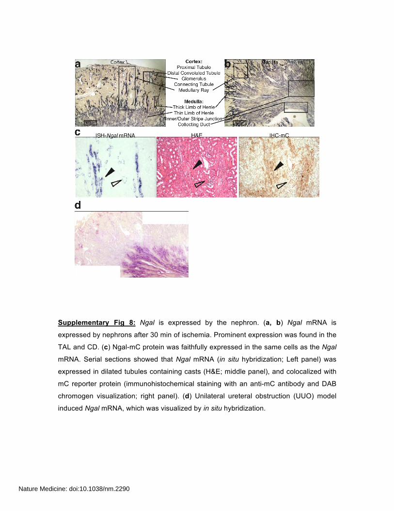
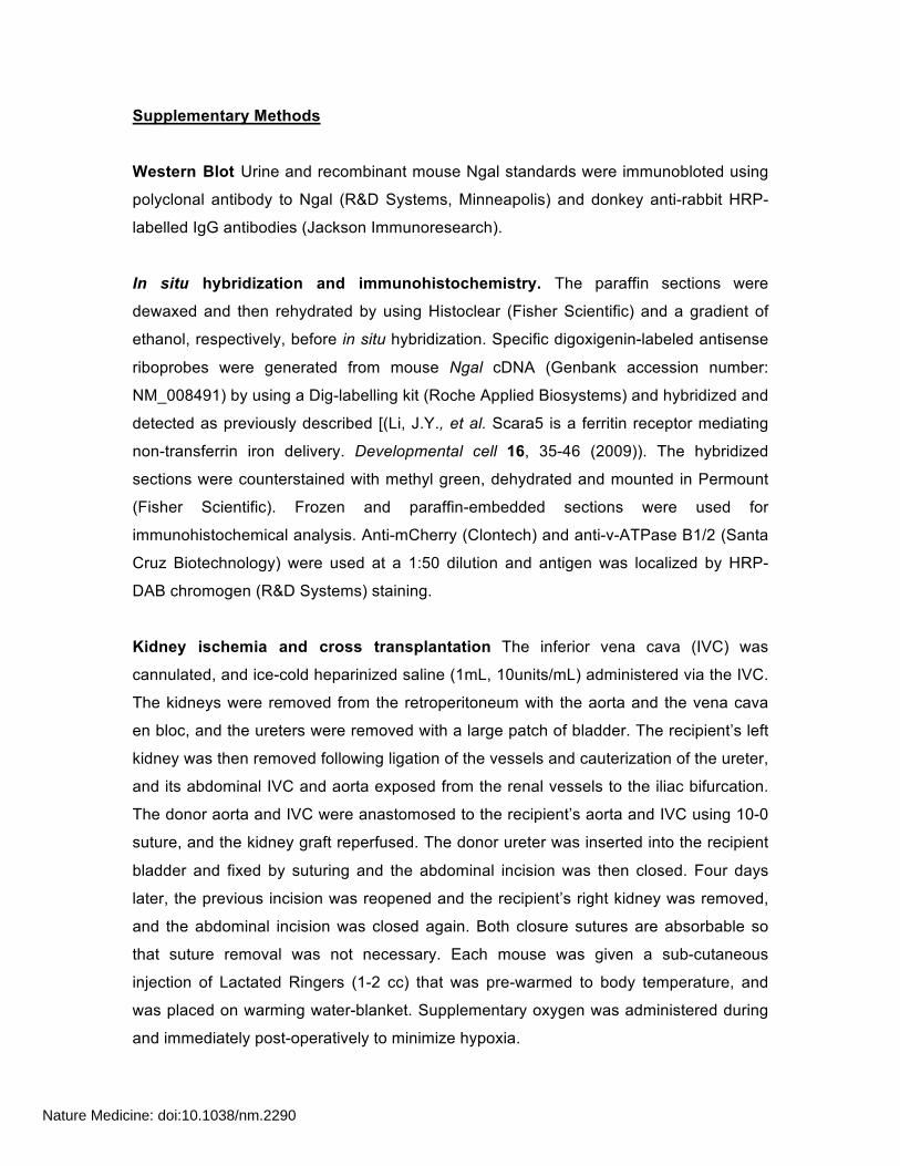
Supplementary Fig 8: Ngal is expressed by the nephron. (a, b) Ngal mRNA is
expressed by nephrons after 30 min of ischemia. Prominent expression was found in the
TAL and CD. (c) Ngal-mC protein was faithfully expressed in the same cells as the Ngal
mRNA. Serial sections showed that Ngal mRNA (in situ hybridization; Left panel) was
expressed in dilated tubules containing casts (H&E; middle panel), and colocalized with
mC reporter protein (immunohistochemical staining with an anti-mC antibody and DAB
chromogen visualization; right panel). (d) Unilateral ureteral obstruction (UUO) model
induced Ngal mRNA, which was visualized by in situ hybridization.
Nature Medicine: doi:10.1038/nm.2290

Supplementary Fig 9: Apoptotic S3 proximal tubule cells (top; boxed area) were
detected by Tunel assay. They do not overlap with Ngal expressing medullary rays and
CD cells (bottom; open arrowheads).
!"#"$%&'()**+,-,./"+'012)#,'3
Nature Medicine: doi:10.1038/nm.2290

Supplementary Fig 10: (a) Ngal-mCherry fluorescence was visible in the TAL (closed
arrowheads), but not in the adjacent thin Limbs of Henle (open arrowheads). (b) Ngal-
mCherry fluorescence was visible in the alternating epithelial cells of the CD revealing
specific expression of Ngal mRNA in α-IC (closed arrowheads).
!"#"$%&'()**+,-,./"+'012)#,'34
! "
Nature Medicine: doi:10.1038/nm.2290
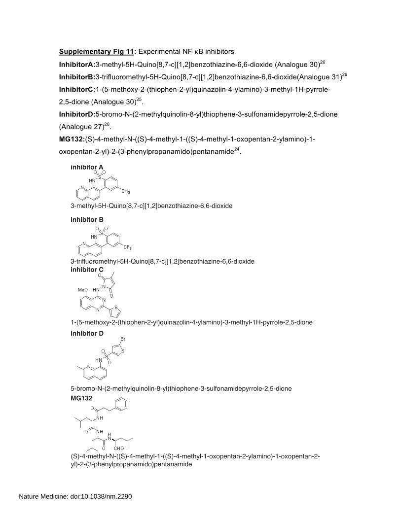
Supplementary Fig 11: Experimental NF-κB inhibitors
InhibitorA:3-methyl-5H-Quino[8,7-c][1,2]benzothiazine-6,6-dioxide (Analogue 30)26
InhibitorB:3-trifluoromethyl-5H-Quino[8,7-c][1,2]benzothiazine-6,6-dioxide(Analogue 31)26
InhibitorC:1-(5-methoxy-2-(thiophen-2-yl)quinazolin-4-ylamino)-3-methyl-1H-pyrrole-
2,5-dione (Analogue 30)25.
InhibitorD:5-bromo-N-(2-methylquinolin-8-yl)thiophene-3-sulfonamidepyrrole-2,5-dione
(Analogue 27)26.
MG132:(S)-4-methyl-N-((S)-4-methyl-1-((S)-4-methyl-1-oxopentan-2-ylamino)-1-
oxopentan-2-yl)-2-(3-phenylpropanamido)pentanamide24.
!"#$%&%"'"()"&*+,-./012%.12"3"-.4+,1%5,*2*"6"70.8%29&1:*5-$$%.*");!":1%2*
6"&*+,-."!<"=012%>3;?"@A>B;)A#*2C%+,19C12*"D;D":1%E1:*
B"(!"&*+,%E-")"(+,1%5,*2")"-.4/0129C%.12"F"-.9&12%4"6"&*+,-."B<"5-$$%.*");!":1%2*
(G4"F"&*+,-."'"((G4"F"&*+,-."B"((G4"F"&*+,-."B"%E%5*2+92")"-.9&12%4"B"%E%5*2+92")"-.4")"(6"5,*2-.5$%5929&1:%45*2+929&1:*
!"#!$!%&'()
!"#!$!%&'(*
!"#!$!%&'(+
!"#!$!%&'(,
-./01
Nature Medicine: doi:10.1038/nm.2290
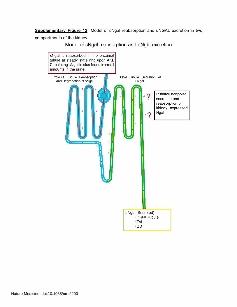
Supplementary Figure 12: Model of sNgal reabsorption and uNGAL excretion in two
compartments of the kidney.
Nature Medicine: doi:10.1038/nm.2290