themode action of aspirin-like drugs: effecton inducible … oxidesynthase ashokr.amin*tt ,...
TRANSCRIPT
Proc. Natl. Acad. Sci. USAVol. 92, pp. 7926-7930, August 1995Medical Sciences
The mode of action of aspirin-like drugs: Effect on induciblenitric oxide synthaseASHOK R. AMIN*tt§, PRANAV VYAS*, MUKUNDAN ATrUR*, JOANNA LESZCZYNSKA-PIZLAK*,INDRAVADAN R. PATELT, GERALD WEISSMANN*, AND STEVEN B. ABRAMSON***Department of Rheumatology, Hospital for Joint Diseases, New York, NY 10003; Departments of tPathology and tMedicine, New York University MedicalCenter, New York, NY 10016; and 1Department of Biochemistry, Glaxo, Inc., Research Triangle Park, NC 27709
Communicated by H. Sherwood Lawrence, New York University Medical Center, New York, NY, May 26, 1995 (received for reviewJanuary 19, 1995)
ABSTRACT Nitric oxide synthesized by inducible nitricoxide synthase (iNOS) has been implicated as a mediator ofinflammation in rheumatic and autoimmune diseases. Wereport that exposure of lipopolysaccharide-stimulated murinemacrophages to therapeutic concentrations of aspirin (IC50 =3 mM) and hydrocortisone (IC50 = 5 ,uM) inhibited theexpression of iNOS and production of nitrite. In contrast,sodium salicylate (1-3 mM), indomethacin (5-20 ,uM), andacetaminophen (60-120 ,uM) had no significant effect on theproduction of nitrite at pharmacological concentrations. Atsuprapharmacological concentrations, sodium salicylate(IC50 = 20 mM) significantly inhibited nitrite production.Immunoblot analysis of iNOS expression in the presence ofaspirin showed inhibition of iNOS expression (IC5s = 3 mM).Sodium salicylate variably inhibited iNOS expression (0-35%), whereas indomethacin had no effect. Furthermore,there was no significant effect of these nonsteroidal anti-inflammatory drugs on iNOS mRNA expression at pharma-cological concentrations. The effect of aspirin was not due toinhibition of cyclooxygenase 2 because both aspirin andindomethacin inhibited prostaglandin E2 synthesis by >75%.Aspirin and N-acetylimidazole (an effective acetylating agent),but not sodium salicylate or indomethacin, also directlyinterfered with the catalytic activity of iNOS in cell-freeextracts. These studies indicate that the inhibition of iNOSexpression and function represents another mechanism ofaction for aspirin, if not for all aspirin-like drugs. The effectsare exerted at the level of translational/posttranslationalmodification and directly on the catalytic activity of iNOS.
Nitric oxide (NO), first identified as an endothelium-derivedrelaxation factor (1), is now recognized to be an intra- andextracellular mediator of cell function (2-5). NO produced bythe constitutive isoform of nitric oxide synthase (NOS) is a keyregulator of homeostasis, whereas the generation of NO byinducible NOS (iNOS) plays an important role in inflamma-tion, host-defense responses, and tissue repair (2-4). NOformation is increased during inflammation (rheumatoid ar-thritis, and ulcerative colitis, Crohn disease), and severalclassic inflammatory symptoms (erythema and vascular leak-iness) are reversed by NOS inhibitors (2-4). Vane and co-workers (6) have implicated NO as an important mediator ofinflammation in animal models. Furthermore, because iNOSis up-regulated by endotoxin, interleukin 1, tumor necrosisfactor, and interferon y, the increased synthesis of NO hasbeen implicated in autoimmune diseases, allograft rejection,graft-versus-host disease, and systemic response to sepsis.Recent studies by Salvemini et al. (7) have shown that NOmodulates the activity of prostaglandin endoperoxide H syn-thase 2 [cyclooxygenase 2 (COX-2)] in a concentration-
dependent manner, through a mechanism independent ofcGMP.Although nonsteroidal antiinflammatory drugs (NSAIDs)
clearly inhibit the synthesis and release of prostaglandins (8, 9),these actions are by no means sufficient to explain all theantiinflammatory effects of NSAIDs. NSAIDs also inhibitactivation of neutrophils (10), which provoke inflammation byreleasing products other than prostaglandins (11). In thesestudies we examined the effect of NSAIDs on NO production.Among the agents studied in an effort to elucidate the effect
of NSAIDs on iNOS expression and function, we have selectedthree: an acetylated salicylate (aspirin, an effective inhibitor ofCOX); a nonacetylated salicylate (sodium salicylate, an inef-fective inhibitor of COX); and a nonacetylated nonsteroidalcompound (indomethacin, a potent inhibitor of COX). Wetested the hypothesis that NSAIDs, which inhibit COX activity,might inhibit inflammation by modifying iNOS expression/activity. Aspirin, sodium salicylate, and indomethacin, whichreach therapeutic concentrations in plasma of 1-3 mM, 1-3mM, and 5-20 ,uM, respectively (12), were tested for theircapacities to inhibit iNOS expression/catalytic activity at theclinically relevant concentrations. In the present study wereport that aspirin and, to a lesser extent, sodium salicylate (butnot indomethacin) inhibit iNOS expression in murine macro-phages activated with lipopolysaccharide (LPS). In addition,aspirin inhibits the catalytic activity of iNOS, an effect mim-icked by N-acetylimidazole (NAI), another acetylating agent.We therefore conclude that the aspirin-like drugs differ intheir mode of action and that acetylation may be a criticaldifference.
MATERIALS AND METHODSCell Lines and Reagents. Murine macrophage cells (RAW
264.7) were obtained from the American Type Culture Col-lection. An antimurine iNOS antibody was obtained fromTransduction Laboratories (Lexington, KY). Aspirin, sodiumsalicylate, indomethacin, acetaminophen, NAI, and imidazolewere obtained from Sigma.Immunoblot Analysis. Equal amounts of protein (50 ,ug)
estimated by bicinchoninic acid reagent (Pierce) were loadedonto SDS/PAGE gels and stained to verify the concentrationsof various protein fractions by examining the intensities of theprotein bands on the gels. Immunoblot analysis was done fromthe same cell extracts. The immunoblotted membrane wasprobed with a specific anti-iNOS monoclonal antibody, asspecified by Transduction Laboratories. The blots were devel-
Abbreviations: COX, cyclooxygenase (prostaglandin endoperoxide Hsynthase 2); NO, nitric oxide; NOS, NO synthase; iNOS, inducibleNOS; NAI, N-acetylimidazole; NSAIDs, nonsteroidal antiinflamma-tory drugs; LPS, lipopolysaccharide(s).§To whom reprint requests should be addressed at: Department ofRheumatology, Hospital for Joint Diseases, 301 East 17th Street,Room 1600, New York, NY 10003.
7926
The publication costs of this article were defrayed in part by page chargepayment. This article must therefore be hereby marked "advertisement" inaccordance with 18 U.S.C. §1734 solely to indicate this fact.

Proc. Natl. Acad. Sci. USA 92 (1995) 7927
oped by using the enhanced chemoluminescence immunoblotsystem (Amersham). Density of the bands was measured witha densitometer from Molecular Dynamics.Northern Blot Analysis. Total RNA was isolated using TRI
reagent (Molecular Research Center, Cincinnati). Northernblot analysis was done as described by Church and Gilbert (13).Thirty micrograms of RNA was subjected to electrophoresis in1% agarose/formaldehyde gel. The gel was then transferredvia capillary action onto a nylon membrane (Zeta probe,Bio-Rad). The membrane was hybridized with [32P]dCTP-labeled iNOS cDNA (4-kb Sma I fragment), from JamesCunningham (Harvard Medical School, Boston). After hybrid-ization, the blot was exposed to Kodak x-ray film for 24-48 hrwith intensifying screens at -70°C. The ,B-actin probe waspurchased from Clontech and probed as described above.Measurement of the intensity of the iNOS/,B-actin bands wasdone by using a Phospholmager (Molecular Dynamics).Assays for iNOS in Cell-Free Extracts. Specific activity of
iNOS was determined in cell-free extracts by monitoring theconversion of L-[3H]arginine to L-[3H]citrulline, as describedby Misko et al. (14) and modified by us (unpublished work).RAW 264.7 cells were induced with LPS in the presence andabsence of NSAIDs for 16-18 hr. After induction, the cellswere pelleted at 4°C and resuspended in Tris buffer (10 mM,pH 7.4) containing chymostatin, antipain, leupeptin, andpepstatin each at 10 ,ug/ml, as well as dithiothreitol and 1 mMphenylmethylsulfonyl fluoride. Cells were lysed in a PolytronPT 1200 homogenizer (Kinematica, Lucerne, Switzerland)after three cycles of rapid freeze-thawing. The lysate wascentrifuged at 30,000 x g for 60 min at 4°C, and the superna-tants were used as cell-free extracts. The protein was measuredby bicinchoninic acid assay reagent using bovine serum albu-min as standard (16). The reaction mixture for iNOS assayconsists of 50 ,uM Tris (pH 7.8); bovine serum albumin at 1mg/ml; 1 mM dithiothreitol; 2 mM CaCl2; 10 ,uM FAD; 10 ,uMtetrahydrobiopterin; 30 ,uM L-arginine; 1 mM NADPH (11).The reaction mixture was treated with 1 Al (250 nM) ofL-[3H]arginine (DuPont/NEN) (1 mCi/ml = 37.0 MBq/ml).After 20 min the assays were terminated by heating thereaction mixture at 90°C for 5 min. The precipitates wereremoved by centrifuging at 27,000 x g for 20 min. Tenmicroliters (-50,000 cpm) of the supernatant was spotted onactivated Avicel TLC plates (Analtech). The TLC plates weredeveloped in a solvent system of ethanol/water/ammonia,80:16:4. The spot for L-[3H]citrulline was quantitated by usinga Bioscan (Washington, DC) system 200 imaging scanner.Briefly, total cpm per lane were averaged, and the cpm of eachlane was then normalized to the mean. The quantity of[3H]arginine converted to [3H]citrulline was calculated fromthe specific activity of [3H]arginine added to the assay mixture(2 cpm = 1 pmol of [3H]arginine or [3H]citrulline).
Assay for COX-2 in Whole Cells. Cells were incubated withLPS (1 ,ug/ml) for 16 hr to induce COX-2, exposed to NSAIDsfor 1 hr, and subsequently harvested. The harvested cells werethen incubated with radiolabeled arachidonic acid (100,000cpm, 57 mCi/mM) in 1 ml of Tris HCl (together with 3 ,uM ofcold arachidonic acid) for 10 min. Specific enzyme activity(whole-cell assays) was measured by the conversion of [14C]ar-achidonic acid to prostaglandin E2 after separation by TLC (6,17). Authentic prostaglandin and monohydroxy standardswere run in parallel. The transformed products were quanti-tated by a Bioscan system 200 imaging scanner, as describedabove.
RESULTS AND DISCUSSIONEffects of NSAIDs on Nitrite Accumulation. Murine mac-
rophage cells (RAW 264.7) were selected because the iNOSregulation in these cells has been well-characterized, both atthe biochemical and molecular levels (2, 18, 19). RAW 264.7cells were activated with LPS at 100 ng/ml to induce iNOS (18)with and without aspirin (1-3 mM), sodium salicylate (1-3mM), and indomethacin (5-20 ,uM). Expression and activity ofiNOS were monitored by estimating the stable end-productnitrite, as described for these cells by other investigators (7, 18,19). Table 1 shows a concentration-dependent inhibition ofnitrite accumulation in cells stimulated with LPS in thepresence of 1-3 mM aspirin. Only 2 and 3 mM concentrationshowed a significant effect on nitrite accumulation. Supra-pharmacological concentrations of aspirin (5 and 10 mM)further inhibited nitrite accumulation [by 50% ± 6 and 80% +5 (p <0.005), respectively] above that seen at 3 mM (data notshown). Sodium salicylate (3 mM) and indomethacin (5 ,uM)did not significantly inhibit nitrite production (-7%). Supra-pharmacological concentrations of sodium salicylate (5 mM)inhibited nitrite accumulation by 15% under identical condi-tions. However, the IC50 of sodium salicylate with respect tonitrite accumulation was 20 mM, whereas its ability to inhibitfMet-Leu-Phe-induced neutrophil aggregation was 3 mM(data not shown) (11). Although indomethacin is effectivetherapeutically at 20 ,M, the extent to which it inhibited nitriteaccumulation (10 ± 9.6%) was only marginally greater thanthat seen with 5 ,zM and was not statistically significant. Ourresults on the effect of indomethacin on nitrite accumulationin RAW 264.7 cells were identical to those seen by Salveminiet al. (7). Acetaminophen (60-120 ,uM), an analgesic agentclosely related to salicylates, failed to block nitrite production(1 ± 1%) in LPS-stimulated macrophages at therapeuticconcentrations. As previously shown by Moncada and cowork-ers (21) in murine macrophages (J774 cells) and as seen here(Table 2), hydrocortisone (5 ,uM) inhibited endotoxin-inducedNO production by >60%.
Table 1. Effect of NSAIDs on nitrite accumulation and specific activity of iNOS in murine macrophages inducedwith LPS
Nitrite released Specific activity
Inhibition, Inhibition,Modulating agent Nitrite, ,uM % P value pmol/min per mg protein % P value
Control (uninduced) 0.5 ± 0.5 - 11.7 ± 1.5LPS-induced 29.2 ± 6.8 310.0 ± 54.6Aspirin (1 mM) 26.6 ± 4.3 10 <0.267 271.7 ± 17.2 12 <0.155Aspirin (2 mM) 22.9 ± 5.3 22 <0.071 231.3 ± 29.8 25 <0.046Aspirin (3 mM) 20.3 ± 3.9 32 <0.025 162.3 ± 25.9 48 <0.006Sodium salicylate (3 mM) 27.1 ± 8.8 7 <0.345 304.0 ± 48.1 2 <0.446Indomethacin (5 ,uM) 27.5 ± 7.7 7 <0.365 304.0 ± 39.4 2 <0.441
Murine macrophage cells (RAW 264.7) were incubated with NSAIDs for 2 hr followed by addition of LPS at 100 ng/ml.After 16-18 hr of incubation, the medium was used to estimate nitrite accumulation by the modified Greiss method (20).Specific activity of iNOS was determined in cell-free extracts at a given time, as described. Nitrite and specific activity dataare representative of means ± SD values, as determined by Student's t test, for four independent experiments. The P valuesindicate comparisons with LPS-stimulated cells.
Medical Sciences: Amin et al.

7928 Medical Sciences: Amin et al.
Table 2. Action of NSAIDs on expression of iNOS and COX-2
Inhibition of iNOS at 16 hr, %
Specific activity in Specific activity Inhibition of COX-2,Modulating agent Nitrite release cell-free extracts Protein expression mRNA in in vitro assay %
Aspirin (3 mM) 32.0* 47t -53 NS -45 (1mM) >75tSodium salicylate (3 mM) 7.0t 2§ 15 NS -1 (1mM) NSIndomethacin (5 ,iM) 7.0T 2§ 0 NS 0(5 I.M) >75tHydrocortisone (5 ,uM) 63.01 ND ND ND ND NDNAI (1 mM) ND ND ND ND -74 (1mM) NDThe data (expressed as percentage inhibition) are compiled from this study. RAW 264.7 cells were induced with LPS at 100 ng/ml to stimulate
iNOS and COX-2 activity. After 16-18 hr of incubation, COX-1/COX-2 activity was assayed, as described by Mitchell et al. (17). COX-1 activitywas not detected in these cells, as described (7). In vitro assay indicates the effect of NSAIDs (at concentrations in parentheses) on catalytic activityof iNOS in cell-free extracts. Protein expression data are represented as approximate percentage inhibition based on the densitometry data fromone of the two representative experiments. ND, not done; NS, not significant. Note P values: §, P <0.45, *, P <0.36, *, P <0.05, 1, P <0.01, t,P <0.006 (see text and Table 1 for absolute values).
We next compared the capacities of selected drugs to inhibitthe specific activity of COX-2 in RAW 264.7 cells exposed toLPS, as shown in Table 2. Aspirin (3 mM) and indomethacin(20 ,M) inhibited the specific activity of COX-2 by 79 ± 6.7%(P <0.001) and 84 ± 4.0% (P <0.002), respectively, whereassodium salicylate (3 mM) had no effect [16 ± 11% (P <0.28)].These data indicate that aspirin does not inhibit nitrite pro-duction by inhibiting COX because aspirin shares this lattereffect with indomethacin.We further examined the mechanism of action of aspirin by
determining its effects on (i) the specific activity of theenzyme, (ii) the synthesis of iNOS at the protein level, (iii) thesynthesis of mRNA, and (iv) the catalytic activity of iNOS incell-free extracts and also by comparing it with sodium salic-ylate and indomethacin.
Effect of NSAIDs on Expression and Catalytic Activity ofiNOS. Because nitrite accumulation, which represents thecumulative effect of iNOS expression from induction of theenzyme, does not directly assess the effects of pharmacologicagents (i.e., NSAIDs) on specific enzyme activity, we analyzedthese two parameters in tandem.The specific enzyme activity of iNOS from cells exposed to
aspirin in cell-free extracts showed a significant inhibition inactivity in a dose-dependent fashion (IC50 = 3 mM). Sodiumsalicylate and indomethacin did not inhibit the specific activityof iNOS (Table 1). These observations raised the followinghypotheses. Aspirin may (i) decrease the expression of iNOSprotein and therefore decrease the specific activity of theenzyme and subsequently the production of nitrite; (ii) de-crease only the catalytic activity of iNOS without influencingthe expression of iNOS protein; or (iii) decrease both thecatalytic activity of iNOS and the expression of iNOS protein,which in turn cumulatively leads to decrease in the accumu-lation of nitrite in the medium.We therefore analyzed (by immunoblot) iNOS protein in
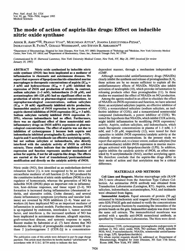
cells treated with LPS with and without NSAIDs for 16-18 hr.Fig. 1A shows a significant decrease in iNOS expression in cellstreated with aspirin, thus accounting, in part, for the decreasein the specific activity of iNOS and thus eliminating hypothesisii described above. Aspirin at 10 mM further decreased iNOSexpression by =70%, as determined by immunoblot analysis(Fig. 1B). Therapeutic concentration of sodium salicylate (2mM) caused =15% inhibition of iNOS expression, whereas 5
,uM indomethacin had no effect, as assessed by immunoblotanalysis. Sodium salicylate (2-3 mM) caused a variable (0-35%) inhibition of iNOS expression at therapeutic concentra-tions in four independent experiments, one of which is pre-sented in Fig. LB. However, increased sodium salicylate con-centration (5 and 20 mM) did not increase inhibition of iNOSexpression, unlike the increasing effects seen with 10 mMaspirin (=70%). These results are not easily interpreted, butwe assume that sodium salicylate at lower concentrationsinterferes with enzyme synthesis, whereas at higher concen-
trations this salicylate inhibits the catalytic activity of iNOS.This biphasic effect would account for a decrease in nitriteproduction without apparent decrements of protein synthesis,as assessed by immunoblot analysis.
Previous studies have shown that induction of iNOS andCOX-2 are both achieved by LPS in RAW 264.7 cells after12-16 hr (7). Indomethacin (5 ,uM) inhibited COX-2 activityby >75% but had no effect on iNOS expression in immunoblotanalysis (Fig. 1A). Furthermore, because indomethacin hadminimal effects on iNOS activity at therapeutic concentra-tions, COX-2 or its products are unlikely to be regulators ofiNOS activity per se, at least in murine macrophages.
Effect of NSAIDs on Expression of iNOS mRNA. Aspirinmay suppress iNOS expression early in the course of enzymeinduction, leading to inhibition or delay in nitrite accumula-tion. This assumption is based on the observation that, inmacrophages, transforming growth factor ,31 suppresses iNOSexpression by decreasing mRNA stability and translation and
A LPS
o E E E e
e* ......~~~~~~~~~~~~~~~. ... ..-,. ....1 2 3 4 5 6
Western blot
- 0 25 47 53 0
LPS
-1g
E E cl
7 r iN(i
1 2 3 4 5 6 7
B LPS
2 E E E e.. N n -C c CL CL co) cn C
0d
0 to 0o
-iNOS
Western blot
- 0 47 69 15 0 0 % Inhibition
actin
1 2 3 4 5 6 7
Northern blot Ratio2 12 12 12 12 14 10 iNOS/4 actin
xl0-3
FIG. 1. Immuno- and Northern blot analyses of iNOS from RAW264.7 cells activated with LPS with and without NSAIDs. (A and B)Western (immunoblot) analysis of cells treated with LPS at 100 ,ug/mlwith or without NSAIDs for 16 hr. (C) RNA blot analysis of iNOS and,B-actin from RAW 264.7 cells activated with LPS and treated withNSAIDs for 16 hr. The iNOS/,B-actin ratio was determined by usinga PhosphoImager. Data represent one of the four representativeexperiments. Asp, aspirin; Indo, indomethacin; NaSal, sodium salic-ylate.
Proc. Natl. Acad. Sci. USA 92 (1995)
Proc. Natl. Acad. Sci. USA 92 (1995) 7929
increasing the degradation of iNOS protein in macrophages(22). There was no significant difference in the expression ofiNOS mRNA (at 16 hr) in cells treated with LPS with or
without NSAIDs because the iNOS mRNA/13-actin mRNAratios were either identical or not significantly different fromcells stimulated with LPS alone (Fig. 1). Recent studies byTetsuka et al. (23) have demonstrated that indomethacinaddition enhanced interleukin 113-induced steady-state level ofiNOS mRNA and nitrite production in rat mesangial cells.Hence, our studies indicate that the effect of indomethacinmay differ in different cell types. Kopp and Ghosh (24) showedthat aspirin (3 mM) or sodium salicylate (5 mM) inhibitNF-KB-dependent transcription, using sensitive assays basedon plasmids containing two IgK-KcB sites driving a luciferasereporter gene. However, in the same studies, the same con-
centrations of aspirin and sodium salicylate had no significanteffect on NF-KB activation, judged by gel-shift assays. Nathanand coworkers (19) have shown that NF-KB expression is oneof the integral components of iNOS transcription/expression,which can be inhibited by an NF-KB inhibitor, pyrrolidinedithiocarbamate, at 30 ,uM. Our studies indicate that 3 mMaspirin is probably not sufficient to block the transcription ofthe iNOS gene, as seen with 30,uM of pyrrolidine dithiocar-bamate, which blocked >90% of nitrite accumulation in our
studies (data not shown). Furthermore, the lack of significanteffects of aspirin and sodium salicylate on iNOS mRNAexpression and the differential effect of aspirin and sodiumsalicylate on iNOS expression support the above notion thataspirin and sodium salicylate have no significant effect on
iNOS expression at the gene level, at least in murine macro-
phages activated with LPS in vitro. These experiments furtherreinforce the notion that the mechanism of action of aspirin (atpharmacological concentrations) in inhibiting iNOS expres-
sion is due to its interference in translational/posttranslationalmodification of the enzyme and/or inhibiting the catalyticactivity of iNOS. However, direct experiments with an iNOSpromoter and a reporter gene are needed to confirm thisobservation.
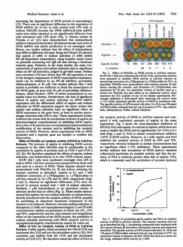
Effect ofNSAIDs on Catalytic Activity ofiNOS in Cell-FreeExtracts. The potency of aspirin in inhibiting iNOS activitycompared to the other NSAIDs may be attributable to theacetylation by aspirin of proteins such as COX-1 and COX-2(25, 26). We therefore examined the effects of aspirin, sodiumsalicylate, and indomethacin in in vitro iNOS enzyme assays.
RAW 264.7 cells were incubated overnight with LPS toinduce iNOS. Cell-free extracts were prepared from these cellsand used as a source of iNOS. These enzyme extracts were
preincubated with NSAIDs for 20 min before initiating theenzyme reactions as described. Aspirin at 0.1 and 1 mMinhibited conversion of L-[3H]arginine to L-[3H]citrulline incell-free extracts by 10-12% and 45-68%, respectively (Figs.2 and 3), whereas no significant differences (7%) were ob-served in extracts treated with 1 mM of sodium salicylate.Similarly, 5,LM indomethacin or an equivalent volume ofabsolute alcohol had no effect (Fig. 2). These studies demon-strated that aspirin, but not sodium salicylate or indomethacin,directly interfered with the catalytic activity of iNOS, possiblyby acetylating an important functional component of theenzyme or its cofactors. However, because sodium salicylate attherapeutic (2 mM) and suprapharmacological (5 and 20 mM)concentrations inhibits accumulation of nitrite (by 6%, 15%,and 50%, respectively) and has only minimal and insignificanteffect on the expression of the iNOS protein, the possibility ofsodium salicylate interfering with iNOS catalytic activity atsuprapharmacological concentrations cannot be ruled out.
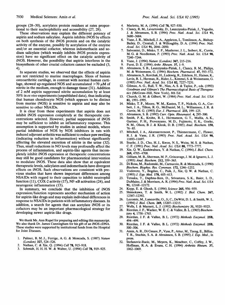
Effect of NAI on Catalytic Activity of iNOS in Cell-FreeExtracts. Unlike aspirin, which acetylates Ser-530 of COX andinactivates the COX and not the peroxidase activity (26), NAIacetylates and inhibits both the COX and the peroxidaseactivity ofCOX (27). We therefore tested the effect ofNAI on
LPS Induced
E E67C E~ a ')CDC a L mL co
r°
co CD 0
Citruline-
Arginine _
Specific Activity
% Inhibition
U,to0-ac
15 -96 168 35 369 400 41- - 68 10 7 0 0
FIG. 2. Effect of NSAIDs on iNOS activity in cell-free extracts.RAW 264.7 cells were induced with LPS for 16 hr, and enzyme extractswere prepared to examine iNOS activity in cell-free extracts bymonitoring conversion of L-[3H]arginine to L-[3H]citrulline by TLC, asdescribed. NSAIDs were preincubated with enzyme extracts 20 minbefore starting the reaction, and formation of L-[3H]citrulline wasmonitored for 30 min. An equivalent volume of alcohol used as asolvent for NSAIDs was also added as an additional control. Datarepresent the TLC analysis of one of two similar experiments. Per-centage inhibition is determined after removing the control value(=15), which represents specific activity of iNOS in uninduced cells.The specific activity of LPS-treated cells after 16-18 hr was 396 pmolof [3H]citrulline released/min per mg of protein at 37°C. Asp, aspirin;Indo, indomethacin; NaSal, sodium salicylate.
the catalytic activity of iNOS in cell-free extracts and com-pared it with equivalent amounts of aspirin in the sameexperiment. Fig. 3 shows the dose-dependent inhibition ofiNOS by NAI and aspirin. In contrast to aspirin, which does notseem to inhibit the iNOS activity significantly (10-12%) at 0.1mM (Figs. 2 and 3), NAI at similar concentrations inhibited-45% of iNOS activity. However, at 1 mM, aspirin and NAIinhibited the catalytic activity of iNOS by 45% and 74%,respectively, whereas imidazole at similar concentrations hadno significant effect (<5% inhibition). These experimentsfurther indicate that acetylation of iNOS [and/or essentialcofactor(s)] inactivates its catalytic activity and that the po-tency of NAI is relatively greater than that of aspirin. NAI,which is commonly used for acetylation of tyrosine hydroxyl
CD)0
.2
Cl)C
100-0* N-Acetylimidazoleo Aspirin
60-
40-
20-
II
0.01 0.05 0.1 0.5
Concentration (mM)
FIG. 3. Effect of acetylating agents aspirin and NAI on catalyticactivity of iNOS in cell-free extracts. The enzyme reactions were setup as described for Fig. 3. Acetylating compounds were incubated withthe enzyme extracts 20 min before starting the reaction and assayed asdescribed. The specific activity of LPS-treated cells after 16-18 hr was178 pmol of [3H]citrulline released/min per mg of protein at37°C. Anequivalent volume of absolute alcohol, which was used as a carrier forthe drugs, inhibited iNOS activity by -10%.
Medical Sciences: Amin et aL
I
7930 Medical Sciences: Amin et al.
groups (28-30), acetylates protein residues at rates propor-tional to their nucleophilicity and accessibility (27, 28).These observations may explain the different potency of
aspirin and sodium salicylate. Aspirin inhibits iNOS by effectson both synthesis of the iNOS protein and on the catalyticactivity of the enzyme, possibly by acetylation of the enzymeand/or an essential cofactor, whereas indomethacin and so-dium salicylate (which weakly inhibits iNOS protein expres-sion) have no significant effect on the catalytic activity ofiNOS. However, the possibility that aspirin interferes in thebiosynthesis of other crucial cofactors cannot be excluded (3,4).
In separate studies, we observed that the effects of aspirinare not restricted to murine macrophages. Slices of humanosteoarthritic cartilage, in contrast with normal human carti-lage, showed up-regulated NOS and accumulated >70 ,uM ofnitrite in the medium, enough to damage tissue (31). Additionof 2 mM aspirin suppressed nitrite accumulation by at least50% in ex vivo experiments (unpublished data), thus indicatingthat human chondrocyte NOS (which appears to be distinctfrom murine iNOS) is sensitive to aspirin and may also besensitive to other NSAIDs.
It is clear from these experiments that aspirin does notinhibit iNOS expression completely at the therapeutic con-centrations selected. However, partial suppression of iNOSmay be sufficient to inhibit an inflammatory response. Thisassumption is supported by studies in animal models, wherepartial inhibition of NOS by NOS inhibitors in rats withinduced adjuvant arthritis was sufficient to reduce paw swelling(indicating reduction in inflammation) without significantlyaffecting the elevated excretion of nitrite in the urine (32).Thus, small reductions in NO levels may profoundly affect theprocess of inflammation, and aspirin-like agents that incom-pletely inhibit iNOS expression at therapeutic concentrationsmay still be good candidates for pharmaceutical interventionto modulate iNOS. These data also show that at equivalenttherapeutic levels, salicylates and indomethacin have divergenteffects on iNOS. Such observations are consistent with pre-vious studies that have shown important differences amongNSAIDs with regard to their capacities to inhibit neutrophilfunction (11), COX-2 activity (17), NF-KB activation (24), andneurogenic inflammation (15).
In summary, we conclude that the inhibition of iNOSexpression/function represents another mechanism of actionfor aspirin-like drugs and may explain individual differences inresponse to NSAIDs in patients with inflammatory diseases. Inaddition, a search for agents that can acetylate iNOS or itscofactors may be an important pharmacological strategy fordeveloping newer aspirin-like drugs.
We thank Ms. Ann Rupel for preparing and editing this manuscript.We also thank Dr. James Cunningham for his gift of an iNOS cDNA.These studies were supported by institutional funds from the Hospitalfor Joint Diseases.
1. Palmer, R. M. J., Ferrige, A. G. & Moncada, S. (1987) Nature(London) 327, 524-526.
2. Nathan, C. & Xie, Q. (1994) Cell 78, 915-918.3. Schmidt, H. H. H. W. & Walter, U. (1994) Cell 78, 919-925.
4. Marletta, M. A. (1994) Cell 78, 927-930.5. Clancy, R. M., Levartovsky, D., Leszczynska-Piziak, J., Yegudin,
J. & Abramson, S. B. (1994) Proc. Natl. Acad. Sci. USA 91,3680-3684.
6. Vane, J. R., Mitchell, J. A., Appleton, I., Tomlinson, A., Bishop-Bailey, D., Croxtall, J. & Willoughby, D. A. (1994) Proc. Natl.Acad. Sci. USA 91, 2046-2050.
7. Salvemini, D., Misko, T. P., Masferrer, J. L., Seibert, K., Currie,M. G. & Needleman, P. (1993) Proc. Natl. Acad. Sci. USA 90,7240-7244.
8. Vane, J. (1994) Nature (London) 367, 215-216.9. Furst, D. E. (1994) Arthr. Rheum. 37, 1-9.
10. Abramson, S. B., Leszczynska-Piziak, J., Clancy, R. M., Philips,M. & Weissmann, G. (1994) Biochem. Pharmacol. 47, 593-572.
11. Abramson, S., Korchak, H., Ludewig, R., Edelson, H., Haines, K,Levin, R. I., Herman, R., Rider, L., Kimmel, S. & Weissmann, G.(1985) Proc. Natl. Acad. Sci. USA 82, 7227-7231.
12. Gilman, A. G., Rall, T. W., Nies, A. S. & Taylor, P., eds. (1993)Goodman and Gilman's The Pharmacological Basis of Therapeu-tics (McGraw-Hill, New York), 8th Ed.
13. Church, G. M. & Gilbert, W. (1984) Proc. Natl. Acad. Sci. USA81, 1991-1995.
14. Misko, T. P., Moore, W. M., Kasten, T. P., Nickols, G. A., Cor-bett, J. A., Tilton, R. G., McDaniel, M. L., Williamson, J. R. &Currie, M. G. (1993) Eur. J. Pharmacol. 233, 199-125.
15. Abramson, S. B. (1992) Curr. Opin. Rheumatol. 4, 295-300.16. Smith, P. K., Krohn, R. I., Hermanson, G. T., Mallia, A. K.,
Gartner, F. H., Provenzano, M. D., Fujimoto, E. K., Goeke,N. M., Olson, B. J. & Klenk, D. B. (1985) Anal. Biochem. 150,76-85.
17. Mitchell, J. A., Akarasereenont, P., Thiemermann, C., Flower,R. J. & Vane, J. R. (1993) Proc. Natl. Acad. Sci. USA 90,11693-11697.
18. Stuehr, J. D., Cho, H. J., Kwon, N. S., Weiss, M. S. & Nathan,C. F. (1991) Proc. Natl. Acad. Sci. USA 88, 7773-7777.
19. Xie, Q. W., Kashiwabara, Y. & Nathan, C. (1994) J. Biol. Chem.269, 4705-4708.
20. Gilliam, M. B., Sherman, M. P., Griscavage, J. M. & Ignarro, L. J.(1993) Anal. Biochem. 212, 359-365.
21. Di Rosa, M., Radomski, M., Carnuccio, R. & Moncada, S. (1990)Biochem. Biophys. Res. Commun. 172, 1246-1252.
22. Vodovotz, Y., Bogdan, C., Paik, J., Xie, Q. W. & Nathan, C.(1993) J. Exp. Med. 178, 605-613.
23. Tetsuka, T., Daphna-Iken, D., Srivastava, S. K., Baier, L. D.,DuMaine, J. & Morrison, A. R. (1994) Proc. Natl. Acad. Sci. USA91, 12168-12172.
24. Kopp, E. & Ghosh, S. (1994) Science 265, 956-959.25. Shimokawa, T. & Smith, W. L. (1992) J. Biol. Chem. 267,
12387-12392.26. Lecomte, M., Laneuville, O., Ji, C., DeWitt, D. L. & Smith, W. L.
(1994) J. Biol. Chem. 269, 13207-13215.27. Wells, I. & Marnett, L. J. (1992) Biochemistry 31, 9520-9525.28. Riordan, J. F., Wacker, W. E. C. & Vallee, B. L. (1965) Biochem-
istry 4, 1758-1765.29. Riordan, J. F. & Vallee, B. L. (1972) Methods Enzymol. 25B,
494-499.30. Riordan, J. F. & Vallee, B. L. (1972) Methods Enzymol. 25B,
500-506.31. Amin, A. R., Di Cesare, P., Vyas, P., Attur, M., Tzeng, E., Billiar,
T. R., Stuchin, S. A. & Abramson, S. B. (1995) J. Exp. Med., inpress.
32. Stefanovic-Racic, M., Meyers, K., Meschter, C., Coffey, J. W.,Hoffman, R. A. & Evans, C. H. (1994) Arthritis Rheum. 37,1062-1069.
Proc. Natl. Acad. Sci. USA 92 (1995)