theemergencycliniciansapproachtothefelinerespiratorydistre ... · decreased fio 2 ! !anesthesia !...
TRANSCRIPT

1/13/12%
1%
THE EMERGENCY CLINICIAN’S APPROACH TO THE FELINE RESPIRATORY DISTRESS PATIENT
Gretchen Lee Schoeffler, DVM, DACVECC Emergency & Critical Care
Overview
! Definitions ! The 5 causes of hypoxemia ! The 8 causes of respiratory distress ! The dyspneic cat ! Case examples ! Summary
Definitions
! Symptom ! Subjective evidence of disease or physical disturbance
observed by the patient ! Clinical sign
! An objective evidence of disease as observed and interpreted by the physician (or vet!) rather than by the patient or lay observer
! Dyspnea ! Symptom ! Patient’s perception of difficult or labored breathing
! Respiratory distress ! Clinical sign ! Our perception of difficult or labored breathing
Medline Plus Medical Dictionary, Merriam-Webster
Respiratory Function
! Ventilation - ability to move air into and out of the alveoli ! Reflected in the partial pressure of carbon dioxide dissolved in
arterial blood (PaCO2) as measured on a blood gas
! Oxygenation - ability to get oxygen into the blood ! Reflected in the partial pressure of oxygen dissolved in arterial
blood (PaO2) as measured on a blood gas ! Reflected in the percent saturation of arterial hemoglobin with
oxygen SaO2 (SpO2) PaO2 SaO2 Implication
5 Causes of Hypoxemia
! Decreased FiO2 ! Anesthesia
! Hypoventilation ! Neurologic ! Anesthesia/sedation ! Obstruction ! Airway disease
! Diffusion Impairment ! Pulmonary fibrosis ! Interstitial disease
(edema/hemorrhage)
! V/Q Mismatch ! Alveolar disease ! Pulmonary hypertension
! Shunt (V/Q = ZERO) ! Atelectasis ! Severe alveolar disease ! No response to O2
supplementation
Respiratory Distress - Signs
! Increased respiratory rate and/or effort ! Abnormal posture (orthopnea) ! Pallor
! Vasoconstriction due to catecholamine ! Anemia
! Cyanosis ! 3-5 g/dl of deoxygenated hemoglobin ! With normal HCT, SpO2 = 73-78%,
PaO2 = 39-44 mmHg ! With LOW HCT, SpO2 is even LOWER
! If due to respiratory disease ! Hypoxemia: PaO2 < 60 mmHg ! Hypercapnia: PaCO2 > 50 mmHg
1/13/12%
2%
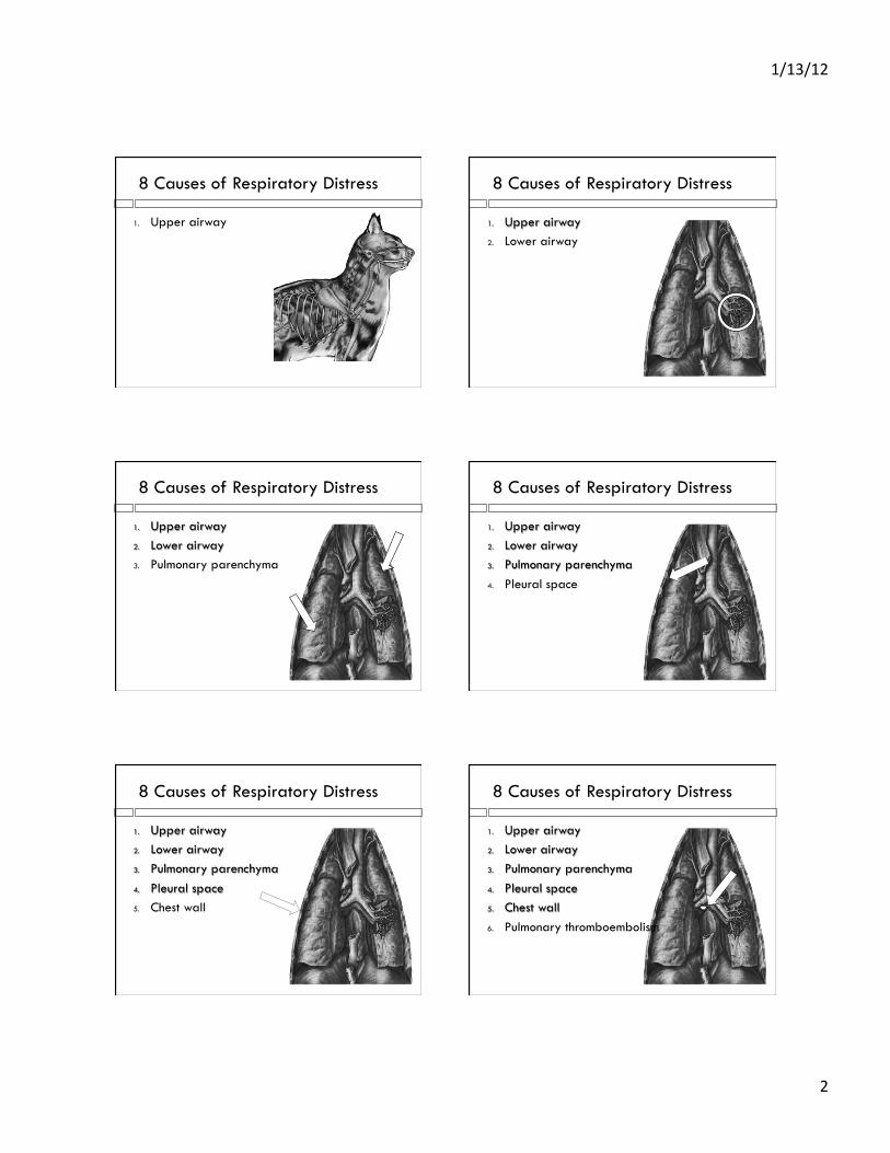
8 Causes of Respiratory Distress
1. Upper airway
8 Causes of Respiratory Distress
1. Upper airway 2. Lower airway
8 Causes of Respiratory Distress
1. Upper airway 2. Lower airway 3. Pulmonary parenchyma
8 Causes of Respiratory Distress
1. Upper airway 2. Lower airway 3. Pulmonary parenchyma 4. Pleural space
8 Causes of Respiratory Distress
1. Upper airway 2. Lower airway 3. Pulmonary parenchyma 4. Pleural space 5. Chest wall
8 Causes of Respiratory Distress
1. Upper airway 2. Lower airway 3. Pulmonary parenchyma 4. Pleural space 5. Chest wall 6. Pulmonary thromboembolism
1/13/12%
3%
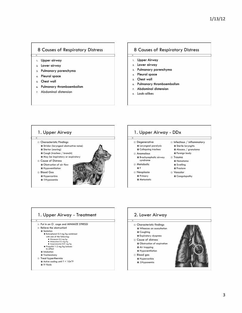
8 Causes of Respiratory Distress
1. Upper airway 2. Lower airway 3. Pulmonary parenchyma 4. Pleural space 5. Chest wall 6. Pulmonary thromboembolism 7. Abdominal distension
8 Causes of Respiratory Distress
1. Upper Airway 2. Lower airway 3. Pulmonary parenchyma 4. Pleural space 5. Chest wall 6. Pulmonary thromboembolism 7. Abdominal distension 8. Look-alikes
1. Upper Airway
! Characteristic Findings ! Stridor (laryngeal obstructive noise) ! Stertor (snoring) ! Cough (trachea / bronchi) ! May be inspiratory or expiratory
! Cause of Distress ! Obstruction of air flow ! Hypoventilation
! Blood Gas ! Hypercarbia ! ±Hypoxemia
1. Upper Airway - DDx
! Degenerative ! Laryngeal paralysis ! Collapsing trachea
! Anomalous ! Brachycephalic airway
syndrome ! Metabolic
! ?
! Neoplasia ! Primary ! Metastatic
! Infectious / inflammatory ! Sterile laryngitis ! Abscess / granuloma ! Foreign body
! Trauma ! Hematoma ! Swelling ! Fracture
! Vascular ! Coagulopathy
1. Upper Airway - Treatment
! Put in an O2 cage and MINIMIZE STRESS! ! Relieve the obstruction!
! Sedation " Butorphanol 0.3 mg/kg combined
with one of the following: " Diazepam 0.2 mg/kg " Midazolam 0.2 mg/kg " Acepromazine 0.01 mg/kg
" Propofol 1-2 mg/kg boluses to effect
! Intubation ! Tracheostomy
! Treat hyperthermia ! Active cooling until T < 104°F ! IV fluids
2. Lower Airway
! Characteristic findings ! Wheezes on auscultation ! Coughing ! Expiratory dyspnea
! Cause of distress ! Obstruction of expiration ! Air trapping ! Hypoventilation
! Blood gas ! Hypercarbia ! ±Hypoxemia
1/13/12%
4%
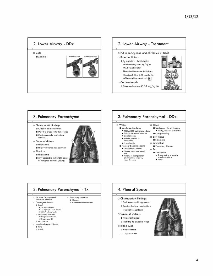
2. Lower Airway - DDx
! Cats ! Asthma!
2. Lower Airway - Treatment
! Put in an O2 cage and MINIMIZE STRESS! ! Bronchodilators
! B2 agonists – best choice " Terbutaline, 0.01 mg/kg IM " Albuterol inhaler
! Phosphodiesterase inhibitors " Aminophylline 5-10 mg/kg IM " Theophylline – oral only
! Corticosteroids ! Dexamethasone SP 0.1 mg/kg IM
3. Pulmonary Parenchymal
! Characteristic findings ! Crackles on auscultation ! May be areas with dull sounds ! Most commonly inspiratory
distress ! Cause of distress
! Hypoxemia ! Hypoventilation less common
! Blood as ! Hypoxemia ! ±Hypercarbia in SEVERE cases
or fatigued animals (young)
3. Pulmonary Parenchymal - DDx
! Water ! Cardiogenic edema
" Perihilar edema " Pulmonary veins > arteries " Cardiomegaly " Murmur, gallop, or
arrhythmia " Hypothermia
! Non-cardiogenic edema " Caudodorsal edema " Normal heart and vessel
size " History of strangulation,
electrocution, seizures, near-drowning
! Blood ! Contusion – hx of trauma
" Patchy, variable distribution ! Coagulopathy
! Soft Tissue ! Neoplasia
! Interstitial ! Pulmonary fibrosis
! Pus ! Pneumonia
" Cranioventral or patchy alveolar pattern
" Fever
ANYWHERE pulmonary edema
3. Pulmonary Parenchymal - Tx
! Put in an O2 cage and MINIMIZE STRESS!
! Cardiogenic Edema ! Lasix!
" 2-4 mg/kg initially " 1-2 mg/kg q 15-30 minutes " CRI 0.7-1.4 mg/kg/hr
! Vasodilator Therapy " Nitroglycerine topical " Nitroprusside CRI
! NO FLUIDS! ! Non-Cardiogenic Edema
! Time ! Lasix?
! Pulmonary contusion ! Oxygen ! Conservative IVF therapy
4. Pleural Space
! Characteristic Findings ! Dull to normal lung sounds ! Rapid, shallow respirations
(restrictive pattern)
! Cause of Distress ! Hypoventilation ! Inability to expand lungs
! Blood Gas ! Hypercarbia ! ±Hypoxemia

1/13/12%
5%
4. Pleural Space - DDx
! Air (pneumothorax) ! COMMON in trauma ! Doesn’t always sound
dull ! Water (hydrothorax)
! Congestive heart failure ! Hypoproteinemia
! Pus (pyothorax) ! External wound may
have already healed ! Pulmonary abscess
! Other exudate ! FIP
! Blood (hemothorax) ! Trauma ! Coagulopathy
! Chyle (chylothorax) ! Idiopathic ! Heart disease ! Neoplasia ! Heartworm disease
4. Pleural Space - Tx
! Flow by O2 and MINIMIZE STRESS! ! Thoracocentesis is a “twofer”
! Therapeutic ! Diagnostic
! Options ! Needle ! Catheter ! Butterfly
4. Pleural Space - Tx
! Indications for emergency thoracostomy tube ! Tension pneumothorax
" Disruption of lung " Tissue acting as “ball valve” " Air enters thorax on
each inspiration ! Continuous pneumothorax
" Hole in lung open to pleural space
" Hole in chest open to pleural space
! Pyothorax " Risk of sepsis " Evacuate abscess, just as if it were in some other location
! Once stable, place in O2
5. Chest Wall
! Characteristic Findings ! Palpation of chest wall defect ! Pain on palpation ! Identification of a flail segment
! Cause of Distress ! Hypoventilation ! Inability to expand lungs
! Blood Gas ! Hypercarbia ! ±Hypoxemia
5. Chest Wall - DDx
! Flail segment is the only chest wall disease that can cause respiratory distress ! Penetrating chest wound = pleural space ! Rib fracture = look alike (pain)
! Two fractures on same rib ! Compromises ventilation
5. Chest Wall - Diagnosis
! Radiographs ! Visualization
1/13/12%
6%
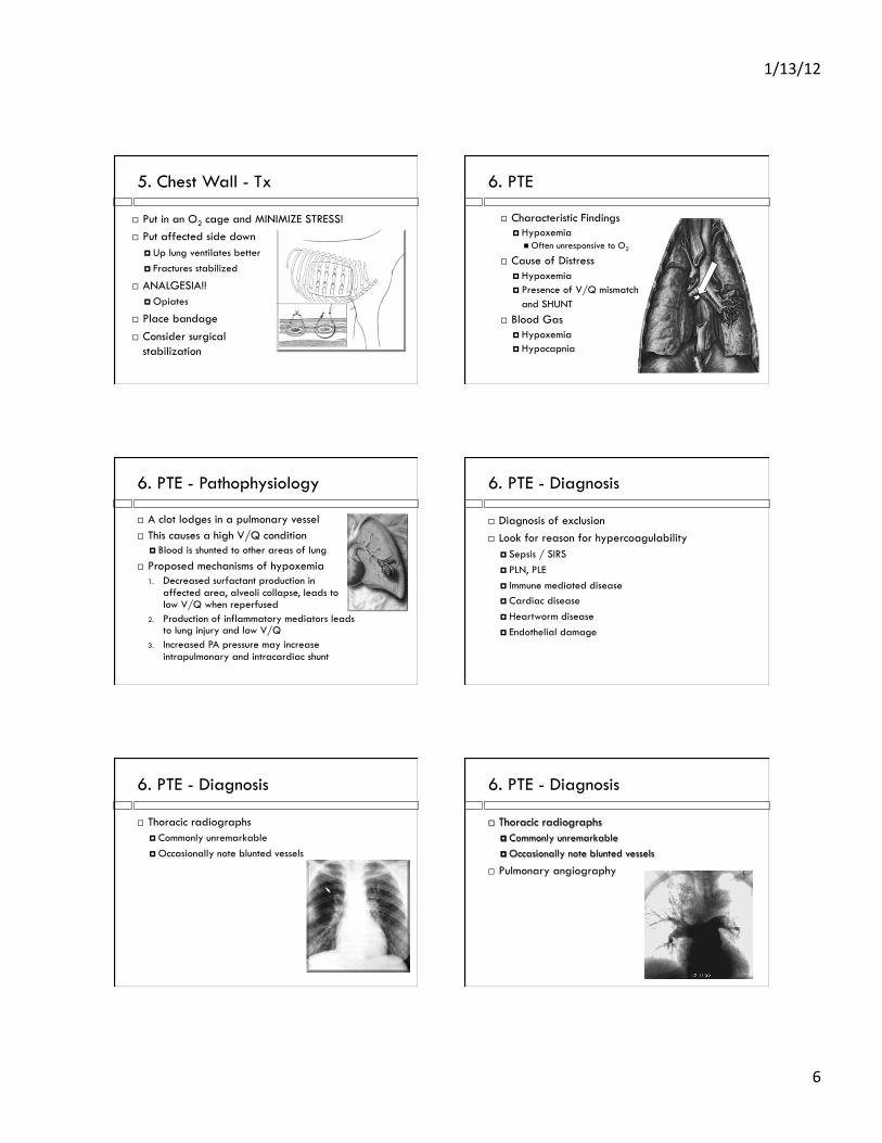
5. Chest Wall - Tx
! Put in an O2 cage and MINIMIZE STRESS! ! Put affected side down
! Up lung ventilates better ! Fractures stabilized
! ANALGESIA!! ! Opiates
! Place bandage ! Consider surgical
stabilization
6. PTE
! Characteristic Findings ! Hypoxemia
" Often unresponsive to O2
! Cause of Distress ! Hypoxemia ! Presence of V/Q mismatch
and SHUNT ! Blood Gas
! Hypoxemia ! Hypocapnia
6. PTE - Pathophysiology
! A clot lodges in a pulmonary vessel ! This causes a high V/Q condition
! Blood is shunted to other areas of lung ! Proposed mechanisms of hypoxemia
1. Decreased surfactant production in affected area, alveoli collapse, leads to low V/Q when reperfused
2. Production of inflammatory mediators leads to lung injury and low V/Q
3. Increased PA pressure may increase intrapulmonary and intracardiac shunt
6. PTE - Diagnosis
! Diagnosis of exclusion ! Look for reason for hypercoagulability
! Sepsis / SIRS ! PLN, PLE ! Immune mediated disease ! Cardiac disease ! Heartworm disease ! Endothelial damage
6. PTE - Diagnosis
! Thoracic radiographs ! Commonly unremarkable ! Occasionally note blunted vessels
6. PTE - Diagnosis
! Thoracic radiographs ! Commonly unremarkable ! Occasionally note blunted vessels
! Pulmonary angiography

1/13/12%
7%
6. PTE - Diagnosis
! Thoracic radiographs ! Commonly unremarkable ! Occasionally note blunted vessels
! Pulmonary angiography ! Contrast CT
! Angiogram sometimes successful ! Often unremarkable
6. PTE - Diagnosis
! Thoracic radiographs ! Commonly unremarkable ! Occasionally note blunted vessels
! Pulmonary angiography ! Contrast CT
! Angiogram sometimes successful ! Often unremarkable
! V/Q Scan
6. PTE - Diagnosis 6. PTE - Diagnosis
! Thoracic radiographs ! Commonly unremarkable ! Occasionally note blunted vessels
! Pulmonary angiography ! Contrast CT
! Angiogram sometimes successful ! Often unremarkable
! V/Q Scan ! Ultrasound
6. PTE - Treatment
! Supportive care ! Heparin
! Does not BREAK DOWN clot ! Decrease growth of clot or new clot formation ! Dosing is a CHALLENGE! ! Monitor PTT, shoot for 150% of normal
! Thrombolytic therapy ! All convert plasminogen to plasmin, which breaks
down clots ! Streptokinase
" Not specifically active at clot sites => risk of bleeding
! Tissue plasminogen activator (tPA) " Increased activity when bound to fibrin (clot) " Can still cause bleeding at high doses
! VERY limited experience with dogs and cats ! $$$$$$$
7. Abdominal Distension
! Characteristic findings ! Abdominal distension on palpation
! Cause of distress ! Hypoventilation ! Sometimes combined with Pickwickian
Syndrome (decreased chest compliance)
! Blood gas ! Hypercapnia ! ±Hypoxemia

1/13/12%
8%
7. Abdominal Distension - DDx
! Fluid (ascites) ! Transudate (±modified) ! Exudate ! Blood
! Organomegaly ! Neoplasia ! Fat
6. Abdominal Distension - Tx
! Most commonly due to ascites when acute ! Abdominocentesis
! Only enough to relieve distress ! Removing large volumes can lead
to fluid shifts " Hemodynamic effects " Electrolyte effects
8. Look Alikes
! Characteristic findings ! Rule out other causes
! Cause of distress ! Neither hypoxemia nor hypercapnia ! A non-respiratory problem that
makes the patient appear to be in respiratory distress
! Blood gas ! Hypocapnia ! Normoxemia
8. Look Alikes - DDx
! Behavioral ! Stress ! Fear ! Pain ! Anxiety
! Metabolic ! Metabolic acidosis
" Compensatory ! Anemia
" Low oxygen content
! Environmental ! Hyperthermia ! Hypothermia (less
common)
8. Look Alikes - Treatment
! Behavioral ! Anxiolytics ! Analgesics
! Metabolic acidosis ! Treat underlying cause ! Sodium bicarbonate
" Bicarb deficit = 0.3 * BW[kg] * BE " NEVER give more than
1/3 – 1/2 of that dose " ONLY give if severe acidemia
and patient can ventilate!
! Anemia ! Transfuse if symptomatic ! 1 ml/kg of pRBCs raises PCV by 1% ! General guideline, 10-15 ml/kg
The Dyspneic Cat
! You auscult and you hear… ! Hmmm, was there a crackle? A wheeze? ! Top three differentials
! CHF with pulmonary edema ! Pleural space disease ! Asthma
! How do you treat? ! Put in an O2 cage ! Lasix IM (2-4 mg/kg) ! LEAVE ALONE! ! Diagnostic / therapeutic
thoracocentesis ! Last resort….
" 2 mg/kg lasix " 0.1 mg/kg dexamethasone SP " 0.01 mg/kg terbutaline

1/13/12%
9%
Blanche
! Sig: 2 yr SF DSH cat – 3.5 kg ! Hx:
! Previously healthy ! < 24 hr history of tachypnea and lethargy ! Indoor cat that lives with 3 other cats, and is up to date on
her vaccinations including FeLV ! PE:
! General: " T = 99.6, HR 200 bpm, RR = 140 " Overt respiratory distress
! EENT: mildly cyanotic, CRT ~ 2 seconds ! CV: IV/VI left sided systolic murmur, gallop rhythm ! Resp: marked tachypnea and effort, minimal BV sounds
Blanche’s Problem List
! Cyanosis ! Respiratory distress
! Minimal lung sounds ! Restrictive pattern
! Heart murmur and gallop rhythm ! Hypothermia
Emergency Stabilization
! Thoracocentesis!!!! ! Furosemide (2 mg/kg IM or IV) ! Oxygen ! Nitroglycerin (1/4” inguinal) ! Minimize stress!
Radiographs
Too MUCH Fluid!!!!
Echocardiography
! What’s your diagnosis?
Feline Hypertrophic
Cardiomyopathy with
Congestive Heart Failure
Can We Stabilize Blanche?
! After our second thoracocentesis Blanche is MUCH more stable
! Principles of management of CHF: ! Improve oxygen delivery
" Supplement!! ! Reduce formation of edema and congestion
" Lasix PRN!! ! Increase cardiac output
" Consider: " �-adrenergic blocker (atenolol) " Calcium channel blocker (diltiazem) " ACE inhibitor (enalapril)
1/13/12%
10%
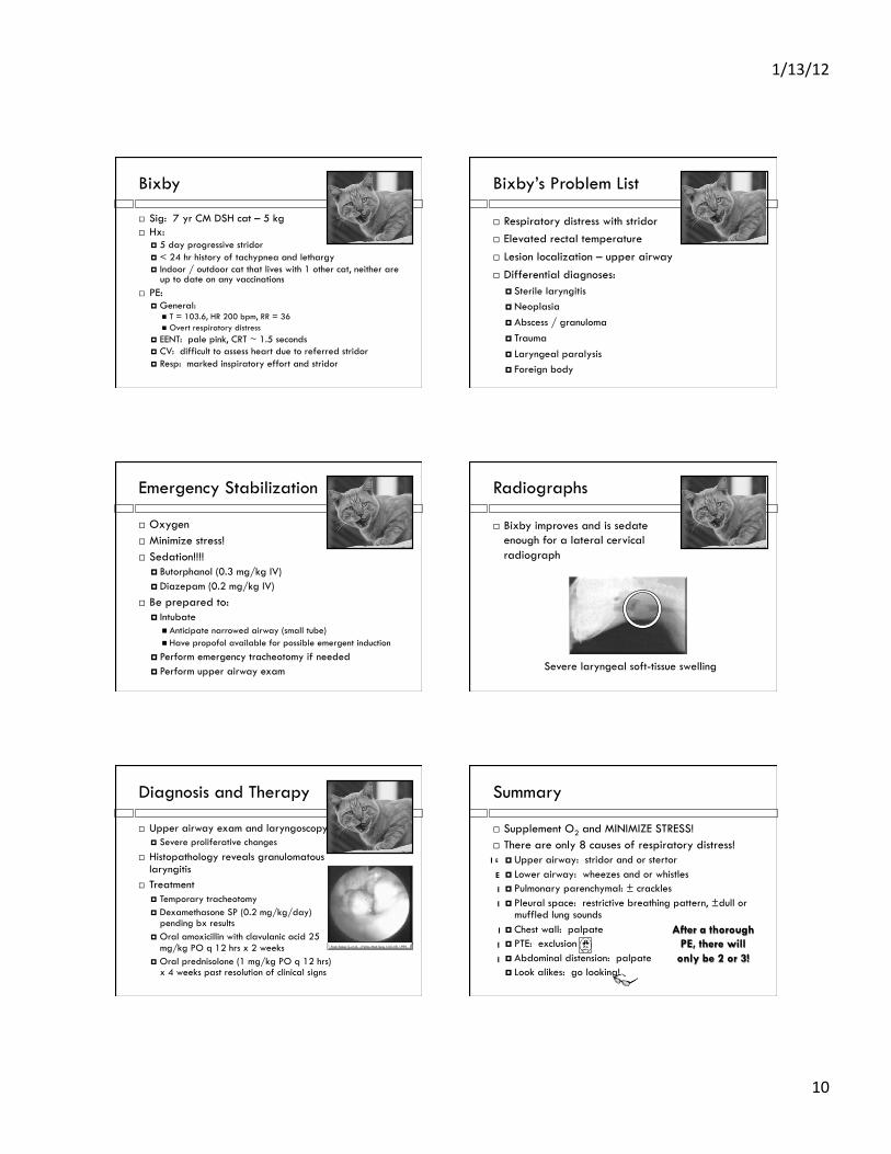
Bixby
! Sig: 7 yr CM DSH cat – 5 kg ! Hx:
! 5 day progressive stridor ! < 24 hr history of tachypnea and lethargy ! Indoor / outdoor cat that lives with 1 other cat, neither are
up to date on any vaccinations ! PE:
! General: " T = 103.6, HR 200 bpm, RR = 36 " Overt respiratory distress
! EENT: pale pink, CRT ~ 1.5 seconds ! CV: difficult to assess heart due to referred stridor ! Resp: marked inspiratory effort and stridor
Bixby’s Problem List
! Respiratory distress with stridor ! Elevated rectal temperature ! Lesion localization – upper airway ! Differential diagnoses:
! Sterile laryngitis ! Neoplasia ! Abscess / granuloma ! Trauma ! Laryngeal paralysis ! Foreign body
Emergency Stabilization
! Oxygen ! Minimize stress! ! Sedation!!!!
! Butorphanol (0.3 mg/kg IV) ! Diazepam (0.2 mg/kg IV)
! Be prepared to: ! Intubate
" Anticipate narrowed airway (small tube) " Have propofol available for possible emergent induction
! Perform emergency tracheotomy if needed ! Perform upper airway exam
Radiographs
! Bixby improves and is sedate enough for a lateral cervical radiograph
Severe laryngeal soft-tissue swelling
Diagnosis and Therapy
! Upper airway exam and laryngoscopy ! Severe proliferative changes
! Histopathology reveals granulomatous laryngitis
! Treatment ! Temporary tracheotomy ! Dexamethasone SP (0.2 mg/kg/day)
pending bx results ! Oral amoxicillin with clavulanic acid 25
mg/kg PO q 12 hrs x 2 weeks ! Oral prednisolone (1 mg/kg PO q 12 hrs)
x 4 weeks past resolution of clinical signs
From Tasker S, et al. J Feline Med Surg 1:53–59, 1999
Summary
! Supplement O2 and MINIMIZE STRESS! ! There are only 8 causes of respiratory distress!
! Upper airway: stridor and or stertor ! Lower airway: wheezes and or whistles ! Pulmonary parenchymal: ± crackles ! Pleural space: restrictive breathing pattern, ±dull or
muffled lung sounds ! Chest wall: palpate ! PTE: exclusion ! Abdominal distension: palpate ! Look alikes: go looking!
After a thorough PE, there will
only be 2 or 3!
I
E
E
E
I
I
I
I
I

1/13/12%
11%
Suggested Reading
! Textbook of Respiratory Disease in Dogs and Cats ! Lesley King
! Small Animal Critical Care Medicine ! Deborah Silverstein and Kate Hopper
! Manual of Small Animal Emergency and Critical Care Medicine ! Douglass Macintire, Kenneth Drobatz, Steve Haskins
and William D. Saxon ! Kirk's Current Veterinary Therapy XIV
! John Bonagura and David Twedt ! Consultations in Feline Internal Medicine, Volume 6
! John R. August