the value of immunofluorescence in the study of …
TRANSCRIPT

THE VALUE OF IMMUNOFLUORESCENCE IN THE STUDY OF HUMAN RENAL DISEASE*
BY ROBERT T. McCLUSKEY
(From the Department of Pathology, State University of New York at Buffalo, Buffalo, New York 14214)
The use of immunofluorescence has resulted in considerable expansion of our understanding of human glomerular diseases. I t has provided evidence that many, perhaps most, forms of glomerulonephritis have an immunologic basis. On the other hand, it has also provided impressive evidence that certain glomerular diseases formerly frequently considered to have an allergic basis almost certainly do not.
I t is, of course, through immunofluorescence observations that giomerular diseases in man are provisionally classified as examples of immune complex disease or anti-glomerular basement membrane (GBM) 1 antibody disease (Table I). I t appears justifiable to consider any glomerular disease character- ized by conspicuous irregular granular, discontinuous deposits of immuno- globulins and complement components as probably an immune complex dis- ease. I t is apparent from observations in experimental models and human diseases that complexes may be deposited in a variety of patterns. The major forms consist of granular deposits along the basement membrane (often sub- epithelial as seen by electron microscopy), broad irregular deposits along the basement membrane (generally subendothelial), granular or irregular mesan- gial deposits, probably interrupted "linear" deposits, various combinations of these, and patterns which defy analysis or description. Although the patterns vary, in all cases the deposits are irregular or discontinuous to some extent. On the other hand, the finding of completely continuous (linear) staining for IgG and complement along the GBM warrants the provisional diagnosis of anti-GBM disease (Fig. 1).
However, certain warnings are in order. For one thing, proof that a disease is due to deposition of immune complexes
requires demonstration that much of the immunoglobulin in glomeruli repre- sents antibody combined with antigen. This has been satisfactorily accom- plished only in cases of lupus nephritis (1), possibly in poststreptococcal (2) and malarial (3) glomerulonephritis, as well as in two cases of chronic glomeru-
* Part of the work reported here was supported by U.S. Public Health Service grant No. AI-09115.
1 Abbreviations used in this paper: GBM, glomerular basement membrane.
242 s THE JOURNAL OF EXPERIMENTAL MEDICINE • VOL. 134, NO. 3, PT. 2, 1971
Dow
nloaded from http://rupress.org/jem
/article-pdf/134/3/242/1415844/242s.pdf by guest on 02 February 2022

~cCLT3SKEY Immunofluorescence in Human Renal Disease 243 s
lar disease in which thyroglobulin was found within deposits. 2 Nevertheless, in conditions in which the deposits are quite characteristic and closely re- semble those seen in experimental models, the presumption is very strong that the disease is due to immune complexes. This is the case with membranous glomerulonephritis, which is rather faithfully mimicked in some rabbits with
TABLE I Human Renal Diseases Presumed or Known to Result from Immunologic Mechanisms
I. Immune complex glomerulonephritis A. Diseases of known etiology
1. Poststreptococcal glomerulonephritis* 2. Glomerulonephritis associated with other bacterial infections: staphylococcal?;
pneumococcal?; syphilis?; bacterial endocarditis? 3. Glomerulonephritis associated with malaria*
B. Glomerular diseases associated with systemic diseases of unknown cause 1. Lupus nephritis$ 2. Nephritis with anaphylactoid purpura? 3. Nephritis in periarteritis nodosa? 4. Nephritis in idiopathic "mixed" cryoglobulinemia?§
C. Primary glomerular diseases of unknown cause 1. Membranous glomerulonephritis? 2. Membranoproliferative glomerulonephritis? 3. Chronic sclerosing glomerulonephritis? 4. Focal glomerulonephritis? 5. IgA-IgG nephropathy (Berger)? 6. Unclassified glomerular diseases?
II. Anti-GBM antibody disease A. Glomerulonephritis associated with Goodpasture's syndrome B. Some cases classified as rapidly progressive or chronic glomerulonephritis
III. Interstitial and/or tubular disease A. Immunoglobulin deposits in tubular basement membranes (immune complexes?) B. Interstitial mononuclear ceil infiltration
1. Due to drugs (methiclllin, penicillin, ampiUicin, and others) 2. Due to other exogenous substances? 3. Autoimmune?
* Antigen tentatively identified in glomerular deposits. :~ Antigen (DNA) definitely identified in glomerular deposits. § Antigen presumably altered IgG.
chronic serum sickness (4) and in rats with Heymann ' s nephrosis. The immuno- fluorescence findings in these conditions are quite distinctive and consist of rather bright granular staining for IgG and usually J31C all along the basement membrane in all glomeruli (Fig. 2). Electron microscopy shows corresponding electron-opaque deposits on the epithelial side of the basement membrane.
2 Koffier, D. Personal communication.
Dow
nloaded from http://rupress.org/jem
/article-pdf/134/3/242/1415844/242s.pdf by guest on 02 February 2022

244 s LVlMU_N'E COMPLEXES AND GLOMERULONEPI{RITIS IN" MAN
Nothing is known of the antigen presumed to be present in the deposits of the human disease. Poststreptococcal glomerulonephritis is another disease whose salient histologic, immunofluorescence, and electron microscopic features are faithfully reproduced in an experimental model, namely acute ("one shot")
Fio. 1. Anti-GBM disease. IgG is seen in a completely continuous (linear) pattern along the glomerular basement membrane.
serum sickness. Therefore, even though there is disagreement as to whether antigenic material derived from streptococci has been demonstrated in glomer- uli, as discussed elsewhere in this symposium by Zabriskie, it seems justifiable to consider this to be an acute immune complex disease. Somewhat less certain is the position of membranoproliferative glomerulonephritis, whose charac- teristic findings are only partially reproduced in some rabbits with chronic serum sickness (4, 5). Similar comments apply to the nephritis seen with ana- phylactoid purpura and some cases classified as focal glomerulonephritis.
Dow
nloaded from http://rupress.org/jem
/article-pdf/134/3/242/1415844/242s.pdf by guest on 02 February 2022

MCCI,USKEY Immunofluorescence in Human Renal Disease 245 s
Finally, there are human glomerular diseases in which, although immuno- globulin deposits may be present, they are generally scanty and their distribu- tion is not distinctive. These include many cases classified as chronic sclerosing (end-stage) glomerulonephritis, some cases classified as focal glomerulonephri- tis, and many undassifiable diseases. In such instances, scanty focal deposits of IgG and/31C are sometimes seen, although often there is no staining at all. These findings cannot be interpreted satisfactorily. The presence of small
Fro. 2. Membranous glomerulonephrifis. IgG is present in granular form all along the basement membrane. X 1000.
amounts of IgG and ¢~1C may indicate immune complexes or just nonspecific sticking of these plasma proteins in damaged glomeruli. On the other hand, the absence of such staining, especially in sclerotic glomeruli, does not exclude the possibility that the original damage was due to immune complexes.
Concerning the immunofluorescence findings in anti-GBM disease three kinds of problems arise. First, if deposits of immune complexes are finely granular, numerous, and contiguous all along the basement membrane, they may present an appearance which is difficult to distinguish from a linear pat-
Dow
nloaded from http://rupress.org/jem
/article-pdf/134/3/242/1415844/242s.pdf by guest on 02 February 2022
246 s IMMUNE COMPLEXES AND GLOMERULONE:PttRITIS IN MAN
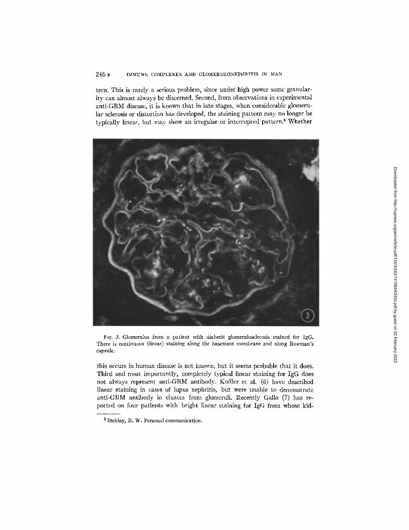
tern. This is rarely a serious problem, since under high power some granular- ity can almost always be discerned. Second, from observations in experimental an t i -GBM disease, it is known that in late stages, when considerable glomeru- lar sclerosis or distortion has developed, the staining pat tern m a y no longer be typically linear, but may show an irregular or interrupted pattern, a Whether
FIG. 3. Glomerulus from a patient with diabetic glomerulosclerosis stained for IgG. There is continuous (linear) staining along the basement membrane and along Bowman's capsule.
this occurs in human disease is not known, but it seems probable that it does. Third and most importantly, completely typical linear staining for IgG does not always represent ant i -GBM antibody. KotTler et al. (6) have described linear staining in cases of lupus nephritis, but were unable to demonstrate an t i -GBM antibody in eluates from glomeruli. Recently Gallo (7) has re- ported on four patients with bright linear staining for IgG from whose kid-
a Steblay, R. W. Personal communication.
Dow
nloaded from http://rupress.org/jem
/article-pdf/134/3/242/1415844/242s.pdf by guest on 02 February 2022

McCLUSKEY Immunofluorescence in Human Renal Disease 247 s
neys anti-GBM antibody could not be eluted. Two of the patients had dia- betic glomerulosclerosis. I have also seen moderately bright linear staining for IgG in a few patients with diabetic glomerulosclerosis (Fig. 3), and in several patients with mild, unclassified glomerular disease. In the patients with dia- betes, linear staining of Bowman's capsule and tubular basement membranes may also be seen. In none of these, nor in the patients reported by Gallo, was there linear staining for ~1C or evidence of circulating anti-GBM antibodies. These findings are unexplained, but it seems clear that typical bright glomeru- lar linear staining for IgG, in the absence of similar staining for complement or circulating anti-GBM antibodies, cannot be accepted as conclusive evidence of anti-GBM disease. In my experience, faint linear staining for IgG without /31C staining is considerably more common and may be seen in a variety of conditions in which there is no other evidence of anti-GBM antibody.
Based on immunofiuorescence observations on a fairly large number (> 1000) of cases of renal disease studied by biopsy or at autopsy, I would conclude that immune complex glomerular disease is rather common and anti-GBM disease is extremely rare.
Immunofluorescence has also been of value in the study of another problem, namely the immunologic mechanisms responsible for damage in renal allo- grafts. As discussed by Milgrom in this symposium, three types of damage are possible: (a) that due to the allograft reaction itself (which may involve cellu- lar or humoral mechanisms), (b) recurrence of the original glomerular disease, and (c) damage due to autoimmune mechanisms, presumably initiated by re- lease or alteration of tissue-specific antigens in the graft. Studies employing immunofluorescence have revealed evidence of all three mechanisms; however, the distinction between rejection phenomena and recurrence of the patient's original glomerular disease has proved to be more difficult than might have been anticipated. Andres et al. (8) have recently reported a study of 34 renal allografts. In some, a diffuse linear pattern of staining for IgG and complement was seen in glomeruli. However, the staining was generally not as sharp as that seen in known anti-GBM disease, and immunoferritin studies indicated that the immunoglobulins localized more in the subendothelial space than in the basement membrane proper, suggesting that the linear staining was not due to anti-GBM antibodies. Andres suggested it may have been due to trans- plantation antibodies (8). Obviously, elution studies will be required to clarify this problem. In other allografts, granular staining for immunoglobulins was encountered; this could result from recurrence of immune complex disease or deposition of complexes containing transplantation antigens or autologous antigens and autoantibodies. Damage due to autoimmune mechanisms has also been observed in allografts involving tubules, as will be discussed.
In addition, immunofiuorescence observations have provided evidence against the possibility that immunologic mechanisms are responsible for cer-
Dow
nloaded from http://rupress.org/jem
/article-pdf/134/3/242/1415844/242s.pdf by guest on 02 February 2022

248 s IMMUNE COMPLEXES AND GLOMERULONEPIq 'RITIS IN MAN
tain glomerular diseases; these are lipoid nephrosis and the glomerular lesions in toxemia of pregnancy. In both conditions immunoglobulins are generally not demonstrable, although they may sometimes be seen as irregular focal deposits which do not form a characteristic pattern (9, 10). to conclude from these findings that an immunologic mechanism is almost certainly not involved seems justified, since in established experimental models immunoglobulins and complement components are generally readily demonstrable in a charac- teristic pattern, at least at some stage of the disease.
Aside from providing evidence concerning immunologic mechanisms, ob- servations made by immunofluorescence support the interpretation that deposi- tion of fibrin or other fibrinogen derivatives may play a pathogenic role in some glomertflar diseases. In toxemia of pregnancy, fibrinogen derivatives are found within and between endothelial and mesangial cells; these and other considerations suggest that the glomerular alterations result from a prolonged low grade state of intravascular coagulation (i0). Further, in some forms of glomerulonephritis, especially rapidly progressive forms, conspicuous fibrin deposits are found in Bowman's space, and based on experimental studies this appears to be a major factor in the development of crescents (9, 11, 12). Depo- sition of fibrin in glomerular tufts in these conditions may also accelerate or intensify glomerular sclerosis (9, 11, 12).
In addition to its value in elucidating pathogenetic mechanisms, immuno- fluorescence can be considered from the point of view of its usefulness as a diag- nostic tool, that is as a sort of "special stain" which helps the pathologist place a case in a recognized category. The classification of glomerular diseases is not very satisfactory, since in most instances etiologic factors are unknown. However, there are several conditions which are fairly well-defined morpho- logic entities. It seems unlikely that these will turn out to be etiologic entities since it is clear from experimental models that different antigens can produce the same morphologic diseases. Despite these considerations, classification is useful because it provides a guide to prognosis and evaluation of therapy. In several conditions, the immunofluorescence findings are highly characteristic if not entirely pathognomonic. However, in most cases this technique cannot be considered essential, since if a diagnosis can be made at all, it can usually be accomplished on the basis of histologic, electron microscopic, clinical, and serological findings. Nevertheless, the technique usually' provides important supporting evidence and is sometimes required for diagnosis, especially if electron microscopy is not available. For example, the differentiation between lipoid nephrosis and membranous glomerulonephritis is not always possible on histologic grounds (even with very good preparations) if the deposits are exceedingly fine. However, the distinction can readily be made by immuno- fluorescence, since in membranous glomerulonephritis bright granular staining for IgG is always seen (Fig. 2). By electron microscopy, corresponding dense
Dow
nloaded from http://rupress.org/jem
/article-pdf/134/3/242/1415844/242s.pdf by guest on 02 February 2022

McCLUSKE¥ Immunofluorescence in Human Renal Disease 249 s
deposits are found. In contrast, in lipoid nephrosis, IgG and electron-opaque deposits are absent. The distinction is important, since lipoid nephrosis gener- ally responds to steroids and has a good prognosis, whereas membranous nephritis generally does not respond and usually runs a chronic unremitting course which frequently ends with chronic progressive renal failure. I t seems almost certain that in the few recorded instances of supposed transition from
Fro. 4. Membranoproliferative glomerulonephritis. IgG is seen along the periphery of lobules as broad irregular deposits and in mesangial regions. (Reprinted by copyright permis- sion from B~dl. N. Y. Acad. Met. 1970. 46:769.)
lipoid nephrosis to membranous nephritis, as seen in serial biopsies, the disease was membranous nephritis from the onset and in all probability the correct initial diagnosis could have been made with immunofluorescence or electron microscopy.
Another situation where immunofluorescence may be useful or essential is in the differential diagnosis between acute poststreptococcal glomerulonephritis and membranoproliferative glomerulonephritis. This is not a frequent prob- lem, since the distinction between these two conditions can usually be made with ease in histologic preparations. Nevertheless, in cases of membrano-
Dow
nloaded from http://rupress.org/jem
/article-pdf/134/3/242/1415844/242s.pdf by guest on 02 February 2022

FIG. 5. Electronmicrograph of a glomerulus from a patient with IgA-IgG deposits. There is increased mesangial matrix and electron-opaque deposits in mesangial regions. X 7000.
250 s
Dow
nloaded from http://rupress.org/jem
/article-pdf/134/3/242/1415844/242s.pdf by guest on 02 February 2022

McCLtlSKEY Immunofluorescence in Human Renal Disease 251 s
proliferative glomerulonephrifis with only mild increase in mesangial matrix and with slight peripheral capillary wall thickening, the differential diagnosis may not be possible without immunofluorescence or electron microscopic ob- servations. Although the immunofluorescence findings are not constant, most patients with irrembranoproliferative glomerulonephritis show numerous elongated as well as granular deposits of/3xC and less often of IgG in a periph-
Fro. 6. Glomerulus from a patient with IgA-IgG deposits. Conspicuous granular deposits of IgA are seen, especially in mesangial regions.
eral lobular pattern (Fig. 4) and in mesangial regions, in contrast to the scanty granular deposits seen in most cases of acute glomerulonephritis. The distinc- tion between the two diseases is important because poststreptococcal glomerulo- nephritis ahnost always resolves, whereas membranoproliferative glomerulo- nephritis runs a chronic, unremitting course. Further, the inclusion of cases of membranoproliferative glomerulonephritis in a series of cases considered to be acute glomerulonephritis has undoubtedly been one factor which has re- sulted in an unduly pessimistic estimate of the recovery rate in poststrepto- coccal glomerulonephritis.
There is at least one glomerular disease which at present can be recognized
Dow
nloaded from http://rupress.org/jem
/article-pdf/134/3/242/1415844/242s.pdf by guest on 02 February 2022

252 S IMMITNE COMPLEXES AND G L O M E R U L O N E P t I R I T I S I N MAN
only through immunofluorescence observations. This is a condition described in 55 patients by Berger, which he refers to as "nephropathy with mesangial IgAIgG deposits" (13). Most of the patients show persistent or recurrent hematuria, with mild proteinuria and normal renal function. There is no asso- ciated systemic disease. The renal disease runs a chronic course and so far only one patient has developed renal insufficiency. The histologic findings are generally not distinctive. Generally slight, irregular increase in mesangial
Fro. 7. Renal biopsy from a patient with lupus nephritis. Granular deposits of IgG are seen along tubular basement membrane.
matrix is found, within which deposits may sometimes be seen with special stains or by electron microscopy (Fig. 5). Slight irregular increase in mesangial cells may be found. Many of the cases might be classified as focal glomerulo- nephritis on histologic grounds, whereas others would ngk warrant any diag- nosis. However, the immunofluorescence findings are quite characteristic and consist of conspicuous granular deposits of IgA, IgG, and 131C, largely in nle- sangial regions (Fig. 6). To my knowledge, a similar picture is seen only in some instances of nephritis with anaphylactoid purpura or rarely in lupus nephritis. The nature of the condition described by Berger is unknown, al- though it has features of chronic immune complex disclose. Nothing is known of
Dow
nloaded from http://rupress.org/jem
/article-pdf/134/3/242/1415844/242s.pdf by guest on 02 February 2022

FIGS. 8 and 9. From a rabbit immunized with homvlogous kidney and Freund's adjuvant Granular staining for IgG is seen along the basement membrane of proximal tubules. The glomeruli contain no deposits.
253 s
Dow
nloaded from http://rupress.org/jem
/article-pdf/134/3/242/1415844/242s.pdf by guest on 02 February 2022

254 S IMMUNE COMPLEXES AND GLOI~ERULONEPHRITIS IN MA.N
etiologic factors. The condition may not be rare; in the past two years I have seen six cases.
Finally, I should like to mention a form of tubular disorder which appears to be due to deposits of immune complexes and which can be recognized with certainty only by immunofluorescence. We have seen four patients (three with lupus nephritis, one with idiopathic hematuria) in whom bright granular deposits of IgG and/31C were found along the basement membrane of con- voluted tubules (Fig. 7); by electron microscopy, dense granular deposits were seen in the basement membrane of proximal convoluted tubules: The de- posits were not recognizable in histologic preparations but were associated with interstitial fibrosis and mononuclear cell infiltration. Lesions with similar ultrastructural findings have been described in patients with membranous glomerulonephritis (14) and in renal allograffs (8). The same type of deposits (Figs. 8 and 9) have been produced in rabbits immunized with homologous kidney as shown by Unanue and Dixon (15) and by Klassen, McCluskey, and Milgrom (16). In this model the most plausible explanation for the deposits is that they result from union of an autologous antigen leaking out from proxi- mal convoluted cells which combines with autoantibody diffusing in from peritubular capillaries (16). The frequency of this kind of tubular immune deposit disease(s) and its functional significance remain to be assessed. How- ever, it seems likely that such lesions may have been overlooked in many instances.
SIYM-MARY
Immunofluorescence is a useful technique in the study of human renal diseases, both from the point of view of elucidating pathogenic mechanisms and as a diagnostic tool.
The finding of characteristic staining patterns for immunoglobulins and complement indicates that many forms of glomerulonephritis are immune complex diseases and that a few are due to anti-GBM antibodies. On the other hand, in lipoid nephrosis and toxemia of pregnancy, deposits of immuno- globulins and complement are generally absent, indicating that immunologic mechanisms are probably not responsible for these glomerular diseases.
The finding of fibrin or other fibrinogen derivatives in glomeruli in toxemia of pregnancy and in certain forms of glomulonephritis supports the interpreta- tion that these substances play a pathogenic role in certain glomerular dis- eases.
The use of immunofluorescence has led to the recognition of two previously unrecognized renal diseases: nephropathy with mesangial IgA-IgG deposits (Berger), and a tubular disorder with deposits of immunoglobulins and com- plement along the basement membrane.
4 Klassen, J., R. T. McCluskey, and F. Milgrom. Unpublished observations.
Dow
nloaded from http://rupress.org/jem
/article-pdf/134/3/242/1415844/242s.pdf by guest on 02 February 2022

~cCLIYSKEY Immunofluorescence in H u m a n Renal Disease 255 s
I would like to thank Dr. G. Andres and Dr. J. Klassen for help in the preparation of the photomicrographs and electronmicrographs and Mrs. Marion Sepulveda for excellent tech- nical assistance.
REFERENCES
1. Koffier, D., P. H. Schur, and H. G. Kunkel. 1967. Immunological studies con- cerning the nephritis of systemic lupus erythematosus. J. Exp. Med. 126:607.
2. Andres, G. A., I. Accinni, K. C. Hsu, J. B. Zabriskie, and B. C. Seegal. 1966. Electron microscopic studies of human glomerulonephritis with ferritin- conjugated antibody. Localization of antigen-antibody complexes in glomerular structures of patients with acute glomerulonephritis. J. Exp. Med. 123:399.
3. Ward, P., and P. Conran. 1969. Immunopathology of renal complications of simian malaria and human quartan malaria. Mil. Med. 134:1228.
4. Dixon, F. J., J. D. Feldman, and J. J. Vazquez. 1961. Experimental glomeru- Ionephritis. The pathogenesis of a laboratory model resembling the spectrum of human glomerulonephritis. J. Exp. Med. 113:899.
5. Germuth, F. G., Jr., L. B. Senterfit, and A. D. Pollack. 1967. Immune complex disease. I. Experimental acute and chronic glomerulonephritis. Johns Hopkins Med. J. 19.0:225.
6. Koffier, D., V. Agnello, R. I. Carr, and H. G. Kunkel. 1969. Variable patterns of immunoglobulin and complement deposition in the kidneys of patients with systemic lupus erythematosus. Amer. J. Pathol. 56:305.
7. Gallo, G. 1970. Elution studies in kidneys with linear deposition of immuno- globulin in glomeruli. Amer. J. Pathol. 61:377.
8. Andres, G. A., L. Accinni, K. C. Hsu, I. Penn, K. A. Porter, J. M. Rendall, B. C. Seegal, and T. E. Stargl. 1970. Human renal transplants. I I I . Immunopatho- logic studies. Lab. Invest. 29.:588.
9. McCluskey, R. T., P. Vassalli, G. Gallo, and D. S. Baldwin. 1966. An immuno- fluorescent study of pathogenic mechanisms in glomerular diseases. N. Engl. J. Med. 274:695.
10. Vassalli, P., R. H. Morris, and R. T. McCluskey. 1963. The pathogenic role of fibrin deposition in the glomerular lesions of toxemia of pregnancy. J. Exp. Med. 118:467.
11. Vassalli, P., and R. T. McCluskey. 1964. The pathogenic role of the coagulation process in rabbit Masugi nephritis. Amer. J. Pathol. 45:653.
12. Vassalli, P., and R. T. McCluskey. 1965. The coagulation process and glomerular disease. Amer. J. Med. 39:179.
13. Berger, J. 1969. IgA glomerular deposits in renal disease. Transplant. Proc. 1: 939.
14. Berger, J., and P. Galle. 1963. D6p6ts denses au sein des membranes basales du rein. Etude en microscopies optique et 61ectronique. Presse Med. 71:2351.
15. Unanue, E. R., and F. J. Dixon. 1967. Experimental allergic glomerulonephritis induced in the rabbit with heterologous renal antigens. J. Exp. Med. 125:149.
16. Klassen, J., R. T. McCluskey, and F. Milgrom. 1971. Nonglomerular renal disease produced in rabbits by immunization with homologous kidney. Amer. J. Pathol. 63:333.
Dow
nloaded from http://rupress.org/jem
/article-pdf/134/3/242/1415844/242s.pdf by guest on 02 February 2022