the usefulness of incorporating non-pathomorphological data recorded in the hospital information...
TRANSCRIPT

The Usefulness of Incorporating Non-pathomorphological Data Recorded in the Hospital Information System into the Pathology Report Form and a Standardized Method of Viewing Digital Pathology Images
Keizo Furuya, Toshiharu Maeda, Takanori KikuchiDepartment of Pathology and Radiology, Ehime Prefectural Central Hospital
古谷敬三 Oct. 25, 2010 Pathology Vision 2010 (San Diego)
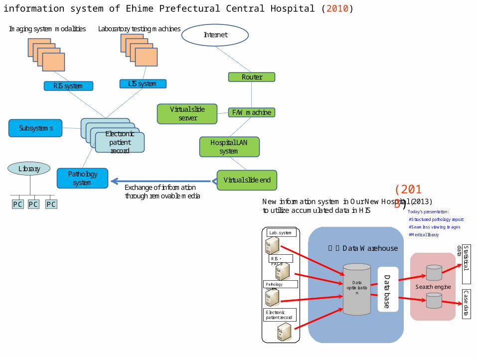
Hospital information system of Ehime Prefectural Central Hospital (2010)
Electronic patient record
RIS system
Pathology system
Virtual slide server
LIS system
Imaging system modalities Laboratory testing machines
Virtual slide end
F/W machine
Router
Internet
Exchange of information through removable media
Subsystems
Hospital LAN system
Library
PC PC PC
S earch engineData
optimization
L ab. s ys tem
R IS ・P AC S
P athology system
E lectronic patient record
専用Data Warehouse
Data base
Statistical
dataC
ase data
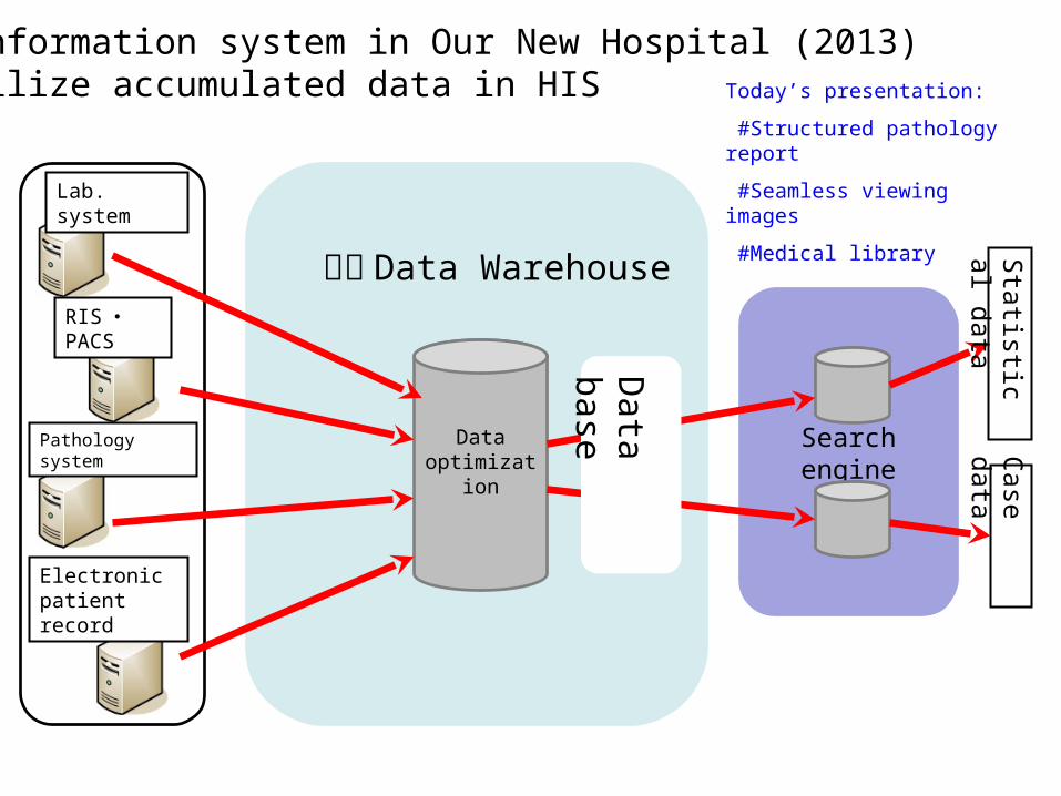
New information sys tem in O ur New Hospital (2013)to utilize accumulated data in HIS Today’s presentation:
#Structured pathology report
#Seamless viewing images
#Medical library
(2013)
Constitution1. Background2. Aim3. Materials and methods 3a. Pathology report form 3b. Seamlessly viewing images 3c. Medical library4. Result5. Conclusion
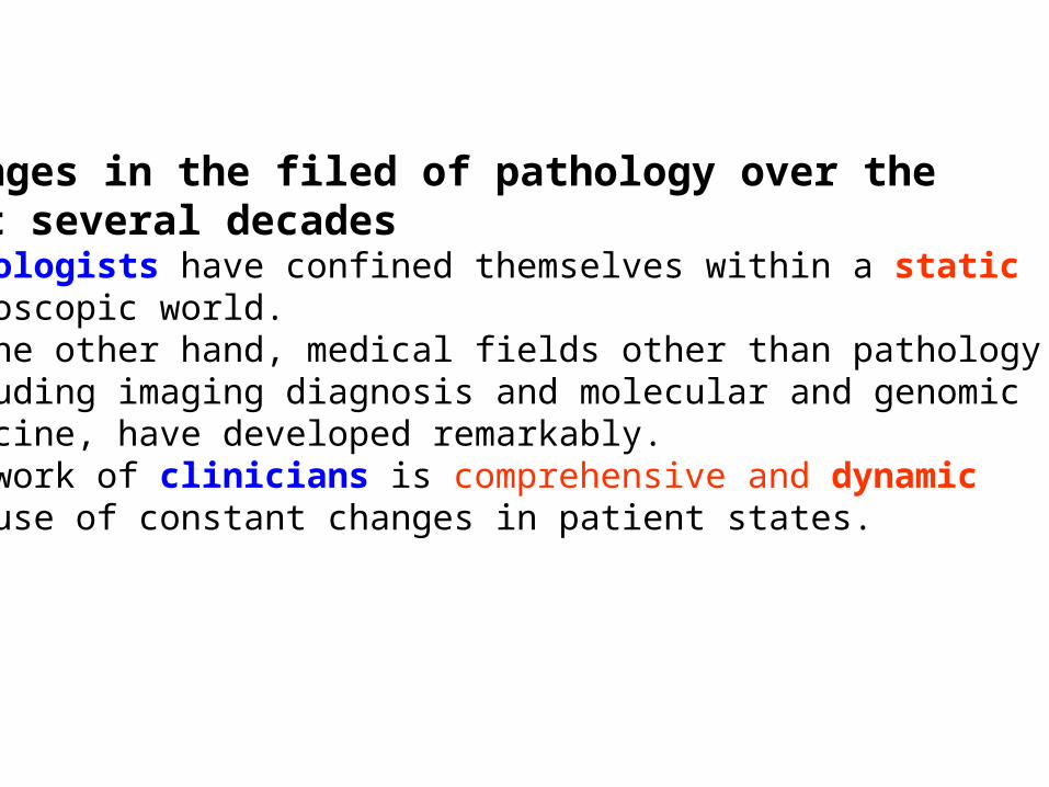
Changes in the filed of pathology over the past several decadesPathologists have confined themselves within a static microscopic world.On the other hand, medical fields other than pathology, including imaging diagnosis and molecular and genomic medicine, have developed remarkably.The work of clinicians is comprehensive and dynamic because of constant changes in patient states.

Medical record description by POMR (problem oriented medical record)First medical examination1. Data base 1) Patient information: patient profile, chief complaint, history of present illness, past history, family history, social history, system review, etc. 2) Physical examination findings: general status (external appearance, mental state, skin condition) specific status 3) Test findings2. Problem list : disease name, signs and symptoms, patient’s condition or findings, etc. 1) #1 title, #2 title, #3 title--- 2) Distinguish active title from inactive title3. Initial plan 1) Diagnostic plan: to select the diagnostic tests and plan a schedule for enforcement, e.g. urine tests, blood tests, blood biochemistry tests, immunoserum tests, chest and abdominal plain X-rays, ECG, urine culture and blood culture tests and abdominal US etc 2) Therapeutic plan 3) Education planSecond medical examination or since the second day after admission4. Progress note 1) Narrative note S: subjective data (patient complaint) O: objective data (physical findings and test findings ): A: assessment (each problem is analyzed on the basis of the information available about the disease and a hypothesis is established on what happens and why) P: plan (make plans to prove the correctness of the hypothesis and describe it) Imaging examinations (CT scan, MRI scan, etc.) and pathological examination 2) Flow chartRecord at discharge5. Discharge summaryProvide a brief description, so it is understood easily by all who read it.
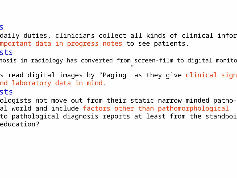
CliniciansAs for the daily duties, clinicians collect all kinds of clinical information and summarize important data in progress notes to see patients.
RadiologistsImaging diagnosis in radiology has converted from screen-film to digital monitor diagnosis.Radiologists read digital images by “Paging” as they give clinical signs and symptoms, and laboratory data in mind.
PathologistsWhy do pathologists not move out from their static narrow minded patho-morphological world and include factors other than pathomorphological findings into pathological diagnosis reports at least from the standpoint of medical education?
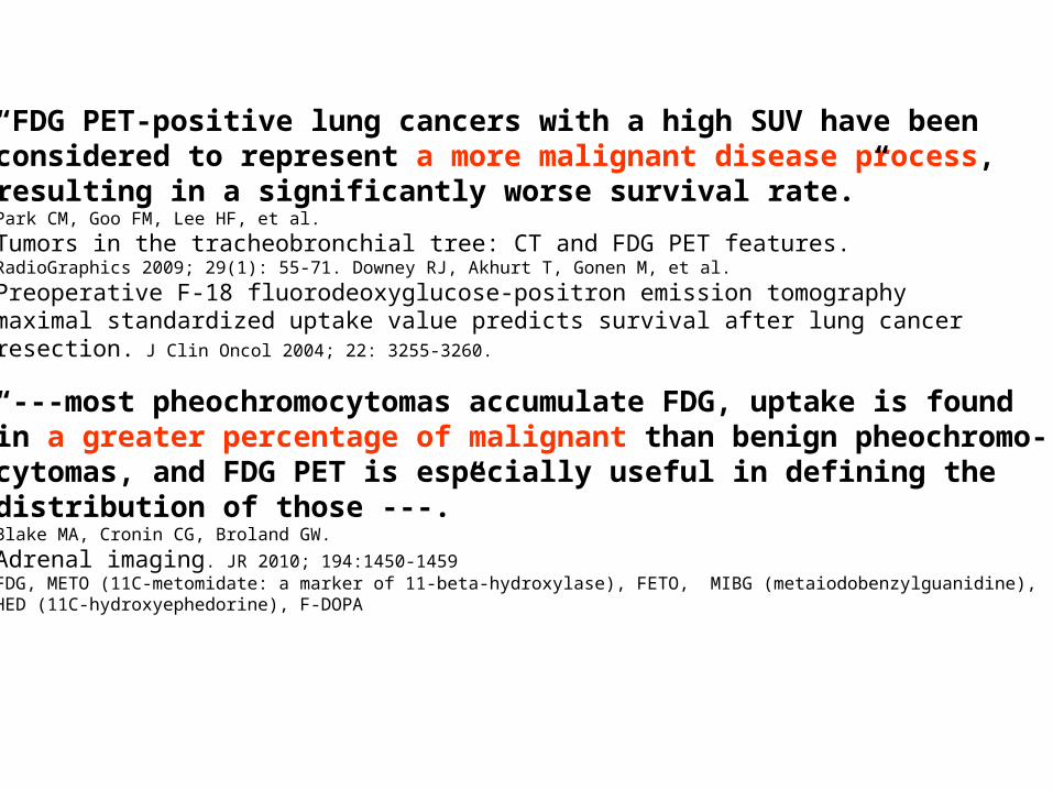
“FDG PET-positive lung cancers with a high SUV have been considered to represent a more malignant disease process, resulting in a significantly worse survival rate.”Park CM, Goo FM, Lee HF, et al.
Tumors in the tracheobronchial tree: CT and FDG PET features. RadioGraphics 2009; 29(1): 55-71. Downey RJ, Akhurt T, Gonen M, et al.
Preoperative F-18 fluorodeoxyglucose-positron emission tomography maximal standardized uptake value predicts survival after lung cancer resection. J Clin Oncol 2004; 22: 3255-3260.
“---most pheochromocytomas accumulate FDG, uptake is found in a greater percentage of malignant than benign pheochromo-cytomas, and FDG PET is especially useful in defining the distribution of those ---.”Blake MA, Cronin CG, Broland GW.
Adrenal imaging. JR 2010; 194:1450-1459FDG, METO (11C-metomidate: a marker of 11-beta-hydroxylase), FETO, MIBG (metaiodobenzylguanidine), HED (11C-hydroxyephedorine), F-DOPA

Constitution1. Background2. Aim3. Materials and methods 3a. Pathology report form 3b. Seamlessly viewing images 3c. Medical library4. Result5. Conclusion
[Aim]To examine the usefulness of incorporating non-pathomorphological data recorded in the HIS (hospital information system) into the pathology report form and the applicability of a standardized method for viewing digital pathology images.

Constitution1. Background2. Aim3. Materials and methods 3a. Pathology report form 3b. Seamlessly viewing images 3c. Medical library4. Result5. Conclusion
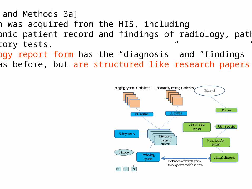
[Materials and Methods 3a] Information was acquired from the HIS, including the electronic patient record and findings of radiology, pathology, and laboratory tests. The pathology report form has the “diagnosis” and “findings” sections, as before, but are structured like research papers.
Electronic patient record
RIS system
Pathology system
Virtual slide server
LIS system
Imaging system modalities Laboratory testing machines
Virtual slide end
F/W machine
Router
Internet
Exchange of information through removable media
Subsystems
Hospital LAN system
Library
PC PC PC
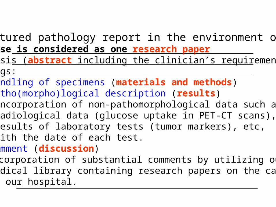
Structured pathology report in the environment of HISOne case is considered as one research paper Diagnosis (abstract including the clinician’s requirements) Findings: 1) Handling of specimens (materials and methods) 2) Patho(morpho)logical description (results) Incorporation of non-pathomorphological data such as radiological data (glucose uptake in PET-CT scans), results of laboratory tests (tumor markers), etc, with the date of each test. 3) Comment (discussion) Incorporation of substantial comments by utilizing our medical library containing research papers on the cases in our hospital.

Structured pathology report: surgically resected cases Case 1: 73-year-old manChief complaint: liver tumor detected at the follow-up CT imaging after gastrectomy for gastric cancerClinical diagnosis: liver cancer suggestive of a combined type HCC (hepatocellular carcinoma), CCC (cholangiocellular carcinoma), or metastatic carcinoma
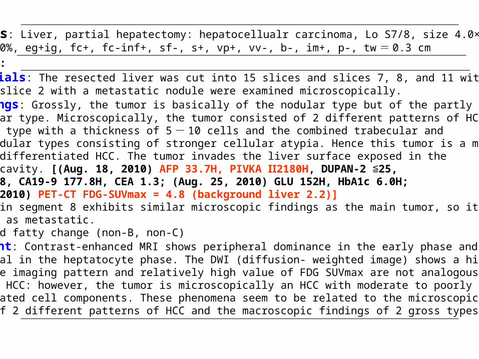
Diagnosis: Liver, partial hepatectomy: hepatocellualr carcinoma, Lo S7/8, size 4.0×3.1 cm, necrosis 10%, eg+ig, fc+, fc-inf+, sf-, s+, vp+, vv-, b-, im+, p-, tw = 0.3 cm
Findings: 1) Materials: The resected liver was cut into 15 slices and slices 7, 8, and 11 with the main tumor and slice 2 with a metastatic nodule were examined microscopically. 2) Findings: Grossly, the tumor is basically of the nodular type but of the partly confluent multinodular type. Microscopically, the tumor consisted of 2 different patterns of HCC: the trabecular type with a thickness of 5 - 10 cells and the combined trabecular and pseudoglandular types consisting of stronger cellular atypia. Hence this tumor is a moderately to poorly differentiated HCC. The tumor invades the liver surface exposed in the abdominal cavity. [(Aug. 18, 2010) AFP 33.7H, PIVKA 2180HⅡ , DUPAN-2 25, ≦SPAN-2 13.8, CA19-9 177.8H, CEA 1.3; (Aug. 25, 2010) GLU 152H, HbA1c 6.0H; (Aug. 19, 2010) PET-CT FDG-SUVmax = 4.8 (background liver 2.2)] The tumor in segment 8 exhibits similar microscopic findings as the main tumor, so it is identified as metastatic.Liver: mild fatty change (non-B, non-C) 3) Comment: Contrast-enhanced MRI shows peripheral dominance in the early phase and a low signal in the heptatocyte phase. The DWI (diffusion- weighted image) shows a high signal. The imaging pattern and relatively high value of FDG SUVmax are not analogous to typical HCC: however, the tumor is microscopically an HCC with moderate to poorly differentiated cell components. These phenomena seem to be related to the microscopic findings of 2 different patterns of HCC and the macroscopic findings of 2 gross types.
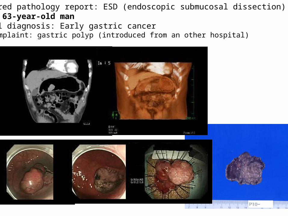
Structured pathology report: ESD (endoscopic submucosal dissection) caseCase 2: 63-year-old man Clinical diagnosis: Early gastric cancerChief complaint: gastric polyp (introduced from an other hospital)
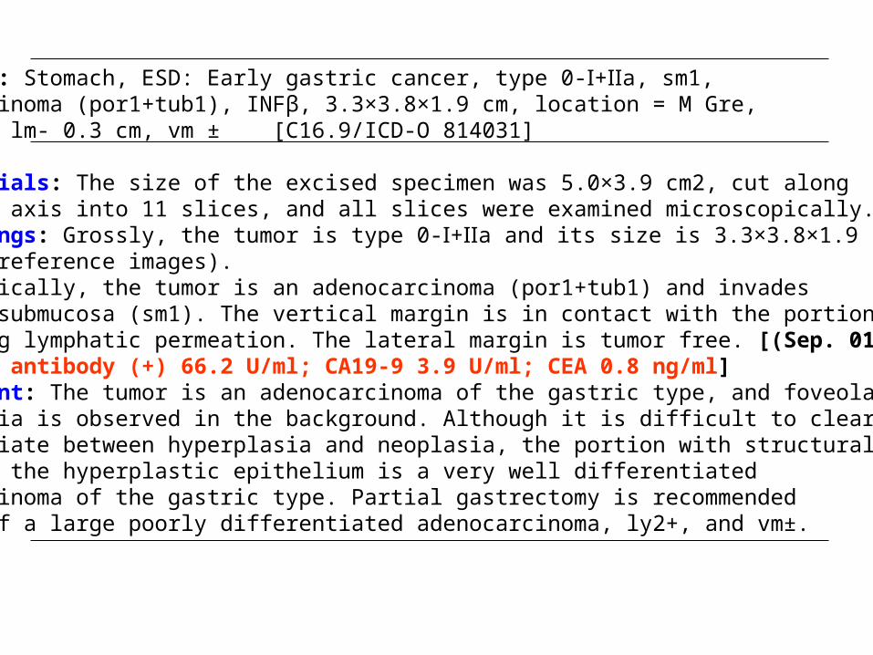
Diagnosis: Stomach, ESD: Early gastric cancer, type 0- + a, sm1, Ⅰ Ⅱadenocarcinoma (por1+tub1), INFβ, 3.3×3.8×1.9 cm, location = M Gre, ly2+, v0, lm- 0.3 cm, vm ± [C16.9/ICD-O 814031]Findings: 1) Materials: The size of the excised specimen was 5.0×3.9 cm2, cut along the short axis into 11 slices, and all slices were examined microscopically. 2) Findings: Grossly, the tumor is type 0- + a and its size is 3.3×3.8×1.9 Ⅰ Ⅱcm3 (see reference images).Microscopically, the tumor is an adenocarcinoma (por1+tub1) and invades into the submucosa (sm1). The vertical margin is in contact with the portion of exhibiting lymphatic permeation. The lateral margin is tumor free. [(Sep. 01, 2010) H. pylori antibody (+) 66.2 U/ml; CA19-9 3.9 U/ml; CEA 0.8 ng/ml] 3) Comment: The tumor is an adenocarcinoma of the gastric type, and foveolar hyperplasia is observed in the background. Although it is difficult to clearly differentiate between hyperplasia and neoplasia, the portion with structural atypia in the hyperplastic epithelium is a very well differentiated adenocarcinoma of the gastric type. Partial gastrectomy is recommended because of a large poorly differentiated adenocarcinoma, ly2+, and vm±.
Constitution1. Background2. Aim3. Materials and methods 3a. Pathology report form 3b. Seamlessly viewing images 3c. Medical library4. Result5. Conclusion

Hospital information system of Ehime Prefectural Central Hospital (2010)
Electronic patient record
RIS system
Pathology system
Virtual slide server
LIS system
Imaging system modalities Laboratory testing machines
Virtual slide end
F/W machine
Router
Internet
Exchange of information through removable media
Subsystems
Hospital LAN system
Library
PC PC PC
Every day clinicians observe various types of images including radiological and gross pathological images using the HIS.

Reference CT and MRI images in the electronic patient record are arranged in the following order: from head to foot, left to right, and top to bottom.
111

[Materials and Methods 3b] All kinds of images including radiological, gross pathological and histopathological images are arranged to be viewed seamlessly as follows.
1) The resected gross specimens were sliced in the same planes as the radiological axial images. 2) The slices were then placed on a photography table in the same order as in the radiological images and were photographed. 3) All the gross slices and histology slides were assigned corresponding numbers and 4) the glass histology slides were digitized by virtual microscopy.
1
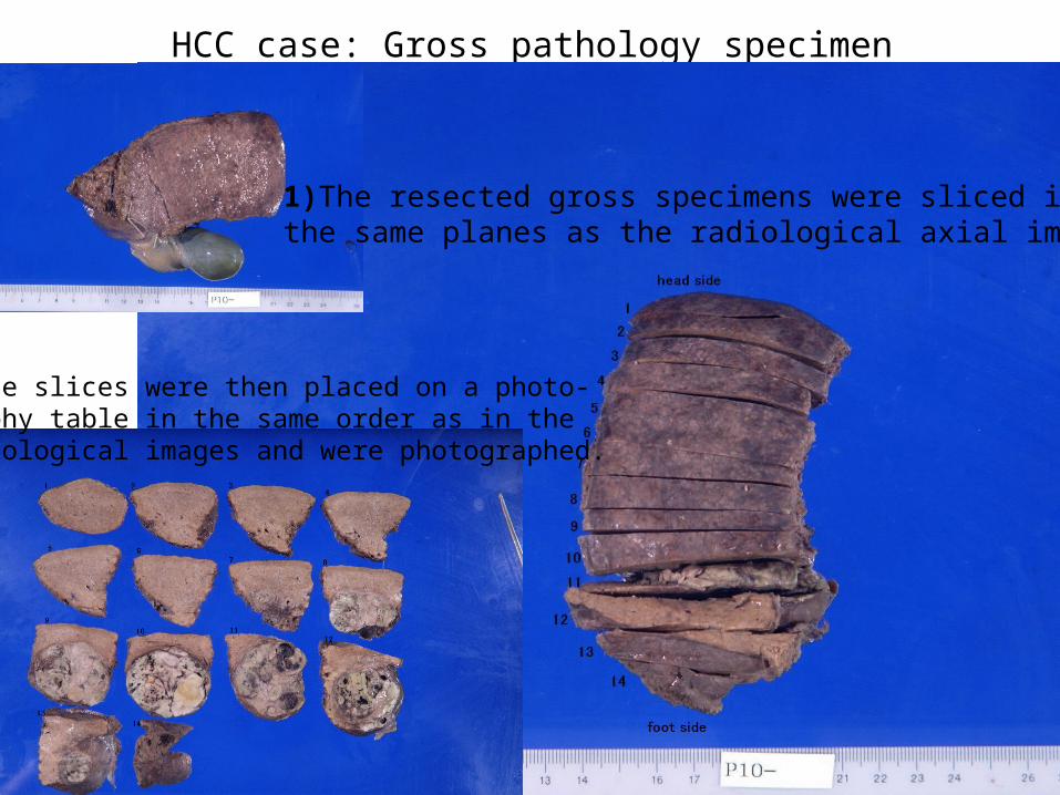
HCC case: Gross pathology specimen
1)The resected gross specimens were sliced in the same planes as the radiological axial images.
2)The slices were then placed on a photo-graphy table in the same order as in the Radiological images and were photographed.

1 3 4
Gross pathology images are arranged in the following order: from head to foot, left to right, and top to bottom.
3) All the gross slices and histology slides were assigned corresponding numbers
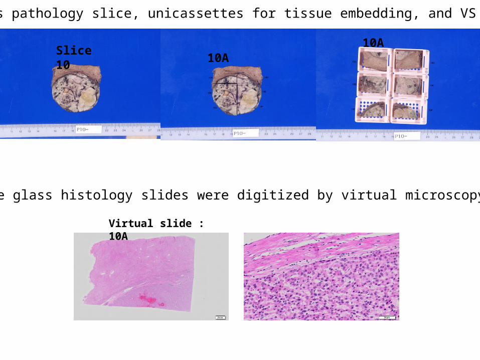
Virtual slide : 10A
A gross pathology slice, unicassettes for tissue embedding, and VS
Slice 1010A
10A
4) the glass histology slides were digitized by virtual microscopy.
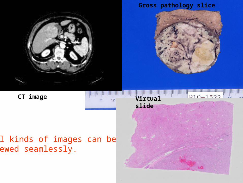
CT image Virtual slide
Gross pathology slice
All kinds of images can be viewed seamlessly.

A resected case of HCC: macroscopically, this case is basically nodular type, but some parts are the confluent multinodular type.

Figure (CT images)
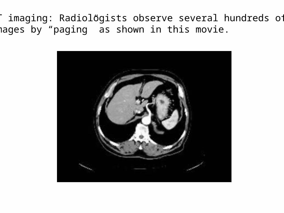
CT imaging: Radiologists observe several hundreds of images by “paging” as shown in this movie.

Movie on the gross pathological images: Just like radiologists, pathologists, too, can observe gross pathological imaging by paging.
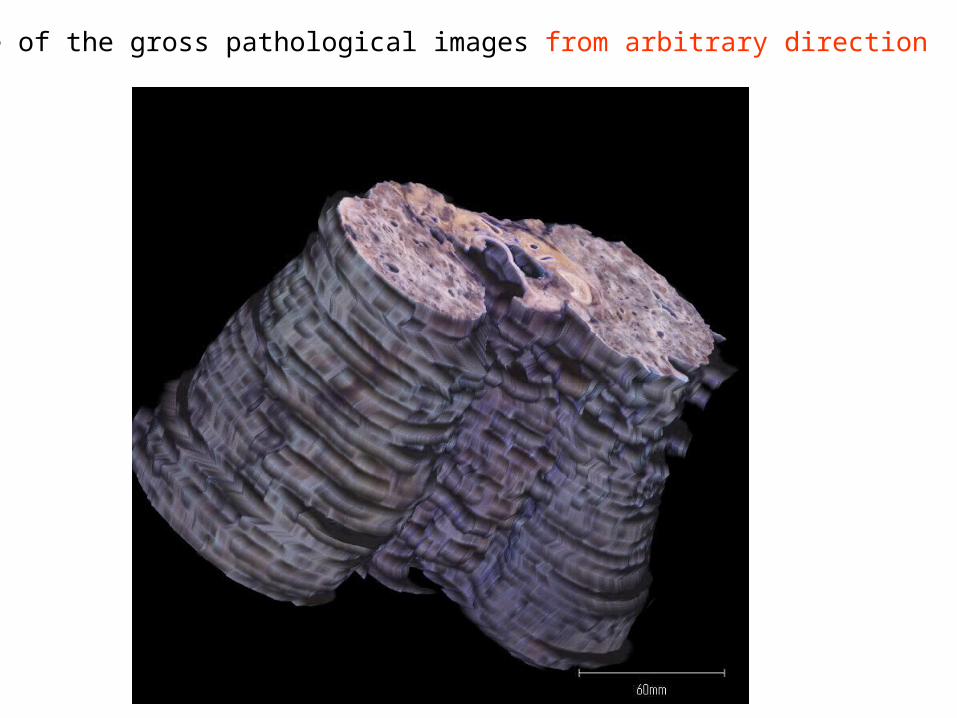
Movie of the gross pathological images from arbitrary direction

Constitution1. Background2. Aim3. Materials and methods 3a. Pathology report form 3b. Seamlessly viewing images 3c. Medical library4. Result5. Conclusion
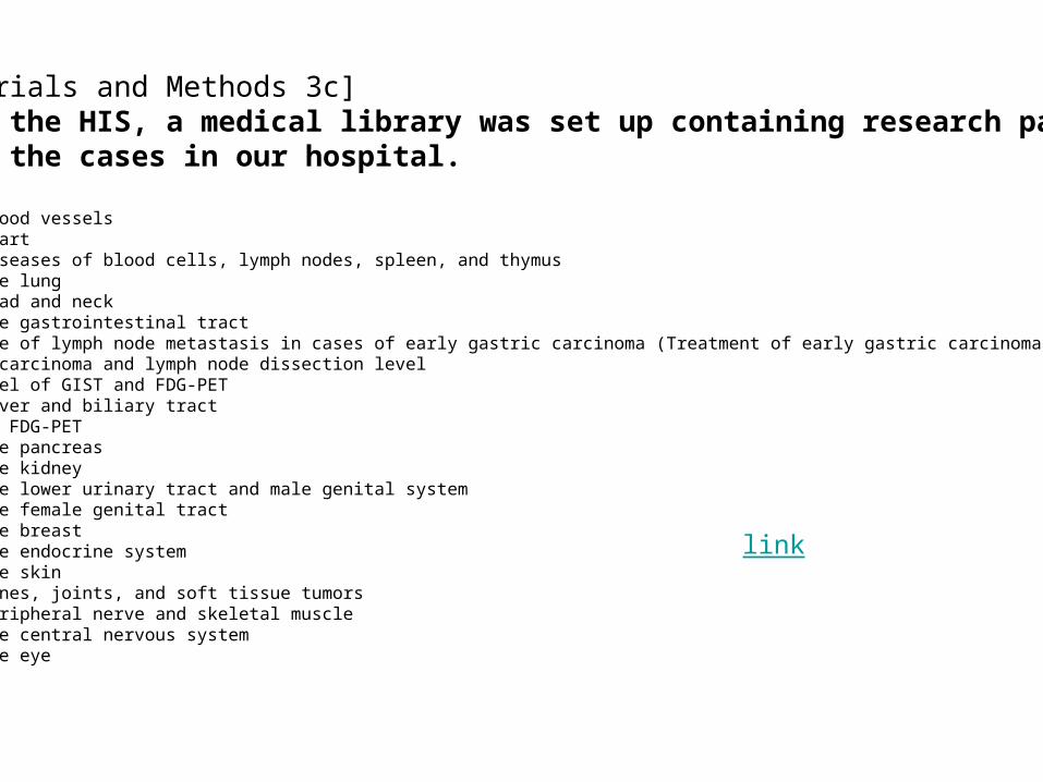
[Materials and Methods 3c]Using the HIS, a medical library was set up containing research papers about the cases in our hospital.
Lib 11 Blood vesselsLib 12 HeartLib 13 Diseases of blood cells, lymph nodes, spleen, and thymusLib 14 The lungLib 15 Head and neckLib 16 The gastrointestinal tract Incidence of lymph node metastasis in cases of early gastric carcinoma (Treatment of early gastric carcinoma by ESD) Gastric carcinoma and lymph node dissection level Risk level of GIST and FDG-PETLib 17 Liver and biliary tract HCC and FDG-PET Lib 18 The pancreasLib 19 The kidneyLib 20 The lower urinary tract and male genital systemLib 21 The female genital tractLib 22 The breastLib 23 The endocrine systemLib 24 The skinLib 25 Bones, joints, and soft tissue tumorsLib 26 Peripheral nerve and skeletal muscleLib 27 The central nervous systemLib 28 The eye
link
Constitution1. Background2. Aim3. Materials and methods 3a. Pathology report form 3b. Seamlessly viewing images 3c. Medical library4. Result5. Conclusion
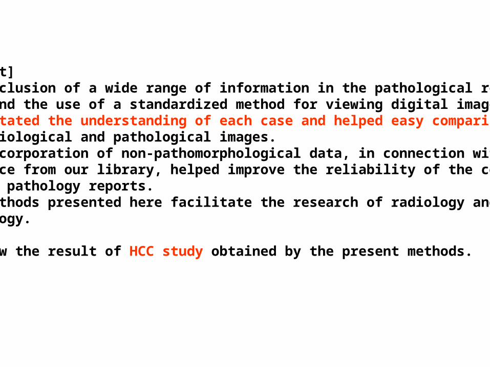
[Result] The inclusion of a wide range of information in the pathological report form and the use of a standardized method for viewing digital images facilitated the understanding of each case and helped easy comparison of radiological and pathological images. The incorporation of non-pathomorphological data, in connection with evidence from our library, helped improve the reliability of the comments in the pathology reports.The methods presented here facilitate the research of radiology and pathology.
We show the result of HCC study obtained by the present methods.
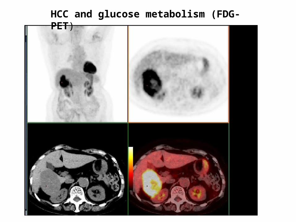
HCC and glucose metabolism (FDG-PET)

Figure. Gross types of HCC: type 1, 2x, 2a, 2b, 3, and 4
Type 1 Type 2x
Type 2a Type 2b
Type 3 Type 4
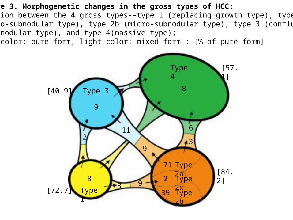
Figure 3. Morphogenetic changes in the gross types of HCC:Relation between the 4 gross types--type 1 (replacing growth type), type 2a (macro-subnodular type), type 2b (micro-subnodular type), type 3 (confluentmultinodular type), and type 4(massive type); deep color: pure form, light color: mixed form ; [% of pure form]
Type 4
Type 3
Type 1
Type 2a
Type 2b
Type 2x3
8
9
2
9
71
2
39
93
8
611
[57.1]
[84.2]
[40.9]
[72.7]

Gross type No. of cases (%) Tumor size (cm) Tumor necrosis (%)
1 2 (2.0) 2.4±0.5 15.0±21.24)
2a 44 (44.9) 3.3±2.2 87.8±17.55)
2b 23 (23.5) 3.9±2.1 69.8±24.76)
2x 5 (5.1) 2.3±1.7 88.0±13.07)
3 15 (15.3) 3.5±2.3 59.3±32.1
4 9 (9.2) 10.6±3.7 43.9±20.68)
Table 4 Relation between HCC gross type, size, and necrosis in HCC cases with TAE
1) HCC: Hepatocellular carcinoma, 2) TAE: transcatherter arterial embolization, 3) Gross type 1: replacing growth type, 2a: macro-subnodular type, 2b: micro-subnodular type, 3: confluent multinodular type, 4: massive type5) vs. 6) P=0.0009, 4) vs. 6) p=0.0059, 4) vs. 7) p=0.0059, 6) vs. 8) p=0.0092
Gross type DD-score DD-G FDG uptake (No. of cases)
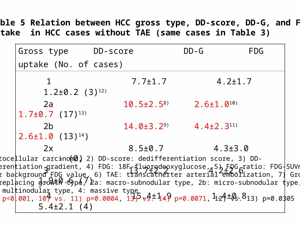
1 7.7±1.7 4.2±1.7 1.2±0.2 (3)12)
2a 10.5±2.58) 2.6±1.010) 1.7±0.7 (17)13)
2b 14.0±3.29) 4.4±2.311) 2.6±1.0 (13)14)
2x 8.5±0.7 4.3±3.0 - (0)
3 13.7±2.2 4.2±2.6 1.9±0.6 (7)
4 15.4±1.9 1.4±0.8 5.4±2.1 (4)
Table 5 Relation between HCC gross type, DD-score, DD-G, and FDG uptake in HCC cases without TAE (same cases in Table 3)
HCC: Hepatocellular carcinoma, 2) DD-score: dedifferentiation score, 3) DD-G: dedifferentiation gradient, 4) FDG: 18F-fluorodeoxyglucose, 5) FDG ratio: FDG-SUVmax of HCC per background FDG value, 6) TAE: transcatherter arterial embolization, 7) Gross type 1: replacing growth type, 2a: macro-subnodular type, 2b: micro-subnodular type, 3: confluent multinodular type, 4: massive type8) vs. 9) p<0.001, 10) vs. 11) p=0.0004, 13) vs. 14) p=0.0071, 12) vs. 13) p=0.0305

Constitution1. Background2. Aim3. Materials and methods 3a. Pathology report form 3b. Seamlessly viewing images 3c. Medical library4. Result5. Conclusion
Conclusion The format of the pathology report form with seamless image viewing described in this study is expected to promote deeper integration of comprehensive information in evolving HIS and contribute positively to education, research, and patient care.

Thank you very much for your attention
ご清聴、ありがとうございました。
Search engineDataoptimization
Lab. system
RIS ・PACS
Pathology system
Electronic patient record
専用 Data Warehouse
Data
base
Statistical data
Case data
New information system in Our New Hospital (2013)to utilize accumulated data in HIS Today’s presentation:
#Structured pathology report
#Seamless viewing images
#Medical library