the university of manitoba faculty of graduate …
TRANSCRIPT
STRUCTURE-FUNCTION PROPERTIES OF FLAXSEED PROTEIN-DERIVED
MULTIFUNCTIONAL PEPTIDES
By
Chibuike Chinedu Udenigwe
A Thesis
Submitted to the Faculty of Graduate Studies
In Partial Fulfillment of the Requirements for the Degree of
Doctor of Philosophy
Food and Nutritional Sciences
University of Manitoba
Winnipeg, Manitoba
Canada
Copyright © October 2010 by Chibuike Chinedu Udenigwe

THE UNIVERSITY OF MANITOBA
FACULTY OF GRADUATE STUDIES
*****
COPYRIGHT PERMISSION
STRUCTURE-FUNCTION PROPERTIES OF FLAXSEED PROTEIN-DERIVED
MULTIFUNCTIONAL PEPTIDES
By
Chibuike Chinedu Udenigwe
A Thesis submitted to the Faculty of Graduate Studies
University of Manitoba
In partial fulfillment of the requirements for the degree of
Doctor of Philosophy
Copyright © October 2010 by Chibuike Chinedu Udenigwe
Permission has been granted to the Library of the University of Manitoba to lend or sell copies of this thesis to the National Library of Canada to microfilm this thesis and to lend or sell copies of the film, and University Microfilms Inc. to publish an abstract
of this thesis.
The reproduction or copy of this thesis has been made available by authority of the copyright owner solely for the purpose of private study and research, and may only be
reproduced and copied as permitted by copyright laws or with express written authorization from the copyright owner.

iii
ABSTRACT
Food protein-derived peptides have increasingly become important sources of
ingredients for the formulation of therapeutic products. The main aim of this work was
to study the in vitro and in vivo bioactive properties of structurally diverse group of
peptides produced through enzymatic hydrolysis of flaxseed proteins (FP). Hydrolysis of
FP with seven proteases followed by fractionation into low-molecular-weight (LMW)
cationic fractions yielded multifunctional peptides that inhibited angiotensin converting
enzyme (ACE) and renin activities, which are molecular targets for antihypertensive
agents. The LMW peptides also exhibited antioxidant properties by scavenging free
radicals and inhibiting amine oxidase activity. The peptide fractions showed inhibition of
calmodulin-dependent phosphodiesterase, an enzyme that has been implicated in the
pathogenesis of several chronic diseases. Moreover, FP hydrolysis with thermolysin and
pronase followed by mixing with activated carbon yielded branched-chain amino acids
(BCAA)-enriched multifunctional peptide mixture (Fischer ratio of 23.65) with
antioxidant properties and in vitro ACE inhibition; Fischer ratio of 20.0 is considered
minimum for therapeutic purposes. The BCAA-enriched peptide product can be used in
clinical nutrition to treat muscle wasting symptoms associated with hepatic diseases.
Furthermore, an arginine-rich peptide mixture (31% arginine versus 11% in the original
flaxseed protein) was produced by hydrolysis of FP with trypsin and pronase followed by
separation using electrodialysis-ultrafiltration. Arginine plays important physiological
roles especially as precursor to vasodilator, nitric oxide. The arginine-rich peptide
mixture exhibited in vitro ACE and renin inhibition and led to decreased systolic blood

iv
pressure (–17.9 and –11.7 mmHg, respectively at 2 and 4 h) after oral administration to
spontaneously hypertensive rats. For the first time in the literature, we showed that
arginine peptides have superior physiological effects when compared to the amino acid
form of arginine. Lastly, quantitative structure-activity relationship studies using partial
least squares (PLS) regression yielded two predictive models for renin-inhibiting
dipeptides with z-scales amino acid descriptors. The PLS models indicated that
hydrophobic and bulky side chain-containing amino acids contribute to renin inhibition
if present at the amino- and carboxyl-terminal of dipeptides, respectively. Based on this
study, Ile-Trp was discovered as potent renin-inhibiting dipeptide, and may serve as a
useful template for the development of potent antihypertensive peptidomimetics.

v
ACKNOWLEDGEMENTS
I am sincerely thankful to my supervisor, Dr. Rotimi Aluko, for giving me the
opportunity to work in his research group and for his mentorship and support
throughout the duration of my doctoral studies. I would also like to thank my thesis
committee members, Drs. Mohammed Moghadasian and Karmin O, for their support
through the years and the external examiner, Dr. Janitha Wanasundara, for devoting her
time to read and evaluate this thesis.
I acknowledge the financial support that we received from the following sources
as research grants or scholarship/fellowship during the course of my doctoral studies:
Natural Sciences and Engineering Research Council of Canada, Advanced Foods and
Materials Network of Centres of Excellence Canada, Canadian Institute of Food Science
and Technology, Institute of Food Technologists (USA), Manitoba Health Research
Council, Manitoba Agriculture and Rural Development Initiative, and the University of
Manitoba Faculty of Graduate Studies. I would also like to thank Dr. Wen-Chi Hou and
his research group at Taipei Medical University for their hospitality during my stay in
Taiwan, and Dr. Laurent Bazinet and his research group at Laval University, Québec for
hosting me in their laboratory to learn the electrodialysis-ultrafiltration technique.
Many thanks are due to Dr. Aluko’s research team and my numerous friends in the
Department of Human Nutritional Sciences for their company. I could not have
completed my research work without the emotional and spiritual support that I received
from my family especially my heartthrob, Ogochukwu, and our baby, Chimamanda.
Above all, I could not have accomplished anything without the amazing grace of God.

vi
FOREWARD
This thesis was prepared using the manuscript format and it is composed of six
manuscripts following the General Introduction and Literature Review chapters. The
manuscripts were written in different journal styles as follows: Manuscript 1 (Journal of
Functional Foods), Manuscript 2 (Food Chemistry), Manuscript 3 (Food Chemistry),
Manuscript 4 (Journal of Agricultural and Food Chemistry), Manuscript 5 (Food
Chemistry) and Manuscript 6 (Amino Acids). Manuscripts 1, 2 and 4 have been
published, Manuscripts 6 is currently under consideration for publication, and
Manuscripts 3 and 5 have been prepared for submission to the journals indicated above.
The manuscripts were linked by transition statements at the end of each manuscript for
coherence. The format for list of references cited for the Introduction and Literature
Review chapters follows that of the journal, Food Chemistry. The last chapter provides a
general summary of the thesis, concluding remarks and future directions of the project.

vii
TABLE OF CONTENTS
Page
ABSTRACT iii
ACKNOWLEDGEMENTS v
FOREWARD vi
TABLE OF CONTENTS vii
LIST OF TABLES xiv
LIST OF FIGURES xvii
LIST OF COPYRIGHTED MATERIALS xxiii
LIST OF ABBREVIATIONS xxiv
CHAPTER ONE GENERAL INTRODUCTION 1
CHAPTER TWO LITERATURE REVIEW 9
2.1. Hypertension and the renin-angiotensin system (RAS) 9
2.2. Reactive oxygen species and free radicals in human health conditions 12
2.2.1. Inflammatory responses 12
2.2.2. Neurodegenerative diseases 15
2.2.3. Oxidative stress, inflammation and hypertension 18
2.3. Calmodulin (CaM) and CaM-dependent enzymes 18
2.4. Food protein-derived bioactive peptides in human health 22
2.4.1. ACE-inhibitory and antihypertensive peptides 26
2.4.2. Antioxidant peptides 29
2.4.3. Hypolipidemic and hypocholesterolemic peptides 32
2.4.4. Anticancer peptides 34
2.4.5. Immunomodulatory and anti-inflammatory peptides 36
2.4.6. Calmodulin (CaM)-binding peptides 37
2.4.7. Branched-chain amino acid-rich peptides and liver diseases 38
2.4.8. Multifunctional peptides 41
2.5. Quantitative Structure-Activity Relationship (QSAR) studies of peptides 42
2.6. Flaxseed 44
viii
2.6.1. Nutritional quality, compositions and health benefits of
flaxseed
45
2.6.2. Flaxseed proteins 46
2.7. References 53
CHAPTER THREE MANUSCRIPT 1
KINETICS OF THE INHIBITION OF RENIN AND ANGIOTENSIN I-
CONVERTING ENZYME BY FLAXSEED PROTEIN HYDROLYSATE
FRACTIONS
68
3.0. Abstract 69
3.1. Introduction 70
3.2. Materials and Methods 73
3.2.1. Materials 73
3.2.2. Isolation of flaxseed proteins 73
3.2.3. Preparation of flaxseed protein hydrolysates 74
3.2.4. Fractionation of flaxseed protein hydrolysates 75
3.2.5. Preparative ion-exchange liquid chromatography 76
3.2.6. Determination of ACE inhibition 77
3.2.7. Determination of kinetics parameters of ACE inhibition 78
3.2.8. Renin inhibition assay 79
3.2.9. Statistical analysis 80
3.3. Results and Discussion 80
3.4. Conclusions 94
3.5. References 95
APPENDIX A 99
TRANSITION STATEMENT
109

ix
CHAPTER FOUR MANUSCRIPT 2
FLAXSEED PROTEIN-DERIVED PEPTIDE FRACTIONS:
ANTIOXIDANT PROPERTIES AND INHIBITION OF
LIPOPOLYSACCHARIDE-INDUCED NITRIC OXIDE PRODUCTION
IN MURINE MACROPHAGES
110
4.0. Abstract 111
4.1. Introduction 112
4.2. Materials and Methods 115
4.2.1. Materials 115
4.2.2. Production of FPH and cationic peptide fractions 115
4.2.3. In vitro antioxidant assays 118
4.2.3.1. DPPH˙ scavenging assay 118
4.2.3.2. Superoxide radical (O2˙– 119 ) scavenging assay
4.2.3.3. Hydroxyl radical (˙OH) scavenging assay 120
4.2.3.4. Nitric oxide (NO) scavenging assay 120
4.2.4. Determination of SSAO inhibition 121
4.2.5. LPS-induced NO production in RAW 264.7 macrophages 122
4.2.5.1. Cell culture and cell treatment 122
4.2.5.2. Nitrite quantification and cell viability assay 122
4.2.6. Statistical analysis 123
4.3. Results and Discussion 123
4.3.1. Peptide production and isolation 123
4.3.2. Peptide-induced scavenging of free radicals 125
4.3.3. Peptide-induced inhibition of SSAO 133
4.3.4. Peptide-induced inhibition of in vitro/ex vivo NO production 135
4.4. Conclusions 140
4.5. References 142
APPENDIX B 147
TRANSITION STATEMENT 152

x
CHAPTER FIVE MANUSCRIPT 3
MULTIFUNCTIONAL PEPTIDES FROM A CATIONIC FRACTION
OF FLAXSEED PROTEIN HYDROLYSATES
153
5.0. Abstract 154
5.1. Introduction 155
5.2. Materials and Methods 157
5.2.1. Production of flaxseed cationic peptide fraction 157
5.2.2. CaM-dependent PDE (CaMPDE) inhibition assay 157
5.2.3. ACE inhibition assay 159
5.2.4. Renin inhibition assay 159
5.2.5. High Performance Liquid Chromatography (HPLC) 160
5.3. Results and Discussion 161
5.4. Conclusions 173
5.5. References 174
TRANSITION STATEMENT 176
CHAPTER SIX MANUSCRIPT 4
ANTIOXIDANT AND ANGIOTENSIN CONVERTING ENZYME-
INHIBITORY PROPERTIES OF A FLAXSEED PROTEIN-DERIVED
HIGH FISCHER RATIO PEPTIDES MIXTURE
177
6.0. Abstract 178
6.1. Introduction 179
6.2. Materials and Methods 182
6.2.1. Materials 182
6.2.2. Enzymatic hydrolysis of flaxseed protein 182
6.2.3. Adsorption of flaxseed protein hydrolysate onto activated
carbon-packed column
183
6.2.4. Adsorption of flaxseed protein hydrolysate onto activated
carbon by mixing
184
6.2.5. Amino acid analysis 184
xi
6.2.6. Gel permeation chromatography 185
6.2.7. Antioxidant assays 185
6.2.8. ACE and renin inhibition assays 187
6.2.9. Statistical analysis 188
6.3. Results and Discussion 188
6.3.1. Enzymatic flaxseed protein hydrolysis 188
6.3.2. Activated carbon treatments, amino acid profiles and Fischer
ratios
189
6.3.3. Estimated peptide MW distribution 195
6.3.4. Antioxidant activity 196
6.3.5. Antihypertensive properties 200
6.4. Conclusions 204
6.5. References 205
APPENDIX C 208
TRANSITION STATEMENT 214
CHAPTER SEVEN MANUSCRIPT 5
SEPARATION OF ARGININE-RICH PEPTIDES FROM FLAXSEED
PROTEIN HYDROLYSATES BY ELECTRODIALYSIS-
ULTRAFILTRATION: ANGIOTENSIN CONVERTING ENZYME,
RENIN-INHIBITORY AND ANTIHYPERTENSIVE ACTIVITIES OF
THE PEPTIDES
215
7.0. Abstract 216
7.1. Introduction 217
7.2. Materials and Methods 221
7.2.1. Materials 221
7.2.2. Enzymatic hydrolysis of flaxseed protein 221
7.2.3. Electrodialysis-ultrafiltration (EDUF) 222
7.2.4. Determination of peptide migration during EDUF 224
7.2.5. Amino acid analysis 224

xii
7.2.6. Liquid chromatography-mass spectrometry (LC-MS) 225
7.2.7. ACE and renin inhibition assays 225
7.2.8. Evaluation of antihypertensive activity in spontaneously
hypertensive rats (SHR)
226
7.2.9. Statistical analysis 227
7.3. Results and Discussion 227
7.3.1. Enzymatic hydrolysis of flaxseed protein 227
7.3.2. Peptide migration during EDUF 228
7.3.3. Amino acid profiles and peptide identification 234
7.3.4. Inhibition of ACE and renin activities by the arginine-enriched
peptide fraction
238
7.3.5. Antihypertensive activity of the arginine-rich peptide fraction 240
7.4. Conclusions 242
7.5. References 244
TRANSITION STATEMENT 248
CHAPTER EIGHT MANUSCRIPT 6
QUANTITATIVE STRUCTURE-ACTIVITY RELATIONSHIP
MODELLING OF HUMAN RENIN-INHIBITING DIPEPTIDES
249
8.0. Abstract 250
8.1. Introduction 251
8.2. Methods 253
8.2.1. Peptide dataset 253
8.2.2. Partial least square (PLS) regression analysis 255
8.2.3. Peptide synthesis 256
8.2.4. Renin inhibition assay 256
8.2.5. ACE inhibition assay 257
8.2.6. Statistical analysis 257
8.3. Results and Discussion 258
8.3.1. QSAR of renin-inhibitory dipeptides by PLS 258

xiii
8.3.2. Peptide prediction 262
8.3.3. Renin inhibition by predicted dipeptides 266
8.3.4. Relationship between renin and ACE inhibition 270
8.4. Conclusions 272
8.5. References 274
APPENDIX D 277
CHAPTER NINE
GENERAL DISCUSSION AND CONCLUSIONS 284

xiv
LIST OF TABLES
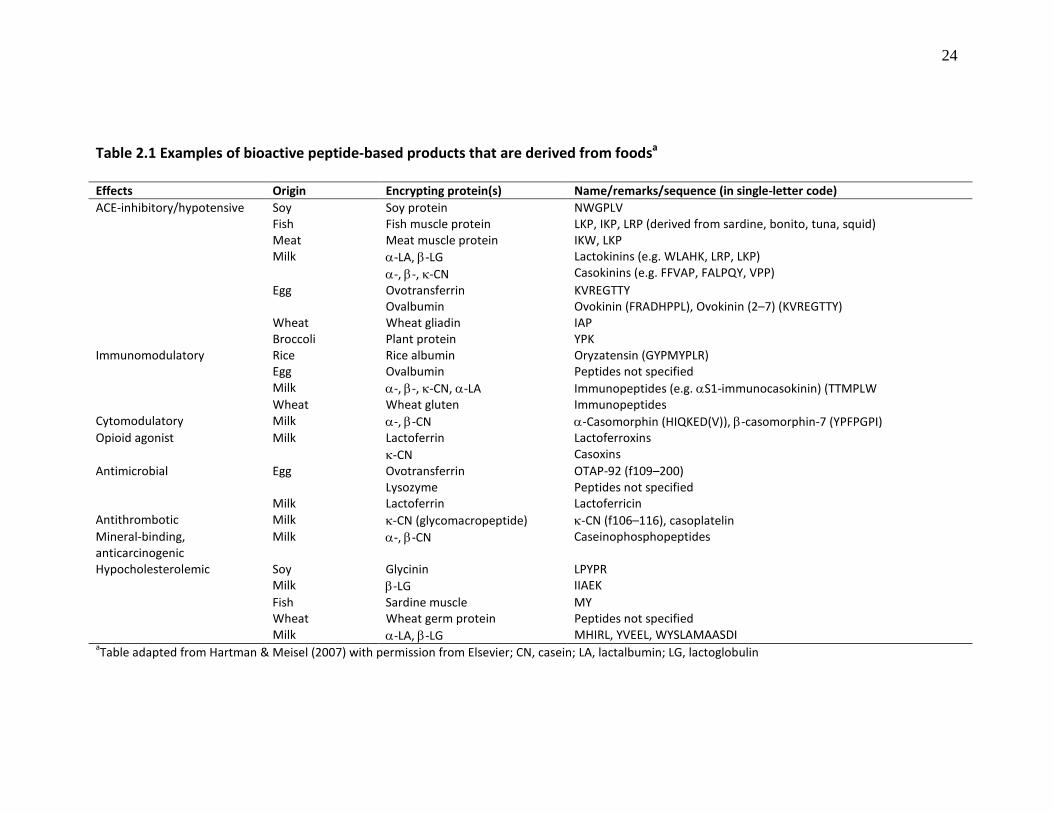
Page Table 2.1 Examples of bioactive peptide-based products that are derived from food
24
Table 2.2 Examples of commercially available functional foods or food ingredients carrying bioactive peptides
25
Table 2.3 Differences in flaxseed protein contents (%) based on different isolation methods and mucilage contents of the starting materials
48
Table 2.4 Amino acid compositions (%) of flaxseed, flaxseed meals and flaxseed protein extracts
50
Table 2.5 Amino acid composition of the two major flaxseed 11-12 S (linin) and 1.6-2 S (conlinin) protein subunits
51
Table 3.1 Reaction conditions for enzymatic hydrolysis of flaxseed proteins; protein contents and yield of the ultrafiltration permeate
83
Table 3.2 Kinetics constants of ACE catalyzed reaction in the absence and presence of different concentrations of Alcalase cationic peptide fractions I and II, and thermolysin hydrolysate (ThermoH); Km and Km′ are Michaelis constants in the absence and presence of inhibitor, respectively; Vmax, and Vmax
′ are maximum reaction velocities in the absence and presence of inhibitor, respectively; CE, catalytic efficiency of ACE; Ki, enzyme-inhibitor dissociation constant
90
Table S3.1 Amino acid composition (%) of flaxseed protein isolate (FPI) compared to soy, egg white, pea, rapeseed and wheat proteins
103
Table S3.2 Essential amino acid composition (mg/g) of flaxseed protein isolate (FPI) from this study and other food proteins compared to FAO/WHO suggested requirements for humans
104
Table 4.1 EC50 (mg protein/ml) values of flaxseed-protein derived peptide fractions in the scavenging of DPPH˙, O2˙– and ˙OH, and IC50
(mg protein/ml) for in vitro inhibition of SSAO; results are expressed as mean ± standard deviation
130
Table 6.1 Fischer ratios (BCAA/AAA) and Phe+Tyr contents of enzymatic flaxseed protein hydrolysates (FPH) passed through a column packed with activated carbon (FPH-col) or mixed with activated carbon inside a beaker (FPH-mix)
192

xv
Table 6.2 Percentage amino acid composition of flaxseed protein isolate (FPI), thermolysin-pronase hydrolysate (FPH), FPH passed through a column packed with activated carbon (FPH-col) and FPH mixed with activated carbon inside a beaker (FPH-mix)
194
Table S6.1 Protein contents (%) and yield (%) of flaxseed protein hydrolysate (FPH) and FPH passed through a column packed with activated carbon (FPH-col) or mixed with activated carbon in a beaker (FPH-mix) for the different enzyme treatments
212
Table S6.2 Amino acid composition (%) of the activated carbon treated FPH samples (FPH-col and FPH-mix)
213
Table 7.1 Amino acid compositions (%) of flaxseed protein isolate (FPI), protein hydrolysate (FPH) and peptides from the anode (KCl 1), cathode (KCl 2) and feed (FPX-X) compartments after electrodialysis-ultrafiltration
236
Table 7.2 Molecular weight identification and potential sequences of peptides that migrated to the KCl 1 (anionic) and KCl 2 (cationic) compartments during electrodialysis-ultrafiltration of flaxseed protein hydrolysates
237
Table 8.1 Dipeptide dataset with in vitro renin-inhibitory activity (RI) at 3.2 mM peptide concentration and log RI of the dipeptides
254
Table 8.2 Summary of the PLS models using the amino acid 3-z scale and 5-z scale. Multiple correlation coefficient (R2) indicates the sum of squares of Y explained and estimate of model fit whereas the cross-validated correlation coefficient (Q2
cv
) indicates the model’s predictive ability
260
Table 8.3 Predicted and observed log percentage renin inhibition (RI) at 3.2 mM peptide concentration for the predicted renin-inhibiting dipeptides and the prediction errors
270
Table 8.4 ACE-inhibitory activities (IC50
) of the Tryptophan (W)-containing dipeptides
272
Table S8.1 Descriptor (3-z) scores for the 20 coded amino acids
277
Table S8.2 Descriptor (5-z) scores for the 20 coded amino acids
278
Table S8.3 Structural properties of the dipeptides used to develop the PLS models based on the 3-z scale; n1 and n2 represent the amino acids at the N- and C-terminal of the dipeptides, respectively
279

xvi
Table S8.4 Structural properties of the dipeptides used to develop the PLS models based on the 5-z scale
280
Table S8.5 Predicted peptide dataset based on the 3-z scale descriptor and their predicted renin inhibitory activity (RI, %)
281
Table S8.6 Predicted peptide dataset based on the 5-z scale descriptor and their predicted renin inhibitory activity (RI)
282

xvii
LIST OF FIGURES
Page Figure 2.1 The renin-angiotensin system (RAS) showing the molecular targets for antihypertensive agents; AT-I, angiotensin I; AT-II, angiotensin II; ACE, angiotensin I-converting enzyme
11
Figure 2.2 Production of ROS/free radicals and oxidative stress-mediated oxidative damage to biological macromolecules
13
Figure 2.3 Production of ROS and free radicals in the brain and their roles in oxidative damages leading to neuroinflammation and neuronal cell death during neurodegenerative diseases
16
Figure 2.4 The role of Ca2+
/calmodulin-dependent phosphodiesterase (CaMPDE) in the regulation of cellular processes
21
Figure 2.5 Bioactive properties of food protein-derived peptides relevant to the promotion of human health and disease prevention
23
Figure 2.6 Targets of branched chain amino acids (BCAA) in the treatment of hepatic diseases. AAA, aromatic amino acids; BBB, blood-brain barrier
40
Figure 3.1 (A) Flaxseed protein-derived peptide fractions exhibit concentration-dependent ACE inhibition; results are expressed as means of ACE activity ± standard deviation of triplicate determinations. (B) 50% inhibitory concentration (IC50) values of peptide fractions in ACE inhibition; Each bar represents the mean of triplicate determinations of IC50
± standard deviation; bars with different letters are significantly different at P< 0.05; 1-3AlcH and 3-5AlcH stand for the 1-3 kDa and 3-5 kDa peptides of alcalase hydrolysate, respectively (For other abbreviations, see Table 3.1)
85
Figure 3.2 Lineweaver-Burk plots of the inhibition of ACE by different concentrations of: (A) cationic peptide fractions I; (B) cationic peptide fractions II; (C) ThermoH, at varying concentrations of FAPGG; V is the initial rate of reaction (∆A345nm
/min)
89
Figure 3.3 50% Inhibitory concentration (IC50
) values of flaxseed protein-derived peptide fractions in inhibiting human recombinant renin activity; n.a, no renin inhibitory activity observed; bars with different letters are significantly different at P<0.05
92
Figure 3.4 Lineweaver-Burk plot of the inhibition of human recombinant renin by flaxseed protein-derived cationic peptide fraction II at 0.5 and 2 mg protein/ml
93

xviii
Figure S3.1 Protein contents (%) of flaxseed protein isolate (FPI) from the different isolation methods; Control, conventional method; FPI-1, pre-treatment of the defatted flaxseed meal with 1% cellulose for 2 h; FPI-1, pre-treatment of the defatted flaxseed meal with 1% cellulose for 4 h; FPI-3, soaking of the defatted flaxseed meal in acidified water for 1 h prior to protein isolation
101
Figure S3.2 Flowchart for flaxseed protein isolation
105
Figure S3.3 Flowchart for enzymatic hydrolysis of flaxseed protein isolate and post-hydrolysis processing. FPH, flaxseed protein hydrolysates
106
Figure S3.4 Fast Protein Liquid Chromatography of the <1kDa flaxseed protein hydrolysate ultrafiltration permeate using a cation exchange column. Column was washed with 0.1 M ammonium acetate buffer to remove unbound peptides, followed by a gradient elution of the bound peptides using 0–50% 0.5 M ammonium carbonate (pH 8.8) in 0.1 M ammonium acetate buffer. F0
, unbound peptides; FI, cationic fraction I; FII, cationic fraction II
107
Figure S3.5 Dose-dependent inhibition of human recombinant renin activity by flaxseed protein-derived peptide fractions; 50% inhibitory concentration (IC50
) values are shown in Figure 3.3
108
Figure 4.1 (A) DPPH˙ scavenging activity of glutathione (GSH), 1-3 kDa (1-3AlcH) and 3-5 kDa Alcalase FPH (3-5AlcH), and <1 kDa papain FPH (PapH). (B) The effects of pH and buffers on the DPPH˙ scavenging activity of the peptide fractions at concentration near their EC50
values; pH 4.0–6.0, 0.1 M acetate buffer; pH 5.5–8.0, 0.1 M phosphate buffer; pH 7.0–10.0, 0.1 M Tris-HCl buffer; Each point represents an average of three determinations
127
Figure 4.2 (A) Flaxseed peptide fractions and glutathione (GSH) with dose-dependent O2˙–
scavenging activities; see Table 1 for abbreviations. (B) The scavenging activities of peptide fractions against ˙OH measured by electron-spin resonance spectroscopy
129
Figure 4.3 Inhibition of semicarbazie-sensitive amine oxidase (SSAO) by flaxseed peptide fractions; inhibitory activity of peptides was dependent on size and nature of the peptides; thus, the higher MW peptides showed the best activity
134
Figure 4.4 In vitro scavenging activities of 0.2 mg/ml flaxseed protein-derived peptide fractions against nitric oxide (NO) produced from sodium nitroprusside
136
Figure 4.5 Effects of flaxseed protein-derived (A) PepH, (B) FicH and (C) PapH on LPS-induced nitric oxide (NO) production in RAW 264.7 cells; the cells were treated as follows: no LPS (control); LPS (100 ng/ml) only; LPS (100 ng/ml) plus

xix
PMB (50 µg/ml); and LPS (100 ng/ml) plus 0.2–1.0 mg protein/ml flaxseed peptide fraction. PMB was used as positive control. The treated cells were incubated at 37°C and 5% CO2
for 24 h followed by determination of cellular production of NO and cell viability. Each bar represents mean of triplicate determinations; bars with the same letter within each graph are not significantly different at p = 0.05 (D) Viability of RAW 264.7 cells in the presence of the peptide fractions, which showed no toxicity against the growth of the macrophages
137
Figure S4.1 Electron spin resonance (ESR) spectra showing concentration-dependent decrease in intensity of DMPO-OH adduct spin resonance by flaxseed protein-derived (A) cationic peptide fraction I (0.04–0.20 mg/ml) and (B) FicH (0.08–0.40 mg/ml); this represents the scavenging of hydroxyl radicals (˙OH) by the flaxseed peptides samples. Details of the ˙OH scavenging activity (%) for all the samples are shown in Figure 4.2B and Table 4.1, and discussed in Section 4.3.2
147
Figure S4.2 Screening for inhibitory activity of the flaxseed peptide samples (0.1 mg/ml) against nitric oxide production in lipopolysaccharide (LPS)-activated RAW 264.7 macrophages; no LPS (control), LPS (100 ng/ml) only, LPS (100 ng/ml) plus polymyxin B (PMB) (50 µg/ml), and LPS (100 ng/ml) plus flaxseed peptide fraction. Bar with different letters are significantly different at P=0.05. The most active samples (PepH, FicH and PapH) were selected for further studies
148
Figure S4.3 Screening for the induction of nitric oxide production in RAW 264.7 macrophages in the absence of lipopolysaccharide (LPS) by flaxseed peptide samples; control (no LPS); polymyxin B (PMB, 50 µg/ml PMB); LPS only (100 ng/ml LPS); and 0.1 mg protein/ml flaxseed peptide fraction plus 50 µg/ml PMB. Bar with different letters are significantly different at P=0.05. Compared to LPS only treatment, none of the samples showed substantial induction of nitric oxide production in the absence of LPS
149
Figure S4.4 Screening for effects of flaxseed peptide samples against the viability of human breast adenocarcinoma cell line (MCF7). Cell viability was measured using the MTT assay. Bar with different letters are significantly different at P=0.05. The samples did not show any considerable inhibition of MCF7 cell viability at 0.1 mg/ml peptide concentration
150
Figure S4.5 Screening for effects of flaxseed peptide samples against the viability of human promyelocytic leukemia cell line (HL-60). Cell viability was measured using the MTT assay. Bar with different letters are significantly different at P=0.05. The samples did not show any considerable inhibition of HL-60 cell viability at 0.1 mg/ml peptide concentration
151

xx
Figure 5.1 Concentration-dependent inhibition of calmodulin-dependent phosphodiesterase (CaMPDE) by flaxseed-derived cationic fractions FI and FII
163
Figure 5.2 Effects of (A) FI and (B) FII on calmodulin-dependent phosphodiesterase (CaMPDE) activity at different concentrations of CaM
164
Figure 5.3 Lineweaver-Burk plots of the binding of (A) FI and (B) FII to CaM with concomitant inhibition of calmodulin-dependent phosphodiesterase (CaMPDE); Vmax, s.moles-1; Km, U/ml; K i
, mg/ml
166
Figure 5.4 High Performance Liquid Chromatography (HPLC) fractionation of the flaxseed cationic peptide fraction II (FII)
168
Figure 5.5 Angiotensin converting enzyme (ACE)-inhibitory activities of the HPLC fractions of flaxseed cationic peptide fraction II (FII) at 0.05 and 0.2 mg/ml
169
Figure 5.6 Renin-inhibitory activities of the HPLC fractions of flaxseed cationic peptide fraction II (FII) at 1 mg/ml
171
Figure 5.7 The inhibition of calmodulin-dependent phosphodiesterase (CaMPDE) activity by the HPLC fractions of flaxseed cationic peptide fraction II (FII) at 0.1 mg/ml
172
Figure 6.1 Flow chart for production of branched-chain amino acids (BCAA)-enriched high Fischer ratio mixture by enzymatic hydrolysis of isolated flaxseed protein and activated carbon treatment
190
Figure 6.2 Size-exclusion gel chromatogram of the high Fischer ratio flaxseed protein hydrolysate mixed with activated carbon (FPH-mix)
195
Figure 6.3 DPPH radical scavenging activity of 5 mg protein/ml of the high Fischer ratio FPH-mix compared to glutathione
197
Figure 6.4 Concentration-dependent scavenging of superoxide radical (O2˙–) by high Fischer ratio FPH-mix with effective concentration (EC50
) value of 1.67 mg protein/ml; bars with different letters are significantly different at p<0.05
197
Figure 6.5 Concentration-dependent scavenging of hydroxyl radical (˙OH) by branched-chain amino acids (BCAA)-rich FPH-mix; bars with different letters are significantly different at p<0.05
198
Figure 6.6 Protective effect of different concentrations of the high Fischer ratio FPH-mix and glutathione against linoleic acid oxidation
200

xxi
Figure 6.7 Concentration-dependent inhibition of ACE activity by high Fischer ratio FPH-mix with 50% inhibitory concentration (IC50
) value of 0.16 mg protein/ml
202
Figure 6.8 Double reciprocal plot of the inhibition of ACE activity by 0.1 and 0.4 mg/ml of the high Fischer ratio FPH-mix indicating non-competitive mode of inhibition
203
Figure S6.1 Optimization of flaxseed protein hydrolysate (FPH) adsorption on activated carbon by mixing in a beaker. A solution of FPH was mixed with different amounts of activated carbon (0.0125–0.15 g) for 10 min followed by centrifugation. The absorbance of the supernatant was measured at 260 nm to represent an approximation of the amount of Tyr, which was used to calculate the % adsorbed aromatic amino acids (AAA). Increase in the amount of activated carbon increase the amount (%) of AAA removed from FPH
208
Figure S6.2 Optimization of flaxseed protein hydrolysate (FPH) adsorption on activated carbon by mixing in a beaker. The absorbance values at 280 nm represent an approximation of the amount of Phe and Trp, and these were also used to calculate the % adsorbed aromatic amino acids (AAA). Increase in the amount of activated carbon increase the amount (%) of AAA removed from FPH
209
Figure S6.3 Optimization of flaxseed protein hydrolysate (FPH) adsorption on activated carbon by mixing in a beaker. The absorbance of the supernatant at 220 nm was measured to represent approximate measurements of the FPH sample peptide concentrations. The activated carbon treatment that efficiently removed most of the aromatic amino acids (AAA) with a high protein yield (0.025 g activated carbon) was selected for scale-up experiments
210
Figure S6.4 Aromatic amino acids (%) removed from the flaxseed protein hydrolysate (FPH) passed through a column packed with activated carbon (FPH-col) or mixed with activated carbon (FPH-mix) for the different enzyme treatments (based on the complete amino acid profile of the FPH samples)
211
Figure 7.1 Configuration of the electrodialysis cell; anionic (P–) and cationic peptides (P+) migrate to recovery compartments KCl 1 and KCl 2, respectively whereas peptides with zero net charge (P±) remain in the feed compartment; AEM, anion exchange membrane; CEM, cation exchange membrane; UFM, ultrafiltration membrane
223
Figure 7.2 Change in conductivity in KCl 1 (anionic) and KCl 2 (cationic) compartments during electrodialysis (n=5); decrease in the conductivity indicates peptide migration to the electrodes during electrodialysis leading to counter-migration of K+ and Cl
–
230

xxii
Figure 7.3 Peptide migrations during electrodialysis of flaxseed protein hydrolysates (n=5) as determined using the microBCATM
assay method; flaxseed protein hydrolysate (FPH) was used as reference in calculating the peptide concentrations
232
Figure 7.4 Mass spectrometry total ion current plot (TIC) indicating migration of peptides from feed compartment (containing flaxseed protein hydrolysates, FPH) to the KCl 1 (anionic) and KCl 2 (cationic) compartments during electrodialysis-ultrafiltration
233
Figure 7.5 ACE-inhibitory activity of flaxseed protein-derived arginine-enriched peptide fraction (KCl 2)
239
Figure 7.6 Renin-inhibitory activity of flaxseed protein-derived arginine-enriched peptide fraction (KCl 2)
240
Figure 7.7 Effects of arginine-rich flaxseed peptides, free arginine, flaxseed protein hydrolysate (FPH), and protein isolate (FPI), each at 200 mg/kg (BW), on systolic blood pressure (SBP) in spontaneously hypertensive rats (SHR); captopril (3 mg/kg BW) and saline were used as positive and blank controls, respectively
242
Figure 8.1 Relationship between the observed and the predicted values of log renin inhibition (RI) using the amino acid (a) 3-z scale and (b) 5-z scale
261
Figure 8.2 Partial least squares (PLS) regression coefficients of the (a) 3-z scale and (b) 5-z scale of dipeptides; the contribution of an X-variable in the models depends on the coefficient value relative to the origin
263
Figure 8.3 Variable Importance for the Projection (VIP) of the (a) 3-z scale and (b) 5-z scale models
265
Figure 8.4 In vitro inhibition of human recombinant renin activity by the predicted dipeptides at a concentration of 3.2 mM; N.A., no renin inhibitory activity; bars with different asterisks have mean values that are significantly different at P=0.05
268
Figure 8.5 Concentration-dependent in vitro inhibition of human recombinant renin by dipeptide IW with IC50
value of 2.32 ± 0.07 mM
269
Figure S8.1 Concentration-dependent inhibition (%) of angiotensin I-converting enzyme activity by the four predicted dipeptides (a) IW, (b) AW, (c) LW and (d) VW
283
xxiii
LIST OF COPYRIGHTED MATERIALS
These materials have been reproduced with permission from the copyright holders
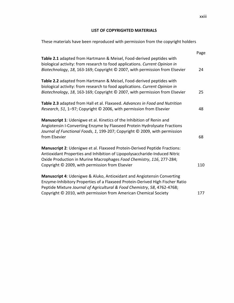
Page Table 2.1 adapted from Hartmann & Meisel, Food-derived peptides with biological activity: from research to food applications. Current Opinion in Biotechnology, 18, 163-169; Copyright © 2007, with permission from Elsevier
24
Table 2.2 adapted from Hartmann & Meisel, Food-derived peptides with biological activity: from research to food applications. Current Opinion in Biotechnology, 18, 163-169; Copyright © 2007, with permission from Elsevier
25
Table 2.3 adapted from Hall et al. Flaxseed. Advances in Food and Nutrition Research, 51, 1–97; Copyright © 2006, with permission from Elsevier
48
Manuscript 1: Udenigwe et al. Kinetics of the Inhibition of Renin and Angiotensin I-Converting Enzyme by Flaxseed Protein Hydrolysate Fractions Journal of Functional Foods, 1, 199-207; Copyright © 2009, with permission from Elsevier
68
Manuscript 2: Udenigwe et al. Flaxseed Protein-Derived Peptide Fractions: Antioxidant Properties and Inhibition of Lipopolysaccharide-Induced Nitric Oxide Production in Murine Macrophages Food Chemistry, 116, 277-284; Copyright © 2009, with permission from Elsevier
110
Manuscript 4: Udenigwe & Aluko, Antioxidant and Angiotensin Converting Enzyme-Inhibitory Properties of a Flaxseed Protein-Derived High Fischer Ratio Peptide Mixture Journal of Agricultural & Food Chemistry, 58, 4762-4768; Copyright © 2010, with permission from American Chemical Society
177

xxiv
LIST OF ABBREVIATIONS
AAA Aromatic amino acids
ACE Angiotensin I-converting enzyme
AEM Anion exchange membrane
AT-I Angiotensin I
AT-II Angiotensin II
BCAA Branched-chain amino acids
BW Body weight
CaM Calmodulin
CaMPDE Calmodulin-dependent phosphodiesterase
CE Catalytic efficiency of enzymes
CEM Cation exchange membrane
DPPH 2,2-diphenyl-1-picrylhydrazyl
DWB Dry weight basis
EC 50% Effective concentration 50
EDUF Electrodialysis-ultrafiltration
E/S Enzyme-substrate ratio
ESR Electron spin resonance
FAPGG N-(3-[2-furyl]acryloyl)-phenylalanylglycylglycine
FIU Flourescence Intensity Unit
FPI Flaxseed protein isolate
FPH Flaxseed protein hydrolysates

xxv
FPLC Fast Protein Liquid Chromatography
GSH Glutathione
HMW High molecular weight
HPLC High Performance Liquid Chromatography
IC 50% Inhibitory concentration 50
K Enzyme-inhibitor dissociation constant i
K Michaelis constant or enzyme-substrate dissociation constant m
Km′, Km Apparent Michaelis constant app
KNOS Kinin-nitric oxide system
LA Linoleic acid
LMW Low molecular weight
LPS Lipopolysaccharide
MW Molecular weight
MWCO Molecular weight cut-off
NO Nitric oxide
˙OH Hydroxyl radical
O2˙ Superoxide radical –
ONOO Peroxynitrite –
PDE Phosphodiesterase
PLS Partial least square projection of latent structure
PMB Polymyxin B
QSAR Quantitative structure-activity relationship

xxvi
RAS Renin-angiotensin system
RAAS Renin-angiotensin-aldosterone system
ROS Reactive oxygen species
RLU Relative Luminescence Unit
SFR Structure-function relationship
SHR Spontaneously hypertensive rats
SNP Sodium nitroprusside
SSAO Semicarbazide-sensitive amine oxidase
UFM Ultrafiltration membrane
V Maximum enzyme reaction rate max
Vmax′, Vmax Maximum apparent enzyme reaction rate app

1
CHAPTER ONE
GENERAL INTRODUCTION
The human body is constantly subjected to physiological imbalances and exposure
to extrinsic toxic substances that perturb the normal functioning of the system leading
to various health conditions, which can be controlled by physiological homeostasis or
through the use of therapeutic agents especially in acute and chronic conditions. It is
generally established that the nutritive and non-nutritive constituents of human diets
can be used to modify the risk of developing or aggravating human disease conditions
(Health Canada, 1998). In this regard, functional foods have emerged during the last two
decades as adjuvant or alternative to chemotherapy especially in prevention and
management of human diseases, and for maintaining optimum human health state.
However, the positive effects of functional foods may not be observed in acute health
conditions following short-term consumption since the levels of the active ingredients
are often below the therapeutic amounts, or not readily biologically available due to
complex interactions with polysaccharides and proteins of the food matrix (Manach et
al., 2004; Parada & Aguilera, 2007). As a result, bioactive ingredients present in
functional foods can be isolated and administered as pills to increase the amount of
these molecules that are absorbed into blood circulation and transported to cellular
targets. These bioactive ingredients are known as nutraceuticals, which were formally
defined by Health Canada (1998) as: “...products isolated or purified from foods that are
generally sold in medicinal forms not usually associated with food. Nutraceuticals are
demonstrated to have physiological benefits or provide protection against chronic

2
diseases”. Nutraceuticals and bioactive compounds-enriched food products have
increasingly become subject of various R&D programs, as the health and well-being of
consumers gradually became the primary focus of the food industry. The growing
market for functional foods and natural health products can be attributed to the
consumers’ understanding of the relationship between diet and disease, safety of the
natural health products and low cost associated with the bioactive molecules compared
to chemotherapeutic agents or drugs (Agriculture and Agri-Food Canada, 2010).
In Canada, the functional foods and nutraceuticals sector has experienced
substantial growth during the last ten years. According to Statistics Canada, a total of
$2.9 billion was reported in annual sales of functional foods and nutraceutical products
by 389 firms in Canada during 2003-2004 (Palinic, 2007); this represents a substantial
increase compared to the 294 firms earlier reported for 2002 (Tebbens, 2005). In 2007,
the number of functional foods and natural health products firms in Canada increased
to 689 with annual revenues of $3.7 billion; these firms produced a variety of products
for Canada and international markets (Cinnamon, 2009). Many of these firms are
recognized worldwide for their bioactive products such as flax and fish-derived omega-3
fatty acids, plant sterols and stanols, soluble fibres, soy proteins, prebiotics and
probiotics (Agriculture and Agri-Food Canada, 2010). In Canada and other parts of the
world, another growing trend in the functional foods and nutraceuticals sector is the use
of food protein-derived peptides for intervention against chronic human disease
conditions and for maintenance of general well-being. This approach involves enzymatic
hydrolysis or fermentation of food proteins to release bioactive peptide sequences

3
followed by simple or complex post-hydrolysis processing to isolate the bioactive
peptides from the complex mixture of other inactive molecules; bioactivity of the
peptides represent physiologically-relevant beneficial properties in the human system
beyond normal and adequate nutrition (Hartmann & Meisel, 2007). The food processing
steps lead to concentration of the active peptides with the enhancement of the
physiological activity of the products, which could also be nutritionally beneficial as a
source of essential amino acids. This approach can provide the opportunity for
diversification of the use of Canada’s major protein-rich agricultural crops, especially
low value crop products, beyond basic nutritional purposes.
Flax (Linum usitatissimum L.) is a major oilseed crop cultivated commercially in
Canada, a leading world producer and exporter of flaxseed (Oomah & Mazza, 1993;
Agriculture and Agri-Food Canada, 2007). Most of the nutritional and health benefits
attributed to flaxseed are due to its omega-3 fatty acid (α-linolenic acid) content, which
is extracted from the crushed oilseed by pressing or use of solvent extraction (Hall et al.,
2006). The by-product of the flaxseed oil extraction process is known as defatted
flaxseed meal, and this contains large amounts of dietary fibre, lignans and proteins.
Both flaxseed-derived dietary fibres and lignans possess human health benefits (Hall et
al., 2006). In 2006-2007, the world flaxseed meal production was estimated at 1.4
million tonnes, and Canada generated annual revenue of $2.5 million from exporting
flaxseed meal (Agriculture and Agri-Food Canada, 2007). Moreover, the Canadian
flaxseed from 2009 harvest was reported to contain 22% protein with a 10-year mean
value of 23.2% protein (Canadian Grain Commission, 2009). Thus, there is a worldwide
4
production of about 0.31 million tonnes of flaxseed proteins per annum. This large
amount of flaxseed proteins has not been optimally utilized in human food systems,
although flaxseed meal has been incorporated into livestock feeds as protein and fibre
supplement (Agriculture and Agri-Food Canada, 2007; Bell & Keith, 1993). The use of
flaxseed meal as feed supplement, and perhaps as protein source in human, has been
hindered due to its constituent anti-nutritional factors especially cyanogenic glycosides
(Oomah & Mazza, 1993), but the development of advanced processing techniques could
encourage removal of these toxic compounds to enhance value of the oilseed meal.
Compared to other food proteins, flaxseed proteins possess rich amino acid profiles
with a high amount of branched-chain and cationic amino acids (Oomah & Mazza, 1995;
Hall et al., 2006). The abundance of low-value flaxseed meal that is underutilized in
human food system underscores the need for conversion of defatted flaxseed meal into
value-added products, especially bioactive peptides, for increased human health and
economic benefits.
Food protein hydrolysates and purified food-derived peptides have exhibited
several in vitro and in vivo physiologically-relevant activities such as antihypertensive,
antioxidant, immunomodulatory, antimicrobial and lipid-lowering activities (Erdmann et
al., 2008; Aluko, 2008a, b; Meisel, 2004; Pihlanto, 2006). Plant proteins are preferred
over animal proteins as source of bioactive peptides because plant proteins are cheaper
and more abundant for large scale peptide production (Aluko, 2008a). The specific
bioactivity of food peptides depends primarily on their structural properties such as
chain length and physicochemical characteristics of the amino acid residues, e.g.
5
hydrophobicity, molecular charge and side chain bulkiness (Pripp et al., 2004a).
Therefore, in-depth knowledge about the structure-function property of peptides can
contribute towards the discovery of more potent peptides and design of appropriate
processing conditions to liberate these peptides from their parent proteins. Previous
peptide quantitative structure-activity relationship (QSAR) studies have focused mainly
on peptide inhibitors of angiotensin I-converting enzyme (ACE), a class of
antihypertensive agents, resulting in the discovery of peptides with more potent
activities (Pripp et al., 2004b, 2005; Pripp, 2005; Wu et al., 2006a,b; Shu et al., 2009).
ACE and renin are key metabolic enzymes of the blood pressure regulating renin-
angiotensin system (RAS). Unlike ACE-inhibiting peptides, there is dearth of literature
information on the inhibition of renin by food-derived peptides as well as their
structural requirements for potency. This information would be beneficial towards
antihypertensive therapy because renin catalyzes the rate-limiting step of the RAS
pathway, and its inhibition can potentially provide better control over elevated blood
pressure than the inhibition of only ACE activity. Moreover, peptides with more than
one (multifunctional) bioactive property such as ACE/renin inhibitors and antioxidants
can be beneficial in diseases with multiple symptoms such as cardiovascular disease.
In this thesis, it was hypothesized, based on the amino acid profile of flaxseed
meal proteins, that controlled enzymatic hydrolysis could generate structurally distinct
peptides with physiologically-relevant multifunctional bioactive properties. Specifically,
it was hypothesized that:

6
(1) Low molecular weight peptides derived from flaxseed proteins would possess
multifunctional bioactive properties as antioxidants, anti-inflammatory and
antihypertensive agents;
(2) Flaxseed peptides with net positive charge (cationic peptides) would bind the
negatively charged calmodulin (CaM) leading to the inhibition of CaM-dependent
phosphodiesterase (CaMPDE), which has been implicated in chronic human
diseases;
(3) Branched-chain amino acid-enriched peptide product could be generated from
flaxseed protein through enzymatic treatments and simple cost-effective
processing;
(4) Arginine-enriched low molecular weight peptide product could be generated from
flaxseed protein, and this product, when efficiently absorbed, could serve as a
precursor of nitric oxide, which possesses physiological vasodilatory effect and
could be used to lower blood pressure during hypertension;
(5) The sequences and structural characteristics of amino acid residues in bioactive
peptides determine their in vitro renin-inhibitory activities.
Based on these hypotheses, the general objectives of this project were to: a)
produce bioactive peptides from flaxseed proteins as means of increasing the value-
added utilization of defatted flaxseed meal, which will provide economic benefits to
Canada and oilseed processing industry; and b) study the structure-function properties
of low molecular weight renin inhibiting peptides. The specific objectives of this project
were to:

7
(1) Optimize the isolation of flaxseed proteins from defatted flaxseed meal and
generate various functional enzymatic hydrolysates from these proteins under
controlled conditions;
(2) Determine kinetics of the inhibition of ACE, renin and CaMPDE by the protein
hydrolysates as well as their ability to scavenge free radicals and inhibit nitric
oxide production in activated macrophages;
(3) Fractionate low molecular weight peptides that can simultaneously inhibit ACE,
renin and CaMPDE activities from the flaxseed protein hydrolysates using
chromatographic techniques;
(4) Study the structure-function properties of renin inhibiting natural dipeptides by
developing QSAR models using the partial least squares (PLS) regression method;
(5) Design a cheap and efficient process for the production of high Fischer ratio
peptide mixture from flaxseed protein isolate and evaluation of the
multifunctional bioactive properties of this product;
(6) Use electrodialysis-ultrafiltration, a novel membrane technology, to separate
arginine-rich peptides from flaxseed protein hydrolysates and evaluate the
peptide product for in vitro (inhibition of ACE and renin) and in vivo
(antihypertensive activity in spontaneously hypertensive rats, SHR) activities.
Therefore, findings from this study could contribute towards value-added
utilization of defatted flaxseed meal, which is an abundant by-product of Canada’s
oilseed processing industry. Results from this work can also provide fundamental
information on the structural requirements for renin inhibition by food peptides, which

8
can serve as template for the design and synthesis of more potent peptide and
peptidomimetic antihypertensive agents.

9
CHAPTER TWO
LITERATURE REVIEW
2.1. Hypertension and the renin-angiotensin system (RAS)
Hypertension or sustained elevated blood pressure is a medical condition
regarded as a controllable risk factor in the development of cardiovascular,
cerebrovascular and other vascular diseases such as stroke, myocardial infarction, left
ventricular and smooth muscle cell hypertrophy (Kannel, 1996). Although not a disease,
hypertension has emerged as a global public health concern that represents a major
cause of death worldwide. A World Health Organization (WHO) report estimated that
7.5 million premature deaths worldwide were due to hypertension, leading other risks
with a total contribution of 12.8% deaths (WHO, 2009). In 2000, 972 million people
worldwide had hypertension and this number was projected to increase to 1.56 billion
in 2025 (Kearney et al., 2005). In U.S., about 31% and 25% of adults are hypertensive
and pre-hypertensive, respectively (National Centre for Health Statistics, 2008; Lloyd-
Jones et al., 2010) and it was estimated that over $76 billion (US) will be spent in 2010 in
healthcare services, medication and other costs associated with hypertension (Lloyd-
Jones et al., 2010). The Canadian Health Measures Survey completed between 2007 and
2009 reported that 19% of the Canadian adult population have hypertension (Wilkins et
al., 2010). Therefore, hypertension has attracted substantial research interests
especially towards the discovery of therapeutic agents and lifestyle modification
approaches to reduce its prevalence. The latter strategy, also known as dietary
approaches to stop hypertension (DASH), has been reported to be successful in lowering

10
blood pressure in hypertensive patients (Colin et al., 2000) while therapeutic agents are
primarily used to target blood pressure-regulating pathways.
Blood pressure is physiologically controlled by the renin-angiotensin system
(RAS) and the kinin-nitric oxide system (KNOS) as shown in Figure 2.1. The RAS involves
activation of angiotensinogen, a zymogen, by the proteolytic activity of renin, an
aspartate protease, which converts it to angiotensin (AT)-I. This reaction is the first and
rate-limiting step of the RAS pathway. AT-I is then cleaved at the histidyl residue from
the C-terminus by the activity of angiotensin-I converting enzyme (ACE) to produce AT-
II. AT-II is a powerful vasoconstrictor that functions by binding to receptors, which are
located in tissues all over the body. Interactions of AT-II with receptors will elicit
physiological reaction cascades that lead to blood vessel contractions that maintain
normal blood pressure. However, in pathological conditions, there is excessive level of
AT-II, which causes severe blood vessel contractions and limited relaxation to produce
high blood pressure. The KNOS system is involved in the production of bradykinin, which
exerts its antihypertensive effects by eliciting reactions that increase intracellular
Ca2+concentration leading to activation of nitric oxide synthases (NOS) that produce
nitric oxide (NO), a powerful vasodilator. ACE degrades bradykinin, and increased
concentration of ACE leads to dual effects, the prevention of vasodilation and activation
of vasoconstriction (Figure 2.1). Based on the roles of ACE in the RAS pathway, inhibitors
of this enzyme have been used as antihypertensive agents (Ibrahim, 2006). Moreover,
direct inhibition of renin can potentially provide better control of elevated blood
pressure than ACE inhibition since it prevents the synthesis of AT-I, which can be

11
converted to AT-II in some tissues via an ACE-independent alternative chymase-
catalyzed pathway (Segall et al., 2007). However, inhibition of renin activity does not
prevent ACE-catalyzed bradykinin degradation (Staessen et al., 2006). Thus, there is a
need to develop therapeutic agents that could provide multiple effects as ACE and renin
inhibitors.
Figure 2.1 The renin-angiotensin system (RAS) showing the molecular targets for
antihypertensive agents; AT-I, angiotensin I; AT-II, angiotensin II; ACE, angiotensin I-
converting enzyme
Angiotensinogen
AT-I
AT-II
AT1, AT2 receptors
Vasoconstriction
Kidney
Bradykinin
Inactive peptides
Chymase
Asp-Arg-Val-Tyr-Ile-His-Pro-Phe-His-Leu-Leu
Asp-Arg-Val-Tyr-Ile-His-Pro-Phe-His-Leu
Asp-Arg-Val-Tyr-Ile-His-Pro-Phe
Arg- Pro-Pro Gly-Phe-Ser-Pro-Phe-Arg
ACE inhibitors
Renin inhibitors
β blockers
Angiotensin Receptor Blockers
(ARB)
Vasodilation (Nitric oxide-mediated)
↓Blood Pressure
↑Blood Pressure
ACE
Renin
ACE
Arg- Pro-Pro Gly-Phe + Ser-Pro-Phe-Arg

12
2.2. Reactive oxygen species and free radicals in human health conditions
2.2.1. Inflammatory responses
In cells, reactive oxygen species (ROS) and free radicals are constantly produced
as by-products of cellular processes, secondary messengers or agents of immune
response reactions (Halliwell, 1991; Turrens, 2003). The human body is also exposed to
exogenous ROS/free radicals from diets and the atmosphere, and to microbial
pathogens that can induce overproduction of ROS (Halliwell, 1991). Upon exposure to
microorganisms or endotoxins, the immune system becomes activated and phagocytic
cells (e.g. macrophages, monocytes and neutrophils) release several pro-inflammatory
cytokines leading to activation of NF-κB transcription factor and subsequent up-
regulation of the production of enormous amounts of ROS to combat the invading
pathogens and protect cells from infection (Dinarello, 2000). Figure 2.2 shows the
production of ROS, their involvement in cellular damages and the role of endogenous
antioxidant system in maintaining normal homeostasis. Upon binding of endotoxins to
surface receptors, phagocytic immune cells produce ROS by activation of the NADPH
oxidase complex during phagocytosis (Heyworth et al., 1999). Activated NADPH oxidase
catalyzes the reduction of molecular oxygen to superoxide radical (O2˙¯). The amount of
O2˙¯, also produced due to electron leakage in the mitochondria, can be controlled by
superoxide dismutase (SOD), which converts O2˙¯ to hydrogen peroxide (H2O2) and
oxygen. H2O2 is in turn detoxified to water and oxygen by catalase or glutathione
peroxidase, but can also be converted to highly reactive hydroxyl radical (˙OH) through
the transition metal ion (e.g. Fe2+)-mediated Fenton reaction, or to other powerful

13
oxidants, such as hypochlorous acid and singlet oxygen (1O2
), by the action of
myeloperoxidase (Klebanoff, 1999; Turrens, 2003).
Figure 2.2 Production of ROS/free radicals and oxidative stress-mediated oxidative
damage to biological macromolecules
In addition, immune activation involves cytokine-mediated activation of the
expression of inducible nitric oxide synthase (iNOS or NOS2), which catalyzes the
production of nitric oxide (NO) from arginine and molecular oxygen, and cycloxygenase-
Mitochondria Electron Transport Mn-SOD
O2 O2˙¯ H2O2
O2˙¯ H2O2
HO˙
ONOO¯ Protein/ lipid/DNA oxidation
Xanthine oxidase
Fenton reaction
Fe2+
O2 Catalase
iNOS
Cu/Zn-SOD
O2
H2O + O2 NADPH oxidase
GSSG GSH
GPx
GR
SOD, superoxide dismutase; GPx, glutathione peroxidase; GR, glutathione reductase GSH, glutathione (reduced); GSSH, glutathione (oxidized); iNOS, inducible NOS
Myelo- peroxidase
1O2
Endotoxin-induced immune activation
NO
O2˙¯
Induces apoptosis

14
2 (COX-2), which produces proinflammatory prostaglandin E2 from unsaturated fatty
acids (Moncada & Higgs, 1993; Pacher et al., 2007). NO is beneficial in signal
transduction and vasodilation, but can react with the abundant O2˙¯ to form highly
reactive peroxynitrite (ONOO¯) that causes most cellular damages attributed to NO
(Pacher et al., 2007); these N-containing oxidants are also referred to as reactive
nitrogen species (Turrens, 2003). These oxidative processes are tightly controlled by the
endogenous antioxidant system, but when produced in large amounts due to chronic or
acute immune activation, ROS can overwhelm the endogenous redox regulatory system
leading to cellular oxidative damages such as degradation of membrane lipids (loss of
membrane integrity), DNA fragmentation, protein modification (e.g., tyrosine nitration)
that causes loss of function and activation of the cysteine proteases (caspases) of the
cell death pathway, resulting in apoptosis or programmed cell death (Ames, 1983; Ames
et al., 1993; Wiseman & Halliwell, 1996; Pacher et al., 2007). The condition whereby
ROS are over-produced in cells is known as “oxidative stress” or “respiratory burst”, to
reflect the increased cellular oxygen consumption associated with the process.
Oxidative stress plays important roles in the aetiology and progression of human
physiological and disease conditions (Ames, 1983; Ames et al., 1993; Pacher et al., 2007;
Torres et al., 2002; Liu et al., 2002). Several studies have established that ROS/free
radicals contribute to initiation, promotion and progression of cancer by causing DNA
damage leading to the inactivation of proto-oncogenes or tumour-suppressor genes,
and modification of other genes and proteins involved in cell proliferation,
differentiation and apoptosis (Wiseman & Halliwell, 1996). In addition, excessive ROS
15
production could trigger inflammatory disease-related tissue damage (Liu et al., 2002)
and pathogenesis of human immunodeficiency virus (HIV)-1 infection (Torre et al.,
2002). Oxidative stress can be surmounted by the consumption of dietary or drug
antioxidants to support the overwhelmed endogenous antioxidant system (Ames et al.,
1993; Halliwell, 1996; Wiseman & Halliwell, 1996).
2.2.2. Neurodegenerative diseases
ROS and free radicals also play important pathophysiological roles in aetiology
and progression of neurodegenerative diseases (Figure 2.3). A number of toxins and
pathogens, such as bacteria, fungi and viruses, are capable of invading the brain cells
through blood circulation, and elicit cellular immune responses. Of the 3 major resident
cells involved in the phagocytosis and clearance of the invading pathogens, the microglia
plays a crucial role as macrophages in the central nervous system (CNS). Microglia
constitutes about 15% of all the cells of the CNS and serves as immune cells in the
defence of brain cells (Rock et al., 2004; Carson et al., 2008). Upon activation, the
microglia releases several pro-inflammatory cytokines that initiate the oxidative
process. It has been shown that, in the glial cells, TNF-α, IL-1β and interferon-γ are the
most potent activators of the expression of iNOS and COX-2 genes (Hunot & Hirsch,
2003), whose protein products catalyze the production of nitric oxide and pro-
inflammatory prostaglandins, respectively; iNOS is not normally produced in brain cells
(Murphy, 2000).

16
Figure 2.3 Production of ROS and free radicals in the brain and their roles in oxidative
damages to cellular macromolecules leading to neuroinflammation and neuronal cell
death during neurodegenerative diseases
Other enzymes induced by this process in the brain immune cells include NADPH
oxidase and myeloperoxidase, leading to excessive production of O2˙¯ and hypochlorous
acid, respectively. These reactive oxygen species are utilized by the macrophage-like
cells in attacking the pathogens. Nitric oxide is less damaging by itself but reacts with
O2
Neurotoxin
˙¯, which is also excessively produced by the macrophages due to mitochondrial
Mitochondrial electron leakage
O2˙¯ H2O2 HO˙
ONOO¯
NO
Activated microglia
Protein oxidation (amyloid-β, α-synuclein)
Lipid and DNA oxidation
Neuronal Cell Death (Necrosis or Apoptosis)
Superoxide dismutase
Fenton reaction
Fe2+
H2O + O2
Catalase
Neuroinflammation
iNOS
O2˙¯
NO
Immune Activation

17
electron leakage, to form ONOO¯ that causes damage to biomacromolecules including
DNA in glial cells and peripheral dopaminergic neurons (Hunot & Hirsch, 2003; Pacher et
al., 2007). Superoxide radical could also cause direct damage to cells by reacting with
polyunsaturated fatty acid components of the biological membrane lipid bi-layer.
Endogenous antioxidant superoxide dismutase utilizes O2˙¯ to produce H2O2
, with
concomitant production of ˙OH through the Fenton reaction. These oxidative processes
are tightly regulated by the endogenous antioxidant system but can be exacerbated in
chronic and acute infection, and other pathological conditions, leading to neuronal cell
damages associated with diseases such as Alzheimer’s and Parkinson’s (Hirsch & Hunot,
2009). Moreover, the brain cells are particularly susceptible to oxidative damage due to
its high oxygen consumption (Halliwell, 2001). Overall, oxidative burst and inflammation
in neuronal cells contribute to pathogenesis in neurodegenerative diseases (Ames et al.,
1993). Thus, it is important to develop therapeutic agents that can reduce oxidative
pathogenesis associated with these diseases by scavenging these reactive species,
inhibiting their production, or by decreasing the levels of the pro-inflammatory
cytokines released during microglial activation. According to Halliwell (2001), it is
important that the reactive species targeted by the therapeutic agent should have a
direct relevance in the disease process, and must be present at the site of injury; thus,
all the aforementioned ROS play important roles in propagating neuronal cell damages
(Hirsch & Hunot, 2009).

18
2.2.3. Oxidative stress, inflammation and hypertension
Hypertension is proinflammatory. Elevated levels of AT-II during hypertension
lead to phospholipase C activation followed by increase in intracellular Ca2+
concentration (causes smooth muscle (SM) contraction and hypertrophy), and increased
SM lipoxygenase activity, which increases oxidation of low density lipoproteins (LDL)
(Ross, 1999). The oxidized LDL (LDL-oxd) in the arterial intima is taken up by scavenger
receptors of macrophages and accumulation of cholesterol causes macrophage
transformation into foam cells (Hansson, 2005). LDL-oxd activates inflammation by
triggering endothelial cells to express vascular cell adhesion molecules that bind
monocytes and T-lymphocytes; the immune cells attracted to the site also express
inflammatory genes (Ross, 1999; Hansson, 2005). Activated macrophages overproduce
ROS, which react with NO to decrease its availability in the vascular endothelium leading
to increased leukocyte adhesion to the endothelium (Ross, 1999). These processes
increase peripheral vascular resistance and blood pressure, which ultimately increases
the risk of cardiovascular disease.
2.3. Calmodulin (CaM) and CaM-dependent enzymes
Calmodulin (CaM) is a ubiquitous negatively-charged 148-amino acid (16.6 kDa)
Ca2+-binding protein that is involved in the activation of several important proteins in
response to increased intracellular Ca2+ concentration (Ikura et al., 1992; Hooks &
Means, 2001). Instead of having multiple Ca2+-sensing proteins in several cells, CaM has
evolved to serve as mediator for detecting Ca2+ signals and activating target proteins in

19
several cells (Lakowski et al., 2007). The activation process involves binding of Ca2+-free
CaM (apo-CaM) to Ca2+, which induces conformational changes that expose a
methionine-rich hydrophobic region on CaM structure for binding a short peptide
sequence of target proteins with high affinity (Crivici & Ikura, 1995; Schauer-Vukasinovic
et al., 1997). Some clinically important enzymes that require Ca2+/CaM activation
include endothelial (eNOS) and neuronal (nNOS) nitric oxide synthase, cyclic nucleotide
phosphodiesterase 1 (CaMPDE), adenosine triphosphatase, phospholipase A2
A total of 11 PDE families (PDE1-11) have been identified in human. CaMPDE is
the isoform from the PDE1 gene family, which gives three distinct gene products namely
PDE1A, PDE1B and PDE1C; they are expressed in various cell types including coronary
vascular smooth muscle cells (SMC) where CaMPDE play a role in the regulation of
vascular tone (Bender & Beavo, 2006; Kass et al., 2007). CaMPDEs possess two binding
sites for Ca
, adenylate
cyclase and protein kinase II (Itano et al., 1980). Thus, CaM plays important roles in
several cellular processes including cell growth, cell proliferation, neurotransmission and
smooth muscle contraction (Cho et al., 1998).
2+/CaM complex, and complete binding is required for up to 10-fold or more
activation of the enzyme (Rybalkin et al., 2003). As shown in Figure 2.4, CaMPDE
catalyzes the conversion of cyclic nucleotide monophosphates (PDE1A/B/C for cGMP
and PDE1C for cAMP) to nucleotide monophosphates (GMP and AMP), thereby
modulating cellular processes mediated by the second messengers (cAMP/cGMP)
including neurotransmission, immune cell activity, vasodilation, cell growth and
proliferation (Cho et al., 1998). Chronic over-expression of CaMPDE has been found in

20
atherosclerosis, heart pressure-load stress, heart failure and vascular proliferation (Kass
et al., 2007). Separate inhibitors of PDE1A and PDE1C expressions and activity in
cultured cells has been reported to decrease PDE1-induced proliferation of human SMC,
which is important towards treatment of atherosclerosis, cancer and other responses
due to SMC proliferation (Rybalkin et al., 2003; Nagel et al., 2006). Moreover, inhibition
of PDE1B expression and activity inhibited proliferation and also induced cAMP-
mediated apoptosis in cultured human leukemic cells, which is favourable for cancer
chemotherapy since normal blood cells do not express PDE1B (Jiang et al., 1996). In
addition, AT-II increases PDE1A1 isoform expressions in SMC through AT-II-mediated
increase in cellular Ca2+
One approach towards reducing these negative effects of CaMPDE include the
use of Ca
concentration (Kim et al., 2001). Thus, CaMPDE-induced
pathogenesis can also be linked to RAS-mediated hypertension; CaMPDE has been
implicated in pulmonary arterial hypertension (Schermuly et al., 2007). PDE1B2
expression and activity are also upregulated during monocyte differentiation, and
contributes to pathogenesis during inflammation, which makes CaMPDE a good target
for anti-inflammation therapy (Bender et al., 2005).
2+ chelating agents to prevent CaM activation thereby decreasing CaMPDE
activity; however, specificity will not be achieved since Ca2+ is involved in numerous
cellular processes. Another approach is the use of CaM-binding agents, which can
specifically regulate the activity of only CaM-dependent PDE isoforms and other CaM-
dependent enzymes. Thus, CaM-binding compounds can be used as therapeutic agents
against diseases induced or exacerbated by increased CaM or CaMPDE expression. Some

21
known potent chemical inhibitors of CaMPDE include vinpocetine, KS-505a, IC224,
SCH51866, W-7 and phenothiazines (Bender & Beavo, 2006). Several natural products
and synthetic peptides have also displayed CaM inhibition (Martínez-Luis et al., 2007).
Details on CaM-binding natural peptides can be found in Section 2.4.6.
Figure 2.4 The role of Ca2+
/calmodulin-dependent phosphodiesterase (CaMPDE) in the
regulation of cellular processes
GP
H
GTP
R
GC
cGMP GMP
Ca2+/CaMPDE (PDE1A/B/C)
AC
H
ATP
R
GP
cAMP
Ca2+/CaMPDE (PDE1C)
Cellular Response Glycogenolysis
Vasodilation Neurotransmission Immune functions
AMP
-P PR
Active
PR
Inactive
-P PR
Active
PR
Inactive
PKG PKG
Inactive Active
Cellular Response Glycogenolysis Brochodilation
Vasodilation SM relaxation
H, hormone; GP, G protein; R, GP-coupled receptors; GC, guanylate cyclase; AC, adenylate cyclase; ATP, adenosine triphosphate; GTP, guanosine triphosphate; PKG, cGMP-dependent protein kinase; PKA, cAMP-dependent protein kinase; PR, target protein; SM, smooth muscles
PKA PKA
Inactive Active
22
2.4. Food protein-derived bioactive peptides in human health
Food protein hydrolysates have been shown to exhibit potent biological activities
(Figure 2.5), which are largely due to their constituent peptides. These peptides are
encrypted in the primary structure of plant and animal proteins as inactive amino acid
sequences but they can be released by fermentation, food processing and enzyme-
catalyzed proteolysis in vitro or in vivo after human consumption (Li et al., 2004;
Hartmann & Meisel, 2007; Aluko, 2008b). In most cases, these protein hydrolysates and
peptides have demonstrated better bioactivities (Figure 2.5) compared to their parent
proteins, and this shows that hydrolysis of peptide bonds is important in liberating the
potent peptides. Several factors affect the bioactive properties of the peptides including
the enzymes used for hydrolysis, processing conditions and the size of the resulting
peptides, which greatly affects their absorption across the enterocytes and
bioavailability in target tissues. Generally, the activity of these peptides against
molecular disease targets are regarded as lower than synthetic peptidomimetics and
drugs, but the use of bioactive peptides in intervention against human diseases offers
many advantages including safety of the natural product, low health cost and the
additional nutritional benefits of the peptides as source of beneficial amino acids. The
current literature contains a vast amount of information on food protein-derived
bioactive peptides with physiologically-relevant bioactive properties including ACE-
inhibiting/antihypertensive, anticancer or cytomodulatory, antioxidant, opioid-like,
hypocholesterolemic, immunomodulatory, antimicrobial and mineral-binding activities
(Table 2.1). These peptides are mostly derived from milk and soy proteins, and range in

23
sizes from di-, tri- and oligopeptides to high molecular weight (HMW) polypeptides
(Hartman & Meisel; 2007; Erdmann et al., 2008; Hernández-Ledesma et al., 2009a).
Based on these bioactivities, several food-derived peptide products have been
developed and commercialized for use as human health promoting agents as shown in
Table 2.2. Details on the biological properties of some of the food-derived peptides are
discussed in the following sub-sections.
Figure 2.5 Bioactive properties of food protein-derived peptides relevant to the
promotion of human health and disease prevention
Food Protein-Derived Peptides
Antioxidant Activities
Anticancer Properties
Anti-inflammatory Properties
Immunomodulatory Properties
Antimicrobial Activity
Lipid-lowering Properties
Antihypertensive Activity
Liver disease treatment (BCAA-enriched peptides)
Multifunctional Properties
24
Table 2.1 Examples of bioactive peptide-based products that are derived from foods
Effects
a
Origin Encrypting protein(s) Name/remarks/sequence (in single-letter code) ACE-inhibitory/hypotensive Soy Soy protein NWGPLV Fish Fish muscle protein LKP, IKP, LRP (derived from sardine, bonito, tuna, squid) Meat Meat muscle protein IKW, LKP Milk α-LA, β-LG
α-, β-, κ-CN Lactokinins (e.g. WLAHK, LRP, LKP) Casokinins (e.g. FFVAP, FALPQY, VPP)
Egg Ovotransferrin Ovalbumin
KVREGTTY Ovokinin (FRADHPPL), Ovokinin (2–7) (KVREGTTY)
Wheat Wheat gliadin IAP Broccoli Plant protein YPK Immunomodulatory Rice Rice albumin Oryzatensin (GYPMYPLR) Egg Ovalbumin Peptides not specified Milk α-, β-, κ-CN, α-LA Immunopeptides (e.g. αS1-immunocasokinin) (TTMPLW Wheat Wheat gluten Immunopeptides Cytomodulatory Milk α-, β-CN α-Casomorphin (HIQKED(V)), β-casomorphin-7 (YPFPGPI) Opioid agonist Milk Lactoferrin Lactoferroxins κ-CN Casoxins Antimicrobial Egg Ovotransferrin
Lysozyme OTAP-92 (f109–200) Peptides not specified
Milk Lactoferrin Lactoferricin Antithrombotic Milk κ-CN (glycomacropeptide) κ-CN (f106–116), casoplatelin Mineral-binding, anticarcinogenic
Milk α-, β-CN Caseinophosphopeptides
Hypocholesterolemic Soy Glycinin LPYPR Milk β-LG IIAEK Fish Sardine muscle MY Wheat Wheat germ protein Peptides not specified Milk α-LA, β-LG MHIRL, YVEEL, WYSLAMAASDI aTable adapted from Hartman & Meisel (2007) with permission from Elsevier; CN, casein; LA, lactalbumin; LG, lactoglobulin

25
Table 2.2 Examples of commercially available functional foods or food ingredients carrying bioactive peptides
Product name
a
Manufacturer Type of food Bioactive peptides Health claim Calpis AMEEL S (Japan) or Calpico (Europe)
Calpis Co., Japan Sour milk VPP, IPP from β- and κ-CN Hypotensive Hypotensive Hypotensive Hypotensive Hypotensive Hypotensive
Evolus Valio, Finland Fermented milk, calcium-enriched
VPP, IPP from β- and κ-CN
BioZate Davisco, USA β-LG hydrolysate Whey peptides C12 Peption DMV, Netherlands Ingredient Casein-derived dodecapeptide
FFVAPFPEVFGK Peptide Soup NIPPON, Japan Soup Bonito-derived peptides Casein DP Peptio Drink Kanebo, Japan Soft drink Casein-derived dodecapeptide
FFVAPFPEVFGK BioPURE-GMP Davisco, USA Whey protein hydrolysate Glycomacropeptide Anticarcinogenic,
antimicrobial, antithrombotic
CholesteBlock Kyowa Hakko, Japan Drink powder Soy peptides bound to
Phospholipids Hypocholesterolemic
CSPHP ProDiet F200
Ingredia, France Milk drink, confectionery αS1-CN (f91–100), YLGYLEQLLR
Reduces stress
Capolac Arla Foods, Denmark Ingredient CPP (caseinophosphopeptide) Helps mineral
absorption Tekkotsu Inryou Suntory, Japan Soft drink CPP Kotsu Kotsu calcium Asahi, Japan Soft drink CPP CE90CPP DMV, Netherlands Ingredient CPP (20%) Glutamine Peption DMV, Netherlands Dry milk protein hydrolysate Glutamine-rich peptides Immunomodulatory WGE80GPA WGE80GPN WGE80GPU aTable adapted from Hartman & Meisel (2007) with permission from Elsevier; CN, casein; LA, lactalbumin; LG, lactoglobulin

26
2.4.1. ACE-inhibitory and antihypertensive peptides
The pioneering work on naturally occurring ACE-inhibiting antihypertensive
peptides from snake (Bothrops jararaca) venom (Ferreira et al., 1970; Ondetti et al.,
1971; Kato et al., 1971) sprouted several investigations on the use of food protein-
derived peptides as antihypertensive agents. Till date, several ACE-inhibiting peptides
have been reported from an enormous list of plant and animal food proteins most
especially milk (casein and whey), fish, egg and soy proteins (Li et al., 2004; FitzGerald et
al., 2004; Aluko et al., 2008a,b; Hartman & Meisel, 2007; Erdmann et al., 2008). Table
2.1 shows some ACE-inhibiting peptide sequences derived from food proteins. The
peptide inhibitory concentration that reduced ACE activity by 50% (IC50) were reported
to be as low as 2, 5 and 9 µM for VAP (αs1-casein f25–27), β-casein-derived IPP (f74-76)
and VPP (f84-86), respectively (Nakamura e al., 1995; FitzGerald et al., 2004). IPP and
VPP are the active ingredients of hypotensive products Calpis AMEEL S and Evolus (Table
2.2), and these tripeptides were reported to reduce systolic blood pressure (SBP) by –
28.3 and –32.1 mmHg, respectively in spontaneously hypertensive rats (SHR) (Nakamura
et al., 1995). In mildly hypertensive human subjects, Calpis (IPP/VPP) reduced SBP and
diastolic blood pressure (DBP) by –14.1 and –6.9 mmHg, after consumption of 95 ml/day
of the sour milk product for 8 weeks (Hata et al., 1996). Likewise, whey-derived peptide
product, BioZate, exhibited hypotensive activity in hypertensive human volunteers by
decreasing SBP (–11 mmHg) and DBP (–7 mmHg) after 6 weeks of consuming 20 g of the
product per day (Pins & Keenan, 2002).

27
The mechanism of ACE inhibition by peptides has been studied using both
synthetic and food protein-derived peptides. Using the Lineweaver-Burk plots for
inhibition kinetics studies, most peptide ACE inhibitors exerted their activities by
competitive mode of inhibition (Sato et al., 2002; Li et al., 2004), characterized by
competition of the peptides with ACE substrate for the active (catalytic) sites of the
enzyme. Moreover, some peptides have also exhibited non-competitive (e.g. LW and IY)
and uncompetitive (e.g. IW and FY) modes of inhibition (Sato et al., 2002). The above
example shows that a single amino acid substitution (even with isomers) can greatly
influence the nature of interactions between peptides and ACE. Thus, an understanding
of the structural basis for potency can lead to the discovery of more potent peptides.
The molecular mechanism of ACE inhibition by peptides is still unclear. In a proposed
binding model between the inhibitor and the active site of zinc metallopeptidase (ACE
belongs to this class of enzyme), three circular cleft subsites or pockets (S1, S1′ and S2′)
located at the active site on ACE structure were designated the major sites for
interaction of the C-terminal tripeptide residues (Ondetti & Cushman, 1982; Li et al.,
2004). These pockets have been reported to contain predominantly hydrophobic amino
acids (Ondetti & Cushman, 1982). This implies that food protein-derived peptides
containing hydrophobic amino acid residues could effectively bind these hydrophobic
pockets where they are held together by hydrophobic interactions. In support of this
proposed mechanism, several reports have indicated that most competitive ACE-
inhibiting peptides contain hydrophobic amino acids (Sato et al., 2002; Li et al., 2004).
QSAR studies have also indicated that, for dipeptides and tripeptides, C-terminal bulky
28
hydrophobic amino acids (e.g. proline, tryptophan, phenylalanine and tyrosine) and N-
terminal aliphatic amino acids (leucine, isoleucine and valine) are necessary structural
features for ACE inhibition by peptides (Wu et al., 2006a). Moreover, structure-function
studies have also showed that the last four C-terminal amino acid residues in 4-10
amino acid-containing peptides are important determinants for ACE inhibition, and that
hydrophobic amino acids mostly occupy these positions in potent peptides (Wu et al.,
2006b). Other amino acids, arginine and lysine with positive charge on the ε-amino
group, also contribute substantially to ACE inhibition if present at the C-terminal of
peptides, possibly by interacting with anionic allosteric binding sites different from the
active site of ACE (Vermeirssen et al., 2004). The overall effect of ACE inhibition using
food protein-derived peptides is decrease in the amount of circulating AT-II and
elevated level of bradykinin with concomitant decrease in elevated blood pressure.
There is consensus that in vitro ACE inhibition by peptides does not generally
translate into in vivo antihypertensive activity, and this is due to concerns about
absorption, bioavailability and susceptibility of the peptides to degradation into inactive
fragments by physiological proteases and peptidases (Vermeirssen et al., 2004). Thus, in
order to use food-derived peptides as enterally potent hypotensive agents, they must
show stability against gastrointestinal proteases following oral administration
(FitzGerald et al., 2004; Li et al., 2004; Vermeirssen et al., 2004). This ensures that the
original peptide sequences that displayed in vitro ACE inhibition are conserved and
delivered to the cellular sites of action. In addition, peptides must also be absorbed
through the enterocytes to the serum without degradation by brush border and serum

29
peptidases (FitzGerald and Meisel, 2003). As an example, peptides derived from milk
proteins such as αs1-casein (f23-27) (Maruyama et al., 1987) and αs1
-casein (f104-109)
(Maeno et al., 1996) were reported to possess potent in vitro ACE-inhibitory activity, but
they showed no effects in decreasing SBP in SHR (FitzGerald et al., 2004). In this regard,
it would be desirable to investigate the ACE-inhibitory properties of small peptides such
as di-, tri-peptides, and 4 amino acid-containing oligopeptides. Peptides of small sizes
have demonstrated significant antihypertensive effects in vivo, resistance to peptidolysis
and can be absorbed intact into blood circulation to target tissues (Abubakar et al.,
1998; Vermeirssen et al., 2004). Moreover, ACE-inhibitory activity of peptides can be
enhanced due to gastrointestinal peptidolysis. For example, intravenous injection of
IVGRPRHQG did not produce hypotensive activity, but when hydrolyzed with trypsin
into IVGRPR and HQG, the products decreased blood pressure in SHR (Li et al., 2004). A
practical application of this concept is an antihypertensive “prodrug” Katsuobushi
oligopeptide (LKPNM), which is converted by digestive enzymes into the active form,
LKP (Hartmann & Meisel, 2007).
2.4.2. Antioxidant peptides
Dietary consumption of antioxidants can supplement the endogenous enzymatic
and non-enzymatic antioxidant systems against oxidative stress (Fang et al., 2002).
Although synthetic food antioxidants have been widely applied in the food industry for
food preservation, the use of food-derived peptides has generated interest as both food
preservative and health products. There is abundant literature information on several

30
food protein hydrolysates and peptides with antioxidant properties in various oxidative
reaction systems in vitro and in cell culture. Plant and animal food protein sources of
antioxidant peptides include pea, soy, fish, quinoa, milk casein, whey and egg (Erdmann
et al., 2008; Humiski & Aluko, 2007; Pihlanto, 2006; Aluko & Monu, 2003). The
antioxidant properties of these peptides include scavenging or quenching of ROS/free
radicals and inhibition of ROS-induced oxidation of biological macromolecules such as
lipids, proteins and DNA. The radical quenching activities of food antioxidants are due to
the ability of the antioxidants to participate in single electron transfer (SET) or hydrogen
atom transfer (HAT) reactions (Huang et al., 2005); thus, the abundance of peptidic
amino acid residues that can transfer electrons or hydrogen atom to ROS/free radicals
at physiological pH can contribute to enhanced antioxidative property. Other
mechanisms of antioxidant activity of peptides include transition metal chelating activity
and ferric reducing power. It is generally agreed upon than peptides possess better
antioxidative activities than their constituent amino acids due to the electron-donating
peptide bond, but some studies also indicated that some amino acids can be more
active than their parent peptides (Erdmann et al., 2008; Kitts & Weiler, 2003).
Factors that affect the antioxidant activity of food protein hydrolysates include
specificity of proteases used for hydrolysis, degree of hydrolysis and the structural
properties of the resulting peptides including molecular size, hydrophobicity and amino
acid composition (Pihlanto, 2006). The amount of histidine, cysteine, proline,
methionine and aromatic amino acids contribute to the antioxidant activity of food
peptides. Structure-function studies revealed that histidine residue of peptides can
31
chelate metal ion, quench active oxygen and scavenge .
In addition to the direct antioxidant activity due to the sulfhydryl functional
group, cysteine residues of peptides can also serve as precursor for the synthesis of
glutathione (γ-L-glutamyl-L-cysteinylglycine), a ubiquitous cellular antioxidant tripeptide,
OH (Chen et al., 1996; Chen et
al., 1998) and these properties were attributed to its imidazole group, which can
participate in HAT and SET reactions (Chan & Decker, 1994). Similar potent antioxidant
activity has also been reported for another histidine-containing dipeptide, carnosine (β-
Ala-His), derived from muscle cells (Chan et al., 1994). Moreover, the addition of
hydrophobic amino acids, proline and leucine, to the N-terminus of a dipeptide His-His
resulted in enhanced in vitro antioxidative property of the peptides, and these new
peptides also displayed synergistic effects when combined with non-peptide
antioxidants (Chen et al., 1996; Kitts & Weiler, 2003). Hydrophobic amino acids are
important for enhancement of the antioxidant properties of peptides since they
increase the accessibility of the antioxidant peptides to hydrophobic cellular targets
such as the polyunsaturated chain of fatty acids of biological membranes (Chen et al.,
1998; Erdmann et al., 2008). Moreover, the electron-dense aromatic rings of
phenylalanine, tyrosine and tryptophan residues of peptides can contribute to the
chelating of pro-oxidant metal ions whereas phenylalanine can also scavenge ˙OH
radicals to form more stable para-, meta- or ortho-substituted hydroxylated derivatives
(Sun et al., 1993). Therefore, the specific contribution of individual amino acid residues
to the antioxidant activity of a peptide depends largely on the nature of the ROS/free
radical and the reaction medium.

32
thereby contributing towards regeneration of the physiological antioxidant defence
system (Meisel, 2005). Food-derived peptides can also display antioxidant property by
induction of gene expression of proteins that protect cellular components from
oxidative stress-induced deterioration. In endothelial cells, a dipeptide Met-Tyr derived
from sardine muscle protein stimulated the expression of heme oxygenase-1 and ferritin
leading to a sustained cellular protection from oxidative stress (Erdmann et al., 2006).
2.4.3. Hypolipidemic and hypocholesterolemic peptides
Protease-assisted hydrolysis of food proteins can also release peptide sequences
that possess cholesterol and lipid-lowering activities. These food protein sources include
soy protein (Nagaoka et al., 1999; Aoyama et al., 2000; Cho et al., 2007), milk protein
(Kirana et al., 2005), buckwheat protein (Kayashita et al., 1997), egg white protein
(Manso et al., 2008) and fish protein (Wergedahl et al., 2004). However, enzymatic
hydrolysis can also lead to reduced lipid-lowering activity of food proteins (Kayashita et
al., 1997). Most literatures on lipid-lowering peptides were focused on soy protein
hydrolysates and peptides. The hypocholesterolemic and hypolipidemic properties of
soy protein hydrolysates reported in animals (Aoyoma et al., 2000) and in humans (Hori
et al., 2001) have been partly attributed to the soy 7S globulin (β-conglycinin). The α+α′
subunit of this protein strongly upregulated the expression of LDL-R in cultured
hepatocytes leading to an increase in LDL uptake and degradation (Lovati et al., 1998).
The peptide region responsible for the activity has been identified from the α′ subunit
and sequenced (Lovati et al., 2000). This 24-amino acid peptide that corresponds to

33
position 127-150 of the α′ subunit displayed potential in modulating cholesterol
homeostasis by increasing LDL-R-mediated LDL uptake in Hep G2 cells (Lovati et al.,
2000). Moreover, Cho et al. (2008) also identified an octapeptide (FVVNATSN) from the
enzymatic digest of soy protein as the most active stimulator of LDL-R transcription in
Hep T9A4 human hepatic cells. Thus, proteolytic digestion of the soy protein was
important for releasing more active small peptides with improved cardioprotective
property. This has also been demonstrated in a study by Mochizuki et al. (2009) that
produced bioactive peptides from purified isoflavone-free soy 7S β−conglycinin using
bacterial proteases. The resulting 7S-peptides showed hypotriglyceridemic properties by
altering gene expressions related to TAG synthesis and also decreased Apo B-100
accumulation in Hep G2 cells partly due to increase in LDL-R mRNA expression
(Mochizuki et al., 2009). Apo B-100 is a functional component of VLDL and its
degradation reduces VLDL synthesis. These observations supported a previous study
which showed that soy β-conglycinin possesses beneficial effects on plasma TAG in
humans (Kohno et al., 2006).
In addition to alterations of gene expressions, soy protein hydrolysates and
constituent peptides also exhibited hypocholesterolemic activity by binding bile acids
and neutral sterols in the intestine leading to increased fecal removal (Cho et al., 2007;
Yang et al., 2007). The ability of the soy protein hydrolysates to bind bile acids may
depend in part on their insoluble high-MW peptide fraction rich in hydrophobic amino
acids (Higaki et al., 2006), as earlier observed for high-MW fraction of the tryptic digest
of buckwheat protein (Kayashita et al., 1997). This shows that even though large

34
bioactive peptides may not be able to cross the intestinal epithelium into circulation to
exert their beneficial effects in the liver, they might be useful in cholesterol homeostasis
by enhancing fecal removal of bile acids and exogenous cholesterol from the intestine
depending on their hydrophobic properties.
2.4.4. Anticancer peptides
Peptides with anticancer properties have also been reported from foods. Most of
the research investigations on anticancer peptides have been focused on lunasin, a 43
amino acid-containing polypeptide (MW 5.4 kDa) found in soy, barley, rye and wheat
(Hernández-Ledesma et al., 2009a; Wang et al., 2008). The anticancer property of
lunasin is predominantly against chemical and viral oncogene-induced cancers, and
based on the modulation of histone (H) acetylation and deacetylation pathways
specifically by inhibiting histone acetyl transferase (HAT). This leads to inhibition of
acetylation of H3 and H4, repression of cell cycle progression (arrest at G1/S phase) and
apoptosis in cancer cells (Hernández-Ledesma et al., 2009a). Although lunasin showed
excellent potential as anticancer agent in cell cultures, its large molecular size raises
questions as to its absorption and use as an orally bioavailable therapeutic agent. Dia et
al. (2009a) recently reported that 4.5% of lunasin was absorbed in human subjects that
consumed lunasin-containing soy protein. They also observed other lunasin-derived
peptide sequences in the plasma, which could be attributed to degradation by
gastrointestinal proteases and plasma peptidases. Another study reported efficient
absorption of lunasin from rye consumption into the liver, kidney and blood, and the

35
tissue-derived extract retained the anticancer HAT-inhibitory property of the parent
molecule (Jeong et al., 2009). It has been suggested that the activity of protease
inhibitors present in lunasin-containing whole food contributed to the resistance of
lunasin against gastrointestinal digestion as opposed to its synthetic form (Hernández-
Ledesma et al., 2009a). Additional studies are needed to understand the mechanism of
lunasin activity, absorption kinetics into the blood circulation and cancer cell targets,
and application as effective food-derived anticancer nutraceutical.
Other soy protein-derived peptides have also shown promising activities for
anticancer therapy. Wang et al. (2008) reported that enzymatic hydrolysates from
different soy varieties inhibited the viability of cultured leukemia cells (L1210) with IC50
values of 3.5–6.2 mg/ml, which were lower than the activity of lunasin (IC50 78 µg/ml or
13.9 µM). Moreover, a lunasin-containing glutelin fraction of Amaranthus
hypochondriacus when digested with trypsin induced apoptosis in HeLa cells by 30% and
38% at 1 and 5 µg/ml, respectively (Silva-Sánchez et al., 2008). Soy protein-derived
hydrophobic peptide fraction also exhibited cytotoxicity (IC50 0.16 mg/ml) against a
macrophage-like murine tumour cell line (P388D1) by arresting cell cycle progression at
the G2/M phases (Kim et al., 2000). Thus, enzymatic hydrolysis of proteins can release
peptides with anticancer properties. This has been clearly demonstrated in another
study where the antiproliferative activity of A. mantegazzianus protein hydrolysates was
twice the activity of the parent protein (Barrio & Añon, 2010). Based on these studies,
animal and clinical human intervention studies are needed to evaluate the safety and
physiological anticancer activities of these peptides.

36
2.4.5. Immunomodulatory and anti-inflammatory peptides
Immunomodulation involves suppression or stimulation of human immune
functions. Immunomodulatory food peptides act by enhancing the functions of immune
system including regulation of cytokine expression, antibody production and ROS-
induced immune functions (Hartmann & Meisel, 2007). For example, a tryptic digest of
rice protein improved immune function by promoting phagocytosis and increasing
superoxide anion production in human polymorphonuclear leukocytes (Takahashi et al.,
1994). Egg-derived peptides also showed immunostimulating activities and were used to
increase immune functions during cancer immunotherapy (Mine & Kovacs-Nolan, 2006).
In human volunteers, consumption of 3 g/day of wheat protein hydrolysate for 6 days
increased activity of natural killer cells (Horiguchi et al., 2005). In addition, whey
protein-derived peptides can activate cellular immune functions (Gauthier et al., 2006).
The anti-inflammatory properties of food-derived peptides have also been
reported mostly in modulating endotoxin-induced production of proinflammatory
responses in macrophages. For example, soy lunasin and lunasin-like peptides exhibited
anti-inflammatory properties by decreasing ROS production, TNF-α, IL-6, IL-1β, NF-κB
levels, and downregulating NO/PGE2 synthesis and iNOS/COX-2 expressions in activated
macrophages (Dia et al., 2009b; Gonzalez de Mejia & Dia, 2009; Hernández-Ledesma et
al., 2009b). The activity of lunasin was due to the suppression of nuclear translocation of
p65/p50 subunits of NF-κB in RAW264.7 macrophage, which reduces binding of NF-κB
to target genes with concomitant inhibition of proinflammatory markers gene activation
and the gene products, e.g. IL-6, iNOS, COX-2 (Gonzalez de Mejia & Dia, 2009).

37
2.4.6. Calmodulin (CaM)-binding peptides
Considering the roles of CaM in human health conditions (see Section 2.3), CaM-
binding agents can be used as multifunctional therapeutic agents. The amino acid
sequences of many natural CaM-binding proteins and peptides revealed the presence of
repeated positively-charged (cationic) and hydrophobic amino acid residues at the CaM-
binding sites (O’Neil & DeGrado, 1990). These structural features are thought to be
more important than the specific amino acid sequence in determining affinity of
peptides for CaM (Kizawa et al., 1995). The affinity of the cationic residues for the net
negatively-charged CaM led to a rationale to use cationic peptides as CaM-binding
agents (Itano et al., 1980; Barnett et al., 1983). A number of food protein-derived
cationic peptides have been reported to bind calmodulin leading to the inhibition of
CaM-dependent enzymes. Earlier works by Kizawa et al. (1995) and Kizawa (1997)
reported the isolation of CaM-binding peptides from casein, specifically αs2-casein
(f164-179, f183-206, f183-207 and f90-109), which inhibited CaMPDE activation with
IC50 values of 38, 6.9, 1.1 and 1.0 µM, respectively, without any effects on the basal PDE
activity. Based on this work, our laboratory has explored other cationic amino acid-rich
food protein sources for the production of CaM-binding peptides. Li and Aluko (2005)
reported that pea protein-derived cationic peptide fraction inhibited CaM-dependent
protein kinase II activity by competitive mode of inhibition. In other studies, two cationic
peptide mixtures fractionated from Alcalase-prepared flaxseed protein hydrolysates
bound CaM with concomitant inhibition of the activities of eNOS and nNOS (Omoni &
Aluko, 2006a,b) via mixed-type and non-competitive modes of inhibition, respectively.

38
The authors reported that these activities were due to decreased α-helix/unfolding of
CaM and increased rigidity of the Ca2+
/CaM complex due to binding of the cationic
peptides. Moreover, a recent study have shown that cationic peptide fractions from egg
white lysozyme can simultaneously inhibit CaMPDE and also act as antioxidants (You et
al., 2010), which makes these peptides good candidates for multiple therapeutic uses
(see Section 2.4.8. for details about multifunctional peptides). These cationic peptides
can be easily purified from inactive enzymatic food protein hydrolysates due to their
unique physicochemical characteristics (Kizawa et al., 1995). Although these peptides
have shown potent interaction with CaM and inhibition of CaM-dependent enzymes,
there is dearth of information on in vivo activity of CaM-binding peptides in ameliorating
human health and disease conditions.
2.4.7. Branched-chain amino acid-rich peptides and liver diseases
Liver disease conditions are associated with plasma amino acid imbalance
especially for those amino acids metabolized in the liver. Thus, there is an observed
increase in the plasma levels of aromatic amino acids (AAA: Tyr, Phe and Trp) and Met
with a concomitant decrease in the levels of branched-chain amino acids (BCAA: Leu, Ile
and Val) (Clemente, 2000). The molar ratio of plasma BCAA to AAA (termed Fischer
ratio) has been used as a parameter to measure abnormal amino acid metabolism and
imbalance especially in liver disease patients. The amino acid imbalance results in
corresponding plasma Fischer ratio of <2.5, <1.5 and <0.8 in liver disease, hepatic coma
and profound coma, respectively as opposed to the normal plasma Fischer ratio of 3.5-4

39
(Fischer et al., 1975). In addition to use as diagnostic parameter during human liver
disease, plasma amino acid Fischer ratio has been used for formulation of therapeutic
amino acid-enriched food products. As shown in Figure 2.6, branched-chain amino acid
products with high Fischer ratio can be used to improve plasma amino acid imbalance
during chronic liver disease with concomitant improvement of associated hepatic
encephalopathy and muscle loss (Fischer, 1990; Okita et al., 1985; Holecek, 2010). This is
due to the fact that BCAAs play pivotal roles in the up-regulation of muscle protein
synthesis, inhibition of AAA transport across the blood-brain barrier (to decrease the
biogenesis of false neurotransmitters in the brain) and removal of excessive ammonia
through glutamine synthesis during chronic liver disease such as hepatic
encephalopathy (Clemente, 2000; Holecek, 2010). BCAAs are also available as carbon
skeleton for peripheral metabolism in advance liver disease and can also be used to
improve muscle mass and strength (Clemente, 2000; Di Pasquale, 2008; Holecek, 2010).
A number of food proteins with high amounts of BCAA and low amounts of AAA
have been reported as good sources of high Fischer ratio peptide product, after
appropriate enzymatic treatments and post-hydrolysis processing using activated
carbon treatment and gel filtration chromatography. Adachi et al. (1993) reported that
hydrolysis of casein with thermolysin followed by papain and subsequent passage of the
hydrolysate solution (pH 2.5) through activated carbon-packed column yielded a peptide
mixture containing 27.5% BCAA with Fischer ratio of 31.6. In addition, a similar approach
of activated carbon treatment was used, in addition to ultrafiltration and desalting using
ion-exchange resin, to prepare an oligopeptide mixture (Fischer ratio=34.71, MW of 1-
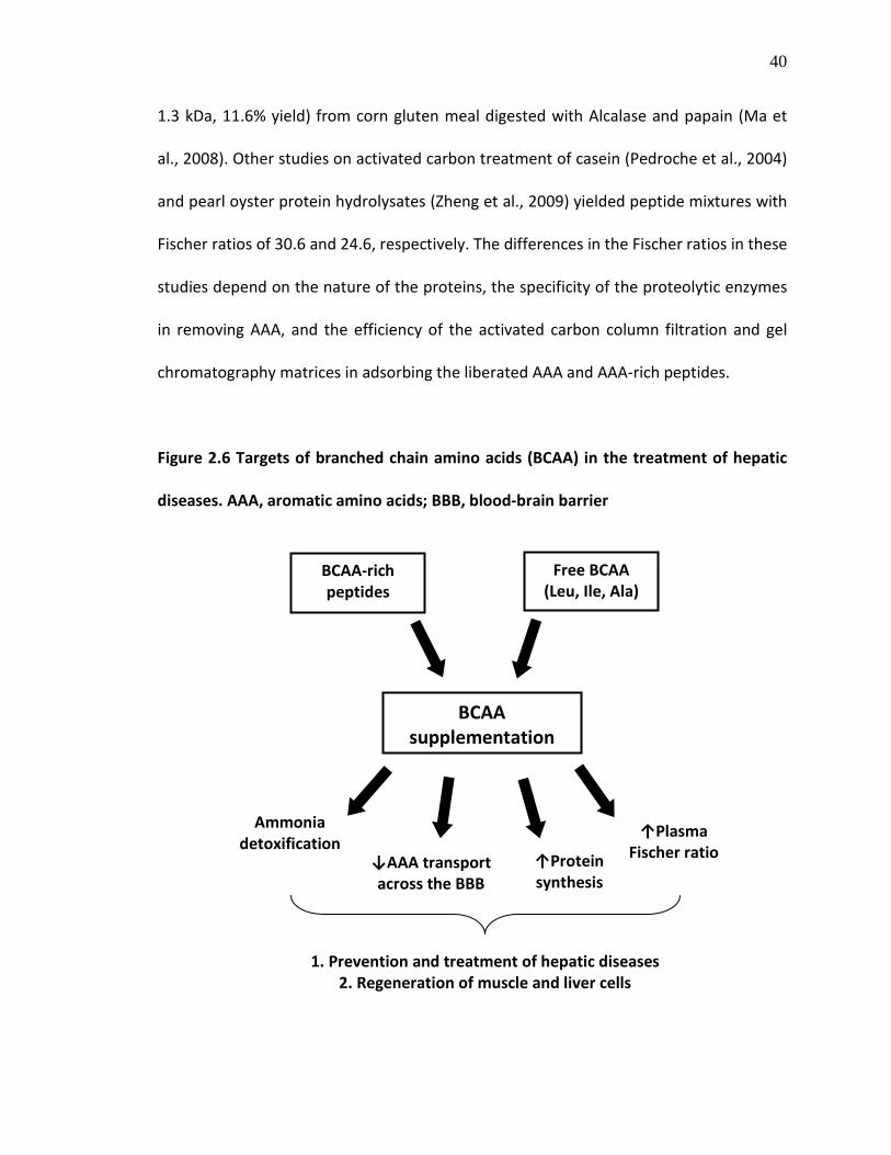
40
1.3 kDa, 11.6% yield) from corn gluten meal digested with Alcalase and papain (Ma et
al., 2008). Other studies on activated carbon treatment of casein (Pedroche et al., 2004)
and pearl oyster protein hydrolysates (Zheng et al., 2009) yielded peptide mixtures with
Fischer ratios of 30.6 and 24.6, respectively. The differences in the Fischer ratios in these
studies depend on the nature of the proteins, the specificity of the proteolytic enzymes
in removing AAA, and the efficiency of the activated carbon column filtration and gel
chromatography matrices in adsorbing the liberated AAA and AAA-rich peptides.
Figure 2.6 Targets of branched chain amino acids (BCAA) in the treatment of hepatic
diseases. AAA, aromatic amino acids; BBB, blood-brain barrier
BCAA-rich peptides
Free BCAA (Leu, Ile, Ala)
BCAA supplementation
Ammonia detoxification
↓AAA transport across the BBB
↑Plasma Fischer ratio ↑Protein
synthesis
1. Prevention and treatment of hepatic diseases 2. Regeneration of muscle and liver cells

41
2.4.8. Multifunctional peptides
Multifunctional peptides have been discovered from some food proteins and
they have been reported to possess more than one significant physiologically-relevant
bioactive property. Studies on milk proteins reported that a hexapeptide (TTMPLW)
derived from αS1-casien (f194-199) by trypsin-catalyzed digestion exhibited both ACE-
inhibitory and immunomodulatory activities (Meisel, 2004) while a β-lactoglobulin-
derived β-lactorphin (YLLF) inhibited ACE activity and also possessed opioid-like activity
(Antila et al., 1991; Mullally et al., 1997). In addition, several other milk-derived peptides
such as α-lactorphin (YGLF), α-immunocasokinin (TTMPLW), β-casomorphin-7 (YPFPGPI)
and β-casokinin (AVPYPQR) are regarded as multifunctional, some possessing in vivo
bioactive properties (Meisel, 2004). Moreover, crude chymotryptic α-casein
hydrolysates displayed several in vitro bioactivities such as ACE and propyl
endopeptidase inhibition, antioxidant, Zn2+-binding and antibacterial activities (Srinivas
& Prakash, 2010). Other partially purified food peptides from quinoa/pea proteins and
hen egg white lysozyme have also displayed multifunctionality as ACE-inhibiting
antioxidants (Aluko & Monu, 2003; Humiski & Aluko, 2007) and CaMPDE-inhibiting
antioxidants (You et al., 2010), respectively. The multiple bioactivities displayed by these
peptides can increase their impact towards the amelioration of more than one disease
target or multiple symptoms of a disease, such as cardiovascular disease, since many
human diseases are interrelated in terms of aetiology and progression. Therefore, the
conditions for generating and processing bioactive food protein hydrolyastes can be
carefully designed to yield multifunctional peptides with diverse applications.

42
2.5. Quantitative Structure-Activity Relationship (QSAR) studies of peptides
Structure-function relationship (SFR) studies of bioactive compounds involve
analysis of structural functionality of the bioactive molecules and relating these
characteristics to their pharmacological activity. This approach is known as quantitative
structure-activity relationship (QSAR) studies, which is a practically reliable method in
chemometrics for SFR studies. In peptide QSAR, potency of bioactive peptides has been
found to depend largely on peptide chain length and physicochemical properties such as
molecular size, charge, hydrophilicity/hydrophobicity and position of amino acid
residues of the peptides (Pripp et al., 2004a). Therefore, the first step in peptide QSAR is
to obtain a library of peptides with varying bioactivity (designated as Y variables) and
different fully-characterized structural features (X variables) relevant to the bioactivity
(Wold et al., 2001). Subsequently, models of the relationship between these variables
are obtained using statistical multiple regression analysis (Wold et al., 2001). Partial
least squares (PLS) method has been widely used in peptide QSAR for this purpose
(Sandberg et al. 1998; Wu et al. 2006a, b). PLS modelling of these data will result in two
important parameters: (1) the multiple correlation coefficients (R2) that indicate the
model fit, and (2) the cross-validation coefficients (Q2cv) that indicate the predictive
power of the model (Hellberg et al., 1987; Wold et al., 2001). Q2cv can be reliably used
for model validation in the absence of external model validation set especially when
working with limited number of observations in the training set (Wold et al., 2001);
combination of both validation methods gives better results on the predictive ability of
PLS models.

43
Some factors to consider prior to QSAR studies using PLS method include the size
of the analogues in peptide datasets, which is determined by the number of positions to
be varied on the peptide chain (Hellberg et al., 1987). In addition, peptide structural
descriptors need to be carefully chosen to reflect the bioactivity under investigation.
The z scale amino acid descriptors are widely used for peptide QSAR studies (Hellberg et
al., 1987; Sandberg et al., 1998). The 3-z scale (z1, z2 and z3) have been developed by
principal component analysis of 29 physicochemical variables of 20 coded amino acids,
and were interpreted to be related to hydrophobicity (z1), steric properties or side chain
bulk (z2) and electronic properties (z3) (Hellberg et al., 1987). This approach has
received some criticisms because of difficulty in interpretation and the lack of
consideration of the peptides’ conformations in the derivation of the principal
properties (Dunn & Wold, 1995). An extended 5-z scale was subsequently developed to
include additional variables (z4 and z5), which are difficult to clearly interpret and are
related to heat of formation, and molecular “hardness” and electrophilicity, respectively
(Sandberg et al., 1998). The z4 and z5 variables contain important information that
improved modelling quality (Sandberg et al., 1998; Siebert, 2003; Wu et al., 2006b).
Other amino acid descriptors have also been proposed for peptide QSAR. Lin et al.
(2008) recently proposed the “V-scale” that used the primary amino acid side chain data
that influence interaction with protein receptors including Van der Waal’s volume, net
charge index and hydrophobic parameter without compression by PCA. They reported
that models generated for bitter-tasting dipeptides using these descriptors (R2=0.948
and 0.903, Q2cv=0.921 and 0.860) were more robust than the z-scale based model

44
(R2
=0.824). However, the descriptors were based on the amino acid side chains only and
not the entire peptide sequence, which limits their use for enzyme inhibition modelling
where the entire peptide functionality is involved. The PLS method has been used to
develop good QSAR models for elastase substrates and neurotensin analogues
(Sandberg et al. 1998), functional properties of polypeptides (Strøm et al., 2001; Siebert,
2003) and ACE-inhibiting peptides (Pripp et al., 2004b; Wu et al. 2006a, b) leading to
accurate predictions of potency.
2.6. Flaxseed
Flaxseed or linseed (Linum usitatissimum L.) is an oilseed that is cultivated in
various parts of the world, especially in Canada, China, USA, India and Argentina (Bhatty,
1995; Agriculture and Agri-Food Canada, 2007; Hall et al., 2006). Flaxseed has been used
for the production of oil, paint and other industrial products, and has increasingly
gained particular interest in the human food system due to its rich nutritional
components and medicinal values. Whole flaxseed is rich in nutrients (e.g. oil, dietary
fibre and proteins), non-nutrients (e.g. lignans) and anti-nutritional factors (e.g.
cyanogenic glycosides and phytic acids) (Oomah & Mazza, 1993, 1995; Bhatty, 1995;
Oomah, 2001; Hall et al., 2006). This section discusses the nutritional composition and
beneficial aspects of flaxseed.

45
2.6.1. Nutritional quality, compositions and health benefits of flaxseed
Flaxseed can be crushed for human consumption, and the oil components can be
extracted by pressing or solvent extraction. In 2009, oil from Canadian-cultivated
flaxseed made up 46% of the whole flaxseed and was comprised mainly of α-linolenic
acid, ALA (58% of oil) with minor levels of linoleic acid (15.6%), oleic acid (16.9%), stearic
acid (3.1%) and palmitic acid (4.8%) (Canadian Grain Commission, 2009); this makes
flaxseed the best known terrestrial food source of omega-3 ALA (Bhatty, 1995). In
addition to ALA, flaxseed also contains high amounts of non-starch polysaccharides
(gum and fibre). Flaxseed contains large amounts of proteins ranging from 10.5% to 31%
with mean value of 21% (Oomah & Mazza, 1995); these proteins serve primarily as
nutrient reservoirs. Canadian annual quality survey showed that flaxseed No. 1 Canada
Western contained 22% proteins (%N × 6.25) in 2009 compared to the 1999-2008 mean
value of 23.3% (Canadian Grain Commission, 2009). Mean protein contents of 28.9%
and 34.1% were also reported for 11 cultivars grown in North Dakota and Canada,
respectively (Oomah & Mazza, 1993; Bhatty, 1995). Thus, the total protein content of
flaxseed protein varies with cultivars and location/environment (Oomah & Mazza, 1995;
Hall et al., 2006). Flaxseed also contains large amounts of lignans, a class of polyphenolic
phytoestrogens, and is considered the richest food source of secoisolariciresinol
diglucoside (SDG), the major flaxseed lignan (Bloedon & Szapary, 2004; Hall et al., 2006).
The current market for whole and ground flaxseed is primarily focused on the
constituent omega-3 ALA, fibres and lignans. These flaxseed components contribute to
its multiple positive effects in cardiovascular health, cancer prevention and diabetes

46
(Oomah, 2001; Bloedon & Szapary, 2004; Hall et al., 2006). Human consumption of
flaxseed resulted in reduction of CVD risk factors including decrease in serum total and
LDL cholesterol concentrations and markers of inflammation (Bloedon & Szapary, 2004).
Flaxseed lignans inhibit oxidative stress and inflammation whereas the insoluble fibre
components improve gut health and also possess hypolipidemic effects (Oomah, 2001;
Bloedon & Szapary, 2004). However, flaxseed proteins have not been well studied for
therapeutic applications. Most studies on flaxseed proteins have focused on
characterization of the structures and functional properties of the proteins.
2.6.2. Flaxseed proteins
The major storage proteins found in various flaxseed cultivars include a globulin-
like salt soluble 11-12S protein with molecular weight (MW) of 252-298 kDa, and an
albumin-type water soluble basic 1.6-2S protein with MW of 16-17 kDa (Madhusudhan
& Singh, 1983, 1985a, b; Oomah & Mazza, 1995). These proteins were named linin and
conlinin, respectively by Vassel and Nesbitt (1945). The 11-12S and the 1.6-2S proteins
account for 56-73.4% and 20-42% of the total flaxseed proteins, respectively (Youle &
Huang, 1981; Madhusudhan & Singh, 1983; 1985a, b; Marcone et al., 1998). The primary
amino acid sequence of flaxseed conlinin has been elucidated and reported to contain
168 or 169 amino acid residues (UniProtKB/TrEMBL accession # Q8LPD4). Protein
sequences of other 142-180 amino acid-containing oil-binding flaxseed proteins, known
as oleosins, have also been reported from gene transcripts (UniProtKB/TrEMBL
accession # A0RZJ4, A0RZJ6, A0RZJ7 and A0RZJ8). Compared to other food proteins such

47
as soy, maize and milk proteins, the amino acid sequences of flaxseed proteins need to
be fully characterized for in-depth understanding of functional properties and food
applications of the proteins.
The first report of flaxseed protein isolation was by T.B. Osborn in 1892 during
the period that he worked on several other food proteins (Vassel & Nesbitt, 1945). Since
the past three decades, several efforts have been made to optimize flaxseed protein
isolation methods and characterize the protein for use in the human food system.
Approaches used for successful isolation of flaxseed protein from defatted meal include
acid precipitation at isoelectric point (pI=4.2) after alkaline solubilisation (Dev &
Quensel, 1988), buffered salt (NaCl) extraction (Oomah et al., 1994), polyphosphate
extraction (Wanasundara & Shahidi, 1996) and micellisation (Krause et al., 2002) with
each process yielding isolates that contained high protein levels. Distinctive similarities
among these extraction methods include the use of laboratory samples of the desired
cultivars of defatted and dehulled flaxseed as starting material, which enhances the
protein isolation process. Moreover, protein analyses were based on total nitrogen (N ×
6.25), which can overestimate proteins due to reaction of other non-protein N-
containing organic compounds. Practically, the oilseed processing industry generates
large amounts of defatted flaxseed meal as by-product after oil extraction from crushed
whole flaxseed, which contains high amounts of mucilage. Mucilage interferes with
protein extraction from flaxseed, but removal of the mucilage from whole flaxseed by
soaking in water or enzymatic treatments improved the protein contents of the extract
(Madhusudhan & Singh, 1983; Wanasundara & Shahidi, 1997). Table 2.3 shows the

48
amount of protein (%) in various flaxseed fractions of varying mucilage content and also
the differences based on extraction methods.
Table 2.3 Differences in flaxseed protein contents (%) based on different isolation
methods and mucilage contents of the starting materials
FRACTION PROTEIN (%)
Seed 19.2
Dehulled seed 21.8–23.9
Hull 17.3–20.3
Meal 22.9-49.0
Laboratory-prepared meal 43.9
Commercial meal 34.7
Dehulled meal 48.9-50.0
PROTEIN EXTRACT
Micelle protein isolate 93.0
Isoelectric-precipitated protein isolate 89.0
LMF 56.4 a
LMPC 59.7 b
HMPC-S 63.4 c
HMPC-EC 65.5 d
LMPI 86.6 e
HMPI 66.3 f aLMF, low mucilage flour; bLMPC, low mucilage protein concentrate; cHMPC-S,
high mucilage protein concentrate from seed; dHMPC-EC, HMPC from expeller
cake; eLMPI, low mucilage protein isolate; fHMPI, high mucilage protein isolate;
data adapted from Hall et al., 2006 with permission from Elsevier
49
Most defatted flaxseed meal produced in Canada are exported to international
markets for livestock feed (Agriculture and Agri-Food Canada, 2007). At the moment,
based on literature information, little emphasis has been placed on value addition to
this industrial by-product and creation of opportunities for local use in Canada, even
though flaxseed meal proteins contain rich amino acid profile. As shown in Table 2.4,
whole flaxseed, flaxseed meals and isolated proteins are rich sources of glutamic
acid/glutamine (Glx), arginine (Arg), branched-chain amino acids (valine, Val and leucine,
Leu) and aromatic amino acid (tyrosine, Tyr and phenylalanine, Phe). There is no
apparent difference between the amino acid profiles of laboratory-prepared and
commercial defatted flaxseed meal (Bhatty, 1995), but the histidine (His) content was
reported to be higher in flaxseed flour compared to the whole seed and defatted meal
(Table 2.4). Defatting and protein extraction processes generally increased Arg, Leu and
cysteine (Cys) compositions (%) compared to the whole seed. Likewise, Arg and Leu
contents (%) of hexane extracted protein were also higher than commercial protein
extracts. Table 2.5 shows the distribution amino acids in the flaxseed protein subunits.
The amounts of Arg in the 1.6 S and 12 S subunits are equal, but tend to be lower in the
2 S proteins. Glx and aspartic acid/asparagine (Asx) are predominant in the 1.6 S and 12
S subunits, respectively, whereas BCAA (Ala+Val) and AAA (Phe+Tyr) are more in the 12
S than the 1.6 S subunits. This information can lead to isolation of a particular flaxseed
protein subunit enriched with amino acids of interest for nutritional and therapeutic
applications.

50
Table 2.4 Amino acid compositions (%) of flaxseed, flaxseed meals and flaxseed protein extracts
Oomah & Mazza, 1995
Dev & Quensel, 1986
Bhatty, 1995
Wanasundara & Shahidi, 1994
AMINO ACIDS
FLAXSEED
a
FLAXSEED
FLOUR
FLAXSEED MEAL FLAXSEED PROTEIN EXTRACT
LABORATORY SAMPLES
COMMERCIAL SAMPLESb
HEXANE EXTRACT b
MAW-HE
c COMMERCIAL
ARG 9.2 10.4 11.8 11.1 11.5 11.20 9.78 HIS 2.2 5.9 2.9 3.1 2.69 2.46 2.36 ILE 4.0 4.6 5.2 5.0 4.78 4.54 4.19 LEU 5.8 6.5 6.8 7.1 6.70 6.39 5.96 LYS 4.0 6.0 4.1 4.3 4.38 4.14 3.92
MET 1.5 3.0 2.2 2.5 1.45 1.41 1.24 CYS 1.1 N.D. 3.8 4.3 3.29 3.39 3.16 PHE 4.6 6.5 5.3 5.3 5.13 4.91 4.63 TYR 2.3 4.6 2.9 3.1 2.21 2.12 1.98 THR 3.6 3.1 4.9 5.1 3.40 3.33 3.00 TRP N.D. N.D. 1.8 1.7 0.46 0.46 0.25 VAL 4.6 4.9 5.6 5.6 5.75 5.64 5.02 ALA 4.4 4.3 5.4 5.5 4.81 4.64 4.61 ASX 9.3 d 8.3 12.5 12.4 9.18 9.16 8.03 GLX 19.6 e 22.8 21.3 26.4 16.70 16.36 14.45 GLY 5.8 4.9 7.0 7.1 6.44 6.26 5.64 PRO 3.5 3.0 5.2 5.5 3.64 3.65 3.32 SER 4.5 4.1 5.8 5.9 4.94 4.99 4.48
aData for NorLin flaxseed variety; bmean values (n=6); cMAW-HE, methanol-ammonia-water/hexane extracted; dASX, aspartic acid and asparagine; eGLX, glutamic acid and glutamine

51
Table 2.5 Amino acid composition of the two major flaxseed 11-12 S (linin) and 1.6-2 S
(conlinin) protein subunits
Madhusudhan &
Singh (1985a)
Youle & Huang
(1981)
Madhusudhan &
Singh (1985b)
AMINO ACIDS 1.6 S 2 S 12 S
ARG 13.1 6.0 12.5
HIS 1.6 1.2 2.5
ILE 2.8 2.9 4.6
LEU 5.4 5.3 5.8
LYS 4.9 6.0 3.1
MET 0.8 1.9 1.7
CYS 3.5 8.2 1.4
PHE 2.4 2.2 5.9
TYR 1.4 1.5 2.3
THR 2.1 3.6 3.9
TRP 2.0 0.8 1.3
VAL 2.6 3.9 5.6
ALA 1.9 5.1 4.8
ASX 5.5 a 6.4 11.3
GLX 35.0 b 23.8 19.8
GLY 8.3 13.8 4.8
PRO 3.0 1.6 4.5
SER 3.9 6.1 5.1 aASX, aspartic acid and asparagine; bGLX, glutamic acid and glutamine

52
Till date, there is little global interest in promoting the value and use of flaxseed
meal proteins despite their rich amino acid profiles. A number of studies have shown
that flaxseed proteins possess potential for therapeutic applications. For example,
peptides derived from enzymatic hydrolysis of flaxseed proteins inhibited CaM-
dependent eNOS/nNOS and ACE activities, and also displayed in vitro antioxidant
activities (Omoni & Aluko, 2006a, b; Marambe et al., 2008). Moreover, a recent study
also reported that a flaxseed protein extract inhibited growth of a wide range of food
spoilage fungi (Xu et al., 2008) but this activity could be due to other non-protein
components, such as lignans, present in the crude protein extracts. As previously stated
in the General Introduction (Chapter 1), it will be worthwhile to investigate the potential
human health benefits of flaxseed protein by exploiting the distinct structural features
of the predominant amino acids (e.g. Arg and BCAA) in producing peptides with
physiologically-relevant bioactive properties.

53
2.7. REFERENCES
Abubakar, A., Saito, T., Kitazawa, H., Kawai, Y. and Itoh, T. (1998). Structural analysis of new antihypertensive peptides derived from cheese whey protein by proteinase K digestion. Journal of Dairy Science, 12, 3131-3138
Adachi, S., Yamanaka, T., Hayashi, S., Kimura, Y., Matsuno, R., & Yokogoshi, H. (1993). Preparation of peptide mixture with high Fischer ratio from protein hydrolysate by adsorption on activated carbon. Bioseparation, 3, 227-232
Agriculture and Agri-Food Canada. (2007). Flaxseed: situation and outlook. Bi-weekly Bulletin, 20(3), AAFC No. 2081/E
Agriculture and Agri-Food Canada. (2010). Functional foods and nutraceuticals. http://www4.agr.gc.ca/AAFC-AAC/display-afficher.do?id=1170856376710 (Accessed on August 15, 2010)
Aluko, R.E. (2008a). Determination of nutritional and bioactive properties of peptides in enzymatic pea, chickpea, and mung bean protein hydrolysates. Journal of AOAC International, 91, 947-956
Aluko, R. E. (2008b). Antihypertensive properties of plant-derived inhibitors of angiotensin I-converting enzyme activity: a review. In: J. N. Govil, & V. K. Singh (Eds.), Recent Progress in Medicinal Plants–Phytopharmacology and Therapeutic Values IV, vol. 22 (pp. 541-561). Houston, Texas: Stadium Press
Aluko, R. E., & Monu, E. (2003). Functional and bioactive properties of quinoa seed protein hydrolysates. Journal of Food Science, 66, 1254-1258
Ames, B. N. (1983). Dietary carcinogens and anticarcinogens: oxygen radicals and degenerative diseases. Science, 221, 1256–1264
Ames, B. N., Shigena, M. K., & Hegen, T. M. (1993). Oxidants, antioxidants and the degenerative diseases of aging. Proceedings of the National Academy of Science U.S.A., 90, 7915–7922
Antila, P., Paakkari, I., Järvinen, A., Mattila, M.J., Laukkanen, M., Pihlanto-Leppälä, A., Mäntsälä, P., & Hellman, J. (1991). Opioid peptides derived from in vitro proteolysis of bovine whey proteins. International Dairy Journal, 1, 215-229
Aoyama, T., Fukui, K., Takamatsu, K., Hashimoto, Y., & Yamamoto, T. (2000). Soy protein isolate and its hydrolysate reduce body fat of dietary obese rats and genetically obese mice (yellow KK). Nutrition, 16, 349-354
Barnett, R.D., & Weiss, B. (1983). Inhibition of calmodulin activity by insect venom peptides. Biochemical Pharmacology, 32, 2929-2933

54
Barrio, D.A., & Añon, M.C. (2010). Potential antitumor properties of a protein isolate obtained from the seeds of Amaranthus mantegazzianus. European Journal of Nutrition, 49, 73-82
Bell, J. M., & Keith, M. O. (1993). Nutritional evaluation of linseed meals from flax with yellow or brown hulls, using mice and pigs. Animal Feed Science and Technology, 43, 1-18
Bender, A.T., & Beavo, J.A. (2006). Nucleotide phosphodiesterase: molecular regulation to clinical use. Pharmacological Reviews, 58, 488-520
Bender, A.T., Ostenson, C.L., Wang, E.H., & Beavo, J.A. (2005). Selective up-regulation of PDE1B2 upon monocyte-to-macrophage differentiation. Proceedings of the National Academy of Science USA, 102, 497-502
Bhatty, R.S. (1995). Nutrient composition of whole flaxseed and flaxseed meal. In: S.C. Cunnane, & L.U. Thompson (Eds.), Flaxseed in Human Nutrition (pp. 22-42). Champaign, Illinois: AOCS Press
Bloedon, L.T., & Szapary, P.O. (2004). Flaxseed and cardiovascular risk. Nutrition Reviews, 62, 18-27
Canadian Grain Commission (2009). Quality of western Canadian flaxseed 2009. http://www.grainscanada.gc.ca/flax-lin/harvest-recolte/2009/hqf09-qrl09-eng.pdf ISSN 1700-2087 (Accessed August 2010)
Carson, M. J., Crane, J., & Xie, A. X. (2008). Modeling CNS microglia: the quest to identify predictive models. Drug Discovery Today: Disease Models, 5, 19–25
Chan K.M., & Decker, E.A. (1994). Endogenous skeletal muscle antioxidants, Critical Reviews in Food Science and Nutrition, 34, 403–426
Chan, W.K.M., Decker, E.A., Lee, J.B., & Butterfield, D.A. (1994). EPR spin-trapping studies of the hydroxyl radical scavenging activity of carnosine and related dipeptides. Journal of Agricultural and Food Chemistry, 42, 1407-1410
Chen, H.M., Muramoto, K., Yamauchi, F., & Nokihara, K. (1996). Antioxidant activity of designed peptides based on the antioxidant peptide isolated from digests of a soybean protein. Journal of Agricultural and Food Chemistry, 44, 2619-2623
Chen, H.M., Muramoto, K., Yamauchi, F., Fujimoto, K., & Nokihara K. (1998). Antioxidant properties of histidine-containing peptides designed from peptide fragments found in the digests of a soybean protein. Journal of Agricultural and Food Chemistry, 46, 49-53

55
Cho, M.J., Vaghy, P.L., Kondo, R., Lee, S.H., Davis, J., Rehl, R., et al. (1998). Reciprocal regulation of mammalian nitric oxide synthase and calcineurin by plant calmodulin isoforms. Biochemistry, 37, 15593-15597
Cho, S., Juillerat, M.A., & Lee, C. (2007). Cholesterol lowering mechanism of soybean protein hydrolysate. Journal of Agricultural and Food Chemistry, 55, 10599-10604
Cho, S.J., Juillerat, M.A., & Lee, C.H. (2008). Identification of LDL-receptor transcription stimulating peptides from soybean hydrolysate in human hepatocytes. Journal of Agricultural and Food Chemistry, 56, 4372-4376
Cinnamon, B. (2009). Results from the functional foods and natural health products survey – 2007. Statistics Canada working paper, Catalogue no. 88F0006X, no. 1, ISBN 978-1-100-13192-4
Clemente, A. (2000). Enzymatic protein hydrolysates in human nutrition. Trends in Food Science and Technology, 11, 254-262
Colin, P.R., Chow, D., Miller, E.R., Svetkey, L.P., Lin, P.H., Harsha, D.W., Moore, T.J., Sask, F.M., & Appel, L.J. (2000). The effect of dietary patterns on blood pressure control in hypertensive patients: results from the Dietary Approaches to Stop Hypertension (DASH) trial. American Journal of Hypertension, 13, 949-955
Crivici, A., & Ikura, M. (1995). Molecular and structural basis of target recognition by calmodulin. Annual Review of Biophysics and Biomolecular Structure, 24, 85-116
Dev, D. K., & Quensel, E. (1986). Functional and microstructural characteristics of linseed (Limun usitatissimum L.) flour and a protein isolate. LWT – Food Science and Technology, 19, 331-337
Dev, D. K., & Quensel, E. (1988). Preparation and functional properties of linseed protein products containing differing levels of mucilage. Journal of Food Science, 53, 1834-1837
Di Pasquale, M.D. (2008). Amino acids and proteins for the athlete: the anabolic edge, 2nd
Dia, V.P., Torres, S., de Lumen, B.O., Erdman Jr., J.W. & Gonzalez de Mejia, E. (2009). Presence of lunasin in plasma of men after soy protein consumption. Journal of Agricultural and Food Chemistry, 57, 1260-1266
edition; CRC Press, Taylor & Francis Group: Boca Raton, FL, 434 pp.
Dia, V.P., Wang, W., Oh, V.L., de Lumen, B.O., & Gonzalez de Mejia, E. (2009). Isolation, purification and characterization of lunasin from defatted soybean flour and in vitro evaluation of its anti-inflammatory activity. Food Chemistry, 114, 108-115
Dinarello, C.A. (2000). Proinflammatory cytokines. CHEST, 118, 503-508

56
Dunn III, W.J., & Wold, S. (1995). SIMCA pattern recognition and classification. In: H. van de Waterbeemd (Ed.), Chemometrics Methods in Molecular Design (pp 179-193). VCH VerlagsgesellshaftmbH, Weinheim
Erdmann, K., Cheung, B. W. Y., & Schroder, H. (2008). The possible roles of food-derived bioactive peptides in reducing the risk of cardiovascular disease. Journal of Nutritional Biochemistry, 19, 643-654
Erdmann, K., Grosser, N., Schipporeit, K., & Schroder, H. (2006). The ACE inhibitory dipeptide Met-Tyr diminishes free radical formation in human endothelial cells via induction of heme oxygenase-1 and ferritin, Journal of Nutrition, 136, 2148-2152
Fang, Y.Z., Yang, S., & Wu, G. (2002). Free radicals, antioxidants, and nutrition. Nutrition, 18, 872-879
Ferreira, S.H., Bartelt, D.C., & Greene, L.J. (1970). Isolation of bradykinin-potentiating peptides from Bothrops jararaca venom. Biochemistry, 9, 2583-93
Fischer, J.E. (1990). Branched-chain-enriched amino acid solutions in patients with liver failure: an early example of nutritional pharmacology. Journal of Parenteral and Enteral Nutrition, 14, 249S-256S
Fischer, J.E., Funovics, J.M., Aguirre, A., James, J.H., Keane, J.M., Wesdorp, R.I., Yoshimura, N., & Westman, T. (1975). The role of plasma amino acids in hepatic encephalopathy. Surgery, 78, 276-290
FitzGerald, R.J. and Meisel, H. (2003). Milk protein hydrolysates and bioactive peptides. In: P.F. Fox, & P.L.H. McSweeney P.L.H. (Eds.), Advanced Dairy Chemistry (pg 675-698), New York: Kluwer Academic/Plenum Publishers
FitzGerald, R.J., Murray, B.A., & Walsh, D.J. (2004). Hypotensive peptides from milk proteins. Journal of Nutrition, 134, 980S-988S
Gauthier, S., Pouliot, Y., & Saint-Sauveur, D. (2006). Immunomodulatory peptides obtained by the enzymatic hydrolysis of whey proteins. International Dairy Journal, 16, 1315-1323
Gonzalez de Mejia, E., & Dia, V.P. (2009). Lunasin and lunasin-like peptides inhibit inflammation through suppression of NF-κB pathway in the macrophage. Peptides, 30, 2388-2398
Hall, C., Tulbek, M. C., & Xu, Y. (2006). Flaxseed. Advances in Food and Nutrition Research, 51, 1-97

57
Halliwell, B. (1991). Reactive oxygen species in living systems: Source, biochemistry, and role in human disease. American Journal of Medicine, 91, 3C-14S-3C-22S
Halliwell, B. (1996). Antioxidants in human health and disease. Annual Review of Nutrition, 16, 33-50
Halliwell, B. (2001). Role of free radicals in the neurodegenerative diseases: therapeutic implications for antioxidant treatment. Drugs and Aging, 18, 685-716
Hansson, G. (2005). Inflammation, atherosclerosis, and coronary artery disease. New England Journal of Medicine, 352, 1685-1695
Hartmann, R., & Meisel, H. (2007). Food-derived peptides with biological activity: from research to food applications. Current Opinion in Biotechnology, 18, 163-169
Hata, Y., Yamamoto, M., Ohni, M., Nakajima, K., Nakamura, Y., & Takano, T. (1996). A placebo-controlled study of the effect of sour milk on blood pressure in hypertensive subjects. American Journal of Clinical Nutrition, 64, 767-771
Health Canada (1998). Nutraceuticals/functional foods and health claims on foods. http://www.hc-sc.gc.ca/fn-an/alt_formats/hpfb-dgpsa/pdf/label-etiquet/nutra-funct_foods-nutra-fonct_aliment-eng.pdf (Accessed on August 15, 2010)
Hellberg, S., Sjostrom, M., Skagerberg, B., & Wold S.J. (1987). Peptide quantitative structure-activity relationships, a multivariate approach. Journal of Medicinal Chemistry, 30, 1126-1135
Hernández-Ledesma, B., Hsieh, C.-C.., & de Lumen, B. O. (2009a). Lunasin, a novel seed peptide for cancer prevention. Peptides, 30, 426-430
Hernández-Ledesma, B., Hsieh, C-C.., & de Lumen, B.O. (2009b). Antioxidant and anti-inflammatory properties of cancer preventive peptide lunasin in RAW 264.7 macrophages. Biochemical and Biophysical Research Communication, 390, 803-808
Heyworth, P.G., Curnutte, J.T., & Badwey, J.A. (1999). Structure and regulation of NADPH oxidase of phagocytic leukocytes: insight from chronic granulomatous disease. In: E.S. Serhan & P.A. Ward (Eds.), Molecular and Cellular Basis of Inflammation (pp. 165-192). New Jersey: Humana Press
Higaki, N., Sato, K., Suda, H., Suzuka, T., Komori, T., Saeki, T., Nakamura, Y., Ohtsuki, K., Iwami, K., & Kanamoto, R. (2006). Evidence for the existence of a soybean resistant protein that captures bile acid and stimulates its fecal excretion. Bioscience, Biotechnology and Biochemistry, 70, 2844-2852
58
Hirsch, E.C., & Hunot, S. (2009). Neuroinflammation in Parkinson's disease: a target for neuroprotection? Lancet Neurology, 8, 382-397
Holecek, M. (2010). Three targets of branched-chain amino acid supplementation in the treatment of liver disease. Nutrition, 26, 482-490
Hooks, S.S., & Means, A.R. (2001). Ca2+
Hori, G., Wang, M-F., Chan, Y-C., Komatsu, T., Wong, Y., Chen, T-H., Yamamoto, K., Nagaoka, S., & Yamamoto, S. (2001). Soy protein hydrolyzate with bound phospholipids reduces serum cholesterol levels in hypercholesterolemic adult male volunteers. Bioscience, Biotechnology and Biochemistry, 65, 72-78
/CaM-dependent kinases: from activation to function. Annual Review of Pharmacology and Toxicology, 41, 471-505
Horiguchi, N., Horiguchi, H., Suzuki, Y. (2005). Effect of wheat gluten hydrolysate on the immune system in healthy human subjects. Bioscience, Biotechnology and Biochemistry, 69, 2445-2449
Huang, D., Ou, B., & Prior, R. L. (2005). The chemistry behind antioxidant capacity assays. Journal of Agricultural and Food Chemistry, 53, 1841-1856
Humiski, L. M., & Aluko, R. E. (2007). Physicochemical and bitterness properties of enzymatic pea protein hydrolysates. Journal of Food Science, 72, S605-S611
Hunot, S., & Hirsch, E.C. (2003). Neuroinflammatory processes in Parkinson's disease. Annals of Neurology, 53, S49-S58
Ibrahim, M.M. (2006). RAS inhibition in hypertension. Journal of Human Hypertension, 20, 101-108
Ikura, M., Clore, G.M., Gronenborn, A.M., Zhu, G., Klee, C.B., Bax A. (1992). Solution structure of a calmodulin-target peptide complex by multidimensional NMR. Science, 256, 632-638
Itano, T., Itano, R., & Penniston, J.T. (1980). Interactions of basic polypeptides and proteins with calmodulin. Biochemical Journal, 189, 455-459
Jeong, H. J., Lee, J. R., Jeong, J. B., Park, J. H., Cheong, Y.-K., & de Lumen, B. O. (2009). The cancer preventive seed peptide lunasin from rye is bioavailable and bioactive. Nutrition and Cancer, 61, 680-686
Jiang, X., Li, J., Paskind, M., & Epstein, P.M. (1996). Inhibition of calmodulin-dependent phosphodiesterase induces apoptosis in human leukemic cells. Proceedings of the National Academy of Science USA, 93, 11236-11241

59
Kannel, W.B. (1996). Blood pressure as a cardiovascular risk factor. Journal of the American Medical Association, 275, 1571-1576
Kass, D.A., Takimoto, E., Nagayama, T., & Champion, H.C. (2007). Phosphodiesterase regulation of nitric oxide signalling. Cardiovascular Research, 75, 303-314
Kato, H., & Suzuki, T. (1971). Bradykinin-potentiating peptides from the venom of Akistrodon halys blomhoffii: isolation of five bradykinin potentiators and the amino acid sequences of two of them, potentiators B and C. Biochemistry, 10, 972-80
Kayashita, J., Shimaoka, I., Nakajoh, M., Yamazaki, M., & Kato, N. (1997). Consumption of buckwheat protein lowers plasma cholesterol and raises fecal neutral sterols in cholesterol-fed rats because of its low digestibility. Journal of Nutrition, 127, 1395-1400
Kearney, P. M., Whelton, M., Reynolds, K., Muntner, P., Whelton, P. K., & He, J. (2005). Global burden of hypertension: analysis of worldwide data. Lancet, 365, 217-223
Kim, D., Rybalkin, S.D., Pi, X., Wang, Y., Zhang, C., Munzel, T., Beavo, J.A., Berk, B.C., & Yan, C. (2001). Upregulation of phosphodiesterase 1A1 expression is associated with the development of nitrate tolerance. Circulation, 104, 2338–2343
Kim, S.E., Kim, H.H., Kim, J.Y., Kang, Y.I., Woo, H.J., & Lee, H.J. (2000). Anticancer activity of hydrophobic peptides from soy proteins. BioFactors, 12, 151-155
Kirana, C., Rogers, P. F., Bennett, L. E., Abeywardena, M. Y., & Patten, G. S. (2005). Naturally derived micelles for rapid in vitro screening of potential cholesterol-lowering bioactives. Journal of Agricultural and Food Chemistry, 53, 4623-4627
Kitts, D.D., & Weiler, K. (2003). Bioactive proteins and peptides from food sources. Applications of bioprocesses used in isolation and recovery. Current Pharmaceutical Design, 9, 1309-1323
Kizawa, K. (1997). Calmodulin binding peptide comprising α-casein exorphin sequence. Journal of Agricultural and Food Chemistry, 45, 1579-1581
Kizawa, K., Naganuma, K., & Murakami, U. (1995). Calmodulin-binding peptides isolated from α-casein peptone. Journal of Dairy Research, 62, 587-592
Klebanoff, S.J. (1999). Myeloperoxidase. Proceedings of the Association of American Physicians, 111, 383-389
Kohno, M., Hirotsuka, M., Kito, M., & Matsuzawa, Y. (2006). Decreases in serum triacylglycerol and visceral fat mediated by dietary soybean beta-conglycinin. Journal of Atherosclerosis and Thrombosis, 13, 247-255

60
Krause, J.P., Schultz, M., & Dudek, S. (2002). Effects of extraction conditions on composition, surface activity and rheological properties of protein isolates from flaxseed (Linum usitatissimum L.). Journal of the Science of Food and Agriculture, 82, 970-976
Lakowski, T.M., Lee, G.M., Lelj-Garolla, B., Okon, M., Reid, R.E. & McIntosh, L.P. (2007). Peptide binding by a fragment of calmodulin composed of EF-hands 2 and 3. Biochemistry, 46, 8525-8536
Li, G-H., Le, G-W., Shi, Y-H., & Shresthe, S. (2004). Angiotensin I-converting enzyme inhibitory peptides derived from food proteins and their physiological and pharmacological effects. Nutrition Research, 24, 469-486
Li, H., & Aluko, R.E. (2005). Kinetics of the inhibition of calcium/calmodulin-dependent protein kinase II by pea protein-derived peptides. Journal of Nutritional Biochemistry, 16, 656-662
Lin, Z.H., Long, H.X., Bo, Z., Wang, Y.Q., & Wu, Y.Z. (2008). New descriptors of amino acids and their application to peptide QSAR study. Peptides, 29, 1798-1805
Liu, B., Gao, H-.M., Wang, J-.Y., Jeohn, G-.H., Cooper, C. L., & Hong, J-.S. (2002). Role of nitric oxide in inflammation-mediated neurodegeneration. Annals of the New York Academy of Sciences, 962, 318–331
Lloyd-Jones D, Adams RJ, Brown TM, et al. (2010). Heart disease and stroke statistics-2010 update. A report from the American Heart Association statistics committee and Stroke Statistics subcommittee. Circulation, 121, e1-e170
Lovati, M. R., Manzoni, C., Gianazza, E., & Sirtori, C.R. (1998). Soybean protein products as regulators of liver low-density lipoprotein receptors. I. Identification of active β-conglycinin subunits. Journal of Agricultural and Food Chemistry, 46, 2474-2480
Lovati, M. R., Manzoni, C., Gianazza, E., Arnoldi, A., Kurowska, E., Carroll, K. K., & Sirtori, C. R. (2000). Soy protein peptides regulate cholesterol homeostasis in Hep G2 cells. Journal of Nutrition, 130, 2543-2549
Ma, Y., Lin, L., & Sun, D-W. (2008). Preparation of high Fischer ratio oligopeptide by proteolysis of corn gluten meal. Czech Journal of Food Science, 26, 38-47
Madhusudhan, K.T., & Singh, N. (1983). Studies on linseed proteins. Journal of Agricultural and Food Chemistry, 31, 959-963
Madhusudhan, K.T., & Singh, N. (1985a). Isolation and characterization of a small molecular weight protein of linseed. Phytochemistry, 24, 2507-2509

61
Madhusudhan, K.T., & Singh, N. (1985b). Isolation and characterization of the major fraction (12 S) of linseed proteins. Journal of Agricultural and Food Chemistry, 33, 673-677
Maeno, M., Yamamoto, N., & Takano, T. (1996). Identification of antihypertensive peptides from casein hydrolysate produced by a proteinase from Lactobacillus helveticus CP790. Journal of Dairy Science, 73, 1316-1321
Manach, C., Scalbert, A., Morand, C., Remesy, C., & Jemenez, L. (2004). Polyphenols: food sources and bioavailability. American Journal of Clinical Nutrition, 79, 727-747
Manso, M. A., Miguel, M., Even, J., Hernández, R., Aleixandre, A., & López-Fandiño, R. (2008). Effects of the long-term intake of an egg white hydrolysate on the oxidative status and blood lipid profile of spontaneously hypertensive rats. Food Chemistry, 109, 361-367
Marambe, P. W. M. L. H. K., Shand, P. J., & Wanasundara, J. P. D. (2008). An in vitro investigation of selected biological activities of hydrolyzed flaxseed (Linum usitatissimum L.) proteins. Journal of the American Oil Chemists’ Society, 85, 1155-1164
Marcone, M.F., Kakuda, Y., & Yada, R.Y. (1998). Salt-soluble seed globulins of dicotyleonous and monocotyledonous plants. II. Structural characterization. Food Chemistry, 63, 265-274
Martínez-Luis, S., Pérez-Vásquez, A., & Mata, R. (2007). Natural products with calmodulin inhibitor properties. Phytochemistry, 68, 1882-1903
Maruyama, S., Matachi, H., Tanaka, H., Tomizuka, N., & Suzuki, H. (1987). Studies on the active site and antihypertensive activity of angiotensin I-converting enzyme inhibitors derived from casein. Agricultural and Biological Chemistry, 51, 1581-1586
Meisel H. (2004). Multifunctional peptides encrypted in milk proteins. BioFactors, 21, 55-61
Meisel, H. (2005). Biochemical properties of peptides encrypted in bovine milk proteins, Current Medicinal Chemistry, 12, 1905-1919
Mine, Y., & Kovacs-Nolan, J. (2006). New insights in biologically active proteins and peptides derived from hen’s egg. World’s Poultry Journal, 62, 87-95
Mochizuki, Y., Maebuchi, M., Kohno, M., Hirotsuka, M., Wadahama, H., Moriyama, T., Kawada, T., & Urade, R. (2009). Changes in lipid metabolism by soy β-conglycinin-
62
derived peptides in HepG2 cells. Journal of Agricultural and Food Chemistry, 57, 1473-1480
Moncada, S., & Higgs, A. (1993). The L-arginine-nitric oxide pathway. New England Journal of Medicine, 329, 2002-2012
Mullally, M. M., Meisel, H., & FitzGerald, R. J. (1997). Identification of a novel angiotensin-I-converting enzyme inhibitory peptide corresponding to a tryptic fragment of bovine beta-lactoglobulin, FEBS Letters, 402, 99-101
Murphy, S. (2000). Production of nitric oxide by glial cells: regulation and potential roles in the CNS. Glia, 29, 1-13
Nagaoka, S., Miwa, K., Eto, M., Kuzuya, Y., Hori, G., & Yamamoto, K. (1999). Soy protein peptic hydrolysate with bound phospholipids decreases micellar solubility and cholesterol absorption in rats and Caco-2 cells. Journal of Nutrition, 129, 1725-1730
Nagel, D.J., Aizawa, T., Jeon, K.I., Liu, W., Mohan, A., Wei, H., et al. (2006). Role of nuclear Ca2+
Nakamura, Y., Yamamoto, N., Sakai, K., Okubo, A., Yamazaki, S. & Takano, T. (1995) Purification and characterization of angiotensin I-converting enzyme inhibitors from a sour milk. Journal of Dairy Science, 78, 777-783
/calmodulin-stimulated phosphodiesterase 1A in vascular smooth muscle cell growth and survival. Circulation Research, 98, 777-784
National Center for Health Statistics (2008). Health, United States, 2008. National Center for Health Statistics, Centres for Disease Control and Prevention, U.S. Department of Health and Human Services
O’Neil, K.T., & DeGrado, W.F. (1990). How calmodulin binds its targets: sequence independent recognition of amphiphilic α-helices. Trends in Biochemical Sciences, 15, 59-64
Okita, M., Watanabe, A., & Nagashima, H. (1985). Nutritional treatment of liver cirrhosis by branched amino acids-enriched nutrient mixture. Journal of Nutritional Science and Vitaminology, 31, 291-303
Omoni, A.O., & Aluko, R.E. (2006a). Effect of cationic flaxseed protein hydrolysate fractions on the in vitro structure and activity of calmodulin-dependent endothelial nitric oxide synthase. Molecular Nutrition and Food Research, 50, 958-966
Omoni, A.O., & Aluko, R.E. (2006b). Mechanism of the inhibition of calmodulin-dependent neuronal nitric oxide synthase by flaxseed protein hydrolysates. Journal of the American Oil Chemists’ Society, 83, 335-340

63
Ondetti, M.A., & Cushman, D.W. (1982). Enzymes of the renin-angiotensin system and their inhibitors. Annual Review of Biochemistry, 51, 283-308
Ondetti, M.A., Williams, N.J., Sabo, E.F., Pluvec, J., Weaver, E.R, & Kocy, O. (1971). Angiotensin-converting enzyme inhibitors from the venom of Bothrops jararaca: isolation, elucidation of structure and synthesis. Biochemistry, 10, 4033-4039
Oomah, B. D., & Mazza, G. (1993). Flaxseed proteins – a review. Food Chemistry, 48, 109-114
Oomah, B.D. (2001). Flaxseed as a functional food source. Journal of the Science of Food and Agriculture, 81, 889-894
Oomah, B.D., & Mazza, G. (1995). Functional properties, uses of flaxseed protein. Inform, 6, 1246-1252
Oomah, B.D., Mazza, G., & Cui, W. (1994). Optimization of protein extraction from flaxseed meal. Food Research International, 27, 355-361
Pacher, P., Beckman, J. S., & Liaudet, L. (2007). Nitric oxide and peroxynitrite in health and disease. Physiological Reviews, 87, 315–424
Palinic, R. (2007). Results from the functional foods and nutraceuticals survey – 2005. Statistics Canada working paper, Catalogue no. 88F0006XIE, no. 003, ISBN: 978-0-662-46010-7
Parada, J., & Aguilera, J.M. (2007). Food microstructure affects the bioavailability of several nutrients. Journal of Food Science, 72, R21-R32
Pedroche, J., Yust, M.M., Lqari, H., Girón-Calle, J., Vioque, J., Alaiz, M., & Millán, F. (2004). Production and characterization of casein hydrolysates with a high amino acid Fischer’s ratio using immobilized proteases. International Dairy Journal, 14, 527-533
Pihlanto, A. (2006). Antioxidative peptides derived from milk proteins. International Dairy Journal, 16, 1306-1314
Pins, J.J., & Keenan, J. M. (2002). The antihypertensive effects of a hydrolysed whey protein isolate supplement (BioZate® 1). Cardiovascular Drugs and Therapy, 16, 68
Pripp, A.H. (2005). Initial proteolysis of milk proteins and its effect on formation of ACE-inhibitory peptides during gastrointestinal proteolysis: A bioinformatic, in silico, approach. European Food Research and Technology, 221, 712-716
64
Pripp, A.H., Isaksson, T., Stepaniak, L., Sørhaug, T., & Ardö, Y. (2004a). Quantitative structure-activity relationship modelling peptides and proteins as a tool in food science. Trends in Food Science and Technology, 16, 484-494
Pripp, A.H., Isaksson, T., Stepaniak, L., & Sørhaug, T. (2004b). Quantitative structure-activity relationship modelling of ACE-inhibitory peptides derived from milk proteins. European Food Research and Technology, 219, 579-583
Rock, R. B., Gekker, G., Hu, S., Sheng, W. S., Cheeran, M., Lokensgard, J. R., & Peterson, P. K. Role of microglia in central nervous system infections. Clinical Microbiology Reviews, 2004, 17, 942-964
Ross, R. (1999). Atherosclerosis – an inflammatory disease. New England Journal of Medicine, 340, 115-126
Rybalkin S.D., Yan, C., Bornfeldt, K.E., & Beavo, J.A. (2003). Cyclic GMP phosphodiesterases and regulation of smooth muscle function. Circulation Research, 93, 280-291
Sandberg, M., Eriksson, L., Jonsson, J., Sjöström, M., & Wold, S. (1998). New chemical descriptors relevant for the design of biologically active peptides. A multivariate characterization of 87 amino acids. Journal of Medicinal Chemistry, 41, 2181-2491
Sarwar, G., Christiansen, D. A., Finlayson, A. T., Friedman, M., Hackler, L. R., McKenzie, S. L., Pellett, P. L., & Tkachuk, R. (1983). Intra- and inter-laboratory variation in amino acid analysis. Journal of Food Science, 48, 526-531
Sato, M., Hosokawa, T., Yamaguchi, T., Nakano, T., Muramoto, K., Kahara, T., Funayama, K., Kobayashi, A., & Nakano, T. (2002). Angiotensin I-converting enzyme inhibitory peptides derived from wakame (Undaria pinnatifida) and their antihypertensive effect in spontaneously hypertensive rats. Journal of Agricultural and Food Chemistry, 50, 6245-6252
Schauer-Vukasinovic, V., Cullen, L., & Daunert, S. (1997). Rational design of a calcium sensing system based on induced conformational changes of calmodulin. Journal of the American Chemical Society, 119, 11102-11103
Schermuly, R.T., Pullamsetti, S.S., Kwapiszewska, G., Dumitrascu R., Tian, X., Weissmann, N., et al. (2007). Phosphodiesterase 1 upregulation in pulmonary arterial hypertension. Target for reverse-remodeling therapy. Circulation, 115, 2331-2339
Segall, L., Covic, A., & Goldsmith, D.J.A. (2007). Direct renin inhibitors: the dawn of a new era, or just a variation on a theme? Nephrology Dialysis Transplantation, 22, 2435-2439

65
Shu, M., Mei, H., Yang, S., Liao, L., & Li, Z. (2009). Structural parameter characterization and bioactivity simulation based on peptide sequence. QSAR Combinatorial Science, 28, 27–35
Siebert, K. J. (2003). Modeling protein functional properties from amino acid composition. Journal of Agricultural and Food Chemistry, 51, 7792-7797
Silva-Sánchez, C., Barba de la Rosa, A.P., León-Galván, M.F., de Lumen, B.O., de León-Rodríguez, A., & Gonzalez de Mejía, E. (2008). Bioactive peptides in amaranth (Amarathus hypochondriacus) seed. Journal of Agricultural and Food Chemistry, 56, 1233-1240
Srinivas, S., & Prakash, V. (2010). Bioactive peptides from bovine milk α-casein: isolation, characterization and multifunctional properties. International Journal of Peptide Research and Therapeutics, 16, 7-15
Staessen, J.A., Li, Y., & Richart, T. (2006). Oral renin inhibitors. Lancet, 368, 1449-1456
Strøm, M. B., Stensen, W., Svendsen, J. S., & Rekdal, Ø. (2001). Increased antibacterial activity of 15-residue murine lactoferricin derivatives. Journal of Peptide Research, 57, 127-139
Sun, J.Z., Kaur, H., Halliwell, B., Li, X.Y., & Bolli, R. (1993). Use of aromatic hydroxylation of phenylalanine to measure production of hydroxyl radicals after myocardial ischemia in vivo. Direct evidence for a pathogenic role of the hydroxyl radical in myocardial stunning. Circulation Research, 73, 534-549
Takahashi, M., Moriguchi, S., Yoshikawa, M., & Sasaki, R. (1994). Isolation and characterization of oryzatensin: A novel bioactive peptide with ileum-contracting and immunomodulating activities derived from rice albumin. Biochemistry and Molecular Biology International, 33, 1151-1158
Tebbens, J. (2002). Functional foods and nutraceuticals: the development of value-added food by Canadian firms. Statistics Canada working paper, Catalogue no. 88F0006XIE, no. 016, ISBN: 0-662-41612-0
Torre, D., Pugliese, A., & Speranza, F. (2002). Role of nitric oxide in HIV-1 infection: friend or foe? Lancet Infectious Diseases, 2, 273–280
Turrens, J.F. (2003). Mitochondrial formation of reactive oxygen species. Journal of Physiology, 552, 335-344
Vassel, B., & Nesbitt, L.L. (1945). The nitrogenous constituents of flaxseed. II. The isolation of a purified protein fraction. Journal of Biological Chemistry, 159, 571-584

66
Vermeirssen, V., Van Camp, J., & Verstraete, W. (2004). Bioavailability of angiotensin I converting enzyme inhibitory peptides. British Journal of Nutrition, 92, 357-366
Wanasundara, J.P.D., & Shahidi, F. (1994). Functional properties and amino-acid composition of solvent-extracted flaxseed meals. Food Chemistry, 49, 45-51
Wanasundara, P.K.J.P.D., & Shahidi, F. (1996). Optimization of hexametaphosphate-assisted proteins using response surface methodology. Journal of Food Science, 61, 604-607
Wanasundara, P.K.J.P.D., & Shahidi, F. (1997). Removal of flaxseed mucilage by chemical and enzymatic treatments. Food Chemistry, 59, 47-55
Wang, W., Bringe, N.A., Berhow, M.A., & Gonzalez de Mejia, E. (2008). β-Conglycinins among sources of bioactives in hydrolysates of different soybean varieties that inhibit leukemia cells in vitro. Journal of Agricultural and Food Chemistry, 56, 4012-4020
Wergedahl, H., Liaset, B., Gudbrandsen, O. A., Lied, E., Espe, M., Muna, Z., Mork, S., and Berge, R. K. (2004). Fish protein hydrolysate reduces plasma total cholesterol, increases the proportion of HDL cholesterol, and lowers acyl-CoA:cholesterol acyltransferase activity in liver of Zucker rats. Journal of Nutrition, 134, 1320-1327
Wilkins, K., Campbell, N. R. C., Joffres, M. R., McAlister, F. A., Nichol, M., Quach, S., Johansen, H. L., & Tremblay, M. S. (2010). Blood pressure in Canadian adults. Statistics Canada, Catalogue no. 82-003-XPE, Health Reports, 21(1), March 2010
Wiseman, H., & Halliwell, B. (1996). Damage to DNA by reactive oxygen and nitrogen species: role in inflammatory disease and progression to cancer. Biochemical Journal, 313, 17-29
WHO (2009). Global health risks: mortality and burden of disease attributable to selected major risks. Geneva: World Health Organization Press (ISBN 978 92 4 156387 1)
Wold, S., Sjostrom, M., & Ericksson L. (2001). PLS-regression: a basic tool of chemometrics. Chemometrics and Intelligent Laboratory Systems, 58, 109-130
Wu, J., Aluko, R.E., & Nakai, S. (2006a). Structural requirements of angiotensin I-converting enzyme inhibitory peptides: quantitative structure-activity relationship study of di- and tripeptides. Journal of Agricultural and Food Chemistry, 54, 732-738
Wu J., Aluko R.E., & Nakai, S. (2006b). Structural requirements of angiotensin I-converting enzyme inhibitory peptides: quantitative structure-activity relationship
67
modelling of peptides containing 4-10 amino acid residues. QSAR Combinatorial Science, 25, 873-880
Xu, Y., Hall III, C., & Wolf-Hall, C. (2008). Antifungal activity stability of flaxseed protein extracts using response surface methodology. Journal of Food Science, 73, M9-M14
Yang, S., Liu, S., Yang, H., Lin, Y., & Chen, J. (2007). Soybean protein hydrolysate improves plasma and liver lipid profiles in rats fed high-cholesterol diet. Journal of the American College of Nutrition, 26, 416-423
You, S.J., Udenigwe, C.C., Aluko, R.E., & Wu, J. (2010). Multifunctional peptides from egg white lysozyme. Food Research International, 43, 848-855
Youle, R.J., & Huang, A.H.C. (1981). Occurrence of low molecular weight and high cysteine containing albumin storage proteins in oilseeds of diverse species. American Journal of Botany, 68, 44-48
Zheng, H., Zhang, C., Cao, W., Liu, S., & Ji, H. (2009). Preparation and characterization of the pearl oyster (Pinctada martensii) meat protein hydrolysates with a high Fischer ratio. International Journal of Food Science and Technology, 44, 1183-1191

68
CHAPTER THREE
MANUSCRIPT 1
KINETICS OF THE INHIBITION OF RENIN AND ANGIOTENSIN I-CONVERTING ENZYME BY
FLAXSEED PROTEIN HYDROLYSATE FRACTIONS
C. C. UDENIGWEa, Y-S. LINb, W-C. HOUc, R. E. ALUKOa,d
aDepartment of Human Nutritional Sciences, University of Manitoba, Winnipeg, MB R3T
2N2, Canada; bSchool of Pharmacy, College of Pharmacy, Taipei Medical University,
Taipei 110, Taiwan; cGraduate Institute of Pharmacognosy, Taipei Medical University,
Taipei 110, Taiwan; d
The Richardson Centre for Functional Foods and Nutraceuticals,
University of Manitoba, Winnipeg, MB R3T 2N2, Canada.
Reprinted from Kinetics of the Inhibition of Renin and Angiotensin I-Converting Enzyme
by Flaxseed Protein Hydrolysate Fractions Journal of Functional Foods, 1, 199-207
Copyright © 2009, with permission from Elsevier

69
3.0. ABSTRACT
Enzymatic hydrolysates from flaxseed protein were investigated for in vitro inhibition of
angiotensin I-converting enzyme (ACE) and renin activities. Pepsin, ficin, trypsin, papain,
thermolysin, pancreatin and Alcalase were used to hydrolyze flaxseed proteins followed
by fractionation using ultrafiltration to isolate low-molecular-weight peptides, and
separation of the Alcalase hydrolysate into cationic peptide fractions. Using N-(3-[2-
furyl]acryloyl)-phenylalanylglycylglycine as substrate, the protein hydrolysates showed a
concentration-dependent ACE inhibition (IC50, 0.0275–0.151 mg/ml) with thermolysin
hydrolysate and Alcalase cationic peptide fraction I (FI) showing the most potent
activity. Flaxseed peptide fractions also showed nil or moderate inhibitory activities
against human recombinant renin (IC50
, 1.22–2.81 mg/ml). Kinetics studies showed that
the thermolysin hydrolysate and FI exhibited mixed-type pattern of ACE inhibition
whereas cationic peptide fraction II inhibited renin in an uncompetitive fashion. These
results show that the protein components of flaxseed meal possess peptide amino acid
sequences that can be exploited as potential food sources of antihypertensive agents.
KEYWORDS: Flaxseed; Protein hydrolysates; Bioactive peptides; Angiotensin
converting enzyme; Renin; IC50;
Enzyme inhibition kinetics

70
3.1. INTRODUCTION
Since the past two decades, the renin-angiotensin-aldoresterone system (RAAS)
has turned out to be an excellent physiological target for the development of
antihypertensive agents. This is due to the fact that RAAS has been reported to play vital
roles in the progression of cardiovascular and chronic kidney diseases in human beings
(Segall et al., 2007; Staessen et al., 2006). The RAAS pathway comprises a series of
reactions that produce molecules capable of regulating blood pressure in humans; renin
and angiotensin I-converting enzyme (ACE) are the two key enzymes in this pathway. In
the RAAS pathway, renin (an aspartyl protease) catalyzes the initial and rate-limiting
step by converting angiotensionogen to angiotensin I (AT-I), a decapeptide.
Subsequently, ACE (peptidyldipeptidase A) catalyzes the conversion of AT-I into a potent
vasoconstrictor octapeptide, angiotensin II (AT-II), and also degrades bradykinin, a
vasodilator (Segall et al., 2007; Yang et al., 1970). ACE inhibitors have found clinical
applications in blocking or suppressing the formation of AT-II thereby reducing the
occurrence of elevated blood pressure. However, various ACE-inhibitory compounds
that have been developed as antihypertensive drugs, on prolonged administration, have
shown some undesirable side effects in human such as cough, taste distortion, skin rash
and angioedema (Acharya et al., 2003; Seseko & Kaneko, 1985). Thus, it is imperative to
discover safer therapeutic compounds for lowering human blood pressure during
hypertension, a risk factor in cardiovascular disease and other associated health
complications. Moreover, it is thought that the direct inhibition of renin activity
provides a better control of elevated blood pressure than ACE inhibition since it

71
prevents the production of AT-I, which could also be converted to a substantial amount
of AT-II in some organs via an ACE-independent pathway catalyzed by chymase;
however, renin inhibition does not prevent ACE-catalyzed bradykinin degradation (Segall
et al., 2007; Staessen et al., 2006). Thus, there is a need to develop therapeutic agents
that could exhibit pleiotropic effects in the inhibition of ACE and renin activities. Several
food protein hydrolysates and constituent peptides have been shown to display in vitro
inhibition of ACE activity (Aluko, 2008) resulting in the lowering of elevated blood
pressure in spontaneously hypertensive rats (Aluko, 2008; Wu & Ding, 2001) and in
hypertensive humans (FitzGerald et al., 2004; Seppo et al., 2003). Synthetic peptides
and peptidomimetic compounds have also been developed and applied parenterally as
renin inhibitors in the clinical treatment of hypertension; moreover, Aliskiren has been
developed as the first non-peptide oral renin inhibitor (Staessen et al., 2006). To the
best of our knowledge, there is no information in the literature regarding the inhibition
of renin activity by enzymatically prepared food protein hydrolysates. The health-
promoting potential of peptides derived from food proteins has remained a subject of
interest to many food scientists as new and underutilized food protein sources are
continuously investigated for bioactive properties.
Flaxseed (Linum usitatissimum, also known as linseed) has long been used as raw
material in the production of oil, paint and other industrial products. In the recent past,
flaxseed has emerged as one of the major focuses of the food and health sectors
worldwide. The emerging economic importance of flaxseed is primarily due to its
constituent lignans, dietary fibre and the Omega-3 fatty acid, α-linolenic acid, which
72
possess potential to provide cardiovascular and other health benefits in humans (Hall III
et al., 2006). However, the underutilized protein components of flaxseed also have
potential therapeutic properties that remain to be explored. Flaxseed meal is generated
as a by-product of industrial flaxseed oil extraction, and has been widely used as a
protein supplement in the formulation of livestock feed (Bell & Keith, 1993). The world
flaxseed meal production is estimated at 1.1–1.4 million tonnes with Canada, USA and
Argentina making up the largest exporters of this product (Agriculture and Agri-Food
Canada, 2007). It has been estimated that flaxseed and its defatted meal contain high
amounts of proteins, which are comparable in amino acid compositions to other high
quality food proteins like soy protein, with a preponderance of basic and branch-chain
amino acids (Bhatty, 1995; Oomah & Mazza, 1993, 1995). In 2007, the protein contents
of Flaxseed, No. 1 Canada Western ranged from 19.4 to 30.8% (N × 6.25), with a mean
value of 24.3% (Canadian Grain Commission, 2007).
Apart from its nutritional significance in containing most of the amino acid
requirements of humans and animals, the intact flaxseed proteins may possess other
biological activities; thus, it was recently reported that flaxseed protein extract showed
antifungal activity against a wide range of food spoilage fungi (Xu et al., 2008). It was
also recently reported that the crude flaxseed protein hydrolysate prepared using
Flavourzyme inhibited ACE in vitro and displayed antioxidant properties in scavenging
hydroxyl radical (Marambe et al., 2008). However, in a bid to improve the value of
defatted flaxseed meal generated as a by-product of the oilseed processing industry,
this study explored the bioactive properties of enzymatically prepared flaxseed protein
73
hydrolysates with respect to their potential use as source of antihypertensive agents.
The objective of this work was to determine the potential ACE and renin inhibitory
activities of low-molecular-weight (LMW) flaxseed protein hydrolysates produced from
hydrolysis of isolated flaxseed proteins using different food-grade proteases.
3.2. MATERIALS AND METHODS
3.2.1. Materials
Defatted flaxseed meal was a gift from Bioriginal Foods and Science Corporation
(Saskatoon, SK, Canada). Cellulase (from Aspergillus niger), Alcalase (protease from
Bacillus licheniformis), thermolysin (from Bacillus thermoproteolyticus rokko), ficin (from
fig tree latex), pepsin (from porcine gastric mucosa), trypsin (from bovine pancreas),
papain (from papaya latex), pancreatin (from porcine pancreas), ACE from rabbit lung
(E.C.3.4.15.1) and N-(3-[2-furyl]acryloyl)-phenylalanylglycylglycine (FAPGG) were
purchased from Sigma-Aldrich (St. Louis, MO, USA). Human recombinant Renin Inhibitor
Screening Assay Kit was purchased from Cayman Chemicals (Ann Arbor, MI, USA). The
ultrafiltration membranes of 1, 3 and 5 kDa molecular weight cut-off (MWCO), the 100
Da MWCO dialysis membrane and other analytical grade reagents were purchased from
Fischer Scientific (Oakville, ON, Canada).
3.2.2. Isolation of flaxseed proteins
The defatted flaxseed meal cake was pulverized using a Retsch ZM 200
ultracentrifugal mill (Retsch GmbH, Haan, Germany) at 14,000 min-1 and the resulting
74
flour stored in an air-tight container at 4°C until used. The protein components of the
meal were isolated according to our newly developed method that significantly (P<0.05)
increased protein yield when compared to the traditional method of Dev and Quensel
(1988). Defatted flaxseed meal (5%, w/v, dry weight basis) was suspended in deionized
water and stirred thoroughly using a magnetic stirrer. The highly viscous suspension was
adjusted to pH 5.0 and temperature of 37o
C, followed by the addition of cellulase (1%,
w/w; activity of powder, 1.44 U/mg) to initiate fibre hydrolysis. After 4 h of reaction, the
resulting less viscous suspension was cooled to 4°C followed by alkaline solubilisation
(pH 10.0) with 0.5 M NaOH and acid-induced flaxseed protein precipitation (pH 4.2) with
0.5 M HCl according to a previous method (Dev & Quensel, 1988). The resulting protein
precipitate was washed thrice with acidified water (pH 4.2), suspended in a small
volume of deionized water and the pH adjusted to 7.0 using 0.5 M NaOH. The
suspension was freeze-dried and stored at –20°C until used as isolated flaxseed protein.
3.2.3. Preparation of flaxseed protein hydrolysates
Hydrolysis of the isolated flaxseed proteins was conducted with pepsin, ficin,
trypsin, papain, thermolysin, pancreatin and Alcalase under different conditions using a
Metrohm 842 Titrando pH-stat connected to a Metrohm 800 Dosino device (Metrohm
AG, Herisau, Switzerland). Flaxseed protein isolate (5%, w/v, protein basis) was
suspended in deionized water in a reaction vessel equipped with a stirrer, heated to the
appropriate temperature and adjusted to the appropriate pH prior to the addition of the
proteolytic enzyme; the reaction conditions are shown in Table 1. The enzymes were

75
added to the slurry at an enzyme-substrate ratio (E/S) of 1:100 (based on the protein
content of the protein isolate) separately except for the Alcalase reaction. Digestion was
performed at the above conditions for 4 h; pH of the reaction mixture was kept constant
by the pH-stat with 2 M NaOH except for the pepsin reaction. Alcalase hydrolysis was
carried out as previously reported at an E/S of 1.5:25 (Omoni & Aluko, 2006). After
digestion, the enzyme was inactivated by immersing the reaction vessel in boiling water
bath (95-100°C) for 15 min and undigested proteins were precipitated by adjusting the
pH to 4.0 with 2 M HCl followed by centrifugation at 14,941 × g for 30 min. The
supernatant containing target peptides was collected for further fractionation. Protein
content of samples was determined by a modified Lowry’s method using bovine serum
albumin as a standard (Markwell et al., 1978).
3.2.4. Fractionation of flaxseed protein hydrolysates
The flaxseed protein hydrolysates (supernatants from section 2.3) were each
separated into the LMW fractions using membrane ultrafiltration. The supernatant
resulting from protein hydrolysis was passed through an Amicon stirred ultrafiltration
cell (Millipore Corporation, Bedford, MA, USA) set-up using a 1 kDa MWCO membrane,
and resulting permeate was collected. The flaxseed protein hydrolysate resulting from
Alcalase digestion was sequentially passed through ultrafiltration membranes first with
MWCO of 1 kDa then the retentate through 3 kDa whose retentate was passed through
5 kDa to separate peptides of sizes <1, 1-3 and 3-5 kDa. The resulting permeates were
dialyzed for 48 h against deionized water at 4°C using a 100 Da MWCO dialysis tube, and

76
dialyzates were freeze-dried and stored at –20°C until used as flaxseed protein
hydrolysate fractions in ACE and renin inhibition studies.
3.2.5. Preparative ion-exchange liquid chromatography
The cationic peptides in the <1 kDa permeate from the Alcalase prepared
flaxseed protein hydrolysate were separated as previously reported (Omoni & Aluko,
2006) with some modifications. Briefly, a solution of the freeze-dried permeate (250
mg/ml in 0.1 M ammonium acetate buffer) was adjusted to pH 7.0 using 2 M
ammonium hydroxide. Four ml of the solution was injected onto an SP-Sepharose High
Performance XK 50/20 cation exchange chromatography column (column volume,
196.35 ml) coupled to an AKTA Fast Protein Liquid Chromatography system (Amersham
Biosciences, Montreal, QC, Canada). Prior to sample injection, the column was pre-
equilibrated with 1.5 column volume (CV) of 0.1 M ammonium acetate buffer (pH 7.5).
After sample was loaded, the column was washed with 1.5 CV of the 0.1 M ammonium
acetate buffer to remove unbound peptides, followed by a gradient elution of the
bound peptides using 0–50% 0.5 M ammonium carbonate (pH 8.8) in 0.1 M ammonium
acetate buffer at a flow rate of 10 ml/min; eluted peptides were monitored at 214 nm.
Two major peaks were observed and fractions within these peaks were pooled and
concentrated by vacuum evaporation at 37°C. Thereafter, the concentrated peptide
solution was dialyzed for 48 h against deionized water at 4°C using a 100 Da MWCO
dialysis tube. The dialyzed solutions were freeze-dried and stored at –20°C until used as
cationic peptide fractions I (FI) and II (FII).
77
3.2.6. Determination of ACE inhibition
The ability of the flaxseed peptide fractions to inhibit the activity of ACE in vitro
was measured according to the spectrophotometric method of Holmquist et al. (1979)
as reported by Lin et al. (2008) using FAPGG as substrate. Briefly, 1 ml of 0.5 mM FAPGG
(dissolved in 50 mM Tris-HCl buffer containing 0.3 mM NaCl, pH 7.5) was mixed with 20
µl of ACE (1 U/ml; final activity of 20 mU) and 200 µl of flaxseed protein hydrolysate in
50 mM Tris-HCl buffer. The decreased absorbance at 345 nm, due to cleavage of the
Phe-Gly peptide bond of FAPGG, was recorded at regular intervals for 2 min at room
temperature. Tris-HCl buffer was used instead of peptide fraction solutions in the blank
experiment. ACE activity was expressed as rate of disappearance of FAPGG (∆A.min-1
)
and inhibitory activity was calculated using Eq. (3.1).
ACE inhibition (%) = 1 – (∆A.min-1(sample) / ∆A.min-1
(blank)
) × 100 (3.1)
∆A.min-1(sample) and ∆A.min-1
(blank) are the reaction rates in the presence and absence of
the peptide fraction, respectively. Dose-dependent ACE inhibition was investigated
using at least four different concentrations of peptide fractions. The concentration of
peptide fractions that inhibited ACE activity by 50% (IC50
) was calculated using a non-
linear regression from a plot of ACE inhibition versus sample concentrations. All
experiments were performed in triplicate.

78
3.2.7. Determination of kinetics parameters of ACE inhibition
The kinetics of ACE-catalyzed conversion of FAPGG to N-(3-[2-furyl]acryloyl)-
phenylalanine was studied in the absence and presence of three different
concentrations of flaxseed protein-derived peptide fractions with 0.0625, 0.1, 0.125,
0.25 and 0.5 mM FAPGG, a range selected to represent concentrations above and below
reported Km for FAPGG (Holmquist et al., 1979; Hou et al., 2003; Lin et al., 2008). The
mode of inhibition of ACE was determined from the Lineweaver-Burk plots. Kinetic
parameters (Km, Km′, Vmax and Vmax
′) were estimated from non-linear regression fit of
the kinetics data to the Michaelis-Menten equation using GraphPad Prism version 4.0
(GraphPad Software, San Diego, CA, USA). The catalytic efficiency (CE) of ACE in the
absence and presence of the peptide fractions was calculated using Eq. (3.2).
CE = Vmax/Km or Vmax′/Km
′ (3.2)
Vmax and Km are the maximum reaction velocity and Michaelis constant, respectively,
whereas Vmax′ and Km′ represent their apparent values in the presence of the inhibitor.
The enzyme-inhibitor dissociation constant (K i
) was determined as the intercept on the
X-axis of a secondary plot of the slope of the Lineweaver Burk plot versus
concentrations of inhibiting peptide fractions. All kinetics experiments were performed
in duplicate.

79
3.2.8. Renin inhibition assay
In vitro inhibition of the activity of human recombinant renin was conducted by
fluorescence spectrometry using the Renin Inhibitor Screening Assay Kit and the method
of Yuan et al. (2006) modified slightly as follows. Briefly, the total assay volume of 190 µl
contained 10 µM Arg-Glu(EDANS)-Ile-His-Pro-Phe-His-Leu-Val-Ile-His-Thr-Lys(Dabcyl)-
Arg (renin substrate dissolved in dimethyl sulphoxide), human recombinant renin and
flaxseed peptide fraction in 50 mM Tris-HCl buffer, pH 8.0, containing 100 mM NaCl. The
Tris-HCl buffer was used instead of the flaxseed peptide solution in the blank
experiment. The renin substrate and flaxseed peptide sample were mixed and pre-
warmed to 37°C for 10 min to attain thermal equilibrium. Thereafter, the reaction was
initiated by adding human recombinant renin to the mixture; the increase in
fluorescence intensity was monitored for 10 min in a JASCO FP-6300 spectrofluorimeter
(Japan Spectroscopic Company, Tokyo, Japan) equipped with a thermostated cell
compartment that was maintained at 37°C using a circulatory water bath. The
spectrofluorimeter parameters were set as follows: excitation wavelength, 340 nm;
emission wavelength, 490 nm; excitation bandwidth, 5 nm; emission bandwidth, 10 nm.
The enzyme activity was expressed as reaction rate, arbitrary fluorescence intensity unit
per min (FIU.min-1
). Renin inhibitory activity of the peptide fractions was calculated
using Eq. (3.3).
Renin inhibition (%) = [(FIU.min-1(blank) – FIU.min-1
(sample)) / FIU.min-1(blank)
(3.3)
] × 100

80
FIU.min-1(blank) and FIU.min-1
(sample)
represent the renin reaction rates in the absence and
presence of the flaxseed peptide fraction, respectively. In addition, kinetic studies was
conducted for the most active flaxseed peptide fraction using 1.25, 2.5, 5 and 10 µM
renin substrate in the absence and presence of the peptide fraction (0.5 and 2 mg
protein/ml). This was followed by the determination of mode of inhibition and kinetics
parameters of the renin reaction as earlier described in the ACE assay.
3.2.9. Statistical analysis
Data are reported as mean ± standard deviation. Statistical significance of
differences was evaluated by Student’s t-test and Duncan’s multiple range test (P<0.05)
using the Statistical Analysis Systems (SAS) software (SAS, Cary, NC, USA).
3.3. RESULTS AND DISCUSSION
In the recent past, food protein-derived peptides have been reported to display
various biological functions towards the management and treatment of human diseases,
including antimicrobial activity (Pellegrini, 2003), immunomodulatory activity (Gauthier
et al., 2006), antioxidant, antithrombotic, hypolipidemic and hypocholesterolemic
activities (Erdmann et al., 2008). In addition, a wide range of food proteins have also
afforded highly potent ACE-inhibiting peptides, which have been actively investigated
for their ability to lower blood pressure in SHR (Aluko, 2008; Erdmann et al., 2008; Wu &
Ding, 2001) and in hypertensive human subjects (FitzGerald et al., 2004; Seppo et al.,
2003). These food proteins include zein (Yano et al., 1996), corn gluten (Suh et al.,

81
2003), milk (FitzGerald et al., 2004; Seppo et al., 2003), soybean, wheat and quinoa
(Aluko, 2008), yam dioscorin (Hsu et al., 2002; Lin et al., 2006), bovine skin gelatin (Kim,
Byun, Park, & Shahidi, 2001) and canola (Wu et al., 2008). To date, there is only one
recent literature regarding ACE-inhibiting hydrolyzed flaxseed proteins generated by
Flavourzyme digestion (Marambe et al., 2008). It is noteworthy that these underutilized
flaxseed proteins also contain peptide sequences with properties that could be
exploited in the development of therapeutic food products.
The potential of food proteins to provide bioactive peptides depends on several
factors including the ability of proteases to digest and release the active peptide
sequences. Thus, the work reported in this paper investigated the hydrolysates
generated from digestion of isolated flaxseed proteins using different proteases for in
vitro effects on ACE and renin activities; the proteases were used at their optimum
reaction conditions. These proteases were considered safe for food applications since
they are either part of the human digestive system or have been used extensively in the
food industry. Following enzymatic hydrolysis, the <1 kDa fractions of the flaxseed
protein hydrolysates were separated using membrane ultrafiltration, and their protein
contents ranged from 77.1 to 99.7% after dialysis with protein yields of between 5.4 and
18.5% (Table 3.1). The interest in short-chain peptides is due to reported evidence that
these peptides are able to withstand proteolytic degradation by the enzymes of the
gastrointestinal tract when orally ingested by humans (Aluko, 2008). Moreover, it is
widely accepted that LMW peptides are more potent in vitro ACE inhibitors than larger
peptides. Partial purification of the 1 kDa ultrafiltration permeate of the Alcalase

82
hydrolysate by fast protein liquid chromatography using a cation-exchange column
yielded two cationic peptide fractions (FI and FII) as previously reported; FII possesses a
stronger cationic character than FI (Omoni & Aluko, 2006). FI and FII contained 95.5 and
70.6% proteins, respectively, after desalting by dialysis. These protein hydrolysates and
cationic peptide fractions were evaluated for ACE and renin inhibitory activities.
The protease treatments of flaxseed proteins liberated bioactive peptides from
the native protein structure, and these peptides inhibited in vitro activity of ACE; the
presence of the peptide fractions in the assay led to a concentration-dependent loss of
ACE activity (Figure 3.1A). The LMW peptides resulting from thermolysin hydrolysis of
flaxseed proteins displayed the most potent activity with the inhibition of 20 and 61%
ACE activity at 0.00625 and 0.05 mg protein/ml, respectively. This extent of ACE
inhibition was also observed for other enzymatic flaxseed protein hydrolysate fractions
but at slightly higher concentrations. The IC50 values of these protein hydrolysate
fractions in ACE inhibition is shown in Figure 3.1B; the thermolysin LMW peptide
fraction gave the least IC50 value, similar to activity previously reported for crude
thermolysin hydrolysates from α-zein, IC50 = 0.021 mg/ml (Miyoshi et al., 1995) and
dried bonito, IC50 = 0.029 mg/ml (Fujita et al., 1995) even though these studies used a
different assay system to measure ACE activity. The peptides resulting from pepsin and
trypsin hydrolyses of flaxseed proteins had similar inhibitory effects on ACE activity.
Among all the enzymatic digests investigated, the pancreatin LWM peptide fraction
showed the weakest ACE inhibition. These data suggest that ACE inhibition observed in
this study could be more associated with the bioactive peptide sequences released

83
Table 3.1 Reaction conditions for enzymatic hydrolysis of flaxseed proteins; protein
contents and yield of the ultrafiltration permeate
Enzyme
Abbreviation
Reaction conditions Ultrafiltration permeate
pH Temp (°C) Protein (%) Yield (%)a b
Pepsin PepH 2.0-2.2 37 77.1 8.7
Ficin FicH 7.0 37 94.7 7.4
Trypsin TrypH 8.0 37 99.7 9.0
Papain PapH 6.5 37 73.4 5.4
Thermolysin ThermoH 8.0 37 97.9 5.6
Pancreatin PancH 8.0 40 87.6 18.5
aProtein contents of <1 kDa permeates after dialysis using a 100 Da MWCO membrane
bProtein basis
during hydrolysis rather than the amount of small peptides, since there was no linear
relationship between the protein yield of the LMW peptide fractions and ACE inhibition;
moreover, the pancreatin hydrolysate fraction with the highest <1 kDa protein yield
gave the lowest ACE inhibition and the reverse was observed for thermolysin
hydrolysate. Besides, these levels of ACE inhibition observed for the flaxseed protein
digests are comparable to inhibitory activities reported for crude and ultrafiltration-
fractionated simulated gastrointestinal digests of pea and whey proteins using similar
assay conditions (Vermeirssen et al., 2005). A combination of the protein yield and IC50

84
values reported here showed that the LMW peptides in thermolysin hydrolysate have
the best potential for industrial application as ACE inhibitor followed by trypsin, pepsin
and ficin digests. When peptides of higher molecular weight (MW) were evaluated, the
Alcalase protein hydrolysate containing peptides of MW range of 3-5 kDa showed better
ACE inhibition than the 1-3 kDa peptides. Moreover, a previous report showed that
when Flavourzyme was used to hydrolyze flaxseed proteins to three different (11.9%,
24.6% and 25.1%) degrees of hydrolysis, the crude hydrolysates showed ACE inhibition
with IC50 values 0.07 and 0.09 mg protein/ml using hippuryl-histidyl-leucine as substrate
(Marambe et al., 2008). Considering that some of the proteases investigated in this
study are present in the gastrointestinal tract of humans, it is possible that these
bioactive peptides could be released during human consumption of intact flaxseed
proteins.

85
Figure 3.1 (A) Flaxseed protein-derived peptide fractions exhibit concentration-
dependent ACE inhibition; results are expressed as means of ACE activity ± standard
deviation of triplicate determinations. (B) 50% inhibitory concentration (IC50) values of
peptide fractions in ACE inhibition; Each bar represents the mean of triplicate
determinations of IC50
± standard deviation; bars with different letters are
significantly different at P< 0.05; 1-3AlcH and 3-5AlcH stand for the 1-3 kDa and 3-5
kDa peptides of alcalase hydrolysate, respectively (For other abbreviations, see Table
3.1)
FI FII
1-3 AlcH
3-5 AlcH PepHFicH
TrypH
PapH
ThermoH
PancH
IC50
val
ue (m
g/m
l)
0.00
0.02
0.04
0.06
0.08
0.10
0.12
0.14
0.16
g
b
d
f f e f
c
g
a
Concentration (mg/ml)0.00 0.05 0.10 0.15 0.20
ACE
activ
ity (∆
A 345
nm/m
in)
0.002
0.004
0.006
0.008
0.010
0.012FIFII1-3 AlcH3-5 AlcHPepH FicH TrpHPapH ThermoHPancH
B
A

86
Food protein-derived cationic peptides have also demonstrated in vitro ACE
inhibition; thus we investigated the ACE-inhibiting potentials of cationic peptides in the
flaxseed protein digests. When the <1 kDa Alcalase protein hydrolysate was separated
into its cationic peptide fractions, one of the resulting fractions showed a strong ACE-
inhibitory activity. Cationic peptide fraction I showed activity (IC50 = 0.0288 mg
protein/ml) similar to that observed for thermolysin hydrolysates with the inhibition of
about 70% ACE activity at 0.065 mg protein/ml whereas cationic peptide fraction II
showed weaker activity with the inhibition of 64% ACE activity at 0.2 mg protein/ml
(IC50
Several studies have reported the mode of inhibition of peptides purified from
food-protein hydrolysates. However, there are few studies in the literature that
investigated the kinetics of ACE inhibition in the presence of food protein hydrolysates.
This information will be necessary to elucidate the synergistic effects of the peptide
mixture on ACE reaction kinetics prior to in vivo studies especially if the protein
= 0.118 mg protein/ml). Thus, it could be concluded from these data that the
ability of these flaxseed protein-derived peptide fractions to inhibit ACE activity in vitro
is independent of their cationic strength. This observation is contrary to a trend
previously reported for ACE-inhibiting cationic peptides derived from pepsin-pancreatin
soy protein hydrolysate (Farzamirad & Aluko, 2008). Based on these enzyme inhibitory
activities, cationic peptide fraction I and the thermolysin hydrolysate fraction were
selected for kinetics studies to elucidate their mode of inhibition of ACE activity.
Cationic peptide fraction II was also studied for comparison.

87
hydrolysates are highly potent ACE inhibitors that do not require further purification. In
this study, Lineweaver-Burk plots were used to estimate the modes of ACE inhibition by
the flaxseed peptides. Figure 3.2A–C shows the double reciprocal plots of ACE-catalyzed
reaction in the absence and presence of flaxseed peptide fractions; ACE inhibition by
various concentrations of flaxseed-protein derived peptides was evident across the
FAPGG concentrations investigated. Using the non-linear regression fit of the kinetics
data, the Km value of ACE activity in the absence of the inhibiting peptides was
determined to be 0.3058 mM FAPGG, which was similar to previously reported values of
0.3 mM (Holmquist et al., 1979) and 0.292 mM FAPGG (Hou et al., 2003). The Alcalase
cationic peptide and thermolysin hydrolysate fractions showed mixed-type inhibition of
ACE activity characterized by various effects on the catalytic parameters at different
peptide concentrations (Table 3.2). These results are similar to the mode of ACE
inhibition reported for glutathione and carnosine under similar assay conditions (Hou et
al., 2003). This shows that the constituent peptides of the fractions exhibited their
activities possibly by binding ACE in both its free and FAPGG-bound forms. This also
means that the peptides could bind ACE at other sites different from the FAPGG binding
site, which might affect the conformation of the active site resulting in loss of activity.
The presence of the Alcalase cationic peptide fraction I in the assay resulted in a two-
fold decrease in the affinity of FAPGG for ACE at 0.0164 mg protein/ml, almost the same
magnitude as the effect reported for 0.0164 mM glutathione (Hou et al., 2003).
Moreover, both the Alcalase cationic peptide fraction II and thermolysin hydrolysate
fraction showed a concentration-dependent increase in Km. The effects of these

88
flaxseed protein hydrolysate fractions on Vmax did not follow any pattern, and was not
concentration-dependent. However, there was an observed linear decrease in the
catalytic efficiency of ACE with increase in the concentration of inhibiting peptides;
Alcalase cationic peptide fraction I and thermolysin protein hydrolysate showed better
effects than fraction II (Table 3.2). These results confirm the ability of the peptides to
dose-dependently reduced the efficiency of the reaction regardless of their mode and
extent of ACE inhibition. Despite having similar IC50 values, the Alcalase cationic peptide
fraction I had more affinity for ACE than the thermolysin hydrolysate as evident in its
lower K i value. However, the K i for cationic peptide fraction II was about twice the value
for thermolysin protein hydrolysate even though the latter was more than four times
more active in ACE inhibition (IC50
Furthermore, the flaxseed protein-derived peptide fractions moderately
inhibited renin activity, and this was also dependent on the proteolytic treatments of
the flaxseed proteins; their IC
).
50 values are shown in Figure 3.3. The trend of renin
inhibition observed for the cationic peptide fraction II and thermolysin hydrolysate
fraction was the opposite of their observed ACE-inhibitory activities, with the cationic
peptide showing the best activity whereas the thermolysin hydrolysate was weakest.
The results suggest that renin inhibition observed in this study was dependent on the
cationic character of the peptide fraction because the weaker cationic fraction (FI) did
not show any renin-inhibitory activity in this assay. In addition, the <1 kDa peptide
fractions from papain and pancreatin hydrolysates, and the high MW Alcalase
hydrolysate fractions, did not show renin inhibition under same assay conditions.

89
Figure 3.2 Lineweaver-Burk plots of the inhibition of ACE by different concentrations
of: (A) cationic peptide fractions I; (B) cationic peptide fractions II; (C) ThermoH, at
varying concentrations of FAPGG; V is the initial rate of reaction (∆A345nm
/min)
1/[FAPGG] (mM-1)-5 0 5 10 15 20
1/V
(m
in/∆
A34
5nm
)
0
200
400
600
800
1000No FI (r2=0.99)FI=0.0164mg/mL (r2=0.99)FI=0.0082mg/mL(r2=0.98) FI=0.0041mg/mL (r2=0.99)
A
1/[FAPGG] (mM-1)-5 0 5 10 15 20
1/V
(min
/∆A
345n
m)
0
200
400
600
800
1000
1200
FII=0.1mg/mL (r2=0.99)FII=0.05mg/mL (r2=0.98) FII=0.025mg/mL (r2=0.98)No FII (r2=0.99)
B
1/[FAPGG] (mM-1)-5 0 5 10 15 20
1/V
(m
in/∆
A34
5nm
)
0
200
400
600
800
1000
1200No ThermoH (r2=0.99)
ThermoH=0.025mg/mL (r2=0.99)
ThermoH=0.0125mg/mL (r2=0.97)
ThermoH=0.00625mg/mL (r2=0.98)
C

90
Table 3.2 Kinetics constants of ACE catalyzed reaction in the absence (control) and presence of different concentrations of
Alcalase cationic peptide fractions I and II, and thermolysin hydrolysate (ThermoH); Km and Km′ are Michaelis constants in the
absence and presence of inhibitor, respectively; Vmax, and Vmax
Catalytic
parameter
′ are maximum reaction velocities in the absence and presence of
inhibitor, respectively; CE, catalytic efficiency of ACE; Ki, enzyme-inhibitor dissociation constant
Control Fraction I (mg/ml) Fraction II (mg/ml) ThermoH (mg/ml)
0.0041 0.0082 0.0164 0.0250 0.0500 0.1000 0.0063 0.0125 0.0250
Km or Km′ (mM) 0.3058 a 0.3964 0.7799 0.6109 0.2790 0.4052 1.764 0.5551 0.6782 0.9444
Vmax or Vmax′ 0.0183 b 0.0166 0.0221 0.0140 0.0129 0.0138 0.0276 0.0195 0.0173 0.0172
CE 0.0599 0.0419 0.0282 0.0229 0.0463 0.0339 0.0156
0.0351 0.0255 0.0182
K i (mg/ml) 0.0065 0.0240 0.0128
aFAPGG as substrate
bVmax, ∆A.min-1

91
The results are consistent with the fact that ACE inhibition is more easily achieved when
compared to renin inhibition and this is reflected in the numerous ACE-inhibitory
peptides already reported in the literature but almost no food protein-derived peptide
inhibitors of renin. Based on this activity, the kinetics of renin inhibition was studied for
Alcalase cationic peptide fraction II.
In this study, the Km value of human recombinant renin in the absence of any
inhibitor was determined to be 4.416 µM for the renin substrate, lower than a value 6.4
µM reported by Yuan et al. (2006) and higher than a value 1.3 µM reported by Wang et
al. (1993). The difference in Km values could be attributed to the different sources of
renin in these studies. The double reciprocal plot (Figure 3.4) showed that the cationic
peptide fraction II exhibited an uncompetitive mode of renin inhibition with large
decrease in Vmax from 39.17 FIU/min for the uninhibited reaction to 24.2 and 10.47
FIU/min in the presence of 0.5 and 2 mg/ml inhibitor sample, respectively. The observed
decrease in Vmax was associated with decrease in Km value to 3.912 and 2.72 µM with
0.5 and 2 mg/ml inhibitor, respectively. Thus, it could be concluded that the cationic
peptide fraction II contained peptides that did not bind to the human recombinant renin
active site but associated with other regulatory sites on the enzyme-substrate complex.
This observation justifies the high IC50 value for the cationic peptide fraction since high
inhibitor concentration would be required to effect a change in the conformation of the
renin active site, depending on its interaction with the inhibitor-binding sites. It was
difficult to make comparison since there are no related renin inhibition kinetics studies
with enzymatic food protein hydrolysates in the literature.

92
Figure 3.3 50% Inhibitory concentration (IC50) values of flaxseed protein-derived
peptide fractions in inhibiting human recombinant renin activity1
; n.a, no renin
inhibitory activity observed; bars with different letters are significantly different at
P<0.05
1 See Figure S3.5 in Appendix A for dose-dependent inhibition of human recombinant renin activity by the flaxseed protein-derived peptide fractions
FI FII
1-3AlcH
3-5AlcH
PepH
FicHTryp
HPap
H
Therm
oHPan
cH0.0
0.5
1.0
1.5
2.0
2.5
3.0
n.a n.a n.a n.a n.a
bb
a,bb
IC50
val
ue (m
g/m
l)
a

93
Figure 3.4 Lineweaver-Burk plot of the inhibition of human recombinant renin by
flaxseed protein-derived cationic peptide fraction II at 0.5 and 2 mg protein/ml
3.4. CONCLUSIONS
1/[S] (µM-1)
-0.4 -0.2 0.0 0.2 0.4 0.6 0.8 1.0
1/V
(min
.FIU
-1)
0.00
0.05
0.10
0.15
0.20
0.25
0.30
No FII (r2=0.98)FII=0.5mg/mL (r2=0.96)FII=2.0mg/mL (r2=0.95)

94
The results of this study clearly indicate that flaxseed protein hydrolysates
possess potential as a food source of antihypertensive agents. The inhibition of the
activities of ACE and human recombinant renin observed in this study is dependent on
the ability of the different proteases used for hydrolysis to release bioactive peptide
sequences from flaxseed proteins rather than the protein yield of low-molecular-weight
peptides. The cationic peptide fraction I and thermolysin hydrolysate gave promising
results due to their ability to inhibit ACE in a mixed-type inhibition pattern at very low
concentrations. In addition, the flaxseed peptide fractions that inhibited both ACE and
renin activities possess better prospects, and potentially could provide better in vivo
lowering of blood pressure when compared to peptides that inhibit ACE alone. Further
work is needed to characterize the effects of these flaxseed protein hydrolysate and
cationic peptide fractions in lowering of blood pressure, and to identify their constituent
bioactive peptides. This will contribute towards increased value-added utilization of
flaxseed meal, currently a low-value by-product of the oilseed processing industry.
ACKNOWLEDGEMENTS
Financial support for this work was provided by operating and equipment grants to REA
from the Advanced Foods and Materials Network of Centre of Excellence (AFMnet) and
the Natural Sciences and Engineering Research Council of Canada (NSERC). CCU is a
recipient of a Manitoba Health Research Council Graduate Scholarship and an NSERC
Summer Research Program in Taiwan Fellowship.
3.5. REFERENCES

95
Acharya, K. R., Sturrock, E. D., Riordan, J. F., and Ehlers, M. R. W. Ace revisited: A new target for structure-based drug design, Nature Reviews Drug Discovery 2 (2003), pp. 891-902.
Agriculture and Agri-Food Canada. Flaxseed: situation and outlook, Bi-weekly Bulletin 20(3) February 23, 2007, AAFC No. 2081/E.
Aluko, R. E. (2008). Antihypertensive properties of plant-derived inhibitors of angiotensin I-converting enzyme activity: a review. In: J. N. Govil, & V. K. Singh, Recent Progress in Medicinal Plants – Phytopharmacology and Therapeutic Values IV, vol. 22 (pp. 541-561). Houston, Texas: Stadium Press.
Bell, J. M., and Keith, M. O. Nutritional evaluation of linseed meals from flax with yellow or brown hulls, using mice and pigs, Animal Feed Science and Technology 43 (1993), pp. 1-18.
Bhatty, R. S. (1995). Nutrient composition of whole flaxseed and flaxseed meal. In: S. C. Cunnane, & L. U. Thompson, Flaxseed in Human Nutrition, pp. 22-42. Champaign, Illinois: AOCS Press.
Canadian Grain Commission. Quality of western Canadian flaxseed. http://www.grainscanada.gc.ca/flax-lin/harvest-recolte/2007/flax-2007-eng.pdf (2007) ISSN 1700-2087 (Accessed August 2008).
Dev, D. K., and Quensel, E. Preparation and functional properties of linseed protein products containing differing levels of mucilage, Journal of Food Science 53 (1988), pp. 1834-1837.
Erdmann, K., Cheung, B. W. Y., and Schroder, H. The possible roles of food-derived bioactive peptides in reducing the risk of cardiovascular disease, Journal of Nutritional Biochemistry 19 (2008), pp. 643-654
Farzamirad, V., and Aluko, R. E. Angiotensin-converting enzyme inhibition and free-radical scavenging properties of cationic peptides derived from soybean protein hydrolysates, International Journal of Food Science and Nutrition 59 (2008), pp. 428-437.
FitzGerald, R. J., Murray, B. A., and Walsh, D. J. Hypotensive peptides from milk proteins, Journal of Nutrition 134 (2004), pp. 980S-988S.
Fujita, H., Yokoyama, K., Yasumoto, R., and Yoshikawa, M. Antihypertensive effect of thermolysin digest of dried bonito in spontaneously hypertensive rat, Clinical and Experimental Pharmacology and Physiology 22 (1995), pp. S304-S305.

96
Gauthier, S. F., Pouliot, Y., and Saint-Sauveur, D. Immunomodulatory peptides obtained by the enzymatic hydrolysis of whey proteins, International Dairy Journal 16 (2006), pp. 1315-1323.
Hall III, C., Tulbek, M. C., and Xu, Y. Flaxseed, Advances in Food and Nutrition Research 51 (2006), pp. 1-97.
Holmquist, B., Bunning, P., and Riordan, J. F. A continuous spectrophotometric assay for angiotensin converting enzyme, Analytical Biochemistry 95 (1979), pp. 540-548.
Hou, W.-C., Chen, H.-J., and Lin, Y.-H. Antioxidant peptides with angiotensin converting enzyme inhibitory activities and applications for angiotensin converting enzyme purification, Journal of Agricultural and Food Chemistry 51 (2003), pp. 1706-1709.
Hsu, F.-L., Lin, Y.-H., Lee, M.-H., Lin, C.-L., and Hou, W.-C. Both dioscorin, the tuber storage protein of yam (Dioscorea ala cv. Tainong No. 1), and its peptic hydrolysates exhibited angiotensin converting enzyme inhibitory activities, Journal of Agricultural and Food Chemistry 50 (2002), pp. 6109-6113.
Kim, S.-K., Byun, H.-G., Park, P.-J., and Shahidi, F. Angiotensin I converting enzyme inhibitory peptides purified from bovine skin gelatin hydrolysate, Journal of Agricultural and Food Chemistry 49 (2001), pp. 2992-2997.
Lin, C.-L., Lin, S.-Y., Lin, Y.-H., and Hou, W.-C. Effects of tuber storage protein of yam (Dioscorea alata cv. Tainong No.1) and its peptic hydrolyzates on spontaneously hypertensive rats, Journal of the Science of Food and Agriculture 86 (2006), pp. 1489-1494.
Lin, S.-Y., Wang, C.-C., Lu, Y.-L., Wu, W.-C., and Hou, W.-C. Antioxidant, anti-semicarbazide-sensitive amino oxidase, and anti-hypertensive activities of geraniin isolated from Phyllanthus urinaria, Food and Chemical Toxicology 46 (2008), pp. 2485-2492.
Marambe, P. W. M. L. H. K., Shand, P. J., and Wanasundara, J. P. D. An in vitro investigation of selected biological activities of hydrolyzed flaxseed (Linum usitatissimum L.) proteins, Journal of the American Oil Chemists’ Society 85 (2008), pp. 1155-1164.
Markwell, M. A. C., Haas, S. M., Biebar, L. L., and Tolbert, N. E. A modification of the Lowry procedure to simplify protein determination in membrane and in protein samples, Analytical Biochemistry 87 (1978), pp. 206-211.
Miyoshi, S., Kaneko, T., Ishikawa, H., Tanaka, H., and Maruyama, S. Production of bioactive peptides from corn endosperm proteins by some proteases, Annals of the New York Academy of Sciences 750 (1995), pp. 429-431.

97
Omoni, A., and Aluko, R. E. Effect of cationic flaxseed protein hydrolysate fractions on the in vitro structure and activity of calmodulin-dependent endothelial nitric oxide synthase, Molecular Nutrition and Food Research 50 (2006), pp. 958-966.
Oomah, B. D., and Mazza, G. Flaxseed proteins – a review, Food Chemistry 48 (1993), pp. 109-114
Oomah, B. D., and Mazza, G. Functional properties, uses of flaxseed protein, International News on Fats, Oils and Related Materials (INFORM) 6 (1995), pp. 1246-1252.
Pellegrini, A. Antimicrobial peptides from food proteins, Current Pharmaceutical Design 9 (2003), pp. 1225-1238
Segall, L., Covic, A., and Goldsmith, D. J. A. Direct renin inhibitors: the dawn of a new era, or just a variation on a theme? Nephrology Dialysis Transplantation 22 (2007), pp. 2435-2439.
Seppo, L., Jauhiainen, T., Poussa, T., and Korpela, R. A fermented milk high in bioactive peptides has a blood pressure-lowering effect in hypertensive subjects, American Journal of Clinical Nutrition 77 (2003), pp. 326-330.
Seseko, S., and Kaneko, Y. Cough associated with the use of captopril, Archives of Internal Medicine 145 (1985), pp. 1524-1529.
Staessen, J. A., Li, Y., and Richart, T. Oral renin inhibitors, Lancet 368 (2006), pp. 1449-1456
Suh, H. J., Whang, J. H., Kim, Y. S., Bae, S. H., and Noh, D. O. Preparation of angiotensin I-converting enzyme inhibitor from corn gluten, Process Biochemistry 38 (2003), pp. 1239-1244.
Vermeirssen, V., Van Camp, J., and Verstraete, W. Fractionation of angiotensin I converting enzyme inhibitory activity from pea and whey protein in vitro gastrointestinal digests, Journal of the Science of Food and Agriculture 85 (2005), pp. 399-405.
Wang, G. T., Chung, C. C., Holzman, T. F., and Krafft, G. A. A continuous fluorescence assay of renin activity, Analytical Biochemistry 210 (1993), pp. 351-359.
Wu, J., and Ding, X. Hypotensive and physiological effect of angiotensin converting enzyme inhibitory peptides derived from soy protein on spontaneously hypertensive rats, Journal of Agricultural and Food Chemistry 49 (2001), pp. 501-506.

98
Wu, J., Aluko, R. E., and Muir, A. D. Purification of angiotensin I-converting enzyme inhibitory peptides from the enzymatic hydrolysate of defatted canola meal, Food Chemistry 111 (2008), pp. 942-950.
Xu, Y., Hall III, C., and Wolf-Hall, C. Antifungal activity stability of flaxseed protein extracts using response surface methodology, Journal of Food Science 73 (2008), pp. M9-M14.
Yang, H. Y. T., Erdos, E. G., and Levive, Y. A dipeptidyl carboxypeptidase that converts angiotensin I and inactivates bradykinin, Biochimica et Biophysica Acta 214 (1970), pp. 374-376.
Yano, S., Suzuki, K., and Funatsu, G. Isolation of α-zein from thermolysin peptides with angiotensin I-converting enzyme inhibitory activity, Bioscience Biotechnology and Biochemistry 60 (1996), pp. 661-663
Yuan, L., Wu, J., Aluko, R. E., and Ye, X. Kinetics of renin inhibition by sodium houttuyfonate analogs, Bioscience, Biotechnology and Biochemistry 70 (2006), pp. 2275-2280.

99
APPENDIX A: SUPPLEMENTAL INFORMATION
The rationale for using microbial cellulase (endo-β(1→4)-glycosidase) devoid of
any proteolytic activity for pre-treatment of the defatted flaxseed meal was to cleave
the β(1→4) glycosidic bonds of the cellulose and hemi -cellulose of the soluble fibre
(mucilage) of defatted flaxseed meal. This treatment reduces viscosity of the aqueous
suspension of flaxseed meal and thus enhances protein solubilisation in alkaline
solution; the longer the fibre polymer, the more viscous the slurry and the lower the
efficiency of the protein extraction process. In theory, soluble cellobiose and shorter
polysaccharides are produced due to cellulase activity (the β(1→4) bond in cellobiose
cannot be hydrolyzed by cellulase) and subsequently removed by washing during
protein isolation. The optimization experiments were conducted with 5% defatted
flaxseed meal (5 g in 100 ml of distilled water).
In addition to this cellulase hydrolysis method (described in details in Section
3.2.2) for mucilage removal prior to protein isolation, the flaxseed mucilage was also
removed by soaking in acidified water. The water was acidified (adjusted to pH 4.2,
isoelectric point for flaxseed protein) to prevent solubility and loss of proteins during
the extraction process. This experiment was chosen to reduce the cost associated with
the use of enzyme in food processing and to enhance protein extractability from
flaxseed by the removal of protein-bound water-soluble fibres. The overall aim of the
process was to improve the protein yield from the high-mucilage defatted flaxseed
meal. Briefly, 5 g of defatted flaxseed meal was dispersed in 100 ml of acidified distilled
water (adjusted to pH 4.2 using 0.5 M HCl). The suspension was gently stirred for 1 h

100
and allowed to settle at room temperature. Thereafter, the suspension was decanted
and the resulting meal rinsed with 100 ml acidified water followed by protein isolation
by the conventional method of direct alkaline solubilisation and acid-induced protein
precipitation, as previously outlined in Section 3.2.2. The protein content of the
resulting freeze dried flaxseed protein isolate (FPI) was compared to the flaxseed
protein isolates obtained by conventional method and cellulase treatments (2 and 4 h).
The protein contents (%) and dry weight yields (%) of the FPI isolated using
different methods are shown in Fig. S3.1. Treatment of defatted flaxseed meal with 1%
cellulase for 4 h prior to protein isolation gave a flaxseed protein isolate with the lowest
yield (9.36%, dry weight basis) and highest protein content of 87.6% compared to the
control with 76.0% protein. This represents a 13.2% increase in the protein content of
the FPI. However, the 2-h treatment of flaxseed meal with 1% cellulase gave an FPI with
67.1% protein; thus, longer cellulase pre-treatment duration is required to ensure the
breakdown of flaxseed mucilage and subsequent release of the proteins from the
complex. On the other hand, pre-treatment of the defatted flaxseed meal by soaking in
acidified water resulted in a product with 55.4% protein. This observation could be due
to the loss of the proteins in the fibre-protein complex during the water extraction of
the mucilage, or due to co-extraction of the acidified water-insoluble fibres during
protein isolation. The latter could have also accounted for the higher dry weight yield of
the control FPI compared to the FPI from cellulase pre-treatment method. Based on
these results, the 4-h cellulase pre-treatment process was adopted for scale-up
production of large amount of flaxseed protein isolate.

101
Figure S3.1 Protein contents (%) and yield (%, dry weight basis) of flaxseed protein
isolate (FPI) from the different isolation methods; Control, conventional method; FPI-
1, pre-treatment of the defatted flaxseed meal with 1% cellulase for 2 h; FPI-2, pre-
treatment of the defatted flaxseed meal with 1% cellulase for 4 h; FPI-3, soaking of the
defatted flaxseed meal in acidified water for 1 h prior to protein isolation
Protein content Dry weight yield0
10
20
30
40
50
60
70
80
90
ControlFPI-1FPI-2FPI-3
%

102
In the scale-up experiment, 75 g of defatted flaxseed meal was suspended in
1500 ml of distilled water followed by the addition of 1% cellulase (w/v); reaction was
carried out under the same conditions described in Section 3.2.2. This scale-up process
produced an FPI with a dry weight yield of 12.01 ± 1.88 (n=15) and 78.9% protein
representing a 22.7% increase in protein content compared to control (61% protein)
under similar conditions. Although the protein content of the FPI from the scale-up
cellulase pre-treated sample was lower than that from the small-scale process, the
magnitude of increase from the control sample was higher when the former process
was used. Thus, the high mucilage defatted flaxseed meal was pre-treated with 1%
cellulase for 4 h to produce a less viscous suspension that yielded the FPI used for the
entire project.
The nutritional quality of the FPI was evaluated by comparing its amino acid
composition with those of other food proteins most importantly soy and egg proteins,
which are good quality plant and animal proteins, respectively (Table S3.1). According to
the FAO/WHO suggested requirements, the essential amino acid composition of egg
protein is superior to FPI except for Trp content whereas soy protein is comparable to
FPI but for the lower amounts of Leu, Lys and aromatic amino acids in FPI (Table S3.2).
Based on these suggestions, it could be observed that FPI satisfies the amino acid
nutritional requirements of adults, but not for infants and older children due to the
limited amounts of Leu and Lys. These observations support the need for large scale
production and commercialization of FPI for use in food systems in countries such as
Canada that generate enormous amounts of protein-rich defatted flaxseed meal.

103
Table S3.1 Amino acid composition (%) of flaxseed protein isolate (FPI) compared to
soy, egg white, pea, rapeseed and wheat proteins
AMINO ACIDS SOY EGGa PEAa RAPESEEDa WHEATa FPIa b
ARG 7.58 5.48 8.25 7.01 4.42 11.28
HIS 2.45 2.17 2.49 2.96 2.24 2.29
ILE 4.55 5.09 4.37 4.23 3.42 4.5
LEU 8.20 8.44 7.84 7.97 7.02 5.8
LYS 6.11 6.73 7.85 5.79 2.73 3.04
MET 1.19 3.69 1.13 2.00 1.69 2.02
CYS 1.15 2.71 1.54 2.50 2.28 1.36
PHE 5.42 5.98 5.10 4.31 4.99 5.47
TYR 3.89 4.24 3.58 2.87 2.98 2.52
THR 3.70 4.51 4.26 4.26 3.00 3.67
TRP 1.10 1.41 0.89 1.52 1.15 1.73
VAL 4.73 6.53 5.07 5.33 4.38 5.21
ALA 4.21 5.77 4.84 4.79 3.57 4.89
ASX 11.37 c 10.08 11.86 7.66 5.17 11.29
GLX 19.95 d 13.49 16.73 20.65 31.72 19.83
GLY 4.07 3.46 4.72 5.29 4.05 5.47
PRO 5.22 3.62 4.41 6.38 10.70 5.29
SER 5.11 6.58 5.05 4.48 4.50 4.34
EAA 42.49 e 51.50 44.14 43.73 35.88 37.61
NEAA 57.51 f 48.50 55.86 56.27 64.12 62.39 aData derived from Sarwar et al., J. Food Sci. 1983, 48, 526-531; bdata from this
work; cASX, aspartic acid+asparagine; dGLX, glutamic acid+glutamine; eEAA,
essential amino acids; fNEAA, non-essential amino acids;

104
Table S3.2 Essential amino acid composition (mg/g) of flaxseed protein isolate (FPI)
from this study and other food proteins compared to FAO/WHO suggested
requirements for humans
AMINO
ACIDS
AMINO ACID COMPOSITION
(mg/g)
a
FAO/WHO suggested
requirementsa
EGG SOY FPI 1 y.o. 2-5 y.o. 10-12 y.o. ADULT
THR 45.1 37.0 36.7 43 34 28 9
CYS+MET 64.0 23.4 33.8 42 25 22 17
VAL 65.3 47.3 52.0 55 35 25 13
ILE 50.9 45.5 45.0 46 28 28 13
LEU 84.4 82.0 58.0 93 66 44 19
TYR+PHE 102.3 93.1 79.9 72 63 22 19
HIS 21.7 24.5 22.9 26 19 19 16
LYS 67.3 61.1 30.4 66 58 44 16
TRP 14.1 11.0 17.3 17 11 9 5
aEgg white and soy proteins data derived from Sarwar et al., J. Food Sci. 1983, 48,
526-531; FAO/WHO data adapted from Friedman, J. Agric. Food Chem. 1996, 44, 6-
29; y.o., year(s) old

105
Figure S3.2 Flowchart for flaxseed protein isolation
Mucilage hydrolysis
Alkaline solubilisation
Centrifugation
Defatted flaxseed meal (5%)
1% Cellulase, pH 5.0, 37°C, 4 h
0.5 M NaOH, pH 10.0, 30 min, r.t.
0.5 M HCl, pH 4.2
14,941 × g, 4°C, 30 min
Discard residue Acid precipitation of supernatant
Centrifugation 14,941 × g, 4°C, 15 min
Discard supernatant Wash precipitate 3 times with acidified water (pH 4.2)
Freeze dry precipitate
Flaxseed protein isolate
Adjusted to pH 7.0 prior to freeze drying

106
Figure S3.3 Flowchart for enzymatic hydrolysis of flaxseed protein isolate and post-
hydrolysis processing. FPH, flaxseed protein hydrolysates
Enzymatic hydrolysis
Membrane ultrafiltration
Centrifugation
Flaxseed protein isolate (5%)
Alcalase, pepsin, ficin, trypsin, papain, thermolysin, or pancreatin For conditions, see Section 3.2.3
1 kDa MWCO membrane
FPH (Alcalase) FPH (Other enzymes)
Desalt by dialysis
Cation exchange chromatography
Freeze dry fractions FI & FII
Cationic peptide fractions FI & FII
Membrane ultrafiltration
1, 3, 5 kDa MWCO membranes
Freeze dry permeates
<1 kDa FPH PepH, FicH, TrypH, PapH,
ThermoH, PancH
Freeze dry permeates
1-3 kDa, 3-5 kDa AlcH
<1 kDa AlcH
Freeze dry
See Fig. S3.4 & Section 3.2.5.
Discard residue Collect supernatant

107
Figure S3.4 Fast Protein Liquid Chromatography of the <1kDa flaxseed protein
hydrolysate ultrafiltration permeate using a cation exchange column. Column was
washed with 0.1 M ammonium acetate buffer to remove unbound peptides, followed
by a gradient elution of the bound peptides using 0–50% 0.5 M ammonium carbonate
(pH 8.8) in 0.1 M ammonium acetate buffer. F0
, unbound peptides; FI, cationic fraction
I; FII, cationic fraction II
From the chromatogram, the least cationic peptide fraction (FI) eluted from 48 min [6-
16% 0.5 M (NH4)2CO3 buffer] while the other 3 cationic peptide fractions (F2-1, F2-2 and
F2-3) eluted at 69, 76 and 80 min, respectively [25-42% 0.5 M (NH4)2CO3
0 100 200 300 400 500 600 700 800 900-500
0
500
1000
1500
2000
2500
3000
3500
mAU %B
0
10
20
30
40
50
60
70
80
90
100F0
FI
FIIF2-1
F2-2
F2-3
Elution volume (ml)
Abs
orba
nce
@ 2
14 n
m (m
AU
)
%B
buffer], and
were pooled together as fraction FII.

108
Figure S3.5 Dose-dependent inhibition of human recombinant renin activity by
flaxseed protein-derived peptide fractions; 50% inhibitory concentration (IC50
) values
are shown in Figure 3.3
0 1 2 3 4 50
10
20
30
40
50
60
70
80
90
100
TrypHThermoHFicHPepHFII
Concentration (mg/ml)
Ren
in in
hibi
tion
(%)
109
TRANSITION STATEMENT
This paper addressed the potential use of flaxseed protein isolated from defatted
flaxseed meal as a source of low-MW antihypertensive peptide fractions, which
modulated the RAS in vitro by inhibiting both ACE and renin activities. In order to
maximize their value and utilization in ameliorating human disease conditions, these
flaxseed peptide products were also investigated for multifunctional properties as
antioxidant and anti-inflammatory agents using in vitro and cell culture assays. Details
can be found in the next chapter. The ROS systems targeted in this part of the project
included O2˙–
, ˙OH, NO and synthetic DPPH radical, as well as suppression of endotoxin-
induced nitric oxide production in murine macrophages and inhibition of semicarbazide-
sensitive amine oxidase reaction. It is believed that RAS-modulating peptides that
possess these other biological activities can be used as a single agent to target and
ameliorate multiple human health and disease conditions, thereby reducing the
expenditures needed to develop multiple nutraceuticals for different diseases.

110
CHAPTER FOUR
MANUSCRIPT 2
FLAXSEED PROTEIN-DERIVED PEPTIDE FRACTIONS: ANTIOXIDANT PROPERTIES AND
INHIBITION OF LIPOPOLYSACCHARIDE-INDUCED NITRIC OXIDE PRODUCTION IN
MURINE MACROPHAGES
C. C. UDENIGWEa, Y-L. LUb, C-H. HANb, W-C. HOUc, R. E. ALUKOa,d
aDepartment of Human Nutritional Sciences, University of Manitoba, Winnipeg, MB R3T
2N2, Canada; bSchool of Pharmacy, College of Pharmacy, Taipei Medical University,
Taipei 110, Taiwan; cGraduate Institute of Pharmacognosy, Taipei Medical University,
Taipei 110, Taiwan; d
The Richardson Centre for Functional Foods and Nutraceuticals,
University of Manitoba, Winnipeg, MB R3T 2N2, Canada.
Reprinted from Flaxseed Protein-Derived Peptide Fractions: Antioxidant Properties and
Inhibition of Lipopolysaccharide-Induced Nitric Oxide Production in Murine
Macrophages Food Chemistry, 116, 277-284
Copyright © 2009, with permission from Elsevier

111
4.0. ABSTRACT
A protein isolate was produced from cellulase-treated defatted flaxseed meal followed
by hydrolysis with seven proteases and evaluation of the hydrolysates for antioxidant
and anti-inflammatory properties. The flaxseed protein hydrolysates (FPH) were
processed by ultrafiltration and ion-exchange chromatography to isolate low molecular
weight (LMW) and cationic peptide fractions, respectively. The peptides showed
antioxidant properties in scavenging 2,2-diphenyl-1-picrylhydrazyl radical, superoxide
anion radical, electron spin resonance-detected hydroxyl radical and nitric oxide. In
addition, all peptide fractions inhibited semicarbazide-sensitive amine oxidase activity.
Antioxidant activities of these peptides were dependent on the specificity of proteases
and size of the resulting peptides. The LMW fractions from pepsin, ficin and papain FPH
also inhibited lipopolysaccharide-induced nitric oxide productions in RAW 264.7
macrophages without apparent cytotoxicity; thus, these peptides may act as anti-
inflammatory agents. Thus, flaxseed protein hydrolysates may serve as potential
ingredients for the formulation of therapeutic products.
KEYWORDS: Antioxidant; Flaxseed protein hydrolysates; nitric oxide; RAW 264.7
macrophages; Semicarbazide-sensitive amine oxidase

112
4.1. INTRODUCTION
Reactive oxygen species (ROS) from dietary sources, cellular processes and
mitogen-activated immune cells have been shown to be associated with the aetiology
and pathogenesis of human physiological and disease conditions due to their roles in
the oxidative degradation of biological macromolecules (Ames, Shigena, & Hegen, 1993;
Ames, 1983; Pacher, Beckman, & Liaudet, 2007). Epidemiological studies have shown a
positive correlation between consumption of fruits and vegetables, which are rich
sources of antioxidants, and reduction of risk of some ROS-mediated diseases (Kris-
Etherton et al., 2002). The predominant ROS and reactive nitrogen intermediates
generated from human physiological processes include hydrogen peroxide (H2O2),
superoxide anion radical (O2˙–), hydroxyl radical (˙OH), nitric oxide (NO) and
peroxynitrite (ONOO–). NO is a signalling molecule responsible for the regulation of
physiological processes including vasodilation and neurotransmission (Pacher et al.,
2007). It is synthesized in various cell types from L-arginine and molecular oxygen by a
group of enzymes known as nitric oxide synthases (NOS). In macrophages and
neutrophils, NO is produced in large amounts by calcium/calmodulin (CaM)-
independent inducible NOS (iNOS) in response to pro-inflammatory cytokines and
endotoxins. In the cells, NO may react with O2˙– to form a more reactive DNA-damaging
ONOO–, which could also be converted to ˙OH (Pacher et al., 2007). These highly
reactive intermediates could trigger degenerative cellular processes including
inflammation-related tissue damage such as arthritis (Ames et al., 1993; Ames, 1983;
Pacher et al., 2007; Torre, Puliese, & Speranza, 2002; Liu, Gao, Wang, Jeohn, Cooper, &

113
Hong, 2002). In addition, overproduction of NO in macrophages has also been shown to
play a crucial role in the pathogenesis of human immunodeficiency virus (HIV)-1
infection (Torre et al., 2002). Therefore, suppression of mitogen-induced cellular nitric
oxide production could constitute a step in treatment of inflammatory diseases (Liu et
al., 2002) and in suppressing virus-induced pathogenesis in HIV-1 infection (Torre et al.,
2002).
Semicarbazide-sensitive amine oxidase (SSAO, E.C.1.4.3.6.) is the name of a
group of enzymes ubiquitously distributed in plants and animals. Amongst its several
physiological roles, it catalyzes the oxidative deamination of various primary amines to
produce their respective aldehydes, ammonia and H2O2 (Göktürk et al., 2003; Stolen et
al., 2004a; Stolen, Yegutkin, Kurkijarvi, Bono, Alitalo, & Jalkanen, 2004b; Magyar,
Mészáros, & Mátyus, 2001). Methylamine and aminoacetone are well-known
endogenous substrates for SSAO; their deamination products are potential cytotoxic
agents and precursors of advanced glycation end-products (AGEs) (Göktürk et al., 2003;
Stolen et al., 2004a, 2004b). Plasma level of SSAO has been reported to be elevated in
disease conditions such as diabetes mellitus, atherosclerosis, cerebral infarction, liver
cirrhosis and congestive heart failure (Göktürk et al., 2003; Stolen et al., 2004a, 2004b;
Magyar et al., 2001; Boomsma, De Kam, Tjeerdsma, Van Den Meiracker, & Van
Veldhuisen, 2000). The increased serum activity of SSAO has been associated with
progression of vascular endothelial damages mediated by AGEs (Stolen et al., 2004b).
Based on these findings, it was proposed that inhibition of SSAO could reduce the
damages observed in these disease conditions (Göktürk et al., 2003; Magyar et al.,

114
2001); moreover, SSAO inhibitory activities have been reported for natural and synthetic
compounds and biomaterials (Lin, Wang, Lu, Wu, & Hou, 2008; Liu, Wu, Liang, & Hou,
2007a).
Flaxseed (Linum usitatissimum) is an oilseed found in various parts of the world
including Canada, producer of the largest amount of the world export market of
flaxseed (Agriculture and Agri-Food Canada, 2007). It has been widely studied for its
abundant α-linolenic acid, dietary fibre and lignan constituents, which have been
reported to possess potential to reduce the risk of cardiovascular disease in human
(Dodin et al., 2008). However, the protein components of flaxseed have not been
optimally utilized especially in human nutrition. Flaxseed proteins contain high amounts
of arginine, lysine and branch-chain amino acids (Oomah & Mazza, 1993; Hall III, Tulbek,
& Xu, 2006). Due to their amino acid profiles, there are considerable interests in value-
added use of flaxseed proteins isolated from defatted flaxseed meal. Thus, previous
studies have reported that cationic peptides from Alcalase-catalyzed flaxseed protein
hydrolysate (FPH) bound and inactivated CaM with concomitant inhibition of
endothelial and neuronal NOS (Omoni & Aluko, 2006a, 2006b). Moreover, it has also
been reported that Flavourzyme-catalyzed FPH inhibited angiotensin-converting enzyme
activity, and showed antioxidant properties in scavenging O2˙– (Marambe, Shand, &
Wanasundara, 2008). The aim of this study was to determine the antioxidant and anti-
inflammatory properties of low molecular weight (LMW) and cationic peptide fractions
from flaxseed proteins that have been hydrolyzed with various food-grade and human
gastrointestinal tract proteases. The inhibitory activity of the peptides against
115
lipopolysaccharide (LPS)-induced nitric oxide production in RAW 264.7 macrophages
was used as a model to determine the potential anti-inflammatory properties of these
flaxseed peptide fractions.
4.2. MATERIALS AND METHODS
4.2.1. Materials
Defatted flaxseed meal was a gift from Bioriginal Foods and Science Corporation
(Saskatoon, SK, Canada). Cellulase (Aspergillus niger), Alcalase (Bacillus licheniformis),
thermolysin (Bacillus thermoproteolyticus rokko), ficin (fig tree latex), pepsin (porcine
gastric mucosa), trypsin (bovine pancreas), papain (papaya latex), pancreatin (porcine
pancreas), pyrogallol (1,2,3-trihydroxybenzene), 2,2′-azino-bis(3-ethylbenzthiazoline-6-
sulphonic acid (ABTS), benzylamine, bovine plasma, 5,5-dimethyl-1-pyrroline-N-oxide
(DMPO), 2,2-diphenyl-1-picrylhydrazyl (DPPH), ferrous sulphate, horseradish peroxidase
(148 units/mg solid), lipopolysaccharide (LPS), sodium nitroprusside (SNP), 3-(4,5-
dimethylthiazol-2-yl)-2,5-diphenyl tetrazoliumbromide (MTT), polymyxin B (PMB) were
purchased from Sigma-Aldrich (St. Louis, MO, USA). Hydrogen peroxide (33%) was from
Wako Pure Chemical Industry (Osaka, Japan). Other analytical grade reagents were
obtained from Fischer Scientific (Oakville, ON, Canada).
4.2.2. Production of FPH and cationic peptide fractions
The protein components of the defatted flaxseed meal were isolated using a new
method as previously reported (Udenigwe, Lin, Hou, & Aluko, 2009). Briefly, defatted

116
flaxseed meal (5%, w/v, dry weight basis) was suspended in deionized water and stirred
thoroughly using a magnetic stirrer. The highly viscous slurry was adjusted to pH 5.0 and
a temperature of 37°C followed by addition of cellulase (1%, w/w; activity of powder,
1.44 U/mg) to initiate fibre hydrolysis. After 4 h of reaction, the resulting suspension
was cooled to 4°C followed by alkaline solubilisation (pH 10.0) with 0.5 M NaOH and
acid-induced protein precipitation at pH 4.2 with 0.5 M HCl following the method of Dev
and Quensel (1988). The resulting protein precipitate was washed thrice with acidified
water (pH 4.2), suspended in a small volume of deionized water and the pH adjusted to
7.0 using 0.5 M NaOH. The suspension was freeze-dried and stored at –20°C.
Hydrolysis of the protein fractions of flaxseed meal was conducted under
different conditions using a pH-stat instrument (Metrohm AG, Herisau, Switzerland) as
reported by Udenigwe et al. (in press). Flaxseed protein isolate (5%, w/v, protein basis)
was suspended in deionized water in a reaction vessel equipped with a stirrer, heated to
37°C and adjusted to the appropriate pH prior to the addition of one of the following
enzymes; pepsin (pH 2.0–2.2), ficin (pH 7.0), trypsin (pH 8.0), papain (pH 6.5) or
thermolysin (pH 8.0). The pancreatin reaction was conducted at temperature of 40°C
and pH 8.0. Each enzyme was added to the suspension at an enzyme-substrate ratio
(E/S) of 1:100 (protein basis). Digestion was performed at the above conditions for 4 h;
pH of the reaction mixture was kept constant by the pH-stat with 2 M NaOH except for
the pepsin reaction. The Alcalase hydrolysis was carried out as previously reported
(Omoni & Aluko, 2006a, 2006b). After digestion, the enzyme was inactivated by
immersing the reaction vessel in boiling water bath (95-100°C) for 15 min and

117
undigested proteins were precipitated by adjusting the pH to 4.0 with 2 M HCl followed
by centrifugation at 14,941 × g for 30 min. The supernatant containing the peptides was
collected for further fractionation. Protein content of samples was determined by a
modified Lowry’s method (Markwell, Haas, Biebar, & Tolbert, 1978). The supernatant
resulting from protein hydrolysis was passed through an Amicon stirred ultrafiltration
cell set-up using a 1 kDa molecular weight cut-off (MWCO) membrane, and resulting
permeate was collected. The FPH resulting from Alcalase digestion was passed through
ultrafiltration membranes with MWCO of 5, 3 and 1 kDa to separate peptides of sizes 3-
5, 1-3 and <1 kDa. To remove residual salts, the resulting permeates were dialyzed for
48 h against deionized water at 4°C using a 100 Da MWCO dialysis membrane and the
retentates freeze-dried and stored at –20°C until needed.
The cationic peptides in the <1 kDa permeate from the Alcalase FPH were
separated as previously reported (Omoni & Aluko, 2006a) with some modifications. A
sample solution (4 ml of 250 mg/ml) of the freeze-dried hydrolysate was loaded onto an
SP-Sepharose High Performance XK 50/20 cation exchange chromatography column
connected to an AKTA Fast Protein Liquid Chromatography system (Amersham-GE
Biosciences, Montreal, Canada). The column was pre-equilibrated with 1.5 column
volume (CV) of 0.1 M ammonium acetate buffer, pH 7.5. After sample loading, the
column was washed with 1.5 CV of 0.1 M ammonium acetate buffer to remove unbound
peptides, followed by a gradient elution of the bound peptides using 0–50% 0.5 M
ammonium carbonate, pH 8.8, in 0.1 M ammonium acetate buffer at a flow rate of 10
ml/min. The elution of peptides was monitored at 214 nm. Two major peaks (FI and FII)

118
were observed and fractions within these peaks were pooled and concentrated by
vacuum evaporation at 37°C. Thereafter, the concentrated peptide solution was
dialyzed for 48 h against deionized water at 4°C. The content of the dialysis bag was
freeze-dried and stored at –20°C until needed.
4.2.3. In vitro antioxidant assays
4.2.3.1. DPPH˙ scavenging assay
The scavenging activity of flaxseed peptide fractions against DPPH˙ was
measured according to a previous method (Hou, Chen, Chen, Lin, Yang, & Lee, 2001),
which was slightly modified. Briefly, 160 µl of 100 µM DPPH˙ in methanol was mixed
with 40 µl of flaxseed peptide fraction in a 96-well microplate for 20 min at room
temperature under light protection. Thereafter, absorbance of the mixture was
measured at 517 nm (As). Distilled water was used instead of the peptide fractions in
blank experiments (Ac) whereas reduced glutathione (GSH) and BSA were used as
positive and negative controls, respectively. The scavenging activity of peptide fractions
was calculated using Eq. (4.1).
(4.1)
Concentration-dependence of the scavenging properties of the active peptide fractions
against DPPH˙ was also determined at four concentrations. The concentration of the
samples that resulted in scavenging of 50% of DPPH˙ was calculated by non-linear
regression and expressed as half maximal effective concentration (EC50
%100Activity ×
−
=Ac
AsAc
). The effect of
119
buffers and pH on the scavenging activities of the peptide fractions against DPPH˙ was
also investigated. The peptide fraction (60 µl at final concentration near the EC50
) was
mixed with 40 µl of 0.1 M acetate buffers (pH 4.0, 4.5, 5.0, 5.5, 6.0), 0.1 M phosphate
buffers (pH 5.5, 6.0, 6.5, 7.0, 7.5, 8.0) and 0.1 M Tris-HCl buffers (pH 7.0, 7.5, 8.0, 8.5,
9.0, 9.5, 10.0) in a 96-well microplate. DPPH˙ scavenging assay was performed as
described above. The buffer solutions of different pH values were used instead of
peptide solutions for control experiments. All assays were performed in triplicate.
4.2.3.2. Superoxide radical (O2˙–
In this assay, O
) scavenging assay
2˙– was generated from autoxidation reaction of pyrogallol
(Marklund & Marklund, 1974; Gao, Yuan, Zhao, & Gao, 1998). Eighty microliters of
flaxseed peptide fraction of various concentrations was mixed with 80 µl of 50 mM Tris-
HCl buffer (pH 8.3) containing 1 mM EDTA in a 96-well microplate followed by the
addition of 40 µl of 1.5 mM pyrogallol in 10 mM HCl. The rate of O2˙–
-induced
polymerization of pyrogallol (∆A/min s) was measured as increase in absorbance at 420
nm for 4 min at room temperature. Tris-HCl buffer was used instead of peptide fraction
in blank experiment (∆A/min c) whereas GSH was used as positive control. All assays
were performed in triplicate, and the scavenging activity of peptide fractions was
calculated using Eq. (4.2).
(4.2) %100
min/min/min/Activity ×
∆∆−∆
=cA
sAcA

120
4.2.3.3. Hydroxyl radical (˙OH) scavenging assay
Flaxseed peptide fractions were assayed for scavenging properties against ˙OH
generated from Fenton’s reaction using electron spin resonance (ESR) spectroscopy (Lin
et al., 2008). Briefly, the reaction mixture (0.5 ml) contained 5 mM DMPO, 0.05 mM
FeSO4, 0.25 mM H2O2
and various concentrations of flaxseed peptide fractions. This
solution was mixed and transferred to an ESR quartz cell. The cell was placed in the
cavity of the ESR spectrometer and the relative intensity of the DMPO-OH spin adduct
signal (INTs) was measured. Deionized water was used instead of sample solution for
blank experiments (INTc). All ESR spectra were recorded at the ambient temperature
(300 K) on a Bruker EMX-6/1 EPR spectrometer under the following conditions: center
field 345.4 ± 5.0 mT; microwave power 8 mW (9.416 GHz); modulation amplitude 5 G;
modulation frequency 100 kHz; time constant 0.65 s; scan time 1.5 min. Scavenging
activity of peptide fractions against ˙OH was calculated as percentage using Eq. (4.3).
(4.3)
4.2.3.4. Nitric oxide (NO) scavenging assay
NO was generated from SNP and measured as nitrite by the Griess reaction
(Fiorentino et al., 2008). The assay mixture contained 5 mM SNP and 0.2 mg/ml flaxseed
peptide fractions in 0.4 ml of 0.1 M phosphate buffer (pH 7.4). The assay mixture was
incubated at 37°C for 2 h. Thereafter, 0.1 ml of the reaction mixture was withdrawn and
%100Activity ×
−
=INTc
INTsINTc

121
added onto a 96-well microplate followed by the addition of Griess reagent (50 µl of
0.1% N-1-napthylethylenediamine dihydrochloride in water, and 50 µl of 1%
sulphanilamide in 5% phosphoric acid). The mixture was kept in the dark for 10 min at
room temperature followed by measurement of the absorbance at 530 nm (A530nm).
Phosphate buffer was used instead of the peptide samples as blank. The scavenging
activity of the samples against NO was expressed as percentage relative to A530nm
of the
blank experiment. All assays were performed in triplicate.
4.2.4. Determination of SSAO inhibition
SSAO inhibition assay was determined based on previous methods (Szutowicz,
Kobes, & Orsulak, 1984; Lin et al., 2008) that were modified as follows. Bovine plasma
(P-4639 reconstituted in 10 ml of deionized water) was used as a source of SSAO. The
reaction mixture (200 µl) contained the following: 50 µl of 8 mM benzylamine, 50 µl of
200 mM phosphate buffer (pH 7.4), SSAO (2.53 units) and flaxseed peptide sample. The
reaction mixture was incubated at 37°C for 1 h followed by heating at 100°C for 5 min to
inactivate the enzyme and terminate the reaction. After cooling to room temperature
and a brief centrifugation, 40 µl of the reaction mixture was withdrawn and added to a
160 µl solution containing 50 µl of 200 mM phosphate buffer (pH 7.4), 25 µl of 1 mM
ABTS solution, 50 µl of horseradish peroxidase (5 µg/ml) and 35 µl of deionized water in
a 96-well microplate. The rate of release of the reaction product was recorded as
change in absorbance at 420 nm for 1 min (∆A/min s). Deionized water was used instead
of peptide sample solutions as blank (∆A/min c). The percent (%) inhibition of SSAO

122
activity was calculated using Eq. (2), and the concentration of the samples that resulted
in inhibition of 50% of the activity of SSAO expressed as half maximal inhibitory
concentration (IC50
). All assays were performed in triplicate.
4.2.5. LPS-induced NO production in RAW 264.7 macrophages
4.2.5.1. Cell culture and cell treatment
RAW 264.7 macrophages were obtained from American Type Culture Collection
(ATCC, Rockville, MD, USA) and cultured in DMEM (GibcoBRL, USA) supplemented with
10% fetal calf serum. The cells were seeded onto a 24-well culture plate at a density of 5
× 105 cells/ml and incubated at 37°C in 5% CO2
for 6 h prior to cell treatment.
Thereafter, the cells were treated with only LPS, or LPS and PMB, or LPS and flaxseed
peptides, in triplicate, and incubated at 37°C for 24 h.
4.2.5.2. Nitrite quantification and cell viability assay
Nitrite formation was used to quantify NO production in the cell cultures. After
24 h incubation, 50 µl were withdrawn from each of the cell cultures and added onto a
96-well microplate followed by measurement of nitrite with Griess reagent as earlier
described. Nitrite standard curve was prepared using 0–200 µM sodium nitrite. The %
inhibition of NO production was calculated using the LPS-only treatment as 100%. The
concentration of active flaxseed peptide fractions that suppressed 50% of NO
production in the macrophages was calculated relative to control (no LPS) treatment
and expressed as IC50. Following the 24 h incubation, viability of the cells in all

123
treatments was determined by MTT staining. The results were expressed as relative cell
viability (%) using the blank (no LPS) treatment as reference (100%).
4.2.6. Statistical analysis
All data are reported as mean ± standard deviation of three separate
determinations. Statistical significance of differences was evaluated by Student’s t-test
and Duncan’s multiple range test (p < 0.05) using the Statistical Analysis Systems
software (Statistical Analysis System, Cary, NC, USA).
4.3. RESULTS AND DISCUSSION
4.3.1. Peptide production and isolation
Despite their nutritionally rich amino acid profile, there is no report of large scale
production of isolated flaxseed proteins for human consumption. This could be
attributed to interferences of the polysaccharide gums of seed hulls during flaxseed
protein isolation, which results in low protein yields (Oomah & Mazza, 1993). Several
authors have attempted to improve the yield of isolated flaxseed protein using various
methods including removal of the mucilage by de-hulling the flaxseed prior to protein
isolation. Recently, Marambe et al. (2008) reported that laboratory de-oiling and
subsequent removal of flaxseed mucilage using salt solution at 50°C resulted in an
isolated flaxseed protein with 80% protein content (N% × 6.25). However, the flaxseed
meal utilized in our study was crushed and defatted for industrial applications without
removal of the seed hull, and mucilage removal from the meal using dilute salt solution

124
will solubilize its protein constituents. Thus, we developed a method to optimize the
isolated protein yield from flaxseed meal by enzymatic removal of the seed mucilage
using cellulase prior to protein isolation by alkaline solubilisation and acid-induced
protein precipitation; this method provided an isolated flaxseed protein with 78.9%
protein based on the modified Lowry protein assay.
The isolated flaxseed proteins were hydrolyzed using seven proteases to
generate potential bioactive peptides followed by fractionation into <1 kDa fractions
using membrane ultrafiltration. The Alcalase hydrolysate was also fractionated into
three fractions of molecular weight ranges 3-5, 1-3 and <1 kDa. The <1 kDa fraction of
Alcalase FPH was further separated by fast protein liquid chromatography on a strong
cationic exchange column, and this resulted in isolation of two cationic peptide
fractions, FI and FII, with the latter possessing stronger cationic character as previously
reported (Omoni & Aluko, 2006a). These peptide fractions were evaluated for
antioxidant and potential anti-inflammatory properties. Since antioxidant activity may
depend on factors such as the type of radical species involved in the reaction and the
functionality of the antioxidant, it is appropriate to evaluate the antioxidant property of
a sample using various assays. In this study, the potential antioxidant properties of
flaxseed protein-derived peptide fractions were evaluated using free radical-scavenging
assays against DPPH˙, O2˙–
, ˙OH and NO, as well as inhibition of SSAO activity.

125
4.3.2. Peptide-induced scavenging of free radicals
DPPH˙ is a stable N-containing free radical widely used in the primary assessment
of antioxidant capacity of food. As shown in Fig. 4.1A, the flaxseed protein-derived
peptides possess concentration-dependent scavenging activities against DPPH˙; EC50 of
the active fractions are shown in Table 1. The 1-3 kDa fraction of Alcalase FPH had the
most potent activity better than those of the 3-5 kDa fraction and GSH. The papain-
hydrolyzed FPH fraction scavenged similar amount of DPPH˙ as GSH at twice the sample
amount. The other peptide fractions and BSA did not scavenge DPPH˙. Previous studies
have reported the DPPH˙-scavenging activities for various enzymatic food protein
hydrolysates (Sakanaka & Tachibana, 2006; Wang, Zhao, Zhao, & Jiang, 2007; Li, Jiang,
Zhang, Mu, & Liu, 2008a; Cumby, Zhong, Naczk, & Shahidi, 2008; Xie, Huang, Xu, & Jin,
2008; Li, Han, & Chen, 2008b). In this study, it could be observed that the release of
DPPH˙-scavenging peptides from flaxseed proteins depends in part on the specificity of
the proteases used in hydrolysis. Moreover, it was also observed that the high molecular
weight (HMW) peptide fractions had better activity in reducing DPPH˙. Similarly, other
studies have reported that the DPPH scavenging activities of food protein hydrolysates
may depend on the size of their constituent peptides (Wang et al., 2007; Li et al., 2008a;
Li et al., 2008b; Wu et al., 2003). Since DPPH˙-scavenging reaction is a single electron
transfer (SET) reaction (Huang, Ou, & Prior, 2005; Prior, Wu, & Schaich, 2005), results
from this study suggest that the HMW peptide fractions contained more amino acid
groups that could readily donate electrons to DPPH˙ when compared to the smaller
peptides.

126
Furthermore, the DPPH˙ scavenging properties of flaxseed peptide fractions were
observed to be dependent on both pH and buffer (Fig. 4.1B). These results show that
peptide fractions exhibited better radical scavenging activity in acetate buffer than in
phosphate and Tris-HCl buffers; similar DPPH˙-scavenging activity patterns were
observed for all peptide samples in the different buffers except for the activity of LMW
peptides from papain FPH at pH 5.5 and 6.0 in acetate buffer. Using Tris-HCl buffer, it
was observed that the peptide fractions exhibited their best activity at pH 7.0, and this
was lost with increase in pH even at 8.0, which is similar to the condition presently used
in most DPPH˙ scavenging assays. Furthermore, it was observed that phosphate buffer is
not suitable for this assay since the peptide fractions lost up to 35% of their DPPH˙
scavenging properties; the most potent DPPH˙ scavenger in this study showed almost no
activity in phosphate buffer at pH 8.0. These results are in agreement with previously
reported evidence that pH affects the DPPH˙-scavenging properties of antioxidants (Liu
et al., 2007a; Lin et al., 2008). In the SET DPPH˙-scavenging reaction, as suggested by Lin
et al. (2008) for geraniin, pH conditions might affect ionization potential and electron
transfer capacity of peptides, which could explain the different activities observed under
different pH. However, due to the stability of DPPH˙, some potential antioxidants may
not show scavenging activity within the assay duration (Huang et al., 2005). Moreover,
DPPH˙ is not functionally similar to highly reactive peroxyl radicals that cause oxidation
of biological macromolecules (Huang et al., 2005; Prior et al., 2005), which raises
questions about the physiological relevance of DPPH˙-scavenging antioxidants. Based on

127
Figure 4.1 (A) DPPH˙ scavenging activity of glutathione (GSH), 1-3 kDa (1-3AlcH) and 3-
5 kDa Alcalase FPH (3-5AlcH), and <1 kDa papain FPH (PapH). (B) The effects of pH and
buffers on the DPPH˙ scavenging activity of the peptide fractions at concentration near
their EC50
values; pH 4.0–6.0, 0.1 M acetate buffer; pH 5.5–8.0, 0.1 M phosphate
buffer; pH 7.0–10.0, 0.1 M Tris-HCl buffer; Each point represents an average of three
determinations
0.0 0.2 0.4 0.6 0.8 1.0 1.2 1.4 1.6 1.8 2.020
30
40
50
60
70
80
GSH
1-3AlcH
3-5AlcH
PapH
Concentration (mg/ml)
DPP
H. s
cave
ngin
g ac
tivity
(%)
3 4 5 6 7 8 9 10 110
10
20
30
40
50
60
70PapH1-3AlcH3-5AlcH
pH
DPP
H. s
cave
ngin
g ac
tivity
(%)
A
B

128
these and other limitations, highly reactive and physiologically-relevant free radicals
were used to evaluate antioxidant properties of the flaxseed peptide fractions.
In alkaline solution, O2˙– induces pyrogallol autoxidation leading to the formation
of conjugation products that can be detected at 420 nm; reducing compounds inhibit
this reaction by acting as O2˙– scavengers; EDTA is used as metal chelator to eliminate
the effects of interfering metal ions (Marklund & Marklund, 1974; Gao et al., 1998). The
O2˙– generated from autoxidation of pyrogallol was used to evaluate the antioxidant
property of the flaxseed peptides, which showed concentration-dependent activity (Fig.
4.2A). As observed in the DPPH˙ assay, activity of the peptide fractions in scavenging
O2˙– differed depending on the protease used in protein hydrolysis and MW ranges of
the fractions; the higher MW peptide fractions showed stronger scavenging activity with
lower IC50 (Table 4.1). Hydrolysis with ficin and papain yielded <1 kDa peptides with
moderate and similar O2˙–-scavenging activities. Moreover, purification of the <1 kDa
fraction of Alcalase FPH using a cation-exchange column yielded cationic peptide
fraction II, which also scavenged O2˙–. The O2˙–-scavenging activities of these peptide
fractions were observed to be weaker than the activity of GSH, which completely
scavenged O2˙– at 0.5 mg/ml. In comparison, the O2˙–-scavenging of Alcalase alfalfa leaf
protein hydrolysate (0–0.9 mg/ml) was reported to range from 0–67% (Xie et al., 2008)
and that of Alcalase chickpea hydrolysate fractions (2.0 mg/ml) from 35–69% (Li et al.,
2008a). In addition, egg yolk proteins hydrolyzed with proteinase from Bacillus sp.

129
Figure 4.2 (A) Flaxseed peptide fractions and glutathione (GSH) with dose-dependent
O2˙–
scavenging activities; see Table 1 for abbreviations. (B) The scavenging activities
of peptide fractions against ˙OH measured by electron-spin resonance spectroscopy
0.00 0.05 0.100
20
40
60
80
100
1 2 3 4
PapH
1-3AlcHGSH
FII3-5AlcH
FicH
O2.- s
cave
ngin
g ac
tivity
(%)
0.0 0.1 0.2 0.3 0.40
20
40
60
80
100
0.8 1.0 1.2
FicHTrypHPapHThermoHPancH
Concentration (mg/ml)
. OH
sca
veng
ing
activ
ity (%
)
A
B
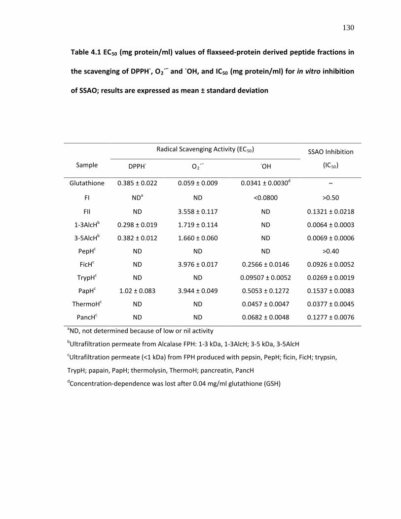
130
Table 4.1 EC50 (mg protein/ml) values of flaxseed-protein derived peptide fractions in
the scavenging of DPPH˙, O2˙– and ˙OH, and IC50
(mg protein/ml) for in vitro inhibition
of SSAO; results are expressed as mean ± standard deviation
Sample
Radical Scavenging Activity (EC50 SSAO Inhibition
(IC
)
50) DPPH˙ O2˙ ˙OH –
Glutathione 0.385 ± 0.022 0.059 ± 0.009 0.0341 ± 0.0030 – d
FI ND ND a <0.0800 >0.50
FII ND 3.558 ± 0.117 ND 0.1321 ± 0.0218
1-3AlcH 0.298 ± 0.019 b 1.719 ± 0.114 ND 0.0064 ± 0.0003
3-5AlcH 0.382 ± 0.012 b 1.660 ± 0.060 ND 0.0069 ± 0.0006
PepH ND c ND ND >0.40
FicH ND c 3.976 ± 0.017 0.2566 ± 0.0146 0.0926 ± 0.0052
TrypH ND c ND 0.09507 ± 0.0052 0.0269 ± 0.0019
PapH 1.02 ± 0.083 c 3.944 ± 0.049 0.5053 ± 0.1272 0.1537 ± 0.0083
ThermoH ND c ND 0.0457 ± 0.0047 0.0377 ± 0.0045
PancH ND c ND 0.0682 ± 0.0048 0.1277 ± 0.0076 aND, not determined because of low or nil activity bUltrafiltration permeate from Alcalase FPH: 1-3 kDa, 1-3AlcH; 3-5 kDa, 3-5AlcH cUltrafiltration permeate (<1 kDa) from FPH produced with pepsin, PepH; ficin, FicH; trypsin,
TrypH; papain, PapH; thermolysin, ThermoH; pancreatin, PancH dConcentration-dependence was lost after 0.04 mg/ml glutathione (GSH)

131
scavenged about 5–90% O2˙– at 0.0313–0.5% protein (Sakanaka & Tachibana, 2006). In
this study, the <1 kDa permeate of trypsin and thermolysin FPH as well as Alcalase
cationic peptide fraction I showed meagre antioxidant properties by scavenging only
12.6, 17.5 and 19.4% O2˙–, respectively at 2 mg protein/ml whereas both the pepsin and
pancreatin treatments yielded LMW peptides that could not scavenge O2˙–
The ˙OH-scavenging property of the flaxseed peptides was evaluated using an
assay similar to the physiological system whereby ˙OH was generated by reaction of Fe
.
2+
and H2O2 (Fenton reaction). The ˙OH produced in the reaction was spin-trapped with
DMPO, and the intensity of the DMPO-OH adducts measured using ESR spectrometer. A
decrease in intensity or disappearance of the DMPO-OH signal is an indication of the
˙OH scavenging activity of antioxidants (Li et al., 2004). It was observed in this study that
most of the FPH fractions were capable of reducing the intensity of DMPO-OH signals at
various protein concentrations. Fig. 4.2B shows that there was a linear relationship
between ˙OH-scavenging activity and the amount of peptide samples in the assay.
Unlike the antioxidant properties observed in the DPPH˙ and O2˙– assays, the LMW
flaxseed peptides showed potent ˙OH-scavenging activities except for the pepsin
hydrolysate (Table 4.1). The thermolysin, pancreatin and ficin hydrolysates scavenged
86.5, 89.9 and 82.3% ˙OH, respectively at 0.2, 0.3 and 0.4 mg/ml, respectively. The least
cationic peptide fraction from Alcalase hydrolysate (FI) also displayed potent ˙OH-
scavenging activity with inhibition of 55.6, 76.7 and 81% ˙OH at 0.08, 0.12 and 0.2
mg/ml, whereas the stronger cationic peptides (FII) did not. This observation is inversely
correlated with their observed activity in the O2˙–-scavenging assay. Moreover, the ˙OH-

132
scavenging properties of these flaxseed peptide fractions reported in this paper are
better than the recently reported activity of crude hydrolysates from Flavourzyme
digestion of flaxseed proteins (IC50, 1.56–3.06 mg/ml) (Marambe et al., 2008). Previous
studies have also reported that, using different spectrophotometric assay systems, the
peptide fractions from Alcalase chickpea protein hydrolysate (1.5 mg/ml) scavenged
38% to 81% ˙OH (Li et al., 2008a) whereas treatment of egg yolk protein with Bacillus sp
proteinase yielded hydrolysates that scavenged 74% of ˙OH at 0.5 mg/ml (Sakanaka &
Tachibana, 2006). In addition, a recent study reported that whey protein hydrolysate
and its peptide fractions showed moderate antioxidant properties against ˙OH and other
free radicals when measured by ESR spectroscopy (Peng, Xiong, & Kong, 2009) similar to
the method used in this present study. Contrary to the results in the DPPH˙ and O2˙–
assays, the HMW flaxseed peptide fractions did not show ˙OH-scavenging activity. It
should be noted that this assay method has a limitation as it is almost impossible to
determine whether the activity of antioxidants is due to scavenging of ˙OH or chelation
of Fe2+, since most potential antioxidants are also good metal chelators that might
decrease ˙OH production (Huang et al., 2005), thus reducing the intensity of the DMPO-
OH adduct. However, Peng et al. (2009) recently reported that the ˙OH-scavenging
activity observed in their study for whey protein hydrolysate fractions could be
attributed to the hydrolysates, which they reported to possess poor Fe2+
chelating
activities.

133
4.3.3. Peptide-induced inhibition of SSAO
The in vitro SSAO inhibitory activities of the flaxseed protein-derived peptide
fractions are shown in Fig. 4.3; all the peptide samples evaluated in this study exhibited
concentration-dependent inhibition of SSAO activity which was also dependent on size
of the peptides. The HMW peptides from Alcalase hydrolysate potently inhibited SSAO
activity at low concentrations. Moreover, the LMW peptide fractions as well as Alcalase
cationic peptide FII also inhibited SSAO activity. The weakest inhibitory activity was
observed for the pepsin hydrolysate LWM fraction and the Alcalase cationic peptide FI.
The IC50 values for the peptide fractions in SSAO inhibition are shown in Table 4.1. To
the best of our knowledge, this work is the first to report the activity of enzymatic food
protein hydrolysates in inhibition of SSAO activity. Since physiological SSAO reaction
generates H2O2 and reactive aldehydes that play important roles in pathogenesis of
disease conditions, its inhibition by these flaxseed peptide fractions may constitute a
step in treatment of inflammation and other related diseases.

134
Figure 4.3 Inhibition of semicarbazie-sensitive amine oxidase (SSAO) by flaxseed
peptide fractions; inhibitory activity of peptides was dependent on size and nature of
the peptides; thus, the higher MW peptides showed the best activity
0.00 0.010
20
40
60
80
0.1 0.2 0.3 0.4 0.5
1-3AlcH3-5AlcHFIIFIPepHFicHTrypHPapHThermoHPancH
Concentration (mg/mL)
% S
SAO
inhi
bitio
n

135
4.3.4. Peptide-induced inhibition of in vitro and ex vivo NO production
The cellular toxicity of NO has been associated with its reaction derivatives
especially ONOO–, which could lead to DNA fragmentation and protein structure
modification. Thus, NO scavengers could lower the risk of cellular and tissue damages
associated with excessive NO production. Fig. 4.4 shows the in vitro scavenging activities
of the flaxseed peptide fractions against NO; at 0.2 mg/ml, the peptide fractions showed
weak NO scavenging properties. Both the thermolysin and pancreatin FPH fractions gave
the strongest activity by scavenging 27.1% and 35.1% NO, respectively. The other
peptide fractions scavenged ≤20.3% NO at the same concentration. Moreover, in order
to evaluate their anti-inflammatory properties, the flaxseed peptide samples were
screened for effects on LPS-induced NO production in macrophages. The treatment of
RAW 264.7 macrophages with LPS resulted in increased NO production in the cell
culture (Fig. 4.5). The addition of PMB to the cell culture prior to LPS activation resulted
in normalized NO production, since PMB binds and inactivates LPS. On treatment of the
macrophage cultures with LPS and the various flaxseed peptides, the pepsin, ficin and
papain FPH fractions were observed to exhibit concentration-dependent inhibition of
LPS-induced NO production in the macrophages with IC50 values of 0.250, 0.504 and
0.215 mg protein/ml, respectively (Fig. 4.5A–C). At 1 mg protein/ml, the pepsin and
papain FPH fractions completely inhibited the endotoxin-induced NO production to the
basal level observed for control and PMB treatments. Moreover, these flaxseed peptide
fractions did not show cytotoxicity against the cells within the range of concentrations
studied using the MTT staining assay (Fig. 4.5D). It is noteworthy that, despite showing

136
Figure 4.4 In vitro scavenging activities of 0.2 mg/ml flaxseed protein-derived peptide
fractions against nitric oxide (NO) produced from sodium nitroprusside
FI FII
1-3AlcH
3-5AlcH
PepH
FicHTryp
HPap
H
Therm
oHPan
cH0
10
20
30
40
% N
O S
cave
ngin
g A
ctiv
ity

137
Figure 4.5 Effects of flaxseed protein-derived (A) PepH, (B) FicH and (C) PapH on LPS-
induced nitric oxide (NO) production in RAW 264.7 cells; the cells were treated as
follows: no LPS (control); LPS (100 ng/ml) only; LPS (100 ng/ml) plus PMB (50 µg/ml);
and LPS (100 ng/ml) plus 0.2–1.0 mg protein/ml flaxseed peptide fraction. PMB was
used as positive control. The treated cells were incubated at 37°C and 5% CO2
for 24 h
followed by determination of cellular production of NO and cell viability. Each bar
represents mean of triplicate determinations; bars with the same letter within each
graph are not significantly different at p = 0.05 (D) Viability of RAW 264.7 cells in the
presence of the peptide fractions, which showed no toxicity against the growth of the
macrophages
Contro
lLP
S
LPS+P
MB 0.2 0.4 0.6 0.8 1.00
2
4
6
8
10
12
PepH (mg/ml)+ LPS (100 ng/ml)
d,e
a
b
e
c
d d,eeN
itrite
( µM
)
Contro
lLP
S
LPS+PMB 0.2 0.4 0.6 0.8 1.0
0
2
4
6
8
10
12
FicH (mg/ml)+ LPS (100 ng/ml)
d
a
b
d
b
c
d
c
Nitr
ite ( µ
M)
A B

138
0.2 0.4 0.6 0.8 1.00
25
50
75
100
125
PepH (mg/ml)
Rel
ativ
e ce
ll vi
abili
ty (%
)
0.2 0.4 0.6 0.8 1.0
FicH (mg/ml)
0.2 0.4 0.6 0.8 1.0
PapH (mg/ml)
Control
LPS
LPS+PMB 0.2 0.4 0.6 0.8 1.00
2
4
6
8
10
12
PapH (mg/ml)+ LPS (100 ng/ml)
e,f
a
b
f
c,dc
f
d,e
Nitr
ite ( µ
M)
C
D
139
the best in vitro NO scavenging activities (Fig. 4.4), the thermolysin and pancreatin FPH
fractions did not affect concentrations of NO in the cell cultures when compared to the
LPS-only treatment (data not shown)2. Thus, the ability of the flaxseed peptides to
scavenge NO in vitro is not correlated with their inhibitory activities against NO
production or their potential NO scavenging properties in the LPS-treated culture. It is
possible that the bioactive peptides present in thermolysin and pancreatin FPH were
metabolized to inactive fragments by the macrophages. It could also be suggested that
the active flaxseed peptide fractions may have altered the macrophage NO synthesis
pathway. It has been shown that the activity of potential therapeutic agents in inhibition
of NO production could be through inhibition of transcription factor NF-κB activation
and subsequent inhibition of iNOS mRNA and protein expressions in macrophages
(Chen, Shen, Lee, Hou, Yang, & Lee, 2001; Ho & Lin, 2008; Pan et al., 2008). To the best
of our knowledge, there is limited information in the literature regarding the effects of
enzymatic food protein hydrolysates on endotoxin-induced NO production in cell
cultures. A previous study reported that a hydrolyzed casein diet fed to young diabetes-
prone BB rats led to restoration of NO production to basal level in interleukin-1β-
induced overproduction of NO in the pancreatic islets (Malaisse, Olivares, Laghmich,
Ladrière, Sener, & Scott, 2000). Recently, lunasin, a naturally occurring oligopeptide
from soybean was reported to possess potential in vitro anti-inflammatory activity due
to its inhibitory effects on the production of NO and prostaglandin E2
2 See Figures S4.2 in Appendix B for data
through the
inhibition of iNOS and cyclooxygenase-2 protein expressions, respectively, in LPS-

140
activated RAW 264.7 macrophages (Dia, Wang, Oh, de Lumen, & Gonzalez de Mejia,
2009). However, a yam storage protein, dioscorin, had been shown to exhibit
immunomodulatory effects partly by inducing NO productions in macrophages in the
absence of LPS (Liu, Shang, Wang, Hsu, & Hou, 2007). Moreover, it was also observed in
this present study that the flaxseed protein-derived peptide fractions did not induce
excessive production of NO in the macrophages when LPS was eliminated with PMB
(data not shown). Furthermore, all the flaxseed protein-derived peptide fractions in this
study were also evaluated for cytotoxicity against two cancer cell lines, HL-60 (human
promyelocytic leukemia, ATCC CCL-240) and MCF7 (human breast adenocarcinoma,
ATCC HTB-22) using the MTT staining assay. The flaxseed peptide fractions did not show
cytotoxicity against these cancer cell lines at 0.2 mg protein/ml after 24 h of incubation
(data not shown)3
.
4.4. CONCLUSIONS
Enzymatic hydrolysis of flaxseed proteins by various proteases resulted in the
release of bioactive peptides with antioxidant and potential anti-inflammatory
properties. Bioactive properties of the flaxseed peptide fractions were dependent on
catalytic specificity of the proteases as well as the size of peptides in the resulting
protein hydrolysate fractions. Papain treatment released multifunctional flaxseed
peptides with MW less than 1 kDa that exhibited comparatively moderate biological
activities in all the in vitro antioxidant assays evaluated in this study. On the other hand,
3 See Figures S4.4 and S4.5 in Appendix B for data

141
pepsin-catalyzed hydrolysis yielded poor free radical-scavenging <1 kDa flaxseed
peptides but with excellent restoration of LPS-induced NO production to basal level in
macrophages. The other <1 kDa flaxseed peptide fractions showed potent or moderate
activities in scavenging free radicals; however, the <1 kDa cationic flaxseed peptide
fractions did not exhibit good antioxidant property except the least cationic fraction (FI)
that potently scavenged ˙OH. Overall, the HMW peptides (1-3 and 3-5 kDa) showed
better antioxidant properties than the LMW peptides except in scavenging ˙OH and
inhibition of NO production in macrophages. These bioactive properties could
encourage increased value-added utilization of flaxseed meal proteins for the
formulation of therapeutic products. Evaluation of the effects of the flaxseed protein-
derived peptides in appropriate animal disease models is required to confirm some of
the preliminary results obtained in this work.
ACKNOWLEDGEMENTS
The present work was supported by operating and equipment grants to REA from the
Advanced Foods and Materials Network of Centres of Excellence (AFMnet) and the
Natural Sciences and Engineering Research Council of Canada (NSERC). CCU is a
recipient of a Manitoba Health Research Council Graduate Scholarship and NSERC
Summer Research Program in Taiwan Fellowship.

142
4.5. REFERENCES
Agriculture and Agri-Food Canada. (2007). Flaxseed: situation and outlook. Bi-weekly Bulletin, 20(3), AAFC No. 2081/E
Ames, B. N. (1983). Dietary carcinogens and anticarcinogens: oxygen radicals and degenerative diseases. Science, 221, 1256–1264
Ames, B. N., Shigena, M. K., & Hegen, T. M. (1993). Oxidants, antioxidants and the degenerative diseases of aging. Proceedings of the National Academy of Science U.S.A., 90, 7915–7922
Boomsma, F., De Kam, P. J., Tjeerdsma, G., Van Den Meiracker, A. H., & Van Veldhuisen, D. J. (2000). Plasma semicarbazide-sensitive amine oxidase (SSAO) is an independent prognostic marker for mortality in chronic heart failure. European Heart Journal, 21, 1859–1863
Chen, Y-.C., Shen, S-.C., Lee, W-.R., Hou, W-.C., Yang, L-.L., & Lee, T. J. F. (2001). Inhibition of nitric oxide synthase inhibitors and lipopolysaccharide induced inducible NOS and cyclooxygenase-2 gene expressions by rutin, quercetin, and quercetin pentaacetate in RAW 264.7 macrophages. Journal of Cellular Biochemistry, 82, 537–548
Cumby, N., Zhong, Y., Naczk, M., & Shahidi, F. (2008). Antioxidant activity and water-holding capacity of canola protein hydrolysates. Food Chemistry, 109, 144–148
Dev, D. K., & Quensel, E. (1988). Preparation and functional properties of linseed protein products containing differing levels of mucilage. Journal of Food Science, 53, 1834–1837
Dia, V. P., Wang, W., Oh, V. L., de Lumen, B. O., & Gonzalez de Mejia, E. (2009). Isolation, purification and characterization of lunasin from defatted soybean flour and in vitro evaluation of its anti-inflammatory activity. Food Chemistry, 114, 108–115
Dodin, S., Cunnane, S. C., Mâsse, B., Lemay, A., Jacques, H., Asselin, G., Tremblay-Mercier, J., Marc, I., Lamarche, B., Légaré, F., & Forest, J.-C. (2008). Flaxseed on cardiovascular disease markers in healthy menopausal women: a randomized, double-blind, placebo-controlled trial. Nutrition, 24, 23–30
Fiorentino, A., Ricci, A., D’Abrosca, B., Pacifico, S., Golino, A., Letizia, M., Piccolella, S., & Monaco, P. (2008). Potential food additives from Carex distacya roots: Identification and in vitro antioxidant properties. Journal of Agricultural and Food Chemistry, 56, 8218–8225

143
Gao, R., Yuan, Z., Zhao, Z., & Gao, X. (1998). Mechanism of pyrogallol autoxidation and determination of superoxide dismutase enzyme activity. Bioelectrochemistry and Bioenergetics, 45, 41–45
Göktürk, C., Nilsson, J., Nordquist, J., Kristensson, M., Svensson, K., Söderberg, C., Isrealson, M., Garpenstrand, H., Sjöquist, M., Oreland, L., Forsberg-Nilsson, K. (2003). Overexpression of semicarbazide-sensitive amine oxidase in smooth muscle cells leads to an abnormal structure of the aortic elastic laminas. American Journal of Pathology, 163, 1921–1928
Hall III, C., Tulbek, M. C., & Xu, Y. (2006). Flaxseed. Advances in Food and Nutrition Research, 51, 1–97
Ho, S-.C., & Lin, C-.C. (2008). Investigation of heat treating conditions for enhancing the anti-inflammatory activity of citrus fruits (Citrus reticulate) peels. Journal of Agricultural and Food Chemistry, 56, 7976–7982
Hou, W.-C., Chen, Y.-C., Chen, H.-J., Lin, Y.-H., Yang, L.-L., & Lee, M.-H. (2001). Antioxidant activities of trypsin inhibitor, a 33 kDa root storage protein of sweet potato (Ipomoea batatas L. Lam cv. Tainong 57). Journal of Agricultural and Food Chemistry, 49, 2978–2981
Huang, D., Ou, B., & Prior, R. L. (2005). The chemistry behind antioxidant capacity assays. Journal of Agricultural and Food Chemistry, 53, 1841–1856
Kris-Etherton, P. M., Hecker, K. D., Bonanome, A., Coval, S. M., Binkoski, A. E., Hilpert, K. F., Griel, A. E., & Etherton, T. D. (2002). Bioactive compounds in foods: their role in the prevention of cardiovascular disease and cancer. American Journal of Medicine, 113, 71S–88S
Li, L., Abe, Y., Kanagawa, K., Usui, N., Imai, K., Mashino, T., Mochizuki, M., & Miyata, N. (2004). Distinguishing the 5,5-dimethyl-1-pyrroline N-oxide (DMPO)-OH radical quenching effect from the hydroxyl radical scavenging effect in the ESR spin-trapping method . Analytica Chimica Acta, 512, 121–124
Li, Y., Jiang, B., Zhang, T., Mu, W., & Liu, J. (2008a). Antioxidant and free radical-scavenging activities of chickpea protein hydrolysate (CPH). Food Chemistry, 106, 444–450
Li, X-.X., Han, L-.J., & Chen, L-.J. (2008b). In vitro antioxidant activity of protein hydrolysates prepared from corn gluten meal. Journal of the Science of Food and Agriculture, 88, 1660–1666
Lin, S.-Y., Wang, C.-C., Lu, Y.-L., Wu, W.-C., & Hou, W.-C. (2008). Antioxidant, anti-semicarbazide-sensitive amino oxidase, and anti-hypertensive activities of

144
geraniin isolated from Phyllanthus urinaria. Food and Chemical Toxicology, 46, 2485–2492
Liu, B., Gao, H-.M., Wang, J-.Y., Jeohn, G-.H., Cooper, C. L., & Hong, J-.S. (2002). Role of nitric oxide in inflammation-mediated neurodegeneration. Annals of the New York Academy of Sciences, 962, 318–331
Liu, D-.Z., Wu, W-.C., Liang, H-.J., & Hou, W-.C. (2007a). Antioxidant and semicarbazide-sensitive amine oxidase inhibitory activities of alginic acid hydroxamates. Journal of the Science of Food and Agriculture, 87, 138–146
Liu, Y-.W., Shang, H.-F., Wang, C.-K., Hsu, F.-L., & Hou, W.-C. (2007b). Immunomodulatory activity of dioscorin, the storage protein of yam (Dioscorea alata cv. Tainong No. 1) tuber. Food and Chemical Toxicology, 45, 2312–2318
Magyar, K., Mészáros, Z., & Mátyus, P. (2001). Semicarbazide-sensitive amine oxidase. Its physiological significance. Pure and Applied Chemistry, 73, 1393–1400
Malaisse, W. J., Olivares, E., Laghmich, A., Ladrière, L., Sener, A., & Scott, F. W. (2000). Feeding a protective hydrolyzed casein diet to young diabetes-prone BB rats affects oxidation of L[U-14
Marambe, P. W. M. L. H. K., Shand, P. J., & Wanasundara, J. P. D. (2008). An in vitro investigation of selected biological activities of hydrolyzed flaxseed (Linum usitatissimum L.) proteins. Journal of the American Oil Chemists’ Society, 85, 1155–1164
C] glutamate in islets and Peyer’s patches, reduces abnormally high mitotic activity in mesenteric lymph nodes, enhances islet insulin and tends to normalize NO production. International Journal of Experimental Diabetes Research, 1, 121–130
Marklund, S., & Marklund, G. (1974). Involvement of the superoxide anion radical in the autoxidation of pyrogallol and a convenient assay for superoxide dismutase. European Journal of Biochemistry, 47, 469–474
Markwell, M. A. C., Haas, S. M., Biebar, L. L., & Tolbert, N. E. (1978). A modification of the Lowry procedure to simplify protein determination in membrane and in protein samples. Analytical Biochemistry, 87, 206–211
Omoni, A. O., & Aluko, R. E. (2006a). Mechanism of the inhibition of calmodulin-dependent neuronal nitric oxide synthase by flaxseed protein hydrolysates. Journal of the American Oil Chemists’ Society, 83, 335–340
Omoni, A. O., & Aluko, R. E. (2006b). Effect of cationic flaxseed protein hydrolysate fractions on the in vitro structure and activity of calmodulin-dependent endothelial nitric oxide synthase. Molecular Nutrition and Food Research, 50, 958–966

145
Oomah, B. D., & Mazza, G. (1993). Flaxseed proteins – a review. Food Chemistry, 48, 109–114
Pacher, P., Beckman, J. S., & Liaudet, L. (2007). Nitric oxide and peroxynitrite in health and disease. Physiological Reviews, 87, 315–424
Pan, M-.H., Chang, Y-.H., Tsai, M-.L., Lai, C-.S., Ho, S.-Y., Badmaev, V., & Ho, C-.T. (2008). Pterostilbene suppressed lipopolysaccharide-induced up-expression of iNOS and COX-2 in murine macrophages. Journal of Agricultural and Food Chemistry, 56, 7502–7509
Peng, X., Xiong, Y. L., & Kong, B. (2009). Antioxidant activity of peptide fractions from whey protein hydrolysates as measured by electron spin resonance. Food Chemistry, 113, 196–201
Prior, R. L., Wu, X., & Schaich, K. (2005). Standardized methods for the determination of antioxidant capacity and phenolics in foods and dietary supplements. Journal of Agricultural and Food Chemistry, 53, 4290–4302
Sakanaka, S., & Tachibana, Y. (2006). Active oxygen scavenging activity of egg-yolk protein hydrolysates and their effects on lipid oxidation in beef and tuna homogenates. Food Chemistry, 95, 243–249
Stolen, C. M., Madanat, R., Marti, L., Kari, S., Yegutkin, G. G., Sariola, H., Zorzano, A., & Jalkanen, S. (2004a). Semicarbazide-sensitive amine oxidase overexpression has dual consequences: insulin mimicry and diabetes-like complications. FASEB Journal, 18, 702–704
Stolen, C. M., Yegutkin, G. G., Kurkijarvi, R., Bono, P., Alitalo, K., & Jalkanen, S. (2004b). Origins of serum semicarbazide-sensitive amine oxidase. Circulation Research, 95, 50–57
Szutowicz, A., Kobes, R. D., & Orsulak, P. J. (1984). Colorimetric assay for monoamine oxidase in tissues using peroxidase and 2′,2-azinodi(3-ethylbenzthiazoline-6-sulfonic acid) as chromogen. Analytical Biochemistry, 138, 86–94
Torre, D., Pugliese, A., & Speranza, F. (2002). Role of nitric oxide in HIV-1 infection: friend or foe? Lancet Infectious Diseases, 2, 273–280
Udenigwe, C. C., Lin, Y.-S., Hou, W.-C., & Aluko, R. E. (2009). Kinetics of the inhibition of renin and angiotensin I-converting enzyme by flaxseed protein hydrolysate fractions. Journal of Functional Foods, 1, 199-207
Wang, J-.S., Zhao, M-.M., Zhao, Q-.Z., & Jiang, Y-.M. (2007). Antioxidant properties of papain hydrolysates of wheat gluten in different oxidation systems. Food Chemistry, 101, 1658–1663

146
Wu, H.-C., Chen, H.-M., & Shiau, C-.Y. (2003). Free amino acids and peptides as related to antioxidant properties in protein hydrolysates of mackerel (Scomber austriasicus). Food Research International, 36, 949–957
Xie, Z., Huang, J., Xu, X., & Jin, Z. (2008). Antioxidant activity of peptides isolated from alfalfa leaf protein hydrolysate. Food Chemistry, 111, 370–376

147
APPENDIX B: SUPPLEMENTAL INFORMATION
Figure S4.1 Electron spin resonance (ESR) spectra showing concentration-dependent
decrease in the intensity of DMPO-OH adduct spin resonance induced by flaxseed
protein-derived (A) cationic peptide fraction FI (0.04–0.20 mg/ml) and (B) FicH (0.08–
0.40 mg/ml); this represents the scavenging of hydroxyl radicals (˙OH) by the flaxseed
peptides samples. Details of the ˙OH scavenging activity (%) for all the samples are
shown in Figure 4.2B and Table 4.1, and discussed in Section 4.3.2
Blank
0.04
0.08
0.12
0.20
(A)
Blank
0.08
0.20
0.30
0.40
(B)

148
Figure S4.2 Screening for inhibitory activity of the flaxseed peptide samples (0.1
mg/ml) against nitric oxide production in lipopolysaccharide (LPS)-activated RAW
264.7 macrophages; no LPS (control), LPS (100 ng/ml) only, LPS (100 ng/ml) plus
polymyxin B (PMB) (50 µg/ml), and LPS (100 ng/ml) plus flaxseed peptide fraction. Bar
with different letters are significantly different at P=0.05. The most active samples
(PepH, FicH and PapH) were selected for further studies
Contro
lLP
S
LPS+P
MB FI FII
1-3AlcH
3-5AlcH
PepH
FicHTryp
HPap
H
Therm
oHPan
cH0
5
10
15
20
25
f f
a,b
c,d,e
a,b
b,c,d
a
b,c a,b
e,d ee,d
e
Nitr
ite p
rodu
ctio
n ( µ
M)
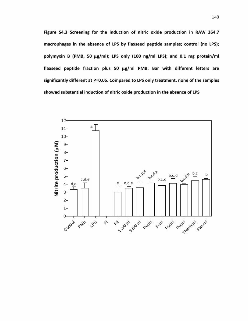
149
Figure S4.3 Screening for the induction of nitric oxide production in RAW 264.7
macrophages in the absence of LPS by flaxseed peptide samples; control (no LPS);
polymyxin B (PMB, 50 µg/ml); LPS only (100 ng/ml LPS); and 0.1 mg protein/ml
flaxseed peptide fraction plus 50 µg/ml PMB. Bar with different letters are
significantly different at P=0.05. Compared to LPS only treatment, none of the samples
showed substantial induction of nitric oxide production in the absence of LPS
Contro
lPMB
LPS FI FII
1-3AlcH
3-5AlcH
PepH
FicHTryp
HPap
H
Therm
oHPan
cH0
1
2
3
4
5
6
7
8
9
10
11
12
Nitr
ite p
rodu
ctio
n ( µ
M)
d,ec,d,e
a
e c,d,eb,c
,d,e
b,c,dbb,c
b,c,d,
eb,c,d
b,c,d,
e
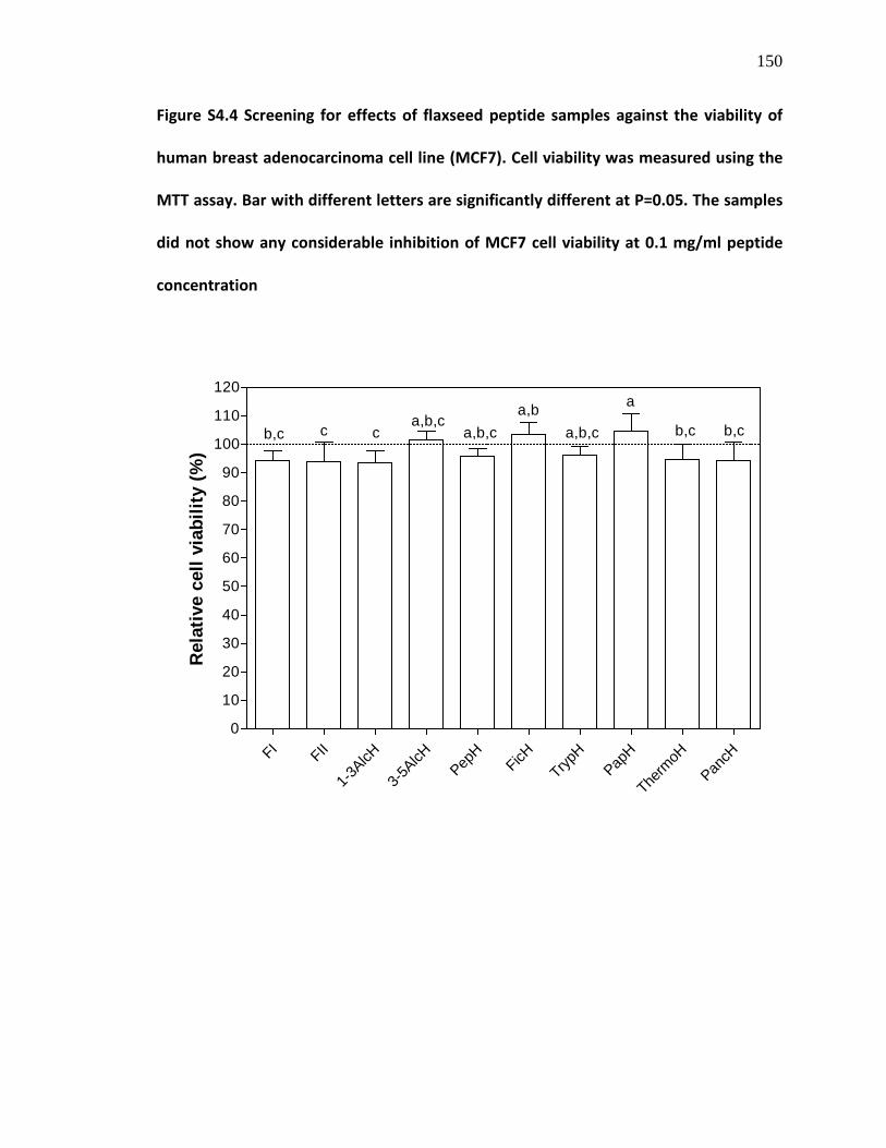
150
Figure S4.4 Screening for effects of flaxseed peptide samples against the viability of
human breast adenocarcinoma cell line (MCF7). Cell viability was measured using the
MTT assay. Bar with different letters are significantly different at P=0.05. The samples
did not show any considerable inhibition of MCF7 cell viability at 0.1 mg/ml peptide
concentration
FI FII
1-3AlcH
3-5AlcH
PepH
FicHTryp
HPap
H
Therm
oHPan
cH0
10
20
30
40
50
60
70
80
90
100
110
120
b,c c ca,b,c
a,b,ca,b
a,b,c
a
b,c b,c
Rel
ativ
e ce
ll vi
abili
ty (%
)

151
Figure S4.5 Screening for effects of flaxseed peptide samples against the viability of
human promyelocytic leukemia cell line (HL-60). Cell viability was measured using the
MTT assay. Bar with different letters are significantly different at P=0.05. The samples
did not show any considerable inhibition of HL-60 cell viability at 0.1 mg/ml peptide
concentration
FI FII
1-3AlcH
3-5AlcH
PepH
FicHTryp
HPap
H
Therm
oHPan
cH0
10
20
30
40
50
60
70
80
90
100
110
120
Rel
ativ
e ce
ll vi
abili
ty (%
)
b,c c ca
b,ca
ca,b
c b,c

152
TRANSITION STATEMENT
The multifunctional bioactive properties of some of the flaxseed peptides produced in
this study have promising application in intervention against multiple human disease
conditions especially hypertension and oxidative stress-induced diseases. In order to
address specific objectives two and three of this thesis, the ACE and renin-inhibiting
flaxseed protein-derived cationic peptide fraction II (FII) was targeted against
calmodulin (CaM) to investigate if the positively-charged peptide fraction could bind the
negatively-charged CaM with concomitant inhibition of CaM-dependent
phosphodiesterase (CaMPDE), which have been implicated in the aetiology and
progression of chronic human diseases. The following chapter discusses the kinetics of
inhibition of CaMPDE by flaxseed cationic peptides (FI and FII) and fractionation of
peptides from FII that could simultaneously inhibit the activities of ACE, renin and
CaMPDE. Multifunctional peptides possess an advantage over single-function peptides
since they can target multiple symptoms and risk factors of chronic human diseases,
thus reducing the cost associated with developing many nutraceutical products.

153
CHAPTER FIVE
MANUSCRIPT 3
MULTIFUNCTIONAL PEPTIDES FROM A CATIONIC FRACTION OF FLAXSEED PROTEIN
HYDROLYSATES
C. C. UDENIGWEa, R. E. ALUKOa,b
aDepartment of Human Nutritional Sciences, University of Manitoba, Winnipeg, MB R3T
2N2, Canada; b
The Richardson Centre for Functional Foods and Nutraceuticals,
University of Manitoba, Winnipeg, MB R3T 2N2, Canada.
To be submitted for publication
154
5.0. ABSTRACT
Alcalase hydrolysis of flaxseed protein fractions liberated cationic peptides, which were
separated into two major fractions (FI and FII) by chromatography using a cation-
exchange column. Due to their cationic property, the peptide fractions bound and
inactivated calmodulin with concomitant inhibition of calmodulin-dependent
phosphodiesterase (CaMPDE); this activity was substantially reduced with increased
CaM concentrations. Enzyme kinetics studies showed competitive inhibition of CaMPDE
for FI and FII with enzyme-inhibitor dissociation constants (K i
) of 0.0202 and 0.0511
mg/ml, respectively. The cationic FII was further fractionated by reverse phase HPLC to
concentrate the active peptides. All the HPLC fractions inhibited CaMPDE, angiotensin
converting enzyme and renin activities at 0.1, 0.2 and 1.0 mg/ml, respectively, but the
multifunctional activities was more pronounced in HPLC fraction F6. The multifunctional
properties of the cationic peptide fractions can increase their use in targeting multiple
symptoms of cardiovascular and other diseases, considering the roles of these enzymes
in aetiology and progression of chronic human diseases.
KEYWORDS: Calmodulin-dependent phosphodiesterase; Angiotensin converting
enzyme; Renin; Multifunctional peptides; Flaxseed proteins; Enzyme inhibition
kinetics

155
5.1. INTRODUCTION
Aberrant production of angiotensin converting enzyme (ACE), renin and
calmodulin (CaM)-dependent phosphodiesterase (CaMPDE) have been observed in
various health conditions, and are related to aetiology and progression of chronic
human diseases. ACE and renin are key enzymes of the blood pressure-regulating renin-
angiotensin system (RAS) pathway and have been widely targeted with chemical
compounds for antihypertensive therapy. CaMPDE is expressed in various cell types
including coronary vascular smooth muscle cells (SMC), brain, kidney and heart (Bender
& Beavo, 2006; Kass et al., 2007) where it catalyzes the conversion of cyclic nucleotide
monophosphates (cGMP and cAMP) to GMP and AMP making it important in regulating
cellular processes mediated by the cyclic nucleotides including neurotransmission,
immune cell activity, vasodilation, cell growth and proliferation (Cho et al., 1998).
Chronic over-expression of CaMPDE has been found in atherosclerosis, heart pressure-
load stress, heart failure and vascular proliferation (Kass et al., 2007). CaMPDE inhibition
decreased CaMPDE-induced human SMC proliferation and also induced cAMP-mediated
apoptosis in cultured human leukemic cells, which are relevant activities toward
treatment of atherosclerosis and cancer (Rybalkin et al., 2003; Nagel et al., 2006; Jiang
et al., 1996). Moreover, angiotensin II (AT-II) increases CaMPDE expressions in SMC
through AT-II-mediated increase in cellular Ca2+ concentration (Kim et al., 2001); thus,
CaMPDE-induced pathogenesis can also be linked to RAS-mediated hypertension.
CaMPDE has also been implicated in pulmonary arterial hypertension (Schermuly et al.,
2007) and inflammation since CaMPDE is upregulated during monocyte differentiation
156
(Bender et al., 2005). Thus, CaM-binding agents can be used as specific CaMPDE
inhibitors in modulating the effects of increased expression of the enzyme.
Food protein-derived peptides have generated particular interests as adjuvant or
alternative therapeutic agents against chronic human health conditions. This has
provided the opportunity for diversification of the use of major protein-rich agricultural
crops beyond basic nutritional purposes. In a bid to improve the value of defatted
flaxseed meal, a number of bioactive peptide fractions have been reported from
enzymatic flaxseed protein hydrolysates and these peptides exhibited physiologically-
relevant activities such as antioxidant, anti-inflammatory, ACE and renin-inhibitory
activities (Marambe et al., 2008; Udenigwe et al., 2009a,b; Udenigwe & Aluko, 2010).
Cationic peptides from flaxseed protein were also reported to bind calmodulin (CaM)
with concomitant inhibition of CaM-dependent nitric oxide syntheses (Omoni & Aluko,
2006a, b). Moreover, food-derived peptides with more than one bioactive property can
be used to target multiple diseases or a disease with more than one symptom, which
will ultimately reduce the cost associated with developing different health promoting
products for particular diseases.
Therefore, the objectives of this project were to (1) determine the kinetics of
inhibition of CaMPDE by ACE and renin-inhibiting CaM-binding cationic peptide fractions
derived from flaxseed protein hydrolysates, and (2) separate the most multifunctional
cationic peptide fraction using high performance liquid chromatography (HPLC) to
concentrate the constituent multifunctional peptides that can simultaneously inhibit
ACE, renin and CaMPDE activities.

157
5.2. METHODS
5.2.1. Production of flaxseed cationic peptide fraction
The flaxseed protein-derived cationic peptide fraction was produced as
previously reported (Udenigwe et al., 2009a). Briefly, flaxseed protein isolate was
hydrolyzed with Alcalase (E/S 1:25) for 4 h followed by centrifugation at 14,941g for 30
min. The supernatant was subjected to nitrogen gas pressure-driven ultrafiltration using
a 1000 Da molecular weight cut-off membrane and the resulting permeate was
collected and freeze dried. A solution of the permeate (4 ml of 250 mg/ml) was loaded
onto an SP-Sepharose High Performance XK 50/20 cation exchange chromatography
column connected to an AKTA Fast Protein Liquid Chromatography system (Amersham-
GE Biosciences, Montreal, Canada). The sample was eluted from the column by first
washing with 1.5 column volume of 0.1 M ammonium acetate buffer (pH 7.5) to remove
unbound peptides followed by elution of the bound peptides using a gradient of 0–50%
0.5 M ammonium carbonate (pH 8.8) in 0.1 M ammonium acetate buffer at a flow rate
of 10 ml/min; elution was monitored as absorbance at 214 nm. After elution of the
bound peptides, two major peaks were observed; fractions within the peak that eluted
at a later time were pooled, freeze dried and subsequently used as cationic peptide
fraction II (FII).
5.2.2. CaM-dependent PDE (CaMPDE) inhibition assay
CaMPDE activity was measured by the luciferin-luciferase technique of Kizawa et
al. (1995) and Weiss et al. (1972) as recently reported by You et al. (2010). The reaction

158
mixture contained 0.05 U CaM, 0.1 mU phosphodiesterase, 0.15 mM CaCl2, 1 U
pyruvate kinase, 1 U myokinase, 1 mM cAMP, 25 mM ammonium acetate, 3 mM MgCl2,
0.26 mM phosphoenolpyruvate, 17 mM dithiothreitol, 0.1 nM guanosine 5′-
triphosphate, 10% bovine serum albumin and flaxseed peptide sample in 300 µl of 50
mM glycine-glycine buffer (pH 8.0). The assay mixture was incubated at 37°C using an
Eppendorf Thermomixer R for 30 min and the reaction was terminated by heating the
mixture in a water bath (95-100°C) to inactivate the enzymes, followed by centrifugation
at 10,000 × g for 10 min. A portion (10 µl) of the protein-free supernatant was placed in
a 1.5 ml microfuge tube followed by the addition of 100 µl of luciferin-luciferase reagent
(Promega ENLITEN® ATP Assay Bioluminescence Detection Kit). Thereafter, luminescence
was measured as relative luminescence unit (RLU) using a Turner Biosystems 20/20n
Luminometer to quantify ATP produced during assay. Blank assays were conducted for
all samples using heat-inactivated (95–100°C) phosphodiesterase whereas control assay
did not contain the peptides. CaMPDE inhibitory activity (%) of the active samples was
calculated as: {[(RLUc–RLUs)/ RLUc] × 100}, where RLUc and RLUs are the RLU of the
control and sample assays, respectively. Kinetics of the inhibition of CaMPDE was also
conducted by varying CaM concentration (0.0832–0.6656 U/ml) at fixed CaMPDE and
peptide concentrations. Enzyme inhibition kinetics parameter (Km, Vmax, Kmapp and
Vmaxapp) and mode of inhibition were determined by non-linear regression (Michaelis-
Menten equation) using GraphPad Prism version 5.00 for Windows (GraphPad Software,
San Diego, California, USA). Lineweaver-Burk double reciprocal plots were used to
represent the kinetics data. The dissociation constant of enzyme-inhibitor complex (K i)

159
was determined as {K i = [I]/α – 1}; [I] = inhibitor concentration, α = Kmapp/ Km
(Marangoni, 2003); Km is the dissociation constant of CaM-PDE complex; Vmax is the
reaction rate at saturating CaM concentration; Kmapp and Vmax
app
represent their
apparent values in the presence of the inhibitor.
5.2.3. ACE inhibition assay
ACE inhibition assay was conducted as previously described using FAPGG as ACE
substrate (Udenigwe et al., 2009a). Briefly, 1 ml of 0.5 mM FAPGG in 50 mM Tris-HCl
buffer containing 0.3 mM NaCl (pH 7.5) was mixed with 200 µl of the buffered peptide
sample (pH 7.5) followed by the addition of 20 µl of 1 U/ml ACE solution. The enzyme
activity was recorded for 2 min at room temperature as rate of decrease in absorbance
at 345 nm (∆A.min-1). The buffer was used instead of peptide fraction solutions in the
blank experiment. ACE inhibition (%) was calculated as: {1 – (∆A.min-1(sample) / ∆A.min-
1(blank)) × 100}, where ∆A.min-1
(sample) and ∆A.min-1(blank)
are the ACE reaction rates in the
presence and absence of the peptide samples, respectively.
5.2.4. Renin inhibition assay
Renin inhibition assay was conducted by fluorescence spectrometry as
previously described (Udenigwe et al., 2009a). Briefly, the total assay volume of 190 µl
contained 20 µl of 10 µM renin substrate, 10 µl of human recombinant renin and the
peptide sample in 50 mM Tris-HCl buffer containing 100 mM NaCl (pH 8.0). The Tris-HCl
buffer was used instead of the peptide solution in the blank experiment. The renin

160
substrate and peptide samples were mixed and pre-warmed to 37°C for 10 min prior to
the assay. Thereafter, the reaction was initiated by adding renin to the mixture. The
increase in fluorescence intensity was measured for 10 min in a JASCO FP-6300
spectrofluorimeter (Japan Spectroscopic Company, Tokyo, Japan) with a thermostated
cell compartment that was maintained at 37°C using a circulatory water bath. The
spectrofluorimeter was set at excitation wavelength of 340 nm, emission wavelength of
490 nm, excitation bandwidth of 5 nm and emission bandwidth of 10 nm. Renin activity
was expressed as reaction rate, fluorescence intensity unit per min (FIU.min-1) and
inhibitory activity (%) of the peptide samples was calculated as: {[(FIU.min-1(blank) –
FIU.min-1(sample)) / FIU.min-1
(blank)] × 100}, where FIU.min-1(blank) and FIU.min-1
(sample)
are
the renin reaction rates in the absence and presence of the peptide samples,
respectively.
5.2.5. High Performance Liquid Chromatography (HPLC)
The multifunctional cationic peptide fraction II (FII) was fractionated using
reverse-phase HPLC (RP-HPLC) technique. An aqueous solution of FII (0.2 ml of 100
mg/ml) was subjected to RP-HPLC on a Phenomenex C12 column (21 mm × 250 mm)
connected to a Waters HPLC system equipped with a photodiode array detector. The
column was pre-equilibrated with 0.1% trifluoroacetic acid (TFA) in water (solvent A),
and both the sample and solvents were filtered with 0.2 µm Fisherbrand general
filtration membrane filters prior to fractionation. The sample was eluted from the
column at a flow rate of 10 ml/min with a linear gradient of 0-50% methanol that also

161
contained 0.1% TFA (solvent B) for 45 min, maintained at 100%B for additional 10 min
followed by equilibration with 100%A for 10 min. Peptide elution was monitored as
absorbance at 214 nm. Fractions were collected using an automated fraction collector
every 0.5 min and pooled into 10 fractions of distinct peaks or groups of peaks. The
pooled fractions were desolventized in a rotary evaporator while the aqueous residues
were freeze dried and stored at -20°C for further use.
5.3. RESULTS AND DISCUSSION
Food protein-derived low MW bioactive peptides have gained particular interest
for use as therapeutic agents in human disease intervention due to safety of the natural
peptide products, low cost and potential efficient absorption through the enterocytes
into blood circulation to exert their activities in target tissues. The activities of these
peptides depend largely on the peptide sequence, molecular size, and physicochemical
properties of the constituent amino acid residues (Pripp et al., 2004). Thus, careful
processing of enzymatic food protein hydrolysates can lead to enrichment of particular
structural features in peptides, which can exert multifunctional physiological activities
depending on the nature of the molecular disease targets. For example, the rich arginine
composition of flaxseed proteins was basis for the production of highly cationic peptide
fractions that bound and inactivated negatively-charged CaM (Omoni & Aluko, 2006a,
b), and exhibited antioxidant activities in addition to inhibition of enzymes of the renin-
angiotensin system (Udenigwe et al., 2009a, b). CaM-binding oligopeptides have been
identified from insect venom (Martínez-Luis et al., 2007), but the high molecular size

162
reduces their prospective use as enteral therapeutic agents due to concerns about low
levels of absorption and bioavailability. Thus, the current study investigated the
bioactive properties of smaller peptides (<1 kDa) in order to increase their potential use
in human.
In this study, two cationic amino acids-enriched fractions from flaxseed proteins
were targeted against CaM in order to inhibit the activity of CaM-dependent PDE. The
cationic peptide fraction FI showed concentration-dependent inhibition of the activity of
CaMPDE in vitro with maximum inhibition of 41.6% at 0.09 mg/ml peptides (Figure 5.1).
Moreover, FII also showed concentration-dependent CaMPDE inhibition resulting in a
loss of 25% of the enzyme activity at 0.1 mg/ml. These activities are slightly lower than
that of previously reported casein-derived cationic peptide fraction, which inhibited 50%
of CaM-activated PDE activity at 0.014 mg/ml (Kizawa et al., 1995). Although FII
contained more cationic peptides that could bind and inactive CaM, the CaMPDE-
inhibitory activity of FII was lower than FI. This is contrary to results of similar previous
investigations on CaM-dependent nitric oxide synthases and protein kinase II, where the
more cationic peptide fractions displayed stronger interactions with CaM leading to
more potent inhibition of CaM-dependent activities of the enzymes (Li & Aluko, 2005;
Omoni & Aluko, 2006a, b). This discrepancy could be due to other peptides and non-
peptides that are present in FI and could bind CaM or directly inhibit PDE leading to
enhanced potency of the peptide fraction. CaM-binding activities have been reported
for various natural products including alkaloids, coumarins, terpenoids and lignans
(Rojas et al., 2003; Martínez-Luis et al., 2007). Therefore, the presence of small amounts

163
of flaxseed lignans in the cationic peptide fraction can contribute to increased CaM-
binding activity. In order to verify that the observed CaMPDE inhibition in this study was
due to CaM inactivation, different peptide concentrations were targeted against
CaMPDE at different CaM concentrations. As shown in Figure 5.2A, the residual
Figure 5.1 Concentration-dependent inhibition of calmodulin-dependent
phosphodiesterase (CaMPDE) by flaxseed-derived cationic fractions FI and FII
CaMPDE activity was dependent on CaM concentration at three different peptide
concentrations; the extent of FI-induced CaMPDE inhibition was significantly (P<0.05)
reduced by increasing CaM concentration. This trend was also observed for FII where
the CaMPDE-inhibiting activity of the peptide fraction at 0.1 mg/ml was overcome by
0
10
20
30
40
50FIFII
0.05
0.06
0.08
0.07
0.01
0.0010.0
90.1
Peptide concentration (mg/ml)
CaM
PDE
inhi
bitio
n (%
)
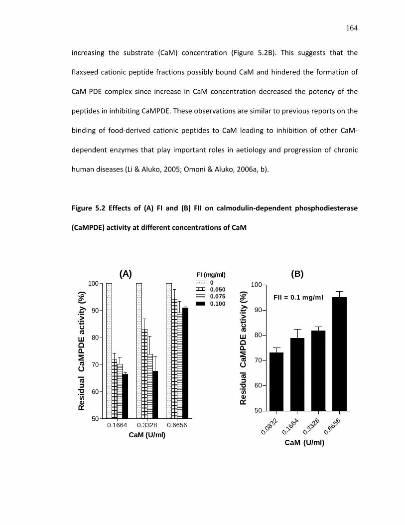
164
increasing the substrate (CaM) concentration (Figure 5.2B). This suggests that the
flaxseed cationic peptide fractions possibly bound CaM and hindered the formation of
CaM-PDE complex since increase in CaM concentration decreased the potency of the
peptides in inhibiting CaMPDE. These observations are similar to previous reports on the
binding of food-derived cationic peptides to CaM leading to inhibition of other CaM-
dependent enzymes that play important roles in aetiology and progression of chronic
human diseases (Li & Aluko, 2005; Omoni & Aluko, 2006a, b).
Figure 5.2 Effects of (A) FI and (B) FII on calmodulin-dependent phosphodiesterase
(CaMPDE) activity at different concentrations of CaM
0.083
2
0.166
4
0.332
8
0.665
650
60
70
80
90
100(B)
FII = 0.1 mg/ml
CaM (U/ml)
Res
idua
l C
aMPD
E ac
tivity
(%)
0.1664 0.3328 0.665650
60
70
80
90
100 00.0500.0750.100
FI (mg/ml)(A)
CaM (U/ml)
Resi
dual
CaM
PDE
activ
ity (%
)
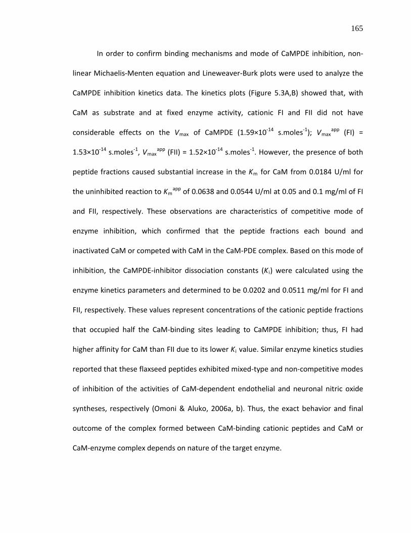
165
In order to confirm binding mechanisms and mode of CaMPDE inhibition, non-
linear Michaelis-Menten equation and Lineweaver-Burk plots were used to analyze the
CaMPDE inhibition kinetics data. The kinetics plots (Figure 5.3A,B) showed that, with
CaM as substrate and at fixed enzyme activity, cationic FI and FII did not have
considerable effects on the Vmax of CaMPDE (1.59×10⁻¹⁴ s.moles-1); Vmaxapp (FI) =
1.53×10⁻¹⁴ s.moles-1, Vmaxapp (FII) = 1.52×10⁻¹⁴ s.moles-1. However, the presence of both
peptide fractions caused substantial increase in the Km for CaM from 0.0184 U/ml for
the uninhibited reaction to Kmapp of 0.0638 and 0.0544 U/ml at 0.05 and 0.1 mg/ml of FI
and FII, respectively. These observations are characteristics of competitive mode of
enzyme inhibition, which confirmed that the peptide fractions each bound and
inactivated CaM or competed with CaM in the CaM-PDE complex. Based on this mode of
inhibition, the CaMPDE-inhibitor dissociation constants (K i) were calculated using the
enzyme kinetics parameters and determined to be 0.0202 and 0.0511 mg/ml for FI and
FII, respectively. These values represent concentrations of the cationic peptide fractions
that occupied half the CaM-binding sites leading to CaMPDE inhibition; thus, FI had
higher affinity for CaM than FII due to its lower K i
value. Similar enzyme kinetics studies
reported that these flaxseed peptides exhibited mixed-type and non-competitive modes
of inhibition of the activities of CaM-dependent endothelial and neuronal nitric oxide
syntheses, respectively (Omoni & Aluko, 2006a, b). Thus, the exact behavior and final
outcome of the complex formed between CaM-binding cationic peptides and CaM or
CaM-enzyme complex depends on nature of the target enzyme.

166
Figure 5.3 Lineweaver-Burk plots of the binding of (A) FI and (B) FII to CaM with
concomitant inhibition of calmodulin-dependent phosphodiesterase (CaMPDE); Vmax,
s.moles-1; Km, U/ml; K i
, mg/ml
-60 -54 -48 -42 -36 -30 -24 -18 -12 -6 0 6 12 18 24 30
2.5×101 3
5.0×101 3
7.5×101 3
1.0×101 4
I = 0I = 0.05 mg/ml FI
I = 0 I = FI I = FIIVmax(×10-14) 1.59 1.53 1.52Km 0.0184 0.0638 0.0544Ki na 0.0202 0.0511
(A)
1/V
(s.m
oles
-1)
1/[CaM] ml/U
-60 -54 -48 -42 -36 -30 -24 -18 -12 -6 0 6 12 18 24 30
2.5×101 3
5.0×101 3
7.5×101 3
1.0×101 4
I = 0I = 0.1 mg/ml FII
(B)
1/V
(s.m
oles
-1)
1/[CaM] ml/U

167
Although FII displayed lower affinity for CaM, it was selected for further
fractionation to concentrate the potent multifunctional peptides since it also displayed
ACE and renin-inhibitory activities as opposed to FI, which did not inhibit renin activity
(Udenigwe et al., 2009a). Peptide fractionation by HPLC using a reverse-phase column
yielded 8 peptide fractions designated as FII-2, FII-3, FII-4, FII-5, FII-6, FII-7, FII-8 and FII-9
(Figure 5.4) with recovery yields of 0.84%, 5.79%, 15.53%, 6.73%, 1.74%, 4.15%, 0.87%
and 2.10% (dry weight basis), respectively; the rest of the sample was composed of
ammonium salts from the FPLC buffers (FII-10) and an oily residue (FII-1). These HPLC
peptide fractions (FII-2–FII-9) were screened for multifunctional properties as ACE, renin
and CaMPDE inhibitors. Figure 5.5 shows the ACE-inhibitory activities of the HPLC
fractions. At 0.05 mg/ml peptides, the HPLC fractions displayed different weak ACE-
inhibitory activities (6.0–17.2%) except FII-5 and FII-7, which were inactive at this
concentration. Moreover, the activities of these HPLC fractions were lower than that of
the parent cationic peptide fraction (FII), which inhibited 31% of ACE activity at that
concentration. However, all the HPLC fractions exhibited moderate to strong ACE-
inhibitory activities at 0.2 mg/ml of the peptides. Compared to the cationic FII, HPLC
fractions FII-6 and FII-8 displayed better ACE inhibition (73.6% and 74.5% respectively)
whereas the other fractions, except FII-4, were significantly (P<0.05) less active. Thus,
FII-6 and FII-8 contained the most potent peptides although the ACE-inhibiting cationic
peptides were distributed in all the HPLC fractions. There was no observed relationship
between ACE inhibition and hydrophobicity of the cationic peptides, represented by
time of elution of the peptide from the C12 reverse-phase HPLC column.

168
Figure 5.4 High Performance Liquid Chromatography (HPLC) fractionation of the flaxseed cationic peptide fraction II (FII)
AU
0.00
0.20
0.40
0.60
0.80
1.00
1.20
1.40
1.60
1.80
2.00
2.20
Minutes0.00 5.00 10.00 15.00 20.00 25.00 30.00 35.00 40.00 45.00 50.00 55.00 60.00 65.00
FII-1
FII-2
FII-3
FII-4 FII-5 FII-6
FII-7
FII-8 FII-9 FII-10
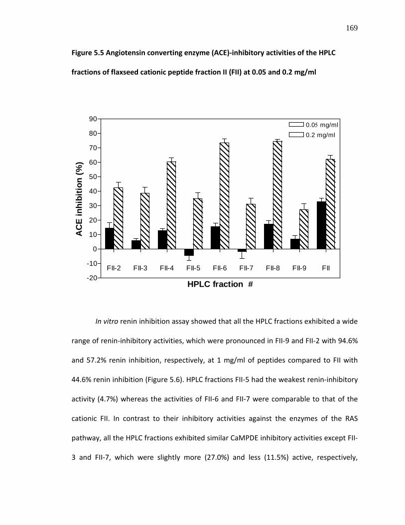
169
Figure 5.5 Angiotensin converting enzyme (ACE)-inhibitory activities of the HPLC
fractions of flaxseed cationic peptide fraction II (FII) at 0.05 and 0.2 mg/ml
In vitro renin inhibition assay showed that all the HPLC fractions exhibited a wide
range of renin-inhibitory activities, which were pronounced in FII-9 and FII-2 with 94.6%
and 57.2% renin inhibition, respectively, at 1 mg/ml of peptides compared to FII with
44.6% renin inhibition (Figure 5.6). HPLC fractions FII-5 had the weakest renin-inhibitory
activity (4.7%) whereas the activities of FII-6 and FII-7 were comparable to that of the
cationic FII. In contrast to their inhibitory activities against the enzymes of the RAS
pathway, all the HPLC fractions exhibited similar CaMPDE inhibitory activities except FII-
3 and FII-7, which were slightly more (27.0%) and less (11.5%) active, respectively,
FII-2 FII-3 FII-4 FII-5 FII-6 FII-7 FII-8 FII-9 FII-20
-10
0
10
20
30
40
50
60
70
80
900.05 mg/ml0.2 mg/ml
HPLC fraction #
AC
E in
hibi
tion
(%)
170
compared to the activity of FII (18.6%) at 0.1 mg/ml of peptides (Figure 5.7). These
activities were slightly less than the activities of similar HPLC fractions of α-casein-
derived cationic peptides, which inhibited 50% of CaMPDE activity at 0.0034–0.077
mg/ml peptides (Kizawa et al., 1995), and hen egg white lysozyme-derived cationic
peptides with up to 43.5% CaMPDE inhibition at 0.1 mg/ml peptides (You et al., 2010).
As observed in ACE inhibition, there was no correlation of the hydrophobic properties of
the peptides with both renin and CaMPDE inhibition, although a combination of
hydrophobicity and cationic properties have been observed in the amino acid sequences
of potent CaM-binding peptides (O’Neil & DeGrado, 1990; Kizawa et al., 1995). Thus, the
bioactivities of the flaxseed peptide fractions could be specifically due to their cationic
characters and amino acid sequences as previously demonstrated in insect venom
peptides, which lost their CaMPDE inhibitory activities after removal of their cationic
functional groups by acetylation (Martínez-Luis et al., 2007). When the results from the
enzyme inhibition assays were compared, HPLC fraction FII-6 showed excellent prospect
for further studies due to its multifunctional activity in inhibiting the three enzymes at
levels comparable to or better than the activity of the original cationic peptide fraction
(FII). Moreover, HPLC fraction FII-9 can be explored for potent renin inhibiting peptides,
FII-8 for potent ACE-inhibiting peptides, and FII-3 for potent CaMPDE inhibiting peptides
due to their enhanced activities in the corresponding assays compared to the activities
of FII.

171
Figure 5.6 Renin-inhibitory activities of the HPLC fractions of flaxseed cationic peptide
fraction II (FII) at 1 mg/ml
FII-2 FII-3 FII-4 FII-5 FII-6 FII-7 FII-8 FII-9 FII0
10
20
30
40
50
60
70
80
90
100
HPLC fraction #
Ren
in in
hibi
tion
(%)

172
Figure 5.7 The inhibition of calmodulin-dependent phosphodiesterase (CaMPDE)
activity by the HPLC fractions of flaxseed cationic peptide fraction II (FII) at 0.1 mg/ml
FII-2 FII-3 FII-4 FII-5 FII-6 FII-7 FII-8 FII-9 FII0
5
10
15
20
25
30
HPLC fraction #
CaM
PDE
inhi
bitio
n (%
)

173
5.4. CONCLUSIONS
In conclusion, ACE and renin-inhibiting CaM-binding cationic peptide fractions
from flaxseed proteins bound and inactivated CaM leading to competitive inhibition of
CaMPDE activity. The multifunctional properties of the cationic peptides were
conserved in the HPLC fractions, but specific inhibitory activities against one particular
enzyme (ACE, renin or CaMPDE) were substantially enhanced in some of the fractions.
The multifunctional nature of these peptide fractions can be used against multiple
symptoms of chronic human diseases such as cardiovascular disease, thus reducing the
cost associated with developing multiple bioactive products. The identity of the
peptides in the most potent multifunctional fraction need to be determined for studying
the structure-function properties of the peptides and for design of processing methods
that can efficiently generate high yields of the bioactive peptides from the primary
sequence of the parent proteins.
ACKNOWLEDGMENTS
Operating and equipment research grants for this project were provided to Dr. R.E.
Aluko by the Natural Sciences and Engineering Research Council of Canada (NSERC) and
Advanced Foods and Materials Network of Centres of Excellence (AFMNet), Canada. C.C.
Udenigwe is a recipient of NSERC Alexander Graham Bell Canada Graduate Scholarship
for doctoral studies.

174
5.5. REFERENCES
Bender, A.T., & Beavo, J.A. (2006). Nucleotide phosphodiesterase: molecular regulation to clinical use. Pharmacological Reviews, 58, 488-520
Bender, A.T., Ostenson, C.L., Wang, E.H., & Beavo, J.A. (2005). Selective up-regulation of PDE1B2 upon monocyte-to-macrophage differentiation. Proceedings of the National Academy of Science USA, 102, 497-502
Cho, M.J., Vaghy, P.L., Kondo, R., Lee, S.H., Davis, J., Rehl, R., et al. (1998). Reciprocal regulation of mammalian nitric oxide synthase and calcineurin by plant calmodulin isoforms. Biochemistry, 37, 15593-15597
Jiang, X., Li, J., Paskind, M., & Epstein, P.M. (1996). Inhibition of calmodulin-dependent phosphodiesterase induces apoptosis in human leukemic cells. Proceedings of the National Academy of Science USA, 93, 11236-11241
Kass, D.A., Takimoto, E., Nagayama, T., & Champion, H.C. (2007). Phosphodiesterase regulation of nitric oxide signalling. Cardiovascular Research, 75, 303-314
Kim, D., Rybalkin, S.D., Pi, X., Wang, Y., Zhang, C., Munzel, T., Beavo, J.A., Berk, B.C., & Yan, C. (2001). Upregulation of phosphodiesterase 1A1 expression is associated with the development of nitrate tolerance. Circulation, 104, 2338–2343
Kizawa, K., Naganuma, K., & Murakami, U. (1995). Calmodulin-binding peptides isolated from α-casein peptone. Journal of Dairy Research, 62, 587-592
Li, H., & Aluko, R.E. (2005). Kinetics of the inhibition of calcium/calmodulin-dependent protein kinase II by pea protein-derived peptides. Journal of Nutritional Biochemistry, 16, 656-662
Marambe, P. W. M. L. H. K., Shand, P. J., & Wanasundara, J. P. D. (2008). An in vitro investigation of selected biological activities of hydrolyzed flaxseed (Linum usitatissimum L.) proteins. Journal of the American Oil Chemists’ Society, 85, 1155-1164
Marangoni, A.G. (2003). Enzyme kinetics. A modern approach. Hoboken, New Jersey: John Wiley & Sons, Inc., 229 pp
Martínez-Luis, S., Pérez-Vásquez, A., & Mata, R. (2007). Natural products with calmodulin inhibitor properties. Phytochemistry, 68, 1882-1903
Nagel, D.J., Aizawa, T., Jeon, K.I., Liu, W., Mohan, A., Wei, H., et al. (2006). Role of nuclear Ca2+/calmodulin-stimulated phosphodiesterase 1A in vascular smooth muscle cell growth and survival. Circulation Research, 98, 777-784
175
O’Neil, K.T., & DeGrado, W.F. (1990). How calmodulin binds its targets: sequence independent recognition of amphiphilic α-helices. Trends in Biochemical Sciences, 15, 59-64
Omoni, A.O., & Aluko, R.E. (2006a). Effect of cationic flaxseed protein hydrolysate fractions on the in vitro structure and activity of calmodulin-dependent endothelial nitric oxide synthase. Molecular Nutrition and Food Research, 50, 958-966
Omoni, A.O., & Aluko, R.E. (2006b). Mechanism of the inhibition of calmodulin-dependent neuronal nitric oxide synthase by flaxseed protein hydrolysates. Journal of the American Oil Chemists’ Society, 83, 335-340
Pripp, A.H., Isaksson, T., Stepaniak, L., Sørhaug, T., & Ardö, Y. (2004). Quantitative structure-activity relationship modelling peptides and proteins as a tool in food science. Trends in Food Science and Technology, 16, 484-494
Rojas, S., Acevedo, L., Macias, M., Toscano, R.A., Bye, R., Timmermann, B., & Mata, R., (2003). Calmodulin inhibitors from Leucophyllum ambiguum. Journal of Natural Products, 66, 221-224
Rybalkin S.D., Yan, C., Bornfeldt, K.E., & Beavo, J.A. (2003). Cyclic GMP phosphodiesterases and regulation of smooth muscle function. Circulation Research, 93, 280-291
Schermuly, R.T., Pullamsetti, S.S., Kwapiszewska, G., Dumitrascu R., Tian, X., Weissmann, N., et al. (2007). Phosphodiesterase 1 upregulation in pulmonary arterial hypertension. Target for reverse-remodeling therapy. Circulation, 115, 2331-2339
Udenigwe, C.C., & Aluko, R.E. (2010). Antioxidant and angiotensin converting enzyme-inhibitory properties of a flaxseed protein-derived high Fischer ratio peptide mixture. Journal of Agricultural and Food Chemistry, 58, 4762-4768
Udenigwe, C.C., Lin, Y-S., Hou, W-C., & Aluko, R.E. (2009a). Kinetics of the inhibition of renin and angiotensin I-converting enzyme by flaxseed protein hydrolysate fractions. Journal of Functional Foods, 1, 199-207
Udenigwe, C.C., Lu, Y-L., Han, C-H., Hou, W-C., & Aluko, R.E. (2009b). Flaxseed protein-derived peptide fractions: Antioxidant properties and inhibition of lipopolysaccharide-induced nitric oxide production in murine macrophages. Food Chemistry, 116, 277-284
Weiss, B., Lehne, R., & Strada, S. (1972). Rapid microassay of adenosine 3′,5′-monophosphate phosphodiesterase activity. Analytical Biochemistry, 45, 222-235
You, S.J., Udenigwe, C.C., Aluko, R.E., & Wu, J. (2010). Multifunctional peptides from egg white lysozyme. Food Research International, 43, 848-855
176
TRANSITION STATEMENT
The three preceding chapters addressed the first three objectives of this project by
establishing that flaxseed protein-derived peptide fractions and cationic peptides
possess multifunctional bioactive properties in vitro and in cell culture. To address the
fifth objective, a unique approach was used to generate a peptide product containing
dipeptides, tripeptides and oligopeptides from flaxseed proteins that could be applied
as a source of branched-chain amino acids (BCAA) for restoration of plasma amino acid
imbalance during the treatment of liver disease. These BCAA-enriched peptide mixtures
are known as high Fischer ratio (molar ratio of BCAA to aromatic amino acids, AAA);
they contain high amounts of BCAA and corresponding low amounts of AAA. This part of
the project is important because this flaxseed protein-derived BCAA-enriched peptide
mixture also possess additional beneficial properties that are relevant to human health
sustenance, which also increases the value of the product, and potentially the value and
utilization of defatted flaxseed meal.

177
CHAPTER SIX
MANUSCRIPT 4
ANTIOXIDANT AND ANGIOTENSIN CONVERTING ENZYME-INHIBITORY PROPERTIES OF
A FLAXSEED PROTEIN-DERIVED HIGH FISCHER RATIO PEPTIDES MIXTURE
C. C. UDENIGWEa, R. E. ALUKOa,b
aDepartment of Human Nutritional Sciences, University of Manitoba, Winnipeg, MB R3T
2N2, Canada; b
The Richardson Centre for Functional Foods and Nutraceuticals,
University of Manitoba, Winnipeg, MB R3T 2N2, Canada.
Reproduced with permission from Antioxidant and Angiotensin Converting Enzyme-
Inhibitory Properties of a Flaxseed Protein-Derived High Fischer Ratio Peptide Mixture
Journal of Agricultural & Food Chemistry, 58, 4762-4768
Copyright © 2010 American Chemical Society
178
6.0. ABSTRACT
Hydrolysis of flaxseed proteins using thermolysin and pronase followed by treatment
with activated carbon yielded a peptide mixture with a Fischer ratio (branched-chain
amino acids/aromatic amino acids) of 23.65 and a phenylalanine+tyrosine content of
1.11%. Gel permeation chromatography showed that the flaxseed peptide sample
contains mainly low molecular weight peptides (<4 kDa). The high Fischer ratio peptide
sample exhibited antioxidant property by scavenging 2,2-diphenyl-1-picrylhydrazyl
radical, superoxide radical, hydroxyl radical, and also by protecting linoleic acid from
oxidation. In addition, the peptide mixture showed antihypertensive property by
inhibiting angiotensin I-converting enzyme in a mixed-type inhibition pattern. Protein
hydrolysates with Fischer ratio higher than 20 and phenylalanine+tyrosine content
lower than 2% has been used to treat patients with hepatic encephalopathy; thus, this
multifunctional flaxseed peptide mixture could be used to formulate food products with
multiple human health benefits in liver diseases, oxidative stress and hypertension.
KEYWORDS: Flaxseed protein; Fischer ratio; Activated carbon; Branched-chain Amino
acids; Aromatic amino acids; Antioxidant; Angiotensin converting enzyme

179
6.1. INTRODUCTION
Acute and chronic liver failure is associated with alterations in protein
metabolism, which can lead to loss of muscle protein mass. Plasma amino acid
imbalance have been found in patients with liver diseases, and this is characterized by
elevated levels of aromatic amino acids (AAA; tyrosine and phenylalanine) and
methionine, and decreased levels of branched-chain amino acids (BCAA; leucine,
isoleucine and valine) (Clemente, 2000). The ratio of plasma BCAA to AAA is termed
Fischer ratio and is used to measure abnormal amino acid metabolism during liver
disease, and also in clinical nutrition for the formulation of amino acid products for
therapeutic purposes. In human beings, the normal plasma Fischer ratio is 3.5–4; this
value decreases to <2.5 in liver diseases, <1.2 in hepatic coma and could drop to below
0.8 in profound coma (Fischer et al., 1975). Clinical nutrition approaches towards
treatment of liver diseases involve feeding patients with BCAA-enriched formulae
(Fischer et al., 1976; Capollo et al., 1992; Khanna & Gopalan, 2007; Schulze et al., 2008)
or protein hydrolysates with Fischer ratio higher than 20 and phenylalanine+tyrosine
content lower than 2% (Okita et al., 1985). BCAAs have been shown to reduce muscle
wasting and plasma concentrations of AAA with concomitant improvement in
encephalopathy (Fischer, 1990). The therapeutic effect of BCAA is due to the fact that
they are preferentially taken up by the muscle, inhibit the transport of AAA across the
blood-brain barrier, and are available for peripheral metabolism in advanced liver
disease (Clemente, 2000). In addition, some athletes also use BCAA as a natural way of
improving muscle mass and strength (Di Pasquale, 2008).
180
A number of studies have reported successful production of high Fischer ratio
peptide mixtures by enzymatic hydrolysis of food proteins and subsequent processing
using activated carbon and gel filtration chromatography (Adachi et al., 1993; Ma et al.,
2008; Pedroche et al., 2004; Pedroche et al., 2006; Zheng et al., 2009). In clinical
nutrition, peptides are preferred over individual amino acids due to the fact that short
peptides have higher rates of intestinal absorption and the osmotic pressure of peptides
is lower than that of corresponding free amino acids (Grimble et al., 1987; Monchi &
Rerat, 1993). The recommended criteria for selecting the starting food protein that will
yield high Fischer ratio peptide mixture include high amounts of BCAA, low amounts of
AAA (Clemente, 2000) and availability of the raw material.
Flaxseed (Linum usitatissimum) is a readily available oilseed predominantly
produced in the Prairie regions of Canada, and a major dietary source of α-linolenic acid,
dietary fibres and lignans (Hall III et al., 2006). These flaxseed components have been
reported to possess various physiological activities relevant to human health sustenance
especially in cardiovascular disease prevention, cancer and diabetes (Hall III et al., 2006).
However, the protein components of flaxseed have been scarcely explored for
nutritional and therapeutic applications. These proteins constitute a major part of
defatted flaxseed meal, which results from industrial flaxseed crushing and oil
extraction, and is comparable in nutritional quality and amino acid composition to other
high quality proteins, e.g. soy protein (Hall III et al., 2006; Oomah & Mazza, 1993).
Moreover, previous studies have reported that enzymatic hydrolysis of flaxseed proteins
afforded low molecular weight (MW) peptides that possess antioxidant and anti-

181
inflammatory properties in cell cultures (Udenigwe et al., 2009a) as well as in vitro
antihypertensive properties (Udenigwe et al., 2009b). Flaxseed proteins contain high
levels of BCAA, with 50, 71 and 56 mg/g of isoleucine, leucine and valine (Oomah &
Mazza, 1995), respectively. Thus, through careful choice of enzymes and processing
conditions, flaxseed proteins have the potential to be converted into protein
hydrolysates enriched with BCAA.
Food protein-derived peptides possess physiological therapeutic roles such as
antioxidant and antihypertensive properties. Endogenous and exogenous reactive
oxygen species and free radicals are implicated in the initiation and progression of
human disease conditions (Ames et al., 1993). The amount of these reactive species is
controlled by endogenous antioxidants until it reaches a level when the antioxidants are
overwhelmed, a state known as oxidative stress. This condition can be surmounted by
the consumption of foods rich in antioxidants. The renin-angiotensin system, which
controls blood pressure in human beings, has been targeted for the treatment of
hypertension through the inhibition of angiotensin I-converting enzyme (ACE) and renin.
ACE inhibitors are thought to possess the ability to lower blood pressure in hypertensive
subjects and animals (Aluko, 2008). Food protein hydrolysates and constituent peptides
with multifunctional bioactive properties against these diseases targets are desirable for
the formulation of food products with human health benefits in multiple physiological
and disease conditions. Therefore, a BCAA-enriched product that possesses other
bioactive properties could, in addition to the positive effects on liver disease, be

182
potentially useful as a single agent to provide simultaneous relief from other associated
disease symptoms.
The objectives of this project were to produce a high Fischer ratio enzymatic
flaxseed protein hydrolysate and determine the ability of the hydrolysate to scavenge
free radicals, inhibit in vitro oxidative reactions and activities of ACE and renin.
6.2. MATERIALS AND METHODS
6.2.1. Materials
Thermolysin (from Bacillus thermoproteolyticus rokko), Alcalase (from B.
licheniformis), ficin (from fig tree latex), papain (from papaya latex), activated carbon,
pyrogallol (1,2,3-trihydroxybenzene) and N-(3-[2-furyl]acryloyl)-phenylalanylglycyl-
glycine (FAPGG) were purchased from Sigma Chemicals (St. Louis, MO). Pronase (from
Streptomyces griseus) was purchased from Roche Diagnostics GmbH (Mannheim,
Germany). All other chemical reagents were of analytical grade.
6.2.2. Enzymatic hydrolysis of flaxseed protein
Flaxseed protein isolate (FPI) was prepared from defatted flaxseed meal as
previously described (Udenigwe et al., 2009b). The overall approach involved hydrolysis
of flaxseed proteins with thermolysin followed by treating the resulting protein
hydrolysate with papain, ficin, alcalase or pronase. FPI (5% w/v) was suspended in
distilled water; the slurry was adjusted to pH 7.5 with 1 M Na2CO3 solution and to a
temperature of 37°C. Thereafter, protein hydrolysis was initiated by adding thermolysin

183
at an E/S ratio of 1:100 (based on protein content). During hydrolysis, the reaction
mixture was maintained at pH 7.5 by adding 1 M Na2CO3 solution using a pH stat
instrument (Metrohm Titrando, Herisau, Switzerland). After 5 h, the enzymatic reaction
was terminated by adjusting the pH to 4.0 using 1M HCl. The temperature and pH of the
reaction mixture was adjusted appropriately and subjected to another hydrolysis using
one of these proteases: papain (pH 6.5, 37°C), ficin (pH 7.0, 37°C), alcalase (pH 8.0, 50°C)
and pronase (pH 7.4, 40°C). These enzymes were separately added to the resulting
hydrolysate for further hydrolysis at E/S ratio of 1:100 (based on protein content); pH
was also maintained with 1 M Na2CO3
solution. After 5 h, the reaction was stopped by
adjusting the pH to 4.0 using 1M HCl. The resulting flaxseed protein hydrolysate (FPH)
was collected by centrifugation at 15000×g, freeze-dried and stored at –20°C until used.
The protein content of the resulting powder was determined by a modified Lowry
protein assay method (Markwell et al., 1978).
6.2.3. Adsorption of flaxseed protein hydrolysate onto activated carbon-packed
column
To separate the BCAA-containing peptides from the AAA-containing peptides,
each of the freeze-dried FPH was dissolved in deionized water at 10 mg/ml and the
solution was adjusted to pH 2.5 using 0.5 M HCl (Adachi et al., 1993) followed by
filtration using Whatman No. 1 filter paper. Thereafter, the resulting acidic filtrate was
passed through a column packed with activated carbon (9.5×80mm) and sample was
eluted using a single-channel Peristaltic pump model P-1 (GE Healthcare, Montreal, PQ,

184
Canada). The unbound peptides (BCAA-rich) were collected in the flow-through solution
and labelled as FPH-col. The column was regenerated for subsequent fractionation by
eluting with absolute ethanol to remove bound peptides (AAA-rich) followed by washing
with distilled water. Peptide elution was monitored using a spectrophotometer by
measuring absorbance at 220, 260 and 280 nm. The collected peptide solutions were
freeze-dried and stored at –20°C until used.
6.2.4. Adsorption of flaxseed protein hydrolysate onto activated carbon by mixing
The AAA-containing peptides and free AAA were also removed from the FPH by
mixing 10 mg of FPH (adjusted to pH 2.5 using 0.5 M HCl) with various amounts of
activated carbon (10, 25, 50, 75, 100 and 150 mg) in 1 ml of deionized water for 10 min
at room temperature in a beaker. Thereafter, the mixture was centrifuged at 15000×g
for 30 min and the supernatant (contain BCAA-rich peptides) filtered through a
Whatman No.1 filter paper to completely remove traces of activated carbon. The filtrate
(FPH-mix) was freeze-dried and stored at –20°C until used. The levels of peptides and
AAA in the FPH-mix peptide product were initially estimated using a spectrophotometric
absorbance at 220 and 260+280 nm, respectively.
6.2.5. Amino acid analysis
An HPLC system was used to determine the amino acid profiles after samples
were hydrolyzed with 6 M HCl as previously reported (Bidlingmeyer et al., 1984). The
cysteine and methionine contents were determined after performic acid oxidation

185
(Gehrke et al., 1985
) and tryptophan content was determined after alkaline hydrolysis
(Landry & Delhaye, 1992). The Fischer ratio was calculated as the ratio of BCAA to AAA.
6.2.6. Gel permeation chromatography
Molecular weight distribution of the peptides present in the FPH-mix with the
highest Fischer ratio was estimated by gel filtration chromatography as previously
reported (Humiski & Aluko, 2007) with some modifications. The peptide sample (10
mg/ml) was filtered using a 0.2 µm membrane and 100 µl of the solution was injected
onto a Superdex 75 100/300L column connected to an AKTA fast protein liquid
chromatography system (GE Healthcare, Montreal, QC, Canada). The column was
equilibrated with 1 column volume (23.5 ml) of 50 mM phosphate buffer containing
0.15 M NaCl (pH 7.0) prior to loading the sample. The peptides were eluted from the
column using a flow rate of 0.75 ml/min and peaks were detected at 214 nm. The
column was calibrated using the following standards: cytochrome c (12.3 kDa), aprotinin
(6.5 kDa), pepstatin A (685.9 Da), glutathione (307.33 Da) and glycine (75 Da); blue
dextran was used to determine the column void volume. The estimated molecular
weights of the eluted peaks were determined from a plot of the elution volume versus
the log10
MW of the standards.
6.2.7. Antioxidant assays
The ability of the FPH-mix peptide sample with the highest Fischer ratio to
scavenge DPPH radical was evaluated as previously reported (Udenigwe et al., 2009a).

186
Superoxide (O2˙–) scavenging assay was also evaluated using the pyrogallol
autooxidation method as previously described (Udenigwe et al., 2009a). Briefly, 80 µl of
the peptide solution of various concentrations was mixed with 80 µl of 50 mM Tris-HCl
buffer (pH 8.3) containing 1 mM EDTA in a 96-well microplate followed by the addition
of 40 µl of 1.5 mM pyrogallol in 10 mM HCl. The rate of O2˙–-induced polymerization
pyrogallol (∆A/min) was measured as increase in absorbance at 420 nm for 4 min at
room temperature. Tris-HCl buffer was used to replace the peptide solution in control
experiment whereas glutathione was used as positive control. O2˙–
In addition, ˙OH scavenging activity of the high Fischer ratio FPH-mix sample was
assayed based on a method described by Li et al. (2008). Peptide samples (50 µl in 0.1 M
sodium phosphate buffer, pH 7.4) were mixed with 50 µl of 3 mM 1,10-phenanthroline
(in phosphate buffer) and 50 µl of 3 mM FeSO
scavenging activity
(%) was calculated as [(∆A/min c – ∆A/min s)/ ∆A/min c] × 100, where c and s represent
control and sample, respectively.
4 (in water) in a 96-well microplate.
Thereafter, 50 µl of 0.01% aqueous H2O2 was added to initiate the Fenton reaction. The
reaction mixture covered and incubated at 37°C for 1 h and the absorbance measured at
536 nm (A536nm s). A control experiment was also conducted by replacing the peptide
sample with buffer (A536nm c). The ˙OH scavenging activity (%) was calculated as [(A536nm
c – A536nm s)/A536nm
The ability of the flaxseed peptide to inhibit linoleic acid (LA) oxidation was also
investigated as previously described (Li et al., 2008) with modifications. LA (1 ml of 50
mM dissolved in 99.5% ethanol) was mixed with 0.5 ml of a solution of different
c] × 100.

187
concentrations of the peptide sample (final concentrations of 5, 2.5 and 1.25 mg/ml of
0.1 M phosphate buffer, pH 7.0) or glutathione (final concentration of 1.25 mg/ml). The
mixture was kept at 60oC in darkness for 6 days. An aliquot of 0.1 ml of the sample
solution was withdrawn after every 24 hr, and mixed with 4.7 ml of 75% aqueous
ethanol, 0.1 ml of ammonium thiocyanate (30% w/v) and 0.1 ml of 20 mM FeCl2
(dissolved in 1 M HCl). The absorbance of the mixture was measured at 500 nm after 3
min incubation at room temperature.
6.2.8. ACE and renin inhibition assays
The potential antihypertensive property of the FPH-mix sample with highest
Fischer ratio was determined based on its ability to inhibit the activities of ACE and renin
in vitro. At various concentrations of the peptide sample, ACE and renin inhibitory
assays were carried out as previously described using FAPGG as ACE substrate and Arg-
Glu(EDANS)-Ile-His-Pro-Phe-His-Leu-Val-Ile-His-Thr-Lys(Dabcyl)-Arg as renin substrate
(20). In addition, the kinetics of inhibition of ACE was also studied to determine the
mode of the peptide-induced ACE inhibition. Kinetics parameters (Km and Vmax , and
their apparent values in the presence of the peptide sample) were estimated from non-
linear regression fits of the kinetics data to the Michaelis-Menten equation using
GraphPad Prism version 4.0 (GraphPad Software, San Diego, CA). The catalytic efficiency
of ACE in the presence and absence of the peptide sample was calculated as Vmax/Km
. All
the kinetics experiments were conducted in duplicate.

188
6.2.9. Statistical analysis
Except where indicated, data were collected in triplicates and subjected to one
way analysis of variance using Statistical Analysis System Software (SAS version 9.2, SAS
Institute, Cary, NC). Significant differences were determined by Duncan’s multiple range
test and accepted at p<0.05.
6.3. RESULTS AND DISCUSSION
6.3.1. Enzymatic flaxseed protein hydrolysis
Fig. 6.1 shows the procedure used in hydrolyzing the flaxseed protein and
processing the resulting FPH. The aim of the enzymatic hydrolysis of flaxseed protein in
this study was to remove the AAA, and also to generate low-MW peptides for efficient
intestinal absorption. Thermolysin was chosen for the first hydrolysis step because of its
specificity in cleaving proteins at the N-terminal regions of hydrophobic amino acids,
phenylalanine, tyrosine, leucine, isoleucine and valine. In order to release the N-
terminal AAA, the resulting protein hydrolysate was subjected to another controlled
hydrolysis using other specific proteases, papain and ficin. Papain has a wide specificity
but preferentially cleaves the X1–X2 peptide bond in Phe– X1–X2, where X1 and X2 are
any amino acids, thereby releasing a Phe-containing dipeptide into the solution. A
combination of thermolysin and papain has been previously used to produce high
Fischer ratio peptide mixtures (Adachi et al., 1991; 1993). Ficin, a papain-like protease,
was used as a control since it has a wide specificity in cleaving the carboxyl side of 10
different amino acid residues, including valine and tyrosine. Moreover, the highly non-

189
specific proteases were used to produce peptides of shorter chain length that could be
easily absorbed in the intestine. Alcalase is an endopeptidase whereas pronase
possesses both endopeptidase and exopeptidease activity and can generate low
molecular weight peptides and free amino acids from native and denatured proteins
and large peptides. Thus, pronase can cleave the N-terminal AAA resulting from the
thermolysin activity. The protein contents of the FPH were 81% (papain), 85.2% (ficin),
66.3% (alcalase) and 60.7% (pronase) based on Lowry assay. The observed low protein
contents of the FPH generated with pronase may be due to their exopeptidase activity,
which released free amino acids that were not detected by the Lowry assay.
6.3.2. Activated carbon treatments, amino acid profiles and Fischer ratios
The FPH was passed through a tightly packed activated carbon column to
remove AAA-containing peptides and recover the BCAA-rich peptides in the flow-
through fraction. To improve the elution of the peptides from the column, the flaxseed
protein hydrolysates were acidified prior to loading onto the activated carbon column.
This treatment was necessary to precipitate any large peptides and residual undigested
proteins, which have high adsorption capacity on activated carbon (Adachi et al., 1993)
and may block the activated carbon matrix thereby decreasing elution efficiency. The
absorbance values of the flow-through sample at 280 and 260 nm, compared to the
crude hydrolysate solution, showed that over 90% of AAA were removed from the FPH.
However, data from amino acid analysis of the freeze-dried powder showed that the
activated carbon column specifically removed 39–57% of AAA from FPH. This resulted in

190
Figure 6.1 Flowchart for production of branched-chain amino acids (BCAA)-enriched
high Fischer ratio mixture by enzymatic hydrolysis of isolated flaxseed protein and
activated carbon treatment
Flaxseed protein (FP) isolation Amino acid analysis
Hydrolysis of FP with thermolysin
Enzymatic Hydrolysis
Centrifugation
Freeze dry supernatant (FPH)
Amino acid analysis
FPH solution passed through activated
carbon column
FPH solution mixed with activated
carbon; filtered
Freeze dry flow-through sample
Freeze dry filtrate
Amino acid analysis
Amino acid analysis
Papain, ficin, Alcalase, pronase See Section 6.2.2 for details

191
an overall increase in the Fischer ratio of the resulting FPH-col peptide samples when
compared with the crude FPH (Table 6.1). However, the Fischer ratios of the FPH-col
flow-through samples were low, ranging from 2.5 to 4.04 representing up to 2.3-fold
increase relative to the crude FPH. The highest Fischer ratio (4.04) was observed for the
hydrolysate product of thermolysin-pronase activity. It could be observed that increase
in the Fischer ratio correlated with increase in the cleavage sites of the enzymes, except
for alcalase. This showed that the less specific enzymes liberated smaller peptides that
passed through the packed activated carbon in the column. The low Fischer ratio
obtained in the flow-through samples indicated that some of the BCAA-containing
peptides may have been trapped in the activated carbon matrix in the column. The
result obtained in this study is contrary to that of a previous study that reported the
preparation of protein hydrolysates with high Fischer ratio of 31.6 by enzymatic
hydrolysis of casein and passage of the resulting acidified hydrolysates through an
activated carbon-packed column (Adachi et al., 1993). Recently, Ma et al. (2008) also
used similar activated carbon treatment to prepare a peptide mixture with Fischer ratio
of 34.71 from Alcalase-papain hydrolyzed corn gluten meal. Due to the low Fischer ratio
of the flaxseed peptide mixture, another approach was adopted to minimize BCAA loss
while effectively removing the AAA-containing peptides and free AAA.
Mixing of the FPH with activated carbon resulted in concentration-dependent
removal of AAA and decrease in the protein content. The optimized condition (2.5%
activated carbon, w/v) that gave the highest peptide yield (unadsorbed peptides as
A220nm) with the removal of most of the AAA was used for the scale-up experiment. Data
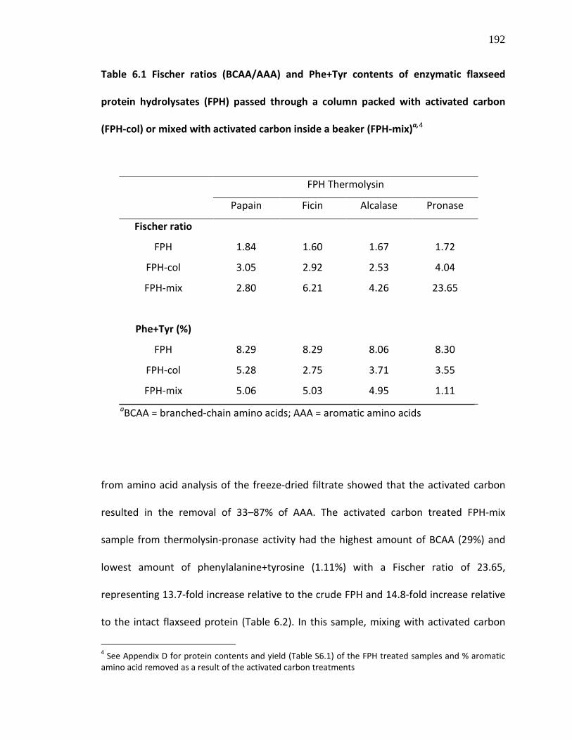
192
Table 6.1 Fischer ratios (BCAA/AAA) and Phe+Tyr contents of enzymatic flaxseed
protein hydrolysates (FPH) passed through a column packed with activated carbon
(FPH-col) or mixed with activated carbon inside a beaker (FPH-mix)a,4
from amino acid analysis of the freeze-dried filtrate showed that the activated carbon
resulted in the removal of 33–87% of AAA. The activated carbon treated FPH-mix
sample from thermolysin-pronase activity had the highest amount of BCAA (29%) and
lowest amount of phenylalanine+tyrosine (1.11%) with a Fischer ratio of 23.65,
representing 13.7-fold increase relative to the crude FPH and 14.8-fold increase relative
to the intact flaxseed protein (Table 6.2). In this sample, mixing with activated carbon
4 See Appendix D for protein contents and yield (Table S6.1) of the FPH treated samples and % aromatic amino acid removed as a result of the activated carbon treatments
FPH Thermolysin
Papain Ficin Alcalase Pronase
Fischer ratio
FPH 1.84 1.60 1.67 1.72
FPH-col 3.05 2.92 2.53 4.04
FPH-mix 2.80 6.21 4.26 23.65
Phe+Tyr (%)
FPH 8.29 8.29 8.06 8.30
FPH-col 5.28 2.75 3.71 3.55
FPH-mix 5.06 5.03 4.95 1.11
aBCAA = branched-chain amino acids; AAA = aromatic amino acids
193
decreased the AAA composition from 9.78% in the crude FPH to 1.23% with up to 2-fold
enrichment of BCAA (Table 6.2). After similar activated carbon treatment, the other
FPH-mix from papain, ficin and alcalase treatments had lower Fischer ratio of 2.8–6.2
due to their high AAA contents, although there was an increase in the Fischer ratio
when compared with the crude FPH and isolated flaxseed protein. Protein hydrolysates
or peptide mixtures of Fischer ratio higher than 20 and phenylalanine+tyrosine value
lower than 2% can be used to treat patients with liver diseases (Okita et al., 1985); thus,
these results show that the highest Fischer ratio flaxseed peptide mixture from
thermolysin-pronase treatment has promising application for use in treating liver
diseases in humans. Other recent studies have reported the production of peptide
mixtures with Fischer ratios 24.58, 28.3 and 30.6 respectively from pearl oyster meat
protein (Zheng et al., 2009), Brassica carinata protein (Pedroche et al., 2006) and bovine
casein (Pedroche et al., 2004) generated through enzymatic hydrolysis, activated carbon
treatment and gel filtration chromatography. Thus, the process reported in this paper
represents a cost-effective method of production of high Fischer ratio peptide mixture
from flaxseed protein since activated carbon is cheap and can be regenerated, and the
process did not require gel filtration chromatography.

194
Table 6.2 Percentage amino acid composition of flaxseed protein isolate (FPI),
thermolysin-pronase hydrolysate (FPH), FPH passed through a column packed with
activated carbon (FPH-col) and FPH mixed with activated carbon in a beaker (FPH-mix)
AMINO ACIDS FPI FPH FPH-col FPH-mix
ASX 11.29 a 10.83 11.40 8.27 THR 3.67 3.60 4.40 4.36 SER 4.34 4.99 6.28 5.79 GLX 19.83 b 21.30 17.79 13.65 PRO 5.29 4.08 4.12 2.57 GLY 5.47 5.22 5.50 4.48 ALA 4.89 4.47 6.70 6.80 CYS 1.36 1.38 0.58 0.18 VAL 5.20 5.72 5.71 10.10 MET 2.02 1.49 1.10 0.76 ILE 4.50 5.14 4.35 7.56 LEU 5.80 5.93 6.61 11.34 TYR 2.52 2.78 1.19 0.28 PHE 5.47 5.52 2.36 0.83 HIS 2.29 2.23 3.12 3.27 LYS 3.04 3.03 4.98 5.97 ARG 11.28 10.79 13.23 13.68 TRP 1.73 1.48 0.57 0.12
AAA 9.73 c 9.78 4.13 1.23 BCAA 15.51 d 16.79 16.68 29.00
FISCHER RATIO 1.59 1.72 4.04 23.65 PHE+TYR 7.99 8.30 3.55 1.11
aAsx, aspartic acid+asparagine; bGlx, glutamic acid+glutamine; cAAA: phenylalanine, tyrosine, tryptophan; dBCAA: valine, leucine, isoleucine

195
6.3.3. Estimated peptide MW distribution
The MW of peptides is important in determining their absorption following oral
intake. It is believed that low-MW peptides can be efficiently transported across the
enterocytes through several peptide transporters. MW estimation using gel permeation
chromatography showed that the high Fischer ratio FPH-mix contains peptides of sizes
<3.96 kDa (Fig. 6.2). The first peak (1) contains oligopeptides of MW 357–3960 Da
whereas peak 2 contains mostly dipeptides, tripeptides and free amino acids as
indicated by the MW range of 68–353 Da. The low-MW peptide content of this sample
increases its potential for use as nutraceutical in treating liver diseases.
Figure 6.2 Size-exclusion gel chromatogram of the high Fischer ratio flaxseed protein
hydrolysate mixed with activated carbon (FPH-mix)
Peak MW range (Da)
1 3960 - 357.82 353.2 - 68.4
0 5 10 15 20 25 300
250
500
750
1000
1250
1500
1750
2000
1 2
Elution volume (ml)
Abs
orba
nce
@ 2
14nm
(mA
U)
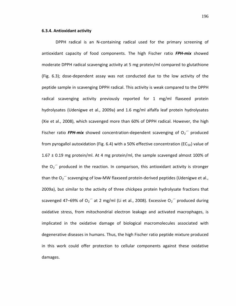
196
6.3.4. Antioxidant activity
DPPH radical is an N-containing radical used for the primary screening of
antioxidant capacity of food components. The high Fischer ratio FPH-mix showed
moderate DPPH radical scavenging activity at 5 mg protein/ml compared to glutathione
(Fig. 6.3); dose-dependent assay was not conducted due to the low activity of the
peptide sample in scavenging DPPH radical. This activity is weak compared to the DPPH
radical scavenging activity previously reported for 1 mg/ml flaxseed protein
hydrolysates (Udenigwe et al., 2009a) and 1.6 mg/ml alfalfa leaf protein hydrolysates
(Xie et al., 2008), which scavenged more than 60% of DPPH radical. However, the high
Fischer ratio FPH-mix showed concentration-dependent scavenging of O2˙– produced
from pyrogallol autoxidation (Fig. 6.4) with a 50% effective concentration (EC50) value of
1.67 ± 0.19 mg protein/ml. At 4 mg protein/ml, the sample scavenged almost 100% of
the O2˙– produced in the reaction. In comparison, this antioxidant activity is stronger
than the O2˙– scavenging of low-MW flaxseed protein-derived peptides (Udenigwe et al.,
2009a), but similar to the activity of three chickpea protein hydrolysate fractions that
scavenged 47–69% of O2˙– at 2 mg/ml (Li et al., 2008). Excessive O2˙– produced during
oxidative stress, from mitochondrial electron leakage and activated macrophages, is
implicated in the oxidative damage of biological macromolecules associated with
degenerative diseases in humans. Thus, the high Fischer ratio peptide mixture produced
in this work could offer protection to cellular components against these oxidative
damages.

197
Figure 6.3 DPPH radical scavenging activity of 5 mg protein/ml of the high Fischer ratio
FPH-mix compared to glutathione
Figure 6.4 Concentration-dependent scavenging of superoxide radical (O2˙–) by high
Fischer ratio FPH-mix with effective concentration (EC50
) value of 1.67 mg protein/ml;
bars with different letters are significantly different at p<0.05
Glutathione FPH-mix0
10
20
30
40
50
60
70 a
bD
PP
H s
cave
ngin
g ac
tivity
(%)
Glutathione 4 2 1 0.50
20
40
60
80
100
FPH-mix (mg/ml)
a a
b
c
d
O2.- -s
cave
ngin
g ac
tivity
(%)

198
In addition, the flaxseed peptide mixture displayed moderate scavenging of ˙OH
in a concentration-dependent fashion with a maximum scavenging of 31.5% at 5 mg
protein/ml whereas glutathione showed similar scavenging activity at a lower
concentration of 1 mg/ml (Fig. 6.5). This activity is weaker than those previously
reported for low-MW peptides from flaxseed protein that scavenged more than 70% of
˙OH at <1.5 mg/ml (Udenigwe et al., 2009a) and chickpea protein hydrolysate fractions,
which scavenged 38–81% of ˙OH at 1.5 mg/ml (Li et al., 2008). The moderate ˙OH
scavenging activity of FPH-mix may be due to the low levels of phenylalanine present in
the peptide mixture. This is because phenylalanine possesses strong radical-scavenging
property since it can trap ˙OH via hydroxylation of the aromatic ring leading to the
formation of stable ortho-, meta- or para-tyrosine (Sun et al., 1993).
Figure 6.5 Concentration-dependent scavenging of hydroxyl radical (˙OH) by branched-
chain amino acid (BCAA)-rich FPH-mix; bars with different letters are significantly
different at p<0.05
Glutathione 5 2.5 1.25 0.6250
5
10
15
20
25
30
35
FPH-mix (mg/ml)
a a
b
c
d
. OH
sca
veng
ing
activ
ity (%
)

199
The FPH-mix with highest Fischer ratio (BCAA-rich) was found to show strong
inhibition of linoleic acid oxidation in an in vitro evaluation system. As observed in Fig.
6.6, the increase in A500nm indicates an increase in linoleic acid oxidation, and this
occurred during the first three days followed by a gradual decrease during the next
three days. On day 1, the BCAA-rich mixture (1.25–5 mg/ml) inhibited approximately
45% of linoleic acid oxidation whereas glutathione (1.25 mg/ml) was over 2-fold more
active. As glutathione lost its activity in protecting linoleic acid from oxidation between
days 2–6, the BCAA-rich sample maintained antioxidant property at the three
concentrations. Glutathione lost its activity with time since its antioxidant function
group, the –SH group of cysteine, could not have been regenerated during the assay. In
the last two days of the experiment, 5 mg/ml of the BCAA-rich sample displayed the
best inhibitory property against linoleic acid oxidation (p<0.05). On the 6th day, 5, 2.5
and 1.25 mg/ml of the peptide mixture concentration-dependently inhibited linoleic
acid oxidation by 48%, 37.5% and 36.4%, respectively whereas glutathione completely
lost its activity. In contrast, a chickpea protein hydrolysate fraction displayed a stronger
activity by inhibiting in vitro linoleic acid peroxidation by 81% at 1 mg/ml after 8 days of
incubation (Li et al., 2008). The oxidation of polyunsaturated fatty acid components of
biological membranes is an important mechanism of cellular damages induced by
reactive oxygen species; thus, the BCAA-rich sample from flaxseed protein may offer
protection to cellular components by reducing oxidative damages.
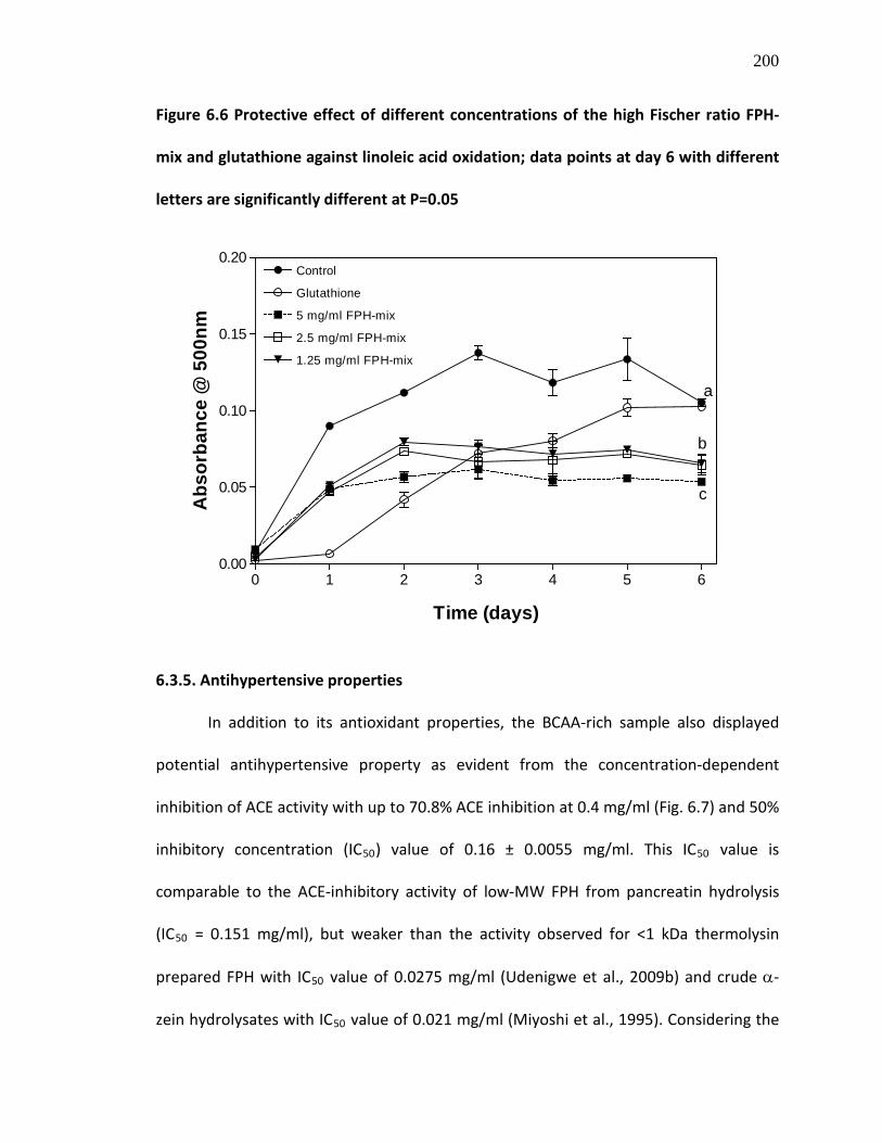
200
Figure 6.6 Protective effect of different concentrations of the high Fischer ratio FPH-
mix and glutathione against linoleic acid oxidation; data points at day 6 with different
letters are significantly different at P=0.05
6.3.5. Antihypertensive properties
In addition to its antioxidant properties, the BCAA-rich sample also displayed
potential antihypertensive property as evident from the concentration-dependent
inhibition of ACE activity with up to 70.8% ACE inhibition at 0.4 mg/ml (Fig. 6.7) and 50%
inhibitory concentration (IC50) value of 0.16 ± 0.0055 mg/ml. This IC50 value is
comparable to the ACE-inhibitory activity of low-MW FPH from pancreatin hydrolysis
(IC50 = 0.151 mg/ml), but weaker than the activity observed for <1 kDa thermolysin
prepared FPH with IC50 value of 0.0275 mg/ml (Udenigwe et al., 2009b) and crude α-
zein hydrolysates with IC50
0 1 2 3 4 5 60.00
0.05
0.10
0.15
0.20Control
Glutathione
5 mg/ml FPH-mix
2.5 mg/ml FPH-mix
1.25 mg/ml FPH-mix
a
b
c
Time (days)
Abs
orba
nce
@ 5
00nm
value of 0.021 mg/ml (Miyoshi et al., 1995). Considering the

201
role of hydrophobic amino acids in ACE inhibition (Wu et al., 2006), it could be that the
decreased amount of AAA in the BCAA-rich sample contributed to the decreased
potency of the peptides in ACE inhibition when compared to the previously reported
flaxseed peptide mixtures (Udenigwe et al., 2009b).
Kinetic studies of ACE inhibition using the Lineweaver-Burk plot showed that the
BCAA-rich sample inhibited ACE in a mixed-type inhibition pattern (Fig. 6.8). This was
characterized by an increase in Km from 0.3121 mM for the uninhibited reaction to
0.6179 and 0.5860 mM in the presence of 0.1 and 0.4 mg/ml peptides, respectively. The
reaction Vmax also decreased in a concentration-dependent fashion from 0.0112 ∆A
/min for the uninhibited reaction to 0.009416 and 0.004521 ∆A /min in the presence of
0.1 and 0.4 mg/ml of the inhibitor, respectively. These results are similar to the mode of
ACE inhibition reported for low-MW crude and cationic peptide fractions from flaxseed
protein (Udenigwe et al., 2009b). This indicates that the constituent peptides in the
BCAA-rich sample inhibited ACE activity by binding both the enzyme active and allosteric
sites; in other words, the peptides (or amino acids) interacted with ACE in both its free
and substrate-bound forms. This shows that ACE inhibition by the BCAA-rich sample
cannot be completely overcome by increasing the substrate concentration. The catalytic
efficiency of ACE was observed to decrease from 0.0358 to 0.0152 and 0.0077 in the
presence of 0.1 and 0.4 mg/ml of the peptides, respectively, which indicates that
inhibition of ACE activity by the BCAA-rich sample was concentration-dependent. In
contrast to these results, Cinq-Mars et al. (2008) reported that both fractionated and
unfractionated Pacific hake fillet hydrolysates exhibited competitive mode of ACE
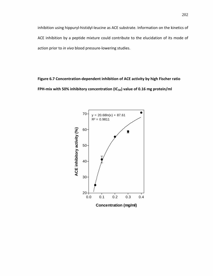
202
inhibition using hippuryl-histidyl-leucine as ACE substrate. Information on the kinetics of
ACE inhibition by a peptide mixture could contribute to the elucidation of its mode of
action prior to in vivo blood pressure-lowering studies.
Figure 6.7 Concentration-dependent inhibition of ACE activity by high Fischer ratio
FPH-mix with 50% inhibitory concentration (IC50
) value of 0.16 mg protein/ml
0.0 0.1 0.2 0.3 0.420
30
40
50
60
70
Concentration (mg/ml)
AC
E in
hibi
tory
act
ivity
(%)
y = 20.68ln(x) + 87.61R² = 0.9811

203
Figure 6.8 Double reciprocal plot of the inhibition of ACE activity by 0.1 and 0.4 mg/ml
of the high Fischer ratio FPH-mix indicating non-competitive mode of inhibition
In addition to ACE inhibition, molecules that can inhibit the activity of renin can
potentially provide better blood pressure lowering activity since renin controls the rate-
limiting step of the renin-angiotensin system. In our present study, the BCAA-rich
sample did not show any considerable inhibitory activity against renin; the peptide
mixture inhibited 7.1% renin activity at 4 mg/ml. In contrast, a recent study reported
that low-MW flaxseed protein-derived peptides moderately inhibited human renin in
vitro by 50% at concentrations ranging from 1.21 to 2.81 mg/ml (Udenigwe et al.,
2009b). The renin-angiotensin system has been widely studied as target for the
treatment of hypertension in humans, and a wide range of food proteins have afforded
ACE-inhibiting peptides, some with antihypertensive activities in animals and humans.
-8 -6 -4 -2 0 2 4 6 8 10 12 14 16
250
500
750
1000
1250
1500
1750
2000 No inhibitor (r2=0.99)0.1 mg/ml (r2=0.99)0.4 mg/ml (r2=0.99)
1/[FAPGG] (mM-1)
1/V
(min
/ ∆A
345n
m)

204
6.4. CONCLUSIONS
In conclusion, treatment of flaxseed protein isolate with thermolysin and
pronase followed by mixing with activated carbon afforded a BCAA-rich peptide mixture
with high Fischer ratio. The multifunctional bioactive properties of the peptide will
potentially enhance the value-added utilization of defatted flaxseed meal by making it
an important raw material in the functional foods and nutraceuticals industry. Animal
feeding experiments using this peptide preparation need to be conducted in animal
model of liver disease to evaluate the in vivo effects of the peptides in improving plasma
Fischer ratio and reducing muscle wasting.
Abbreviations Used. AAA, aromatic amino acids; ACE, angiotensin I-converting enzyme;
BCAA, branched-chain amino acids; DPPH, 2,2-diphenyl-1-picrylhydrazyl; FAPGG, N-(3-
[2-furyl]acryloyl)-phenylalanylglycylglycine; FPI, flaxseed protein isolate; FPH, flaxseed
protein hydrolysates; LA, linoleic acid; MW, molecular weight
ACKNOWLEDGMENTS
Operating and equipment research grants for this project were provided to REA by
Manitoba Agriculture and Rural Development Initiative (ARDI), and the Natural Sciences
and Engineering Research Council of Canada (NSERC). CCU is a recipient of NSERC
Alexander Graham Bell Canada Graduate Scholarship (CGS D).

205
6.5. REFERENCES
Adachi, S.; Kimura, Y.; Murakami, K.; Matsuno, R.; Yokogoshi, H. Separation of peptide groups with definite characteristics from enzymatic protein hydrolysate. Agric. Biol. Chem. 1991, 55, 925-932.
Adachi, S.; Yamanaka, T.; Hayashi, S.; Kimura, Y.; Matsuno, R.; Yokogoshi, H. Preparation of peptide mixture with high Fischer ratio from protein hydrolysate by adsorption on activated carbon. Bioseparation 1993, 3, 227-232.
Aluko, R.E. Antihypertensive properties of plant-derived inhibitors of angiotensin I-converting enzyme activity: a review. In: Recent Progress in Medicinal Plants – Phytopharmacology and Therapeutic Values IV, Govil, J.N., Singh, V.K., Eds.; Stadium Press: Houston, Texas, 2008; Vol. 22, pp. 541-561).
Ames, B. N.; Shigena, M. K.; Hegen, T. M. Oxidants, antioxidants and the degenerative diseases of aging. Proc. Natl. Acad. Sci. U.S.A. 1993, 90, 7915-7922.
Bidlingmeyer, B.; Cohen, S.; Tarvin, T. Rapid analysis of amino acids using pre-column derivatization. J. Chrom. 1984, 336, 93-104.
Campollo, O.; Sprengers, D.; McIntyre, N. The BCAA/AAA ratio of plasma amino acids in three different groups of cirrhotics. Rev. Invest. Clin. 1992, 44, 513-518.
Cinq-Mars, C.D.; Hu, C.; Kitts, D.D.; Li-Chan, E.C.Y. Investigations into inhibitor type and mode, simulated gastrointestinal digestion, and cell transport of the angiotensin I-converting enzyme-inhibitory peptides in Pacific hake (Merluccius productus) fillet hydrolysate. J. Agric. Food Chem. 2008, 56, 410-419.
Clemente, A. Enzymatic protein hydrolysates in human nutrition. Trends Food Sci. Technol. 2000, 11, 254-262.
Di Pasquale, M.D. Amino acids and proteins for the athlete: the anabolic edge, 2nd
Fischer, J.E. Branched-chain-enriched amino acid solutions in patients with liver failure: an early example of nutritional pharmacology. J. Parenter. Enteral. Nutr. 1990, 14, 249S-256S.
edition; CRC Press, Taylor & Francis Group: Boca Raton, FL, 2008, 434 pp.
Fischer, J.E.; Funovics, J.M.; Aguirre, A.; James, J.H.; Keane, J.M.; Wesdorp, R.I.; Yoshimura, N.; Westman, T. The role of plasma amino acids in hepatic encephalopathy. Surgery 1975, 78, 276-290.
Fischer, J.E.; Rosen, H.M.; Ebeid, A.M. The effect of normalization of plasma amino acids on hepatic encephalopathy in man. Surgery 1976, 80, 77-80.
206
Grimble, G.K.; Rees, R.G.; Keohane, P.P. Effects of peptide chain length on absorption of egg protein hydrolysates in the normal human jejunum. Gastroenterology 1987, 92, 136-142.
Gehrke, C.; Wall, L.; Absheer, J.; Kaiser F.; Zumwalt, R. Sample preparation for chromatography of amino acids: acid hydrolysis of proteins. J. Assoc. Off. Anal. Chem. 1985, 68, 811-821.
Hall III, C.; Tulbek, M. C.; Xu, Y. Flaxseed. Adv. Food Nutr. Res. 2006, 51, 1-97.
Humiski, L.M.; Aluko, R.E. Physicochemical and bitterness properties of enzymatic pea protein hydrolysates. J. Food Sci. 2007, S605-S611.
Khanna, S.; Gopalan, S. Role of branched-chain amino acids in liver diseases: the evidence for and against. Curr. Opin. Clin. Nutr. Metab. Care 2007, 10, 297-303.
Landry, J.; Delhaye, S. Simplified procedure for the determination of tryptophan of foods and feedstuffs from barytic hydrolysis. J. Agric. Food Chem. 1992, 40, 776-779.
Li, Y.; Jiang, B.; Zhang, T.; Mu, W.; Liu, J. Antioxidant and free radical-scavenging activities of chickpea protein hydrolysate (CPH). Food Chem. 2008, 106, 444-450.
Ma, Y.; Lin, L.; Sun, D-W. Preparation of high Fischer ratio oligopeptide by proteolysis of corn gluten meal. Czech J. Food Sci. 2008, 26, 38-47.
Markwell, M.A.C.; Haas, S.M.; Biebar, L.L.; Tolbert, N.E. A modification of the Lowry procedure to simplify protein determination in membrane and in protein samples. Anal. Biochem. 1978, 87, 206-211.
Miyoshi, S.; Kaneko, T.; Ishikawa, H.; Tanaka, H.; Maruyama, S. Production of bioactive peptides from corn endosperm proteins by some proteases. Ann NY Acad Sci. 1995, 750, 429-431.
Monchi, M.; Rerat, A.A. Comparison of net protein utilization of milk protein mild enzymatic hydrolysates and free amino acid mixtures with a close pattern in the rat. J. Parenteral. Enteral. Nutr. 1993, 17, 355-363.
Okita, M.; Watanabe, A.; Nagashima, H. Nutritional treatment of liver cirrhosis by branched amino acids-enriched nutrient mixture. J. Nutr. Sci. Vitaminol. 1985, 31, 291-303.
Oomah, B.D.; Mazza, G. Flaxseed proteins – a review. Food Chem. 1993, 48, 109-114.
Oomah, B.D.; Mazza, G. Functional properties, uses of flaxseed protein. Inform 1995, 6, 1246-1252.

207
Pedroche, J.; Yust, M.M.; Lqari, H.; Girón-Calle, J.; Vioque, J.; Alaiz, M.; Millán, F. Production and characterization of casein hydrolysates with a high amino acid Fischer’s ratio using immobilized proteases. Int. Dairy J. 2004, 14, 527-533.
Pedroche, J.; Yust, M.M.; Lqari, H.; Megías, C.; Girón-Calle, J.; Alaiz, M.; Vioque, J.; Millán, F. Production of Brassica carinata protein hydrolyzates with a high Fischer’s ratio using immobilized proteases. J. Agric. Food Chem. 2006, 54, 7621-7627.
Schulz, G.J.; Campos, A.C.L.; Coelho, J.C.U. The role of nutrition in hepatic encephalopathy. Curr. Opin. Clin. Nutr. Metab. Care 2008, 11, 275-280
Sun, J.Z.; Kaur, H.; Halliwell, B.; Li, X.Y.; Bolli, R. Use of aromatic hydroxylation of phenylalanine to measure production of hydroxyl radicals after myocardial ischemia in vivo. Direct evidence for a pathogenic role of the hydroxyl radical in myocardial stunning. Circ. Res. 1993, 73, 534-549.
Udenigwe, C.C.; Lin, Y-S., Hou, W-C.; Aluko, R.E. Kinetics of the inhibition of renin and angiotensin I-converting enzyme by flaxseed protein hydrolysate fractions. J. Funct. Foods 2009, 1, 199-207.
Udenigwe, C.C.; Lu, Y-L.; Han, C-H.; Hou, W-C.; Aluko, R.E. Flaxseed protein-derived peptide fractions: Antioxidant properties and inhibition of lipopolysaccharide-induced nitric oxide production in murine macrophages. Food Chem. 2009, 116, 277-284.
Wu. J.; Aluko, R.E.; Nakai, S. Structural requirements of angiotensin I-converting enzyme inhibitory peptides: quantitative structure-activity relationship study of di- and tripeptides. J. Agric. Food Chem. 2006, 54, 732-738.
Xie, Z.; Huang, J.; Xu, X.; Jin, Z. Antioxidant activity of peptides isolated from alfalfa leaf protein hydrolysate. Food Chem. 2008, 111, 370-376.
Zheng, H.; Zhang, C.; Cao, W.; Liu, S.; Ji, H. Preparation and characterization of the pearl oyster (Pinctada martensii) meat protein hydrolysates with a high Fischer ratio. Int. J. Food Sci. Technol. 2009, 44, 1183-1191.

208
APPENDIX C: SUPPLEMENTAL INFORMATION
Figure S6.1 Optimization of flaxseed protein hydrolysate (FPH) adsorption on activated
carbon by mixing in a beaker. A solution of FPH was mixed with different amounts of
activated carbon (0.0125–0.15 g) for 10 min followed by centrifugation. The
absorbance of the supernatant was measured at 260 nm to represent an
approximation of the amount of Tyr, which was used to calculate the % adsorbed
aromatic amino acids (AAA). Increase in the amount of activated carbon increase the
amount (%) of AAA removed from FPH
0.000 0.025 0.050 0.075 0.100 0.125 0.15070
80
90
100
PapainFicinAlcalasePronase
Activated carbon (g)
% A
dsor
bed
AA
A (f
rom
A26
0nm
)

209
Figure S6.2 Optimization of flaxseed protein hydrolysate (FPH) adsorption on activated
carbon by mixing in a beaker. The absorbance values at 280 nm represent an
approximation of the amount of Phe and Trp, and these were also used to calculate
the % adsorbed aromatic amino acids (AAA). Increase in the amount of activated
carbon increase the amount (%) of AAA removed from FPH
0.000 0.025 0.050 0.075 0.100 0.125 0.15070
80
90
100
PapainFicinAlcalasePronase
Activated carbon (g)
% A
dsor
bed
AA
A (f
rom
A28
0nm
)

210
Figure S6.3 Optimization of flaxseed protein hydrolysate (FPH) adsorption on activated
carbon by mixing in a beaker. The absorbance of the supernatant at 220 nm was
measured to represent approximate measurements of the FPH sample peptide
concentrations. The activated carbon treatment that efficiently removed most of the
aromatic amino acids (AAA) with a high protein yield (0.025 g activated carbon) was
selected for scale-up experiments
0.000 0.025 0.050 0.075 0.100 0.125 0.1500
2
4
6
8
10
12
14
PapainFicinAlcalasePronase
Activated carbon (g)
% U
nads
orbe
d pe
ptid
es(f
rom
A22
0nm
)
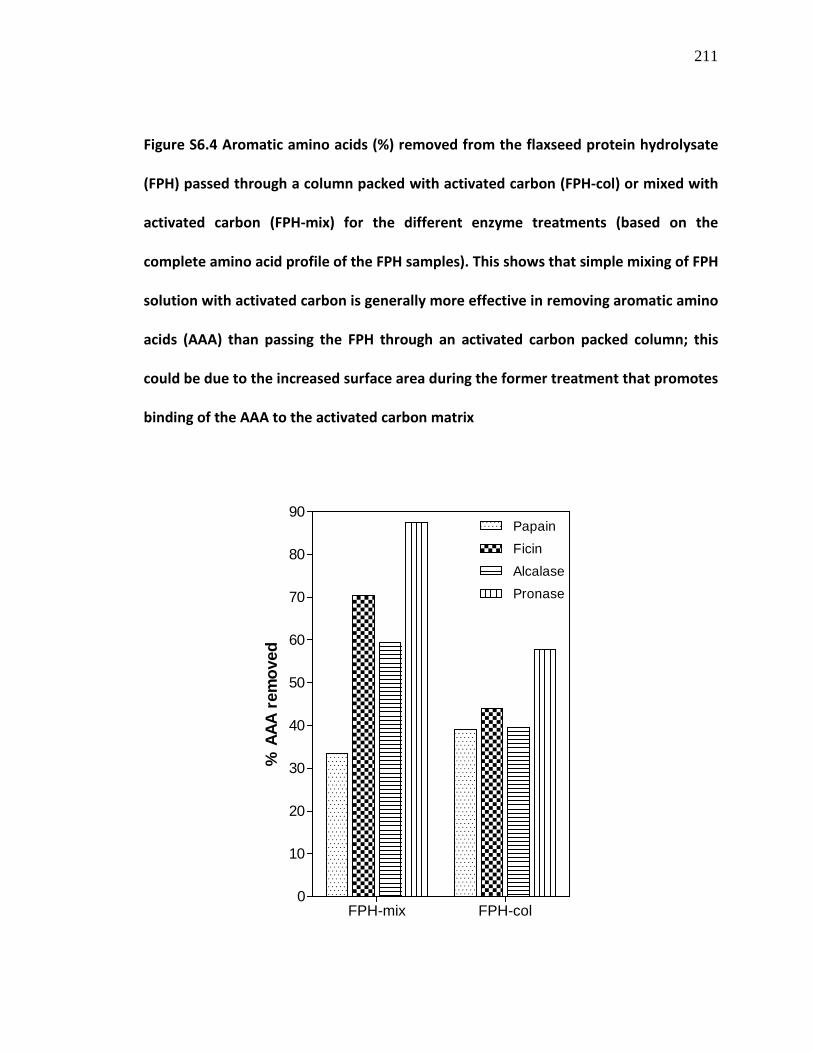
211
Figure S6.4 Aromatic amino acids (%) removed from the flaxseed protein hydrolysate
(FPH) passed through a column packed with activated carbon (FPH-col) or mixed with
activated carbon (FPH-mix) for the different enzyme treatments (based on the
complete amino acid profile of the FPH samples). This shows that simple mixing of FPH
solution with activated carbon is generally more effective in removing aromatic amino
acids (AAA) than passing the FPH through an activated carbon packed column; this
could be due to the increased surface area during the former treatment that promotes
binding of the AAA to the activated carbon matrix
FPH-mix FPH-col
0
10
20
30
40
50
60
70
80
90PapainFicinAlcalasePronase
% A
AA r
emov
ed

212
Table S6.1 Protein contents and yield of flaxseed protein hydrolysate (FPH), FPH
passed through a column packed with activated carbon (FPH-col) or mixed with
activated carbon in a beaker (FPH-mix) for the different enzymatic treatments
FPH Thermolysin
Papain Ficin Alcalase Pronase
Protein content (%)
FPH 81.0 a 85.2 66.3 60.7
FPH-col 90.1 b 97.8 72.6 77.4
FPH-mix 77.1 b 83.1 60.0 48.0
% Yield (DWB) c
FPH 90.6 a 96.9 88.2 96.6
FPH-col 15.5 b 15.0 22.0 16.0
FPH-mix 23.0 b 16.5 24.0 18.0
%Yield (Protein basis)
FPH 73.4 a 82.6 58.5 58.6
FPH-col 13.9 b 14.7 15.9 12.4
FPH-mix 17.7 b 13.7 14.4 8.6
aFrom flaxseed protein isolate; bFrom the corresponding flaxseed protein
hydrolysates (FPH); cDWB, dry weight basis

213
Table S6.2 Amino acid composition (%) of the activated carbon treated FPH samples (FPH-col and FPH-mix)
Amino acids FPH FPH-mixe FPH-cole FPHe FPH-mixf FPH-colf FPHf FPH-mixg FPH-colg g
Asx 10.98 a 10.39 11.93 11.17 11.06 11.62 11.05 10.37 11.27
Thr 3.59 4.30 3.96 3.65 3.88 4.12 3.61 4.12 3.92 Ser 5.11 6.20 6.06 5.01 5.63 5.84 5.07 6.15 6.53 Glx 21.57
b 17.12 20.79 21.28 19.31 19.33 21.51 20.17 21.00 Pro 4.44 4.67 4.24 4.02 4.30 4.63 3.96 3.92 4.27 Gly 5.60 5.86 6.12 5.52 5.58 5.69 5.56 5.97 6.79 Ala 4.71 6.48 5.62 4.80 5.45 6.10 4.76 5.99 6.16 Cys 1.44 0.26 0.72 1.31 0.74 0.65 1.34 0.53 0.98 Val 5.03 6.12 5.19 5.70 5.66 5.75 5.37 5.88 4.87 Met 1.29 0.99 1.34 1.23 1.06 1.30 1.18 0.91 1.25 Ile 4.47 4.73 4.45 4.77 4.97 4.81 4.52 4.41 3.96 Leu 5.92 6.78 6.01 5.99 6.08 6.09 5.97 6.13 5.68 Tyr 2.74 0.78 1.75 2.66 1.64 1.70 2.65 1.16 1.68 Phe 5.55 1.97 3.28 5.63 3.64 3.36 5.41 2.55 3.27 His 2.24 3.40 2.67 2.25 2.85 2.73 2.44 3.15 2.58 Lys 3.11 5.27 3.46 3.03 4.08 4.00 3.10 4.84 3.88 Arg 10.94 14.59 12.09 11.32 13.39 11.90 11.06 13.61 11.13 Trp 1.26 0.09 0.32 0.68 0.68 0.40 1.43 0.15 0.79
AAA 9.55 c 2.84 5.35 8.96 5.96 5.46 9.49 3.86 5.74
BCAA 15.42 d 17.63 15.65 16.46 16.71 16.65 15.85 16.43 14.51
Fischer ratio 1.61 6.21 2.92 1.84 2.80 3.05 1.67 4.26 2.53 aAsx, aspartic acid+asparagine; bGlx, glutamic acid+glutamine; cAAA: phenylalanine, tyrosine, tryptophan; dBCAA: valine, leucine, isoleucine; eThermolysin-ficin sample; fThermolysin-papain sample; gThermolysin-Alcalase sample

214
TRANSITION STATEMENT
In addition to its rich BCAA content, flaxseed protein also contains a high amount of
arginine, a basic/cationic amino acid. Arginine plays several physiological roles especially
as a precursor for the synthesis of nitric oxide (NO), a vasodilating gaseous agent
previously known as endothelial-derived relaxation factor (EDRF). It is thought that
arginine-enriched peptides could serve as vehicles to deliver arginine into the vascular
endothelium for increased NO production, and that this could lead to antihypertensive
activity and other beneficial physiological activities. Therefore, the sixth objective of this
project, discussed in the next chapter, was to use controlled enzymatic hydrolysis to
release arginine-rich peptides from flaxseed proteins and to separate these peptides
using electrodialysis-ultrafiltration, a novel membrane technology that separates
biomolecules based on a combination of molecular size and net molecular charge.

215
CHAPTER SEVEN
MANUSCRIPT 5
SEPARATION OF ARGININE-RICH PEPTIDES FROM FLAXSEED PROTEIN HYDROLYSATES
BY ELECTRODIALYSIS-ULTRAFILTRATION: ANGIOTENSIN CONVERTING ENZYME, RENIN-
INHIBITORY AND ANTIHYPERTENSIVE ACTIVITIES OF THE PEPTIDES
C. C. UDENIGWEa,b, A. P. ADEBIYIa,b, A. DOYENc, L. BAZINETc, R. E. ALUKOa,b
aDepartment of Human Nutritional Sciences, University of Manitoba, Winnipeg, MB R3T
2N2, Canada; bThe Richardson Centre for Functional Foods and Nutraceuticals,
University of Manitoba, Winnipeg, MB R3T 2N2, Canada; c
Institute of Nutraceuticals and
Functional Foods, Department of Food and Nutrition, Laval University, Québec, Canada.
To be submitted for publication
216
7.0. ABSTRACT
Flaxseed protein isolate (FPI) contains high amount of arginine, which plays important
physiological roles especially as nitric oxide precursor in the vascular endothelium. In
this study, enzymatic hydrolysis of FPI with trypsin and pronase resulted in a hydrolysate
(FPH), which was fractionated using electrodialysis-ultrafiltration (EDUF) to concentrate
the arginine-rich peptides. The EDUF experiment resulted in migration of anionic (KCl 1)
and cationic peptides (KCl 2) to the recovery compartments of the set-up with 1.10 ±
0.15% and 1.46 ± 0.09% peptide yields, respectively; more FPH peptides migrated to KCl
2 than KCl 1 and this was confirmed by mass spectrometry scan of the fractions.
Compared to FPI and FPH with 11% arginine, about one-third of KCl 2 was composed of
arginine. The arginine-rich peptides moderately inhibited angiotensin converting
enzyme and renin activities, and decreased systolic blood pressure in spontaneously
hypertensive rats. Thus, the multifunctional properties of this peptide product will be
relevant in clinical intervention against human health conditions especially
hypertension.
KEYWORDS: Electrodialysis; ultrafiltration; bioactive peptides; flaxseed protein;
arginine; angiotensin converting enzyme; renin

217
7.1. INTRODUCTION
Apart from its role as precursor for proline and glutamate synthesis, arginine (2-
amino-5-guanidinopentanoic acid), a conditionally essential basic/cationic amino acid, is
converted to nitric oxide (NO) and citrulline in the human vascular endothelium by a
Ca2+/calmodulin-dependent endothelial nitric oxide synthase (Moncada & Higgs, 1993;
Marin & Sessa, 2007; Pacher, Beckman & Liaudet, 2007). The NO produced from the L-
arginine-NO pathway is a significant factor for decreasing vascular resistance and can be
used to treat disease symptoms such as hypertension, vasospasm, cerebral ischemia,
preeclampsia, male impotence and bronchial asthma (Moncada & Higgs, 1993, 1995;
Marin & Sessa, 2007; Pacher et al., 2007; Coman, Yaplito-Lee & Boneh, 2008; Pasquale,
2008; Heffernan, Fahs, Ranadive, & Patvardhan, 2010). NO can also react with
homocysteine, a key indicator for the risk of cardiovascular disease, to form the non-
toxic S-nitroso-homocysteine, which also possesses vasodilatory property (Perna,
Ingrosso, & De Santo, 2003). In addition, L-arginine is required for hepatic synthesis of
creatine which is subsequently transported to the muscle where it is phosphorylated to
form phosphocreatine, an important energy source for cellular energy and muscular
contraction (Pasquale, 2008). It was demonstrated through several reports that L-
arginine plays important roles in enhancing immune system function (Park, Hayes,
Garlick, Sewell & Eremin, 1991; Delage et al., 2010), neurotransmission, cell signalling,
ammonia detoxification through the urea cycle (Coman et al., 2008), and amelioration
of experimental kidney diseases (Ketteler, Border & Noble, 1994). Some products of the
arginine metabolism pathways also function in prevention and inhibition of early stages

218
of certain cancers (Lowell, Parnes & Blackburn, 1990; Delage et al., 2010; Ma et al.,
2010). Moreover, human clinical interventions with L-arginine in free amino acid form
have resulted in endothelium-dependent vascular dilation in patients with endothelial
dysfunction but not in healthy individuals during short and long-term intakes (Heffernan
et al., 2010). These physiological effects of arginine have elicited our interest in the
development of arginine-enriched nutraceutical peptide products, from low value
flaxseed processing by-product, that can be used to increase physiological arginine
levels when needed, with concomitant increase in the active endogenous NO levels
especially in patients with endothelial dysfunction.
A recent survey reported that Canadian flaxseed from 2009 harvest contained
about 22% proteins (N × 6.25) with a 10-year mean of 23.2% (Canadian Grain
Commission, 2009). In 2007-2008, the world flaxseed meal production was estimated at
1.4 million tonnes (Agriculture and Agri-Food Canada, 2007); this translates to about
0.31 million tonnes of flaxseed proteins. This large amount of flaxseed proteins
produced annually has not been efficiently incorporated into the human food system,
but flaxseed meal has been fed to livestock for protein and fibre supplementation
(Agriculture and Agri-Food Canada, 2007; Bell & Keith, 1993). Flaxseed protein contains
a high amount of arginine (up to 11.2%) (Oomah & Mazza, 1995; Udenigwe & Aluko,
2010), when compared with other food proteins such as soy (7.6%), pea (8.2%),
rapeseed (7.0%), wheat (4.4%) and egg white (5.48%) (Sarwar et al., 1983). Therefore,
the global flaxseed meal production can generate more than 0.034 million tonnes (34
million kg) of arginine per year. Therefore, flaxseed proteins represent an excellent

219
source for the production of nutraceutical peptides enriched with arginine. It is
preferable to use peptides as a vehicle to deliver arginine into physiological locations as
opposed to the use of individual amino acids because small peptides are more
efficiently absorbed in the intestine and also due to the fact that osmotic pressures of
peptides are lower than those of their individual amino acid constituents (Grimble, Rees
& Keohane, 1997; Monchi & Rerat, 1993). Arginine-containing peptides have also been
shown to have cell penetrating capacity without the need for cellular carriers (Kosuge,
Takeuchi, Nakase, Jones, & Futaki, 2008; Schmidt, Mishra, Lai, & Wong, 2010).
Therefore, arginine in the form of peptides can be more readily delivered into cells
when compared to the free amino acid form. In addition, peptides may possess other
physiologically-relevant bioactive properties due to the peptide bonds, configuration
and sequence of amino acids as opposed to single amino acids.
Enzymatic hydrolysis has been extensively used in food processing to generate
bioactive peptides, which are often further separated into fractions with enhanced
activity. The choice of techniques for separating bioactive peptides from the complex
mixture depends on molecular properties of the peptides. Since peptides resulting from
extensive enzymatic hydrolysis of proteins often possess a narrow molecular size range,
it is imperative to use alternative processing methods especially when the separation is
not solely based on size (Firdaous et al., 2009). Electrodialysis-ultrafiltration (EDUF) is a
novel membrane technology used in food processing to separate valuable biomolecules
from complex mixture based on their electrical charge and molecular size (Bazinet,
Amiot, Poulin, Labbé & Tremblay, 2005). This process can be used to separate peptide

220
mixtures into fractions containing cationic, anionic and neutral peptides, and has been
previously applied in fractionation of tryptic digest of β-lactoglobulin (Poulin, Amiot &
Bazinet, 2006) and alfalfa white protein (ribulose-1,5-bisphosphate carboxylase)
hydrolysates (Firdaous et al., 2009). In both studies, the electroseparation process
resulted in the migration of cationic and anionic peptides to the cathode and anode,
respectively with the recovery of antihypertensive and angiotensin converting enzyme
(ACE)-inhibiting peptides in the cationic compartments. The advantages of the EDUF
process include simultaneous separation and concentration of the peptides, and the
absence of fouling of ultrafiltration membranes as opposed to the conventional
pressure-driven ultrafiltration and nanofiltration processes (Lapointe, Gauthier, Pouliot
& Bouchard, 2005; Firdaous et al., 2009).
The objectives of this present study were to (1) use a controlled enzymatic
process to hydrolyze flaxseed protein in order to release arginine-containing peptides,
(2) use electrodialysis-ultrafiltration to concentrate these arginine-rich peptides into a
fraction, (3) characterize the molecular constituents of the peptide fraction, and (4)
determine the potential bioactive properties, specifically antihypertensive activity in
spontaneously hypertensive rats and in vitro inhibition of the activities of ACE and renin,
which are two important enzymes of the blood pressure regulating renin-angiotensin
system.

221
7.2. MATERIALS AND METHODS
7.2.1. Materials
Bovine pancreas trypsin was purchased from Sigma-Aldrich (St. Louis, MO, USA)
and pronase from Streptomyces griseus was purchased from Roche Diagnostics GmbH
(Mannheim, Germany). NaCl and KCl were obtained from Laboratoire MAT (Québec, QC,
Canada). The CMX-SB and AMX-SB membranes were purchased from Eurodia (Wissous,
France) and the ultrafiltration membranes of 20 kDa molecular weight cut-off (MWCO)
purchased from Spectrum Laboratories Inc. (Rancho Dominguez, CA, USA). All other
chemical reagents were of analytical grade.
7.2.2. Enzymatic hydrolysis of flaxseed protein
Flaxseed protein hydrolysis was carried out using trypsin and pronase. Previously
prepared flaxseed protein isolate (Udenigwe, Lin, Hou & Aluko, 2009) was suspended in
distilled water (5% flaxseed protein, w/v) and the slurry was adjusted to pH 7.0 with 0.5
M NaOH and temperature of 37°C. Trypsin was added at an E/S of 1:100 to initiate
protein hydrolysis, and the reaction mixture incubated for 2 h. During hydrolysis, the
reaction was maintained at pH 7.0 with 0.5 M NaOH using a pH-Stat instrument
(Metrohm Titrando, Herisau, Switzerland). The reaction was stopped by adjusting the
mixture to pH 4.0 using 0.5 M HCl. Thereafter, the mixture was readjusted to pH 7.4 and
temperature of 40°C, followed by the addition of pronase at E/S of 1:100; hydrolysis was
continued for additional 2 h and terminated with 0.5 M HCl (pH 4.0). The resulting
mixture was cooled to room temperature and centrifuged at 15,000 x g for 1 h. The
222
supernatant was collected as the flaxseed protein hydrolysate (FPH), adjusted to pH 6.5
using 0.5 M NaOH, freeze dried and stored at –20°C for further processing.
7.2.3. Electrodialysis-ultrafiltration (EDUF)
EDUF was used to separate the cationic peptides of flaxseed protein hydrolysate
in order to obtain an arginine-enriched fraction. The experiment was conducted using a
modified version of a previously described method (Firdaous et al., 2009) as shown in
Figure 7.1. The electrodialysis cell had an effective surface area of 200 cm2 and
consisted of stacked membranes: CMX-SB (cation-exchange), AMX-SB (anion-exchange)
membranes, and two neutral ultrafiltration membranes with MWCO of 20 kDa. The
EDUF cell configuration consisted of two electrode rinsing compartments (containing 2 L
of 0.34 M NaCl), one feed compartment (containing the 1 L of 0.5% FPH) and two
permeate or recovery compartments (each containing 2 L of 0.0026 M KCl) connected to
an IONICS Electrodialyzer (IONICS Inc., Watertown, MA, USA). Each compartment was
connected to a separate external reservoir and re-circulated through the electrodialysis
stack using four pumps at flow rates of 3 L/min for the NaCl pump and 1 L/min for the
pumps connected to the feed and recovery compartments. Voltage (6–10V) and current
(0.2-0.35A) were supplied to the set-up using a direct current generator. The
experimental set-up was equipped with a pH meter model SP20 (Thermo Orion, West
Chester, PA, USA) and an YSI conductivity meter model 3252 (Yellow Springs Instrument
Co., Yellow Springs, OH, USA) for pH and conductivity measurements, respectively.

223
Figure 7.1 Configuration of the electrodialysis cell; anionic (P–) and cationic peptides
(P+) migrate to recovery compartments KCl 1 and KCl 2, respectively whereas peptides
with zero net charge (P±) remain in the feed compartment; AEM, anion exchange
membrane; CEM, cation exchange membrane; UFM, ultrafiltration membrane
The electrodialysis experimental runs were conducted for 360 min (n=5) in a cold
room (4°C). During the experiments, conductivity and pH of the solutions in the recovery
and feed compartments were monitored, and about 1 ml aliquots of samples were
withdrawn from the recovery compartments to determine peptide migration. After each
electroseparation, the electrodialysis cell was thoroughly cleaned for 30 min using an
alkaline solution followed by rinsing with distilled water, cleaning with acid solution for
another 30 min and thorough rinsing with distilled water to neutral pH.
UFM UFM AEM CEM
– +
0.5% FPH solution
KCl 1 (0.2 g/L)
NaCl (20 g/L)
NaCl (20 g/L)
KCl 2 (0.2 g/L)
P±
P+
P– P±
Cl–
K+ Cl–
K+ Anode Cathode
224
7.2.4. Determination of peptide migration during EDUF
The concentration of flaxseed peptides in the recovery compartments was
monitored during EDUF. A 1-ml aliquot was withdrawn from the KCl 1 and KCl 2
compartments at intervals of 0, 30, 60, 120, 180, 240, 300 and 360 min during each
electroseparation experiment. Peptide concentration in the solutions was determined in
duplicates using the MicroBCATM
Protein Assay Kit (Pierce Biotechnology Inc., Rockford,
IL, USA) following the manufacturer’s procedure. The peptide sample (100 µl) was
mixed with 100 µl of the assay working reagent in a microplate and the mixture
incubated for 2 h at 37°C. Thereafter, the solution was cooled to room temperature and
the absorbance measured at 562 nm using a microplate reader. The peptide
concentration (µg/ml) was calculated from a calibration curve prepared using the crude
FPH as a reference standard sample.
7.2.5. Amino acid analysis
Amino acid profiles of the FPH and the flaxseed peptide fractions from EDUF was
determined using the HPLC method after samples were hydrolyzed with 6 M HCl as
previously reported (Bidlingmeyer, Cohen & Tarvin, 1984). The cysteine and methionine
contents were determined after performic acid oxidation (Gehrke, Wall, Absheer, Kaiser
& Zumwalt, 1985) and tryptophan content was determined after alkaline hydrolysis
(Landry & Delhaye, 1992). The peptide samples were analyzed for amino acid
composition by separation on an analytical column packed with a sulphonated
polystyrene cation-exchange resin, followed by post-column derivatization with
225
ninhydrin (2,2-dihydroxy-1,3-indandione) at 135°C using a SYKNM Amino Acid Analyzer
S433 system (SYKAM GmbH, Eresing, Germany). The amino acid composition was
expressed as percentage of total amino acid content of each sample.
7.2.6. Liquid chromatography-mass spectrometry (LC-MS)
Molecular masses of peptides recovered in the KCl 1 and KCl 2 permeates after
EDUF and hydrolysate solution were determined by LC-MS using an LC-MSD QUAD
Agilent 1100 series system (Agilent Technologies, Palo Alto, CA, USA) equipped with two
pumps (bin G1323A), an automatic sample injector (G1329A) and an in-line degasser.
Peptides were loaded on a Luna 5 µm Phenomenex C18
column (2 × 250 mm) and
separated by gradient elution with 0.11% trifluoroacetic acid (TFA) in water (solvent A)
and solvent B, which was a mixture of acetonitrile/water/TFA (90/10/0.1% v/v/v), at a
flow rate of 0.2 mL/min. A linear gradient of 2–100% solvent B was applied for 115 min;
the eluting peptides were detected with DAD G1315A detector at a wavelength of 214
nm (Firdaous et al., 2009). The MS signals were recorded in positive mode using a 90-V
fragmentation with a scan range of 300–3000 m/z.
7.2.7. ACE and renin inhibition assays
The arginine-enriched peptide fraction was also evaluated for modulation of the
RAS pathway through the in vitro inhibition of ACE and renin activities. The sample was
desalted using an SPE column prior to the assays. The ACE and renin inhibition assays
were conducted in duplicates as previously reported using N-(3-[2-furyl]acryloyl)-

226
phenylalanylglycyl-glycine (FAPGG) as ACE substrate and a fluorogenic peptide as renin
substrate (Udenigwe et al., 2009). The ACE and renin inhibitory activities of the peptide
sample were reported as percent inhibition at various sample concentrations and also
as the concentration that resulted in the inhibition of 50% enzyme activity (IC50
),
determined from non-linear regression plot.
7.2.8. Evaluation of antihypertensive activity in spontaneously hypertensive rats (SHR)
Animal experiments were carried out following the Canadian Council on Animal
Care ethics guidelines with a protocol approved by the University of Manitoba Animal
Protocol and Management Review Committee. The spontaneously hypertensive rats
(SHR) were kept in the Animal Housing Facility at the Richardson Centre for Functional
Foods and Nutraceuticals, University of Manitoba under a 12-h day and night cycle at
21oC and were fed a regular chow diet and tap water, ad libitum. The SHR were divided
into 6 groups that received the following treatments (dissolved in phosphate buffered
saline, pH 7.2): arginine-enriched flaxseed peptide fraction (n=3), free arginine amino
acid (n=6), FPH (n=3), FPI (n=6), captopril (positive control, n=12), saline (negative
control, n=12). The samples (each at 200 mg/kg body weight, BW) and captopril (3
mg/kg BW) were administered to the SHR by oral gavage followed by measurement of
systolic blood pressure (SBP) at 2, 4, 6, 8 and 24 h by the tail-cuff method in slightly
anesthetised rats. Prior to sample administration, the baseline (time zero) SBP was
determined. In order to mitigate the blood pressure (BP) depression effect of
isofluorane, the gas flow was optimized such that rats became conscious usually within

227
3-4 min after removal from the chamber, which provided enough time to perform the
blood pressure measurement. Rats were first anesthetised in a chamber (maintained at
about 40°
C) with 4% isofluorane for 4 min. They were then removed from the
isofluorane chamber and tail-cuff measurement of blood pressure performed in the
unconscious state. The change in SBP (∆SBP, mmHg) was determined by subtracting the
data for the different time points from their respective baseline data.
7.2.9. Statistical analysis
The experimental data were collected in replicates as indicated above and
subjected to one way analysis of variance using Statistical Analysis System Software (SAS
version 9.2, SAS Institute, Cary, NC). Significant differences were determined by
Duncan’s multiple range test and accepted at p<0.05.
7.3. RESULTS AND DISCUSSION
7.3.1. Enzymatic hydrolysis of flaxseed protein
The flaxseed protein isolate was hydrolyzed with trypsin followed by pronase at
the optimum reaction conditions of the enzymes to release peptides of interest. Trypsin
was used for the first reaction because it specifically cleaves the carboxyl-terminal of the
peptide bond that contains arginine or lysine leading to the release of several peptides
with C-terminal arginine residues. Pronase was then used to generate low molecular
weight (LMW) peptides from the resulting trypsin-treated hydrolysate since it possesses
both endopeptidase and exopeptidease activities. It is expected that most of the LMW
228
peptides will contain at least a C-terminal arginine in their sequences. LMW peptides
were produced during the process in order to increase the physiological relevance of the
product since these peptides can be absorbed intact without gastrointestinal
peptidolysis and transported to the cellular locations were activity is needed (Mathews
and Adibi 1976; Roberts, Burney, Black, & Zaloga, 1999; Vermeirssen, Van Camp &
Verstraete, 2004). After the trypsin-pronase hydrolysis, the resulting freeze-dried
hydrolysates had a protein content of 84% and a dry weight yield of 80.9%.
7.3.2. Peptide migration during EDUF
The hydrolysate was adjusted to pH 7.0 prior to EDUF since the side (R) group of
arginine (pI = 10.76, pKaR = 12.48) and some other amino acids such as lysine (pI = 9.74,
pKaR = 10.53) and histidine (pI = 7.59, pKaR = 6.00) will be positively charged at that pH
while all the remaining amino acids will be negatively charged or neutral (Damodaran,
1996). This will ensure that peptides containing arginine, depending on their net
charges, will migrate to the cathode during EDUF to yield an arginine-rich fraction that
may also contain substantial amounts of lysine and tryptophan. During
electroseparation of the flaxseed protein hydrolysates, peptide migration into the
recovery compartments was monitored indirectly using conductivity and directly by
measuring actual peptide concentrations in the compartments. As shown in Figure 7.2,
the conductivity of solutions in the recovery compartments (KCl 1 and KCl 2) decreased
after every 60 min over the 360-min duration of electroseparation of the peptides. The
decrease in conductivity indicates peptide migration into the KCl compartments during

229
electrodialysis leading to counter-migration of K+ and Cl–
into the NaCl and feed
compartments (Figure 7.1), as previously demonstrated in EDUF fractionation of other
food protein hydrolysates (Poulin et al., 2006; Firdaous et al., 2009). This counter-
migration of the ions in the process was also confirmed by the observed 20.5% increase
in the conductivity (from 994.6 ± 59.3 to 1198.4 ± 56.6 µS/cm) of the FPH solution in the
feed compartment after EDUF. The conductivity of the KCl 1 solution decreased by 11%,
16% and 22% at 60, 180 and 360 min, respectively, whereas demineralization of the KCl
2 compartment occurred at a higher rates of 20%, 37% and 55%, at the respective time
points. This observation indicated that migration of the peptides to the cathode was
over 2 times more than migration of peptides to the anode at pH 7.0, and this
information also reflects on the final amounts of peptides in both compartments.

230
Figure 7.2 Change in conductivity in KCl 1 (anionic) and KCl 2 (cationic) compartments
during electrodialysis (n=5); decrease in the conductivity indicates peptide migration
to the electrodes during electrodialysis leading to counter-migration of K+ and Cl
–
The peptide concentration in the compartments was also directly monitored during
EDUF. As shown in Figure 7.3 using crude FPH solution as reference, there was an
increase in the peptide concentration (µg/ml) in the KCl compartments with time. This
observation also confirms the migration of flaxseed peptides to the electrodes at pH 7.0,
and indicates that applied electrical field was the major force responsible for the
separation of the peptides (Bargeman, Houwing, Recio, Koops & van der Horst, 2002;
Firdaous et al., 2009). In addition, Figure 7.3 showed that, at the end of the experiment,
the peptides that migrated to the cathode (73.2 ± 4.5 µg/ml) were significantly more in
0 60 120 180 240 300 360200
300
400
500
600
700
800
900
1000
1100
1200
KCl 1KCl 2
R2=0.9217
R2=0.9820
Time (min)
Con
duct
ivity
( µS/
cm)

231
amount than the peptides that migrated to the anode (55.2 ± 6.7 µg/ml); this supports
the conductivity data, which showed that peptide migration was more to the cathode
than to the anode. This was confirmed by the MS total ion current plot (TIC), shown in
Figure 7.4, which indicated that 13 or more peptides migrated to the KCl 2 compartment
while only 3 peptide peaks were observed in KCl 1. Thus, the FPH contained more net
positively-charged peptides than negatively-charged peptides at approximately pH 7.0.
The peptide distribution in the compartments observed in the TIC clearly showed the
effective selectivity of the EDUF process in separating peptides of similar MW and other
properties (e.g. elution time) but different net molecular charge. Based on the peptide
concentrations, the entire EDUF process (n=5) yielded a total of 551.9 mg and 732.7 mg
of the net anionic (KCl 1) and net cationic (KCl 2) peptides, respectively representing a
total yield of 1.10 ± 0.15% and 1.46 ± 0.09% from the original FPH.

232
Figure 7.3 Peptide migrations during electrodialysis of flaxseed protein hydrolysates
(n=5) as determined using the microBCATM
assay method; flaxseed protein hydrolysate
(FPH) was used as reference in calculating the peptide concentrations
60 120 180 240 300 360
-20
-10
0
10
20
30
40
50
60
70
80
KCl 1KCl 2
R2=0.9823
R2=0.9931
Time (min)
Pept
ide
conc
entr
atio
n ( µ
g/m
l)

233
Figure 7.4 Mass spectrometry total ion current plot (TIC) indicating migration of
peptides from feed compartment (containing flaxseed protein hydrolysates, FPH) to
the KCl 1 (anionic) and KCl 2 (cationic) compartments during electrodialysis-
ultrafiltration
min20 40 60 80 1000
200000
400000
600000
800000
1000000
1200000
4.0
98 4
.560
4.7
93 5
.293
5.6
01 6
.157 6
.330
6.7
54 7
.240 7
.322
8.3
54 8
.459
8.8
08 9
.205
9.3
86 9
.892
24.
206
24.
363
24.
628
24.
748
24.
922
25.
245
27.
181
27.
565
29.
877
30.
086
30.
241
31.
636
31.
772
33.
726
34.
468
34.
766
34.
907
35.
113
35.
186
35.
587
38.
279
38.
719
40.
120
40.
709
40.
793
41.
395
41.
877
42.
303
42.
806
42.
931
43.
062
43.
620
43.
934
44.
366
44.
611
44.
708
44.
907
45.
065
45.
588
45.
679
45.
961
46.
466
46.
813
47.
596
47.
996 48.
088
48.
216
48.
674
49.
203
49.
507 4
9.68
7 5
0.31
7 5
0.60
6 5
1.24
2
52.
500
52.
596
52.
694
53.
538
53.
955
54.
155
54.
579
54.
704
55.
756
57.
184
57.
293
59.
507
61.
010
62.
491
65.
504
66.
617
69.
973
70.
553
103
.484
FPH
min20 40 60 80 1000
200000
400000
600000
800000
1000000
1200000
3.4
85 3
.790
4.2
72 4
.436
4.7
22
7.8
53
11.
834
30.
593 4
3.26
3 4
3.37
2
50.
701
51.
032
54.
624
59.
407
59.
605
72.
293
72.
563
83.
100
103
.224
103
.491
103
.853
103
.942
104
.331
104
.649
104
.746
105
.101
105
.282
105
.530
105
.746
105
.890
106
.345
106
.525
106
.797 1
07.0
85 1
07.4
36 1
07.5
66 1
08.1
35 1
08.5
26 1
08.7
46 1
08.8
78 1
09.0
83 1
09.3
05 1
09.8
24 1
0993
5
1 2 3
KCl 1
min20 40 60 80 100
0
200000
400000
600000
800000
1000000
1200000
3.4
62 3
.840
4.7
31 5
.179
5.6
40 6
.358
6.4
65 6
.860
7.6
47 8
.215
8.8
13 9
.032
9.2
31
10.
749
11.
470
11.
650
12.
110
23.
331
23.
602
23.
762
23.
952
28.
065
28.
284 2
9.71
8 2
9.86
7
32.
140
32.
247
35.
540
40.
783
41.
936
42.
537
42.
643
42.
917
43.
247
43.
854
45.
921
50.
626
51.
422
55.
842
55.
942
58.
852
58.
983
59.
133
59.
546
62.
571
65.
486
65.
662
67.
176 7
2.34
9 7
2.66
3
80.
068
80.
166
80.
255
100
.815
102
.577 1
06.3
76 1
07.0
41 1
07.3
38 1
07.8
63 1
08.1
42 1
09.1
961 2
3 4 5 6 7
8 9
10
11 12 13
KCl 2

234
7.3.3. Amino acid profiles and peptide identification
The amino acid profiles of flaxseed protein isolate, FPH and the EDUF samples
were determined to confirm peptide migration and enrichment of the KCl 2 fraction
with peptides containing arginine, which are the peptides of interest in this study. As
shown in Table 7.1, about one-third (31.2%) of the freeze-dried KCl 2 sample resulting
from EDUF comprised of arginine compared to the original FPH and flaxseed protein
isolate, which contained 11.1% and 11.3% of arginine, respectively. This represents
about 3-fold increase in the amount of arginine in the samples. On the other hand, the
KCl 1 sample contained 2.61% of arginine, which migrated to the anode probably due to
the net negative molecular charge of their parent peptides. These results confirmed that
EDUF is an efficient process for the separation of arginine-rich peptides from enzymatic
flaxseed protein hydrolysates. It could also be observed that the process of enzymatic
hydrolysis of flaxseed protein did not affect arginine composition; thus, the arginine-
containing peptides were mostly retained in the FPH. Generally, it could be observed
that the total amount of positively charged amino acids (arginine, lysine and histidine)
was more in the KCl 2 fraction (43.2%) than in the KCl 1 fraction (7.6%) whereas the
acidic amino acids (Asx and Glx) were concentrated in the KCl 1 fraction (22%) compared
to the KCl 2 fraction (9.6%). However, it was observed that the remaining FPH in the
feed compartment after EDUF contained a substantial amount (9.32%) of arginine; thus,
the EDUF process used in this study did not separate all the arginine from FPH. This
observation could be due to the net zero charge or large size of these arginine-
containing peptides, or due to the EDUF conditions used in our study. A previous study

235
reported that the strength of the electrical field applied and the effective membrane
surface area independently affect peptide migration during EDUF (Poulin et al., 2007).
The peaks observed in the TIC of the flaxseed peptide samples were tentatively
identified from the MS data using the ExPASy Proteomics Server FindPept tool (Swiss
Institute of Bioinformatics) and the primary sequence of flaxseed conlinin, the only
major flaxseed storage protein with reported amino acid sequence (UniProtKD/TrEMBL
# Q8LPD3). Table 7.2 shows the MW identification and potential sequences of the KCl 1
and KCl 2 peptides; 10 peptides were identified in KCl 2 and 6 peptides in KCl 1 based on
the MS data, net charge and calculated isoelectric point (pI) of the peptides at pH 7.0.
The net charge of the peptides at the experimental pH corresponded to their migration
to the electrodes. All of the possible KCl 1 peptides contained arginine in their sequence,
except peak 9, while all the KCl 1 peptides contained aspartic acid/glutamic acid, which
contributed strongly to their net charge. However, some peptides (peaks # 2, 4, 6, 8 and
10) tentatively identified in KCl 2 possessed zero net charge with pI values of 6.1 to 6.8.
This observation could possibly be due to fluctuations in pH (values decreased to the pI
of these peptides, data not shown) observed during EDUF leading to temporary change
in net charge and migration of the peptides to the cathode. This explanation is
supported by the net negative charge observed for all the potential peptides in KCl 1,
which possessed calculated pI values of 2.9 to 3.1 (lower than the experimental pH
conditions). It is important to note that these peptide sequences are tentative since
flaxseed contains another major storage protein, linin, which could have also
contributed substantially to the peptide pool.

236
Table 7.1 Amino acid compositions (%) of flaxseed protein isolate (FPI), protein
hydrolysate (FPH) and peptides from the anode (KCl 1), cathode (KCl 2) and feed (FPH-
X) compartments after electrodialysis-ultrafiltration
AMINO ACID FPI FPH a FPH-X FPH (KCl 1) FPH (KCl 2)
ASX 11.29 11.65 11.10 8.86 5.19
THR 3.67 3.97 3.64 7.16 3.17
SER 4.34 5.08 4.34 5.23 2.88
GLX 19.83 20.12 19.87 13.18 4.41
PRO 5.29 4.15 4.07 12.05 3.54
GLY 5.47 5.65 5.98 7.27 5.03
ALA 4.89 5.00 5.19 6.70 4.41
CYS 1.36 1.15 1.17 0.80 0.12
VAL 5.20 5.57 6.47 8.18 6.14
MET 2.02 1.70 1.88 0.34 0.00
ILE 4.50 4.82 5.53 4.77 5.48
LEU 5.80 6.02 6.55 6.82 6.72
TYR 2.52 2.75 3.00 3.41 2.93
PHE 5.47 5.78 6.33 7.61 6.72
HIS 2.29 2.34 2.44 3.41 2.06
LYS 3.04 3.14 3.13 1.59 9.97
ARG 11.28 11.10 9.32 2.61 31.23
CATIONIC 14.32 16.59 14.88 7.61 43.26
ACIDIC 31.12 31.77 30.96 22.05 9.60 aData derived from Udenigwe and Aluko (2010); FPH, flaxseed protein hydrolysate; FPH-X,
flaxseed protein hydrolysate remaining in feed compartment after electrodialysis; FPH (KCl
1), peptides that migrated towards the anode (anionic); FPH (KCl 2), peptides that migrated
towards the cathode (cationic)
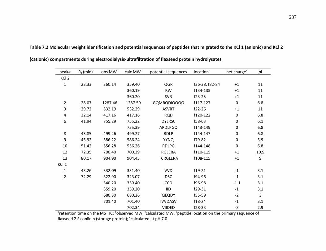
237
Table 7.2 Molecular weight identification and potential sequences of peptides that migrated to the KCl 1 (anionic) and KCl 2
(cationic) compartments during electrodialysis-ultrafiltration of flaxseed protein hydrolysates
peak# Rt (min) obs MWa b calc MW c potential sequences location net charged pI e KCl 2
1 23.33 360.14 359.40 QGR f36-38, f82-84 +1 11
360.19 RW f134-135 +1 11
360.20 SVR f23-25 +1 11
2 28.07 1287.46 1287.59 GQMRQDIQQQG f117-127 0 6.8 3 29.72 532.19 532.29 ASVRT f22-26 +1 11 4 32.14 417.16 417.16 RQD f120-122 0 6.8 6 41.94 755.29 755.32 DYLRSC f58-63 0 6.1
755.39 ARDLPGQ f143-149 0 6.8
8 43.85 499.26 499.27 RDLP f144-147 0 6.8 9 45.92 586.22 586.24 YYNQ f79-82 0 5.9
10 51.42 556.28 556.26 RDLPG f144-148 0 6.8 12 72.35 700.40 700.39 RGLERA f110-115 +1 10.9 13 80.17 904.90 904.45 TCRGLERA f108-115 +1 9
KCl 1 1 43.26 332.09 331.40 VVD f19-21 -1 3.1
2 72.29 322.90 323.07 DSC f94-96 -1 3.1
340.20 339.40 CCD f96-98 -1.1 3.1
359.20 359.20 IID f29-31 -1 3.1
680.30 680.26 QEQDY f55-59 -2 3
701.40 701.40 IVVDASV f18-24 -1 3.1
702.34 VIIDED f28-33 -3 2.9
aretention time on the MS TIC; bobserved MW; ccalculated MW; dpeptide location on the primary sequence of flaxseed 2 S conlinin (storage protein); ecalculated at pH 7.0

238
7.3.4. Inhibition of ACE and renin activities by the arginine-enriched peptide fraction
Food-derived peptides that can modulate the renin-angiotensin system by
inhibiting ACE and renin activities can be used in human clinical interventions against
hypertension and associated cardiovascular diseases. In this study, the arginine-rich
flaxseed peptide sample (KCl 2) was found to exhibit concentration-dependent
moderate inhibitory activity against ACE in vitro (Figure 7.5). A maximum of 80%
inhibition of ACE activity was observed at 1 mg/ml of the desalted KCl 2, which gave an
IC50 value of 0.38 ± 0.01 mg/ml. Previous studies on flaxseed protein-derived peptides
yielded more active ACE inhibitors especially LMW peptide fraction from thermolysin-
catalyzed flaxseed protein hydrolysates with IC50
On the other hand, the arginine-rich flaxseed peptide sample displayed a weak
renin-inhibitory activity, which was also observed to be concentration-dependent with a
maximum of 44.5% renin inhibition at 7.5 mg/ml of the peptide sample (Figure 7.6). This
value of 0.0275 mg/ml (Udenigwe et
al., 2009). The structural requirements for ACE inhibition include the presence of
hydrophobic and bulky amino acids in dipeptides, and C-terminal aromatic amino acid
with middle cationic amino acid in tripeptides (Wu, Aluko & Nakai, 2006). Thus, the low
amounts of hydrophobic and aromatic amino acids in the KCl 2 sample (Table 7.1) could
have contributed to its low ACE-inhibitory activity. The LMW peptides released from
flaxseed protein in this study contain arginine located mostly at the C-terminal of the
peptides due to the activity of trypsin; the tentative peptide sequences in KCl 2 contain
arginine at both terminals and within the sequence, but these arrangements may not
have contributed to potency in ACE inhibition.

239
activity is weaker than those previously reported for various LMW peptides generated
from flaxseed proteins (Udenigwe et al., 2009) and C-terminal arginine-containing
dipeptides Ile-Arg (IR), Leu-Arg (LR) and Asn-Arg (NR) (Udenigwe, Li, & Aluko,
unpublished – see Chatpter 8). A recent study reported that amino-terminal small
hydrophobic amino acids and C-terminal bulky amino acids contribute to enhanced
potency of renin inhibiting dipeptides (Udenigwe et al., unpublished – see Chatpter 8).
Therefore, it was expected that a C-terminal arginine-containing peptide could potently
inhibit renin activity at low concentrations if the N-terminal is occupied by a
hydrophobic amino acid.
Figure 7.5 ACE-inhibitory activity of flaxseed protein-derived arginine-enriched
peptide fraction (KCl 2)
0.0 0.2 0.4 0.6 0.8 1.00
10
20
30
40
50
60
70
80
90
100
y = 23.82ln(x) + 74.09R2=0.9365IC50=0.38 ± 0.01 mg/ml
KCl 2 (mg/ml)
AC
E in
hibi
tion
(%)

240
Figure 7.6 Renin-inhibitory activity of flaxseed protein-derived arginine-enriched
peptide fraction (KCl 2)
7.3.5. Antihypertensive activity of the arginine-rich peptide fraction
Results from the animal study indicated that, at 200 mg/kg BW, the arginine-rich
flaxseed peptide fraction decreased SBP by –17.9 and –11.7 mmHg, respectively after 2
and 4 h following oral gavage when compared to the effect of free arginine (∆SBP of –
10.3 and –2.8 mmHg, respectively) (Figure 7.7). The hypotensive activity of the arginine-
rich peptides was similar to that of captopril after 2 h, which indicates ability to provide
fast relief from hypertension on a short-term basis. The hypotensive effect observed for
the arginine-enriched peptide product was greater than those observed for the amino
acid form of arginine as well as the flaxseed protein isolate. The results support our
0.0 1.5 3.0 4.5 6.0 7.5-10
0
10
20
30
40
50
60
70
80
90
100
y = 5.8129x - 2.0462R2 = 0.9837
KCl 2 (mg/ml)
Ren
in in
hibi
tion
(%)

241
hypothesis that peptides could be a better form of delivering fast therapeutic effects
when compared to free amino acids and large-size proteins. The hypotensive effect of
the arginine-enriched peptide product was observed to be long-lasting compared to the
free amino acid; this indicated a more efficient absorption of the peptides compared to
free arginine. The observed activity could be due to the vasodilatory nitric oxide
synthesized from the arginine or possibly due to ACE and renin inhibition by the cationic
peptides. Moreover, the crude FPH showed similar hypotensive effects to the arginine-
enriched peptide product with sustained decrease in SBP during the first 6 h. Therefore,
the FPH could be used as a source of the arginine-containing peptides, which would
obviate the need for the EDUF process with substantial savings in processing costs.
Surprisingly, FPI also exhibited hypotensive activity with a maximum ∆SBP of –18.4
mmHg at 6 h. The slow-acting effect of the FPI was expected since digestion of the
inherent large proteins must precede absorption of the bioactive peptides. In contrast,
the FPH and arginine-enriched peptide product are already pre-digested, hence
absorption proceeded quickly and they provided faster hypotensive effects than the FPI.
Detailed short-term and long-term studies are underway in our laboratory to
characterize dose-response of these treatments on SBP, circulating AT-II, renin and ACE
protein levels and activities in SHR tissues, especially with the arginine-rich peptides,
crude FPH and FPI.

242
Figure 7.7 Effects of arginine-rich flaxseed peptides, free arginine, flaxseed protein
hydrolysate (FPH) and protein isolate (FPI), each at 200 mg/kg body weight (BW), on
systolic blood pressure (SBP) in spontaneously hypertensive rats (SHR); captopril (3
mg/kg BW) and saline were used as positive and blank controls, respectively
7.4. CONCLUSIONS
It can be concluded that flaxseed meal protein is an excellent protein source for
the production of arginine-rich peptides by controlled enzymatic hydrolysis, and that
combined electrodialysis and ultrafiltration process can be used to concentrate these
peptides into a fraction. The low yield obtained for the separated fraction and the high

243
arginine level in the feed compartment after separation suggest that the efficiency of
both the enzymatic hydrolysis and EDUF processes need to be further investigated and
improved. However, the EDUF process may not be required since the FPH produced
similar hypotensive effects to the cationic peptides. But the peptide product is still a
good candidate for further studies because of its beneficial physiological hypotensive
activity in SHR and activity in moderately inhibiting the enzymes of the blood pressure-
regulating pathway. The fast-acting nature of the FPH and arginine-enriched peptide
product could be an advantage over the original protein isolate by providing quick relief
from high blood pressure. The hypotensive activities observed for FPI and crude FPH will
encourage further studies on the use of these products as therapeutic agents without
further processing. These studies will ultimately contribute to value addition to defatted
flaxseed meal generated from the oilseed industry.
ACKNOWLEDGEMENTS
Operating and equipment research grants for this project were provided to Dr. RE Aluko
by Manitoba Agriculture and Rural Development Initiative (ARDI) and the Natural
Sciences and Engineering Research Council of Canada (NSERC). CC Udenigwe is a
recipient of NSERC Alexander Graham Bell Canada PhD Scholarship. The authors are
grateful to Monica Araya-Farias of Université Laval, Québec, for technical assistance.

244
7.5. REFERENCES
Agriculture and Agri-Food Canada. Flaxseed: situation and outlook, Bi-weekly Bulletin 20(3) February 23, 2007, AAFC No. 2081/E
Bargeman, G., Houwing, J., Recio, I., Koops, G.-K., & van der Horst, C. (2002). Electromembrane filtration for the selective isolation of bioactive peptides from an αs2
Bazinet, L., Amiot, J., Poulin, J.-F., Labbé, D., & Tremblay, D. (2005). Process and system for separation of organic charged compounds. International Patent WO 2005/082495A1
-casein hydrolysate. Biotechnology and Bioengineering, 80, 559-609
Bell, J. M., & Keith, M. O. (1993). Nutritional evaluation of linseed meals from flax with yellow or brown hulls, using mice and pigs. Animal Feed Science and Technology, 43, 1-18
Bidlingmeyer, B., Cohen, S., & Tarvin, T. (1984). Rapid analysis of amino acids using pre-column derivatization. Journal of Chromatography, 336, 93-104
Canadian Grain Commission. Quality of western Canadian flaxseed 2009. http://www.grainscanada.gc.ca/flax-lin/harvest-recolte/2009/hqf09-qrl09-eng.pdf ISSN 1700-2087 (Accessed August 2010).
Coman, D., Yaplito-Lee, D., & Boneh, A. (2008). New indications and controversies in arginine therapy. Clinical Nutrition, 27, 489-496
Damodaran S. (1996). Amino acids, Peptides, and Proteins. In O. R. Fennema (Ed.), Food Chemistry, 3rd
Delage, B., Fennell, D. A., Nicholson, L., McNeish, I., Lemoine, N. R., Crook, T., & Szlosarek, P. W. (2010). Arginine deprivation and argininosuccinate synthetase expression in the treatment of cancer. International Journal of Cancer, 126, 2762-2772
ed. (pp. 321-430). New York: Marcel Dekker Inc.
Di Pasquale, M. G. (2008). Amino acids and proteins for the athlete: the anabolic edge. (2nd
Firdaous, L., Dhulster, P., Amiot, J., Gaudreau, A., Lecouturier, D., Kapel, R., Lutin, F., Vézina, L.-P., & Bazinet, L. (2009). Concentration and selective separation of bioactive peptides from an alfalfa white protein hydrolysate by electrodialysis with ultrafiltration membranes. Journal of Membrane Science, 329, 60-67
ed.). New York: CRC Press, (Chapter 8)
Gehrke, C., Wall, L., Absheer, J., Kaiser F., & Zumwalt, R. (1985). Sample preparation for chromatography of amino acids: acid hydrolysis of proteins. Journal of the Association of Official Analytical Chemists, 68, 811-821

245
Grimble, G. K., Rees, R. G., & Keohane, P. P. (1987). Effects of peptide chain length on absorption of egg protein hydrolysates in the normal human jejunum. Gastroenterology, 92, 136-142
Heffernan, K. S., Fahs, C. A., Ranadive, S. M., & Patvardhan, E. A. (2010). L-arginine as a nutritional prophylaxis against vascular endothelial dysfunction with aging. Journal of Cardiovascular Pharmacology and Therapeutics, 15, 17-23
Ketteler, M., Border, W. A., & Noble, N. A. (1994). Cytokines and L-arginine in renal injury and repair. American Journal of Physiology - Renal Fluid and Electrolyte Physiology, 267, F197-207
Kosuge, M., Takeuchi, T., Nakase, I., Jones, A.T., & Futaki, S. (2008). Cellular internalization and distribution of arginine-rich peptides as a function of extracellular peptide concentration, serum, and plasma membrane associated proteoglycans. Bioconjugate Chemistry, 19, 656-664
Landry, J., & Delhaye, S. (1992). Simplified procedure for the determination of tryptophan of foods and feedstuffs from barytic hydrolysis. Journal of Agricultural and Food Chemistry, 40, 776-779
Lapointe, J.-F., Gauthier, S. F., Pouliot, Y., & Bouchard, C. (2005). Fouling of a nanofiltration membrane by a β-lactoglobulin tryptic hydrolysate: impact on the membrane sieving and electrostatic properties, Journal of Membrane Science, 253 89-102
Lowell, J. A., Parnes, H. L., & Blackburn, G. L. (1990). Dietary immunomodulation: Beneficial effects on oncogenesis and tumor growth. Critical Care Medicine, 18, S145-148
Ma, Q., Wang, Z., Zhang, M., Hu, H., Li, J., Zhang, D., Guo, K., & Sha, H. (2010). Targeting the L-arginine-nitric oxide pathway for cancer treatment. Current Pharmaceutical Design, 16, 392-410
Marin, E., & Sessa, W. C. (2007). Role of endothelial-derived nitric oxide in hypertension and renal disease. Current Opinion in Nephrology and Hypertension, 16, 105-110
Mathews, D. M., & Adibi, S. A. (1976). Peptide absorption. Gastroenterology, 71, 151-161
Moncada, S., & Higgs, A. (1993). The L-arginine-nitric oxide pathway. New England Journal of Medicine, 329, 2002-2012
Moncada, S., & Higgs, A. (1995). Molecular mechanisms and therapeutic strategies related to nitric oxide. FASEB Journal, 9, 1319-1330

246
Monchi, M., & Rerat, A. A. (1993). Comparison of net protein utilization of milk protein mild enzymatic hydrolysates and free amino acid mixtures with a close pattern in the rat. Journal of Parenteral and Enteral Nutrition, 17, 355-363
Oomah, B. D., & Mazza, G. (1995). Functional properties, uses of flaxseed protein. Inform, 6, 1246-1252
Pacher, P., Beckman, J. S., & Liaudet, L. (2007). Nitric oxide and peroxynitrite in health and disease. Physiological Reviews, 87, 315–424
Park, K. G. M., Hayes, P. D., Garlick, P. J., Sewell, H., & Eremin, O. (1991). Stimulation of lymphocyte natural cytotoxicity by L-arginine. Lancet, 337, 645-646
Perna, A. F., Ingrosso, D., & De Santo, N. G. (2003). Homocysteine and oxidative stress. Amino Acids, 25, 409-417
Poulin, J.-F., Amiot, J., & Bazinet, L. (2006). Simultaneous separation of acid and basic bioactive peptides by electrodialysis with ultrafiltration membrane. Journal of Biotechnology, 123, 314-328
Poulin, J.-F., Amiot, J., & Bazinet, L. (2007). Improved peptide fractionation by electrodialysis with ultrafiltration membrane: Influence of ultrafiltration membrane stacking and electrical field strength. Journal of Membrane Science, 299, 82-90
Roberts, P. R., Burney, J. D., Black, K. W., & Zaloga, G. P. (1999). Effect of chain length on absorption of biologically active peptides from the gastrointestinal tract. Digestion, 60, 332-337
Sarwar, G., Christiansen, D. A., Finlayson, A. T., Friedman, M., Hackler, L. R., McKenzie, S. L., Pellett, P. L., & Tkachuk, R. (1983). Intra- and inter-laboratory variation in amino acid analysis. Journal of Food Science, 48, 526-531
Schmidt, N., Mishra, A., Lai, G.H., & Wong, G.C.L. (2010). Arginine-rich cell-penetrating peptides. FEBS Letters, 584, 1806-1813
Udenigwe C. C., & Aluko, R. E. (2010). Antioxidant and angiotensin converting enzyme-inhibitory properties of a flaxseed protein-derived high Fischer ratio peptide mixture. Journal of Agricultural & Food Chemistry, 58, 4762-4768
Udenigwe, C. C., Li, H., & Aluko, R. E. Quantitative structure-activity relationship modeling of renin-inhibiting dipeptides (submitted)
Udenigwe, C. C., Lin, Y.-S., Hou, W.-C., & Aluko, R. E. (2009). Kinetics of the inhibition of renin and angiotensin I-converting enzyme by flaxseed protein hydrolysate fractions. Journal of Functional Foods, 1, 199-207

247
Vermeirssen, V., Van Camp, J., & Verstraete, W. (2004). Bioavailability of angiotensin I converting enzyme inhibitory peptides. British Journal of Nutrition, 92, 357–366
Wu, J., Aluko, R. E., & Nakai, S. (2006). Structural requirements of angiotensin I-converting enzyme inhibitory peptides: quantitative structure-activity relationship study of di- and tripeptides. Journal of Agricultural & Food Chemistry, 54, 732-738

248
TRANSITION STATEMENT
The structure of peptides determines their interactions with biological molecular
disease targets and invariably influences their physiological activities. This project has so
far shown that low-molecular size peptides derived from flaxseed proteins possess
prospects as therapeutic agents especially as antihypertensive agents due to their ability
to inhibit ACE and renin. Till date, several studies have reported some structural
requirements of peptides of various sizes for ACE inhibition, and this has contributed
towards the discovery of more potent ACE inhibitors. However, there is scanty
information in the literature on the structure-function properties of renin-inhibiting
natural peptides. The next chapter reports on the quantitative structure-activity
relationship (QSAR) studies on renin-inhibiting food-derived dipeptides. Information
derived from this study will be invaluable in the design and synthesis of potent orally
bioavailable antihypertensive peptides and peptidomimetics.

249
CHAPTER EIGHT
MANUSCRIPT 6
QUANTITATIVE STRUCTURE-ACTIVITY RELATIONSHIP MODELLING OF RENIN-
INHIBITING DIPEPTIDES
C. C. UDENIGWE, H. LI, R. E. ALUKO
Department of Human Nutritional Sciences, University of Manitoba, Winnipeg, MB R3T
2N2, Canada; The Richardson Centre for Functional Foods and Nutraceuticals, University
of Manitoba, Winnipeg, MB R3T 2N2, Canada.
Submitted to Amino Acids

250
8.0. ABSTRACT
Partial least squares regression method was used to analyze a peptide dataset and
construct inhibitory models for renin inhibitory natural dipeptides. The models were
computed with the renin inhibitory activity as dependent variable (Y) and the peptide
structural properties as predictors (X); validation was conducted using cross-validation
and permutation tests. The amino acid descriptors were based on the 3- and 5-z scales
of 20 coded amino acids to produce models that explained 71.6% of Y with a 33.8%
predictive ability and 75.2% of Y with a predictive power of 50.8%, respectively. In both
models, low molecular size amino acids with hydrophobic side chains were preferred at
the N-terminus, while amino acids with bulky side chains were preferred at the C-
terminus for potency. Based on the 5-z model, four Trp-containing antihypertensive
dipeptides (IW, LW, VW and AW) were predicted as the most potent renin inhibitors.
The peptides were synthesized and in vitro inhibition assay showed that IW and LW
inhibited 70% (IC50
KEYWORDS: Renin; Dipeptides; Quantitative structure-activity relationship (QSAR);
Partial least squares (PLS); Amino acids descriptors
, 2.3 mM) and 37% renin activity at 3.2 mM, respectively whereas VW
and AW were inactive. There was no correlation between the observed renin-inhibitory
activities and angiotensin-converting enzyme inhibitory activities of the dipeptides. We
concluded that the structural similarities between isoleucine and leucine could have
contributed to their distinct inhibitory activity when compared with alanine and valine.
Therefore, IW may be a useful template for the development of advanced forms of
highly active low molecular size antihypertensive peptides and peptidomimetics.

251
8.1. INTRODUCTION
The renin-angiotensin system (RAS) has provided key molecular targets for
therapeutic agents towards the treatment and management of hypertension, a
contributing risk factor to human cardiovascular diseases and a major public health
concern especially in the Western world. This is primarily due to the role of the RAS
pathway in controlling blood pressure through the production of angiotensin (AT)-II, a
potent vasoconstrictor, from AT-I by the action of angiotensin I-converting enzyme
(ACE). Consequently, ACE inhibitors have been widely used to control blood pressure
during hypertension, and several food protein-derived peptides have shown both ACE-
inhibitory and antihypertensive activities (Aluko 2008). Another important molecular
target for antihypertension drug discovery is the renin-catalyzed conversion of
angiotensinogen to AT-I, which is the rate-limiting step of RAS (Staessen et al. 2006). The
inhibition of renin activity can provide better blood pressure control during
hypertension than ACE inhibition since it prevents the formation of AT-I, which can be
converted to AT-II in some cells via an ACE-independent chymase-catalyzed reaction
(Staessen et al. 2006; Segall et al. 2007). Moreover, since angiotensinogen is the only
known renin substrate, targeting renin in the RAS pathway ensures high specificity in
controlling elevated blood pressure as opposed to ACE inhibition, which can also affect
other biochemical pathways leading to toxic effects (Acharya et al. 2003; Staessen et al.
2006). Several synthetic peptides and non-peptides have been reported to possess renin
inhibitory activity but their application as orally-active antihypertensive agents has been
hindered due to poor pharmacokinetic attributes (Rahuel et al. 2000; Fischer and

252
Hollenberg 2001; Wood et al. 2006). Till date, aliskiren is the only known clinically-
relevant non-peptide renin-inhibiting antihypertensive agent (Sepehrdad et al. 2007);
current research efforts are continuously directed towards the discovery of potent non-
toxic renin inhibitors. Recent studies have shown that food protein-derived peptides
and enzymatic hydrolysates possess the ability to inhibit renin activity in vitro
(Udenigwe et al. 2009; Li 2010). These research efforts could lead to the discovery of a
new generation of cheaper renin inhibitors with no side effects since the peptides were
generated from food proteins. Moreover, knowledge of the structure-function property
of these food-derived peptides can lead to the design and synthesis of more potent
renin inhibiting antihypertensive agents, especially peptidomimetics. To the best of our
knowledge, there is scanty information in the literature on the structural requirements
for renin inhibiting food-derived peptides that contain only coded amino acids.
Quantitative structure-activity relationship (QSAR) modelling is a practical and
reliable method in chemometrics for studying the relationship between molecular
structures of therapeutic agents and biological activities. In peptide QSAR, statistical
multiple regression analysis such as partial least squares projection of latent structure
(PLS) have been widely used to develop models that relate the molecular structures to
variation in the biological activities. In other words, bioactivity data can be modelled as
a function of molecular structures of peptides (Sandberg et al. 1998; Wu et al. 2006a).
Using amino acid z scale descriptors, PLS has been successfully applied in developing
good models in QSAR study of various bioactive peptides (Hellberg et al. 1987) including
models that can explain the structural requirements for ACE-inhibiting peptides of

253
various chain lengths (Wu et al. 2006a, 2006b). QSAR models for peptides’ bitterness
property (Wu and Aluko 2007) and for the study of the functional properties of
polypeptides (Siebert 2003) have also been reported.
The objectives of this study were to (i) develop QSAR models to explain the
structure-activity relationship of a group of natural renin inhibiting dipeptides, (ii) use
the models to predict potent renin-inhibiting peptides followed by in vitro evaluation of
their bioactivity, and (iii) evaluate the ACE-inhibitory activity of the most potent
predicted peptides in order to determine if there is a similarity between the structural
requirements for ACE and renin inhibition.
8.2. METHODS
8.2.1. Peptide dataset
The peptide dataset consisted of thirteen renin-inhibiting dipeptides that were
originally identified in our lab (Li 2010) from mass spectrometry analysis of an enzymatic
pea (Pisum sativum) protein hydrolysate (Table 8.1). The renin inhibitory activities (RI,
%) were measured at 3.2 mM peptide concentration and were log-transformed prior to
modelling. Amino acid sequences of the peptides, RI and log
RI are presented in Table
8.1.

254
Table 8.1 Dipeptide dataset with in vitro renin-inhibitory activity (RI) at 3.2 mM
peptide concentration and log RI of the dipeptides
No. Peptide sequence RI (%) log RI
1 IR 49.11 1.69
2 LR 33.97 1.53
3 NR 25.36 1.40
4 KF 28.78 1.46
5 EF 22.36 1.35
6 QF 12.04 1.08
7 RF 6.35 0.80
8 SF 15.9 1.20
9 YA 15.13 1.18
10 FK 8.92 0.95
11 FE 1.81 0.25
12 FQ 8.64 0.93
13 FT 20.4 1.31

255
8.2.2. Partial least squares (PLS) method
The peptide QSAR was carried out using PLS analysis as previously reported for
other bioactive peptides (Wold et al. 2001; Wu et al. 2006a, 2006b). This approach to
peptide QSAR employs the amino acid 3-z scale and the extended 5-z scale to describe
the structural features of the amino acid components of the dipeptides, and this
constitutes the multivariate peptide descriptor (X) matrix. Due to the wide range of data
for the pea protein-derived dipeptides, bioactivity data was expressed as log RI and this
constitutes the dependent variable (Y) matrix. In order to ensure consistency in data
interpretation, both the 3-z scale and the 5-z scale amino acid descriptors used in this
study were reported by the same research group (Hellberg et al. 1987; Sandberg et al.
1998), and the same approach was also used for the bioactivity data. The 3-z scale (z1,
z2, z3) of the amino acids were previously calculated by principal component analysis
from a matrix that consisted of 29 physicochemical variables of each of the 20 coded
amino acids including molecular weight, 1H- and 13C-NMR data, pKa, pI, substituent van
der Waals volume, and so on. The z scales are interpreted to be related to
hydrophobicity (z1), steric properties or side chain bulk/molecular size (z2) and
electronic properties (z3) (Hellberg et al. 1987) whereas the additional descriptors (z4
and z5) in the 5-z scale, which are difficult to clearly interpret, are related to heat of
formation, and “hardness” and electrophilicity, respectively (Sandberg et al. 1998). The
amino acid at the N-terminus of the dipeptides was designated as n1 and its structural
properties were described as n1z1, n1z2 and n1z3 for the 3-z scale or n1z1, n1z2, n1z3,
n1z4 and n1z5 for the 5-z scale (Wu et al. 2006a, 2006b). Likewise, the amino acid at the

256
C-terminus was designated as n2 and its properties were described as n2z1, n2z2 and so
on. The relationship between the renin inhibition data (Y) and the peptide structural
properties (X) was modeled by PLS using SIMCA-P software version 11.0.0.0 (Umetrics
AB, Umeå, Sweden). All variables were centered and scaled to unit variance to ensure
equal contribution in the models. The models were theoretically validated using a
combination of cross-validation and permutation tests as previously described (Wold
and Eriksson 1995; Wold et al. 2001; Wu et al. 2006a).
8.2.3. Peptide synthesis
The most potent predicted peptides (IW, LW, AW and VW) were synthesized by
Genscript Corp. (Piscataway, NJ, USA). The purity (95–99%) of each peptide was
measured by high performance liquid chromatography (HPLC) and their structures were
confirmed by mass spectrometry (Genscript Corp.).
8.2.4. Renin inhibition assay
Renin inhibition assay was conducted by fluorescence spectrometry using the
Renin Inhibitor Screening Assay Kit (Cayman Chemicals, Ann Arbor, MI, USA) as
previously described (Udenigwe et al. 2009). Briefly, the assay mixture contained 10 µM
of renin substrate, human recombinant renin and the peptide sample in 50 mM Tris-HCl
buffer containing 100 mM NaCl (pH 8.0). The renin substrate and peptide samples were
mixed and pre-warmed to 37°C for 10 min prior to the assay followed by the addition of
renin to the mixture to initiate the reaction. The increase in fluorescence intensity was

257
measured for 10 min at 37°C. The spectrofluorometer was set at excitation wavelength
of 340 nm, emission wavelength of 490 nm, excitation bandwidth of 5 nm and emission
bandwidth of 10 nm. Renin activity (RA) was expressed as fluorescence intensity unit per
min and inhibitory activity (%) of the peptides was calculated as {[(RA(blank) –
RA(sample))/RA(blank)] × 100}, where RA(blank) and RA(sample)
are the renin reaction rates in
the absence and presence of the peptide samples, respectively.
8.2.5. ACE inhibition assay
The predicted peptides were also evaluated for ACE inhibition in order to
investigate if there are similarities in the structural requirements of the peptides for
renin and ACE inhibition. The ACE inhibition assay was conducted as previously reported
using N-(3-[2-furyl]acryloyl)-phenylalanylglycyl-glycine (FAPGG) as ACE substrate
(Udenigwe et al. 2009). The activities of the dipeptides were reported as the
concentration that resulted in 50% inhibition of ACE activity (IC50
), which was calculated
from non-linear regression analysis of a plot of ACE inhibition (%) versus peptide
concentration.
8.2.6. Statistical analysis
The enzyme inhibition data were reported as mean ± standard deviation of 2 or
3 replicated assays and all data subjected to one-way analysis of variance. Statistical
significance of differences was evaluated by Duncan’s multiple range test (P=0.05) using
the Statistical Analysis Systems (SAS) software version 9.2 (SAS Institute, Cary, NC, USA).

258
8.3. RESULTS AND DISCUSSION
8.3.1. QSAR of renin-inhibitory dipeptides by PLS
Low molecular size peptides, especially di- and tripeptides, with biological
properties are excellent therapeutic candidates since they are usually resistant to
gastrointestinal proteolysis and can be absorbed intact into blood circulation to sites
where physiological activity is needed (Mathews and Adibi 1976; Vermeirssen et al
2004). Thus, peptides containing only two amino acid residues were chosen for this
QSAR study in order to increase the physiological relevance of the resulting predicted
potent renin-inhibiting peptides. The dipeptides used in the dataset were originally
identified from a renin-inhibiting pea protein hydrolysate fraction, and these peptides
individually showed varying renin inhibitory activities (Table 8.1). The unique structural
properties (X) and the wide range of bioactive properties (Y) of the peptides were used
to develop QSAR models to explain the relationship between X and Y, and also to predict
novel peptides with enhanced renin-inhibitory activity. The first modelling of the 3-z
scale amino acid descriptors with Y resulted in a three-component PLS model that
explained 35.6% of the sum of squares in Y-variance with a predictive ability of 10.5% of
the dipeptides whereas the 5-z scale provided a one-component model that could
explain 46.2% of Y with a predictive power of 9.53% (derived from cross-validation
coefficient, Q2cv). The t1/u1 (tu) PLS scores plots, which shows the relationship between
X and Y, revealed the presence of two outliers (No. 9 and 13 in Table 8.1) in these
models, which were removed from the peptide dataset (Wold et al. 2001).
Subsequently, the new peptide dataset was used to construct second models in an
259
attempt to increase their predictive powers. The summary of these new PLS models are
shown in Table 8.2. The second modelling resulted in improved models, (1) a one-
component model based on the 3-z scale descriptors that could explain 71.6% of the Y
sum of squares (predictive ability of 33.8%) and (2) another one-component PLS model
based on the 5-z scale descriptors that explains 75.2% of the renin inhibition data with
50.8% predictive ability of the peptide dataset. These models are illustrated in Figure
8.1, which shows the relationship between the observed and predicted renin inhibitory
activity (log RI) of the peptide dataset. The multiple correlation coefficients (R2
One approach used to validate QSAR models is to use part of the dataset as
training set in developing the model and the other part as test sets in testing the
predictive capability of the model (Hellberg et al. 1987; Wold et al. 2001). This approach
was not applied to this present work due to the limited number of observations.
Instead, the z scale-based PLS models were validated initially by cross-validation during
modelling and their predictive power also validated by permutation, where the Y data
were each randomly permuted a number of times but with unaltered X-variable
followed by a QSAR modelling of each permutation (Wold and Eriksson 1995). Twenty
permutation rounds yielded cumulative R
) of the
models were higher than 0.70 with the 5-z scale model showing higher value than the 3-
z scale model. Thus, the robustness of the model was substantially improved when the
5-z scale was used compared to the 3-z scale, which is in agreement with previous QSAR
studies of ACE-inhibiting tetra- and penta-peptides (Wu et al. 2006b) and protein
functional properties (Siebert 2003).
2 (R2cum) and Q2
cv intercept values of 0.181 and

260
-0.093 for the 3-z scale, and 0.191 and -0.177 for the 5-z scale, respectively (Table 8.2).
R2cum and Q2
cv intercepts are measures of model fit and it was suggested that R2cum
intercept < 0.3 and Q2cv intercept < 0.05 constitute a desirable limit for a valid model
(Sandberg et al. 1998; Andersson et al. 1998). Thus, the R2cum and Q2
cv
intercept values
observed for these two models indicate that the models can be considered valid and
could not have resulted by chance. These models are regarded as good models
considering that the modelling was based on random peptide dataset that were not
statistically designed.
Table 8.2 Summary of the PLS models using the amino acid 3-z scale and 5-z scale.
Multiple correlation coefficient (R2) indicates the sum of squares of Y explained and
estimate of model fit whereas the cross-validated correlation coefficient (Q2cv
)
indicates the model’s predictive ability
Model N A R2 QY 2 Intercept
R
cv
2
Intercept
Qcum 2
RMSEP
cv
3-z scale 11 1 0.7161 0.3381 0.181 -0.093 0.1981
5-z scale 11 1 0.7524 0.5086 0.191 -0.177 0.1929
N = number of peptides in the dataset; A = number of significant components; RMSEP
= root mean square error of prediction

261
Figure 8.1 Relationship between the observed and the predicted values of log renin
inhibition (RI) using the amino acid (a) 3-z scale and (b) 5-z scale
0.0 0.5 1.0 1.5 2.00.0
0.5
1.0
1.5
2.0
y= 1(x) + 6.694E-8R2=0.7162, Q2
cv=0.3381A=1, n=11
11
12
7
10
6
4
5
1
2
3
8
(a)
Obs
erve
d lo
g R
I
0.0 0.5 1.0 1.5 2.00.0
0.5
1.0
1.5
2.0
y= 1(x) - 1.164E-9R2=0.7524, Q2
cv=0.5086A=1, n=11
(b)
12
7
10 6
45
1
2
3
8
11
Predicted log RI
Obs
erve
d lo
g R
I

262
8.3.2. Peptide prediction
The models were used to determine structural properties of the dipeptides that
contribute substantially to renin inhibition, and were subsequently applied in predicting
peptides with putatively enhanced activity. The PLS coefficient plot shows the extent of
contribution of the peptide structural descriptor (X) to modelling of the bioactive
property (Figure 8.2). The contribution of an X-variable in the models depends on the
coefficient value relative to the origin in the loading space (Wold et al. 2001), that is, the
higher the coefficients in both directions, the more the contribution of the X-variable in
explaining or predicting Y, and the sign indicates the direction of the relationship
(Sandberg et al. 1998). The coefficient plot of the 3-z scale model (Figure 8.2a) shows
that n1z1 and n2z2 are positively correlated to Y whereas n2z1, n1z2, n1z3 and n2z3 are
negatively correlated to bioactivity. This pattern was also observed for the z1-z3
descriptors in the 5-z scale model, whose coefficient plot also indicated a positive
relationship of renin inhibition (Y) with n2z4 and n1z5, and inverse correlation with n1z4
and n2z5 (Figure 8.2b). In order to determine the importance of the variables in terms of
relative contributions of the descriptors (X) in modelling both Y and X, the Variable
Importance for the Projection (VIP) plots were obtained for the models. A VIP value that
is greater than one indicates an important X variable with above average contribution
while VIP values of less than 0.5 indicate unimportant peptide descriptors; values
between 0.5–1 could be important or not depending on the size of the dataset (SIMCA-P
11 Software Analysis Advisor 2005). For this work, X is regarded as important
contributor only when its VIP > 1. The VIP plots indicate that n2z2 and n1z2 are very

263
Figure 8.2 Partial least squares (PLS) regression coefficients of the (a) 3-z scale and (b)
5-z scale of dipeptides; the contribution of an X-variable in the models depends on the
coefficient value relative to the origin
n1z1 n2z1 n1z2 n2z2 n1z3 n2z3-0.50
-0.25
0.00
0.25
0.50
(a)C
oeff
icie
nts
n1z1
n2z1
n1z2
n2z2
n1z3
n2z3
n1z4
n2z4
n1z5
n2z5
-0.2
-0.1
-0.0
0.1
0.2 (b)
Variable ID (Primary)
Coe
ffic
ient
s

264
important descriptors in both models, in addition to n2z4 and n1z5 contributions to the
5-z scale model (Figure 8.3); this also shows the contributions of z4 and z5 to model
quality. Based on the coefficient and VIP plots of these PLS models, it could be observed
that steric properties or side chain bulk/molecular size (z2) of the amino acid plays the
most important role in determining the potency of the peptides as renin inhibitors
regardless of the location of the amino acid residue on the dipeptide; thus, both n1 (N-
terminus) and n2 (C-terminus) of the peptides are equally important. New models that
excluded unimportant X-variables with VIP < 0.5 resulted in improved model quality and
predictive abilities of the PLS models especially the 5-z scale model (N=11, A=1,
R2Y=0.777, Q2cv=0.614); 3-z scale model (N=11, A=1, R2Y=0.665, Q2
cv
According to the old and new models, bulky or high molecular size amino acid
residues decrease the renin inhibitory property of a dipeptide if the amino acid is
located at the N-terminus of the peptide whereas the same amino acids could
substantially increase the potency of the peptide if located at the C-terminus. In
addition, the N-terminus amino acid residue must be highly hydrophobic for increased
renin inhibition by the dipeptide. Based on the PLS regression coefficients (Figure 8.2),
electronic properties of the amino acid residues at both terminal of the dipeptides are
negatively correlated to log RI. Accordingly, these amino acids should be non-polar for
increased potency, even though their contributions are regarded as less important in
the VIP plot (Figure 8.3b). Therefore, in order to observe potent renin inhibitory activity,
these models showed that amino acids Val, Leu, Ile and Ala (↑ z1 ↑z5, ↓z2 ↓z3) are
preferred at the C-terminal of dipeptides. Consequently, different combinations of these
=0.446).

265
Figure 8.3 Variable Importance for the Projection (VIP) of the (a) 3-z scale and (b) 5-z
scale models
n2z2 n1z2 n2z3 n1z1 n1z3 n2z10.0
0.5
1.0
1.5
2.0
(a)VI
P
n2z2
n2z4
n1z2
n1z5
n1z4
n2z3
n1z3
n1z1
n2z5
n2z1
0.0
0.5
1.0
1.5
2.0
(b)
Variable ID (Primary)
VIP

266
amino acids yielded 12 dipeptides with a range of predicted log RI of 1.17–1.98 for the
3-z scale model and 1.33–1.91 for the 5-z scale model, which translate to 15.0–96.5%
and 21.7–81.0% renin-inhibitory activity, respectively at 3.2 mM peptide concentration.
It was observed that the most potent predicted peptides all possess Trp at the C-
terminus and include VW, AW, IW and LW with predicted renin inhibition of 96.5%,
82.2%, 72.5% and 62.5%, respectively for the 3-z scale model and 71.1%, 81.2%, 66.2%
and 69.0%, respectively for the 5-z scale model, at 3.2 mM peptide concentration.
Conversely, peptides with C-terminus Phe showed the weakest predicted renin
inhibition compared to those with C-terminus Trp or Tyr. The most potent Trp-
containing peptides were synthesized for confirmation of the predicted activities and for
testing of the robustness of the models.
8.3.3. Renin inhibition by predicted dipeptides
In vitro enzyme inhibition assay using human recombinant renin showed that
dipeptides IW and LW inhibited 70% and 37% of renin activity at 3.2 mM peptide
concentration, respectively whereas VW and AW were inactive under the same assay
conditions (Figure 8.4). The activity observed for IW was similar to the values predicted
using the two PLS models but that of LW was lower than the predicted values (Table
8.3). In addition, Figure 8.5 showed that IW displayed a concentration-dependent
inhibition of renin activity with 50% inhibitory concentration (IC50) value of 2.3 ± 0.07
mM (0.72 ± 0.02 mg/ml). Although the most potent predicted peptide (IW) was not as
active as high molecular size peptide and non-peptide renin inhibitors (Fischer and

267
Hollenberg 2001; Rahuel et al. 2000; Staessen et al. 2006; Wood et al. 2003), possible
efficient absorption across the enterocytes due to size and resistance to gastrointestinal
proteolysis in vitro (Enari et al. 2008) could encourage its potential use as oral
antihypertensive agent. Moreover, IW was found to be more active than all the
dipeptides in the original dataset used to develop the PLS models. Thus, the z scale
models reported in this study were relevant towards the discovery of a new renin-
inhibiting dipeptide. Conversely, the PLS models did not correctly predict the renin
inhibitory potential of dipeptides VW and AW, which showed high prediction errors
(Table 8.3). It is important to note that the bioactive predicted dipeptides contain
branched-chain aliphatic amino acid isomers, Leu or Ile, at the amino end of the
peptides contrary to the inactive peptides that possess N-terminal Val or Ala.
Consequently, the pattern of renin inhibition by the dipeptides correlated with
hydrophobicity of the N-terminal amino acids where Ile>Leu>Val>Ala (z1 values of -4.44,
-4.19, -2.69 and 0.07, respectively). This observation confirms the need for the presence
of a highly hydrophobic, low molecular size N-terminal amino acid in renin-inhibiting
dipeptides, as suggested by the PLS models. Moreover, since the N-terminal
hydrophobic amino acids possess different conformations that affect their interaction
with the enzyme, the lack of contribution of peptide conformational state to the derived
principal properties (Dunn and Wold 1995) also constitutes a limitation in the predictive
power of the z scale-based PLS models.

268
Figure 8.4 In vitro inhibition of human recombinant renin activity by the predicted
dipeptides at a concentration of 3.2 mM; N.A., no renin inhibitory activity; bars with
different asterisks have mean values that are significantly different at P=0.05
VW IW AW LW0
10
20
30
40
50
60
70
80
N.A. N.A.
*
**
Dipeptides
Ren
in in
hibi
tion
(%)

269
Figure 8.5 Concentration-dependent in vitro inhibition of human recombinant renin by
dipeptide IW with IC50
value of 2.32 ± 0.07 mM
0.0 1.6 3.2 4.8 6.40
10
20
30
40
50
60
70
80 y= 33.34Ln(x) + 18.37R2=0.9174
IW concentration (mM)
Ren
in in
hibi
tion
(%)

270
Table 8.3 Predicted and observed log percentage renin inhibition (RI) at 3.2 mM
peptide concentration for the predicted renin-inhibiting dipeptides and the prediction
errors
8.3.4. Relationship between renin and ACE inhibition
Since there are similarities between the predicted structural requirements for
renin inhibition reported in this study and predicted requirements for ACE inhibition by
dipeptides (Cheung et al. 1980; Wu et al. 2006a), the renin inhibition data were
compared to the ACE-inhibitory activities of the synthesized dipeptides. Literature data
on the ACE-inhibitory activities of the four predicted dipeptides were inconsistent; a
wide range of log IC50 were reported for these peptides (Wu et al. 2006a), which
translate to IC50
values of 1.4–10.8 µM for VW, 10–18.6 µM for AW, 1.5–12.4 µM for IW
and 6.7–50.1 µM for LW. Due to this inconsistency, the dipeptides were evaluated for
log RI
PREDICTED OBSERVED PREDICTION ERROR
PEPTIDE SEQUENCE 3-z scale 5-z scale 3-z scale 5-z scale
IW 1.86 1.82 1.84 -0.02 0.02
LW 1.79 1.83 1.57 -0.22 -0.26
AW 1.91 1.91 0.00 -1.91 -1.91
VW 1.98 1.85 0.00 -1.98 -1.85

271
ACE inhibition in our laboratory under the same assay conditions. This present study
confirmed that the dipeptides inhibited ACE activity in concentration-dependent
fashions with IC50
values of 4.74 µM for IW, 7.1 µM for VW, 34.8 µM for AW and 38.9
µM for LW. Table 4 shows that this trend of ACE inhibition by the dipeptides is in
agreement with a previous report (Sato et al. 2002) and the activity of IW is exactly the
same as reported by Ono et al. (2006). Based on these data and the aforementioned
literature information, there was no observed relationship between the renin and ACE-
inhibitory activities of the dipeptides. Moreover, IW was found to be generally more
active than the other Trp-containing dipeptides in inhibiting both enzymes of the RAS
pathway. All the predicted dipeptides have been previously shown to lower blood
pressure in spontaneously hypertensive rats (Fujita et al. 2000; Sato et al. 2002; Nii et al.
2008). The in vivo antihypertensive effect of IW was observed to be more pronounced
than the other dipeptides; it reduced systolic blood pressure (SBP) by 6.3% (-14.6
mmHg) and 6.4% (-13.8 mmHg) at 0.1 and 1 mg/kg body weight (BW), respectively after
9 h of administration (Sato et al. 2002), and in another study, by 22 mmHg at 60 mg/kg
BW (Fujita et al. 2000). Therapeutic agents with dual effects as ACE and renin inhibitors
can provide better blood pressure-lowering effects during hypertension than
compounds that inhibit only ACE activity. Thus, the combined moderate renin inhibition
and strong ACE inhibition displayed by IW may have contributed to its effects in SHR
leading to pronounced blood pressure-lowering activity.

272
Table 8.4 ACE-inhibitory activities (IC50
) of the Tryptophan (W)-containing dipeptides
IC50 (µM)
PEPTIDE SEQUENCE A B C
IW 4.74 ± 0.04 4.7 1.5
LW 38.92 ± 1.97 17.4 23.6
AW 34.86 ± 1.42 6.4 18.8
VW 7.08 ± 0.08 2.5 3.3
A, data from the present study using FAPGG as ACE substrate; B, data
adapted from Ono et al. (2006); C, data adapted from Sato et al. (2002). B
and C used hippuryl-L-histidyl-L-leucine (HHL) as ACE substrate
8.4. CONCLUSIONS
This is the first QSAR study towards elucidation of structural requirements for renin
inhibition by food protein-derived peptides using the z scale amino acid descriptors.
Based on these models, we concluded that hydrophobic amino acid residue at the N-
terminus (e.g. Ile) and bulky amino acid at the C-terminus (e.g. Trp) contribute to the
potency of renin-inhibiting dipeptides. The fact that only IW and LW were effective
against renin suggests that the required configuration for inhibition is very specific since
these peptides have the same size but are isomers. The high activity of IW suggests that
it may serve as a useful template for developing more efficient renin-inhibitory peptides
and peptidomimetics.

273
ACKNOWLEDGEMENTS
Operating and equipment research grants for this project were provided to Dr. RE Aluko
by the Natural Sciences and Engineering Research Council of Canada (NSERC) and the
Advanced Foods and Materials Network of Centre of Excellence (AFMNet) Canada. CC
Udenigwe is a recipient of NSERC Alexander Graham Bell Canada PhD Scholarship.

274
8.5. REFERENCES
Acharya KR, Sturrock ED, Riordan JF, Ehlers MRW (2003) Ace revisited: A new target for structure-based drug design. Nat Rev Drug Discov 2:891–902
Aluko RE (2008) Antihypertensive properties of plant-derived inhibitors of angiotensin I-converting enzyme activity: a review. In: Govil JN, Singh VK (eds) Recent Progress in Medicinal Plants – Phytopharmacology and Therapeutic Values IV, Vol 22. Stadium Press, Houston, Texas, pp 541–561
Andersson PM, Sjostrom M, Lundstedt T (1998) Preprocessing peptide sequences for multivariate sequence-property analysis. Chemom Intell Lab Syst 42:41–50
Cheung HS, Wang FL, Ondetti MA, Sabo EF, Chushman DW (1980) Binding of peptide substrates and inhibitors of angiotensin-converting enzyme: importance of the COOH-terminal dipeptide sequence. J Biol Chem 255:401–407
Dunn III WJ, Wold, S (1995) SIMCA pattern recognition and classification. In: van de Waterbeemd H (ed) Chemometrics Methods in Molecular Design. VCH VerlagsgesellshaftmbH, Weinheim, pp 179–193
Enari H, Takahashi Y, Kawarasaki M, Tada M, Tatsuta K (2008) Identification of angiotensin I-converting enzyme inhibitory peptides derived from salmon muscle and their antihypertensive effect. Fishery Sci 74:911-920
Fischer NDL, Hollenberg NK (2001) Is there a future for renin inhibitors? Exp Opin Invest Drugs 10:417–426
Fujita H, Yokoyama K, Yoshikawa M (2000) Classification andantihypertensive activity of angiotensin I-converting enzyme inhibitory peptides derived from food proteins. J Food Sci 65:564–569
Hellberg S, Sjostrom M, Skagerberg B, Wold SJ (1987) Peptide quantitative structure-activity relationships, a multivariate approach. J Med Chem 30:1126–1135
Li H (2010) Identification and characterization of bioactive peptides derived from pea protein. Ph.D. Dissertation, University of Manitoba
Mathews DM, Adibi SA (1976) Peptide absorption. Gastroenterology 71:151–161
Nii Y, Fukuta K, Yoshimoto R, Sakai K, Ogawa T (2008) Determination of antihypertensive peptides from an izumi shrimp hydrolysate. Biosci Biotechnol Biochem 72:861–864
Ono S, Hosokawa M, Miyashita K, Takahashi K (2006) Inhibition properties of dipeptides from salmon muscle hydrolysate on angiotensin I-converting enzyme. Int J Food Sci Technol 41:383–386

275
Rahuel J, Rasetti V, Maibaum J, Rüeger H, Göschke R, Cohen N-C, Stutz S, Cumin F, Fuhrer W, Wood JM, MG Grütter (2000) Structure-based drug design: the discovery of novel nonpeptide orally active inhibitors of human renin. Chem Biol 7:493–504
Sandberg M, Eriksson L, Jonsson J, Sjcstrcm M, Wold S (1998) New chemical descriptors relevant for the design of biologically active peptides. A multivariate characterization of 87 amino acids. J Med Chem 41:2181–2491
Sato M, Hosokawa T, Yamaguchi T, Nakano T, Muramoto K, Kahara T, Funayama K, Kobayashi A, Nakano T (2002) Angiotensin I-converting enzyme inhibitory peptides derived from wakame (Undaria pinnatifida) and their antihypertensive effect in spontaneously hypertensive rats. J Agric Food Chem 50:6245–6252
Segall L, Covic A, Goldsmith DJA (2007) Direct renin inhibitors: the dawn of a new era, or just a variation on a theme? Nephrol Dial Transplant 22:2435–2439
Sepehrdad R, Frishman WH, Stier CT Jr, Sica DA (2007) Direct inhibition of renin as a cardiovascular pharmacotherapy: focus on aliskiren. Cardiol Rev 15:242–256
Siebert KJ (2003) Modelling protein functional properties from amino acid composition. J Agric Food Chem 51:7792–7797
SIMCA-P 11 Software Analysis Advisor (2005) Umetrics AB, Umeå, Sweden
Staessen JA, Li Y, Richart T (2006) Oral renin inhibitors. Lancet 368:1449-1456
Udenigwe CC, Lin YS, Hou WC, Aluko RE (2009) Kinetics of the inhibition of renin and angiotensin I-converting enzyme by flaxseed protein hydrolysate fractions. J Funct Foods 1:199–207
Vermeirssen V, Van Camp J, Verstraete W (2004) Bioavailability of angiotensin I converting enzyme inhibitory peptides. Br J Nutr 92:357–366
Wold S, Eriksson L (1995) Statistical validation of QSAR results. In: van de Waterbeemd H (ed) Chemometrics Methods in Molecular Design. VCH VerlagsgesellshaftmbH, Weinheim, pp 309–318
Wold S, Sjostrom M, Ericksson L (2001) PLS-regression: a basic tool of chemometrics. Chemom Intell Lab Syst 58:109–130
Wood JM, Maibaum J, Rauel J et al (2003) Structure-based design of aliskiren, a novel orally effective renin inhibitor. Biochem Biophys Res Comm 308:698–705

276
Wu J, Aluko R (2007) Quantitative structure-activity relationship study of bitter di- and tri-peptides with angiotensin I-converting enzyme inhibitory activity. J Pept Sci 13:63–69
Wu J, Aluko RE, Nakai S (2006a) Structural requirements of angiotensin I-converting enzyme inhibitory peptides: quantitative structure-activity relationship study of di- and tripeptides. J Agric Food Chem 54:732–738
Wu J, Aluko RE, Nakai S (2006b) Structural requirements of angiotensin I-converting enzyme inhibitory peptides: quantitative structure-activity relationship modelling of peptides containing 4-10 amino acid residues. QSAR Comb Sci 25:873–880
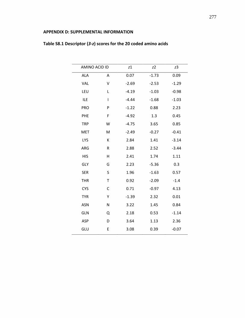
277
APPENDIX D: SUPPLEMENTAL INFORMATION
Table S8.1 Descriptor (3-z) scores for the 20 coded amino acids
AMINO ACID ID z1 z2 z3
ALA A 0.07 -1.73 0.09
VAL V -2.69 -2.53 -1.29
LEU L -4.19 -1.03 -0.98
ILE I -4.44 -1.68 -1.03
PRO P -1.22 0.88 2.23
PHE F -4.92 1.3 0.45
TRP W -4.75 3.65 0.85
MET M -2.49 -0.27 -0.41
LYS K 2.84 1.41 -3.14
ARG R 2.88 2.52 -3.44
HIS H 2.41 1.74 1.11
GLY G 2.23 -5.36 0.3
SER S 1.96 -1.63 0.57
THR T 0.92 -2.09 -1.4
CYS C 0.71 -0.97 4.13
TYR Y -1.39 2.32 0.01
ASN N 3.22 1.45 0.84
GLN Q 2.18 0.53 -1.14
ASP D 3.64 1.13 2.36
GLU E 3.08 0.39 -0.07

278
Table S8.2 Descriptor (5-z) scores for the 20 coded amino acids
AMINO ACID ID z1 z2 z3 z4 z5
ALA A 0.24 -2.32 0.6 -0.14 1.3
VAL V -2.59 -2.64 -1.54 -0.85 -0.02
LEU L -4.28 -1.3 -1.49 -0.72 0.84
ILE I -3.89 -1.73 -1.71 -0.84 0.26
PRO P -1.66 0.27 1.84 0.7 2
PHE F -4.22 1.94 1.06 0.54 -0.62
TRP W -4.36 3.94 0.59 3.44 -1.59
MET M -2.85 -0.22 0.47 1.94 -0.98
LYS K 2.29 0.89 -2.49 1.49 0.31
ARG R 3.52 2.5 -3.5 1.99 -0.17
HIS H 2.47 1.95 0.26 3.9 0.09
GLY G 2.05 -4.06 0.36 -0.82 -0.38
SER S 2.39 -1.07 1.15 -1.39 0.67
THR T 0.75 -2.18 -1.12 -1.46 -0.4
CYS C 0.84 -1.67 3.71 0.18 -2.65
TYR Y -2.54 2.44 0.43 0.04 -1.47
ASN N 3.05 1.62 1.04 -1.15 1.61
GLN Q 1.75 0.5 -1.44 -1.34 0.66
ASP D 3.98 0.93 1.93 -2.46 0.75
GLU E 3.11 0.26 -0.11 -3.04 -0.25

279
Table S8.3 Structural properties of the dipeptides used to develop the PLS models
based on the 3-z scale; n1 and n2 represent the amino acids at the N- and C-terminal
of the dipeptides, respectively
PEPTIDE ID n1z1 n2z1 n1z2 n2z2 n1z3 n2z3 log RI
IR -4.44 2.88 -1.68 2.52 -1.03 -3.44 1.69
LR -4.19 2.88 -1.03 2.52 -0.98 -3.44 1.53
NR 3.22 2.88 1.45 2.52 0.84 -3.44 1.40
KF 2.84 -4.92 1.41 1.3 -3.14 0.45 1.46
EF 3.08 -4.92 0.39 1.3 -0.07 0.45 1.35
QF 2.18 -4.92 0.53 1.3 -1.14 0.45 1.08
RF 2.88 -4.92 2.52 1.3 -3.44 0.45 0.80
SF 1.96 -4.92 -1.63 1.3 0.57 0.45 1.20
YA -1.39 0.07 2.32 -1.73 0.01 0.09 1.18
FK -4.92 2.84 1.3 1.41 0.45 -3.14 0.95
FE -4.92 3.08 1.3 0.39 0.45 -0.07 0.25
FQ -4.92 2.18 1.3 0.53 0.45 -1.14 0.93
FT -4.92 0.92 1.3 -2.09 0.45 -1.4 1.31

280
Table S8.4 Structural properties of the dipeptides used to develop the PLS models based on the 5-z scale
PEPTIDE ID n1z1 n2z1 n1z2 n2z2 n1z3 n2z3 n1z4 n2z4 n1z5 n2z5 log RI
IR -3.89 3.52 -1.73 2.5 -1.71 -3.5 -0.84 1.99 0.26 -0.17 1.69
LR -4.28 3.52 -1.3 2.5 -1.49 -3.5 -0.72 1.99 0.84 -0.17 1.53
NR 3.05 3.52 1.62 2.5 1.04 -3.5 -1.15 1.99 1.61 -0.17 1.40
KF 2.29 -4.22 0.89 1.94 -2.49 1.06 1.49 0.54 0.31 -0.62 1.46
EF 3.11 -4.22 0.26 1.94 -0.11 1.06 -3.04 0.54 -0.25 -0.62 1.35
QF 1.75 -4.22 0.5 1.94 -1.44 1.06 -1.34 0.54 0.66 -0.62 1.08
RF 3.52 -4.22 2.5 1.94 -3.5 1.06 1.99 0.54 -0.17 -0.62 0.80
SF 2.39 -4.22 -1.07 1.94 1.15 1.06 -1.39 0.54 0.67 -0.62 1.20
YA -2.54 0.24 2.44 -2.32 0.43 0.6 0.04 -0.14 -1.47 1.3 1.18
FK -4.22 2.29 1.94 0.89 1.06 -2.49 0.54 1.49 -0.62 0.31 0.95
FE -4.22 3.11 1.94 0.26 1.06 -0.11 0.54 -3.04 -0.62 -0.25 0.25
FQ -4.22 1.75 1.94 0.5 1.06 -1.44 0.54 -1.34 -0.62 0.66 0.93
FT -4.22 0.75 1.94 -2.18 1.06 -1.12 0.54 -1.46 -0.62 -0.4 1.69

281
Table S8.5 Predicted peptide dataset based on the 3-z scale descriptor and their
predicted renin inhibitory activity (RI, %)
Predicted
PEPTIDE ID n1z1 n2z1 n1z2 n2z2 n1z3 n2z3 log RI (%) RI
VW -2.69 -4.75 -2.53 3.65 -1.29 0.85 1.98 96.55
VY -2.69 -1.39 -2.53 2.32 -1.29 0.01 1.64 43.95
VF -2.69 -4.92 -2.53 1.3 -1.29 0.45 1.37 23.35
LW -4.19 -4.75 -1.03 3.65 -0.98 0.85 1.79 62.53
LY -4.19 -1.39 -1.03 2.32 -0.98 0.01 1.45 28.47
LF -4.19 -4.92 -1.03 1.3 -0.98 0.45 1.18 15.13
IW -4.44 -4.75 -1.68 3.65 -1.03 0.85 1.86 72.50
IY -4.44 -1.39 -1.68 2.32 -1.03 0.01 1.52 33.01
IF -4.44 -4.92 -1.68 1.3 -1.03 0.45 1.24 17.54
AW 0.07 -4.75 -1.73 3.65 0.09 0.85 1.91 82.22
AY 0.07 -1.39 -1.73 2.32 0.09 0.01 1.57 37.43
AF 0.07 -4.92 -1.73 1.3 0.09 0.45 1.29 19.89

282
Table S8.6 Predicted peptide dataset based on the 5-z scale descriptor and their predicted renin inhibitory activity (RI)
Predicted
PEPTIDE ID n1z1 n2z1 n1z2 n2z2 n1z3 n2z3 n1z4 n2z4 n1z5 n2z5 log RI RI (%)
VW -2.59 -4.36 -2.64 3.94 -1.54 0.59 -0.85 3.44 -0.02 -1.59 1.85 71.09
VY -2.59 -2.54 -2.64 2.44 -1.54 0.43 -0.85 0.04 -0.02 -1.47 1.44 28.03
VF -2.59 -4.22 -2.64 1.94 -1.54 1.06 -0.85 0.54 -0.02 -0.62 1.36 23.33
LW -4.28 -4.36 -1.3 3.94 -1.49 0.59 -0.72 3.44 0.84 -1.59 1.84 69.01
LY -4.28 -2.54 -1.3 2.44 -1.49 0.43 -0.72 0.04 0.84 -1.47 1.43 27.21
LF -4.28 -4.22 -1.3 1.94 -1.49 1.06 -0.72 0.54 0.84 -0.62 1.35 22.65
IW -3.89 -4.36 -1.73 3.94 -1.71 0.59 -0.84 3.44 0.26 -1.59 1.82 66.18
IY -3.89 -2.54 -1.73 2.44 -1.71 0.43 -0.84 0.04 0.26 -1.47 1.41 26.09
IF -3.89 -4.22 -1.73 1.94 -1.71 1.06 -0.84 0.54 0.26 -0.62 1.33 21.72
AW 0.24 -4.36 -2.32 3.94 0.6 0.59 -0.14 3.44 1.3 -1.59 1.91 81.20
AY 0.24 -2.54 -2.32 2.44 0.6 0.43 -0.14 0.04 1.3 -1.47 1.50 32.02
AF 0.24 -4.22 -2.32 1.94 0.6 1.06 -0.14 0.54 1.3 -0.62 1.42 26.65
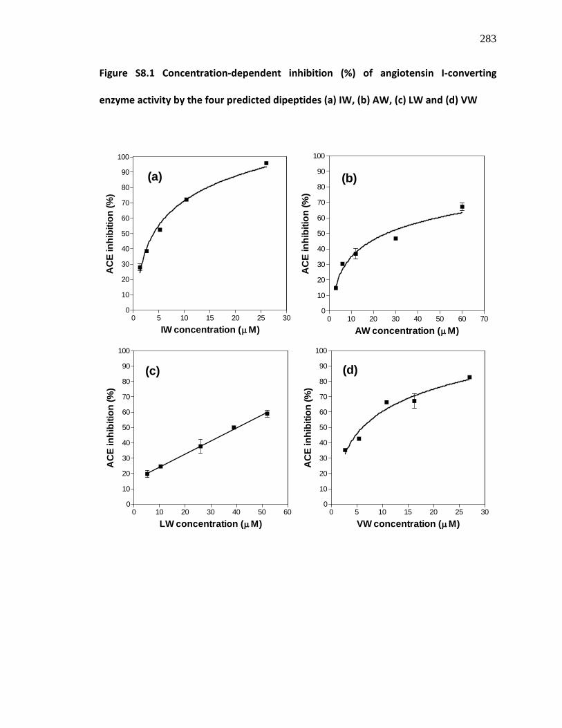
283
Figure S8.1 Concentration-dependent inhibition (%) of angiotensin I-converting
enzyme activity by the four predicted dipeptides (a) IW, (b) AW, (c) LW and (d) VW
0 5 10 15 20 25 300
10
20
30
40
50
60
70
80
90
100
(a)
IW concentration (µM)
AC
E in
hibi
tion
(%)
0 10 20 30 40 50 60 700
10
20
30
40
50
60
70
80
90
100
(b)
AW concentration (µM)
AC
E in
hibi
tion
(%)
0 10 20 30 40 50 600
10
20
30
40
50
60
70
80
90
100
(c)
LW concentration (µM)
AC
E in
hibi
tion
(%)
0 5 10 15 20 25 300
10
20
30
40
50
60
70
80
90
100
(d)
VW concentration (µM)
AC
E in
hibi
tion
(%)

284
CHAPTER NINE
GENERAL DISCUSSION AND CONCLUSIONS
In a bid to utilize flaxseed meal proteins in human foods, we took a number
approaches to produce peptide mixtures with structural properties that can be applied
towards management and treatment of chronic human diseases. Experimental
approach included in vitro and in vivo methods, which provided fundamental as well as
applied knowledge relating to the potential use of flaxseed proteins and peptides as
functional foods and nutraceuticals. Therefore, our work has provided a potential
platform for the comprehensive utilization of flaxseed proteins by the foods and
nutrition industries. This work also contributed to the understanding of structural
requirements for renin inhibition by food protein-derived dipeptides that may be useful
for biologists and chemists interested in developing novel active pharmaceutical
ingredients. The key outcomes of this thesis are as follows:
(1) Development of a novel processing method for isolation of flaxseed proteins from
highly viscous aqueous suspension of defatted flaxseed meal. This approach
involved the use of a food grade cellulolytic enzyme in removing interfering flaxseed
mucilage prior to protein isolation by conventional method resulting in a higher
yield of isolated flaxseed protein when compared to previously reported methods.
This process distinctly utilizes industrially produced high mucilage defatted flaxseed
meal by-product as opposed to reported methods for flaxseed protein isolation
from laboratory-prepared samples that usually contain less mucilage after

285
dehulling. Thus, our method confirmed that industrial flaxseed meal protein can be
isolated and potentially utilized in the food system.
(2) Controlled hydrolysis of the isolated flaxseed proteins using specific and non-
specific proteases, and processing using a combination of membrane ultrafiltration
and ion-exchange chromatography to yield LMW peptides with multifunctional
bioactive properties. Multifunctional peptides have the advantage of modulating
more than one disease-related cellular processes thereby targeting more than one
disease condition at a time. The LMW peptides in our studies showed antioxidant
activities against various free radicals implicated in pathogenesis of human diseases,
suppressed nitric oxide production induced by bacterial lipopolysaccharide in
cultured murine macrophages, and also exhibited ACE and renin inhibition in vitro.
Enzyme inhibition kinetics studies showed, for the first time, that the food peptide
fraction inhibited human recombinant renin via an uncompetitive inhibition pattern;
the peptides potentially exhibited both active site and allosteric inhibition of human
renin. Thus, these flaxseed peptides possess multifunctional properties with
relevance to the management and treatment of hypertension and associated
cardiovascular disease, including oxidative stress and inflammation-related
diseases. Most bioactive peptides are designed to target one disease symptom at a
time, thus, the multifunctionality of the flaxseed peptides could potentially reduce
the cost associated with the development of multiple bioactive peptides for
multiple diseases.

286
(3) Successful development of a cheap, efficient and industrially-feasible method to
enzymatically generate and fractionate BCAA-rich high Fischer ratio peptides from
flaxseed proteins. Due to the high amount of BCAA and low amount of AAA, this
peptide product can be used in clinical nutrition to increase plasma amino acid
Fischer ratio in patients with chronic liver diseases with concomitant improvement
of associated hepatic encephalopathy and muscle loss. Unlike previously reported
methods, our method involves a simple mixing of peptide products with activated
carbon in a container followed by centrifugation. In our opinion, this process is
cheaper and less time-consuming than traditionally reported methods that involved
various forms of expensive column chromatography.
(4) Successful development of a production process to enzymatically release Arginine
(Arg)-containing peptides from flaxseed proteins and separation of these peptides
based on their net molecular charge using a novel membrane technology,
electrodialysis-ultrafiltration. The resulting peptide product contained high amounts
of Arg and decreased systolic blood pressure in spontaneously hypertensive rat
either through the vasodilatory activity of nitric oxide produced from Arg, or
through the inhibition of ACE and renin activities. This is a novel approach towards
managing hypertension using processed food ingredients, different from
modulation of RAS or blocking AT-II receptors. To the best of our knowledge, this
will be the first report of the production and utilization of food protein-derived
arginine-enriched peptide mixture for the management of hypertension. An
attractive advantage of the product is the ability to attenuate high blood pressure at
287
a fast rate (< 2 h) when compared to amino acid form of arginine and the protein
isolate. The work also successfully showed for the first time that flaxseed proteins in
the unhydrolyzed form can modulate blood pressure, though at a slower rate than
the peptide product. Therefore, flaxseed proteins may find utilization in food
formulations designed to remedy cardiovascular impairment, especially
hypertension. Successful incorporation and adoption of flaxseed proteins in foods
could result in significant economic impact specifically on the Prairie Provinces and
generally on the Canadian economy.
(5) Moreover, this study was the first to report the structural requirements for renin
inhibition by food protein-derived peptides using QSAR. The PLS models reported in
this thesis provided a potent dipeptide, Ile-Trp (IW), which can serve as template for
the design and synthesis of peptidomimetics with potent renin-inhibitory and in vivo
antihypertensive properties. One of the significant outcomes is the fact that renin
inhibition differs for peptides that have same molecular weight and amino acid
sequence but in which the amino acids are geometric isomers.
It could be concluded from this thesis work that flaxseed proteins contain
multifunctional bioactive peptide sequences that can be released by enzymatic
hydrolysis, and that the structure of dipeptides determine their in vitro renin inhibitory
and antihypertensive properties. Thus, this project has uniquely contributed knowledge
that can enhance value-added utilisation of defatted flaxseed meal generated from
Canada’s oilseed processing industry, and to the elucidation of structure-function
properties of renin-inhibiting natural dipeptides. Since these peptides are generated
288
from edible food sources, these contributions to research are important in the discovery
of a new generation of non-toxic therapeutic agents for clinical intervention against
chronic human diseases especially hypertension and cardiovascular disease, renal and
inflammatory diseases. The fact that the arginine-rich peptides exhibited blood
pressure-lowering activity provides preliminary evidence of the in vivo bioavailability of
the flaxseed protein-derived peptides especially in the vascular endothelium where
activity is required. Moreover, more extensive animal studies are needed to analyze the
animal tissues for the presence of intact peptides following oral intake of therapeutic
amounts of these flaxseed peptides.
Based on the outcome of this project, future studies include pilot scale isolation
of flaxseed proteins using the method reported in this thesis, to investigate the
feasibility for food industry production and practical application of the proteins in the
human food system. As a follow-up to the short-term antihypertensive studies, animal
feeding trials using the flaxseed protein isolates are ongoing in our laboratory to
investigate the effects of long-term consumption of the intact flaxseed proteins on SBP
in SHR. Moreover, animal studies are also needed to confirm the positive effects of the
BCAA-enriched flaxseed peptides during chronic hepatic diseases. Due to low yield of
the arginine-enriched peptides, future studies should focus on optimization of the
enzymatic hydrolysis process to produce a more potent flaxseed protein hydrolysate,
and electrodialysis-ultrafiltration process to increase yield of the final product, as well as
long-term animal studies to investigate sustained positive effects of the peptide product
on elevated blood pressure. A long-term study to determine the sustained hypotensive

289
effects of flaxseed protein hydrolysate containing arginine peptides is warranted. Future
work should also determine the contributions of arginine-enriched peptides to the nitric
oxide content of blood vessels and major organs. It will be important to determine
whether arginine-enriched peptides work as hypotensive agents through enhanced
nitric oxide production or inhibition of the renin-angiotensin system or both. Finally,
peptide chemometrics studies should focus on synthesis of structural variants of IW and
evaluation of the renin-inhibitory and antihypertensive activities of these
peptidomimetics.
The consumption of flaxseed for human health benefits is increasingly gaining
global attention especially in Canada, and this focus is mostly due to the presence of
bioactive constituents including omega-3 oil, dietary fibres and polyphenolic lignans.
The outcome of this project has added a new dimension to the potential health benefits
derived from flaxseed components, and could provide the Canadian consumer with an
alternative choice of healthy food products for the prevention and management of
many chronic human diseases and health conditions.