the transmissible gastroenteritis coronavirus contains a spherical
TRANSCRIPT

JOURNAL OF VIROLOGY, July 1996, p. 4773–4777 Vol. 70, No. 70022-538X/96/$04.0010Copyright q 1996, American Society for Microbiology
The Transmissible Gastroenteritis Coronavirus Contains aSpherical Core Shell Consisting of M and N Proteins
CRISTINA RISCO,1 INES M. ANTON,2† LUIS ENJUANES,2
AND JOSE L. CARRASCOSA1*
Macromolecular Structure Department1 and Molecular and Cell Biology Department,2
Centro Nacional de Biotecnologıa, Consejo Superior de Investigaciones Cientıficas,Campus Universidad Autonoma, 28049 Madrid, Spain
Received 10 January 1996/Accepted 2 April 1996
Coronaviruses are enveloped RNA viruses involved in a variety of pathologies that affect animals andhumans. Existing structural models of these viruses propose a helical nucleocapsid under the virion envelopeas the unique internal structure. In the present work, we have analyzed the structure of the transmissiblegastroenteritis coronavirus. The definition of its organization supports a new structural model for coronavi-ruses, since a spherical, probably icosahedral, internal core has been characterized. Disruption of these coresinduces the release of N-protein-containing helical nucleocapsids. Immunogold mapping and protein analysisof purified cores showed that they consist of M and N proteins, M being the main core shell component. Thissurprising finding, together with the fact that M protein molecules are also located in the virion envelope,indicates that a reconsideration of the assembly and maturation of coronaviruses, as well as a study ofpotential M-protein subclasses, is needed.
Coronaviruses have been described as enveloped, round par-ticles with a prominent layer of surface spikes consisting ofcomplexes of S glycoprotein. These surface structures form thetypical “corona” that gives the name to the viral family (5, 22).The viral envelope of coronaviruses contains other glycopro-teins, such as the integral membrane protein (M), the smallmembrane protein (sM), and in some members of the family,the hemagglutinin esterase protein (HE) (9, 13, 21). The nu-cleocapsid (N) protein is located inside the virion, complexedwith the viral RNA (11). Structural models of coronavirusesare based on studies of detergent-treated viral particles and onthe analysis of the in vitro interaction of subviral componentsfrom disrupted virions. These studies reported the release ofhelical ribonucleoprotein molecules from mouse hepatitis virusand the 229E strain of the human coronavirus (4, 14). Accord-ingly, the unique internal structure of coronaviruses is consid-ered to be a helical nucleocapsid constituted by N protein andRNA (5, 21). Sturman et al. (24) proposed that these helicalcomplexes might interact with the intraviral domains of theenvelope proteins. The helical RNP of the infectious bronchitisvirus has been very difficult to visualize (6, 14, 24), and a previousstudy (8) failed to release the ribonucleoprotein from transmis-sible gastroenteritis coronavirus (TGEV). In the present work,we have analyzed the structure of TGEV, a virus that causessevere illness in newborn pigs and that has been the subject ofnumerous efforts to define the molecular basis of its epidemi-ology (7). Intact and detergent-treated TGEV virions, whichwere purified from infected cultures of ST cells (10), have beencharacterized by negative staining, ultrathin sectioning, freezefracture, immunogold mapping, and cryoelectron microscopy.In Figure 1 we show representative fields chosen after morethan 500 viral particles were studied with each visualizationmethod. Negative staining with 2% uranyl acetate or 2% so-
dium phosphotungstate was done according to standard pro-cedures (17). Uranyl acetate shows virions (Fig. 1A) thatpresent more extended peplomers when contrasted with so-dium phosphotungstate (Fig. 1B). This staining agent some-times penetrates the viral particles, revealing a compact inter-nal component (Fig. 1C). Ultrathin sections of osmicatedvirions, obtained as previously described (19), also reveal acontinuous structure separated from the envelope that en-closes the ribonucleoprotein (Fig. 1D). When TGEV virionsare visualized after freeze fracture and platinum-carbon shad-owing according to established procedures (18), two differentfracture planes are observed. The one that goes along the viralenvelope renders large particles (arrows in Fig. 1E), while asecond plane along the interior of the virus shows smallerparticles (arrowheads in Fig. 1E). The visualization of thesesmaller structures reveals the presence of an internal shell inTGEV virions.Cryoelectron microscopy allows the visualization of vitrified,
unstained hydrated material (2). This technique avoids theartifacts potentially associated with chemical fixation and stain-ing with heavy metals, allowing the visualization of structuresclose to their native state. The image of vitrified specimens isthus directly formed by the mass of the different components ofthe biological structure. The observation of vitrified TGEVvirions confirmed the presence of an internal structure insidethe virus (Fig. 1F and G). This structure exhibits a polygonalcontour (arrowheads in Fig. 1F and G). After 104 viral parti-cles had been measured, a size of 144.8 6 7.2 nm was calcu-lated for the whole virion (including the extended peplomers),while the internal core had a diameter of 64.5 6 5.5 nm. Thesevalues have to be very close to the real dimensions and clearlydiffer from the size calculated for the whole virion by negativestaining (115.6 6 10.5 nm), a method that induces a moreheterogeneous population of virions. A suspension of latexspheres of homogeneous size (0.0910 6 0.0058 mm) was usedas an internal standard for measurements. A clear gap sepa-rated the internal core from the envelope in vitrified virions.The direct visualization of these internal structures was doneafter a short treatment (3 min at 48C) of TGEV virions with
* Corresponding author. Mailing address: Centro Nacional de Bio-tecnologıa (CSIC), Campus Universidad Autonoma, 28049 Madrid,Spain. Phone: 34-1-5854509. Fax: 34-1-5854506. Electronic mail ad-dress: [email protected].† Present address: Children’s Hospital, Boston, MA 02115.
4773
Dow
nloa
ded
from
http
s://j
ourn
als.
asm
.org
/jour
nal/j
vi o
n 04
Dec
embe
r 20
21 b
y 91
.221
.134
.154
.

two detergents, Triton X-100 (0.025% in phosphate-bufferedsaline [PBS]) and Nonidet P-40 (NP-40) (0.05% in PBS). Thelatter was particularly effective, and at the described concen-tration it removed the envelope, releasing an internal compactstructure of proteinaceous nature (Fig. 2A). Immunogold la-beling of these structures with specific monoclonal antibodies
(MAbs) and a 5-nm colloidal gold conjugate was performed aspreviously described (16). MAbs specific for M and N proteins(20) clearly reacted with this internal component (Fig. 2B andC). A detectable amount of M-protein molecules remainedassociated with the disrupted envelopes (Fig. 2B). In contrast,S protein was totally removed since specific MAbs, which pro-
FIG. 1. Structure of purified infective TGEV virions. Negative staining with uranyl acetate (A) or sodium phosphotungstate (B) shows the surface of TGEVparticles, whose peplomers are better defined and extended with sodium phosphotungstate (arrowheads). This staining agent frequently penetrates the viral particles(C), revealing an internal structure (arrowhead) separated from the envelope (env). (D) Ultrathin sections reveal the internal organization of these viruses thatcomprises a continuous layer of material (arrowhead) surrounding the dense ribonucleoprotein (asterisk). (E) Freeze fracture images show two different fracture planesin TGEV virions. One of them goes along the viral envelope (arrows), and the other one reveals a smaller inner shell (arrowheads). (F and G) Cryoelectron microscopyvisualization of vitrified TGEV shows particles of homogeneous size that contain an internal structure of geometrical periphery (arrowheads) inside the viral envelopeand well-extended peplomers (p). Note the significant gap that separates the internal structure from the envelope. Bars, 100 nm.
FIG. 2. Release of TGEV internal cores from purified virions by treatment with NP-40. (A) A 3-min incubation at 48C with NP-40 (0.05% in PBS) induces thedisruption of the viral envelope (env), which opens up and releases an internal component of a proteinaceous nature (arrow). Epitopes of M (B) and N (C) proteinsare localized on the surface of these internal cores, as revealed by immunogold labeling using specific MAbs. A detectable amount of M molecules remain in thedisrupted envelopes (arrowheads in panel B). Higher concentrations of NP-40 (0.25 to 1%) and longer treatments (15 to 30 min) completely removed the viralenvelopes, and only particles with a geometrical contour were seen, as visualized by negative staining (D). Immunogold characterization of these particles shows thatthey react with anti-N (E) and anti-M (F) MAbs, while no signal was obtained with anti-S MAbs (G). (H) Freeze fracture of the particles followed by etching andreplication with platinum-carbon showed pointed shadows (arrowhead) consistent with icosahedral structures. (I) Triton X-100-disrupted cores (c) released a materialthat strongly reacts with anti-N MAbs (arrowheads). This material consisted of helical aggregates that did not react with anti-M MAbs (J) but contained linearlyarranged N protein, as indicated by immunogold labeling (arrowheads in panel K). Bars, 70 nm.
4774 NOTES J. VIROL.
Dow
nloa
ded
from
http
s://j
ourn
als.
asm
.org
/jour
nal/j
vi o
n 04
Dec
embe
r 20
21 b
y 91
.221
.134
.154
.
vide a strong signal in intact virions, did not label the disruptedenvelopes or the released particles (not shown).At higher concentrations of NP-40 (0.25 to 1% in PBS) and
with longer treatments (15 to 30 min at 48C) the viral envelopeswere totally removed and destroyed, with the release of theinternal structure. The released particles had polygonal con-tours (Fig. 2D) and a diameter of 68.6 6 4.8 nm, similar to theinternal structures visualized in vitrified virions. Immunogoldlabeling provided a weak signal associated with N protein onthe surface of these particles, while M protein covered most oftheir surface (Fig. 2E and F). No signal associated with Sprotein was obtained (Fig. 2G). Freeze-etching and shadowing(18) showed pointed (Fig. 2H) or blunt-ended (not shown)
shadows consistent with icosahedral structures. When TGEVvirions were treated with 0.025% Triton X-100 (3 min at 48C),similar cores, which clearly reacted with anti-M MAbs, werevisualized (not shown). When disrupted by extensive detergenttreatment (30 min), these cores released a material that wasclearly labeled with anti-N antibodies (Fig. 2I, arrowheads).Once separated from the core, this material was visualized ashelical particulated molecules of homogeneous size (Fig. 2J) sim-ilar to the ribonucleoprotein molecules described for mouse hep-atitis virus and the human coronavirus (14). The helical extendedstructures did not react with anti-M MAbs (Fig. 2J). Instead,they contained N-protein molecules arranged in a linear pat-tern as revealed by immunogold labeling (Fig. 2K).
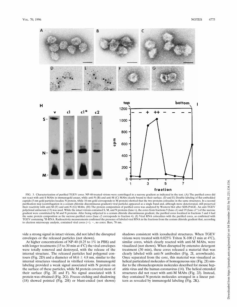
FIG. 3. Characterization of purified TGEV cores. NP-40-treated virions were centrifuged in a sucrose gradient as indicated in the text. (A) The purified cores didnot react with anti-S MAbs in immunogold assays, while anti-N (B) and anti-M (C) MAbs clearly bound to their surface. (D and E) Double-labeling of flat-embeddedcapsids (5-nm gold particles localize N protein, while 10-nm gold corresponds to M protein) showed that the two proteins colocalize in the same structures. In a secondpurification step (centrifugation in a cesium chloride discontinuous gradient) viral particles appeared as a single band and, although more deteriorated, still preservedtheir reactivity with anti-M (F) and anti-N (G) MAbs. (H) The protein composition of purified cores was analyzed by Western blot after SDS-PAGE. An anti-TGEVpolyclonal antiserum (13) was used. While the intact virions contained S, M, and N proteins (lane v), the cores from fractions 9 (lane c1) and 19 (lane c1*) of the sucrosegradient were constituted by M and N proteins. After being subjected to a cesium chloride discontinuous gradient, the purified cores localized in fractions 3 and 4 hadthe same protein composition as the sucrose-purified cores (lane c2 corresponds to fraction 4). (I) Viral RNA colocalizes with the purified cores, as confirmed withTGEV containing 3H-RNA. Radioactivity measurements confirmed the presence of labeled viral RNA in the fractions from the cesium chloride gradient that, accordingto electron microscopy analysis, contained viral cores (1). 2, no cores. Bars, 70 nm.
VOL. 70, 1996 NOTES 4775
Dow
nloa
ded
from
http
s://j
ourn
als.
asm
.org
/jour
nal/j
vi o
n 04
Dec
embe
r 20
21 b
y 91
.221
.134
.154
.
TGEV internal cores were purified by centrifugation in asucrose gradient. NP-40-treated virions (0.5% NP-40, 20 minat room temperature) were centrifuged for 45 min through alinear 15 to 40% sucrose gradient, at 27,000 rpm in a BeckmanSW 55Ti rotor at 48C. The fractions from the gradient werecentrifuged for 8 min at 90,000 rpm in a Beckman TLA 120.1rotor. Pellets were resuspended in PBS containing proteaseinhibitors (0.1 mM N- to syl-L-phenylalanine chloromethyl ke-tone, 0.1 mM Na-p-to syl-L-lysine chloromethyl ketone, 1mg/ml pepstatin A, and 1 mM phenylmethylsulfonyl fluoride)and processed for immunogold and negative staining electronmicroscopy, polyacrylamide gel electrophoresis in the presenceof sodium dodecyl sulfate (SDS-PAGE), and Western blot(immunoblot) (3, 12, 16). Individual cores or small aggregateswere found in an intermediate position in the gradient (frac-tion 9 from the top), while aggregated groups of particles werelocated at the bottom of the tube (fraction 19). Cores fromboth fractions were indistinguishable by negative staining andimmunogold labeling and were also similar to freshly deter-gent-released particles (Fig. 3A to C). They did not react withanti-S MAbs (Fig. 3A), moderately reacted with anti-N MAbs
(Fig. 3B), and exhibited a stronger signal with anti-M MAbs(Fig. 3C) in immunogold assays. Double immunogold labelingof the cores was done by combining preembedding detection ofN protein by specific MAbs and a 5-nm colloidal gold conju-gate with postembedding labeling of M protein (detected witha 10-nm colloidal gold conjugate) on sections of flat-embeddedparticles (17). These experiments showed that M and N pro-teins were simultaneously located in the same structures (Fig.3D and E). SDS-PAGE and Western blot analysis showed thatwhile the purified intact virions presented the three majorstructural proteins, S, M, and N (Fig. 3H, lane v), the corescontained in fractions 9 and 19 from the sucrose gradientconsisted of M and N proteins (Fig. 3H, lanes c1 and c1*). Thepurified cores were submitted to a second centrifugation, in acesium chloride discontinuous gradient (of 1.15, 1.3, 1.5, and1.7 g/ml) containing the protease inhibitors indicated aboveand 0.4 U of the Amersham ribonuclease inhibitor per ml.Centrifugation was performed at 48C for 45 min at 32,000 rpmin a Beckman TLS-55 rotor. The particles were found in asingle band, and their appearance was similar to that in Fig. 3Ato C, except that they were slightly deteriorated. They clearlyreacted with anti-M MAbs (Fig. 3F), while the signal associ-ated with N protein was weaker (Fig. 3G). The relativeamounts of M and N proteins in the cores were maintained, asdetermined by Western blot analysis after SDS-PAGE (Fig.3H, lane c2). These data indicate that, after two purificationsteps, M-protein molecules remain tightly associated with theviral cores in large amounts, covering most of their surface.The purification of cores was also done with a preparation ofTGEV labeled with 3H-RNA. This virus was obtained by met-abolic labeling of TGEV RNA with [5-3H]uridine followed byvirus purification. Infected cultures were incubated in the pres-ence of the radiolabeled precursor, [5-3H]uridine, at a finalconcentration of 20 mCi/ml from 1 h after virus inoculationuntil virus was harvested (26 h postinoculation). Purification ofthe labeled virus was done as previously described (10). Whenthis labeled TGEV was used for purifying the cores, viral RNAcolocalized with them in the gradients (Fig. 3I).We can conclude that TGEV contains a spherical core inside
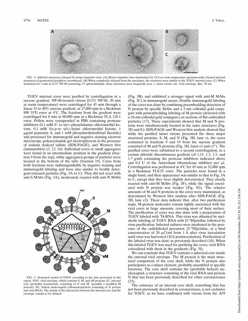
the external viral envelope. The M protein is the main struc-tural component of the core shell, while the N protein alsoparticipates as a minor element, probably assembled at specificlocations. The core shell contains the (probably helical) nu-cleocapsid, a structure consisting of the viral RNA and proteinN that has been previously described for other coronaviruses(23).The existence of an internal core shell, something that has
not been previously described in coronaviruses, is not exclusivefor TGEV, as we have confirmed with virions from the A59
FIG. 4. Subviral structures released by mouse hepatitis virus. (A) Mouse hepatitis virus maintained for 24 h at room temperature spontaneously released internalstructures of geometrical periphery (arrowhead). (B) When completely released from the envelopes, the structures were similar to the TGEV internal cores. (C) Whenincubated for 3 min in 0.1% NP-40 containing 1% glutaraldehyde, these structures were frequently seen. v, intact virion; env, viral envelope. Bar, 70 nm.
FIG. 5. Structural model of TGEV according to the data presented in thisreport. ENV, viral envelope, which contains S, M, and sM proteins; IC, internalcore (probably icosahedral), consisting of N and M9 (possibly a modified Mprotein); NC, helical nucleocapsid (ribonucleoprotein consisting of N proteinand viral RNA). The details of the interaction between the internal core and theenvelope remain to be defined.
4776 NOTES J. VIROL.
Dow
nloa
ded
from
http
s://j
ourn
als.
asm
.org
/jour
nal/j
vi o
n 04
Dec
embe
r 20
21 b
y 91
.221
.134
.154
.

strain of mouse hepatitis virus. These virions, which were pu-rified from infected BALB/3T3 cells (23), spontaneously re-leased an internal core when maintained for 24 h at roomtemperature (Fig. 4A and B). Incubation with NP-40 (0.05% inPBS for 3 min at 48C) destroyed the virions, but when 1%glutaraldehyde was present in the detergent solution similarcores were also found (Fig. 4C).The data presented in this report support the structural model
for coronaviruses proposed in Fig. 5. The most striking aspect ofthis organization is the presence of M protein as the major struc-tural component of the internal core shell. Since M is an integralmembrane protein of the virion envelope (1, 16), its presence onthe surface of the internal core might be the result of stabilizinginteractions from the intravirion domains of M-protein moleculeslocated in the envelope. If these interactions from the envelopedo exist, the possibility that the removal of lipids could eventuallylead to a massive collapse of M-protein molecules on the internalcore cannot be excluded. It is important to note, however, that asignificant gap separates the internal core from the envelope invitrified virions, although focal contacts between both structurescould exist (Fig. 1F and G). A second option would be, then, theexistence of two M subpopulations at different locations withinthe viral particle, a possibility suggested by Fig. 2B. In fact, theheterogeneity of M protein as seen with SDS-PAGE, suggests theexistence of M subclasses. In infected cells, M-protein moleculescould incorporate into viral particles in two different steps orundergo major reorganization in the virion after a single-stepincorporation. Since the simultaneous presence of a viral struc-tural protein in two different locations within the virion has notbeen previously described for other viruses, the assembly processof coronaviruses during morphogenesis should be carefully recon-sidered. Studies on the in vivo and in vitro assembly of TGEV arenow in progress.The existence of an internal core shell in coronaviruses can-
not be considered a surprising finding, as all positive-strandRNA viruses have a spherical (or icosahedral) core (15). In theparticular case of the enveloped positive-strand RNA viruses(togavirus, flavivirus, and arterivirus) the spherical core struc-tures seem to involve a close interaction between protein andRNA. Coronavirus then, would represent a different case, inwhich the core is built up by an external shell that encloses thehelical ribonucleoprotein complex or nucleocapsid. The deter-mination of whether the core shell of coronavirus is truly ico-sahedral (as expected from our data) demands further studiesat higher resolution to check for the presence of the charac-teristic crystallographic symmetries, with either three-dimen-sional reconstruction from cryoelectron microscopy (presentlyunder way) or X-ray diffraction from crystals.
This work was partly supported by grant PB91-0109 from the Comi-sion Interministerial de Ciencia y Tecnologıa to J.L.C. and by grantsfrom the Comision Interministerial de Ciencia y Tecnologıa and theEuropean Union (projects Science and Biotech) to L.E.
REFERENCES1. Armstrong, J., H. Nieman, S. Smeekens, P. Rottier, and G. Warren. 1984.Sequence and topology of a model intracellular membrane protein, E1
glycoprotein, from a coronavirus. Nature 308:751–752.2. Booy, F. P. 1993. Cryoelectron microscopy, p. 21–54. In J. Bentz (ed.), Viralfusion mechanisms. CRC Press, Inc., Boca Raton, Fla.
3. Burnette, W. N. 1981. Western-blotting: electrophoretic transfer from so-dium dodecyl sulfate-polyacrylamide gels to unmodified nitrocellulose andradiographic detection with radioiodinated protein A. Anal. Biochem. 112:195–203.
4. Caul, E. O., C. R. Ashley, M. Ferguson, and S. I. Egglestone. 1979. Prelim-inary studies on the isolation of coronavirus 229E nucleocapsids. FEMSMicrobiol. Lett. 5:101–105.
5. Cavanagh, D., and The Coronaviridae Study Group of the InternationalCommittee on Taxonomy of Viruses. 1994. Revision of the taxonomy of theCoronavirus, Torovirus, and Arterivirus genera. Arch. Virol. 135:226–237.
6. Davies, H. A., R. R. Dourmashkin, and R. MacNaughton. 1981. Ribonucle-oprotein of avian infectious bronchitis virus. J. Gen. Virol. 53:67–74.
7. Enjuanes, L., and B. A. M. Van der Zeijst. 1995. Molecular basis of thetransmissible gastroenteritis virus epidemiology, p. 337–376. In S. G. Siddell(ed.), The Coronaviridae. Plenum Press, New York.
8. Garwes, D. J., D. H. Pocock, and B. V. Pike. 1976. Isolation of subviralcomponents from transmissible gastroenteritis virus. J. Gen. Virol. 32:283–294.
9. Godet, M., R. L’Haridon, J. F. Vautherot, and H. Laude. 1992. TGEVcorona virus ORF 4 encodes a membrane protein that is incorporated intovirions. Virology 188:666–675.
10. Jimenez, G., I. Correa, M. P. Melgosa, M. J. Bullido, and L. Enjuanes. 1986.Critical epitopes in transmissible gastroenteritis virus neutralization. J. Virol.60:131–139.
11. Kapke, P. A., and D. A. Brian. 1986. Sequence analysis of the porcinetransmissible gastroenteritis coronavirus nucleocapsid protein gene. Virol-ogy 151:41–49.
12. Laemmli, U. K. 1970. Cleavage of structural proteins during the assembly ofthe head of bacteriophage T4. Nature 227:680–685.
13. Lai, M. M. C. 1990. Coronavirus. Organization, replication, and expressionof genome. Annu. Rev. Microbiol. 44:303–333.
14. MacNaughton, M. R., H. A. Davies, and M. V. Nermut. 1978. Ribonucle-oprotein-like structures from coronavirus particles. J. Gen. Virol. 39:545–549.
15. Murphy, F. A., C. M. Fauquet, D. H. L. Bishop, S. A. Ghabrial, A. W. Jarvis,G. P. Martinelli, M. A. Mayo, and M. D. Summers (ed.). 1995. Virus tax-onomy, classification and nomenclature of viruses, p. 23. Springer-Verlag,New York.
16. Risco, C., I. M. Anton, C. Sune, A. M. Pedregosa, J. M. Martın-Alonso, F.Parra, J. L. Carrascosa, and L. Enjuanes. 1995. Membrane protein mole-cules of transmissible gastroenteritis coronavirus also expose the carboxy-terminal region on the external surface of the virion. J. Virol. 69:5269–5277.
17. Risco, C., J. L. Carrascosa, A. M. Pedregosa, C. D. Humphrey, and A.Sanchez-Fauquier. 1995. Ultrastructure of the human astrovirus serotype 2.J. Gen. Virol. 76:2075–2080.
18. Risco, C., and P. Pinto da Silva. 1995. Cellular functions during activationand damage by pathogens: immunogold studies of the interaction of bacte-rial endotoxins with target cells. Microsc. Res. Tech. 31:141–158.
19. Risco, C., C. Romero, M. A. Bosch, and P. Pinto da Silva. 1994. Type IIpneumocytes revisited: intracellular membranous systems, surface charac-teristics, and lamellar body secretion. Lab. Invest. 70:407–417.
20. Sanchez, C. M., G. Jimenez, M. D. Laviada, I. Correa, C. Sune, M. J. Bullido,F. Gebauer, C. Smerdou, P. Callebaut, J. M. Escribano, and L. Enjuanes.1990. Antigenic homology among coronaviruses related to transmissiblegastroenteritis virus. Virology 174:410–417.
21. Siddell, S. G. 1995. The Coronaviridae. An introduction, p. 1–10. In S. G.Siddell (ed.), The Coronaviridae. Plenum Press, New York.
22. Spaan, W., D. Cavanagh, and H. C. Horzinek. 1988. Coronaviruses: structureand genome expression. J. Gen. Virol. 69:2939–2952.
23. Sturman, L. S., and K. V. Holmes. 1983. The molecular biology of corona-viruses. Adv. Virus Res. 28:35–112.
24. Sturman, L. S., K. V. Holmes, and J. Behnke. 1980. Isolation of coronavirusenvelope glycoproteins and interaction with the viral nucleocapsid. J. Virol.33:449–462.
VOL. 70, 1996 NOTES 4777
Dow
nloa
ded
from
http
s://j
ourn
als.
asm
.org
/jour
nal/j
vi o
n 04
Dec
embe
r 20
21 b
y 91
.221
.134
.154
.