the transcriptional repressor bcl-6 directs t follicular helper cell lineage commitment
TRANSCRIPT

Immunity
Article
The Transcriptional Repressor Bcl-6 DirectsT Follicular Helper Cell Lineage CommitmentDi Yu,1,2 Sudha Rao,2 Louis M. Tsai,1 Sau K. Lee,2 Yiqing He,2 Elissa L. Sutcliffe,2 Monika Srivastava,2 Michelle Linterman,2
Lei Zheng,1 Nicholas Simpson,2 Julia I. Ellyard,2 Ian A. Parish,2 Cindy S. Ma,1 Qi-Jing Li,3 Christopher R. Parish,2
Charles R. Mackay,1,4,5,* and Carola G. Vinuesa2,*1Immunology and inflammation, Garvan Institute of Medical Research, Sydney, NSW 2010, Australia2Immunology and Genetics, John Curtin School of Medical Research, Canberra, ACT 2601, Australia3Department of Immunology, Duke University Medical Center, Durham, NC 27710, USA4Faculty of Medicine, St. Vincent’s Clinical School, UNSW, Darlinghurst, NSW 2010, Australia5Faculty of Medicine, Nursing and Health Services, Monash Univesity, Clayton, VIC 3800, Australia*Correspondence: [email protected] (C.R.M.), [email protected] (C.G.V.)
DOI 10.1016/j.immuni.2009.07.002
SUMMARY
Follicular helper T (Tfh) cells provide selection signalsto germinal center B cells, which is essential for long-lived antibody responses. High CXCR5 and low CCR7expression facilitates their homing to B cell folliclesand distinguishes them from T helper 1 (Th1), Th2,and Th17 cells. Here, we showed that Bcl-6 directsTfh cell differentiation: Bcl-6-deficient T cells failed todevelop into Tfh cells and could not sustain germinalcenter responses, whereas forced expression ofBcl-6 in CD4+ T cells promoted expression of the hall-mark Tfh cell molecules CXCR5, CXCR4, and PD-1.Bcl-6 bound to the promoters of the Th1 and Th17cell transcriptional regulators T-bet and RORgt andrepressed IFN-g and IL-17 production. Bcl-6 alsorepressed expression of many microRNAs (miRNAs)predicted to control the Tfh cell signature, includingmiR-17-92, which repressed CXCR5 expression.Thus, Bcl-6 positively directs Tfh cell differentiation,through combined repression of miRNAs and tran-scription factors.
INTRODUCTION
Naive T cells differentiate into distinct effector subsets, such as T
helper 1 (Th1), Th2, and Th17 cells, producing specialized cyto-
kines to provide protection from various types of pathogenic
challenge (Ho and Glimcher, 2002; Murphy and Reiner, 2002;
Reiner, 2007; Weaver et al., 2007) and expressing diverse che-
mokine receptors for homing to different tissue microenviron-
ments (Bromley et al., 2008; Sallusto et al., 2000). T cells also
provide help for B cell responses, and germinal centers (GCs)
in lymphoid tissues serve as special sites where T cells select
mutated high-affinity B cells and promote their differentiation
to memory B cells and long-lived plasma cells during T cell-
dependent antibody responses (Allen et al., 2007). Although
the phenomenon of T cell help for B cells was described over
30 years ago, the nature of T helper cells for GC B cells has been
controversial. The identification of the molecules responsible for
follicular T cell-B cell interactions and colocalization, such as the
I
costimulatory molecule CD40L (Casamayor-Palleja et al., 1995),
ICOS (Hutloff et al., 1999), and the chemokine receptor CXCR5
(Ansel et al., 1999), has enabled the identification of a specialized
CD4+ helpercell subset thatenters the folliclesduringT cell-depen-
dent immune responses: follicular B helper T (Tfh) cells (Breitfeld
et al., 2000; Kim et al., 2001; Schaerli et al., 2000). In both mice
and humans, these cells are phenotypically distinct from other
CD4+ helper cell subsets (Chtanova et al., 2004; Kim et al., 2004;
King et al., 2008; Vinuesa et al., 2005a; Vinuesa et al., 2005b). In
particular, they express high amounts of CXCR5, which facilitates
Tfh cell localization to follicles in which the ligand CXCL13 is ex-
pressed. The development of Tfh cells represents an important
yet poorly understood step in T cell differentiation.
T cells differentiate to CD4+ effector subsets through the
actions of different transcription factors, such as T-bet for Th1
cell (Szabo et al., 2000), GATA-3 for Th2 cell (Zheng and Flavell,
1997), RORgt for Th17 cell (Ivanov et al., 2006), and FoxP3 for
regulatory T (Treg) cell (Hori et al., 2003). The transcription factor
responsible for Tfh differentiation, and the associated gene
expression, remains undefined. Selective expression of the tran-
scription factor Bcl-6 by human follicular T cells (Cattoretti et al.,
1995; Chtanova et al., 2004) prompted us to hypothesize that
Bcl-6 may regulate Tfh lineage commitment.
The Bcl-6 proto-oncogene encodes a POZ-zinc finger tran-
scription factor that acts as a sequence-specific repressor of
transcription through recruitment of a silencing mediator for reti-
noid and thyroid hormone receptors (SMRT) and histone deace-
tylase-containing complex (Dhordain et al., 1997). To date, Bcl-6
function has been mostly associated with B cell fate. Within the B
lineage, Bcl-6 expression is largely confined to GC B cells (All-
man et al., 1996; Cattoretti et al., 1995), and Bcl6�/� mice lack
GCs and as a consequence display defective T cell-dependent
antibody responses and demonstrate no antibody affinity matu-
ration (Dent et al., 1997; Ye et al., 1997; Fukuda et al., 1997).
Bcl-6 acts cell-autonomously in B cells to inhibit the terminal
differentiation of GC B cells to plasma cells or memory cells,
through the repression of Prdm1, the gene encoding Blimp-1
(Reljic et al., 2000; Shaffer et al., 2000). Bcl-6 also operates
during T cell differentiation. For instance, Bcl-6 represses the
Th2 cell transcription factor GATA-3 to inhibit Th2 cell differenti-
ation (Kusam et al., 2003), controls long-term CD4+ T cell
memory (Ichii et al., 2007), and also regulates central memory
CD8+ T cell development (Ichii et al., 2002).
mmunity 31, 457–468, September 18, 2009 ª2009 Elsevier Inc. 457

Immunity
Bcl-6 Directs Tfh Cell Differentiation
These findings suggest that the high expression of Bcl-6 found
in Tfh cells might be important for Tfh cell lineage specification.
However, one conundrum is the well-established repressor
activity of Bcl-6. The transcriptional regulators of all the other
T cell subsets positively promote transcription of their target
genes, and in doing so specify cell fate. Here, we established
the central role of Bcl-6 in Tfh cell lineage commitment and
showed that Bcl-6 acted autonomously to promote Tfh cell
differentiation and repress Th1 and Th17 cytokine production.
Bcl-6 achieved this through direct repression of the transcription
factors RORgt and T-bet together with repression of numerous
microRNAs (miRNAs) including miR-17-92 that control the Tfh
gene expression signature.
RESULTS
Bcl-6 Is Required for Tfh Cell Formation In VivoBcl-6-deficient mice are unable to form GCs (Dent et al., 1997;
Ye et al., 1997). In addition to its expression in GC B cells,
Bcl-6 is also expressed in Tfh cells, which led us to ask whether
Bcl-6 acts autonomously in T cells to induce Tfh cells. We gener-
ated mixed chimeras with fetal liver cells from Bcl6+/+ CD45.1
and Bcl6�/� CD45.2 E16 embryos. A control group was gener-
ated with a mix of Bcl6+/+ CD45.1 and Bcl6+/+ CD45.2 fetal liver
cells. Eight weeks after reconstitution, the recipient mice were
immunized with sheep red blood cells (SRBCs). Control mice
mounted robust GC responses after immunization (Figure 1A
and Figure S1 available online) with comparable proportions of
CD45.1- and CD45.2-derived GC B cells and Tfh cells. Analysis
of GC formation in Bcl6+/+ CD45.1 and Bcl6�/� CD45.2 mixed
chimeras confirmed that Bcl6�/� B cells were unable to differen-
tiate into GC cells (Figure 1A). Strikingly, Bcl6�/� T cells were
also completely unable to differentiate into Tfh cells (Figure 1B),
whereas Bcl6+/+ T cells formed normal Tfh cell numbers in the
same chimeric mice. To confirm the absence of Bcl6�/� T cells
within GCs, we stained spleen sections from SRBC-immunized
chimeric mice. Bcl6+/+ CD45.2 CD4+ T cells could be identified
within GCs (yellow cells in Figure 1C, right panel), but there
was a complete absence of Bcl6�/� CD45.2 CD4+ T cells at
these sites (Figure 1C, left panel). To assess a possible gene
dose effect of Bcl-6 in Tfh cell differentiation, we performed a
mixed bone marrow chimera experiment using a 1:1 mix of either
Bcl6+/+ CD45.1 and Bcl6+/� CD45.2 or control Bcl6+/+ CD45.1
and Bcl6+/+ CD45.2 bone marrow. Halving the gene dose of
Bcl6 resulted in 20%–30% reduction in the generation of Tfh
cells (p < 0.05, Figure 1D). Together, these data indicate that
Bcl-6 is essential for the generation of Tfh cells and that it func-
tions in a gene dose-dependent manner.
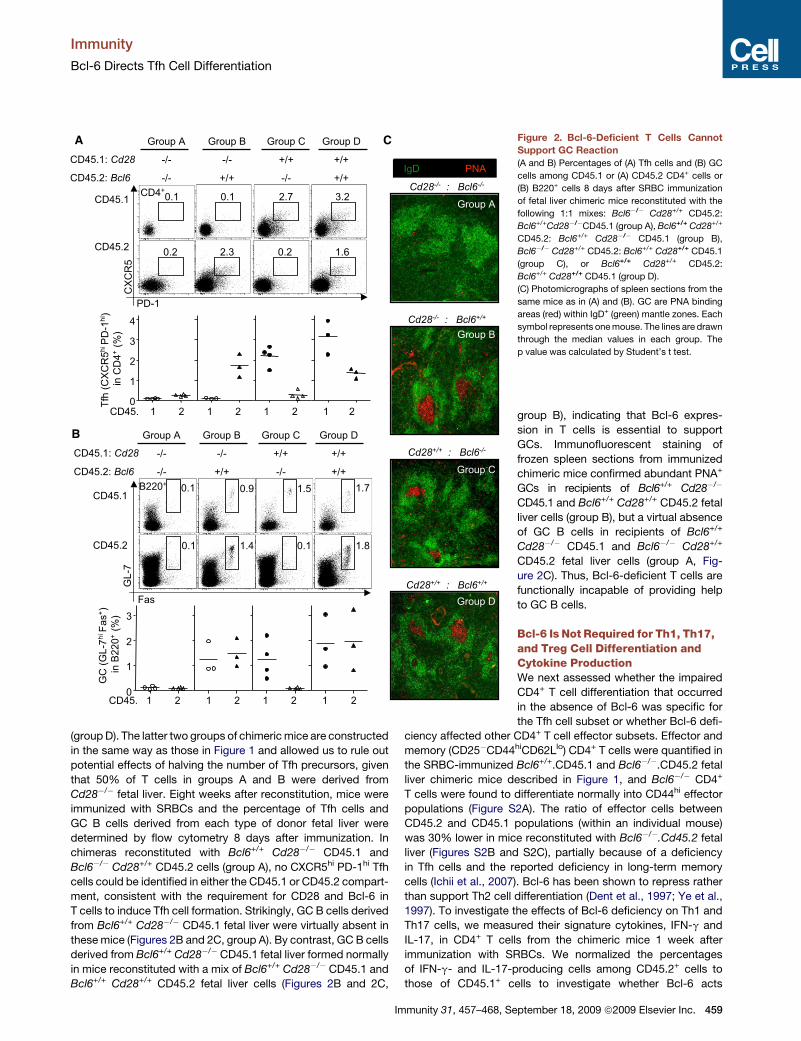
Bcl-6-Deficient T Cells Fail to Support GC ReactionsWe next examined whether deficiency of Bcl-6, specifically in
T cells, results in a deficiency in GC formation. For these exper-
iments, CD28-deficient fetal liver cells were chosen as a source
for T cells unable to differentiate into Tfh cells (Linterman et al.,
2009) and a source of B cells competent to differentiate into
GC B cells. Sublethally irradiated Rag1�/� mice were reconsti-
tuted with a 1:1 mix of Bcl6�/� Cd28+/+ CD45.2+ and
Bcl6+/+ Cd28�/� CD45.1 fetal liver cells (Figure 2, group A). In
these chimeric mice, Tfh cells are not expected to form from
458 Immunity 31, 457–468, September 18, 2009 ª2009 Elsevier Inc.
Cd28�/� T cells or Bcl6�/� T cells; thus, if Bcl-6 in T cells is
essential to sustain GC reactions, GCs derived from Cd28�/�
B cells will not be supported. As a control, Rag1�/� mice were
reconstituted with a 1:1 mix of Bcl6+/+ Cd28+/+ CD45.2 and
Bcl6+/+ Cd28�/� CD45.1 fetal liver cells (group B). In this group
of mice, Tfh cells derived from the Bcl6+/+ fetal liver cells are ex-
pected to support GCs derived from both Cd28�/� and Cd28+/+
B cells. A parallel set of control chimeras consisted of a mix of
Bcl6�/� Cd28+/+ CD45.2 and Bcl6+/+.Cd28+/+ CD45.1 (group C)
and a mix of Bcl6+/+ Cd28+/+ CD45.2 and Bcl6+/+.Cd28+/+ CD45.1
A
B
C
D
Figure 1. Bcl-6 Is Required for Tfh Cell Formation In Vivo
(A and B) Percentages of (A) GC cells and (B) Tfh cells 1 week after SRBC
immunization of Bcl6+/+ CD45.1: Bcl6+/+ CD45.2 (group A) or Bcl6+/+ CD45.1:
Bcl6�/� CD45.2 fetal liver chimeric mice (group B).
(C) Photomicrographs of spleen sections from the same mice as in (A) and (B).
The GC areas within IgD+ (blue) mantle zones have been delineated.
Bcl6+/+ CD45.2 T cells within GCs (right) appear yellow. Bcl6�/� Cd45.2
T cells (yellow) are absent from GCs (left), which only contain Bcl6+/+ CD45.1
CD4+ T cells (green). T, T cell zone; M, mantle zone.
(D) Ratio of Tfh cells 1 week after SRBC immunization of Bcl6+/+ CD45.1:
Bcl6+/+ CD45.2 (left) or Bcl6+/+ CD45.1: Bcl6+/� CD45.2 (right) mixed bone
marrow chimera mice. The ratio was calculated by dividing the percentage
of Tfh cells among CD45.2 T cells (Bcl6+/+ or Bcl6+/�) by the percentage of
Tfh cells among CD45.1 T cells (Bcl6+/+) in each individual mouse.
Each symbol represents one mouse. The lines are drawn through the median
values in each group. p value was calculated by Student’s t test.
Immunity
Bcl-6 Directs Tfh Cell Differentiation
(group D). The latter two groups of chimeric mice are constructed
in the same way as those in Figure 1 and allowed us to rule out
potential effects of halving the number of Tfh precursors, given
that 50% of T cells in groups A and B were derived from
Cd28�/� fetal liver. Eight weeks after reconstitution, mice were
immunized with SRBCs and the percentage of Tfh cells and
GC B cells derived from each type of donor fetal liver were
determined by flow cytometry 8 days after immunization. In
chimeras reconstituted with Bcl6+/+ Cd28�/� CD45.1 and
Bcl6�/� Cd28+/+ CD45.2 cells (group A), no CXCR5hi PD-1hi Tfh
cells could be identified in either the CD45.1 or CD45.2 compart-
ment, consistent with the requirement for CD28 and Bcl-6 in
T cells to induce Tfh cell formation. Strikingly, GC B cells derived
from Bcl6+/+ Cd28�/� CD45.1 fetal liver were virtually absent in
these mice (Figures 2B and 2C, group A). By contrast, GC B cells
derived from Bcl6+/+ Cd28�/� CD45.1 fetal liver formed normally
in mice reconstituted with a mix of Bcl6+/+ Cd28�/� CD45.1 and
Bcl6+/+ Cd28+/+ CD45.2 fetal liver cells (Figures 2B and 2C,
A
B
C Figure 2. Bcl-6-Deficient T Cells Cannot
Support GC Reaction
(A and B) Percentages of (A) Tfh cells and (B) GC
cells among CD45.1 or (A) CD45.2 CD4+ cells or
(B) B220+ cells 8 days after SRBC immunization
of fetal liver chimeric mice reconstituted with the
following 1:1 mixes: Bcl6�/� Cd28+/+ CD45.2:
Bcl6+/+Cd28�/�CD45.1 (group A), Bcl6+/+ Cd28+/+
CD45.2: Bcl6+/+ Cd28�/� CD45.1 (group B),
Bcl6�/� Cd28+/+ CD45.2: Bcl6+/+ Cd28+/+ CD45.1
(group C), or Bcl6+/+ Cd28+/+ CD45.2:
Bcl6+/+ Cd28+/+ CD45.1 (group D).
(C) Photomicrographs of spleen sections from the
same mice as in (A) and (B). GC are PNA binding
areas (red) within IgD+ (green) mantle zones. Each
symbol represents one mouse. The lines are drawn
through the median values in each group. The
p value was calculated by Student’s t test.
group B), indicating that Bcl-6 expres-
sion in T cells is essential to support
GCs. Immunofluorescent staining of
frozen spleen sections from immunized
chimeric mice confirmed abundant PNA+
GCs in recipients of Bcl6+/+ Cd28�/�
CD45.1 and Bcl6+/+ Cd28+/+ CD45.2 fetal
liver cells (group B), but a virtual absence
of GC B cells in recipients of Bcl6+/+
Cd28�/� CD45.1 and Bcl6�/� Cd28+/+
CD45.2 fetal liver cells (group A, Fig-
ure 2C). Thus, Bcl-6-deficient T cells are
functionally incapable of providing help
to GC B cells.
Bcl-6 Is Not Required for Th1, Th17,and Treg Cell Differentiation andCytokine ProductionWe next assessed whether the impaired
CD4+ T cell differentiation that occurred
in the absence of Bcl-6 was specific for
the Tfh cell subset or whether Bcl-6 defi-
ciency affected other CD4+ T cell effector subsets. Effector and
memory (CD25�CD44hiCD62Llo) CD4+ T cells were quantified in
the SRBC-immunized Bcl6+/+.CD45.1 and Bcl6�/�.CD45.2 fetal
liver chimeric mice described in Figure 1, and Bcl6�/� CD4+
T cells were found to differentiate normally into CD44hi effector
populations (Figure S2A). The ratio of effector cells between
CD45.2 and CD45.1 populations (within an individual mouse)
was 30% lower in mice reconstituted with Bcl6�/�.Cd45.2 fetal
liver (Figures S2B and S2C), partially because of a deficiency
in Tfh cells and the reported deficiency in long-term memory
cells (Ichii et al., 2007). Bcl-6 has been shown to repress rather
than support Th2 cell differentiation (Dent et al., 1997; Ye et al.,
1997). To investigate the effects of Bcl-6 deficiency on Th1 and
Th17 cells, we measured their signature cytokines, IFN-g and
IL-17, in CD4+ T cells from the chimeric mice 1 week after
immunization with SRBCs. We normalized the percentages
of IFN-g- and IL-17-producing cells among CD45.2+ cells to
those of CD45.1+ cells to investigate whether Bcl-6 acts
Immunity 31, 457–468, September 18, 2009 ª2009 Elsevier Inc. 459

Immunity
Bcl-6 Directs Tfh Cell Differentiation
cell-autonomously to influence Th1 and Th17 cell differentia-
tion. A near 3-fold increase in the ratio of IFN-g-producing cells
between CD45.2 and CD45.1 cells could be observed in mice
reconstituted with Bcl6�/� CD45.2 fetal liver (Figure S3A, group
B) compared with control chimeric mice (group A). A similar
increase was observed in IL-17-producing cells (Figure S3B).
IFN-g and IL-17 production was also investigated among
sorted effector CD25� CD44hi CD62lo effector cells from the
same chimeric mice restimulated ex vivo (sorting strategy
shown in Figure S4A). A higher proportion of Bcl6�/� T cells
produced IFN-g compared with Bcl6+/+ cells in the same
mice (Figures S3C and S3D). The increase in the produc-
tion of IL-17-producing cells was smaller (Figure S3D). As
a complementary strategy, naive CD25�CD44loCD62Lhi CD4+
T cells were sorted and polarized ex vivo under Th1 or Th17
cell conditions for 3 days. Again, the proportion of Bcl6�/�
CD4+ T cells producing IFN-g was higher than that of Bcl6+/+
B
A
D E
C
Figure 3. Bcl-6 Expression Suppresses Th1
and Th17 Cell Cytokine Production
(A) The upper panel shows cytokine production by
tonsil human CD4+ T cells (gates drawn around
CXCR5hi, CXCR5int and CXCR5lo populations).
The lower panel presents histograms showing
each cytokine expression in the three populations.
Values are the percentages of cytokine-producing
cells. Data are representative of more than three
individual samples.
(B) Cytokine production by mouse splenic CD4+
T cells 1 week after SRBC immunization. Data
are representative of more than three experiments.
(C) Bcl6 mRNA expression assessed by quantita-
tive RT-PCR in mouse CD4+ cell subsets (T naive,
non-Tfh effectors, and Tfh cells), and sorted GFPhi
cells from CD4+ T cells were transduced with either
empty vector or Bcl-6 under Th0 or Th17 cell differ-
entiation conditions. Data shown represent mean
values ± SD, with n = 3.
(D and E) IFN-g (D) and IL-17 (E) production by
naive CD4+ T cells transduced with either a Bcl-
6-expressing retrovirus or a vector control and
differentiated under Th1 (D) or Th17 (E) cell differ-
entiation conditions for the times indicated. Trans-
duced cells were divided into three groups—no
(�), intermediate (int), and high (hi)—according to
expression of GFP (Bcl-6). Day 5 is representative
flow cytometry plots for each of the three groups
are shown. Data shown represent mean values ±
SD, with n = 3.
cells (Figure S3E) with a very modest
increase in IL-17 (Figure S3F). In addi-
tion, there were no obvious differences
between the percentages of Foxp3+ Treg
cells derived from Bcl6�/� or Bcl6+/+
cells in chimeric mice, suggesting that
Bcl-6 deficiency does not affect the
generation of Treg cells (Figure S5).
These results, together with work pub-
lished by others on Bcl-6 in the context
of Th2 cell responses, suggest that Bcl-
6 specifically promotes Tfh cell differen-
tiation and is not required for Th1, Th2, Th17, and Treg cell
formation.
Forced Bcl-6 Expression Represses IFN-g and IL-17ProductionIn vivo and ex vivo data suggest that Bcl-6 expression induces
a parallel specific suppression of Th1 and Th17 effectors.
Because Tfh cells are the highest Bcl-6 expressers, we exam-
ined Th1, Th2, and Th17 cell cytokine production by Tfh cells.
We chose to do this in human tonsils because of their enrichment
in Th1, Th2, Th17, and Tfh cells. Tfh cells, expressing the highest
amounts of CXCR5, produced minimal amounts of IFN-g or IL-17
and low amounts of IL-4 compared to cells expressing interme-
diate or low amounts of CXCR5. By contrast, Tfh cells produced
high amounts of IL-21 and IL-2 comparable to CXCR5int cells
(Figure 3A). These findings coincide with the cytokine production
profiles in spleens from SRBCs immunized mice: compared to
460 Immunity 31, 457–468, September 18, 2009 ª2009 Elsevier Inc.

Immunity
Bcl-6 Directs Tfh Cell Differentiation
CXCR5lo or int cells, CXCR5hi Tfh cells produced the lowest
amounts of IFN-g and IL-17 but were still able to produce
substantial amounts of IL-2 (Figure 3B).
We examined the effect of Bcl-6 overexpression on cells
polarized to differentiate under Th1 or Th17 cell conditions.
Sorted naive CD4+ T cells were transduced by a Bcl-6-express-
ing retrovirus or a control vector. To assess Bcl-6 overexpres-
sion, we quantified Bcl-6 mRNA in transduced cells cultured in
Th0 or Th17 cell conditions and found it to be 44.4 and 12.9-
fold higher in cells overexpressing high levels of Bcl-6 compared
to cells transduced with the empty vector, respectively (Fig-
ure 3C). This expression was comparable to that observed in
Tfh cells (Figure 3C), which expressed 10- to 20-fold higher
amounts of Bcl6 mRNA than naive or non-Tfh effector
(CD44hi CXCR5lo or int PD-1lo or int, Figure S4C) cells.
Transduced cells were cultured under either Th1 or Th17 cell-
inducing conditions. IFN-g- and IL-17-secreting cells were
measured after 2, 3, 4, and 5 days. Transduced cells were
divided into Bcl-6hi, Bcl-6int, and Bcl-6� groups according to
their expression of GFP from the bicistronic vector. Overexpres-
sion of Bcl-6 suppressed Th1 and Th17 cell differentiation in
a dose-dependent manner: on day 5, high amounts of Bcl-6
expression, compared to empty vector, led to a 25% decrease
in IFN-g-producing cells (Figure 3D) and a 50% decrease in
IL-17-producing cells (Figure 3E). Similar suppression of Th1
and Th17 cytokines could be seen even when the cells had
been polarized for 2 days toward those lineages ex vivo, before
being transduced with Bcl-6 (Figure S6). We also observed that
despite strong Bcl-6-mediated suppression of IL-17, IL-2, a cyto-
kine produced by all helper T cells, was produced at much higher
amounts by the same Bcl-6hi cells (Figure S7), indicating that
Bcl-6 specifically repress IFN-g and IL-17 production rather
than causing a general suppression of cytokine production. In
summary, Bcl-6 expression can antagonize IFN-g and IL-17
production even in committed Th1 and Th17 cells, respectively,
and raises the possibility that in vivo, Bcl-6 may direct Tfh cell
differentiation from other helper T cell populations.
Bcl-6 Targets the Transcription Factors for Th1and Th17 Cell DifferentiationIn B cells, Bcl-6 determines GC B cell lineage commitment by re-
pressing its target genes, including the transcriptional regulator
of plasma cells, Blimp-1 (Reljic et al., 2000; Shaffer et al.,
2000). In CD4+ cells, Bcl-6 has been shown to repress GATA-3
expression (Kusam et al., 2003). Given our findings that Bcl-6
overexpression represses Th1 and Th17 cytokine production,
we hypothesized that Bcl-6 might also repress the transcription
factors directing Th1 and Th17 cell cytokine production. Bioin-
formatic analysis revealed the presence of high-affinity Bcl-6
binding sites within both mouse and human promoter regions
of critical transcription factors required for Th1 (T-bet) and
Th17 (RORgt) cell differentiation (Figure 4A and Table S1). To
validate the predicted binding of Bcl-6 to these promoter
regions, we performed Bcl-6 chromatin immunoprecipitation
(ChIP) assays on human Tfh cells and B cells sorted from tonsils
(Figure S5B) by using a high-quality ChIP grade Bcl-6 antibody
(Parekh et al., 2007). Importantly, no promoter enrichment by
Bcl-6 binding was detected in the B cell population (Figure 4B).
Marked Bcl-6 enrichment with ChIP ratios of 80 and 180 on the
Im
T-bet and RORgt promoters, respectively, was seen in Tfh cells,
but no specific enrichment was visible in tonsillar B cells, which
are enriched for Bcl-6+ cells. ChIP enrichment ratios greater than
2 are considered to be substantial binding above background
(Pokholok et al., 2006); hence, the enrichment values observed
across the Tbx21 (encoding T-bet) and Rorc (encoding RORgt)
promoters are highly relevant.
The results above suggest that Tbx21 and Rorc mRNA might
be reduced in Tfh cells (CXCR5hi), which express the highest
amounts of Bcl-6, compared with non-Tfh effectors (CXCR5int).
This was confirmed by quantitative RT-PCR on human tonsillar
T cell subsets (Figure 4C).
Next, we quantified the effects of Bcl-6 overexpression on
T-bet and GATA-3 protein expression. Naive mouse CD4+
T cells, differentiated ex vivo under Th1 cell polarizing conditions
and transduced by a Bcl-6-expressing retrovirus, displayed
�50% reduction in T-bet expression and �20% of the trans-
duced cells became T-bet negative (Figure 4D). Similarly, with
Th2 cell polarization, GATA-3 expression was reduced by
�60% in the Bcl-6 overexpressing cells and �30% of the cells
became GATA-3 negative (Figure S9).
Finally, we investigated T-bet and GATA-3 protein expression
on naive, Tfh, and non-Tfh effector cells that were Bcl-6 sufficient
or Bcl-6 deficient, by using CD45 congenic fetal liver chimeric
mice as shown in Figure 1. T-bet and GATA-3 were upregulated
in non-Tfh effector (CD44hiCXCR5int or loPD-1int or lo) CD4+ cells in
both Bcl-6-sufficient and Bcl-6-deficient mice, compared with
T naive (CD44lo) cells (Figure 4E and Figure S9B). T-bet expres-
sion in Tfh cells was nearly as low as that in naive T cells, whereas
GATA-3 was still expressed in Tfh cells. Interestingly, in the
absence of Bcl-6, T-bet expression was higher in non-Tfh
effector cells (Figure 4E), consistent with the increased IFN-g
expression shown in Figure S3.
Increased Expression of Bcl-6 Promotes the Tfh CellGene Expression SignatureHaving shown that Bcl-6 is essential for Tfh cell formation and
turning down Th1, Th2, and Th17 cell cytokine profiles, we
next examined whether Bcl-6 expression could also actively
induce the characteristic gene expression signature of Tfh cells
(Chtanova et al., 2004; Kim et al., 2004; Vinuesa et al., 2005a).
Upregulation of CXCR5 and downregulation of CCR7 on Tfh cells
determine their follicular positioning and thus are important for
their function (Hardtke et al., 2005; Haynes et al., 2007).
CXCR4, the receptor for follicular stromal cell-derived chemo-
kine CXCL12, is also upregulated on Tfh cells and may assist
in their localization, proximal to the follicular dendritic cell
network (Estes et al., 2004). The T cell costimulatory receptors
ICOS and PD-1 are expressed at the highest levels on Tfh cells
(Haynes et al., 2007; Hutloff et al., 1999).
We first confirmed selective high CXCR4, CCR7, PD-1, and
ICOS expression in human and mouse Tfh cells compared with
other effector CD4+ T populations. In resected human tonsils,
most CD4+ T cells are antigen experienced and therefore do
not express CD45RA (Figure 5A). Among these activated cells,
Tfh cells identified by the highest expression of CXCR5 also dis-
played the highest expression of PD-1, ICOS, and CXCR4 and
the lowest expression of CCR7 (Figure 5A). In mice, robust Tfh
cell responses were generated upon transfer of OVA-specific
munity 31, 457–468, September 18, 2009 ª2009 Elsevier Inc. 461

Immunity
Bcl-6 Directs Tfh Cell Differentiation
A
D
C
E
B Figure 4. Bcl-6 Suppresses Transcription
Factors for Th1, Th2, and Th17 Cell Differen-
tiation
(A) Bcl-6 binding sites as predicted by MatInspec-
tor (Genomatix Software) within �600 bp to
�100 bp, relative to the transcription start sites
(TSS), of promoter regions in humans and mice.
Detailed information listed in Table S1.
(B) Binding of Bcl-6 to putative high-affinity binding
sites in the promoters regions. Bcl-6 ChIP assays
were performed on human tonsil Tfh cells and
B cells. The results are presented as either
‘‘ChIP enrichment ratio’’ or ‘‘percentage of total
(genomic) input’’ and represent the mean of dupli-
cate assays from two independent experiments,
with error bars being SD.
(C) The mRNA expression of indicated genes as-
sessed by quantitative real-time RT-PCR in sorted
human tonsil CD4+ T cell subsets as shown in
Figure 3A (CXCR5lo, CXCR5int, and CXCR5hi).
Data shown represent mean values ± SD, with
n = 3.
(D) Intracellular T-bet expression in sorted mouse
naive CD4+ T cells transduced with either a Bcl-
6-expressing retrovirus or a vector control and
cultured in Th1 cell differentiation conditions for
2 days (left panels). Bar graphs show geometric
mean fluorescent intensity (Geom MFI) and posi-
tive percentages in transduced (GFP+) and non-
transduced (GFP�) cells (right panels). Data shown
represent mean values ± SD, with n = 3. p values
were calculated by Student’s t test.
(E) Representative flow cytometric profiles sho-
wing CD4 versus CD44 expression and CXCR5
versus PD-1 from Bcl6+/+ CD45.1: Bcl6�/� CD45.2
mixed chimeric mice (described in Figure 1), used
to gate T naive (Tn), Tfh, and non-Tfh effectors (left
panel). The histograms (middle panel) show T-bet
expression in Bcl6+/+ (top panel) and Bcl6�/�
(bottom panel) CD4+ T cells from the chimeric
mice; numbers indicate mean fluorescence inten-
sity in each population. The graph (right panel)
shows T-bet MFI in Bcl6+/+ TN, Tfh, and non-Tfh
effectors from four individual mice.
Thy1.1 OT-II cells into Thy1.2 recipient mice and immunization
with OVA in alum (Figure 5B). One week after immunization,
more than 75% of the transferred Thy1.1+ cells had become
effectors, expressing high CD44 (Figure 5B). As observed in
human Tfh cells, mouse Tfh cells—expressing the highest
amounts of CXCR5—also expressed the highest amounts of
PD-1 and CXCR4 and the lowest CCR7 (Figure 5B). ICOS
expression was high in all transferred OT-II cells, regardless of
CXCR5 expression (Figure 5B); this concurs with the report
that ICOS can be expressed at high amounts by other effector
T cell subsets (Lohning et al., 2003).
To test whether high Bcl-6 expression could induce the PD-1hi
CXCR5hi CXCR4hi and CCR7lo Tfh cell signature, we transduced
sorted mouse naive CD4+ T cells with a retrovirus expressing
Bcl-6 or empty vector and cultured them with anti-CD3 and
anti-CD28 under Th0 (no cytokines or antibodies), Thn (anti-
IFN-g and anti-IL-4), Th1, Th2, and Th17 cell conditions for
462 Immunity 31, 457–468, September 18, 2009 ª2009 Elsevier Inc.
2 days. Given our previous observation that Bcl-6 functions in
a gene dose-dependent manner for both Tfh cell development
(Figure 1D) and suppression of other helper T cell lineages
(Figures 3D and 3E; Figures S6 and S7), transduced cells were
again divided according to GFP-Bcl-6 expression into high and
intermediate expressers. Even though PD-1 is normally upregu-
lated after T cell stimulation, Bcl-6 overexpression further
increased the expression of PD-1 in all culture conditions (Fig-
ure 5C and Figure S10). Similarly, Bcl-6 enhanced the expression
of CXCR5 and CXCR4 by 2- to 3-fold across all conditions.
Downregulation of CCR7 by Bcl-6 was also seen under Th17
cell differentiation conditions (Figure 5C and Figure S10). Thus,
Bcl-6 can induce upregulation of PD-1 and the chemokine
receptors required for follicular homing, in both unpolarized
T cells, and in T cells differentiating under Th1, Th2, and Th17
cell conditions. These findings, together with the repression of
Th1 and Th17 cell cytokines in polarized Th1 and Th17 cells

Immunity
Bcl-6 Directs Tfh Cell Differentiation
shown above, suggest Bcl-6 expression may enable all types of
T helper cells—including Th1 and Th17 effectors—to enter the
follicles, change their cytokine production pattern, and become
bona fide Tfh cells.
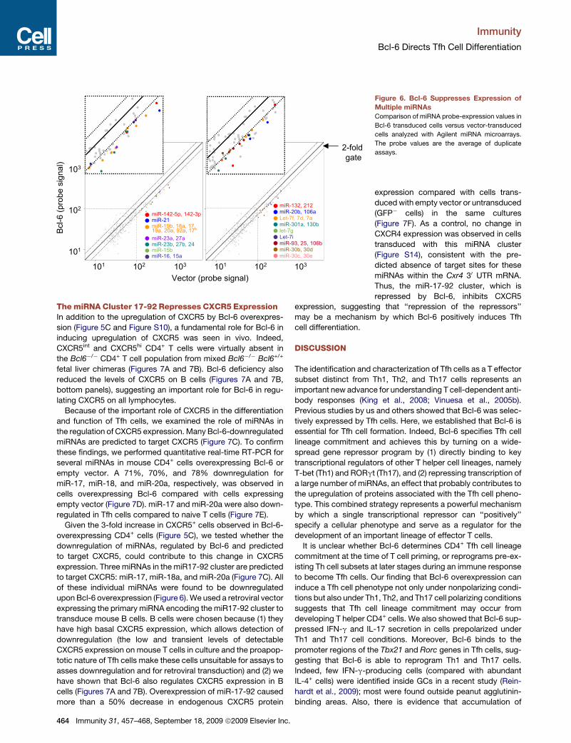
Bcl-6 Widely Represses miRNA ExpressionTo understand the mechanism by which Bcl-6, a transcription
repressor, determines the Tfh cell signature, we performed mi-
croarray analyses to compare the gene expression profiles
between naive CD4+ T cells retrovirally transduced with Bcl-6
or an empty vector. In addition to increased CXCR5, CXCR4,
and PD-1 mRNA and decreased CCR7 mRNA, intriguingly,
a large number of primary miRNA transcripts were downregu-
lated in cells overexpressing Bcl-6 (data not shown). miRNAs
are �23 nucleotide endogenous single-stranded noncoding
RNAs that pair to mRNA mainly through the 30 untranslated
region (30 UTR) to direct the posttranscriptional repression of
these mRNAs by translational inhibition and/or mRNA decay
(Baek et al., 2008). These results suggested that Bcl-6 might
promote the Tfh gene expression signature by repressing the
miRNAs that normally suppress these genes.
We used microarrays to profile mature miRNA expression in
mouse CD4+ T cells transduced with retroviruses expressing
Bcl-6 or an empty vector. Bcl-6 overexpression caused more
than 50% downregulation of 34 miRNas out of the 65 that
were abundantly expressed (i.e. with raw values >100, Table
A
B
C
Figure 5. Bcl-6 Promotes the Tfh Cell Gene
Expression Signature
(A) CXCR5 versus CD45RA expression by purified
human tonsillar CD4+ T cells (left). Expression
(Geom MFI) of PD-1, ICOS, CCR7, and CXCR4
analyzed in CXCR5hi, CXCR5int, and CXCR5lo pop-
ulations (right). Data are representative from more
than three individual samples.
(B) Thy1.1 versus CD44 expression on splenic
CD4+ T cells from C57BL/6 mouse transferred
with Thy1.1+ OT-II cells and immunized with
OVA-alum (Hutloff et al., 1999). Expression
(Geom MFI) of PD-1, ICOS, CCR7, and CXCR4
analyzed in CXCR5hi, CXCR5int, and CXCR5lo pop-
ulations is shown. Data are representative of more
than three experiments.
(C) PD-1, CXCR5, CXCR4, and CCR7 expression
on sorted mouse naive CD4+ T cells transduced
by either a Bcl-6-expressing retrovirus or a vector
control and cultured under Th0 or Th17 cell differ-
entiation conditions for 2 days.
Numbers beside the histograms in (A) and (B)
represent Geom MFI values of PD-1 or percent-
ages of chemokine receptors (CXCR5, CXCR4,
and CCR7) on cells expressing no, intermediate,
or high GFP (Bcl-6), respectively. Data are repre-
sentative of two experiments.
S2). There was not a single miRNA upre-
gulated by more than 2-fold (Table S3).
Of the 34, 31 were transcribed from clus-
ters encoding other miRNA(s) that were
also downregulated (Figure 6 and Table
S2). In control, nontransduced cells
(sorted GFP- cells from the same cultures), there was only one
miRNA with raw values > 100 regulated by more than 2-fold
(Figure S11). This global repression of miRNAs by Bcl-6 provides
a putative mechanism by which Bcl-6 may induce the Tfh gene
expression signature. This idea was further supported by a bioin-
formatic prediction of the multiple target sites within the 30 UTRs
of CXCR5, CXCR4, and PD-1 for those miRNAs markedly down-
regulated by Bcl-6 (Figure S12). Although 25% of these miRNAs
are encoded from transcripts contained within protein-encoding
transcripts, the majority are located in intergenic regions or within
primary transcripts that are nonprotein coding (Table S4), arguing
against the possibility that the observed downregulation is not
specific for miRNAs but a consequence of Bcl-6 repressing other
target genes. We have previously reported that two miRNAs,
miR-101 and miR-103, repressed the Tfh cell feature molecule
ICOS (Yu et al., 2007). Both miR-101 and miR-103 were down-
regulated in cells overexpressing Bcl-6 (Table S3, miR-101 was
expressed at low levels and therefore not included).
The expression of miR-101 and miR-103 was confirmed to be
downregulated by more than 50% in cells transduced with Bcl-
6-expressing retroviral vector and also downregulated in Tfh
cells by quantitative RT-PCR (Figure S13). Together, these
data suggest Bcl-6 may promote Tfh cell differentiation through
the repression of multiple miRNAs that normally prevent effector
T cells from expressing maximal amounts of CXCR5, CXCR4,
PD-1, and other Tfh cell signature molecules.
Immunity 31, 457–468, September 18, 2009 ª2009 Elsevier Inc. 463
Immunity
Bcl-6 Directs Tfh Cell Differentiation
The miRNA Cluster 17-92 Represses CXCR5 ExpressionIn addition to the upregulation of CXCR5 by Bcl-6 overexpres-
sion (Figure 5C and Figure S10), a fundamental role for Bcl-6 in
inducing upregulation of CXCR5 was seen in vivo. Indeed,
CXCR5int and CXCR5hi CD4+ T cells were virtually absent in
the Bcl6�/� CD4+ T cell population from mixed Bcl6�/� Bcl6+/+
fetal liver chimeras (Figures 7A and 7B). Bcl-6 deficiency also
reduced the levels of CXCR5 on B cells (Figures 7A and 7B,
bottom panels), suggesting an important role for Bcl-6 in regu-
lating CXCR5 on all lymphocytes.
Because of the important role of CXCR5 in the differentiation
and function of Tfh cells, we examined the role of miRNAs in
the regulation of CXCR5 expression. Many Bcl-6-downregulated
miRNAs are predicted to target CXCR5 (Figure 7C). To confirm
these findings, we performed quantitative real-time RT-PCR for
several miRNAs in mouse CD4+ cells overexpressing Bcl-6 or
empty vector. A 71%, 70%, and 78% downregulation for
miR-17, miR-18, and miR-20a, respectively, was observed in
cells overexpressing Bcl-6 compared with cells expressing
empty vector (Figure 7D). miR-17 and miR-20a were also down-
regulated in Tfh cells compared to naive T cells (Figure 7E).
Given the 3-fold increase in CXCR5+ cells observed in Bcl-6-
overexpressing CD4+ cells (Figure 5C), we tested whether the
downregulation of miRNAs, regulated by Bcl-6 and predicted
to target CXCR5, could contribute to this change in CXCR5
expression. Three miRNAs in the miR17-92 cluster are predicted
to target CXCR5: miR-17, miR-18a, and miR-20a (Figure 7C). All
of these individual miRNAs were found to be downregulated
upon Bcl-6 overexpression (Figure 6). We used a retroviral vector
expressing the primary miRNA encoding the miR17-92 cluster to
transduce mouse B cells. B cells were chosen because (1) they
have high basal CXCR5 expression, which allows detection of
downregulation (the low and transient levels of detectable
CXCR5 expression on mouse T cells in culture and the proapop-
totic nature of Tfh cells make these cells unsuitable for assays to
asses downregulation and for retroviral transduction) and (2) we
have shown that Bcl-6 also regulates CXCR5 expression in B
cells (Figures 7A and 7B). Overexpression of miR-17-92 caused
more than a 50% decrease in endogenous CXCR5 protein
Figure 6. Bcl-6 Suppresses Expression of
Multiple miRNAs
Comparison of miRNA probe-expression values in
Bcl-6 transduced cells versus vector-transduced
cells analyzed with Agilent miRNA microarrays.
The probe values are the average of duplicate
assays.
expression compared with cells trans-
duced with empty vector or untransduced
(GFP� cells) in the same cultures
(Figure 7F). As a control, no change in
CXCR4 expression was observed in cells
transduced with this miRNA cluster
(Figure S14), consistent with the pre-
dicted absence of target sites for these
miRNAs within the Cxr4 30 UTR mRNA.
Thus, the miR-17-92 cluster, which is
repressed by Bcl-6, inhibits CXCR5
expression, suggesting that ‘‘repression of the repressors’’
may be a mechanism by which Bcl-6 positively induces Tfh
cell differentiation.
DISCUSSION
The identification and characterization of Tfh cells as a T effector
subset distinct from Th1, Th2, and Th17 cells represents an
important new advance for understanding T cell-dependent anti-
body responses (King et al., 2008; Vinuesa et al., 2005b).
Previous studies by us and others showed that Bcl-6 was selec-
tively expressed by Tfh cells. Here, we established that Bcl-6 is
essential for Tfh cell formation. Indeed, Bcl-6 specifies Tfh cell
lineage commitment and achieves this by turning on a wide-
spread gene repressor program by (1) directly binding to key
transcriptional regulators of other T helper cell lineages, namely
T-bet (Th1) and RORgt (Th17), and (2) repressing transcription of
a large number of miRNAs, an effect that probably contributes to
the upregulation of proteins associated with the Tfh cell pheno-
type. This combined strategy represents a powerful mechanism
by which a single transcriptional repressor can ‘‘positively’’
specify a cellular phenotype and serve as a regulator for the
development of an important lineage of effector T cells.
It is unclear whether Bcl-6 determines CD4+ Tfh cell lineage
commitment at the time of T cell priming, or reprograms pre-ex-
isting Th cell subsets at later stages during an immune response
to become Tfh cells. Our finding that Bcl-6 overexpression can
induce a Tfh cell phenotype not only under nonpolarizing condi-
tions but also under Th1, Th2, and Th17 cell polarizing conditions
suggests that Tfh cell lineage commitment may occur from
developing T helper CD4+ cells. We also showed that Bcl-6 sup-
pressed IFN-g and IL-17 secretion in cells prepolarized under
Th1 and Th17 cell conditions. Moreover, Bcl-6 binds to the
promoter regions of the Tbx21 and Rorc genes in Tfh cells, sug-
gesting that Bcl-6 is able to reprogram Th1 and Th17 cells.
Indeed, few IFN-g-producing cells (compared with abundant
IL-4+ cells) were identified inside GCs in a recent study (Rein-
hardt et al., 2009); most were found outside peanut agglutinin-
binding areas. Also, there is evidence that accumulation of
464 Immunity 31, 457–468, September 18, 2009 ª2009 Elsevier Inc.

Immunity
Bcl-6 Directs Tfh Cell Differentiation
IL-17-producing cells in GCs is associated with autoimmunity in
BXD2 mice (Hsu et al., 2008). This may explain how long-lived
antibody-producing cells are generated against typical Th1,
Th2, and Th17 cell-type pathogens while minimizing the potential
damaging effects of excessive IFN-g and IL-17 production in
GCs. This is conceivable, in light of recent work suggesting
there may be much more plasticity during T cell differentiation
than was previously appreciated (Zhou et al., 2009; Tsuji et al.,
2009).
Restricted expression of inflammatory cytokines by Tfh cells in
GCs serves to regulate appropriate isotype switching. B cell
switching toaberrant isotypes in the contextofGCreactions poses
a greater threat than during extrafollicular responses because the
latter produce comparably low-affinity antibodies (MacLennan
et al., 2003), and also the offspring of GC B cells may live for
decades in an individual. Thus, Tfh cells turn down IFN-g but do
produce IL-21, which serves as a potent B cell stimulator, and
favors switching to IgG1while repressing IgE (Ozaki et al., 2002).
A B
C
E
D
F
Figure 7. The miRNA Cluster 17-92
Represses CXCR5 Expression
(A) CXCR5 expression on splenic CD4+ (Hutloff
et al., 1999) and B220+ cells (bottom) from
Bcl6+/+ CD45.1 : Bcl6+/+ CD45.2 or Bcl6+/+
CD45.1 : Bcl6�/� CD45.2 fetal liver chimeras 1
week after SRBC immunization (left).
(B) Graphs showing the percentage of CXCR5int or hi
CD4+ cells (Hutloff et al., 1999) or the Geo MFI of
CXCR5 on B220+ cells (bottom) among CD45.1 or
CD45.2 cells from the same mice. Each symbol
represents one mouse and the lines are drawn
through the median values in each group. The
p values were calculated by Student’s t test.
(C) Predicted binding sites within the 30 untrans-
lated region (30 UTR) of mouse CXCR5 by miRNAs
downregulated over 50% by Bcl-6 overexpression
(Table S2). Underlined miRNAs are expressed
within the miR-17-92 cluster.
(D and E) Mature miR-17, mir-18a, and miR-20
expression quantified by real-time RT-PCR relative
to U6 small nuclear RNA (U6) in mouse CD4+ cells
transduced with empty vector or Bcl-6 (top panel)
and in mouse splenic T naive versus Tfh CD4+
cells (bottom panel). Data shown represent mean
values ± SD, with n = 3.
(F) Histograms showing CXCR5 fluorescence on
mouse B220+ cells transduced with retrovirus ex-
pressing empty vector (filled histogram) or miR-
17-92 (empty histogram). The bar graphs inside
the histograms show CXCR5 geometric mean fluo-
rescence intensity in the same cells. Data shown
represent mean values ± SD, with n = 3.
Recent data has shown that IL-21 is regu-
lated by c-Maf, a transcription factor also
expressed by Th2, Th17, and Tfh cells
(Bauquet et al., 2009; Pot et al., 2009). It
is still not clear whether IL-21 is also regu-
lated by Bcl-6.
Bcl-6 also profoundly modifies chemo-
kine receptor expression. Upon priming,
CXCR5 is induced, and further upregula-
tion of CXCR5 in addition to downregulation of CCR7 are
required for the migration of CD4+ T cells into follicles to become
Tfh cells (Hardtke et al., 2005; Haynes et al., 2007). Bcl-6
enhanced the expression of CXCR5 and decreased CCR7
expression in activated T cells, whereas Bcl6�/� T cells were
unable to upregulate CXCR5 in vivo. Interestingly, Bcl6�/�B cells
also displayed lower expression of CXCR5, suggesting that
Bcl-6 regulates CXCR5 in both B cells and T cells through a
common mechanism, consistent with our results showing the
Bcl-6-regulated miR-17-92 family reduces CXCR5 expression
in mouse B cells. CXCR4 expression was also enhanced by
Bcl-6. Deficiency of CXCR5 on T cells severely impairs but
doesn’t totally abolish their participation in the GC reaction
(Arnold et al., 2007), suggesting synergetic control of multiple
chemokine receptors by Bcl-6 regulates the migration of Tfh
cells to the follicles.
Interestingly, enhanced Bcl-6 expression did not increase
ICOS or CD40L expression (data not shown). This was not
Immunity 31, 457–468, September 18, 2009 ª2009 Elsevier Inc. 465

Immunity
Bcl-6 Directs Tfh Cell Differentiation
entirely unexpected because high expression of ICOS and
CD40L can also be found on extrafollicular helper T cells (Ode-
gard et al., 2008) and other T helper cell lineages (Lohning
et al., 2003). In vivo, we did not observe any apparent decrease
in ICOS expression in Bcl6�/� CD4+ T cells (data not shown),
suggesting that ICOS expression may be mainly controlled by
other transcriptional regulators, cytokines, or signals from the
microenvironment.
A key aspect of Bcl-6-mediated gene regulation that has
emerged in this study is its ability to downregulate more than
half of abundantly expressed miRNAs by 50% or more in acti-
vated T cells. Most miRNAs downregulated by Bcl-6 (31 of 34)
belonged to clusters whose members are transcribed from
a single transcript and therefore are coregulated. Several
miRNAs within the same cluster frequently recognize similar
seed sequences with 30 UTRs and thus target the same
mRNAs, which explains why a mere 50% downregulation in
the amounts of individual mature miRNAs can result in substan-
tial increases in the expression of target genes. Remarkably,
not a single miRNA was upregulated (>2-fold) by Bcl-6.
miRNAs are emerging as key regulators of cell fate by fine-
tuning gene expression (Baek et al., 2008; Lodish et al.,
2008). Tfh cells are unique among T helper cell subsets in
that, despite having a very distinct cell function compared to
other effector T cells, they have a phenotype that is only distin-
guished from non-Tfh effectors by a quantitative increase in the
expression of CXCR5, CXCR4, PD-1, ICOS, IL-21 and other
molecules (King et al., 2008; Vinuesa et al., 2005b). Seventeen
out of thirty-four miRNAs repressed by Bcl-6 were potential
regulators of these molecules. This is particularly important in
light of recent findings demonstrating that miRNAs regulate
proteins that typically function over a narrow range of concen-
trations during normal immune responses and adds to the
evidence that miRNA-mediated increases or decreases in the
expression of these proteins may lead to marked changes in
cell function.
We found that overexpression of the miR-17-92 cluster
comprising seven miRNAs that are all downregulated in Bcl-6-
overexpressing cells leads to a reduction of CXCR5 in mouse
primary cells. Given that at least three miRNAs in this cluster
are predicted to target CXCR5, the net reduction effect of
CXCR5 protein is likely to be a consequence of the cumulative
effects of these different miRNAs. It is interesting to note that
mice transgenic for miR-17-92 display abnormal compartmen-
talization and segregation of B cells and T cells in secondary
lymphoid organs and paucity of B cells within follicular structures
(Xiao et al., 2008). Our results suggest this is due, at least in part,
to lower CXCR5 expression in both B cells and T cells.
Identifying Bcl-6 as the transcription factor that specifies Tfh
cell lineage commitment firmly establishes Tfh cells as a bona
fide T effector subset. Bcl-6 emerges as an important arbiter of
T cell lineage fate, acting through repression of Th1, Th2, and
Th17 cell transcription factors, as well numerous miRNAs that
are predicted to affect expression of Tfh cell signature mole-
cules. Bcl-6 therefore connects the differentiation of an impor-
tant effector T cell subset with the regulation of chemokine
receptors and other molecules. This enables the proper place-
ment of these T cells in the GC microenvironment to facilitate
T cell-dependent antibody responses.
466 Immunity 31, 457–468, September 18, 2009 ª2009 Elsevier Inc.
EXPERIMENTAL PROCEDURES
Mice and Immunizations
Bcl6�/� (Dent et al., 1997), Thy1.1 OT-II, CD45.1 mice were maintained on
a C57BL/6 background and housed in SPF conditions at the Australian
National University (ANU) and Garvan Institute animal facilities. All animal
experiments were carried under protocols approved by the institutes’ Animal
Ethics Committees. For fetal liver and bone marrow reconstitution, 2 to 5 3
106 cells from E15-E17 fetal liver or adult bone marrow were injected intrave-
nously (i.v.) into 8- to 12-week-old CD45.1 or Rag1�/� mice that had been
sublethally irradiated. Mice were analyzed 8 weeks after reconstitution. For
generating T cell-dependent antibody responses, mice were immunized i.p.
with 2 3 109 SRBC (Veterinary Services, IMVS). For induction of T cell-depen-
dent responses after adoptive transfer of OT-II cells, mice were i.v. injected
with splenocytes containing 3 to 5 3 104 OVA323–339-specific CD4+ T cells
and immunized intraperitoneally (i.p.) with 100 mg of OVA in alum. All these
procedures were approved by the Ethics Committee of The Australian National
University.
Human Lymphocyte Preparations
Tonsillar cells were isolated by mechanical disruption and Ficoll-Plaque
density gradient centrifugation. Human studies were approved by the insti-
tutes’ human research ethics committees.
CD4+ T Cell Isolation, Culture, Stimulation, and Differentiation
Mouse CD4+ T cells were purified with CD4 positive selection (Miltenyi Biotec)
and sorted into naive (CD44lo CD62Lhi CD25�) and activated (CD44hi CD62Llo
CD25�) populations on FACSAria (>97% purity). For ChIP, human naive
(CD45RO�CXCR5lo), Tfh (CD45RO+CXCR5hi PD-1hi), and non-Tfh effector
(CD45RO+ CXCR5int or o PD-1int or lo) CD4+ T cells, and an enriched B (CD4�
CXCR5hi) cell population were sorted from isolated tonsil cells (90%–95%
purity). For other purposes, human CD4+ T cells were purified with the Dynal
CD4 Positive Cell Isolation Kit (Invitrogen) (>98% purity). For ex vivo differen-
tiation, mouse naive T cells were cultured in completed RPMI1640 media (Yu
et al., 2008) and stimulated with plate-bound anti-CD3 (5 mg/ml) plus anti-
CD28 (2 mg/ml) under Th0: no cytokines and antibodies; Thn: 10 mg/ml anti-
IFN-g and 10 mg/ml anti-IL-4; Th1: 10 ng/ml IL-12, and 10 mg/ml anti-IL-4;
Th2: 10 ng/ml IL-4, 10 mg/ml anti-IFN-g, and 10 mg/ml anti-IL-12/IL-23 p40;
and Th17: 2 ng/ml TGF-b, 20 ng/ml IL-6, 10 mg/ml anti-IFN-g, and 10 mg/ml
anti-IL-4. For intracellular cytokine staining, CD4+ T cells were restimulated
for 4–6 hr with 50 ng/ml PMA (Sigma), 1 mg/ml ionomycin (Sigma), 3 mg/ml bre-
feldin A (eBioscience), and 2 mM monensin (eBioscience).
Flow Cytometry
Antibodies were listed in the Supplemental Data. The staining procedure has
been described (Yu et al., 2008), except that anti-mouse CCR7 staining was
performed at 37�C. Intracellular cytokines and transcription factors were
stained with the BD Cytofix/Cytoperm kit (BD PharMingen) and Mouse Regu-
latory T Cell Staining Kit (eBioscience), respectively. Flow cytometric analysis
was performed on LSR II or FACSCanto II and analyzed with FlowJo software
(Tree Star).
Immunofluorescence
Frozen spleen sections were fixed in cold acetone, stained, mounted in
VECTASHIELD (Vector Laboratories), and viewed with a Zeiss Axiovert
200M microscope. The images were acquired with AxioVision (release 4.7.1)
software (Carl Zeiss).
Quantitative Real-Time RT-PCR
MiRNA expression was assessed by real-time RT-PCR with the TaqMan
miRNA assay protocol (Applied Biosystems). Gene and miRNA expression
were detected with the ABI 7900 Prism and fold changes in expression were
determined by the 2�DDCt method, with the results normalized to RNU 6 (U6)
for miRNA and b-actin for gene expression.
Retrovirus Production and Transduction
Retrovirus preparation has been described before (Yu et al., 2007). Bcl-6 cDNA
was PCR amplified and cloned into the vector (Bcl-6-IRES-GFP). For retroviral

Immunity
Bcl-6 Directs Tfh Cell Differentiation
transduction, sorted mouse naive CD4+ T or B cells were stimulated with plate-
bound anti-CD3 (5 mg/ml) plus anti-CD28 (2 mg/ml) and 10 ng/ml of IL-2 (for
T cells) or soluble LPS (10 mg/ml) (for B cells) for 2 days. Cells were spun
with retroviral supernatants and 4 mg/ml of polybrene (Sigma) at 800 g-force
for 1 hr at 30�C and then cultured in fresh medium before analysis at the times
indicated.
Chromatin Immunoprecipitation
Chromatin immunoprecipitation (ChIP) assays were performed in accordance
with the instructions (Upstate Biotechnology) as detailed in the Supplemental
Data. Ct values from the PCR amplification plots were converted to arbitrary
copy number with the formula 105/2(Ct �17). We then normalized sample data
to the corresponding total input before fold change above the average no anti-
body control was calculated to yield the ChIP enrichment ratio as previously
described (Pokholok et al., 2006). In addition, we calculated the data to yield
the percentage of total input. All ChIP experiments were performed at least
in duplicate.
miRNA Microarrays
Mouse naive CD4+ T cells were transduced with a Bcl-6-expressing retrovirus
or the control vector and cultured in Th0 condition for 2 days. Cells expressing
medium to high levels of GFP were sorted, and total RNAs were isolated with
the mirVana miRNA Isolation Kit (Applied Biosystems). MiRNA expression was
profiled with Agilent 8x15K mouse miRNA microarray miRNA-v1_95_May07
(miRBase Release 9.2) by Ramaciotti Centre for Gene Function Analysis (Syd-
ney). Each sample was run in duplicate. For analysis, probes were quality-
controlled by deleting any probe that had no positive signals; the average of
each probe was calculated for replicates and probes with negative signals
were set to 0.
Bioinformatic Promoter and miRNA Binding Analysis
Bcl-6 binding site frequency and density in promoter regions were performed
with Gene2Promoter (release 4.2) and GEMS Launcher (release 4.3) within the
Genomatix Suite. We used MatInspector (release 7.4.3) to identify Bcl-6 sites
with optimized core similarity and matrix similarity score settings. MiRNA
target sequences in 30 UTR were predicted by miRanda (September 2008
Release, http://www.microrna.org/microrna/home.do) and Targetscan (Re-
lease 4.2, http://www.targetscan.org/vert_42/) with default settings.
ACCESSION NUMBERS
The miRNA array data for this paper have been deposited in GEO under acces-
sion number GSE17000.
SUPPLEMENTAL DATA
Supplemental Data include Supplemental Experimental Procedures, 14
figures, and 4 tables and can be found with this article online at http://www.
cell.com/immunity/supplemental/S1074-7613(09)00314-8.
ACKNOWLEDGMENTS
We thank X. Hu and J. Hogan (animal experiments), N. Chevalier and C. Wang
(miRNA experiments), A. Wilson and M. Cook (obtaining tonsils), R. Brink and
D. Gatto (OT-II transfer experiments), F. Sierro (immunofluorescence), H.
Speirs (RNA preparation), and H. Vora, C. Brownlee, and Y. Sontani (cell sort-
ing) for their help. This work was funded by a Viertel Senior Medical Research
Fellowship to C.G.V., National Health and Medical Research Council (NHMRC)
Fellowships to D.Y., C.S.M., and C.R.M, Cooperative Research Centre Schol-
arship to L.M.T., a Cancer Institute NSW Fellowship to D.Y., a NHMRC project
grant to S.R. and NHMRC program grants to C.G.V., C.R.M., and C.R.P.
Received: December 22, 2008
Revised: June 18, 2009
Accepted: July 7, 2009
Published online: July 23, 2009
I
REFERENCES
Allen, C.D., Okada, T., and Cyster, J.G. (2007). Germinal-center organization
and cellular dynamics. Immunity 27, 190–202.
Allman, D., Jain, A., Dent, A., Maile, R.R., Selvaggi, T., Kehry, M.R., and Staudt,
L.M. (1996). BCL-6 expression during B-cell activation. Blood 87, 5257–5268.
Ansel, K.M., McHeyzer-Williams, L.J., Ngo, V.N., McHeyzer-Williams, M.G.,
and Cyster, J.G. (1999). In vivo-activated CD4 T cells upregulate CXC chemo-
kine receptor 5 and reprogram their response to lymphoid chemokines. J. Exp.
Med. 190, 1123–1134.
Arnold, C.N., Campbell, D.J., Lipp, M., and Butcher, E.C. (2007). The germinal
center response is impaired in the absence of T cell-expressed CXCR5. Eur. J.
Immunol. 37, 100–109.
Baek, D., Villen, J., Shin, C., Camargo, F.D., Gygi, S.P., and Bartel, D.P. (2008).
The impact of microRNAs on protein output. Nature 455, 64–71.
Bauquet, A.T., Jin, H., Paterson, A.M., Mitsdoerffer, M., Ho, I.C., Sharpe, A.H.,
and Kuchroo, V.K. (2009). The costimulatory molecule ICOS regulates the
expression of c-Maf and IL-21 in the development of follicular T helper cells
and TH-17 cells. Nat. Immunol. 10, 167–175.
Breitfeld, D., Ohl, L., Kremmer, E., Ellwart, J., Sallusto, F., Lipp, M., and For-
ster, R. (2000). Follicular B helper T cells express CXC chemokine receptor
5, localize to B cell follicles, and support immunoglobulin production. J. Exp.
Med. 192, 1545–1552.
Bromley, S.K., Mempel, T.R., and Luster, A.D. (2008). Orchestrating the
orchestrators: Chemokines in control of T cell traffic. Nat. Immunol. 9,
970–980.
Casamayor-Palleja, M., Khan, M., and MacLennan, I.C. (1995). A subset of
CD4+ memory T cells contains preformed CD40 ligand that is rapidly but tran-
siently expressed on their surface after activation through the T cell receptor
complex. J. Exp. Med. 181, 1293–1301.
Cattoretti, G., Chang, C.C., Cechova, K., Zhang, J., Ye, B.H., Falini, B., Louie,
D.C., Offit, K., Chaganti, R.S., and Dalla-Favera, R. (1995). BCL-6 protein is
expressed in germinal-center B cells. Blood 86, 45–53.
Chtanova, T., Tangye, S.G., Newton, R., Frank, N., Hodge, M.R., Rolph, M.S.,
and Mackay, C.R. (2004). T follicular helper cells express a distinctive tran-
scriptional profile, reflecting their role as non-Th1/Th2 effector cells that
provide help for B cells. J. Immunol. 173, 68–78.
Dent, A.L., Shaffer, A.L., Yu, X., Allman, D., and Staudt, L.M. (1997). Control of
inflammation, cytokine expression, and germinal center formation by BCL-6.
Science 276, 589–592.
Dhordain, P., Albagli, O., Lin, R.J., Ansieau, S., Quief, S., Leutz, A., Kerckaert,
J.P., Evans, R.M., and Leprince, D. (1997). Corepressor SMRT binds the BTB/
POZ repressing domain of the LAZ3/BCL6 oncoprotein. Proc. Natl. Acad. Sci.
USA 94, 10762–10767.
Estes, J.D., Thacker, T.C., Hampton, D.L., Kell, S.A., Keele, B.F., Palenske,
E.A., Druey, K.M., and Burton, G.F. (2004). Follicular dendritic cell regulation
of CXCR4-mediated germinal center CD4 T cell migration. J. Immunol. 173,
6169–6178.
Fukuda, T., Yoshida, T., Okada, S., Hatano, M., Miki, T., Ishibashi, K., Okabe,
S., Koseki, H., Hirosawa, S., Taniguchi, M., et al. (1997). Disruption of the Bcl6
gene results in an impaired germinal center formation. J. Exp. Med. 186,
439–448.
Hardtke, S., Ohl, L., and Forster, R. (2005). Balanced expression of CXCR5 and
CCR7 on follicular T helper cells determines their transient positioning to lymph
node follicles and is essential for efficient B-cell help. Blood 106, 1924–1931.
Haynes, N.M., Allen, C.D., Lesley, R., Ansel, K.M., Killeen, N., and Cyster, J.G.
(2007). Role of CXCR5 and CCR7 in follicular Th cell positioning and appear-
ance of a programmed cell death gene-1high germinal center-associated
subpopulation. J. Immunol. 179, 5099–5108.
Ho, I.C., and Glimcher, L.H. (2002). Transcription: Tantalizing times for T cells.
Cell 109 (Suppl ), S109–S120.
Hori, S., Nomura, T., and Sakaguchi, S. (2003). Control of regulatory T cell
development by the transcription factor Foxp3. Science 299, 1057–1061.
mmunity 31, 457–468, September 18, 2009 ª2009 Elsevier Inc. 467

Immunity
Bcl-6 Directs Tfh Cell Differentiation
Hsu, H.C., Yang, P., Wang, J., Wu, Q., Myers, R., Chen, J., Yi, J., Guentert, T.,
Tousson, A., Stanus, A.L., et al. (2008). Interleukin 17-producing T helper cells
and interleukin 17 orchestrate autoreactive germinal center development in
autoimmune BXD2 mice. Nat. Immunol. 9, 166–175.
Hutloff, A., Dittrich, A.M., Beier, K.C., Eljaschewitsch, B., Kraft, R., Anagnosto-
poulos, I., and Kroczek, R.A. (1999). ICOS is an inducible T-cell co-stimulator
structurally and functionally related to CD28. Nature 397, 263–266.
Ichii, H., Sakamoto, A., Hatano, M., Okada, S., Toyama, H., Taki, S., Arima, M.,
Kuroda, Y., and Tokuhisa, T. (2002). Role for Bcl-6 in the generation and main-
tenance of memory CD8+ T cells. Nat. Immunol. 3, 558–563.
Ichii, H., Sakamoto, A., Arima, M., Hatano, M., Kuroda, Y., and Tokuhisa, T.
(2007). Bcl6 is essential for the generation of long-term memory CD4+
T cells. Int. Immunol. 19, 427–433.
Ivanov, I.I., McKenzie, B.S., Zhou, L., Tadokoro, C.E., Lepelley, A., Lafaille,
J.J., Cua, D.J., and Littman, D.R. (2006). The orphan nuclear receptor ROR-
gammat directs the differentiation program of proinflammatory IL-17+ T helper
cells. Cell 126, 1121–1133.
Kim, C.H., Lim, H.W., Kim, J.R., Rott, L., Hillsamer, P., and Butcher, E.C.
(2004). Unique gene expression program of human germinal center T helper
cells. Blood 104, 1952–1960.
Kim, C.H., Rott, L.S., Clark-Lewis, I., Campbell, D.J., Wu, L., and Butcher, E.C.
(2001). Subspecialization of CXCR5+ T cells: B helper activity is focused in
a germinal center-localized subset of CXCR5+ T cells. J. Exp. Med. 193,
1373–1381.
King, C., Tangye, S.G., and Mackay, C.R. (2008). T follicular helper (TFH) cells
in normal and dysregulated immune responses. Annu. Rev. Immunol. 26,
741–766.
Kusam, S., Toney, L.M., Sato, H., and Dent, A.L. (2003). Inhibition of Th2 differ-
entiation and GATA-3 expression by BCL-6. J. Immunol. 170, 2435–2441.
Linterman, M.A., Rigby, R.J., Wong, R.K., Yu, D., Brink, R., Cannons, J.L.,
Schwartzberg, P.L., Cook, M.C., Walters, G.D., and Vinuesa, C.G. (2009).
Follicular helper T cells are required for systemic autoimmunity. J. Exp. Med.
206, 561–576.
Lodish, H.F., Zhou, B., Liu, G., and Chen, C.Z. (2008). Micromanagement of the
immune system by microRNAs. Nat. Rev. Immunol. 8, 120–130.
Lohning, M., Hutloff, A., Kallinich, T., Mages, H.W., Bonhagen, K., Radbruch,
A., Hamelmann, E., and Kroczek, R.A. (2003). Expression of ICOS in vivo
defines CD4+ effector T cells with high inflammatory potential and a strong
bias for secretion of interleukin 10. J. Exp. Med. 197, 181–193.
MacLennan, I.C., Toellner, K.M., Cunningham, A.F., Serre, K., Sze, D.M.,
Zuniga, E., Cook, M.C., and Vinuesa, C.G. (2003). Extrafollicular antibody
responses. Immunol. Rev. 194, 8–18.
Murphy, K.M., and Reiner, S.L. (2002). The lineage decisions of helper T cells.
Nat. Rev. Immunol. 2, 933–944.
Odegard, J.M., Marks, B.R., DiPlacido, L.D., Poholek, A.C., Kono, D.H., Dong,
C., Flavell, R.A., and Craft, J. (2008). ICOS-dependent extrafollicular helper
T cells elicit IgG production via IL-21 in systemic autoimmunity. J. Exp. Med.
205, 2873–2886.
Ozaki, K., Spolski, R., Feng, C.G., Qi, C.F., Cheng, J., Sher, A., Morse, H.C.,
3rd, Liu, C., Schwartzberg, P.L., and Leonard, W.J. (2002). A critical role for
IL-21 in regulating immunoglobulin production. Science 298, 1630–1634.
Parekh, S., Polo, J.M., Shaknovich, R., Juszczynski, P., Lev, P., Ranuncolo,
S.M., Yin, Y., Klein, U., Cattoretti, G., Dalla Favera, R., et al. (2007). BCL6
programs lymphoma cells for survival and differentiation through distinct
biochemical mechanisms. Blood 110, 2067–2074.
Pokholok, D.K., Zeitlinger, J., Hannett, N.M., Reynolds, D.B., and Young, R.A.
(2006). Activated signal transduction kinases frequently occupy target genes.
Science 313, 533–536.
468 Immunity 31, 457–468, September 18, 2009 ª2009 Elsevier Inc.
Pot, C., Jin, H., Awasthi, A., Liu, S.M., Lai, C.Y., Madan, R., Sharpe, A.H., Karp,
C.L., Miaw, S.C., Ho, I.C., and Kuchroo, V.K. (2009). IL-27 induces the tran-
scription factor c-Maf, cytokine IL-21, and the costimulatory receptor ICOS
that coordinately act together to promote differentiation of IL-10-producing
Tr1 cells. J. Immunol. 183, 797–801.
Reiner, S.L. (2007). Development in motion: Helper T cells at work. Cell 129,
33–36.
Reinhardt, R.L., Liang, H.E., and Locksley, R.M. (2009). Cytokine-secreting
follicular T cells shape the antibody repertoire. Nat. Immunol. 10, 385–393.
Reljic, R., Wagner, S.D., Peakman, L.J., and Fearon, D.T. (2000). Suppression
of signal transducer and activator of transcription 3-dependent B lymphocyte
terminal differentiation by BCL-6. J. Exp. Med. 192, 1841–1848.
Sallusto, F., Mackay, C.R., and Lanzavecchia, A. (2000). The role of chemokine
receptors in primary, effector, and memory immune responses. Annu. Rev.
Immunol. 18, 593–620.
Schaerli, P., Willimann, K., Lang, A.B., Lipp, M., Loetscher, P., and Moser, B.
(2000). CXC chemokine receptor 5 expression defines follicular homing T cells
with B cell helper function. J. Exp. Med. 192, 1553–1562.
Shaffer, A.L., Yu, X., He, Y., Boldrick, J., Chan, E.P., and Staudt, L.M. (2000).
BCL-6 represses genes that function in lymphocyte differentiation, inflamma-
tion, and cell cycle control. Immunity 13, 199–212.
Szabo, S.J., Kim, S.T., Costa, G.L., Zhang, X., Fathman, C.G., and Glimcher,
L.H. (2000). A novel transcription factor, T-bet, directs Th1 lineage commit-
ment. Cell 100, 655–669.
Tsuji, M., Komatsu, N., Kawamoto, S., Suzuki, K., Kanagawa, O., Honjo, T.,
Hori, S., and Fagarasan, S. (2009). Preferential generation of follicular B helper
T cells from Foxp3+ T cells in gut Peyer’s patches. Science 323, 1488–1492.
Vinuesa, C.G., Cook, M.C., Angelucci, C., Athanasopoulos, V., Rui, L., Hill,
K.M., Yu, D., Domaschenz, H., Whittle, B., Lambe, T., et al. (2005a). A RING-
type ubiquitin ligase family member required to repress follicular helper
T cells and autoimmunity. Nature 435, 452–458.
Vinuesa, C.G., Tangye, S.G., Moser, B., and Mackay, C.R. (2005b). Follicular B
helper T cells in antibody responses and autoimmunity. Nat. Rev. Immunol. 5,
853–865.
Weaver, C.T., Hatton, R.D., Mangan, P.R., and Harrington, L.E. (2007). IL-17
family cytokines and the expanding diversity of effector T cell lineages.
Annu. Rev. Immunol. 25, 821–852.
Xiao, C., Srinivasan, L., Calado, D.P., Patterson, H.C., Zhang, B., Wang, J.,
Henderson, J.M., Kutok, J.L., and Rajewsky, K. (2008). Lymphoproliferative
disease and autoimmunity in mice with increased miR-17–92 expression in
lymphocytes. Nat. Immunol. 9, 405–414.
Ye, B.H., Cattoretti, G., Shen, Q., Zhang, J., Hawe, N., de Waard, R., Leung, C.,
Nouri-Shirazi, M., Orazi, A., Chaganti, R.S., et al. (1997). The BCL-6 proto-
oncogene controls germinal-centre formation and Th2-type inflammation.
Nat. Genet. 16, 161–170.
Yu, D., Cook, M.C., Shin, D.M., Silva, D.G., Marshall, J., Toellner, K.M., Hav-
ran, W.L., Caroni, P., Cooke, M.P., Morse, H.C., et al. (2008). Axon growth
and guidance genes identify T-dependent germinal centre B cells. Immunol.
Cell Biol. 86, 3–14.
Yu, D., Tan, A.H., Hu, X., Athanasopoulos, V., Simpson, N., Silva, D.G., Hutloff,
A., Giles, K.M., Leedman, P.J., Lam, K.P., et al. (2007). Roquin represses auto-
immunity by limiting inducible T-cell co-stimulator messenger RNA. Nature
450, 299–303.
Zheng, W., and Flavell, R.A. (1997). The transcription factor GATA-3 is neces-
sary and sufficient for Th2 cytokine gene expression in CD4 T cells. Cell 89,
587–596.
Zhou, L., Chong, M.M., and Littman, D.R. (2009). Plasticity of CD4+ T cell
lineage differentiation. Immunity 30, 646–655.