the tetravalent manganese oxides: identification, hydration, and ... · the tetravalent manganese...
TRANSCRIPT
American Mineralogist, Volume 64, pages I199-1218, 1979
The tetravalent manganese oxides: identification, hydration, and structural relationshipsby infrared spectroscopy
RussBrr M. Porrnn'AND GpoRcn R. RossueN
Division of Geological and Planetary SciencefCalifurnia Institute of Technology
Pasadena, Califurnia 9 I 1 25
Abstract
A compilation of the infrared powder absorption spectra of most naturally occurring tet-ravalent and trivalent manganese oxides is presented which is intended to serve as a basis forthe spectroscopic identiflcation of these minerals in both ordered and disordered varieties, in-cluding those too disordered for X-ray diffraction studies. A variety of synthetic manganeseoxides are also included for comparison to the natural phases. The samples include: aurorite,birnessite, braunite, buserite, chalcophanite, coronadite, cryptomelane, groutite, hausman-nite, hollandite, lithiophorite, maoganite, manganese(Ill) manganate(IV), manganosite,manjiroite, marokite, nsutite, partridgeite, pyrolusite, quenselite, rancieite, ramsdellite, ro-manechite, sodium manganese(Il,flI) manganate(Iv), todorokite, and woodruffite. Thespectra indicate that well-ordered water occurs in ramsdellite, chalcophanite, and most ro-manechites. Disordered water is observed in the spectra of nsutite, hollandites, birnessite, to-dorokite, buserite, and rancieite. The infrared spectra of well-ordered todorokite, birnessiteand rancieite differ which indicates that they possess diflerent structures and should be re-garded as distinct mineral species. Much variation is observed in the spectra of pyrolusites,nsutites, birnessites, and todorokites which is interpreted as arising from structural disorder.Spectral trends suggest that todorokite, birnessite and rancieite have layered structures.
Introduction
The finely-particulate and disordered nature ofmanganese oxides in many of their concentrations inthe weathering environment has made identificationof their mineralogy by means of X-ray diffractiondifficult and sometimes impossible. Characteristic X-ray powder diffraction lines are frequently broad, in-distinct, or absent altogether and do not serve as asatisfactory basis for identification. The frequent oc-currence of silicate phases admixed with the low-temperature manganese oxides is an additional com-plication because their X-ray lines can sometimes beconfused with those of the manganese oxides.
Problems of particle size and disorder are a char-acteristic of pure manganese oxides and have beenresponsible for our poor understanding of manganese
I Present address: Owens-Corning Fiberglas Corporation, Gran-ville, Ohio 43023.
2 Contribution N o. 3126.
0np3-{0/'X/7 9 / I I I 2- I 199$02.00
oxide crystal structures and structural relationships.X-ray powder diffraction patterns are the only struc-tural data available on several of the oxides impor-tant in the weathering environment, and these gener-ally show lines which are few in number, broad, andsometimes variable in position. This has caused un-certainty in the structural relationships among syn-thetic and natural samples and a consequent con-fusion in their classification.
This work will show that infrared (IR) spectros-copy is often a necessary alternative and generally auseful supplement to X-ray diffraction for mineral-ogical analysis of the manganese oxides. Because it issensitive to amorphous components and those withshort-range order as well as to material with long-range order, IR spectroscopy yields a more completeand reliable description of materials such as the man-ganese oxides, where crystalline disorder may be ex-pected. In addition, it is sensitive to the structural en-vironment of the hydrous components, which is
POTTER AND ROSSMAN: TETRAVALENT MANGANESE OXIDES
frequently diagnostic of the manganese oxide mrner-alogy.
Because IR spectroscopy is not a primary struc-tural technique like X-ray diffraction, it is necessaryto "calibrate" it against well-crystallized materialswhose mineralogy has been previously determinedby X-ray diffraction. Once a mineral's characteristicIR spectrum has been determined, it can then beused to identify whether that phase is present in moredisordered samples.
More than 20 predominantly tetravalent manga-nese oxide phases are recognized as valid mineralspecies. X-ray criteria for the identification of thewell-crystallized oxides are well established (Cole etal., 1947; Burns and Burns, 1977a). This paper pre-sents the basis for determination of their mineralogyby IR spectroscopy. The IR spectra of many of themanganese oxides have been published (Gattow andGlerrser, l96la, b; Glemser et al., 196l; Moenke,1962;Yalarelh et a1.,1968; Agiorgitis, 1969; Kolta elal., l91l; van der Marel and Beutelspacher, 1976),but the quality of the data are generally too poor toshow clear differences among the oxides. Using im-proved instrumentation and sample preparationtechniques, we have found that the differences in theIR spectra of the tetravalent manganese oxides aresufficiently diagnostic to permit their identification inmanganese oxide concentrations of the natural envi-ronment even when the oxides are highly disordered.We have used these spectra as the basis for the deter-mination of manganese mineralogy in a variety ofoccurrences including desert varnish, manganesedendrites, stream deposits, and concretions (Potterand Rossman, 1979a,b, c).
This paper also contains the results of researchinto some fundamental problems of tetravalent man-ganese oxide mineralogy. We have used our spectro-scopic results in conjunction with a variety of othertechniques to investigate structural variations withinthe manganese oxides, to address questions about thedoubtful structures, to propose structural models forstructures which are as yet unknown, and to test thevalidity of synthetic materials as analogs of naturalsamples.
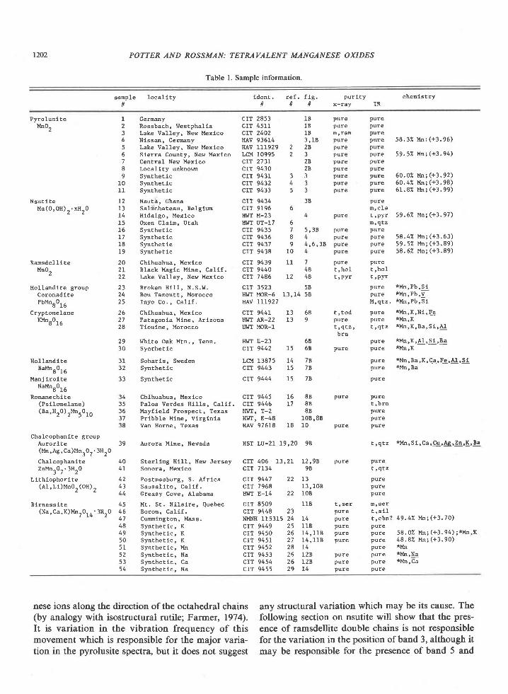
The presentation of these results follows the classi-fication scheme of Burns and Burns (1975,l977a,b),which is based on the nature of the polymerization ofMnOu units, in which six oxygens surround a centralmanganese cation in approximately octahedral coor-dination. For each structure representative spectraare included with the text. Spectra of other samplesare contained in Appendix B as indicated in Table l.
Appendix figures are indicated by an "A" or "B" fol-lowing the figure number.' The intensities of IRspectra may vary by a factor of 2 or 3 due to differ-ences in sample particle size and dispersion in thepellet. For this reason we have presented the IRspectra at concentrations such that all spectra havethe same maximum intensity in the 1400 cm-' to 200cm-' region. Each spectrum in the 4000 cm-'to 1400cm-' region is presented at 4 times the concentrationof its lower energy spectrum. The presentation in-tensities listed in the figure captions allow the origi-nal intensity of the spectra to be calculated. The pre-sentation intensity is 100 times the intensity in thefigure divided by the intensity measured using thestandard preparation techniques (Section 2).
Experimental details
Purity and mineralogy were determined by X-raypowder diffraction using CrKa radiation and aDebye-Scherrer camera. The IR spectrum of someminerals is so distinctive that after an initial correla-tion was made between the X-ray diffraction patternand the IR spectrum, further X-ray work was notneeded. When necessary, qualitative chemical analy-sis was used in addition to X-ray diffraction to deter-mine mineralogy.
IR spectra were obtained with a Perkin-Elmermodel 180 spectrophotometer on 2.0 mg of powderedsample dispersed in TlBr pellets for the 4(XX) cm-' to1400 cm-' region and on 0.5 mg in TlBr and KBr pel-lets for the 1400 cm-' to 200 cm-' region. Pellets of13 mm diameter were pressed for I minute at 19,000psi under vacuum. Pellets were prepared withoutevacuation to verify that dehydration of the manga-nese oxide did not occur under these conditions.Since KBr is hygroscopic, it was not used in the 4000cm-' to 1400 cm-' region, where water and hydroxideabsorption occurs. TlBr is preferable to KBr becauseit is non-hygroscopic. Also, because its refractive in-dex better matches most manganese oxides, it givesspectra of better quality. The figures presented arefrom TlBr pellets. Where the corresponding spec-trum in KBr differs significantly, it is included in Ap-pendix B. A vacuum dewar with KBr windows wasused to obtain IR spectra at liquid nitrogen temper-ature in the 4000 cm-' to 1400 cm-' region. Qualita-tive chemical analyses were done with an SEM
3 To receive a copy of Appendic.es A and B and a table of IRband positions, order Document AM-79-ll7 from the BusinessOffice, Mineralogical Society of America, 2000 Florida Avenue,NW, Washington, D. C. 20009. Please remit $1.00 in advance forthe microfiche.
equipped for energy dispersive X-ray analysis. Man-ganese oxidation state was determined by room tem-perature dissolution in excess 0.05M Fe2* in 0.5MHrSO4 followed by back titration of excess Fe2* with0.002M KMnOo and spectrophotometric determina-tion of total Mn as MnO;. This procedure is a modi-fication of that of Moore et al. (195O). Far-infraredspectra in the region 200 cm-' to 35 cm-' were ob-tained from a petroleum jelly mull of l0 mg ofsample spread on a polyethylene plate.
The chain structures: pyrolusite, nsutite, ramsdellite
Pyrolusire
Two structural forms of pyrolusite are thought toexist (de Woltr, 1959): a tetragonal form, which ischaracteristic of pyrolusite of primary origin, and anorthorhombic modification characteristic of pyrolu-site formed by alteration of manganite. A single-crys-tal X-ray structural determination has not been donefor pyrolusite. The tetragonal structure (Fig. l) isbased on X-ray powder diffraction data. MnOu octa-hedra share edges to form single chains linked to oneanother by shared vertices along the c axis. De Wolffpostulated the existence of an orthorhombic modifi-cation from broadening of several X-ray powderlines. Electron microscopy has revealed oriented la-mellar pores in secondary pyrolusites which could beresponsible for this distortion (Champness, 197l).
The X-ray powder patterns of Figure 2 confirm theexistence of an orthorhombic pyrolusite. The pyrolu-site patterns are arranged in order of increasingbroadness of the following lines (indices in parenthe-sis): 2.204 (200), 1.97 (210), 1.62 (2ll), 1.391 (310),1.305 (301), l.2O (202). The (301) line is superim-,posed on that of (l l2), which does not split. For py-rolusite #4 several of these lines are clearly-resolveddoublets, and the overall pattern can be indexed toan orthorhombic cell bearing the following relationto the undistorted pyrolusite cell:
tetragonal
a : 4 . 4 2 A
c : 2 . 8 7 A
orthorhombic
c : 4 . 4 4 Aa: 4 .364b : 2 . 8 7 A
The IR spectra of this pyrolusite series show con-siderable variation. The order of the spectra in Fig-ure 3 is the same as that of the powder patterns inFigure 2. The spectral variations are not well corre-lated with X-ray patterns, although two trends aresuggested in going from top to bottom in Figure 3;
l20l
the resolution of bands 3,4, and 5 improves, and theintensity of band 3 grows with respect to band 4.
All the pyrolusite powder diffraction patterns con-tain lines at 3.40,2.63,2.32, 1.78, and l.7lA. Thesecannot be indexed on the tetragonal pyrolusite cellnor on a superlattice of it. They can all be attributedto manganite and account for its four strongest lines.The +3.99 manganese oxidation state of pyrolusite#l I limits its possible manganite contamination to Ipercent. Although other pyrolusite samples couldhave up to 8 percent manganite, their unindexed X-ray lines are not measurably stronger than those ofpyrolusite #1. IR sp€ctroscopy limits the amount ofmanganite impurity to less than 5 percent for allsamples, based on the intensity of the band near I100cm-'. This feature may alternatively be attributed tosome other hydroxide ion impurity or to an overtoneof the intense absorption at lower wavenumber. Atpresent it seems likely that most pyrolusite samplescontain a small amount of manganite impurity, butwe do not feel that the evidence is conclusive.
The variation in the pyrolusite IR spectra of Fig-ure 3 is representative for pyrolusite samples in gen-eral and is a problem in its own right. The primarydifference among the spectra is the position and rela-tive intensity of band 3. Farmer (1974) has consid-ered the effect particle shape would theoreticallyhave on the IR spectra of rutile, which is isostruc-tural with pyrolusite and has a spectrum similar to it.He suggested that shape may be the cause of largevariations in the IR spectra of different powdered ru-tile samples. The major variation in going from aplaty to a needle-like morphology is predicted to be adecrease of several hundred wavenumbers in the po-sition of band 3, which is similar to the variations weobserve for pyrolusite. Although scanning electronmicroscopy did show variation in particle shape fromneedles to equant particles, this variation could begenerated for a single sample by different grindingtechniques with no effect on the position of band 3. Itthus appears that particle shape is not responsible forthe variation in pyrolusite IR spectra.
Polarized reflectance spectra of pyrolusite #5 in-dicate that bands l, 2, and 4 are polarized per-pendicular to the c axis and that band 3 is polarizedparallel to the c axis. Band 5 is not present in thespectra of pyrolusite #5, which is our only samplewith single crystals large enough to yield reflectancespectra. These results are in complete agreement withthe predictions offactor group analysis for the tetra-gonal pyrolusite structure. Band 3 is due to the dis-placement of the oxygen ions relative to the manga-
POTTER AND ROSSMAN: TETRAVALENT MANGANESE OXIDES
1202 POTTER AND ROSSMAN: TETRAVALENT MANGANESE OXIDES
Table L Sample information
sample 1oca l l t ytl
lden t . re f . fLg . pur l t y cheEis t ryll ll ll x-rav IR
Pyro lus l teMn0,
N s u t i l e
Ransde l l l teMn0,
Holland iteBaMnr0rU
Manj iroiteNt*8016
Ronanechite
8 Locality unknom9 Synthe t ic
10 Synthe t ic11 Synthe t lc
12 Nsuta, chana
15 Oxen Clain, Utaht6 Synthe t lcL7 SyntheEicl8 Synthetlc19 Synthe t lc
48 Synthe t lc , K49 Synthe t lc , K50 Synthe t ic , K5L Synthe t lc , Mn52 S)mthet lc , Na53 Synthe t ic , Ca54 SynEhet ic , Na
I Germny CIT 2853 lB pure pure2 Rossbach, Westpha l la c IT 4511 1B pure pure3 Lake Valley, New Mexlco CIT 2402 18 E'ram Pure4 Nlssan, cemny HAv 93614 3 ,1B pure Pure 58 . 32 Mn : (+3 . 96)5 Lake Valley, New Mexico HAV 111929 2 2R pure pure
5 S ie r ra County , New Mex lco LCM 10995 2 3 pure pure 59 .52 Mni (+3 .94)7 Central New Mexlco CIT 2731 2R pure pure
PUreMn(0 ,0H) . .xH^0 13 Sak ichateau, Be lg lu CIT 9196 6 n ,c la
14 H ida lgo , Mex lco I I I f f M-23 4 pure t ,PYr 59 .62 Mn; (+3 .97)
I lo l land i te g roup 23 Broken H111, N.S.W. CIT 3523 58Coronad l te?bMn80t6
CryptonelaneKMn^0. .
d r b
29 l, lhtte oak Mtn., Tenn. HwT E-23 5830 Synthetic
31 Soharls, Sweden32 Synthe t lc
33 Synthetlc
CIT 9430 2R pure pureCIT 9431 3 3 pure pure 60.02 ! ' tn; (+3.92)CIT 9432 4 3 pure pure 50.42 Mn;(+3.98)CIT 9433 5 3 pure pure 61.82 l 'h ; (+3. 99)
ctT 9434 38
l{IilT UT-17 6 E , q r zCIT 9435 1 5,3R pure pureCIT 9436 8 4 pure pure 58.42 un;(+3.63)C IT 9437 9 4 ,5 ,38 pu re pu re 59 .52 Mn i (+3 .89 )CIT 9438 10 4 pure pure 58.62 Mn;(+3.89)
pure *Mn,Pb,S ipure *Mn,Pb,V
M , q t z . * M n , P b , 5 1
t , q t z , t , q t z * l ' t n , K , B a , S t ' A !
brnp u r e * M n , K , A 1 , 5 1 , B a
20 Chlhuahua, Mexlco cIT 9439 ll 7 pure Pure2I B lack Mag lc Mine , Ca l l f . C IT 9440 48 t 'ho l t ' ho l22 Lake Valley, New Mexlco CIT 7486 L2 48 t 'pyr t,PYr
24 Bou Tazoutt, Morocco HI'IT MOR-5 13,14 582 5 I n y o C o , , C a l l f . nAv 111927
26 Ch lhuahua, Mex ico CIT 9441 13 68 ! , tod pure * l ' l n ,K ,N1,Fe
27 Patagonia Mine, Arizona HWT AR-22 13 9 pure Pure *ltn,K
28 Tioulne, Morocco HI''T MOR-1
CIT 9442 15 68 pure pure *Mn,K
CIT 9444 15 7R pure
34 Chihuahua, Mexlco CIT 9445 15 8B pure Pure
LCM 13875 14 7Rcr"t 9443 15 7R
p u r e * M n , B a , K , C a , F e , A l , S lpure *l in, Ba
t , b r npurepure
t , q t z * M n , S 1 , C a , C u , 4 g , Z n , K ' B e
t , q t z
Purepurepure
CIT 9449 25 11B pure pureC I T 9 4 5 0 2 6 1 4 , 1 1 8 p u r e p u r e 5 8 . 0 2 M n ; ( + 3 . 9 4 ; ; * y . ' X
CIT 9451 27 14 , 11B pure Pure 48 . 82 Mn; (+3 . 90)crr 9452 28 L4 pure *Mn
CIT 9453 26 12B pure pure *l ln,!9
CTT 9454 26 128 pure pure tMn,ca
c IT 9455 29 14 pure pure
38 Van Horne, Texaa HAv 97518 18 10 pure pure
Chalcophanlte groupAuror l te 39 Aurora Mlne , Nevade HST LU-21 19 ,20 98(Mn ,Ag ,Ca )Mnr0 r . 3Hr0
Chalcophani te 40 Ster l lng H111, New Jersey CIT 406 13,21 L2,98 pure pureZnMnrOr .3HrO 4 I Sonora , Mex lco crr 7134 98
(Ps i lone lane) 35 Pa los Verdes H111s, Ca1 l f . C IT 9446 L7 88(Ba,Hr0) rMn.0r^ 36 Mayf ie ld Prospec t , Texas HwT, T-2 88
37 Pr lbb le Mlne , v l rg ln la H! rT ' E-48 10B '8B
L i th lophor l te 42 Pos tmasburg , S . A f r l ca CIT 9447 22 13( A 1 , L i ) M n 0 " ( O H ) , 4 3 S a u s a l i t o , C a l i f . C I T 7 9 6 8 1 3 , 1 0 8
44 Greasy Cove, Alabama HWT E-14 22 I0B
45 Mt , s t . H i la l re , Quebec CIT 8509 118 t ' ser E ,se lB l rness i te( N a , C a , K ) M n , 0 , r ' 3 H . 0 4 6 B o r o n , C a l i f . C I T 9 4 4 8 2 3 p u r e t , s l l
t Lq ' 47 cumington , Mass , NMNH 115315 24 L4 pure t ,cba? 49 .4"1 Mn; (+3 .70)
nese ions along the direction of the octahedral chains any structural variation which may be its cause. The(by analogy with isostructural rutile; Farner, 1974). following section on nsutite will show that the pres-It is variation in the vibration frequency of this enoe of ramsdellite double chains is not responsiblemovement which is responsible for the major varia- for the variation in the position of band 3, although ittion in the pyrolusite spectra, but it does not suggest may be responsible for the presence of band 5 and
POTTER AND ROSSMAN: TETMVALENT MANGANESE OXIDES
Table l. (continued)
1203
Todorok i te g roup 55Todorok i te 56( M n , C a , M g ) M n i 0 7 . H r 0 5 1
) 6
5 9606 1
Woodru f f l te 62( Z n , M n ) M n ^ 0 - . H ^ 0
J t z
Ranc le l te 63(Ca ,Mn)Mn, 0^ ' 3H^0 64q t z
6 56 6
B u s e r l t e(Na, ! , ln )Mnr0r .xHr0
Pa los Verdes H i I Is , Ca l i f .Chihuahua, MexicoChihuahua, MexicoBonbay, IndiaMlramati Mine, JapanCharco Redondo, CubaUnknom locallty
Ster l lng H111, New Jersey
Putonrs Cave, V i rg ln laRanc i€ Mtns . , F rance
Or ien ta Prov ince , CubaI t ia , Greece
Synthe t i cSynthe t lc , CoSynthe t lc , Mn
cYI 9456 L7 15,138crr 9457 11 138crr 9458 11 15crr 9459 15crT 9460 138crT 9733 30 15HWT Mn-24 30 15
NMNH 114158 31 138
E r S l r {puret , s l lpurem,ngnpurepure
pure *Mn,Zn,Cu,Ca,A l
Puret , s i l
m , b r n
PUre
PurePurePUre
67b d
69
NMNH 120601NMNH 128319
HAV 110334crT 9430
ctT 9622crr 9623crT 9617
L7 pure
, l l t , t o d
1 7 , 1 4 8 n , b r n148
16 pureI5B pure1 5 B m , b i r ,
lnp
2T323 233
343434
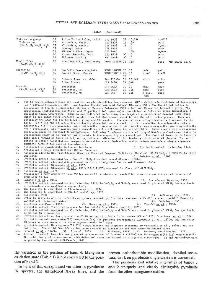
1. The fo l low ing abbrev ia t ions are used fo r sap le ldent l f i ca t ion numbers : CIT = Ca l i fo rn la Ins t i tu te o f Techno logy ,HAV = Harvard UnLversity, LCM = Los Angeles County ltuseun of Natural Hlstory, HWI = The Hewett Collectlon lnPossess ion o f the U. S , Geo log ica l Survey a t Denver , Co lorado, NMNH = Nat iona l l , luseun o f Natura l H is lo ry , TheSmi thson ian Insc l tu t ion ; fo r X- ray and IR pur l ty M ind lca tes na jo r lnpur i t les , m ind ica tes mlnor inpur l t les , tlnd ica tes t race lnpur i t ies . Samples are lnd ica ted to be X- ray puredesp i te the presence o f severa l weak l lneswhlch do no t match o ther s tandard sanp les prov ided tha t these cannot be a t t r ibu ted to o ther phases . Th ls wasgenera l l y the case fo r the ho l land l te g roup and b l rness l te . The spec la l case o f pyro lus l te 1s d lscussed in thetex t ' For x - ray and IR pur l ty the fo l low ing abbrev la t ions are used: b l r = b l rness l te , b rn = braun i le , cbn -carbonate , c la = c lay n inera ls , ho l = ho l l .and i te , inp = un ident i f ied impur i ty , mgn = nangan i te , pyc = pyrochro l te ,pyr = pyro lus i te , qLz = quar tz , ser = serand i te , s i1 = s i l l ca te , tod = todorok l te . Under chemis t ry the nanganeaeox ida t ion s ta te l s enc losed l -n Darentheses . Fo l low ing * , e lements de tec ted bv qua l i ta t i ve ana lys is a re l l s ted lnapProx ina te o rder o f concent ra t lon ; e lements in t race quant l t y a re under l ined . The chen ica l fomulae lnc luded lnthis table should be taken as only an approxlmate lndication of the chemistry of the nineral phases. The vari-a l lons in ca t lon subs t i tu t ion , manganese ox ida t lon s ta te , hydra t ion , and s t ruc tu re p rec lude a s lng le r lgorouschen lca l fo rmula fo r many o f the minera ls ,
2 . M is labe led as ramsde l l i te in the co l lec t ions . 3 . Synthe t ic ne thod: McKenz ie , 1971.4 , Ar t i f i c ia l 6 -Mn02 i f ron Nat lona l Carbon Company.5 . Manganese d iox ide HP; f ron DLanond Shamrock Chen ica l Companv, Ba l t imore , Mary land; 99 .9"A| f t iO2,0 .0052 Fe by the l r
a n a l y s i s . 6 , z w i c k e r e t a r . , 1 9 6 2 .7 . Synthe t ic ne thod: p repara t lon a fo r q " - Mnor f rom Gat tow and Glenser , 1961a.8 ' Synthe t ic method: e lec t ro ly t i c p repara t ion f i r n - Mn02 f rom ca t tow and Glenser , 196Ia .9 . S y n t h e t i c n e t h o d : G i o v a n o l i e t a l . , 1 9 6 7 .10 . Synthe t ic method; c lovano l i e t a1 . , 1967, IO.O N HNO3 was used in p lace o f 1 .0 N HNO3.1 1 . F i n k e l m a n e t a l . . ) . 9 7 4 ,L2 . Apparent ly a poor sanp le o f Lake Va l ley ransde l l l te s ince the ransde l l l te s t ruc tu re was de temlned on na ter ia l
f rom Lake Va l tey .13 , pa lanche er a1 . , 1944. 14 . Bysr r i jm and Bysr r6n , 1950.15 . S)mthet ic ne thod: p repara t ion / /1 , McKenz ie , 1971; Ba(MnOq)2 and NaMnO4 were used in p lace o f KMn04 fo r syn thes is
o f ho l land i te and mnj i ro i te respec t ive ly .16 . The loca l i t y i s descr ibed in F lnke lman e t aL , , 1974.17 . The loca l l t y i s descr lbed in Mi rche l an i -C6: rey , 1973.1 8 , F l e i s c h e r , 1 9 6 0 1 9 . R a d t k e e t a l . , 1 9 6 7 .20 . P l io r to ana lys is ma jor ca lc i te tnpur i ty was removed by 10 n inu te t rea tment w l th d l lu te ace t lc ac id fo l lowed by
wash ing w l th de ion ized water : 2L . Wads ley , 1955.2 2 . F l e i s c h e r a n d F a u s t , 1 9 6 2 . 2 3 . B r o m e r a l . , 1 9 7 1 . 2 4 . F r o n d e l e t a I . , 1 9 6 0 a .25 . Synthe t lc ne thod: The f i rs t p repara t ion fo r 6 -MnO2 f rom Glenser e t a l . , 1951.26 ' Synthe t ic ne lhod: p repara t lon /11 , McKenz ie , 1971; Ca(MnOa)2 and Na l lnOq were used in p lace o f KMn04 fo r syn thes ls
o f Ca and Na b i rness i tes .Synthes is ne thod: as fo r p repara t ion / , l8 (Buser e t a l . , Tab le 4 ) bu t no les HCl = 0 .125; f rom Buser e t a l . , 1954.Synthe t ic ne thod: nanganese( I l l ) manganate ( IV) uas prepared accord ing to G iovano l l e t a l . , 1970b, and was dr ied10 hours a t room tempera ture under approx imate ly IO-5 to r r .Syntbe t ic method: Na mrnganese( I I , I I I ) nanganate( IV) was prepared accord ing to c iovano l - i e t a l . , 1970a, bu t wasnot d r ied ' The so l id f rom OoC ox ida t lon vas washed by f i l t ra t ion and kept under de ion ized water .Fronde l ec a l . ' 1960b. 31 . Fronde l , 1953. 32 . R ichnond, 1969. 33 . Bardossy and Br indLey , 1978.Synthe t ic method: buser i te was prepared by the method o f G lovano l i (1970a) fo r Na manganese( I I , I I f ) nanganate( IV) .The ox ida t ion produc t was washed w l th de ion ized water and s to red as an aqueous suspens lon . Co and l , ln ana logs wereprepared by the ne thod o f MeKenz ie . 197I .
28 .
29 .
3 0 .3 4 .
the variation in the position of band 4. Manganeseoxidation state (Table l) is not correlated to the posi-tion of band 3.
In light of this unexplained variation in pyrolusiteIR spectra, the unindexed X-ray lines, and the
proven orthorhombic modification, detailed struc-tural work on pyrolusite single crystals is warranted.
The positions and relative intensities of bands Iand 2 uniquely and clearly distinguish pyrolusitefrom the other manganese oxides.
POTTER AND ROSSMAN: TETRAVALENT MANGANESE OXIDES
pyro lus i te
Eromsde l l i te
E
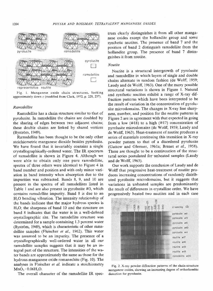
Fig. l . Manganese oxide chain structures, lookingapproximately down c (modified from Clark, 1972, p.228,237).
Ramsdellite
Ramsdellite has a chain structure similar to that ofpyrolusite. In ramsdellite the chains are doubled bythe sharing of edges between two adjacent chains;these double chains are linked by shared vertices(Bystrom, 1949).
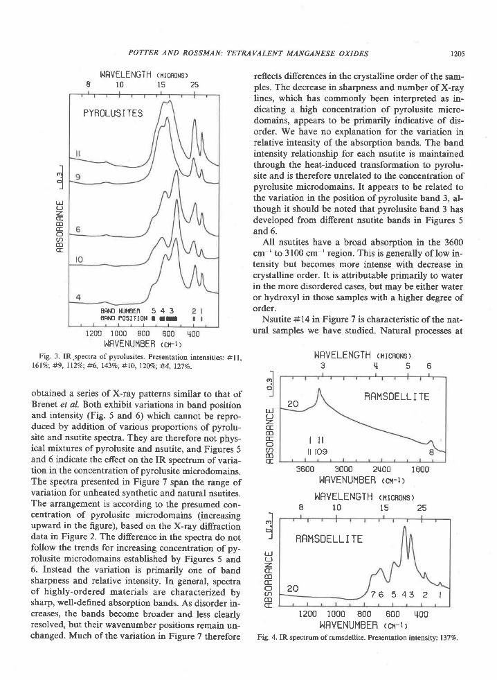
Ramsdellite has been thought to be the only otherstoichiometric manganese dioxide besides pyrolusite.We have found that it invariably contains a singlecrystallographically-ordered water. The IR spectrumof ramsdellite is shown in Figure 4. Although wewere able to obtain only one pure ramsdellite,spectra of three others were identical to Figure 4 inband number and position and with only minor vari-ation in band intensity when absorption due to theimpurities was subtracted. Bands 8, 9, and l0 arepresent in the spectra of all ramsdellites listed inTable I and are also present in pyrolusite #3, whichcontains ramsdellite impurity. Band 8 is due to anHrO bending vibration. The intensity relationship ofthe bands indicate that the major hydrous species isHrO; the sharpness of band l0 and the structure onband 8 indicates that the water is in a well-definedcrystallographic site. The ramsdellite structure wasdetermined for a sample containing 1.3 percent water
@ystrom, 1949), which is characteristic of other rams-dellite samples (Fleischer et al., 1962). This waterwas assumed to be an impurity. The presence of acrystallographically well-ordered water in all ourramsdellite samples suggests that it may be an in-tegral part of the structure. The intensities of the wa-ter bands are approximately the same as those for thehydrous manganese oxide romanechite (Fig. l0). Theanalyses in Fleischer et al. indicate a stoichiometryMnO,'0.06H,O.
The overall character of the ramsdellite IR spec-
trum clearly distinguishes it from all other manga-nese oxides except the hollandite group and somesynthetic nsutites. The presence of band 7 and theposition of band 2 distinguish ramsdellite from thehollandite group. The presence of band 7 distin-guishes it from nsutite.
Nsutite
Nsutite is a structural intergrowth of pyrolusiteand ramsdellite in which layers of single and doublechains alternate in random fashion (de Wol-ff, 1959;Laudy and de Wolff, 1963). One of the many possiblestructural variations is shown in Figure l. Naturaland synthetic nsutites exhibit a range of X-ray dif-fraction patterns which have been interpreted to bethe result of variation in the concentration of pyrolu-site microdomains. The changes in X-ray line sharp-ness, number, and position for the nsutite patterns inFigure 2 arcin agreement with that expected in goingfrom a low (#18) to a high (#17) concentration ofpyrolusite microdomains (de Wolff, 1959; Laudy andde Wolff, 1963). Heat-treatment of nsutite produces aseries of materials continuing this transition in X-raypowder pattern to that of a disordered pyrolusite(Gattow and Glemser, l96la; Brenet et al., 1958).These are thought to be a continuation of the struc-tural series postulated for unheated samples (Laudyand de Woltr, 1963).
Our work supports the conclusion of Laudy and deWolff that progressive heat-treatment of nsutite pro-duces increasing concentrations of randomly distrib-uted pyrolusite microdomains, but it suggests thatvariations in unheated samples are predominantlythe result of differences in crystalline order. We haveprogressively heated two nsutites and in each case
Fig. 2. X-ray powder diffraction patterns of the chain-structuremanganese oxides, showing an increasing degree of orthorhombicdistortion for pyrolusite.
pyro lus i le
. , ' H .;
PYBOLUSI TES
E N N O N U H B E R 5 4 3ENNO FOSITION T II
POTTER AND ROSSMAN:
NRVELENGTH (HrcBoNs)8 l 0 l s 2 5
1200 1000 800 600 q00I,,lFVENUMBER <or-t>
Fig. 3. IR.spectra of pyrolusites. Presentation intensities: #ll.16l%o: #9, ll2Vo; #6, l43%o; #10, l2Vo; #4, l27%o.
obtained a series of X-ray patterns similar to that ofBrenet et al. Both exhibit variations in band positionand intensity (Fig. 5 and 6) which cannot be repro-duced by addition of various proportions of pyrolu-site and nsutite spectra. They are therefore not phys-ical mixtures of pyrolusite and nsutite, and Figures 5and 6 indicate the effect on the IR spectrum of varia-tion in the concentration of pyrolusite microdomains.The spectra presented in Figure 7 span the range ofvariation for unheated synthetic and natural nsutites.The arrangement is according to the presumed con-centration of pyrolusite microdomains (increasingupward in the figure), based on the X-ray diffractiondata in Figure 2. The diference in the spectra do notfollow the trends for increasing concentration of py-rolusite microdomains established by Figures 5 and6. Instead the variation is primarily one of bandsharpness and relative intensity. In general, spectraof highly-ordered materials are characterized bysharp, well-defined absorption bands. As disorder in-creases, the bands become broader and less clearlyresolved, but their wavenumber positions remain un-changed. Much of the variation in Figure 7 therefore
TE7:RAVALENT MANGANESE OXIDES I2O5
reflects differences in the crystalline order of the sam-ples. The decrease in sharpness and nu'r,ber of X-raylines, which has commonly been interpreted as in-dicating a high concentration of pyrolusite micro-domains, appears to be primarily indicative of dis-order. We have no explanation for the variation inrelative intensity of the absorption bands. The bandintensity relationship for each nsutite is maintainedthrough the heat-induced transformation to pyrolu-site and is therefore unrelated to the concentration ofpyrolusite microdomains. It appears to be related tothe variation in the position of pyrolusite band 3, al-though it should be noted that pyrolusite band 3 hasdeveloped from different nsutite bands in Figures 5and 6.
All nsutites have a broad absorption in the 3600cm-' to 3100 cm-' region. This is generally of low in-tensity but becomes more intense with decrease incrystalline order. [t is attributable primarily to waterin the more disordered cases, but may be either wateror hydroxyl in those samples with a higher degree oforder.
Nsutite #14 in Figure 7 is characteristic of the nat-ural samples we have studied. Natural processes at
NRVELENGTH <rrcnoHs>3q56
BRMSDELLI TE20
l t lil ro9
s600 3000 2q00 1800NFVENUMBEB <or-1>
NRVELENGTH <nrcnoHsl8101525
BRMSDELL I TE
207 6 5 4 3 2 |
1200 1000 800 600 q00NFVENUMBEB <or-1:
Fig. 4. IR spectrum oframsdellite. Presentation intensity: 1377o.
-]ol
u.J(Jz.CE6CtrclaCDCE
I(r'Iol
I
l!(Jz.CEcoccOamCE
IololI
UJ(Jz.CEm(EOacnCE
HERT-TBERTEDNSUTI TE
1206
[^IRVELENGTH <NrcnoNs r8 1 0 1 5 2 s
1200 1000 800 600 '{00I,,IRVENUMBEB <cx-1>
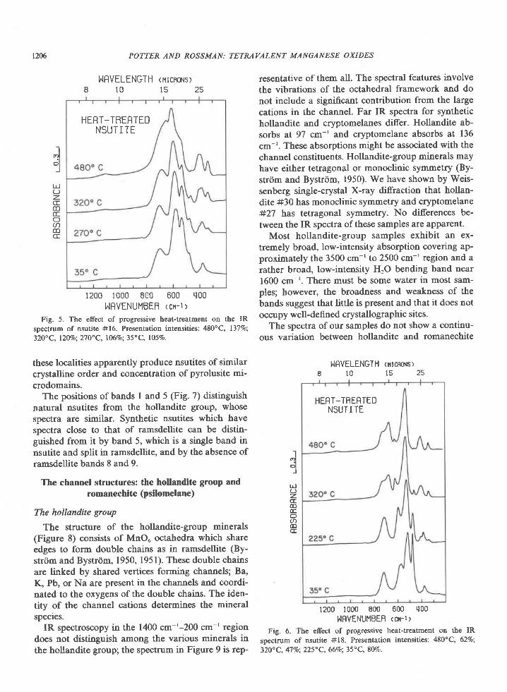
Fig. 5. The effect of progressive heat-treatment on the IRspectrum of nsutite #16. Presentation intensities: 48O"C, l37%o;32O" C, l20%o; 21 O" C, l06vo; 35" C, 1059o.
these localities apparently produce nsutites of similarcrystalline order and concentration of pyrolusite mi-crodomains.
The positions of bands I and 5 (Fig. 7) distinguishnatural nsutites from the hollandite group, whosespectra are similar. Synthetic nsutites which havespectra close to that of ramsdellite can be distin-guished from it by band 5, which is a single band innsutite and split in ramsdellite, and by the absence oframsdellite bands 8 and 9.
The channel structures: the hollandite group andromanechite (psilomelane)
The hollandite group
The structure of the hollandite-group minerals(Figure 8) consists of MnOu octahedra which shareedges to form double chains as in ramsdellite (By-strtjm and Bystrdm, 1950, 1951). These double chainsare linked by shared vertices forming channels; Ba,K, Pb, or Na are present in the channels and coordi-nated to the oxygens of the double chains. The iden-tity of the channel cations determines the mineralspecies.
IR spectroscopy in the 1400 cm-'-200 cm-' regiondoes not distinguish among the various minerals inthe hollandite group; the spectrum in Figure 9 is rep-
POTTER AND ROSSMAN: TETRAVALENT MANGANESE OXIDES
J:
UJOz.CEcoccO(ncoCE
resentative of them all. The spectral features involvethe vibrations of the octahedral framework and donot include a significant contribution from the largecations in the channel. Far IR spectra for synthetichollandite and cryptomelanes differ. Hollandite ab-sorbs at 97 cm-' and cryptomelane absorbs at 136cm-'. These absorptions might be associated with thechannel constituents. Hollandite-group minerals mayhave either tetragonal or monoclinic symmetry (By-strrim and Bystrdm, 1950). We have shown by Weis-senberg single-crystal X-ray diffraction that hollan-dite #30 has monoclinic symmetry and cryptomelane#27 has tetragonal symmetry. No differences be-tween the IR spectra of these samples are apparent.
Most hollandite-group samples exhibit an ex-tremely broad, low-intensity absorption covering ap-proximately the 3500 cm-' to 2500 cm-' region and arather broad, low-intensity HrO bending band near1600 cm-'. There must be some water in most sam-ples; however, the broadness and weakness of thebands suggest that little is present and that it does notoccupy well-defined crystallographic sites.
The spectra of our samples do not show a continu-ous variation between hollandite and romanechite
NRVELENGTH (iltcnoNs)8 1 0 1 5 2 5
1200 1000 800 600 q00
!.IFVENUMBEB <Cr-I >Fig. 6. The effect of progressive heat-treatment on the IR
spectrum of nsutite #18. Presentation intensities: 480"C, 62Vo;
320o C, 47 Vo; 225" C, 66Vo; 35" C, 8Ma
Jqlrl
zCE(D(Eo(t)(DCE
HERT-TRERTEDNSUT I TE
NSUT I TES
BFI.IO MJI{BER 5BRI'ID POSITION T
POTTER AND ROSSMAN:
]^IRVELENGTH <rrcnors I8 1 0 1 5 2 5
1200 1000 800 600 q00NRVENUMBEB <cr-r>
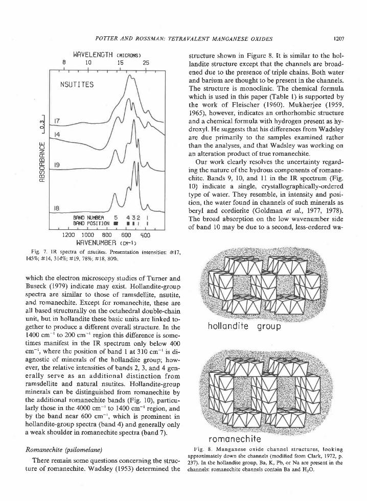
Fig. 7. IR spectra of nsutites. Presentation intensities: #17,l45%o; #l4, 3l4Vo; #19, 7 8Vo: #18, 807o.
which the electron microscopy studies of Turner andBuseck (1979) indicate may exist. Hollandite-groupspectra are similar to those of ramsdellite, nsutite,and romanechite. Except for romanechite, these areall based structurally on the octahedral double-chainunit, but in hollandite these basic units are linked to-gether to produce a different overall structure. In the1400 cm-' to 200 cm-' region this difference is some-times manifest in the IR spectrum only below 400cm-', where the position of band I at 310 cm-'is di-agnostic of minerals of the hollandite group; how-ever, the relative intensities of bands 2, 3, and 4 gen-erally serve as an additional distinction fromramsdellite and natural nsutites. Hollandite-groupminerals can be distinguished from romanechite bythe additional romanechite bands (Fig. l0), particu-larly those in the 4000 cm-' to 1400 cm-'region, andby the band near 600 cm-', which is prominent inhollandite-group spectra (band 4) and generally onlya weak shoulder in romanechite spectra (band 7).
Romane c hit e (p sil o mel ane )There remain some questions concerning the struc-
ture of romanechite. Wadsley (1953) determined the
TETRAVALENT MANGANESE OXIDES
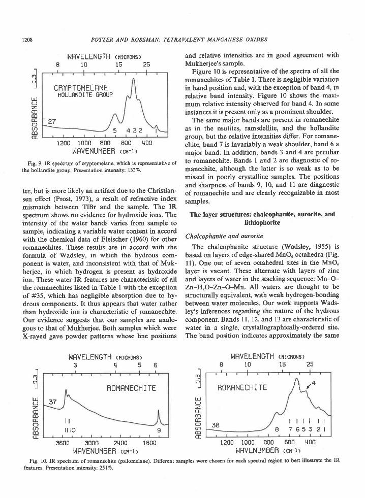
structure shown in Figure 8. It is similar to the hol-landite structure except that the channels are broad-ened due to the presence of triple chains. Both waterand barium are thought to be present in the channels.The structure is monoclinic. The chemical formulawhich is used in this paper (Table l) is supported bythe work of Fleischer (1960). Mukherjee (1959,1965), however, indicates an orthorhombic structureand a chemical formula with hydrogen present as hy-droxyl. He suggests that his differences from Wadsleyare due primarily to the samples examined ratherthan the analyses, and that Wadsley was working onan alteration product of true romanechite.
Our work clearly resolves the uncertainty regard-ing the nature of the hydrous components of romane-chite. Bands 9, 10, and I I in the IR spectrum (Fig.l0) indicate a single, crystallographically-orderedtype of water. They resemble, in intensity and posi-tion. the water found in channels of such minerals asberyl and cordierite (Goldman et al., 1977, 1978).The broad absorption on the low wavenumber sideof band l0 may be due to a second, less-ordered wa-
Fig. 8. Manganese oxide channel structures, lookingapproximately down the channels (modified from Clark, 1972, p.237). kr the hollandite group, Ba, K, Pb, or Na are present in thechannels: romanechite channels contain Ba and H,O.
l2u
Iol
lrJL)z.CE(D(EOacoCE
hol londi te group
romonech i te
1208 POTTER AND ROSSMAN:
NRVELENGTH <rtcnoHs>810152s
CBYPTOMELRNEHOLLRNOITE GffOUP
275 4 3 2
1200 1000 800 600 tl00NRVENUMBEB <cx-l>
Fig. 9. IR spectrum of cryptomelane, which is representative ofthe hollandite group. Presentation intensity: 1337o.
ter, but is more likely an artifact due to the Christian-sen effect (Prost, 1973), a result of refractive indexmismatch between TlBr and the sample. The IRspectrum shows no evidence for hydroxide ions. Theintensity of the water bands varies from sample tosample, indicatin8 a variable water content in accordwith the chemical data of Fleischer (1960) for otherromanechites. These results are in accord with theformula of Wadsley, in which the hydrous com-ponent is water, and inconsistent with that of Muk-herjee, in which hydrogen is present as hydroxideion. These water IR features are characteristic of allthe romanechites listed in Table I with the exceptionof #35, which has negligible absorption due to hy-drous components. It thus appears that water ratherthan hydroxide ion is characteristic of romanechite.Our evidence suggests that our samples are analo-gous to that of Mukherjee. Both samples which wereX-rayed gave powder patterns whose line positions
hIFVELENGTH <rrcnoHsr3 r l 5
BOMRNECHI TE
37
t li l l o
3600 3000 2tt00 1800I^IFVENUMBEB tcx-1>
TETRAVALENT MANGANESE O XI DES
and relative intensities are in good agreement withMukherjee's sample.
Figure l0 is representative of the spectra of all theromanechites of Table l. There is negligible variationin band position and, with the exception of band 4, inrelative band intensity. Figure l0 shows the maxi-mum relative intensity observed for band 4. In someinstances it is present only as a prominent shoulder.
The same major bands are present in romanechiteas in the nsutites. ramsdellite. and the hollanditegroup, but the relative intensities differ. For romane-chite, band 7 is invariably a weak shoulder, band 6 amajor band. In addition, bands 3 and 4 are peculiarto romanechite. Bands I and 2 are diagnostic of ro-manechite, although the latter is so weak as to bemissed in poorly crystalline samples. The positionsand sharpness ofbands 9, 10, and ll are diagnosticof romanechite and are clearly recognizable in mostsamples.
The layer structures: chalcophanite, aurorite, andlithiophorite
Chalcophanite and aurorite
The chalcophanite structure (Wadsley, 1955) isbased on layers of edge-shared,MnO. octahedra (Fig.ll). One out of seven octahedral sites in the MnO"layer is vacant. These alternate with layers of zincand layers of water in the stacking sequence: Mn-O-Zn-HrO-Zn-O-Mn. All waters are thought to bestructurally equivalent, with weak hydrogen-bondingbetween water molecules. Our work supports Wads-ley's inferences regarding the nature of the hydrouscomponent. Bands ll, 12, and 13 are characteristic ofwater in a single, crystallographically-ordered site.The band position indicates approximately the same
NRVELENGTH <rtcnonsr810 l s25
ROMRNECHITE I \ ,O
38r t l l r l
a 7 6 5 3 2 1
1200 1000 800 600 q00
I^IFVENUMBEB <cx-lr
dl t l(JzCEm(tr(tra6CE
I.lol
Lrl(Jz.cfcoccOacoCE
dul(JzCEm(Eo(nCDCE
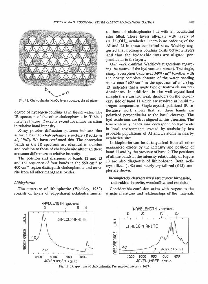
Fig. 10. IR spectrum of romanechite (psilomelane). Diferent samples were chosen for each spectral region to best illustrate the IRfeatures. Presentation intensitv: 251%.
POTTER AND ROSSMAN: TETMVALENT MANGANESE OXIDES
b\--o
Fig. ll. Chalcophanite MnO5, layer structure, the aD plane.
degree of hydrogen-bonding as in liquid water. TheIR spectrum of the other chalcophanite in Table Imatches Figure 12 exactly except for minor variationin relative band intensity.
X-ray powder diffraction patterns indicate thataurorite has the chalcophanite structure (Radtke etal., 1967). We have confirmed this. The absorptionbands in the IR spectrum are identical in numberand position to those of chalcophanite although thereare some differences in relative intensity.
The position and sharpness of bands 12 and 13and the sequence of four bands in the 550 cm-' to4@ cm-' region distinguish chalcophanite and auro-rite from all other manganese oxides.
Lithiophorite
The structure of lithiophorite (Wadsley, 1952)consists of layers of edge-shared octahedra similar
NRVELENGTH <nrcnors>3 ' l 5
CHRLCOPHRNITE
40
t3t2
3500 3000 2'100 1E00IIRVENUMBEB <cx-l>
to those of chalcophanite but with all octahedralsites filled. These layers alternate with layers of(Al,LiXOH)o octahedra. There is no ordering of theAl and Li in these octahedral sites. Wadsley sug-gested that hydrogen bonding exists between layersand that the hydroxide ions are aligned per-pendicular to the layers.
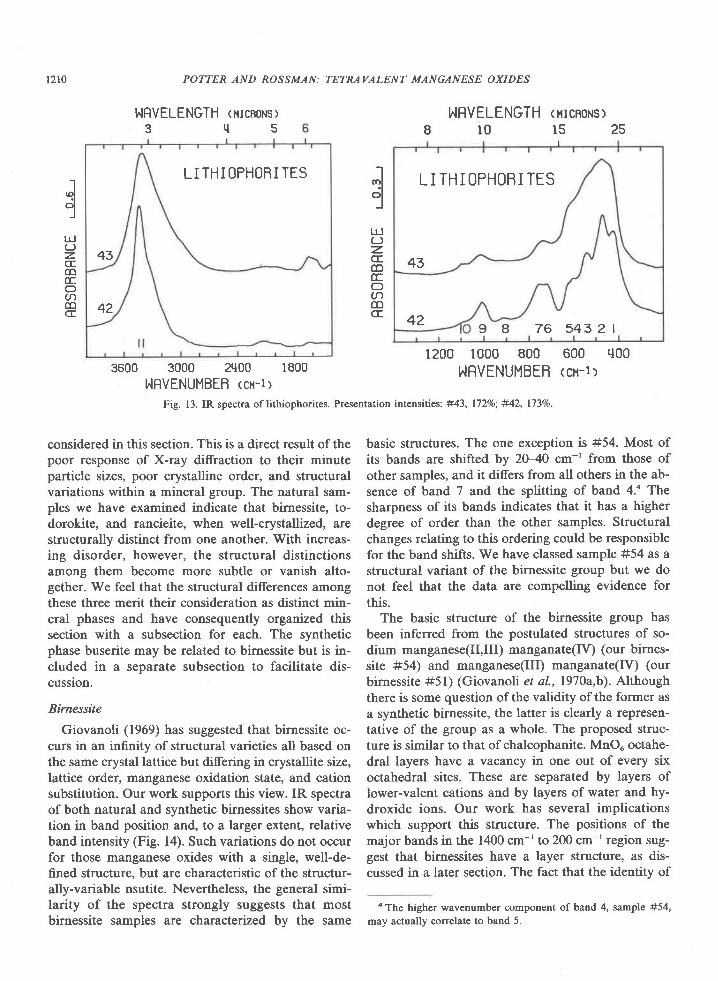
Our work gsnfirms Wadsley's suggestions regard-ing the nature of the hydrous component. The single,sharp, absorption band near 3400 cm-' together withthe nearly complete absence of the water bendingmode near 1600 cm-' in the spectrum of #42 (Fig.13) indicates 11p1a single type of hydroxide ion pre-dominates. In addition, in the well-crystallizedsample there are two weak shoulders on the low-en-ergy side of band I I which are resolved at liquid ni-trogen temperature. Single-crystal, polarized IR re-flectance work shows that all three bands arepolarized perpendicular to the basal cleavage. Thehydroxide ions are thus aligned in this direction. Thelower-intensity bands may oorrespond to hydroxidein local environments created by statistically lessprobable populations of Al and Li atoms in nearbyoctahedral sites.
Lithiophorite can be distinguished from all othermanganese oxides by the intensity and position ofband I I and by the presence ofband 9. The positionsof all the bands in the intensity relationship of Figure13 are also diagnostic of lithiophorite. Both well-crystallized (#42) and poorly-crystallized (#43) sam-ples are shown.
Incompletely characterized structures: birnessite,todorokite, buserite, woodruffite, and rancieite
Considerable confusion exists with respect to thestructural natures and relationships of the materials
NRVELENGTH <rtcnoHsl8101525
CHRLCOPHRN I TE
40 lo 947 6543 2l
1200 1000 800 600 q00
hIRVENUMBER <cn-r >
-lr.rl-l
l-r.l(J=CECDccacoCE
dUJ
z.CECD(EoacoCE
Fig. 12. IR spectrum ofchalcophanite. Presentation intensity: 16l%.
1210 POTTER AND ROSSMAN:TETRAVALENT MANGANESE OXIDES
",lcilJ
lrl(Jz.CECDE.oa(DCE
dlrl(JzCEco(tOacctCE
NRVELENGTH <rtcnoHsl3 t l 5
LI THI OPHOBI TES
43
42
3600 3000 2q00 1800I,,IFVENUMBEB ccx-l >
Fig. 13. IR spectra oflithiophorites.
NRVELENGTH <ucnoNs>8101525
L I THI OPHORI TES
43
429 8 7 6 5 4 3 2 1
1200 1000 800 600 q00hIRVENUMBEB <cx-l>
Presentation intensities: #43, l72Vo; #42, l73%o.
considered in this section. This is a direct result of thepoor response of X-ray diffraction to their minuteparticle sizes, poor crystalline order, and structuralvariations within a mineral group. The natural sam-ples we have examined indicate that birnessite, to-dorokite, and rancieite, when well-crystallized, arestructurally distinct from one another. With increas-ing disorder, however, the structural distinctionsamong them become more subtle or vanish alto-gether. We feel that the structural differences amongthese three merit their consideration as distinct min-eral phases and have consequently organized thissection with a subsection for each. The syntheticphase buserite may be related to birnessite but is in-cluded in a separate subsection to facilitate dis-cussion.
Birnessite
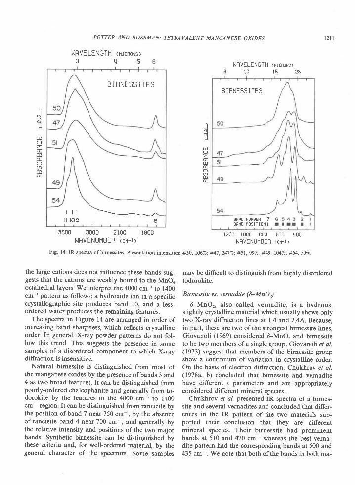
Giovanoli (1969) has suggested that birnessite oc-curs in an infinity of structural varieties all based onthe same crystal lattice but difering in crystallite size,lattice order, manganese oxidation state, and cationsubstitution. Our work supports this view. IR spectraof both natural and synthetic birnessites show varia-tion in band position and, to a larger extent, relativeband intensity (Fig. l4). Such variations do not occurfor those manganese oxides with a single, well-de-fined structure, but are characteristic of the structur-ally-variable nsutite. Nevertheless, the general simi-larity of the spectra strongly suggests that mostbirnessite samples are characterized by the same
basic structures. The one exception is #54. Most ofits bands are shifted by 2040 cm-' from those ofother samples, and it differs from all others in the ab-sence of band 7 and the splitting of band 4.' Thesharpness of its bands indicates that it has a higherdegree of order than the other samples. Structuralchanges relating to this ordering could be responsiblefor the band shifts. We have classed sample #54 as astructural variant of the birnessite group but we donot feel that the data are compelling evidence forthis.
The basic structure of the birnessite group hasbeen inferred from the postulated structures of so-dium manganese(Il,Ill) manganate(IV) (our birnes-site #54) and manganese(Il! manganate(IV) (ourbirnessite #51) (Giovanoli et al.,l970a,b). Althoughthere is some question of the validity of the former asa synthetic birnessite, the latter is clearly a represen-tative of the group as a whole. The proposed struc-ture is similar to that of chalcophanite. MnOu octahe-dral layers have a vacancy in one out of every sixoctahedral sites. These are separated by layers oflower-valent cations and by layers of water and hy-droxide ions. Our work has several implicationswhich support this structure. The positions of themajor bands in the 1400 cm-' to 200 cm-r region sug-gest that birnessites have a layer structure, as dis-cussed in a later section. The fact that the identity of
a The higher wavenumber component of band 4, sample #54,
may actually correlate to band 5.
BIBNESS I TES
l l l - - -= - - - -_J- .
i l t o g I
POTTER AND ROSSMAN: TETMVALENT MANGANESE OXIDES t2tl
NRVELENGTH <urcnorus>3 r l 5
3600 3000 21100 1800HRVENUMBER <cu-r>
the large cations does not influence these bands sug-gests that the cations are weakly bound to the MnOuoctahedral layers. We interpret the 4000 cm-' to 1400cm-' pattern as follows: a hydroxide ion in a specificcrystallographic site produces band 10, and a less-ordered water produces the remaining features.
The spectra in Figure 14 are ananged in order ofincreasing band sharpness, which reflects crystallineorder. In general, X-ray powder patterns do not fol-low this trend. This suggests the presence in somesamples of a disordered component to which X-raydiffraction is insensitive.
Natural birnessite is distinguished from most ofthe manganese oxides by the presence ofbands 3 and4 as two broad features. It can be distinguished frompoorly-ordered chalcophanite and generally from to-dorokite by the features in the 4000 cm-' to 1400cm-' region. It can be distinguished from rancieite bythe position of band 7 near 750 cm-', by the absenceof rancieite band 4 near 700 cm-', and generally bythe relative intensity and positions of the two majorbands. Synthetic birnessite can be distinguished bythese criteria and, for well-ordered material, by thegeneral character of the spectrum. Some samples
hIRVELENGTH (HrcBoNS)8 1 0 1 5 2 5
1200 1000 800 600 rl00I,If lVENUMBEB <cx-t >
may be difficult to distinguish from highly disorderedtodorokite.
Birnessite vs. vernadite (6-MnOr)
6-MnOr, also called vernadite, is a hydrous,slightly crystalline material which usually shows onlytwo X-ray diffraction lines at 1.4 and 2.4A. Because,in part, these are two of the strongest birnessite lines,Giovanoli (1969) considered &-MnO, and birnessiteto be two members of a single group. Giovanoli et al.(1973) suggest that members of the birnessite groupshow a continuum of variation in crystalline order.On the basis of electron diffraction, Chukhrov et a/.(1978a, b) concluded that birnessite and vernaditehave different c parameters and are appropriatelyconsidered different mineral species.
Chukhrov et al. presetted IR spectra of a birnes-site and several vernadites and concluded that differ-ences in the IR pattern of the two materials sup-ported their conclusion that they are diferentmineral species. Their birnessite had prominentbands at 510 and 47O cm-' whereas the best verna-dite pattern had the corresponding bands at 500 and435 cm-'. We note that both of the bands in both ma-
-]dl
r!(Jz.CEmGO
rDCE
-tI(?rl
olJ
UJ(-)z-coccOacoCE
Fig. 14. IR spectra of birnessites. Presentation intensities: #50, 1069o; #47, 247Va; #51, 99Vo; #49, lMVo; #54, 53Vo
B I BNESSI TES
BFNO NUHtsEB 7ERNO POSITION I
t z l 2
terials fall within the range of variation in the spectraof our birnessite samples. In particular, sample #47,our best-quality, natural birnessite would probablybe considered a vernadite by the IR criteria ofChukhrov et aI. The spectrum of 6-MnO, is not in-cluded in our compilation because all the syntheticand natural "6-MnOr" samples which we examinedhad a'l A line in their X-ray powder camera data andwere therefore classified as birnessites.
Buserite
We have found buserite to be structurally analo-gous to its partially dehydrated product, sodiummanganese(Il,Ill) manganate(IV). The shift froml0A to 7A in the X-ray patterns is the result of waterloss alone, rather than a structural rearrangement ofthe manganese octahedral framework. Buserite isthus analogous to some clay minerals which can un-dergo a collapse in basal spacing due to loss of water.
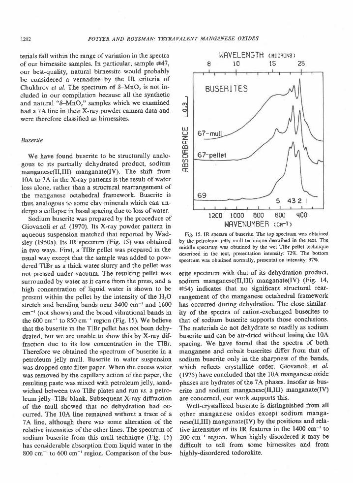
Sodium buserite was prepared by the procedure ofGiovanoli et al. (1970). Its X-ray powder pattern inaqueous suspension matched that reported by Wad-sley (1950a). Its IR spectrum (Fig. 15) was obtainedin two ways. First, a TlBr pellet was prepared in theusual way except that the sample was added to pow-dered TlBr as a thick water slurry and the pellet wasnot pressed under vacuum. The resulting pellet wassurrounded by water as it came from the press, and ahigh concentration of liquid water is shown to bepresent within the pellet by the intensity of the HrOstretch and bending bands near 3400 cm-' and 1600cm-' (not shown) and the broad vibrational bands inthe 600 cm-' to 850 cm-' region (Fig. l5). We believethat the buserite in the TlBr pellet has not been dehy-drated, but we are unable to show this by X-ray dif-fraction due to its low concentration in the TlBr.Therefore we obtained the spectrum of buserite in apetroleum jelly mull. Buserite in water suspensionwas dropped onto filter paper. When the excess waterwas removed by the capillary action of the paper, theresulting paste was mixed with petroleum jelly, sand-wiched between two TlBr plates and run vs. a petro-leum jelly-TlBr blank. Subsequent X-ray diffractionof the mull showed that no dehydration had oc-curred. The l0A line remained without a trace of a7A line, although there was some alteration of therelative intensities of the other lines. The spectrum ofsodium buserite from this mull technique (Fig. 15)has considerable absorption from liquid water in the800 cm-' to 600 cm-' region. Comparison of the bus-
POTTER AND ROSSMAN: TETRAVALENT MANGANESE OXIDES
Jc;lI
l!(Jz.CECD(EO,f,,oct
NRVELENGTH <utcnonsr810 ls25
BUSERI TES
67-mul
67-pel lel
695 432 |
1200 1000 800 600 q00
NRVENUMBEB <cn-l>Fig. 15. IR spectra of buserite. The top spectrum was obtained
by the petroleum jelly mull technique described in the text. Themiddle spectrum was obtained by the wet TlBr pellet techniquedescribed in the text, presentation intensity: 72Vo. Thc bottomspectrum was obtained normally, presentation intensily: 97Vo.
erite spectrum with that of its dehydration product,sodium manganese(Il,Ill) manganate(IV) (Fig. 14,#54) indicates that no significant structural rear-rangement of the manganese octahedral frameworkhas occurred during dehydration. The close similar-ity of the spectra of cation-exchanged buserites tothat of sodium buserite supports those conclusions.The materials do not dehydrate so readily as sodiumbuserite and can be air-dried without losing the l0Aspacing. We have found that the spectra of bothmanganese and cobalt buserites di,ffer from that ofsodium buserite only in the sharpness of the bands,which reflects crystalline order. Giovanob et al.(1975) have concluded that the l0A manganese oxidephases are hydrates ofthe 7A phases. Insofar as bus-erite and sodium manganese(Il,Ill) manganate(IV)are concerned, our work supports this.
Well-crystallized buserite 15 dislinguished from allother manganese oxides except sodium manga-nese(II,IIf manganate(Iv) by the positions and rela-tive intensities of its IR features in the 1400 cm-' to200 cm-' region. When highly disordered it may bedifficult to tell from some birnessites and fromhighly-disordered todorokite.
POTTER AND ROSSMAN: TETRAVALENT MANGANESE OXIDES t2t3
Todorokite and woodruffie
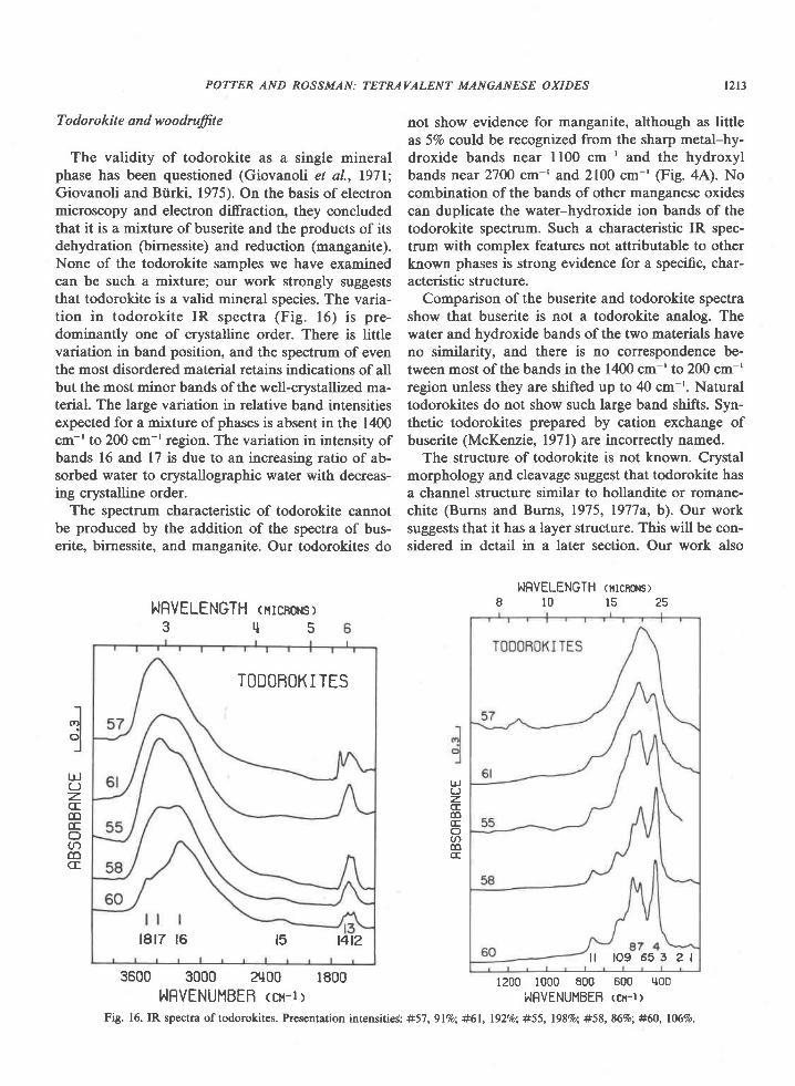
The validity of todorokite as a single mineralphase has been questioned (Giovanoli et al., l97l;Giovanoli and Btirki, 1975). On the basis of electronmicroscopy and electron diffraction, they concludedthat it is a mixture of buserite and the products of itsdehydration (birnessite) and reduction (manganite).None of the todorokite samples we have examinedcan be such a mixture; our work strongly suggeststhat todorokite is a valid mineral species. The varia-tion in todorokite IR spectra (Fig. 16) is pre-dominantly one of crystalline order. There is littlevariation in band position, and the spectrum of eventhe most disordered material retains indications of allbut the most minor bands of the well-crystallized ma-terial. The large variation in relative band intensitiesexpected for a mixture of phases is absent in the 1400cm-'to 2@ cm-'region. The variation in intensity ofbands 16 and 17 is due to an increasing ratio of ab-sorbed water to crystallographic water with decreas-ing crystalline order.
The spectrum characteristic of todorokite cannotbe produced by the addition of the spectra of bus-erite, birnessite, and manganite. Our todorokites do
not show evidence for manganite, although as littleas 57o could be recognized from the sharp metal-hy-droxide bands near ll00 cm-' and the hydroxylbands near 27OO cm-' and 2100 cm-' (Fig. 4A). Nocombination of the bands of other manganese oxidescan duplicate the water-hydroxide ion bands of thetodorokite spectrum. Such a characteristic IR spec-trum with complex features not attributable to otherknown phases is strong evidence for a specific, char-acteristic structure.
Comparison of the buserite and todorokite spectrashow that buserite is not a todorokite analog. Thewater and hydroxide bands of the two materials haveno similarity, and there is no correspondence be-tween most of the bands in the 1400 cm-' to 200 cm-'region unless they are shifted up to 40 cm-r. Naturaltodorokites do not show such large band shifts. Syn-thetic todorokites prepared by cation exchange ofbuserite (McKenzie, l97l) are incorrectly named.
The structure of todorokite is not known. Crystalmorphology and cleavage suggest that todorokite hasa channel structure similar to hollandite or romane-chite (Burns and Burns, 1975, 197'7a, b). Our worksuggests that it has a layer structure. This will be con-sidered in detail in a later section. Our work also
UJCJz.CE(D(Eoan(DCE
r lolJ
ttJ(Jz.cfcoccO(t)6CE
hIRVELENGTH <rrcnoHsr3 q 5
3600 3000 21100 1800hIFVENUMBEB <cr-l>
NRVELENGTH (ilrcnoNs)8 1 0 1 5 2 5
1200 1000 800 600 q00
I,IRVENUMBEB <cn-t:
TODOBOK I TES
1817 t6 t5 t4t2l f l o 9 6 5 3 2 l
Fig. 16. IR spectra of todorokites. Presentation intensitieC: #57, glVo; #61, l92o/o; #55, l98Vo; #58, 86%; #60, 106%o.
RRNCIEITES
12l4 POTTER AND ROSSMAN:TETRAVALENT MANGAN ESE O XI DES
NRVELENGTH <nrcnoHsl3 q 5
NRVELENGTH ortcRonsr810152s
RFNCIEITES
65
64
63 4 3 2 l
1200 1000 800 600 q00I^IRVENUMBEB <cN-r:
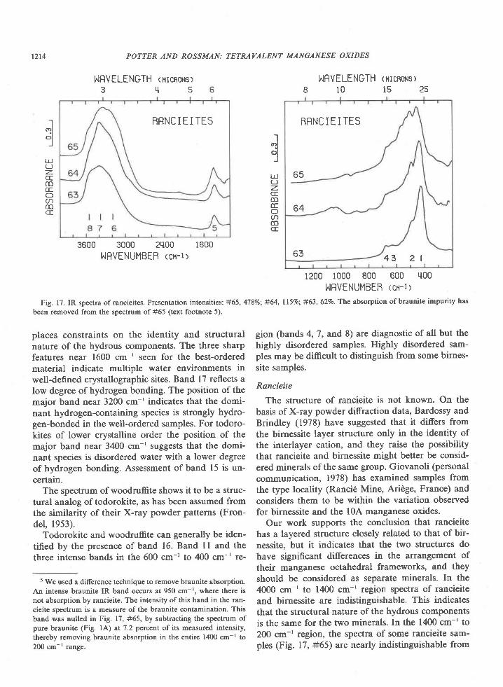
Fig. 17. IR spectra of rancieites. Presentation intensities: #65, 4'l8%o; #64, ll59o; #63, 62Vo. The absorption of braunite impurity has
been removed from the spectrum of #65 (text footnote 5).
Ic)l. lolI
ul(-)z.CEco(Ec)a(DCE
-t(r)l
cil-l
IJJ(J
z.CEcoccOacocf
3600 s000 2q00 1800I,'IFVENUMBEB <cu-l >
places constraints on the identity and structuralnature of the hydrous components. The three sharpfeatures near 1600 cm-' seen for the best-orderedmaterial indicate multiple water environments inwell-defined crystallographic sites. Band l7 reflects alow degree ofhydrogen bonding. The position ofthemajor band near 3200 cm-' indicates that the domi-nant hydrogen-containing species is strongly hydro-gen-bonded in the well-ordered samples. For todoro-kites of lower crystalline order the position of themajor band near 3400 cm-' suggests that the domi-nant species is disordered water with a lower degreeof hydrogen bonding. Assessment of band 15 is un-certain.
The spectrum of woodruffite shows it to be a struc-tural analog of todorokite, as has been assumed fromthe similarity of their X-ray powder patterns (Fron-del, 1953).
Todorokite and woodruffite can generally be iden-tified by the presence of band 16. Band 11 and thethree intense bands in the 600 cm-' to 400 cm-' re-
5 We used a difference techniqu€ to remove braunite absorption.An intense braunite IR band occurs at 950 cm-r. where there isnot absorption by rancieite. The intensity ofthis band in the ran-cieite spectrum is a measure of the braunite contamination. Thisband was nulled in Fig. 17, #65, by subtracting the spectrum ofpure braunite (Fig. lA) at 7.2 percnnt of its measured intensity,thereby removing braunite absorption in the entire 1400 cm-' to200 cm-r range.
gion (bands 4,7, and 8) are diagnostic of all but thehighly disordered samples. Highly disordered sam-ples may be difficult to distinguish from some birnes-site samples.
Rancieite
The structure of rancieite is not known. On thebasis of X-ray powder diffraction data, Bardossy andBrindley (1978) have suggested that it differs fromthe birnessite layer structure only in the identity ofthe interlayer cation, and they raise the possibilitythat rancieite and birnessite might better be consid-ered minerals of the same group. Giovanoli (rersonalcommunication, 1978) has examined samples fromthe type locality (Ranci6 Mine, Aridge, France) andconsiders them to be within the variation observedfor birnessite and the l0A manganese oxides.
Our work supports the conclusion that rancieitehas a layered structure closely related to that of bir-nessite, but it indicates that the two structures dohave significant differences in the arrangement oftheir manganese octahedral frameworks, and theyshould be considered as separate minerals. In the4000 cm-' to 1400 cm-' region spectra of rancieiteand birnessite are indistinguishable. This indicatesthat the structural nature of the hydrous componentsis the same for the two minerals. In the 1400 cm-' to200 cm-' region, the spectra of some rancieite sam-ples (Fig. 17, #65) are nearly indistinguishable from
those of some birnessites, but other rancieite spectra(Fig. 17, #63) differ significantly. Those rancieitesamples with the most distinctive IR patterns arethose for which the characteristic rancieite softnessand color (Palache et al., 1944, p. 572; Richmond etal., 1969\ are most apparent. The structural implica-tion of the rancieite spectra will be considered morefully in the next section.
Most rancieite samples can be distinguished fromother manganese oxides by the high relative intensityof the band l. Some samples can easily be confusedwith disordered todorokite or birnessite; however,rancieite band 4 at 680 cm-' distinguishes rancieitefrom these other two, which absorb at760 cm-' and750 cm-' respectively.
General relations of infrared spectra to structureThe IR spectra of the manganese oxides, except for
lithiophorite, are dependent only on the MnOu oc-tahedral framework. If bands in the spectra are theresult of vibrations in which the large cations partici-pate significantly, then changes in the mass of thesecations should be reflected in the spectra. However,substitution in the hollandite group and the birnes-sites leaves the spectra in the mid-infrared region un-changed. Neither could the effect of cation sub-stitution be seen for chalcophanite and aurorite andfor todorokite and woodruffite, although the extentof the change is less since it involves incomplete sub-stitution involving elements of relatively similarmass. Therefore we conclude that the large cationsare not involved in the bands in this region ofspectra. Only for lithiophorite, where light atomsother than manganese form a major part of the crys-tal structure, are bands in the 1400 cm-' to 200 cm-,region clearly attributable to other cations. Lith-iophorite bands near 1000 cm-' and 900 cm-' areprobably due to (AI,LD-OH. The reason for this in-sensitivity may be that the bands are relatively weakand therefore absorb at lower energies. Hollanditeand cryptomelane spectra difer in the 200 cm-' to 30cm-' region, and the difference may be due to theidentity of the channel cations. An alternative ex-planation is that the large cations are at too low aconcentration in the structures to give observablebands. Although mid-infrared spectroscopy is in-sensitive to the ihannel and interlayer cations, it isdiagnostic of the structural group.
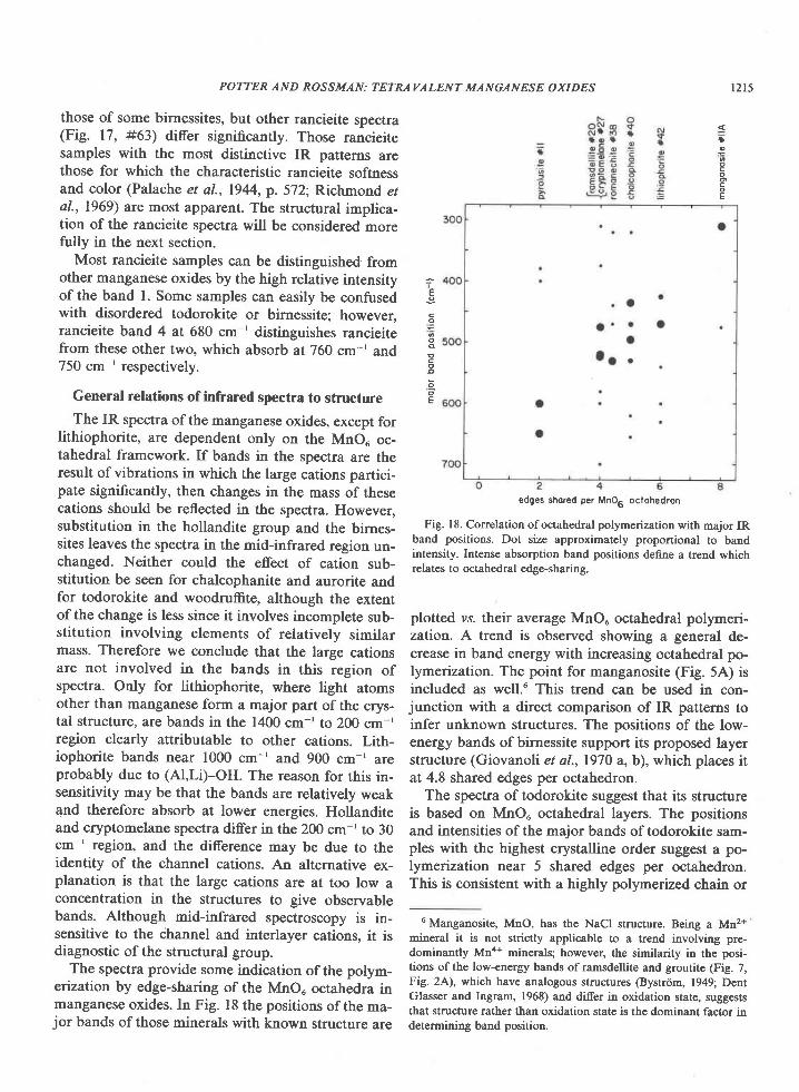
The spectra provide some indication of the polym-erization by edge-sharing of the MnO6 octahedra inmanganese oxides. In Fig. 18 the positions of the ma-jor bands of those minerals with known structure are
t2t5
edges shored per MnOa oclohedron
Fig. 18. Correlation of octahedral polymerization with major IRband positions. Dot size approximately proportional to bandintensity. Intense absorption band positions define a trend whichrelates to octahedral edge-sharing.
plotted vs. their average MnOu octahedral polymeri-zation. A trend is observed showing a general de-crease in band energy with increasing octahedral po-lymerization. The point for manganosite (Fig. 5A) isincluded as well.u This trend can be used in con-junction with a direct comparison of IR patterns toinfer unknown structures. The positions of the low-energy bands ofbirnessite support its proposed layerstructure (Giovanoli et al., 1970 a, b), which places itat 4.8 shared edges per octahedron.
The spectra of todorokite suggest that its structureis based on MnOu octahedral layers. The positionsand intensities of the major bands of todorokite sam-ples with the highest crystalline order suggest a po-lymerization near 5 shared edges per octahedron.This is consistent with a highly polymerized chain or
6 Manganosite, MnO, has the NaCl structure. Being a Mn2+mineral it is not strictly applicable to a trend involving pre-dominantly Mna+ minerals; however, the similarity in the posi-tions of the low-energy bands of ramsdellite and groutitc (Fig. 7,Fig. 2A), which have analogous stnrctur€s (Bystrdm, 1949; DentGlasser and Ingram, 1968) and differ in oxidation state, suggeststhat structure rather than oxidation state is the dominant factor indetermining band position.
POTT:ER AND ROSSMAN: TETRAVALENT MANGANESE OXIDES
*e
o
o
E
E
co
o
Eo
o
E

t2t6 POTT:ER AND ROSSMAN: TETRAVALENT MANGANESE OXIDES
channel structure and with a layer structure contain-ing some vacancies. In order to attain a polymeriza-tion of 5 shared edges per octahedron in a chain orchannel structure it is necessary to build them fromunits whose average polymerization is 5. This corre-sponds to quadruple chains. No chain or channelmanganese oxides composed of such large units areknown to exist, although isolated quadruple chainshave been observed in hollandite in electron micro-scope images (Turner and Buseck, 1979). It appearsthat for highly polymerized structures a layer struc-ture is preferred. Direct comparison of todorokitespectra to those of other manganese oxides supportsthis. All known layer structures have a strong band inthe 400 cm-' to 450 cm-' region, which is analogousto todorokite band 4 at 430 cm-'. Strong absorptionin this region does not occur for the chain and chan-nel structures. These suggestions contrast with pre-vious work which suggests that todorokite has achannel structure (Burns and Burns, 1977a). Thiswas based on the needle-like morphology of todoro-kite and the presence of two perfect cleavage planesseen under the electron microscope. Todorokite oc-curs in other morphologies as well. Todorokite #58has a platy morphology reminiscent of birnessite.Weissenberg single-crystal X-ray diffraction indicatesthat the sample is ordered in the same plane as theplates but disordered in planes perpendicular tothem. This suggests that it consists of a random su-perposition of single-crystal todorokite plates andeliminates the possibility that it is a mass of needlesthat appear morphologically as a plate.
The spectra of rancieite suggest a polymerizationnear 6 shared edges per octahedron, which corre-sponds to a filled octahedral layer.
Spectra of samples of birnessite, todorokite, andrancieite may be similar in the 1400 cm-' to 200 cm-'region. For birnessite and todorokite this similarity isrelated to crystalline disorder. For the most dis-ordered birnessite and todorokite, the IR spectra aresimilar (e.g.,#50 and #57).Interestingly, because theX-ray order does not necessarily follow the IR order,sample #57 is readily identified as todorokite fromthe X-ray pattern, whereas sample #50 has a diffuseX-ray pattern, barely recognizable as birnessite. Asthe order increases, the spectra of the two mineralsbecome more distinct from one another. This sug-gests that birnessite and todorokite are built of fun-damentally similar units and that it is the ordering ofthese units which makes the minerals distinct fromone another. We believe that this is the case for ran-cieite as well, but our data could also be interpreted
as evidence for a structural continuum from well-
crystallized birnessite (Fig. 14, #54) to well-crystal-
lized rancieite (Fig. 17, #63).In this case the smooth
change in relative intensity and position of rancieite
band I (Fig. 17) would reflect the degree of birnessite
"character."
The lower valent manganese oxides
We have included as Appendix A spectra of well-
characterned samples of the following lower valent
manganese oxides: braunite, groutite, hausmannite,
manganite, manganosite, partridgeite and quense-
lite.3 In some cases these are necessary for the inter-
pretation of the earlier sections of the paper. Others
are included so that this paper may serve as a com-
pilation of the spectra of all manganese oxides com-
monly encountered in nature. They illustrate the
ability of infrared spectroscopy to produce patterns
diagnostic for each mineral phase for the whole
range of naturally occurring manganese oxides. It ex-
tends the usefulness ofthis paper as a data base for
further structural work on the manganese oxide.
Conclusions
Infrared spectroscopy has proven to be a useful
tool for the mineralogical identification of the tet-
ravalent manganese oxides. Different oxides can be
distinguished by absorption patterns due to vibra-
tions of the MnOu octahedral framwork in the 1400
cm-' to 200 cm-' region. The 4000 cm-'to 1400 cm-'
region is often diagnostic due to absorption associ-
ated with the hydrous components of the oxides.
Because of its sensitivity to short range order, in-
frared spectroscopy gives more reliable information
than X-ray diffraction when applied to disordered
and finely particulate samples. With X-ray diffrac-
tion a small amount of well-crystallized material in a
disordered or finely particulate matrix can give the
impression that the whole sample is well-crystallized.
The lack of correspondence between the degree of
order indicated by these two techniques for our bir-
nessite samples as a manifestation of this effect. X-
ray diffraction data alone could lead to an error in
mineralogical identification of the disordered mate-
rial if a well-crystallized minor component of differ-
ent mineralogy is present. For minerals which have
only a few characteristic X-ray diffraction lines, such
as birnessite, todorokite, and rancieite, infrared spec-
troscopy has shown that X-ray diffraction alone may
be an insufficient test for the validity of synthetic an-
alogs.IR spectroscopy can also contribute to the determi-

nation of the structures and structural relationshipsamong the tetravalent manganese oxides. We havebeen able to suggest features of the unknown oxidestructures based on their IR spectra and the relationof these spectra to those of manganese oxides withknown structure. We have applied IR spectroscopyto the role of water and hydroxide ion, whose pres-ence and structural orientations can only be inferredfrom X-ray and chemical data.
Now that IR spectra of well-charactet'aed manga-nese oxide samples are available to serve as stan-dards, this technique should find wider applicationfor mineralogical identification of manganese oxidesin the terrestrial and aquatic environments.
Acknowledgments
We thank the following for providing samples for this study:J. S. White, Jr., Smithsonian Institution; C. G. Cunningham, U. S.Geological Survey; A. R. Kampf, Los Angeles County Museum ofNatural History; R. G. Burns and V. M. Burns, Massachusetts In-stitute of Technology; A. J. Bauman, Jet propulsion Laboratory;G. W. Brindley, Pennsylvania State University; A. S. Corey, pasa-dena City College; V. Morgan, Boron, California; L. Dalbec,Ridgecrest, California; and R. Currier, Arcadia, California. Asmall portion of the funding for this work was provided by theL. S. B. Leakey Foundation and the John A. McCarthy Founda-tion. Helpful critical reviews were provided by R. G. Burns and R.Giovanoli, Berne.
References
Agiorgitis, G. (1969) Uber differential-thermoanalytische und in-frarotspektroscopische Untersuchungen von Mangan-Mineral-ien. Tschermaks Mineral. Petrogr. Mitt., I j,2'13-283.
Bardossy, G. and G. W. Brindley (1978) Rancieite associated witha karstic bauxite deposit. Am. Mineral., 63, i63-76j.
Brenet, J. P., J. P. Gabano and M. Seigneurin (1958). Transforma-tions thermiques d'oxydes de manganese. ln Papers presentedTo The Section On Inorganic Chemistry (I6th Internatianal Con-gress Of Pure And Applied Chemistry, Paris, 1957), p. 69-80.Butterworth Scientific Publications, Londoq.
BrowrL F. H., A. Pabst and D. L. Sawyer (1971) Bimessite oncolemanite at Boron, California. Am. Mineral., 56, lO57-1M4.
Burns, R. G. and V. M. Burns (1975) Structural relationships be-twe€n the manganese(IV) oxides. In A. Kozawa and R. J.Brodd, Eds., Manganese Dioxide Symposium, Vol. l, p. 306-32j.The Electrochemical Society, Cleveland.
- and - (1977a) Mineralogy. In G. P. Glasby, 8d,., Ma-rine Manganese Deposits, Chapt. 7. Elsevier, Amsterdam.
- and - (1977b) The mineralogy and crystal chemistryof deep-sea manganese nodules, a polymetallic resource of thetwenty-first century. Phil. Trans. R. Soc. Lond., A286,283-301.
Buser, W., P. Graf and W. Feitknecht (1954) Beitrag zur Kenntnisder Mangan(Il)-manganite und des 6-MnO2, Helv. Chim. Acta,37.2322-2333.
Bystrtim, A. M. (1949) The crystal structure of ramsdellite, an or-thorhombic modification of MnO2. Acta Chim. Scand., 3, 163-173.
12t7
Bystrdm, A. and A. M. Bystrcim (1950) The crystal structure ofhollandite, the related manganese oxide minerals, and a-MnOr.Acta Crystallogr., 3, 146-154.
_ and _ (1951) The positions of the barium atoms inhollandite. Acta Crystallogr., 4, 469.
Champness, P. E. (1971) The transformation manganite + py-rolusite. M ineral. Mag., 38, 245-248.
Chukrov, F. V., A. I. Gorshkov, E. S. Rudnitskaya, V. V. Ber-ezovskaya and A. V. Sivtsov (1978a) On vernadite. (in Russian)Izvest. Akad. Nark. SSS& Ser. Geol.,5-19.
E. S. Rudnitskaya and A. V. Sivtsov (1978b) The charac-teristics of birnessite. (in Russian) Izvest. Akad. Naz&. SSSR,Ser. Geol.,67-'16.
Clark, G. M. (1972) The Structures of Non-molecalar Solids: ACoordinated Polyhedron Approach. Applied Science Publishers,London.
Cole, W. F., A. D. Wadsley and A. Walkley (1947) An X-ray dif-fraction study of manganese dioxide. Trans. Electrochem. Soc.,92, 133-158.
de WolS P. M. (1959) Interpretation of some 7-MnO2 diffractionpatterns. Acta Crystallogr., I 2, 341-345.
Dent Glasser, L. S. and L. Ingram (1968) Refinement of the crys-tal structure of groutite, a-MnOOH. Acta Crystallogr., 24, 1233-1236.
Farmer, V. C. (Ed.) (1974) The Infrared Spectra of Minerals. Min-eral Society of London, London.
Finkelman, R. 8., H. T. Evans, Jr. and J. J. Matzdo (1974) Man-ganese minerals in geodes from Chihuahua, Mexico. Mineral.Mag., 39,549-558.
Fleischer, M. (1960) Studies of the manganese oxide minerals. III.Psilomelane. Am. Mineral., 45, l'16-18'1.
- and G. T. Faust (1963) Studies on manganes€ oxide miner-als VII. Lithiophorite. Schweiz. Mineral. Petrogr. Mitt.,43, 197-216.
W. E. Richmond and H. T. Evans, Jt. (1962) Studies ofthe manganese oxides V. Ramsdellite, MnO2, an orthorhombicdimorph of pyrolusite. Am. Mineral., 47, 47-51.
Frondel, C. (1953) New manganese oxides hydrohausmannite andwoodruffite. Am. Mineral., 38, 761-7 69.
U. B. Marvin and J. Ito (1960a) New data on birnessiteand hollandite. Am. Mineral., 45, 87 1-875.
_ and _ (1960b) New occurrences of todoro_kite. Am. Mineral., 45, 1167-1173.
Gattow, G. and O. Glemser (196la) Darstellung und Eigenshaftenvon Braunsteinen. II (Die y- und 4-Gruppe der Braunsteine). Z.anorg. allg. Chem., 309,20-36.
_ and _ (l96lb) Darstellung und Eigenshaften vonBraunsteinen. III (Die e, p, und a-Gruppe der Braunsteine, uberRamsdellite und uber Urnwandlungen der Braunsteine). Z.anorg. allg. Chem., 309, l2l-232.
Giovanoli, R. (1969) A simplified scheme for the polymorphism inthe manganese dioxides. Chimia, 23,47M72.
- and P. Burki (1975) Comparison ofX-ray evidence of ma-rine manganese nodules and nonmarine manganese ore depos-ias. chimia, 29, 266-269.
nodules. Report #33a, Universitat Bern. [English tradition of in-troduction. Quoted in Burns and Burns (1977a)1.
- M. Giotrredi and W. Stumm (1975) Layer struc-tured manganese oxide hydroxides. IV: The buserite group,structure stabilization by transition elements. Chimia, 29, 517-520.
POTTER AND ROSSMAN: TETRAVALENT MANGANESE OXIDES
12 l8
- W. Feitknecht and F. Fischer (1971) Reduktion von Man-
gan(Ill)-manganat(IV) mit Zimtalkohol. Helv. Chim. Acta, 54,
ltt2-1t24.R. Maurer and W. Feitknecht (1967) Zur Struktur des 7-
MlO2. Helv. Chem. Acta.,50, 1072-1080.
E. Stahli and W. Feitknecht (1970a) Uber Oxidhydroxide
des vierwertigen Mangansmit Schichtengitter l. Mitteilung:
Natrium-mangan(Il,Ill) manganat(Iv). H elv. Chim. Acta, 5 3,
209-220._ and _ (l9?0b) Uber Oxidhydroxide des vier-
w€rtigen Mangans mit Schiechtengitter 2. Mitteilung: Man-
gan(Ill)-manganat(IV). Helv. Chim. Acta, 53, 453-464.
Glemser, O, G. Gattow and H. Meisiek (1961) Darstellung und
Eigenschaften von Braunsteinen. I (Die 6-Gruppe der Brauns-
teire). Z. anorg. allg. Chem., 309, l-19.
Goldman, D. S., G. R. Rossman and W. A. Dollase (1977) Chan-
nel constituents in cordierite. Am. Mineral., 62, 1144-1157.- and K. M. Parkin (1978) Channel constituents in
beryl. Plrys. Chem. Mineral., 3,225-235.
Gruner, J. W. (1947) Groutit€, HMnO2, a new mineral of the dias-
pore-goethite group. Am. M ineral., 3 2, 654-659.
Kolta, G. A., F. M. A. Kerim and A. A. A. Azim (1971) Infrared
absorption spectra of some manganese dioxide modifications
and their thermal products. Z. anorg. allg. Chem., 384,260_266.
Laudy, J. H. A. and P. M. de Woltr (1963) X-ray investigation of
the 6-B transformation of MnO2. Appl. Sci. Res. Sect., 810, 157-
168.McKenzie, R. M. (1971) The synthesis of birnessite, cryptomelane,
and some other oxides and hydroxides of manganese. Mineral.
Mag., 38,294-502.Mitchell, R. S. and A. S. Corey (1973) Braunite and surassite from
Los Angeles County, California. M ineral. Rec., 4, 29O-293.
Moenke, H. (1962) Mineral Specten. Akademie-Verlag, Berlin.
Moore, T. E., M. Ellis and P. W. Selwood (1950) Solid oxides and
hydroxides of manganese. J. Am Chem. Soc., 72,856-8'12.
Mukherjee, B. (1959) X-ray study of psilomelane and cryptome-
lane. Mineral. Mag., 32, 166-171.- (1967) Crystallography of psilomelane, A3)QMn3Oq6. Mrz-
eral. Mag., 35, 645-655.
Palache, C., H. Berman and C. Frondel (1944) Dana's System of
Mineralogy, Vol. I. Wiley, New York.
Potter, R. M. and G. R. Rossman (1979a) The manganese and
POTTER AND ROSSMAN: TETRAVALENT MANGANESE OXIDES
iron oxide mineralogy of desert varnish' Chem. Geol.' 2 5, 7 9-94.
- and - (1979b) A magnesium analogue of chalcopha-
nite in manganese-rich concretions from Baja Califonia- Am.
M ineral., 64, 1227 -1229.
- and - (lg'lgc) Mineralogy of manganese den-
drites and coatings. Am. Mineral.,64' 1219-1226.
Prost, R. (1973) The influence of the Christiansen effect on I' R'
spectra of powders. Clays Clay. Mineral.,2l'363-368.
Radtke, A. S., C. M. Taylor and D. F. Hewett (1967) Aurorite, ar-
gentian todorokite and hydrous silver-bearing lead manganese
oxide. Econ. Geol., 62,186-206'
Richmond, W. E., M. Fleischer and M. E. Mrose (1969) Studies
on manganese oxide minerals. IX rancieite. Bull. Soc. fr. Min-
eral. Cristallogr., 92, l9l-195.
Segeler, C. G. (1959) Notes on a second occurrence of groutite'
Am. M ineral., 44, 8'17 -878.
Turner, S. and P. R. Buseck (1979) Manganese oxide tunnel struc-
tures and their intergrowths. Science, 203' 456458.
Valarelli, J. V., M. Perrier and G. Vicentini (1968) Infrared
spectra of some manganese minerals. ln. Acad' brasil Ci€nc.' 40'
289-296.van der Marel, H. W. and H. Beutelspacher (1976) Atlas of In-
frared Spectra of Clay Minerals and Their Admixtures' Elsevier,
Amsterdam.Vaux, G. and H. Bennet (1937) X-ray studies on pyrolusite (in-
cluding polianite) and psilomelane. M ineral. M ag., 24, 521'526.
Wadsley, A. D. (1950a) A hydrous manganese oxide with ex-
change properties. J. Am. Chem. Soc.,72' 1782-1784'_- (1950b) Synthesis of some hydrated manganese minerals.
Am. Mineral., 35, 485499.- (1952\ The structure of lithiophorite, (Al,Li)Mno2(oH)2.
A cta Cryst allo gr., 5, 6'1 6-680.
(1953) The crystal s t ructure of psi lomelane,
(Ba,H2O)2Mn5O 6. Acta Crystallogr', 6' 433438.
(1955 ) The c r ys ta l s t r uc tu re o f cha l cophan i t e ,
Zn.Mn3O,' 3HrO. Acta Crystallog', 8, |65-l'12.
Zwicker, W. K., W. O. J. Groeneveld Meijer and H. W. Jaffe
(1962) Nsutite-a widespread manganes€ oxide mineral. 42.
Mineral., 47,246-266.
Manuscript received, February 1 2, 1979;
accepted for publication, June 25, 1979.