the tarantula toxins protx-ii and hwtx-iv differentially...
TRANSCRIPT
MOL #66332
1
The tarantula toxins ProTx-II and HWTX-IV differentially interact with human
Nav1.7 voltage-sensors to inhibit channel activation and inactivation
Yucheng Xiao, Kenneth Blumenthal, James O. Jackson II, Songping Liang and Theodore
R. Cummins
Department of Pharmacology and Toxicology, Indiana University School of Medicine,
Indianapolis, Indiana 46202 (Y.X., J.O.J., T.R.C.),
Department of Biochemistry, School of Medicine and Biomedical Sciences, State
University of New York, Buffalo, New York 14214, USA (K.B.)
and
Key Laboratory of Protein Chemistry and Developmental Biology of the Ministry of
Education, College of Life Sciences, Hunan Normal University, Changsha, Hunan
410081, China (Y.X., S. L.)
Molecular Pharmacology Fast Forward. Published on September 20, 2010 as doi:10.1124/mol.110.066332
Copyright 2010 by the American Society for Pharmacology and Experimental Therapeutics.
This article has not been copyedited and formatted. The final version may differ from this version.Molecular Pharmacology Fast Forward. Published on September 20, 2010 as DOI: 10.1124/mol.110.066332
at ASPE
T Journals on Septem
ber 9, 2019m
olpharm.aspetjournals.org
Dow
nloaded from

MOL #66332
2
Running title: Inhibition of hNav1.7 gating by tarantula toxins
Corresponding author: Dr. Theodore R. Cummins, Department of Pharmacology and
Toxicology, Stark Neurosciences Research Institute, Indiana University School of
Medicine, Indianapolis, Indiana 46202, USA. Email: [email protected].
Number of Text Pages: 33
Number of Tables: 2
Number of Figures: 7
Number of References: 32
Number of Words in Abstract: 248
Number of Words in Introduction: 580
Number of Words in Discussion: 1555
Abbreviations:
HWTX-IV, huwentoxin-IV; TTX, tetrodotoxin; Nav, voltage-gated sodium channel; WT, wild
type; HEK293, human embryonic kidney 293; domains I-IV, DI-DIV; transmembrane
segments 1-6, S1-S6
This article has not been copyedited and formatted. The final version may differ from this version.Molecular Pharmacology Fast Forward. Published on September 20, 2010 as DOI: 10.1124/mol.110.066332
at ASPE
T Journals on Septem
ber 9, 2019m
olpharm.aspetjournals.org
Dow
nloaded from
MOL #66332
3
Abstract The voltage-gated sodium channel Nav1.7 plays a crucial role in pain, and drugs that inhibit hNav1.7 may
have tremendous therapeutic potential. ProTx-II and huwentoxin-IV (HWTX-IV), cystine knot peptides
from tarantula venoms, preferentially block hNav1.7. Understanding the interactions of these toxins with
sodium channels could aid the development of novel pain therapeutics. While both ProTx-II and HWTX-
IV have been proposed to preferentially block hNav1.7 activation by trapping the domain-II voltage-
sensor in the resting configuration, we show that specific residues in the voltage-sensor paddle of domain-
II play substantially different roles in determining the affinities of these toxins to hNav1.7. The mutation
E818C increases ProTx-II’s and HWTX-IV’s IC50 for block of hNav1.7 currents by 4- and 400-fold,
respectively. In contrast, the mutation F813G decreases ProTx-II affinity by 9-fold, but has no effect on
HWTX-IV affinity. Importantly, we also show that ProTx-II, but not HWTX-IV, preferentially interacts
with hNav1.7 to impede fast-inactivation by trapping the domain-IV voltage-sensor in the resting
configuration. Mutations E1589Q and T1590K in domain-IV each decreased ProTx-II’s IC50 for
impairment of fast-inactivation by ~6-fold. In contrast mutations D1586A and F1592A in domain-IV
increased ProTx-II’s IC50 for impairment of fast-inactivation by ~4-fold. Our results show that 1) while
ProTx-II and HWTX-IV binding determinants on domain-II may overlap, domain-II plays a much more
crucial role for HWTX-IV and 2) contrary to what has been proposed to be a guiding principle of sodium
channel pharmacology, molecules do not have to exclusively target the domain-IV voltage-sensor in order
to influence sodium channel inactivation.
This article has not been copyedited and formatted. The final version may differ from this version.Molecular Pharmacology Fast Forward. Published on September 20, 2010 as DOI: 10.1124/mol.110.066332
at ASPE
T Journals on Septem
ber 9, 2019m
olpharm.aspetjournals.org
Dow
nloaded from

MOL #66332
4
Introduction
Voltage-gated sodium channels play important roles in action potential generation and propagation. As
Nav1.7 is a crucial contributor to pain sensation (Cox et al., 2006; Cummins et al., 2007), drugs that
selectively target human Nav1.7 (hNav1.7) could be ideal analgesics. Unfortunately drugs targeting
sodium channels typically have broad spectrum sodium channel activity and narrow therapeutic windows
(Cummins and Rush, 2007). Therefore there is substantial interest in identifying compounds that
selectively target hNav1.7 and determining their molecular mechanisms of action.
ProTx-II and Huwentoxin-IV (HWTX-IV) are tarantula toxins that target voltage-gated sodium channels.
These toxins belong to the inhibitory cystine knot family and are stabilized by the same disulfide frame
(C1-C4, C2-C5 and C3-C6) (Middleton et al., 2002; Peng et al., 2002). However they show limited
sequence similarity (Fig. 1A). Although ProTx-II inhibits multiple sodium channel subtypes (Nav1.1-
1.8), it has been reported to be ~100-fold more selective for Nav1.7 (Schmalhofer et al., 2008; Smith et
al., 2007). HWTX-IV preferentially inhibits tetrodotoxin (TTX)-sensitive neuronal subtypes (including
Nav1.7), does not inhibit TTX-resistant neuronal subtypes, and has little effect on skeletal muscle
(Nav1.4) and cardiac (Nav1.5) subtypes (Xiao et al., 2008). Although these toxins have divergent
properties, both are classified as voltage-sensor modifiers (Sokolov et al., 2008; Xiao et al., 2008).
Voltage-sensor modifiers target the voltage sensors of ion channels. The pore-forming sodium channel α-
subunit consists of four domains (DI - DIV), each having six transmembrane segments (S1-S6) (Catterall
et al., 2003). The S5-S6 segments form the channel pore and the S1-S4 segments form voltage sensor
modules. The S4 segments, rich in positive residues, sense membrane depolarization and move outward
to induce channel gating. Scorpion toxins have been extensively characterized as voltage-sensor
modifiers, and understanding the molecular determinants of their interactions with voltage-gated sodium
channels has provided invaluable insight into channel structure-function relationships. Scorpion α-toxins
This article has not been copyedited and formatted. The final version may differ from this version.Molecular Pharmacology Fast Forward. Published on September 20, 2010 as DOI: 10.1124/mol.110.066332
at ASPE
T Journals on Septem
ber 9, 2019m
olpharm.aspetjournals.org
Dow
nloaded from

MOL #66332
5
interact with the DIV S3-S4 linker to stabilize DIV-S4 in the closed state, impeding fast inactivation.
Scorpion β-toxins bind to the DII S3-S4 linker, trapping the DII-S4 in the activated state and enhancing
channel activation. The binding sites for these scorpion toxins are defined as neurotoxin receptor sites 3
and 4, respectively (Cestele and Catterall, 2000). These data, in conjunction with other studies, indicate
that the S4 segments in DI, DII and DIII are determinants of channel activation while that of DIV is
predominantly involved in channel inactivation (Cestele et al., 2001; Cha et al., 1999; Sheets and Hanck,
2007). Both ProTx-II and HWTX-IV have been proposed to inhibit activation by trapping the DII
voltage-sensor in the resting configuration. However, their binding determinants may not be identical.
While HWTX-IV may selectively bind to neurotoxin site 4 (Xiao et al., 2008), ProTx-II may interact with
novel binding sites on Nav1.5 (Smith et al., 2007). Recently it was suggested that ProTx-II inhibits
activation of rNav1.2a by interacting with the voltage-sensor “paddles” (S3b-S4 motifs) of DI, DII and
DIV (Bosmans et al., 2008). This finding was somewhat surprising given the presumed role of DIV in
inactivation, leading to the proposal that for a toxin to alter inactivation it must exclusively interact with
the voltage-sensor paddle of DIV (Bosmans et al., 2008).
We investigated the interactions of ProTx-II and HWTX-IV with the voltage-sensor paddles in hNav1.7
DI, DII and DIV. Our data show that these two tarantula toxins differ substantially in their interactions
with hNav1.7. Although they may not be ideal as analgesic drugs, understanding the molecular
determinants of their complicated interactions with voltage-gated sodium channels should aid the
development of novel hNav1.7 blockers.
This article has not been copyedited and formatted. The final version may differ from this version.Molecular Pharmacology Fast Forward. Published on September 20, 2010 as DOI: 10.1124/mol.110.066332
at ASPE
T Journals on Septem
ber 9, 2019m
olpharm.aspetjournals.org
Dow
nloaded from

MOL #66332
6
Materials and Methods
Toxins - ProTx-II was recombinantly produced as described by Smith et al. (Smith et al., 2007).
HWTX-IV was purified from the crude venom of the female tarantula O. huwena as described by Peng et
al. (Peng et al., 2002). The purity of ProTx-II and HWTX-IV was determined to be over 99% by high
pressure liquid chromatography and matrix-assisted laser desorption ionization time-of-flight analysis.
Plasmids of sodium channels - The cDNA genes encoding rat (r) Nav1.2, rNav1.3 and rNav1.4 were
inserted into the vectors pRC-CMV, pcDNA3.1-mod and pRBG4, respectively (Cummins et al., 2001;
O'Leary, 1998; Ukomadu et al., 1992). The cDNA genes encoding human (h) Nav1.5 and hNav1.7 were
subcloned into the vectors pcDNA3.1 and pcDNA3.1-mod, respectively (Klugbauer et al., 1995).
Auxiliary subunits hβ1 and hβ2 were inserted into an internal ribosome entry site vector (Lossin et al.,
2002).
Site-directed mutagenesis of Nav1.7 - All hNav1.7 mutations in this study were constructed using the
QuikChange II XL Site-Directed Mutagenesis kit according to the manufacture's instruction. All
constructs were sequenced to confirm that the appropriate mutations were made.
Transient Transfection - Transient transfections of hNav1.7 wild type (WT) and mutant constructs into
human embryonic kidney 293 (HEK293) cells were performed using the calcium phosphate precipitation
method. HEK293 cells were grown under standard tissue culture conditions (5% CO2 and 37 °C) in
Dulbecco's modified Eagle's medium supplemented with 10% fetal bovine serum. The WT and mutant
hNav1.7 channels were cotransfected with the hβ1 and hβ2 subunits to increase the current density. The
calcium phosphate-DNA mixture (channel constructs and a green fluorescent protein reporter plasmid)
was added to the cell culture medium and left for 3 h, after which the cells were washed with fresh
medium. Cells with green fluorescent protein fluorescence were selected for whole-cell patch clamp
This article has not been copyedited and formatted. The final version may differ from this version.Molecular Pharmacology Fast Forward. Published on September 20, 2010 as DOI: 10.1124/mol.110.066332
at ASPE
T Journals on Septem
ber 9, 2019m
olpharm.aspetjournals.org
Dow
nloaded from

MOL #66332
7
recordings 36-72 h after transfection. Stably transfected cell lines containing WT rNav1.2, rNav1.3,
rNav1.4 and hNav1.5 without any β subunit or green fluorescent protein reporter plasmid were prepared
using the method described previously (Xiao et al., 2008).
Whole-cell Patch Clamp Recordings - Whole-cell patch clamp recordings were carried out at room
temperature (~21 °C) using an EPC-10 amplifier (HEKA, Lambrecht, Germany). Data were acquired on a
Pentium IV computer using the Pulse program (version 8.31; HEKA). Fire-polished electrodes were
fabricated from 1.7-mm capillary glass (VWR, West Chester, PA) using a P-97 puller (Sutter, Novato,
CA). The standard pipette solution contained (in mM): 140 CsF, 1 EGTA, 10 NaCl and 10 HEPES, pH
7.3. The standard bathing solution was (in mM) 140 NaCl, 3 KCl, 1 MgCl2, 1 CaCl2 and 10 HEPES, pH
7.3. After filling with pipette solution, the access resistance of electrode pipette ranged from 0.8 to 1.4
MΩ and the average resistance was 0.98 ± 0.02 MΩ (n=250). The liquid junction potential for these
solutions was <8 mV; data were not corrected to account for this offset. The offset potential was zeroed
before contacting the cell. After establishing the whole-cell recording configuration, the resting potential
was held at -100 mV for 5 min to allow adequate equilibration between the micropipette solution and the
cell interior. Linear leak subtraction, based on resistance estimates from four to five hyperpolarizing
pulses applied before the depolarizing test potential, was used for all voltage clamp recordings.
Membrane currents were usually filtered at 5 kHz and sampled at 20 kHz. Voltage errors were minimized
using 80% series resistance compensation, and the capacitance artifact was canceled using the computer-
controlled circuitry of the patch clamp amplifier. The stock solutions for ProTx-II and HWTX-IV were
made at 1 mM using bathing solution containing 1 mg/ml BSA, and aliquots were stored at -20 °C. Before
use, the solution was diluted to the concentrations of interest with fresh bathing solution. Toxin was
diluted into the recording chamber (volume of 300 µl) and mixed by repeatedly pipetting 30 µl to achieve
the specified final concentration. The extent of the inhibitory effect of toxin was typically assessed around
10 - 20 min after toxin treatment.
This article has not been copyedited and formatted. The final version may differ from this version.Molecular Pharmacology Fast Forward. Published on September 20, 2010 as DOI: 10.1124/mol.110.066332
at ASPE
T Journals on Septem
ber 9, 2019m
olpharm.aspetjournals.org
Dow
nloaded from

MOL #66332
8
Data Analysis - Data were analyzed using the Pulsefit (HEKA) and GraphPad Prism 4 (GraphPad
Software) programs. All data points are shown as mean ± S.E. and n is presented as the number of the
separate experimental cells. Steady-state activation and inactivation curves were fitted using Boltzmann
equation: y = 1/(1+exp((V1/2-V)/k), in which V1/2, V and k represented midpoint voltage of kinetics, test
potential and slope factor, respectively. Concentration-response curves to determine IC50 values were
fitted using the Hill equation: y = fbottom(1- fbottom)/(1+([Tx]/IC50)nH), where nH is Hill coefficient, IC50 is
half maximal inhibitory concentration, and fbottom is the fraction of current resistant to inhibition at high
toxin (Tx) concentration. For HWTX-IV, the nH was set to 1 because our mutagenesis data have shown
that the toxin had a single high affinity binding site in hNav1.7. For ProTx-II slowing fast-inactivation, the
nH was also set to 1 because only sodium channel DIV is involved in channel inactivation gating.
This article has not been copyedited and formatted. The final version may differ from this version.Molecular Pharmacology Fast Forward. Published on September 20, 2010 as DOI: 10.1124/mol.110.066332
at ASPE
T Journals on Septem
ber 9, 2019m
olpharm.aspetjournals.org
Dow
nloaded from

MOL #66332
9
Results
ProTx-II and HWTX-IV block hNav1.7 channels at nanomolar concentrations. Although ProTx-II and
HWTX-IV have previously been tested against hNav1.7 channels (Schmalhofer et al., 2008; Xiao et al.,
2008), their activity has not been directly compared. Therefore we first compared the effects of ProTx-II
and HWTX-IV on wild-type (WT) hNav1.7 channels expressed in HEK293 cells using whole cell
voltage-clamp recordings. Although both toxins blocked the peak transient sodium currents conducted by
hNav1.7, ProTx-II exhibited a 30-fold higher affinity for WT hNav1.7 than did HWTX-IV (Fig. 1D). The
IC50 values for ProTx-II and HWTX-IV were determined to be 0.7 and 22.7 nM, respectively. Consistent
with previous findings (Schmalhofer et al., 2008; Smith et al., 2007), 100 nM ProTx-II not only shifted
channel activation in the depolarizing direction by 31.1 mV, but also increased the slope factor by
twofold. In contrast, in the presence of 100 nM HWTX-IV, neither the activation nor the slope factor was
obviously modified in the range of voltages tested in the present study (Fig. 1BC and Table 1-2).
Because 100 nM is not a saturating concentration of HWTX-IV, it is likely that the residual currents
shown in Fig. 1B result from channels that did not bind HWTX-IV. Indeed, in our previous study we
showed that saturating concentrations of HWTX-IV shift the voltage-dependence of activation by at least
200 mV in the depolarizing direction (Xiao et al., 2008).
Comparison of the electrophysiological properties of WT and mutant hNav1.7 channels. To explore the
molecular determinants of ProTx-II and HWTX-IV interactions with hNav1.7, we made specific
mutations in the S3-S4 regions of DI, DII and DIV. We mainly focused on residues in the S3-S4 regions
that previous studies have indicated were important for the inhibitory activity of either ProTx-II or
HWTX-IV. The electrophysiological properties of mutant channels, expressed in HEK293 cells, were
characterized under the whole-cell recording configuration and the voltage-dependent properties were
compared to WT hNav1.7. The electrophysiological parameters of activation and steady-state inactivation,
estimated by fitting the data with Boltzmann equations, are summarized in Supplemental Table S1. These
This article has not been copyedited and formatted. The final version may differ from this version.Molecular Pharmacology Fast Forward. Published on September 20, 2010 as DOI: 10.1124/mol.110.066332
at ASPE
T Journals on Septem
ber 9, 2019m
olpharm.aspetjournals.org
Dow
nloaded from

MOL #66332
10
data indicate that acidic residues in extracellular S3-S4 linkers of DI and DII can modulate voltage-
dependent activation of hNav1.7. Although the mutations L201V/N206D, F813G and F204A/F813G
shifted the midpoint potentials of activation of hNav1.7 by less than +5 mV, the mutations of acidic
residues E203K/E818C, E818C and F813G/E818C shifted activation by +17.6, +9.8 and +9.2 mV,
respectively. This finding is consistent with previous reports that voltage sensors of DI and DII are
important for channel activation (Cestele et al., 2001; Cha et al., 1999). By contrast, consistent with the
finding that DIVS4 is mainly responsible for fast-inactivation (Cestele et al., 2001; Sheets and Hanck,
2007), mutations of most residues (D1586A, D1586E, E1589Q, T1590K, F1592A, and D1586A/T1590K)
in DIV had limited effect on channel activation. Only the double mutation T1590K/F1592A substantially
shifted channel activation, by +8.2 mV (Supplemental Table S1). None of mutations significantly altered
steady-state inactivation. The slope factors for steady-state activation and inactivation also did not change
compared to WT hNav1.7 (Supplemental Table S1).
Because the inhibition of hNav1.7 by ProTx-II is voltage dependent in the range of physiological voltages
(Smith et al., 2007), the shifting of the current-voltage relationship caused by channel mutation might
affect the assessment of toxin affinity when measured at the same test pulse potential. To precisely
measure toxin affinity under similar activation conditions, the test pulse potential to activate hNav1.7 WT
and mutant construct channels was set between -10 and +10 mV for the various constructs to ensure that
~90% channel conductance was available.
Mutations in DIIS3-S4 linker differentially decreased toxin affinities for hNav1.7. In previous studies,
two residues in the DIIS3-S4 linker, F813 and E818 (Supplemental Fig. S1C), were shown to be
important for hNav1.7 block by ProTx-II and HWTX-IV, respectively (Schmalhofer et al., 2008; Xiao et
al., 2008). However, it is not known if F813 is important for HWTX-IV block or if E818 is important for
ProTx-II block of hNav1.7. To determine whether these two tarantula toxins share the same binding site
on the DIIS3-S4 linker of hNav1.7, we measured the IC50 values of the two toxins on two single
This article has not been copyedited and formatted. The final version may differ from this version.Molecular Pharmacology Fast Forward. Published on September 20, 2010 as DOI: 10.1124/mol.110.066332
at ASPE
T Journals on Septem
ber 9, 2019m
olpharm.aspetjournals.org
Dow
nloaded from

MOL #66332
11
mutations, F813G and E818C using the whole-cell patch-clamp technique. As shown in Fig. 2, the F813G
mutation decreased ProTx-II affinity for hNav1.7 by 9-fold with the IC50 value estimated to be 6.0 nM,
but the value of HWTX-IV (28.2 nM) for this mutant was close to that (22.7 nM) for WT channels (see
Tables 1 and 2). This result indicates that the residue F813 in hNav1.7 might interact structurally with
ProTx-II but not HWTX-IV. Our previous work demonstrated that the neutralizing mutation E818Q could
decrease HWTX-IV affinity by 63-fold (Xiao et al., 2008). Interestingly, when this acidic residue was
substituted with Cys (E818C) in our present study, hNav1.7 current became substantially more resistant to
HWTX-IV. Even when exposed to the toxin at concentrations up to 10 μM, hNav1.7-E818C current was
only inhibited by 50.3 ± 3.0% (n = 3). The IC50 value was estimated to be 9.1 μM (Fig. 2B and Table 2),
indicating that the E818C mutation decreased the sensitivity of hNav1.7 to HWTX-IV by at least 400-
fold. By contrast, this mutation was found to only decrease ProTx-II affinity by 4-fold, with an IC50 value
of 2.9 nM (Table 1). Given the weak decrease of ProTx-II block by E818C and the proximity of F813 to
E818, it is possible that the decrease caused by the E818C mutation results from a change in the
orientation of F813 within the DIIS3-S4 linker. To further examine this possibility, we constructed a
double mutant F813G/E818C. The IC50 value of ProTx-II for the double mutant was estimated to be 29.8
nM in Fig. 2A (see Table 1). The decrease in ProTx-II affinity (42-fold) for the double mutant
F813G/E818C is additive relative to the effects of the two single mutations from which it derived.
Overall, these data strongly indicate that although the binding determinants of ProTx-II and HWTX-IV
may partially overlap, they are not identical on hNav1.7 DII.
Mutations in DIS3-S4 linker did not significantly change toxin affinities for hNav1.7. As DII mutations
only partially reduced ProTx-II block, and a previous study (Bosmans et al., 2008) showed that ProTx-II
is likely to interact with multiple voltage sensors of rNav1.2a including DI, we next asked if the
extracellular DIS3-S4 linker contributes to the sensitivity of hNav1.7 for these two toxins. We first
focused on E203 and F204 in DIS3-S4 of hNav1.7 (Supplemental Fig. S1C) because mutation of the
corresponding residues in rNav1.2a (E207 and F208) was reported to reduce the binding affinity of ProTx-
This article has not been copyedited and formatted. The final version may differ from this version.Molecular Pharmacology Fast Forward. Published on September 20, 2010 as DOI: 10.1124/mol.110.066332
at ASPE
T Journals on Septem
ber 9, 2019m
olpharm.aspetjournals.org
Dow
nloaded from

MOL #66332
12
II for rNav1.2a by 2.7- and 13.5-fold, respectively (Bosmans et al., 2008). Here we constructed two
double mutants of hNav1.7 E203K/E818C and F204A/F813G, with the expectation that mutations that
reduced binding at DII might help identify the contributions of residues in DI. The IC50 values of ProTx-II
for E203K/E818C (4.6 nM) and F204A/F813G (8.2 nM) were not different from those of single mutants
E818C (2.9 nM) and F813G (6.0 nM), respectively (Fig. 2A; Tables 1 and 2). These data suggested that
although hNav1.7-E203 may have a weak interaction with ProTx-II, hNav1.7-F204 does not seem to play
a role in ProTx-II inhibition of hNav1.7.
We were somewhat surprised that the E203K and F204A mutations did not significantly alter the effect of
ProTx-II on hNav1.7, given the reported effect of the corresponding mutations on rNav1.2a. One
possibility was that the difference between the relative impact of the DIS3-S4 substitutions in rNav1.2a
and hNav1.7 could be a result of overall sequence differences in the DIS3-S4 linker region. Although this
linker region is highly conserved among voltage-gated sodium channel isoforms, rNav1.2a differs from
the hNav1.7 construct that we used at several positions in the DIS3-S4 linker. Interestingly, this linker is
subject to alternative splicing in both rNav1.2 and hNav1.7 (Raymond et al., 2004). Splicing of exon 5
changes L201 and N206 (present in the variant that we have been testing) to Val and Asp in the D1S3-S4
linker, respectively (Chatelier et al., 2008). Sequence alignment shows that the V201 and D206 in the
alternative splice variant hNav1.7a are conserved at the corresponding positions in rNav1.2a (V204 and
D209) (see Supplemental Fig. S1). The effect of Ala-substitutions at these residues on rNav1.2a
sensitivity to ProTx-II was previously examined and, although the V204A mutation did not affect ProTx-
II block of rNav1.2a/Kv2.1 chimeras, the D209A mutation decreased block in the chimeric channels by
~threefold (Bosmans et al., 2008). Therefore we examined if alternative splicing of DIS3-S4 of hNav1.7
could impact the sensitivity to ProTx-II. Fig. 2A shows that the IC50 value of ProTx-II was measured to
be 1.0 nM for the variant hNav1.7a (L201V/N206D), which is close to the value for WT hNav1.7.
Although our results do not completely rule out the possibility that ProTx-II interacts with DIS3-S4 in
hNav1.7, it indicates that such an interaction is less important than in rNav1.2a.
This article has not been copyedited and formatted. The final version may differ from this version.Molecular Pharmacology Fast Forward. Published on September 20, 2010 as DOI: 10.1124/mol.110.066332
at ASPE
T Journals on Septem
ber 9, 2019m
olpharm.aspetjournals.org
Dow
nloaded from
MOL #66332
13
As shown in Fig. 2B, the concentration dependencies of HWTX-IV inhibition almost completely overlap
for the mutants E818C and E203K/E818C, as did the curves for WT, F813G and F204A/F813G Nav1.7
channels. Our data also indicate that alternative splicing of DIS3-S4 in hNav1.7 does not alter block by
HWTX-IV (Fig. 2B and Table 1). Overall these data suggest that the DIS3-S4 linker is not a major
determinant of either ProTx-II or HWTX-IV interactions with hNav1.7.
ProTx-II preferentially interacts with hNav1.7 to increase sustained currents. In the presence of 100
nM ProTx-II, 4.9 ± 0.6% of sodium channels could still be activated at -10 mV (Fig. 2A, n = 4). In an
attempt to completely eliminate the hNav1.7 sodium current, we increased the ProTx-II concentration to 1
μM. However, no further block was observed, suggesting that the toxin effect on hNav1.7 activation
saturates at a concentration of around 100 nM. Intriguingly, 1 μM ProTx-II was detected to significantly
increase sustained currents generated by WT hNav1.7 (Fig. 3AB). The sustained currents did not decay
completely during at least 50 ms. By contrast, in the presence of 1 (or even 10) μM HWTX-IV, no
alternation of fast-inactivation of WT (or mutant E818C) Nav1.7 channels was detected (see Supplemental
Fig. S2). Since hNav1.7 currents induced at -10 mV inactivate completely within 10 ms in the absence of
ProTx-II, we assayed the efficacy of toxin impeding fast-inactivation by measuring the I10 ms/Ipeak ratio,
which gives an estimate of the probability for the channel to generate sustained currents after 10 ms.
Sustained currents induced by 1 μM ProTx-II were detectable at voltages ranging from -40 to +70 mV,
but at voltages more positive than +75 mV sustained currents were not evident (Fig. 3BD), indicating that
the ProTx-II enhancement of sustained currents in hNav1.7 was voltage-dependent. Importantly, the
sustained currents were blocked completely by 200 nM TTX, providing evidence that these sustained
ionic currents were indeed fluxing through the hNav1.7 channel pore (Fig. 3CE).
We next wanted to estimate the concentration-response relationship of the apparent ProTx-II effect on the
sustained current on multiple sodium channel subtypes expressed in HEK293 cells. Fig. 4A shows
This article has not been copyedited and formatted. The final version may differ from this version.Molecular Pharmacology Fast Forward. Published on September 20, 2010 as DOI: 10.1124/mol.110.066332
at ASPE
T Journals on Septem
ber 9, 2019m
olpharm.aspetjournals.org
Dow
nloaded from

MOL #66332
14
representative current traces for five subtypes (rNav1.2a, rNav1.3, rNav1.4, hNav1.5 and hNav1.7) before
and after application of 1 μM ProTx-II. As reported previously (Schmalhofer et al., 2008; Smith et al.,
2007), the toxin IC50 for inhibition of activation of hNav1.7 was ~70-fold higher than for the other four
subtypes rNav1.2a-hNav1.5 (Fig. 4B). In addition, while the sustained currents induced by 1 μM ProTx-II
in hNav1.7 was 85.3 ± 3.4% of the peak current (n = 7), in rNav1.2a, rNav1.3, rNav1.4 and hNav1.5 it was
only 18.0 ± 2.5%, 14.5 ± 1.1%, and 19.2 ± 4.0% (n = 3-4) of the peak current, respectively. As can be
seen in Fig. 4C, fitting the data on the relative amplitude of the sustained currents induced by ProTx-II
with the Hill equation yielded apparent IC50 values of 4.5, 5.6, 4.2, 4.1 and 0.24 μM for rNav1.2a, rNav1.3,
rNav1.4, hNav1.5 and hNav1.7, respectively (Supplemental Table S2). It is difficult to accurately
determine the IC50 values for apparent inhibition of inactivation. Comparisons of the apparent IC50 values
are further complicated here because ProTx-II inhibits activation to different degrees for the different
sodium channel isoforms. Despite these caveats, our data clearly indicate that ProTx-II preferentially
induces sustained currents in hNav1.7 in addition to the preferential inhibition of hNav1.7 activation.
The voltage-dependent induction of sustained currents in hNav1.7 by ProTx-II is somewhat similar to the
voltage-dependent inhibition of activation by ProTx-II. One explanation for the increased sustained
currents is that ProTx-II might induce what appears to be sustained current by variably prolonging the
latency to activation. However, the sustained currents are also similar to those induced by scorpion α-
toxins that inhibit inactivation of voltage-gated sodium channels (Strichartz and Wang, 1986). Therefore
an alternative explanation is that ProTx-II, in addition to its ability to inhibit activation, also inhibits
inactivation, possibly by interacting with the hNav1.7 DIV voltage-sensor associated with inactivation in a
manner similar to that of scorpion α-toxins (Rogers et al., 1996). Although all of the previous studies on
ProTx-II have indicated that ProTx-II inhibits only activation of voltage-gated sodium channels (Bosmans
et al., 2008; Middleton et al., 2002; Schmalhofer et al., 2008; Smith et al., 2007; Sokolov et al., 2008),
Bosmans et al. reported that ProTx-II could interact with the DIV voltage sensor of rNav1.2a.
Surprisingly, they found that specific substitutions in the S3-S4 linker region of DIV of rNav1.2a
This article has not been copyedited and formatted. The final version may differ from this version.Molecular Pharmacology Fast Forward. Published on September 20, 2010 as DOI: 10.1124/mol.110.066332
at ASPE
T Journals on Septem
ber 9, 2019m
olpharm.aspetjournals.org
Dow
nloaded from
MOL #66332
15
substantially reduce the ability of ProTx-II to inhibit activation of rNav1.2a channels expressed in
Xenopus oocytes. Our results suggested that, at least for sodium channels expressed in mammalian cells,
ProTx-II might be inducing sustained currents by interacting with DIV. If this effect is due to binding of
DIV, the apparent affinity of ProTx-II binding for hNav1.7 DIV may be ~17-fold higher than for other
subtypes.
Mutations in DIV alter ProTx-II’s impact on sustained currents generated in hNav1.7. To further
investigate how ProTx-II preferentially induced sustained sodium currents in hNav1.7, we compared the
amino acid sequences of DIVS3-S4 linkers from eight sodium channel subtypes Nav1.1-Nav1.8 (Fig. 5).
The most striking difference in this region is the unique presence of Thr-1590 in hNav1.7; the residue at
the corresponding position in rNav1.1-hNav1.6 is Lys and Gln in hNav1.8. The residue at this position can
be important in modulating the effect of scorpion α-toxins on sodium channels (Leipold et al., 2004).
Importantly, D1586, E1589 and F1592 in hNav1.7 are conserved at the corresponding positions among six
other subtypes (Nav1.1-Nav1.6). The residues at these positions are interesting as previous studies have
determined that the first is crucial for the ability of site 3 scorpion α-toxins and sea anemone toxins to
modify sodium channel inactivation and the latter two have been implicated in rNav1.2a interactions with
ProTx-II (Bosmans et al., 2008; Rogers et al., 1996). Therefore we next investigated if single
substitutions at these four residues in hNav1.7 (D1586A, E1589Q, T1590K and F1592A) might be
important determinants of ProTx-II preferentially inducing sustained currents in hNav1.7.
Current traces for these mutant channels were elicited by a 20-ms depolarizing potential of -10 or -5 mV
from a holding potential of -100 mV (Fig. 6A). It is important to note that none of the four mutations
altered the ability of ProTx-II to block hNav1.7 activation. As shown in Fig. 6B, the fit of the Hill
equation yielded the IC50 values for ProTx-II inhibition of activation to be 1.0 nM (D1586A), 0.5 nM
(E1589Q), 0.9 nM (T1590K) and 0.7 nM (F1592A). In contrast, these mutations had distinct effects on
the ability of ProTx-II to induce hNav1.7 sustained currents (Fig. 6A and Supplemental Fig. S4). Two of
This article has not been copyedited and formatted. The final version may differ from this version.Molecular Pharmacology Fast Forward. Published on September 20, 2010 as DOI: 10.1124/mol.110.066332
at ASPE
T Journals on Septem
ber 9, 2019m
olpharm.aspetjournals.org
Dow
nloaded from

MOL #66332
16
the mutations significantly decreased the enhancement of sustained currents by ProTx-II. The sustained
currents induced by ProTx-II (1 μM) in E1589Q and T1590K channels were 40.3 ± 1.7% (n = 4) and 37.2
± 10.0% (n = 3) of the peak current, respectively. The other two DIV mutations that we tested increased
the ability of ProTx-II to enhance sustained currents. When treated only with 100 nM toxin, the sustained
currents generated in D1586A and F1592A channels was 74.5 ± 2.6% (n = 4) and 61.1 ± 12.8% (n = 4) of
the peak current, respectively, which are close to the value obtained for 1 μM ProTx-II on WT hNav1.7.
As ProTx-II inhibits activation of the DIV mutant channels and WT channels to the same extent, we can
confidently compare the relative effect on inhibition of inactivation by measuring the current amplitude
10 ms into the depolarizing pulse (I10ms) and calculating the ratio (I10ms/ Ipeak), where Ipeak is the peak
current remaining after ProTx-II treatment. In Fig. 6C, the apparent IC50 values for ProTx-II inhibition of
fast-inactivation were estimated to be 48.3 nM, 1.6 μM, 1.4 μM, 70.1 nM and 240 nM for D1586A,
E1589Q, T1590K, F1592A and WT hNav1.7, respectively (see Supplemental Table S2). Therefore, our
data show that while two mutations E1589Q and T1590K selectively decreased ProTx-II ability to induce
sustained currents in hNav1.7 DIV by ~6-fold, the other two mutations D1586A and F1592A selectively
increased ProTx-II ability to induce sustained currents by ~4-fold.
These findings are very different from those of a previous study which found that the conserved mutations
E1614A and F1620A in rNav1.2a decreased ProTx-II inhibition of rNav1.2a activation by over 6-fold and
decreased ProTx-II affinity for the rNav1.2a DIV paddle motif by over 10-fold (Bosmans et al., 2008).
Again in these previous studies, carried out with rNav1.2a, ProTx-II reportedly had no effect on sustained
currents or inactivation. As can be seen in Fig. 5, there are only 2 differences in the DIVS3-S4 linkers of
hNav1.7 and rNav1.2a: Asp-1586 and Thr-1590 of hNav1.7 are substituted with Glu and Lys, respectively,
in rNav1.2a. When we replaced Asp-1586 with Glu in hNav1.7, the ability of ProTx-II to inhibit channel
activation or induce sustained currents were not changed compared to WT channels (Fig. 6BC and Table
1), indicating that the sequence difference at this residue is not important in determining the differences
between our observations and those of Bosmans et al. (2008). However, it has been clearly shown that
This article has not been copyedited and formatted. The final version may differ from this version.Molecular Pharmacology Fast Forward. Published on September 20, 2010 as DOI: 10.1124/mol.110.066332
at ASPE
T Journals on Septem
ber 9, 2019m
olpharm.aspetjournals.org
Dow
nloaded from

MOL #66332
17
DIV residues can have substantial combinatorial effects on the interaction of scorpion α-toxins with
specific sodium channel isoforms (Leipold et al., 2004). Therefore, we next asked whether the residue at
position 1590 (Thr versus Lys) was the key factor for reversing the structural interactions of D1586 and
F1592 with ProTx-II. Using the mutant T1590K as a model, we constructed two double mutations
D1586A/T1590K and T1590K/F1592A. As seen in Fig. 7, the IC50 values for ProTx-II inducing sustained
currents were estimated to be 0.26 µM for D1586A/T1590K and 0.18 µM for T1590K/F1592A, which
were 5-fold and 7-fold smaller than the value for the single mutation T1590K, respectively (Supplemental
Table S2). Therefore, together with the data on two single mutations D1586A and F1592A (Fig. 7C),
these results suggest that the Thr to Lys exchange at position 1590 could not reverse the structural
interaction of D1586 and F1592 with ProTx-II. In addition, neither double mutation altered the ability of
ProTx-II to inhibit channel activation (Fig. 7B and see Table 1).
Finally, we asked whether decreasing the inhibition of action of hNav1.7 by ProTx-II would alter the
apparent effects of ProTx-II on inactivation. Therefore we used the F813G/E818C double mutation,
which reduces inhibition of activation by approximately 42-fold, and examined the impact of the E1589Q
and F1592A mutations (Supplemental Fig. S5). In the F813G/E818C background, the E1589Q and
F1592A mutations still have no effect on the inhibition of activation by ProTx-II, but the E1589Q
mutation decreased the inhibition of inactivation by 5-fold and the F1592A mutation increased the
inhibition of inactivation by 8-fold (Supplemental Fig. S5C). Thus these DIV mutations have nearly
identical effects on ProTx-II inhibition of inactivation for WT and F813G/E818C mutant Nav1.7
channels. Collectively, our data show that while mutations in DIV of hNav1.7 do not alter the inhibition
of activation by ProTx-II, they do substantially modulate the ability of ProTx-II to induce sustained
sodium currents in hNav1.7. This is similar to the DIV dependent inhibition of inactivation by scorpion α-
toxins, indicating that ProTx-II can inhibit both activation and inactivation of voltage-gated sodium
channels.
This article has not been copyedited and formatted. The final version may differ from this version.Molecular Pharmacology Fast Forward. Published on September 20, 2010 as DOI: 10.1124/mol.110.066332
at ASPE
T Journals on Septem
ber 9, 2019m
olpharm.aspetjournals.org
Dow
nloaded from

MOL #66332
18
Discussion
We investigated the molecular determinants of the interactions of two tarantula toxins with hNav1.7.
ProTx-II and HWTX-IV have a similar cysteine knot structure and both exhibit higher affinities for
blocking hNav1.7 than for other subtypes (Schmalhofer et al., 2008; Xiao et al., 2008). However, these
two toxins differ quite substantially in their non-cysteine sequence and our data indicate that they are very
different in the extent and functional consequences of their interactions with hNav1.7.
Differential interactions of ProTx-II and HWTX-IV with hNav1.7 voltage-sensors. ProTx-II and
HWTX-IV are classified as voltage-sensor modifiers and both have been proposed to selectively inhibit
channel activation by trapping the DII voltage-sensor in the closed state (Sokolov et al., 2008; Xiao et al.,
2008). Our results reveal that a single mutation (E818C) reduced the sensitivity of hNav1.7 for HWTX-
IV by over 400-fold (Table 1). Our results further show that mutations of residues in the S3-S4 linkers of
DI and DIV did not alter the inhibition of hNav1.7 by HWTX-IV. The DII voltage-sensor is clearly the
main determinant of action for HWTX-IV inhibition of activation of hNav1.7.
In contrast, ProTx-II is likely to interact with multiple regions of sodium channels. In a study of ProTx-II
interactions with Nav1.5, we concluded that ProTx-II may not make critical interactions with extracellular
linker regions of Nav1.5, suggesting the existence of a novel toxin binding site (Smith et al., 2007). On the
other hand, two studies with rNav1.2a (Sokolov et al., 2008) and hNav1.7 (Schmalhofer et al., 2008)
suggested that ProTx-II might specifically interact with the DII voltage-sensor. However, using chimeric
Kv2.1 channels containing the voltage sensor paddle regions of rNav1.2a, Bosmans et al. (2008) found
that ProTx-II can interact with the voltage sensors from three domains (DI, DII and DIV) of rNav1.2a.
Consistent with this finding, we demonstrate that ProTx-II interacts with the voltage sensors of two
domains (DII and DIV) in hNav1.7. However, our data suggest that the DIS3-S4 linker is less important
in determining hNav1.7 sensitivity to ProTx-II than might be predicted by the chimeric paddle approach
This article has not been copyedited and formatted. The final version may differ from this version.Molecular Pharmacology Fast Forward. Published on September 20, 2010 as DOI: 10.1124/mol.110.066332
at ASPE
T Journals on Septem
ber 9, 2019m
olpharm.aspetjournals.org
Dow
nloaded from

MOL #66332
19
and raises the possibility that other regions of the DI voltage-sensor (such as the S1-S2 linkers) may
influence the isoform specific interactions.
The binding determinants of ProTx-II and HWTX-IV partially overlap on hNav1.7 DII. ProTx-II is at
least 70-fold more selective for hNav1.7 over other subtypes that we tested, consistent with previous
findings (Schmalhofer et al., 2008; Smith et al., 2007). Our data supports the assertion (Schmalhofer et
al., 2008) that this selectivity might result from the higher sensitivity of hNav1.7 DII for ProTx-II. In DII,
F813 is unique in hNav1.7. When F813 is substituted with Gly, the corresponding residue in most other
sodium channel subtypes, we found that ProTx-II affinity for hNav1.7 is decreased by 9-fold. This
decrease is smaller than that previously reported, where the F813G mutation completely abolished
selectivity of ProTx-II for hNav1.7. It is not clear what accounts for this quantitative difference; however
in the previous study the reverse substitution in hNav1.2 (G839F) did not significantly increase ProTx-II
inhibition of hNav1.2 (Schmalhofer et al., 2008), indicating that the residue at this position is not the only
determinant of hNav1.7’s high sensitivity to ProTx-II. Interestingly, F813 is specific for ProTx-II’s
interaction and does not seem to play any role in HWTX-IV’s interactions. Furthermore, we show that
although E818 interacts with both toxins, it is much more important for HWTX-IV. Therefore our data
indicate that the DIIS3-S4 linker is important in determining the higher selectivity of these toxins for
hNav1.7, but the binding determinants of ProTx-II and HWTX-IV only partially overlap in this region.
This observation, in combination with the striking lack of identity between ProTx-II and HWTX-IV,
suggests that DII of hNav1.7 may be an excellent target for development of hNav1.7 specific inhibitors.
Molecular mechanism for ProTx-II inhibition of fast-inactivation. We observed that ProTx-II induced
sustained hNav1.7 currents. Because ProTx-II dissociates from hNav1.7 at depolarized potentials, one
explanation for the sustained current could have been that ProTx-II simply prolonged the latency to
channel opening without any effect on inactivation. However, mutations in hNav1.7 DIV that
substantially modulated the sustained current induced by ProTx-II had no effect on the inhibition of
This article has not been copyedited and formatted. The final version may differ from this version.Molecular Pharmacology Fast Forward. Published on September 20, 2010 as DOI: 10.1124/mol.110.066332
at ASPE
T Journals on Septem
ber 9, 2019m
olpharm.aspetjournals.org
Dow
nloaded from

MOL #66332
20
activation by ProTx-II, in both the WT and F813G/E818C backgrounds. Interestingly, no effect was seen
on the voltage-dependence of steady-state inactivation with ProTx-II (Fig. 1C); however, it should be
noted that disparate effects on the voltage-dependence of steady-state inactivation and the rate of open-
state fast-inactivation are frequently observed for DIV manipulations (Bendahhou et al., 1999; Yang et
al., 1994). Scorpion α-toxins often have no effect on the voltage-dependence of steady-state fast-
inactivation but dramatically inhibit open-state fast-inactivation of voltage-gated sodium channels
(Maertens et al., 2006) and this action is dependent on the DIVS3-S4 linker (Rogers et al., 1996). Based
on these previous studies and our data showing that the DIV mutations selectively alter the ability of
ProTx-II to induce sustained currents in hNav1.7, we conclude that ProTx-II not only has the ability to
inhibit activation, but also inhibits fast-inactivation of sodium channels through a mechanism similar to
neurotoxin receptor site 3 toxins such as α-toxins.
Our data indicate that the effects on activation and inactivation are independent. Mutations in domain IV
that substantially alter the effect on the inhibition of inactivation have no effect on the inhibition of
activation. Mutations in domain II preferentially impact the inhibition of activation. This suggests that
ProTx-II can simultaneously interact with two independent sites, one in DIV and the other possibly in
DII. The estimated IC50 for inhibition of activation is ~400-fold smaller than the apparent IC50 for
inhibition of inactivation, providing additional evidence that there are likely two independent interaction
sites. Despite this differential in IC50‘s, the interaction with DIV is able to induce measurable sustained
currents because the inhibition of activation is incomplete, even at relatively high concentrations.
These findings have some important implications. First, they point out potential limitations in the
usefulness of ProTx-II as a research tool. Although ProTx-II may help determine if Nav1.7 contributes to
peak transient sodium currents in neurons, our results indicate that it would be problematic to use ProTx-
II to determine the contribution of Nav1.7 to sustained sodium currents. Second, it must also be used with
care when examining the contribution of Nav1.7 to action potential firing properties. Excitatory toxins
This article has not been copyedited and formatted. The final version may differ from this version.Molecular Pharmacology Fast Forward. Published on September 20, 2010 as DOI: 10.1124/mol.110.066332
at ASPE
T Journals on Septem
ber 9, 2019m
olpharm.aspetjournals.org
Dow
nloaded from

MOL #66332
21
such as scorpion α-toxins and the tarantula toxin Jingzhaotoxin-I that impede fast-inactivation can
prolong action potential duration and increase repetitive action potential firing (Rogers et al., 1996; Xiao
et al., 2007; Xiao et al., 2005). Sustained Nav1.7 currents induced by ProTx-II, even small ones, could be
problematic. Sensory neurons that express Nav1.7 often express other sodium channel isoforms such as
Nav1.8 that are less sensitive to ProTx-II (Middleton et al., 2002), and the persistent Nav1.7 currents
induced by ProTx-II in conjunction with Nav1.8 currents could have complicated effects on excitability.
The multiple effects of ProTx-II on hNav1.7 activation and inactivation would be expected to complicate,
if not contraindicate, the use of ProTx-II administration to inhibit pain.
A previous report concluded that although ProTx-II interacts with the voltage-sensor paddles of DI, DII
and DIV, ProTx-II only inhibited rNav1.2a activation, not inactivation (Bosmans et al., 2008). As
mutations in rNav1.2a DI, DII and DIV paddles all affected the extent of inhibition of activation of
rNav1.2a, it was concluded that drugs targeting any of the paddle motifs in the first three domains would
only influence channel activation, regardless of any interaction with DIV. Furthermore, as DIV mutations
altered the ability of ProTx-II to inhibit activation of rNav1.2a channels expressed in Xenopus oocytes, it
was concluded that toxins need to exclusively interact with the voltage-sensor of DIV in order to alter
inactivation of voltage-gated sodium channels (Bosmans et al., 2008). Our results challenge the broad
applicability of these conclusions. It is not entirely clear what accounts for the seemingly opposite
actions of ProTx-II on the DIV’s of rNav1.2a and hNav1.7. We found that ProTx-II could impede fast-
inactivation in multiple isoforms expressed in HEK293 cells, including rNav1.2a. Because the effects on
fast-inactivation are much smaller in rNav1.2a, rNav1.3, rNav1.4 and hNav1.5 than in hNav1.7, they might
have been overlooked in previous studies. Alternatively, the differential effects of ProTx-II could result
from differences between Xenopus oocytes and mammalian cells. Differences in the lipid composition of
the membrane could be a factor as ProTx-II exhibits substantial lipid binding activity (Smith et al., 2005).
Indeed, the sensitivity of potassium channels to tarantula toxins can be modulated by differences in the
This article has not been copyedited and formatted. The final version may differ from this version.Molecular Pharmacology Fast Forward. Published on September 20, 2010 as DOI: 10.1124/mol.110.066332
at ASPE
T Journals on Septem
ber 9, 2019m
olpharm.aspetjournals.org
Dow
nloaded from

MOL #66332
22
composition of the lipid bilayer (Schmidt and MacKinnon, 2008). Differences in posttranslational
modifications, such as glycosylation, might also modulate the sensitivity of sodium channels to ProTx-II.
In summary, in this study we extensively investigated the interaction of two tarantula toxins with hNav1.7.
HWTX-IV selectively inhibits activation of hNav1.7 and this is specifically determined by residues in the
DII voltage sensor. ProTx-II also interacts with DII, but our data indicates that ProTx-II inhibits both
activation and inactivation of hNav1.7. These data show that, contrary to what has previously been
proposed to be a guiding principle of sodium channel pharmacology, toxins do not have to exclusively
target the DIV voltage sensor in order to influence sodium channel inactivation. Molecules that interact
with the multiple voltage-sensors of sodium channels can impede both activation and inactivation, and
these complex interactions need to be carefully considered when targeting the voltage-sensors of sodium
channels.
This article has not been copyedited and formatted. The final version may differ from this version.Molecular Pharmacology Fast Forward. Published on September 20, 2010 as DOI: 10.1124/mol.110.066332
at ASPE
T Journals on Septem
ber 9, 2019m
olpharm.aspetjournals.org
Dow
nloaded from
MOL #66332
23
References
Bendahhou S, Cummins TR, Kwiecinski H, Waxman SG and Ptacek LJ (1999) Characterization of a new sodium
channel mutation at arginine 1448 associated with moderate Paramyotonia congenita in humans. J Physiol
518 ( Pt 2):337-344.
Bosmans F, Martin-Eauclaire MF and Swartz KJ (2008) Deconstructing voltage sensor function and pharmacology
in sodium channels. Nature 456:202-208.
Catterall WA, Goldin AL and Waxman SG (2003) International Union of Pharmacology. XXXIX. Compendium of
voltage-gated ion channels: sodium channels. Pharmacol Rev 55:575-578.
Cestele S and Catterall WA (2000) Molecular mechanisms of neurotoxin action on voltage-gated sodium channels.
Biochimie 82:883-892.
Cestele S, Scheuer T, Mantegazza M, Rochat H and Catterall WA (2001) Neutralization of gating charges in
domain II of the sodium channel alpha subunit enhances voltage-sensor trapping by a beta-scorpion toxin. J
Gen Physiol 118:291-302.
Cha A, Ruben PC, George AL, Jr., Fujimoto E and Bezanilla F (1999) Voltage sensors in domains III and IV, but
not I and II, are immobilized by Na+ channel fast inactivation. Neuron 22:73-87.
Chatelier A, Dahllund L, Eriksson A, Krupp J and Chahine M (2008) Biophysical properties of human Nav1.7
splice variants and their regulation by protein kinase A. J Neurophysiol 99:2241-2250.
Cox JJ, Reimann F, Nicholas AK, Thornton G, Roberts E, Springell K, Karbani G, Jafri H, Mannan J, Raashid Y,
Al-Gazali L, Hamamy H, Valente EM, Gorman S, Williams R, McHale DP, Wood JN, Gribble FM and
Woods CG (2006) An SCN9A channelopathy causes congenital inability to experience pain. Nature
444:894-898.
Cummins TR, Aglieco F, Renganathan M, Herzog RI, Dib-Hajj SD and Waxman SG (2001) Nav1.3 sodium
channels: rapid repriming and slow closed-state inactivation display quantitative differences after
expression in a mammalian cell line and in spinal sensory neurons. J Neurosci 21:5952-5961.
Cummins TR and Rush AM (2007) Voltage-gated sodium channel blockers for the treatment of neuropathic pain.
Expert Rev Neurother 7:1597-1612.
This article has not been copyedited and formatted. The final version may differ from this version.Molecular Pharmacology Fast Forward. Published on September 20, 2010 as DOI: 10.1124/mol.110.066332
at ASPE
T Journals on Septem
ber 9, 2019m
olpharm.aspetjournals.org
Dow
nloaded from
MOL #66332
24
Cummins TR, Sheets PL and Waxman SG (2007) The roles of sodium channels in nociception: Implications for
mechanisms of pain. Pain 131:243-257.
Klugbauer N, Lacinova L, Flockerzi V and Hofmann F (1995) Structure and functional expression of a new
member of the tetrodotoxin-sensitive voltage-activated sodium channel family from human neuroendocrine
cells. Embo J 14:1084-1090.
Leipold E, Lu S, Gordon D, Hansel A and Heinemann SH (2004) Combinatorial interaction of scorpion toxins Lqh-
2, Lqh-3, and LqhalphaIT with sodium channel receptor sites-3. Mol Pharmacol 65:685-691.
Lossin C, Wang DW, Rhodes TH, Vanoye CG and George AL, Jr. (2002) Molecular basis of an inherited epilepsy.
Neuron 34:877-884.
Maertens C, Cuypers E, Amininasab M, Jalali A, Vatanpour H and Tytgat J (2006) Potent modulation of the
voltage-gated sodium channel Nav1.7 by OD1, a toxin from the scorpion Odonthobuthus doriae. Mol
Pharmacol 70:405-414.
Middleton RE, Warren VA, Kraus RL, Hwang JC, Liu CJ, Dai G, Brochu RM, Kohler MG, Gao YD, Garsky VM,
Bogusky MJ, Mehl JT, Cohen CJ and Smith MM (2002) Two tarantula peptides inhibit activation of
multiple sodium channels. Biochemistry 41:14734-14747.
O'Leary ME (1998) Characterization of the isoform-specific differences in the gating of neuronal and muscle
sodium channels. Can J Physiol Pharmacol 76:1041-1050.
Peng K, Shu Q, Liu Z and Liang S (2002) Function and solution structure of huwentoxin-IV, a potent neuronal
tetrodotoxin (TTX)-sensitive sodium channel antagonist from Chinese bird spider Selenocosmia huwena. J
Biol Chem 277:47564-47571.
Raymond CK, Castle J, Garrett-Engele P, Armour CD, Kan Z, Tsinoremas N and Johnson JM (2004) Expression of
alternatively spliced sodium channel alpha-subunit genes. Unique splicing patterns are observed in dorsal
root ganglia. J Biol Chem 279:46234-46241.
Rogers JC, Qu Y, Tanada TN, Scheuer T and Catterall WA (1996) Molecular determinants of high affinity binding
of alpha-scorpion toxin and sea anemone toxin in the S3-S4 extracellular loop in domain IV of the Na+
channel alpha subunit. J Biol Chem 271:15950-15962.
This article has not been copyedited and formatted. The final version may differ from this version.Molecular Pharmacology Fast Forward. Published on September 20, 2010 as DOI: 10.1124/mol.110.066332
at ASPE
T Journals on Septem
ber 9, 2019m
olpharm.aspetjournals.org
Dow
nloaded from
MOL #66332
25
Schmalhofer WA, Calhoun J, Burrows R, Bailey T, Kohler MG, Weinglass AB, Kaczorowski GJ, Garcia ML,
Koltzenburg M and Priest BT (2008) ProTx-II, a selective inhibitor of Nav1.7 sodium channels, blocks
action potential propagation in nociceptors. Mol Pharmacol 74:1476-1484.
Schmidt D and MacKinnon R (2008) Voltage-dependent K+ channel gating and voltage sensor toxin sensitivity
depend on the mechanical state of the lipid membrane. Proc Natl Acad Sci U S A 105:19276-19281.
Sheets MF and Hanck DA (2007) Outward stabilization of the S4 segments in domains III and IV enhances
lidocaine block of sodium channels. J Physiol 582:317-334.
Smith JJ, Alphy S, Seibert AL and Blumenthal KM (2005) Differential phospholipid binding by site 3 and site 4
toxins. Implications for structural variability between voltage-sensitive sodium channel domains. J Biol
Chem 280:11127-11133.
Smith JJ, Cummins TR, Alphy S and Blumenthal KM (2007) Molecular interactions of the gating modifier toxin
ProTx-II with NaV 1.5: implied existence of a novel toxin binding site coupled to activation. J Biol Chem
282:12687-12697.
Sokolov S, Kraus RL, Scheuer T and Catterall WA (2008) Inhibition of sodium channel gating by trapping the
domain II voltage sensor with protoxin II. Mol Pharmacol 73:1020-1028.
Strichartz GR and Wang GK (1986) Rapid voltage-dependent dissociation of scorpion alpha-toxins coupled to Na
channel inactivation in amphibian myelinated nerves. J Gen Physiol 88:413-435.
Ukomadu C, Zhou J, Sigworth FJ and Agnew WS (1992) muI Na+ channels expressed transiently in human
embryonic kidney cells: biochemical and biophysical properties. Neuron 8:663-676.
Xiao Y, Bingham JP, Zhu W, Moczydlowski E, Liang S and Cummins TR (2008) Tarantula huwentoxin-IV
inhibits neuronal sodium channels by binding to receptor site 4 and trapping the domain II voltage sensor in
the closed configuration. J Biol Chem 283(40): 27300-27313
Xiao Y, Li J, Deng M, Dai C and Liang S (2007) Characterization of the excitatory mechanism induced by
Jingzhaotoxin-I inhibiting sodium channel inactivation. Toxicon 50:507-517.
Xiao Y, Tang J, Hu W, Xie J, Maertens C, Tytgat J and Liang S (2005) Jingzhaotoxin-I, a novel spider neurotoxin
preferentially inhibiting cardiac sodium channel inactivation. J Biol Chem 280:12069-12076.
This article has not been copyedited and formatted. The final version may differ from this version.Molecular Pharmacology Fast Forward. Published on September 20, 2010 as DOI: 10.1124/mol.110.066332
at ASPE
T Journals on Septem
ber 9, 2019m
olpharm.aspetjournals.org
Dow
nloaded from

MOL #66332
26
Yang N, Ji S, Zhou M, Ptacek LJ, Barchi RL, Horn R and George AL, Jr. (1994) Sodium channel mutations in
paramyotonia congenita exhibit similar biophysical phenotypes in vitro. Proc Natl Acad Sci U S A
91:12785-12789.
This article has not been copyedited and formatted. The final version may differ from this version.Molecular Pharmacology Fast Forward. Published on September 20, 2010 as DOI: 10.1124/mol.110.066332
at ASPE
T Journals on Septem
ber 9, 2019m
olpharm.aspetjournals.org
Dow
nloaded from
MOL #66332
27
Footnotes
This work was supported by National Institutes of Health grants [NS054642 and NS053422], the 973 Research
Program of China [Contract 2010CB529800] and the Program for New Century Excellent Talents in University
[Contract NCET-07-0279].
This article has not been copyedited and formatted. The final version may differ from this version.Molecular Pharmacology Fast Forward. Published on September 20, 2010 as DOI: 10.1124/mol.110.066332
at ASPE
T Journals on Septem
ber 9, 2019m
olpharm.aspetjournals.org
Dow
nloaded from

MOL #66332
28
Legends for Figures
Figure 1. Effects of ProTx-II and HWTX-IV on WT Nav1.7 expressed in HEK293 cells. (A), Sequence
alignment of ProTx-II and HWTX-IV. Six conserved cysteines are identified by the encompassing rectangles in the
sequence alignment. (B), Differential effects of two toxins on the current-voltage relationships of WT Nav1.7. Cells
were held at -100 mV. Nav1.7 currents were elicited by 50-ms depolarization steps to various voltages ranging from
-80 to +100 mV in 5-mV increments. Currents elicited before and after application of 100 nM ProTx-II (left panel)
or 100 nM HWTX-IV (right panel) were normalized to the maximum amplitude of control peak current. (C),
Effects of the two toxins on normalized steady-state activation and inactivation of WT Nav1.7. Channel
conductances before and after application of 100 nM ProTx-II or 100 nM HWTX-IV were calculated with the
equation: G(V) = I/(V-Vrev), in which I, V and Vrev represented inward current elicited as described in (B), test
potential and reversal potential, respectively. Data are plotted as a fraction of the maximum conductance. The
voltage dependence of steady-state inactivation was estimated using a standard double-pulse protocol, in which a
20-ms depolarizing test potential of 0 mV followed a 500-ms prepulse at potentials that ranged from -130 to -10
mV with a 10-mV increment. Cells were held at -100 mV. All curves were fit with the Boltzmann equation as
described under “Experimental Procedures”. (D), concentration-dependent inhibition of WT Nav1.7 by two toxins.
Data points (mean ± S.E., each from 3 - 4 cells) were fit with the Hill equation as described under “Experimental
Procedures”. The values of IC50, slope factor (nH) and fbottom yielded were shown in Table 1 and Table 2.
Figure 2. Concentration-response inhibitory curves of ProTx-II (A) and HWTX-IV (B) on DI and DII
mutant Nav1.7 channels. Sodium current was induced at 5 s intervals by a 20-ms depolarization from a holding
potential of -100 mV. The test pulse potentials to activate channels were set to -10 mV (WT and L201V/N206D), -5
mV (F204A/F813G, F204A/F813G, F813G, E1589Q), 0 mV (E818C and F813G/E818C) and +10 mV
(E203K/E818C), respectively. The residual current after toxin treatment was plotted as fraction of the control
current. Data points (mean ± S.E., each from 3 - 7 cells) were fit with a Hill equation as described under
“Experimental Procedures”. The values of IC50, slope factor (nH) and fbottom yielded are shown in Table 1 and Table
2.
This article has not been copyedited and formatted. The final version may differ from this version.Molecular Pharmacology Fast Forward. Published on September 20, 2010 as DOI: 10.1124/mol.110.066332
at ASPE
T Journals on Septem
ber 9, 2019m
olpharm.aspetjournals.org
Dow
nloaded from
MOL #66332
29
Figure 3. ProTx-II significantly impeded fast inactivation of WT Nav1.7 expressed in HEK293 cells. Cells
were held at -100 mV. Families of current traces before (A) and after application of 1 μM ProTx-II (B) or 200 nM
TTX (C) were induced by 50-ms depolarizing steps to various potentials ranging from -100 to +100 mV in 5-mV
increments. (D) and (E), Effects of 1 μM ProTx-II (D) or 200 nM TTX (E) on the current-voltage (I-V) relationship
of WT Nav1.7. All currents induced before and after toxin treatment were plotted as fraction of the maximum
amplitude of control peak current. The dotted line indicates the control I-V curve. The red filled circles indicate the
peak I-V curves after application of 1 μM ProTx-II. I10ms (blue open diamond) was shown as the current
inactivated at 10 ms after application of 1 μM ProTx-II. (E), Effects of 200 nM TTX on the current-
voltage (I-V) relationship of WT Nav1.7 sustained currents induced by ProTx-II. All currents induced
before and after toxin treatment were plotted as fraction of the maximum amplitude of control peak
current. The dotted line indicates the control I-V curve. The green open circles indicate the I-V curve after
application of 200 nM TTX. I10ms (blue open diamond) was shown as the current inactivated at 10 ms after
application of 1 μM ProTx-II.
Figure 4. ProTx-II differentially inhibited both activation and inactivation of sodium channel subtypes
expressed in HEK293 cells. Cells were held at -100 mV. (A), Currents through WT Nav1.2, Nav1.3, Nav1.4 and
Nav1.7 were induced by a 20-ms depolarizing potential of -10 mV. Nav1.5 current was elicited at -30 mV. The
dotted lines show the residual current in the presence of 1 μM ProTx-II after normalization to the maximum
amplitude of control current. (B), Concentration-response inhibitory curves of ProTx-II on the activation of five
sodium channel subtypes (Nav1.2, Nav1.3, Nav1.4, Nav1.5 and Nav1.7). The residual current after ProTx-II
treatment was plotted as a fraction of control current. Data points (mean ± S.E., each from 3-7 cells) were fit with
Hill equation as described under “Experimental Procedure”. The IC50 values were estimated to be 52.9 ± 1.1
(Nav1.2a), 109.9 ± 7.1 (Nav1.3), 107.6 ± 7.7 (Nav1.4) and 79.4 ± 40.7 (Nav1.5) nM, respectively. The slope factor
(nH) ranged from 0.9 to 1.1. (C), Concentration-response inhibitory curves of ProTx-II on the fast-inactivation of
five sodium channel subtypes. The I10ms was plotted as a fraction of the residual current after ProTx-II treatment.
Inhibition of fast-inactivation increases the ratio of I10ms/ Ipeak. Data points (mean ± S.E., each from 3-7 cells) were
This article has not been copyedited and formatted. The final version may differ from this version.Molecular Pharmacology Fast Forward. Published on September 20, 2010 as DOI: 10.1124/mol.110.066332
at ASPE
T Journals on Septem
ber 9, 2019m
olpharm.aspetjournals.org
Dow
nloaded from

MOL #66332
30
fit with Hill equation as described under “Experimental Procedure”. The IC50 values were shown in Supplementary
Table 2.
Figure 5. Amino acid sequence alignment of the DIV S3-S4 linkers of seven α subunit isoforms from human.
(A), Crucial determinants of neurotoxin receptor 3 are located in the S3-S4 linker of sodium channel domain II. The
positions of amino acid residues of interest are shaded in grey. (B), Schematic diagram of sodium channel α
subunit. The voltage sensor (the 4th segment) of each domain is shaded in gray and marked with “+ +”. The amino
acid sequence of DIV S3-S4 linker is shown in the square frame of (A) as indicated by arrows.
Figure 6. Mutations in DIV S3-S4 linker differentially alter the effect of ProTx-II on fast-inactivation of
hNav1.7 channels expressed in HEK293 cells. (A) Representative current traces for five mutant (D1586A,
D1586E, E1589Q, T1590K and F1592A) hNav1.7 channels. The test pulse potential was -10 mV (D1586A and
D1586E) and -5 mV (E1589Q, T1590K and F1592A), respectively. Cells were held at -100 mV. The dotted line
shows the residual current in the presence of 0.1 or 1 μM ProTx-II after normalization to the maximum amplitude
of control current. (B), Concentration-response inhibitory curves of ProTx-II on the activation of WT and five
mutant (D1586A, D1586E, E1589Q, T1590K and F1592A) Nav1.7 channels. Residual current after toxin treatment
was plotted as a fraction of control peak current amplitude. Data points (mean ± S.E., each from 3-6 cells) were
fitted with Hill equation as described under “Experimental Procedure”. The calculated values of IC50, slope factor
(nH) and fbottom are shown in Table 1. (C), Concentration-response inhibitory curves of ProTx-II on fast-inactivation
of WT and five mutant (D1586A, D1586E, E1589Q, T1590K and F1592A) Nav1.7 channels. The I10ms was plotted
as a fraction of the residual current after ProTx-II treatment. Data points (mean ± S.E., each from 3-6 cells) were
fitted with Hill equation as described under “Experimental Procedure”.
Figure 7. Two mutations (D1586A and F1592A) enhanced the ProTx-II slowing fast-inactivation of hNav1.7
in the presence of K1590. (A) Representative current traces for two double mutant (D1586A/T1590K and
T1590K/F1592A) hNav1.7 channels. The test pulse potential was -5 mV for D1586A/T1590K and 0 mV for
T1590K/F1592A, respectively. Cells were held at -100 mV. The dotted lines show the residual current in the
This article has not been copyedited and formatted. The final version may differ from this version.Molecular Pharmacology Fast Forward. Published on September 20, 2010 as DOI: 10.1124/mol.110.066332
at ASPE
T Journals on Septem
ber 9, 2019m
olpharm.aspetjournals.org
Dow
nloaded from

MOL #66332
31
presence of 1 μM ProTx-II after normalization to the maximum amplitude of control current. (B), Concentration-
response inhibitory curves of ProTx-II on the activation of three mutant (T1590K, D1586A/T1590K and
T1590K/F1592A) Nav1.7 channels. Residual current after toxin treatment was plotted as a fraction of control peak
current amplitude. The calculated values of IC50, slope factor (nH) and fbottom are shown in Table 1. (C),
Concentration-response inhibitory curves of ProTx-II on fast-inactivation of three mutant (T1590K,
D1586A/T1590K and T1590K/F1592A) Nav1.7 channels. The I10ms was plotted as a fraction of the residual current
after ProTx-II treatment. The IC50 values are shown in Supplementary Table 2. In (B) and (C), data points (mean ±
S.E., each from 3 - 4 cells) are fit with a Hill equation as described under “Experimental Procedure”.
This article has not been copyedited and formatted. The final version may differ from this version.Molecular Pharmacology Fast Forward. Published on September 20, 2010 as DOI: 10.1124/mol.110.066332
at ASPE
T Journals on Septem
ber 9, 2019m
olpharm.aspetjournals.org
Dow
nloaded from
MOL #66332
32
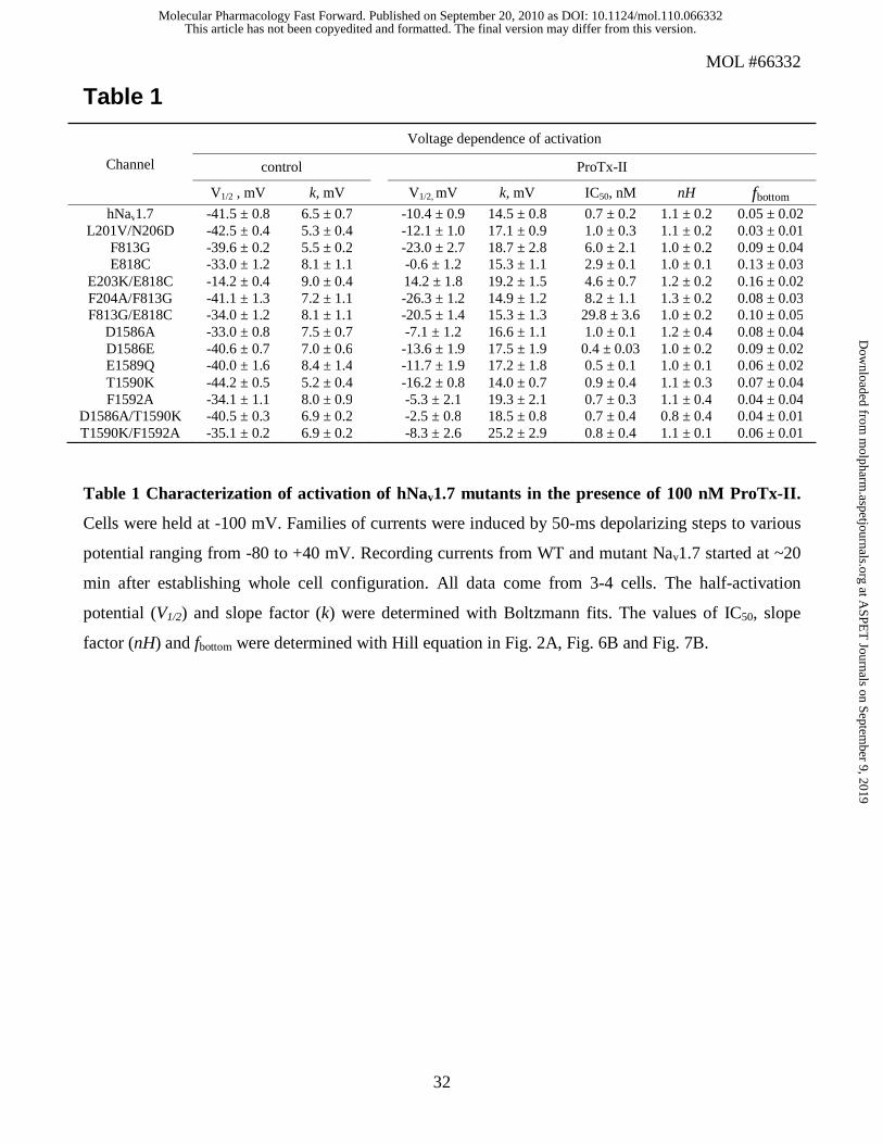
Table 1
Channel
Voltage dependence of activation
control
ProTx-II
V1/2 , mV k, mV V1/2, mV k, mV IC50, nM nH fbottom hNav1.7 -41.5 ± 0.8 6.5 ± 0.7 -10.4 ± 0.9 14.5 ± 0.8 0.7 ± 0.2 1.1 ± 0.2 0.05 ± 0.02
L201V/N206D -42.5 ± 0.4 5.3 ± 0.4 -12.1 ± 1.0 17.1 ± 0.9 1.0 ± 0.3 1.1 ± 0.2 0.03 ± 0.01 F813G -39.6 ± 0.2 5.5 ± 0.2 -23.0 ± 2.7 18.7 ± 2.8 6.0 ± 2.1 1.0 ± 0.2 0.09 ± 0.04 E818C -33.0 ± 1.2 8.1 ± 1.1 -0.6 ± 1.2 15.3 ± 1.1 2.9 ± 0.1 1.0 ± 0.1 0.13 ± 0.03
E203K/E818C -14.2 ± 0.4 9.0 ± 0.4 14.2 ± 1.8 19.2 ± 1.5 4.6 ± 0.7 1.2 ± 0.2 0.16 ± 0.02 F204A/F813G -41.1 ± 1.3 7.2 ± 1.1 -26.3 ± 1.2 14.9 ± 1.2 8.2 ± 1.1 1.3 ± 0.2 0.08 ± 0.03 F813G/E818C -34.0 ± 1.2 8.1 ± 1.1 -20.5 ± 1.4 15.3 ± 1.3 29.8 ± 3.6 1.0 ± 0.2 0.10 ± 0.05
D1586A -33.0 ± 0.8 7.5 ± 0.7 -7.1 ± 1.2 16.6 ± 1.1 1.0 ± 0.1 1.2 ± 0.4 0.08 ± 0.04 D1586E -40.6 ± 0.7 7.0 ± 0.6 -13.6 ± 1.9 17.5 ± 1.9 0.4 ± 0.03 1.0 ± 0.2 0.09 ± 0.02 E1589Q -40.0 ± 1.6 8.4 ± 1.4 -11.7 ± 1.9 17.2 ± 1.8 0.5 ± 0.1 1.0 ± 0.1 0.06 ± 0.02 T1590K -44.2 ± 0.5 5.2 ± 0.4 -16.2 ± 0.8 14.0 ± 0.7 0.9 ± 0.4 1.1 ± 0.3 0.07 ± 0.04 F1592A -34.1 ± 1.1 8.0 ± 0.9 -5.3 ± 2.1 19.3 ± 2.1 0.7 ± 0.3 1.1 ± 0.4 0.04 ± 0.04
D1586A/T1590K -40.5 ± 0.3 6.9 ± 0.2 -2.5 ± 0.8 18.5 ± 0.8 0.7 ± 0.4 0.8 ± 0.4 0.04 ± 0.01 T1590K/F1592A -35.1 ± 0.2 6.9 ± 0.2 -8.3 ± 2.6 25.2 ± 2.9 0.8 ± 0.4 1.1 ± 0.1 0.06 ± 0.01
Table 1 Characterization of activation of hNav1.7 mutants in the presence of 100 nM ProTx-II.
Cells were held at -100 mV. Families of currents were induced by 50-ms depolarizing steps to various
potential ranging from -80 to +40 mV. Recording currents from WT and mutant Nav1.7 started at ~20
min after establishing whole cell configuration. All data come from 3-4 cells. The half-activation
potential (V1/2) and slope factor (k) were determined with Boltzmann fits. The values of IC50, slope
factor (nH) and fbottom were determined with Hill equation in Fig. 2A, Fig. 6B and Fig. 7B.
This article has not been copyedited and formatted. The final version may differ from this version.Molecular Pharmacology Fast Forward. Published on September 20, 2010 as DOI: 10.1124/mol.110.066332
at ASPE
T Journals on Septem
ber 9, 2019m
olpharm.aspetjournals.org
Dow
nloaded from
MOL #66332
33
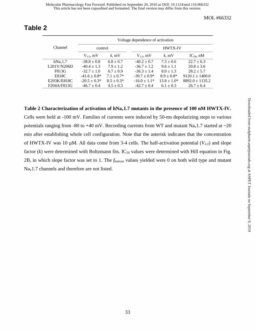
Table 2
Channel
Voltage dependence of activation
control HWTX-IV
V1/2, mV k, mV V1/2, mV k, mV IC50, nM
hNav1.7 -38.8 ± 0.8 6.8 ± 0.7 -40.2 ± 0.7 7.3 ± 0.6 22.7 ± 6.3 L201V/N206D -40.4 ± 1.3 7.9 ± 1.2 -36.7 ± 1.2 9.6 ± 1.1 20.8 ± 3.6
F813G -32.7 ± 1.0 6.7 ± 0.9 -36.3 ± 1.4 8.0 ± 1.3 28.2 ± 5.7 E818C -41.6 ± 0.8* 7.1 ± 0.7* -39.7 ± 0.9* 8.9 ± 0.8* 9120.1 ± 1400.0
E203K/E818C -20.5 ± 0.3* 8.5 ± 0.3* -16.0 ± 1.1* 13.8 ± 1.0* 8892.0 ± 1135.2 F204A/F813G -46.7 ± 0.4 4.5 ± 0.3 -42.7 ± 0.4 6.1 ± 0.3 26.7 ± 6.4
Table 2 Characterization of activation of hNav1.7 mutants in the presence of 100 nM HWTX-IV.
Cells were held at -100 mV. Families of currents were induced by 50-ms depolarizing steps to various
potentials ranging from -80 to +40 mV. Recording currents from WT and mutant Nav1.7 started at ~20
min after establishing whole cell configuration. Note that the asterisk indicates that the concentration
of HWTX-IV was 10 µM. All data come from 3-4 cells. The half-activation potential (V1/2) and slope
factor (k) were determined with Boltzmann fits. IC50 values were determined with Hill equation in Fig.
2B, in which slope factor was set to 1. The fbottom values yielded were 0 on both wild type and mutant
Nav1.7 channels and therefore are not listed.
This article has not been copyedited and formatted. The final version may differ from this version.Molecular Pharmacology Fast Forward. Published on September 20, 2010 as DOI: 10.1124/mol.110.066332
at ASPE
T Journals on Septem
ber 9, 2019m
olpharm.aspetjournals.org
Dow
nloaded from

This article has not been copyedited and formatted. The final version may differ from this version.Molecular Pharmacology Fast Forward. Published on September 20, 2010 as DOI: 10.1124/mol.110.066332
at ASPE
T Journals on Septem
ber 9, 2019m
olpharm.aspetjournals.org
Dow
nloaded from

This article has not been copyedited and formatted. The final version may differ from this version.Molecular Pharmacology Fast Forward. Published on September 20, 2010 as DOI: 10.1124/mol.110.066332
at ASPE
T Journals on Septem
ber 9, 2019m
olpharm.aspetjournals.org
Dow
nloaded from

This article has not been copyedited and formatted. The final version may differ from this version.Molecular Pharmacology Fast Forward. Published on September 20, 2010 as DOI: 10.1124/mol.110.066332
at ASPE
T Journals on Septem
ber 9, 2019m
olpharm.aspetjournals.org
Dow
nloaded from

This article has not been copyedited and formatted. The final version may differ from this version.Molecular Pharmacology Fast Forward. Published on September 20, 2010 as DOI: 10.1124/mol.110.066332
at ASPE
T Journals on Septem
ber 9, 2019m
olpharm.aspetjournals.org
Dow
nloaded from

This article has not been copyedited and formatted. The final version may differ from this version.Molecular Pharmacology Fast Forward. Published on September 20, 2010 as DOI: 10.1124/mol.110.066332
at ASPE
T Journals on Septem
ber 9, 2019m
olpharm.aspetjournals.org
Dow
nloaded from

This article has not been copyedited and formatted. The final version may differ from this version.Molecular Pharmacology Fast Forward. Published on September 20, 2010 as DOI: 10.1124/mol.110.066332
at ASPE
T Journals on Septem
ber 9, 2019m
olpharm.aspetjournals.org
Dow
nloaded from
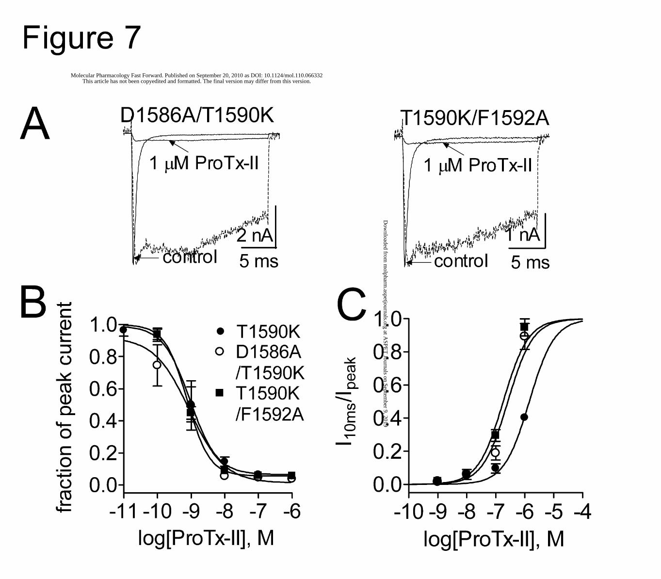
This article has not been copyedited and formatted. The final version may differ from this version.Molecular Pharmacology Fast Forward. Published on September 20, 2010 as DOI: 10.1124/mol.110.066332
at ASPE
T Journals on Septem
ber 9, 2019m
olpharm.aspetjournals.org
Dow
nloaded from