the synthesis of new oxindoles as analogs of natural...
TRANSCRIPT

Turk J Chem
(2018) 42: 332 – 345
c⃝ TUBITAK
doi:10.3906/kim-1706-51
Turkish Journal of Chemistry
http :// journa l s . tub i tak .gov . t r/chem/
Research Article
The synthesis of new oxindoles as analogs of natural product
3,3 ′ -bis(indolyl)oxindole and in vitro evaluation of the enzyme activity of G6PD
and 6PGD
Sinan BAYINDIR1,∗, Adnan AYNA1, Yusuf TEMEL2, Mehmet CIFTCI11Department of Chemistry, Faculty of Sciences and Arts, Bingol University, Bingol, Turkey
2Department of Health Services, Vocational Schools, Bingol University, Bingol, Turkey
Received: 22.06.2017 • Accepted/Published Online: 30.10.2017 • Final Version: 27.04.2018
Abstract: Natural and synthetic derivatives that contain an indole core are being used in medical treatments and
technological processes. Therefore, the development of new synthetic methods for the synthesis of indole derivatives is
very popular. In this study, new oxindoles with reaction of 4,7-dihydro-1H -indole (2) and isatin (4) were synthesized as
analogs of natural product 3,3 ′ -bis(indolyl)oxindole. The biological properties of the compounds obtained during this
study were also studied, showing that compounds 5, 7, and 12 inhibited the activity of G6PD with an IC50 of 99 µM,
231 µM, and 304 µM respectively. The activity of rat erythrocyte 6PGD was increased in the presence of 5 and 7 and
was inhibited in the presence of 12. As indole derivative 5 was an activator of 6PGD and inhibitor of G6PD, it was
selected for docking studies to understand the mechanism of activation and inhibition.
Key words: Oxindoles, natural product, 3,3 ′ -bis(indolyl)oxindole, enzyme, 6PGD, G6PD
1. Introduction
Natural compounds with more than one indole skeleton have been reported to show anticancer activity by
interacting with different cellular targets.1−8 The 3,3 ′ -bisindole oxindole and 3-substituted oxindole skeleton
belongs to a class of privileged heterocyclic frameworks, which constitutes the core structures of many bioactive
compounds (Figure 1). Additionally, 2-alkylindoles and 2.2 ′ -bis(indolyl)methanes represent quite an interesting
class and are also known to play an important role in numerous biological and technological processes.9−12
Therefore, a great deal of attention has been given to the development of effective, facile, and innovative
synthetic strategies for the enhancement of indole chemistry. Indole is an electron-rich heteroaromatic system,
and although various methods for synthesis of C3-substituted derivatives are well known, the synthesis of
C2-substituted derivatives continues to be a difficult task.13−24 While a number of methods have already
been published for the synthesis of 3-substituted indole derivatives, there are no methods for the synthesis
of 2-substituted indole derivatives through the indole ring. An alternative method that utilizes 4,7-dihydro-
1H -indole (2) derivatives as synthetic equivalents (synthons) for easy synthesis of 2-substituted indoles was
developed by Saracoglu and coworkers25−28 (Figure 2). The indole (1) exhibits reactivity at the C-3 position
against the electrophiles and forms the 3-substituent indole derivatives at the end of the reaction. The advantage
of this strategy developed by Saracoglu and coworkers is that the derivatives 2 and 3 obtained as a result of
∗Correspondence: [email protected]
332

BAYINDIR et al./Turk J Chem
reduction of the indole (1) are also pyrrole derivatives. The obtained molecules 2 and 3 electrophilically react
with C-2 carbon to give 2-substituted indole derivatives (Figure 2).
Figure 1. Chemical structures of compounds containing 3,3 ′ -bisindole and indolin-2-one framework.
NH2
NH
NH
NH1
E
E
[H]
E : electrophiles
synthons
C3
C2
1H-pyrrole
N
H3
Figure 2. Reactivity of indole (1) and 4,7-dihydro-1H -indole (2).
The technological applications of synthetic indole derivatives have attracted a great deal of interest
in recent years.29−31 In this context, the indole skeleton is a highly used unit for biological chemosensor
applications. Previous studies of bisindole derivatives revealed that they alter the enzymatic activity of some
enzymes like human carbonic anhydrase isoforms I-II and α/β -glycosidase. In a recent study, the antioxidant
activities of 1,4-bis(indolin-1-ylmethyl)benzene derivatives were reported by Talaz and coworkers.32 However,
there is no study investigating the effect of such compounds on metabolic enzymes including glucose-6-phosphate
dehydrogenase (G6PD; E.C.1.1.1.49) and 6-phosphogluconate dehydrogenase (6PGD; E.C.1.1.1.44). Therefore,
one aim of the work described in this paper is to investigate the effect of indole derivatives on the activity of
the corresponding enzymes. G6PD is the rate-limiting enzyme of the pentose phosphate pathway. It catalyzes
the irreversible conversion of glucose 6-phosphate to 6-phosphoglucono-δ -lactone in the presence of NADP+ .33
6PGD is the third enzyme in the pentose phosphate pathway. It is a well-known oxidative carboxylase that
catalyzes conversion of 6-phosphogluconate into ribulose 5-phosphate.34 The pentose phosphate pathway is
one of the key components of cellular metabolism. It is strongly connected to glycolysis as a major consumer
of glucose. The primary roles of the pathway are generating NADPH as a source of reducing power and the
synthesis of ribose 5-phosphate, which is required for the synthesis of nucleic acids.35,36 In the absence of these
333

BAYINDIR et al./Turk J Chem
enzymes the erythrocyte is susceptible to oxidative damage. G6PD is also associated with some human diseases
including cancer, metabolic disorders, and cardiovascular diseases.37−39 It was also reported that suppression of
6PGD decreased lipogenesis and RNA biosynthesis and increased reactive oxygen levels in cancer cells, lessening
cell proliferation and tumor growth and suggesting that 6PGD could be an anticancer target.40
In our previous studies, we developed an efficient, facile, and atom-economical protocol for the preparation
of 2-alkylated indoles and bis(2-indolyl)methanes derivatives through addition of one or two equivalents of 4,7-
dihydro-1H -indole (2) using ketones as electrophiles followed by an oxidation step28 (Scheme 1a). Herein,
Bi(NO3).3 5H2O-catalyzed reactions of 4,7-dihydro-1H -indole (2) with isatin (4) (Scheme 1b) and the obtained
molecules were examined along with investigating their effects on the enzymatic activity of rat erythrocyte
G6PD and 6PGD and docking studies.
Scheme 1. Strategies for the reactions of 4,7-dihydro-1H -indole (2) with isatin (4).
2. Results and discussion
2.1. Chemistry
Our research interest in the synthesis of new oxindole derivatives with 1,2- and 1,3-diketones of 4,7-dihydro-1H -
indole (2) encouraged us to ascertain the behavior of ketones having a different nature against the condensation
reactions of 4,7-dihydro-1H -indole (2). In this context, initially 4,7-dihydro-1H -indole, which was used as
an output molecule in the reactions, was synthesized via Birch reduction reaction of indole with Li in liquid
ammonia. The Birch reduction, a very powerful reducing system, has been reported to yield a mixture of
4,7-dihydro-1H -indole (2) and 4,5,6,7-tetrahydroindole (3) in a 4:1 ratio according to NMR, which could be
best separated by recrystallization or distillation.25−28 Isatin (4) as an 1,2-diketone derivative has provided
an opportunity to investigate the effect of the carbonyl group on the condensation of 4,7-dihydro-1H -indole
(2) with ketone (Scheme 2). As shown in Table 1, the reaction of 4,7-dihydro-1H -indole with isatin (4) was
334

BAYINDIR et al./Turk J Chem
first assayed with the Bi(NO3).3 5H2O catalyst at room temperature. However, no product formation was
observed under these conditions (Table 1, entry 5). The bismuth nitrate-catalyzed reaction of 4,7-dihydro-1H -
indole with isatin (4) in CH3CN at reflux temperature gave alcohol derivative 3-(4,7-dihydro-1H -indol-2-yl)-
3-hydroxyindolin-2-one (5) in 98% yield instead of the expected product 6 (Table 1, entry 6). Additionally, a
very complex mixture with trifluoroacetic acid (TFA), ZrCl4 , AlCl3 , and PhCOOH catalysts was obtained at
room temperature (Table 1, entries 1–4). As a result of the reaction, the synthesis of indole derivative 6 was
expected by elimination of 1 mol of water and 1 mol of protons from alcohol derivative 5. However, it was
observed that the intermediate product, alcohol derivative 5, was more stable, and it was obtained as the main
product (Scheme 2). It was seen that NH groups resonated at 10.4 and 10.2 ppm in the 1H NMR spectrum of
unexpected bisindole derivative 5. One proton available in the alcohol units resonated at 5.26 ppm. All these
signals support the recommended structure.
Scheme 2. The synthesis of 5 and 7.
Surprisingly, the oxidation of alcohol derivative 5 with MnO2 produced unexpected amine derivative
(2-aminophenyl)(1H -indol-2-yl)methanone (7) in 81% yield instead of the expected product 8 (Schemes 2
and 3). The same results were obtained with p -benzoquinone (pBQ) or DDQ (2,3-dichloro-5,6-dicyano-1,4-
benzoquinone) in 25% and 12% yields, respectively. It was seen that one NH group resonated at 9.29 ppm in
the 1H NMR spectrum of unexpected amine derivative 7. The two protons available in amine units resonated at
5.71 ppm. Also, while the 13C NMR resonance signal was available at 177.0 ppm in the 13C NMR spectrum of
unexpected amine derivative 7, the signal shows the existence of a ketone group in the molecule and 14 carbon
resonance signals in the aromatic area, supporting the recommended structure (Scheme 2). Because of the
sensitivity of the carbonyl group toward moisture the hydration of the carbonyl group undergoes carbon/nitrogen
bond cleavage to give unexpected amine derivative 7 and carbon dioxide (Scheme 3).
Bis/tris-indole derivatives are an important class of biological active indoles. In this context, the reaction
of 2 equivalents of 4,7-dihydro-1H -indole (2) with one equivalent of isatin (4) was also performed under
similar conditions (Scheme 4). The 2,2 ′ -bis(indolyl)oxindole (12), which resembles anticancer agent 3,3 ′ -
bis(indolyl)oxindole, was synthesized by condensation of 4,7-dihydro-1H -indole (2, 2 equiv.) with isatin (4, 1
335

BAYINDIR et al./Turk J Chem
Table 1. Optimization of reaction conditions.a
Entry Catalyst Solvent Temp. (◦C) Time Yieldb (%)
1 TFA CH2Cl2 RT 30 min. 0[c,d]
2 AlCl3 CH2Cl2 RT 30 min. 0[c,d]
3 PhCOOH CH2Cl2 RT 30 min. 0[c,d]
4 ZrCl4 CH2Cl2 RT 30 min. 0[c,d]
5 Bi(NO3).35H2O CH2Cl2 RT 12h 0
6 Bi(NO3).35H2O MeCN Reflux 5h 98
7 Cu(OTf)2 MeCN Reflux 5h 56[c,d]
8 InCl3 MeCN Reflux 5h 11[c,d]
9 Zn(OTf)2 MeCN Reflux 5h 61[c,d]
10 BiCl3 MeCN Reflux 5h 72a:Conditions: 4,7-dihydro-1H-indole (2, 1 equiv.), isatin (4, 1 equiv.), catalyst (0.1 mmol), and solvent (10 mL).b:Isolated yields of 5. c:Complex reaction mixture. d:Under N2.
Scheme 3. Proposed mechanism for synthesis of unexpected
equiv.) catalyzed by Bi(NO3).3 5H2O in CH3CN, followed by an oxidation reaction by p -benzoquinone over
4,7-dihydro-1H -indole derivative 11 (Scheme 4). Despite all attempts, 4,7-dihydro-1H-indole derivative 11,
which was seen from NMR spectra, could not be isolated.
Additionally, the reactions of 4,7-dihydro-1H -indole (2) with other 1,2- and 1,3-diketones such as 2,3-
butanedione (13), acenaphthoquinone (14), and cyclohexane-1,3-dione (15) were examined under the same
conditions (Figure 3). No significant results were obtained from the studies of 1,2-diketones. While the reaction
of diketones 13 and 14 gave no isolable product, no reaction was observed with cyclohexane-1,3-dione (15).
336

BAYINDIR et al./Turk J Chem
Scheme 4. The synthesis of 12.
Figure 3. The chemical structures of other 1,2- and 1,3-diketones examined.
2.2. G6PD and 6PGD inhibition/activations studies
Following synthesis of the bisindole derivatives 5, 7, and 12, investigation of the effects of these derivatives
on rat blood erythrocyte G6PD and 6PGD was conducted. In order to achieve that, first G6PD and 6PGD
were purified from rat blood erythrocytes by 2 ′ ,5 ′ -ADP Sepharose 4B affinity chromatography.41−43 Following
purification, the in vitro effects of ligands 5, 7, and 12 on the activity of both enzymes were investigated. The
inhibition effect of the compounds on enzymes is expressed in IC50 values and K i constants. Derivatives 5, 7,
and 12 inhibited the activity of G6PD with an IC50 of 99 µM, 231 µM, and 304 µM, respectively (Table 2,
Figure S8, ESI). To further understand the mechanism by which these compounds inhibited these enzymes, the
inhibitory modes of the derivatives were studied. The collected raw data were described as Lineweaver–Burk
diagrams and suggested that all derivatives except 5 inhibited the activity of G6PD noncompetitively with
respect to G6P as the reaction rate was decreased and Km remained unchanged while 5 was a competitive
inhibitor of G6PD. Their K i values were calculated based on this diagram to be 48 µM, 369 µM, and 304 µM
respectively (Figure S9, ESI). As suggested by the IC50 and K i values of each compound, inhibition is most
powerful by ligand 5. This could be attributed to the hydrogen bonding capacity of the compounds with the
amino acid residues of the enzyme. Indole derivative 5 has three groups that can make hydrogen bonds and
it also has oxygen, which is more electronegative than other atoms, which makes it form stronger hydrogen
bonds with the amine group of the amino acid residues. These properties may make indole derivative 5 a more
powerful inhibitor than the others.
337

BAYINDIR et al./Turk J Chem
Table 2. Determination of IC50 and K i values and inhibition types of the compounds.
G6PD
Ligand IC50 (µM) Ki (µM) Type of inhibition
5 99 48 Competitive
7 231 369 Noncompetitive
12 304 304 Noncompetitive
6PGD
Ligand IC50 (µM) Ki (µM) Type of inhibition
5 N/A N/A N/A
7 N/A N/A N/A
12 209 N/A N/A
The effects of these compounds on 6PGD were also tested. Unlike G6PD, the activity of 6PGD was
increased approximately fourfold (380%) in the presence of 250 µM indole derivative 5 and the activity was
increased approximately twofold (176%) in the presence of 1500 µM indole derivative 7. However, activity was
inhibited in the presence of indole derivative 12 with an IC50 of 209 µM (Table 2, Figure S10, ESI).
2.3. Molecular docking analysis
The inhibition studies revealed that compound 5 inhibited the activity of G6PD competitively. To elucidate the
binding modes of 5 in rat erythrocyte G6PD, a homology model of the structure of the enzyme was generated
and ligand 5 was docked. Docking experiments gave approximately 250 poses. Careful examination of each pose
revealed that the ligand is located in the NADP+ binding domain and fits in the position of NADP+ , making
weak interactions with the residues around the binding site (Figures 4a–4c and S12, ESI). This result supports
experimental studies demonstrating that the enzyme is competitively inhibited by ligand 5. The oxygen atom
of the ketone group in 5 makes a hydrogen bond (4 A) with the NH group of Gly41. Similarly, the OH group
of 5 will form a hydrogen bond (3.4 A) with the N atom of Asp42. While the C4 -H (acidic proton) of the
4,7-dihydro indole unit makes a hydrogen bond (2.9 A) with the NH group of Pro143, the C7 -H (acidic proton)
atom of the 4,7-dihydro indole unit forms a hydrogen bond (3.9 A) with the NH group of the pyrrole ring in
Pro143 (Figure 4b). NADP+ makes similar interactions with the residues around it. The inhibition of the
enzyme might be caused by the replacement of NADP+ by 5.
In contrast to its effect on G6PD, the presence of the ligand increased the activity of 6PGD. To further
understand the effect, the structure of the enzyme was generated by homology modeling and docking studies
were performed. The results of docking studies were analyzed manually (Figures 5a–5d and S11, ESI). It
was revealed that the ligand was located close to the substrate and ligand binding site without interacting
with any residues around it. It was postulated that the ligand could assist in increasing the activity of the
enzyme by reducing NADP+ to NADPH. In catalysis, two residues, one acting as an acid (Glu) and the other
as a base (Lys), are proposed to contribute to all three catalytic steps of the reaction catalyzed by 6PGD:
dehydrogenation, decarboxylation, and keto-enol tautomerization. Lys is thought to be unprotonated in the
enzyme/substrate complex, where it takes a proton from the 3-OH of 6PGA as a hydride that is transferred
from the C3 of 6PGA to NADP+ . The resultant 3-keto-6PGA intermediate is subsequently decarboxylated to
338

BAYINDIR et al./Turk J Chem
Figure 4. a) Representation of the binding site of ligand 5, b) zoomed view of binding site of ligand 5, and c) NADP+
binding site of G6PD.
form the enediol of 5-phospho-ribulose. At this stage, Glu donates a proton to the C3 carbonyl group of the
keto intermediate to facilitate decarboxylation. Both a base and an acid are essential in the tautomerization
of the enediol intermediate to form ribulose 5-phosphate, with Glu giving a proton to the C1 of the enediol
intermediate and the base accepting a proton from its 2-OH.44−47
As shown in Figures 5a–5d and Scheme 5, it is proposed that NADP+ will assist in converting 6PGA
to R5P. Following that, it will oxidize the 4,7-dihydro indole unit in ligand 5 to an indole unit. The amino
group of an arginine residue located in the close vicinity of 5 will behave as a base and attack the C7 -H of 4,7-
dihydro-1H -indole, forming an aromatic ring, which is then receiving protons from C4 -H and reduces NADP+
to NADPH, thus increasing the activity of the enzyme (Scheme 5).
In conclusion, we have reported the synthesis of possible biologically active new indoles 3-(4,7-dihydro-
1H -indol-2-yl)-3-hydroxyindolin-2-one (5), (2-aminophenyl)(1H -indol-2-yl)methanone (7), and 1H ,1 ′′H -[2,3 ′ :
3 ′ ,2 ′′ -terindol]-2 ′(1′H)-one (12) from the reaction of 4,7-dihydro-1H -indole (2) with isatin (4) and have
discussed the formation mechanism. In addition to synthesis, effects of indole derivatives 5, 7, and 12 on the
activity of erythrocyte G6PD and 6PGD have also been investigated in in vitro conditions. These studies showed
that 5, 7, and 12 inhibited the activity of G6PD with an IC50 of 99 µM, 231 µM, and 304 µM, respectively.
However, studies on rat erythrocyte 6PGD indicated that the activity was increased in the presence of 5 and
339

BAYINDIR et al./Turk J Chem
Figure 5. a) Representation of NADP+ (PDB-ID: 1PGN), b) 6PGA (PDB-ID: 1PGP), c) binding site of 5, and d)
zoomed view of binding sites in 6PGD. The binding sites of NADP+ and 6PGA were derived from crystal structures
and the binding site for 5 was calculated via docking.
Scheme 5. Proposed mechanism for oxidation of 5 with 6PGD.
7 and was inhibited in the presence of 12. As ligand 5 inhibited one enzyme and activated the other, it was
selected for docking experiments. The analyses of docking results suggested that the ligand binds the NADP+
binding site in G6PD while it is located in the close vicinity of the substrate and NADP+ binding sites donating
a hydride to NADP, hence increasing the activity of 6PGD.
3. Experimental
All chemicals, reagents, and solvents were commercially available from Sigma-Aldrich or Merck and were used
as received. 2 ′ ,5 ′ -ADP Sepharose 4B was purchased from Pharmacia. Melting points were determined on a
340

BAYINDIR et al./Turk J Chem
Buchi 539 capillary melting apparatus and are uncorrected. Infrared spectra were recorded on a Mattson 1000
FT-IR spectrophotometer. 1H NMR and 13C NMR spectra were recorded on a 400 (100)-MHz Varian and
Bruker spectrometer and are reported in terms of chemical shift (δ , ppm) with SiMe4 as an internal standard.
Data for 1H NMR are recorded as follows: chemical shift (δ , ppm), multiplicity (s: singlet, d: doublet,
t: triplet, q: quartet, p: pentet, m: multiplet, bs: broad singlet, bd: broad doublet, qd: quasi doublet) and
coupling constant (s) in Hz, integration. Elemental analyses were carried out on a LECO CHNS-932 instrument.
Column chromatography was carried out on silica gel 60 (230–400 mesh ASTM). The reaction progress was
monitored by thin-layer chromatography (TLC) (0.25-mm-thick precoated silica plates: Merck Fertigplatten
Kieselgel (60 F254)). UV-Vis spectra were recorded on a Shimadzu UV-3101PL UV-Vis-NIR spectrometer.
3.1. Synthesis of 4,7-dihydro-1H-indole (2)
Compound 2 was prepared according to the literature method.25,28 Liquid ammonia (500 mL) was distilled
under N2 into a predried three-necked flask. Then the solution of indole (1, 25 g, 0.21 mol) in dry Et2O
(100 mL) was added and the resulting solution was cooled to –35 ◦C and stirred mechanically. The resulting
solution was treated with lithium metal (6 g, 0.84 mol) added in small pieces for 5–10 min, which reacted very
rapidly. The resulting deep blue solution was stirred at the same temperature for 1 h and then the mixture
was transferred to room temperature. After the excess ammonia had evaporated, Et2O (200 mL), NH4Cl (5
g), and H2O (300 mL) were carefully added to the reaction mixture. The layers were separated, the aqueous
layer was extracted with Et2O (2 × 200 mL), and the combined organic layers were washed with NaHCO3
(2 × 100 mL), dried (MgSO4), filtered, and concentrated. The 1H NMR spectrum of the residue showed the
formation of 4,7-dihydro-1H -indole (2) and 4,5,6,7-tetrahydro-1H -indole (3) in a 4:1 ratio. The crude product
(23 g) was recrystallized with hexane to give 4,7-dihydro-1H -indole (2) (white solid, mp: 38–39 ◦C, 19 g, 75%)
as colorless crystals.
1H NMR (400 MHz, CDCl3): δ 7.70 (m, NH, 1H), 6.72 (t, J = 2.5 Hz, =CH, 1H), 6.07 (t, J = 2.5
Hz, =CH, 1H), 5.95 (bd, J = 10.1 Hz, =CH, 1H), 5.87 (bd, J = 10.1 Hz, =CH, 1H), 3.30 (bs, CH2 , 4H); 13C
NMR (100 MHz, CDCl3): δ 128.0, 127.9, 125.98, 118.3, 115.9, 108.8, 27.1, 26.0.
3.2. The reaction of 4,7-dihydro-1H -indole (2, 1 equiv.) with isatin (4, 1 equiv.)
To a solution of 4,7-dihydro-1H -indole (2, 200 mg, 1.68 mmol) in MeCN (5 mL), indoline-2,3-dione (4, isatin,
247 mg, 1.68 mmol) and Bi(NO3).35H2O (0.1 mmol) were added. The reaction mixture was stirred magnetically
in a flask at 100 ◦C. The reaction was monitored by TLC. After the completion of the reaction, the mixture
was diluted with ethyl acetate (30 mL) and washed with water (2 × 50 mL), and the organic phase was dried
over Na2SO4 . The crude product was purified by silica gel column chromatography and the isolated compound
was given according to the elution sequence (EtOAc/Hexane; v/v: 1/3). After purification, 3-(4,7-dihydro-
1H -indol-2-yl)-3-hydroxyindolin-2-one (5, 437 mg, 98%) was obtained as a pale yellow solid (mp: 165–166 ◦C
(crystallized over CH2Cl2 /hexane)).
1H NMR (400 MHz, DMSO-d6): δ 10.42 (bs, NH, 1H), 10.21 (s, NH, 1H), 7.38 (d, J = 7.6 Hz, =CH,
1H), 7.20 (t, J = 7.6 Hz, =CH, 1H), 6.97 (t, J = 7.6 Hz, =CH, 1H), 6.79 (d, J = 7.6 Hz, =CH, 1H), 7.31 (s,
=CH, 1H), 5.77 (m, =CH, 2H), 5.27 (qd, J = 2.5 Hz, OH, 1H), 3.30–3.27 (m, CH2 , 2H), 3.17–3.13 (m, CH2 ,
2H); 13C NMR (100 MHz, DMSO-d6): δ 178.3, 142.2, 133.1, 129.6, 129.4, 126.0, 125.7, 125.6, 124.2, 122.2,
341

BAYINDIR et al./Turk J Chem
112.3, 110.2, 105.5, 74.1, 25.1, 24.6; IR (KBr, cm−1): 3426, 3403, 3342, 3036, 2918, 2876, 2851, 2725, 2685,
1714, 1605, 1469, 1208, 870, 740; Anal. Calcd. for C16H14N2O2 : C, 72.17; H, 5.30; N, 10.52; found: C, 72.57;
H, 5.31; N, 10.97; TLC: Rf = 0.12 (EtOAc/hexane (v/v: 1/3), 254 nm).
3.3. The oxidation of 3-(4,7-dihydro-1H -indol-2-yl)-3-hydroxy indolin-2-one (5)
Procedure A: To a solution of 3-(4,7-dihydro-1H -indol-2-yl)-3-hydroxyindolin-2-one (5, 300 mg, 1.13 mmol)
in 10 mL of CH2Cl2 was added MnO2 (980 mg, 11.3 mmol). After stirring for 12 h at room temperature, the
mixture was diluted with EtOAc (30 mL) and washed with water (3 × 30 mL), and the organic phase was dried
over Na2SO4 . The crude product (230 mg) was eluted on silica gel (25 g) with EtOAc/hexane (v/v: 3/7).
After purification, (2-aminophenyl)(1H -indol-2-yl)methanone (7, 215 mg, 81%) was obtained as a white solid
(mp: 115–116 ◦C (crystallized over CH2Cl2/ hexane)).
Procedure B: (2-Aminophenyl)(1H -indol-2-yl)methanone (7) was obtained as a white solid (67 mg,
25%) from the reaction of 3-(4,7-dihydro-1H -indol-2-yl)-3-hydroxyindolin-2-one (5, 300 mg, 1.13 mmol) with
p -benzoquinone (304 mg, 2.82 mmol) in CH2Cl2 at room temperature for 12 h.
Procedure C: (2-Aminophenyl)(1H-indol-2-yl)methanone (7) was obtained as a white solid (32 mg,
12%) from the reaction of 3-(4,7-dihydro-1H -indol-2-yl)-3-hydroxyindolin-2-one (5, 300 mg, 1.13 mmol) with
DDQ (640 mg, 2.82 mmol) in CH2Cl2 at room temperature for 12 h.
1H NMR (400 MHz, CDCl3): δ 9.29 (bs, NH, 1H), 8.01 (d, J = 8.1 Hz, =CH, 1H), 7.71 (d, J = 7.3
Hz, =CH, 1H), 7.45 (d, J = 8.1 Hz, =CH, 1H), 7.36–7.31 (m, =CH, 2H), 7.16 (t, J = 7.3 Hz, =CH, 1H),
7.11 (s, =CH, 1H), 6.78–7.74 (m, =CH, 2H), 5.71 (s, NH2 , 2H);13C NMR (100 MHz, CDCl3): δ 177.0, 149.7,
137.0, 135.2, 133.8, 132.5, 127.8, 126.0, 123.0, 120.9, 119.3, 117.0, 116.2, 112.0, 111.7; IR (KBr, cm−1): 3437,
3401, 3038, 2918, 2876, 2851, 2725, 1725, 1635, 1424, 1205, 777; Anal. Calcd. for C15H12N2O: C, 76.25; H,
5.12; N, 11.86; found: C, 76.37; H, 5.11; N, 11.97; TLC: Rf = 0.32 (EtOAc/hexane (v/v: 3/7), 254 nm).
3.4. The reaction of 4,7-dihydro-1H -indole (2, 2 equiv.) with isatin (4, 1 equiv.)
To a solution of 4,7-dihydro-1H -indole (2; 300 mg, 2.50 mmol) in MeCN (5 mL) was added isatin (4, 185 mg,
1.26 mmol) and Bi(NO3).3 5H2O (0.1 mmol). The reaction mixture was stirred magnetically in a flask at 100 ◦C.
The reaction was monitored by TLC. After the completion of the reaction, the mixture was diluted with ethyl
acetate (30 mL) and washed with water (2 × 50 mL), and the organic phase was dried over Na2SO4 . The crude
product was dissolved in CH2Cl2 (15 mL) and p -benzoquinone (330 mg, 3.06 mmol) was added. The mixture
was stirred at room temperature overnight. After completion of the reaction, the solvent was evaporated, the
crude product was dissolved with ethyl acetate (30 mL), and the organic phase was washed with NaOH (2 N,
2 × 30 mL) and brine (30 mL) and dried over Na2SO4 . The crude product was purified by silica gel column
chromatography and isolated compounds was given according to elution sequence (EtOAc/hexane; v/v: 3/17).
After purification, 1H ,1 ′′H -[2,3 ′ :3 ′ ,2 ′′ -terindol]-2 ′ (1 ′ H)-one (12, 382 mg, 85%) was obtained as a pale red
solid (mp: 137–138 ◦C (crystallized over CH2Cl2/hexane)).
1H NMR (400 MHz, CDCl3): δ 8.76 (bs, NH, 2H), 8.30 (bs, NH, 1H), 7.60 (d, J = 7.6 Hz, =CH, 1H),
7.52 (d, J = 7.6 Hz, =CH, 2H), 7.33–7.26 (m, =CH, 3H), 7.20–7.13 (m, =CH, 3H), 7.06 (t, J = 7.6 Hz, =CH,
2H), 6.99 (d, J = 7.6 Hz, =CH, 1H), 6.42 (s, =CH, 2H); 13C NMR (100 MHz, CDCl3): δ 177.0, 140.3, 136.8,
135.1, 130.8, 129.4, 128.0, 126.1, 123.6, 122.7, 120.9, 120.4, 111.4, 110.9, 102.4, 79.3; IR (KBr, cm−1): 3451,
342
BAYINDIR et al./Turk J Chem
3195, 3115, 3061, 2816, 1760, 1738, 1698, 1589, 1270, 1203, 1145, 887, 870, 770; Anal. Calcd. for C24H17N3O:
C, 79.32; H, 4.72; N, 11.56, found: C, 79.57; H, 4.51; N, 11.31; TLC: Rf = 0.16 (EtOAc/hexane (v/v: 3/17),
254 nm).
3.5. Preparation of hemolysate
Fresh blood samples were obtained from rats and placed in EDTA-containing tubes. In order to distinguish
erythrocytes from plasma, blood samples were filtered to remove any impurities, then centrifuged for 15 min at
2500 × g to remove plasma. After that the precipitated erythrocytes were washed three times with 0.16 M
KCl and hemolyzed with 5 volumes of cold water. Then, to remove the cell membranes and intact cells, 30 min
of centrifugation at 10,000 × g was performed.
3.6. 2 ′ ,5 ′ -ADP Sepharose 4B Affinity chromatography
Following preparation of the hemolysate, the sample was passed through a 2 ′ ,5 ′ -ADP Sepharose 4B Affinity
column, which was equilibrated with 50 mM KH2PO4 , 1 mM EDTA, and 1 mM DTT at pH 7.3. The protein
was eluted with 80 mM KH2PO4 , 10 mM EDTA, 80 mM KCI, and 5 mM NADP+ at pH 7.3. All procedures
were carried out at 4 ◦C.41−43
3.7. In vitro enzyme inhibition studies
G6PD and 6PGD enzyme activities were measured at 25 ◦C spectrometrically at 340 nm by following the rate
of appearance of NADPH. Assays were initiated by the addition of enzyme to 1.0 mL of 100 mM Tris-HCl, pH
8, containing 0.5 mM EDTA, 0.01 mM MgCl2 , 0.6 mM G6P/6PGA, and 0.2 mM NADP+ . To determine the
effect of compounds 5, 7, and 12 on the activity of rat erythrocyte G6PD and 6PGD, different concentrations
of the corresponding compounds were added in the assay mixture given above. The enzyme activities in the
absence of compounds were taken as 100%. Activity % vs. compound concentration graphs were drawn and
used to calculate the drug concentrations causing a 50% decrease in enzyme activity (IC50 values). The types
of inhibition and K i constants were determined via Lineweaver–Burk graphs.
3.8. Structure preparation, homology modeling, and ligand docking
The sequences of G6PD and 6PGD of rat erythrocytes were retrieved from UniProt (http://www.uniprot.org/
uniprot) in the FASTA format. The sequences were then submitted to PHYRE2 (Protein Homology/analogy
Recognition Engine V 2.0) for protein structure prediction.49 The X-ray crystal structures of human G6PD
(PDB-ID: 1QKI) and sheep 6PGD (PDB-ID: 1PGP) were used as starting structures for molecular modeling.
PROCHECK48 was used for model validation. The molecular structure of compound 5 was built in Chem3D
Pro 12.0 and energy-minimized prior to docking studies. Ligand docking calculations were performed in
SwissDock.49 The docked structures were examined manually and the best pose for the inhibitor was selected
on the basis of the DG value, the scaffold conformation, and the hydrogen bonds formed between residues and
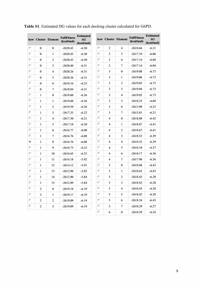
the inhibitor. Estimated DG values for each docking cluster are given in Table S1. UCSF Chimera and PyMol
were used for molecular visualization.50,51
343

BAYINDIR et al./Turk J Chem
Acknowledgements
The authors are indebted to the Department of Chemistry at Bingol University (BAP-209-324-2015) and would
also like to thank Prof Dr Nurullah Saracoglu for practical support and helpful discussions.
References
1. Humphrey, G. R.; Kuethe, J. T. Chem. Rev. 2006, 106, 2875-2911.
2. Cacchi, S.; Fabrizi, G. Chem. Rev. 2005, 105, 2873-2920.
3. Kamal, A.; Srikanth, Y. V. V.; Khan, M. N. A.; Shaik, T. B.; Ashraf, M. Bioorg. Med. Chem. Lett. 2010, 20,
5229-5231.
4. Sayed, M. T.; Mahmoud, K.; Hilgeroth, A.; Fakhr, I. M. I. J. Heterocyclic Chem. 2016, 53, 188-196.
5. Huber, K.; Schemies, J.; Uciechowska, U.; Wagner, J. M.; Rumpf, T.; Lewrick, F.; Suss, R.; Sippl, W.; Jung, W.;
Bracher, F. J. Med. Chem. 2010, 53, 1383-1386.
6. Giannini, G.; Marzi, M.; Marzo, M. D.; Battistuzzi, G.; Pezzi, R.; Brunetti, T. Bioorg. Med. Chem. Lett. 2009,
19, 2840-2844.
7. Gaisina, I. N.; Gallier, F.; Ougolkov, A. V.; Kim, K. H.; Kurome, T.; Guo, S.; Holzle, D.; Luchini, D. N.; Blond,
S. Y.; Billadeau, D. D. et al. J. Med. Chem. 2009, 52, 1853-1863.
8. Paira, P.; Hazra, A.; Kumar, S.; Paira, R.; Sahu, K. B.; Naskar, S.; Saha, P.; Mondal, S.; Maity, A.; Banerjee, S.
et al. Bioorg. Med. Chem. Lett. 2009, 19, 4786-4789.
9. Shiri, M.; Zolfigol, M. A.; Kruger, H. G.; Tanbakouchian, Z. Chem. Rev. 2010, 110, 2250-2293.
10. Safe, S.; Papineni, S.; Chintharlapalli, S. Cancer Lett. 2008, 269, 326-338.
11. Lee, J. Y.; Lee, M. H.; Jeong, K. S. Supramol. Chem. 2007, 19, 257-263.
12. Chmielewski, M. J.; Charon, M.; Jurczak, J. Org. Lett. 2004, 6, 3501-3504.
13. Kilic, H.; Aydin, O.; Bayindir, S.; Saracoglu, N. J. Heterocyclic Chem. 2016, 53, 2096-2101.
14. Hong, L.; Liu, C.; Sun, W.; Wang, L.; Wong, K.; Wang, R. Org. Lett. 2009, 10, 2177-2180.
15. Gao, J.; Shao, Y.; Zhu, J.; Zhu, J.; Mao, H.; Wang, X.; Lv, J. Org. Chem. 2014, 79, 9000-9008.
16. Zhao, F.; Zhang, D.; Nian, Y.; Zhang, L.; Yang W.; Liu, H. Org. Lett. 2014, 16, 5124-5127.
17. Lu, B. Z.; Wei, H. X.; Zhang, Y.; Zhao, W.; Dufour, M.; Li, G.; Farina, V.; Senanayake, C. H. J. Org. Chem. 2013,
78, 4558-4562.
18. Gogoi, A.; Guin, S.; Rout, S. K.; Patel, B. K. Org. Lett. 2013, 15, 1802-1805.
19. Prasad, B.; Adepu, R.; Sandra, S.; Rambabu, D.; Krishna, G. R.; Reddy, C. M.; Deora, G. S.; Misra, P.; Pal, M.
Chem. Commun. 2012, 48, 10434-10436.
20. Bayindir, S.; Erdogan, E.; Kilic, H.; Saracoglu, N. Synlett 2010, 10, 1455-1458.
21. Kilic, H.; Bayindir, S.; Erdogan, E.; Saracoglu, N. Tetrahedron 2012, 68, 5619-5630.
22. Bayindir, S.; Erdogan, E.; Kilic, H.; Aydin, O.; Saracoglu, N. J. Heterocyclic Chem. 2015, 52, 1589-1594.
23. Aydin, O.; Kilic, H.; Bayindir, S.; Saracoglu, N. J. Heterocyclic Chem. 2016, 53, 1540-1553.
24. Kilic, H.; Bayindir, S.; Erdogan, E.; Agopcan, C. S.; Konuklar, F. A. S.; Bali, S. K.; Saracoglu, N.; Aviyente, V.
New J. Chem. 2017, 41, 9674-9687.
25. Cavdar, H.; Saracoglu, N. Tetrahedron 2005, 61, 2401-2405.
26. Cavdar, H.; Saracoglu, N. J. Org. Chem. 2006, 71, 7793-7799.
27. Kilic, H.; Bayindir, S.; Saracoglu, N. Curr. Org. Chem. 2014, 11, 167-181.
344

BAYINDIR et al./Turk J Chem
28. Bayindir, S.; Saracoglu, N. RSC Adv. 2016, 6, 72959-72967.
29. Wang, L. T.; He, X. M.; Guo, Y.; Xu, J. A.; Shao, S. J. Org. Biomol. Chem. 2011, 9, 752-757.
30. Gale, P. A.; Garrido, S. E. G.; Garric, J. Chem. Soc. Rev. 2008, 37, 151-190.
31. Ghosh, K.; Kar, D.; Joardar, S.; Samander, A.; Bukhsh, A. R. K. RSC Adv. 2014, 4, 11590-11597.
32. Talaz, O.; Cavdar, H.; Durdagi, S.; Azak, H.; Ekinci, D. Bioorg. Med. Chem. 2013, 21, 1477-1482.
33. Gupte, S. A. Curr. Opin. Investig Drugs 2008, 9, 993-1000.
34. Beutler, E. N. Engl. J. Med. 1991, 324, 169-174.
35. Kovarova, J.; Barrett, M.P. Trends Parasitol. 2016, 32, 8, 622-634.
36. Krebs, H. A.; Eggleston, L. V. Adv. Enzyme Regul. 1978, 12, 421-433.
37. Zhang, C.; Zhang, Z.; Zhu, Y.; Qin, S. Anticancer Agents Med. Chem. 2014, 14, 280-289.
38. Park, J.; Rho, H. K.; Kim, K. H.; Choe, S. S.; Lee, Y. S.; Kim, J. B. Mol. Cell Biol. 2005, 25, 5146-5157.
39. Park, J.; Choe, S. S.; Choi, A. H.; Kim, K. H.; Yoon, M. J.; Suganami, T.; Ogawa, Y.; Kim, J. B. Diabetes 2006,
55, 2939-2949.
40. Lin, R.; Elf, S.; Shan, C.; Kang, H. B.; Ji, Q.; Zhou, L.; Hitosugi, T.; Zhang, L.; Zhang, S.; Seo, J. H. et al. Nat.
Cell Biol. 2015, 17, 1484-1496.
41. Ozmen, I.; Kufrevioglu, O. I.; Gul, M. Drug Chem. Toxicol. 2005, 28, 433-445.
42. Temel, Y.; Kocyigit, U. M. J. Biochem Mol Toxicol. 2017, 31, 21927-21931.
43. Beydemir, S.; Ciftci, M.; Yılmaz, H.; Kufrevioglu, O. I. Turk. J. Vet. Anim. Sci. 2004, 28, 707-714.
44. Adams, M. J.; Ellis, G. H.; Gover, S.; Naylor, C. E.; Phillips, C. Structure 1994, 2, 651-668.
45. Zhang, L.; Chooback, L.; Cook, P. F. Biochem. J. 1999, 38, 11231-11238.
46. Karsten, W. E.; Chooback, L.; Cook, P. F. Biochem. J. 1998, 37, 15691-15697.
47. Kelley, L. A.; Mezulis, S.; Yates, C. M.; Wass, M. N.; Sternberg, M. J. Nat Protoc. 2015, 10, 845-858.
48. Laskowski, R. A.; MacArthur, M. W.; Moss, D. S.; Thornton, J. M. J. App. Cryst. 1993, 26, 283-291.
49. Grosdidier, A.; Zoete, V.; Michielin, O. Nucleic Acids Res. 2011, 39, 270-277.
50. Pettersen, E. F.; Goddard, T. D.; Huang, C. C.; Couch, G. S.; Greenblatt, D. M.; Meng, E. C.; Ferrin, T. E. J
Comput Chem. 2004, 25, 1605-1612.
51. Delano, W. L. The PyMOL Molecular Graphics System; Schrodinger: Cambridge, MA, USA, 2002.
345

1
Supporting Information
Figure S1. 1H NMR (400 MHz) spectra of 3-(4,7-dihydro-1H-indol-2-yl)-3-hydroxyindolin-2-one (5) (DMSO-d6).

2
Figure S2. 13C NMR (100 MHz) spectra of 3-(4,7-dihydro-1H-indol-2-yl)-3-hydroxyindolin-2-one (5) (DMSO-d6).
Figure S3. 1H NMR (400 MHz) spectra of (2-aminophenyl)(1H-indol-2-yl)methanone (7) (CDCl3).

3
Figure S4. 13C NMR (100 MHz) spectra of (2-aminophenyl)(1H-indol-2-yl)methanone (7) (CDCl3).
Figure S5. 1H NMR (400 MHz) spectra of [2,3':3',2''-terindolin]-2'-one (12) (CDCl3).
4
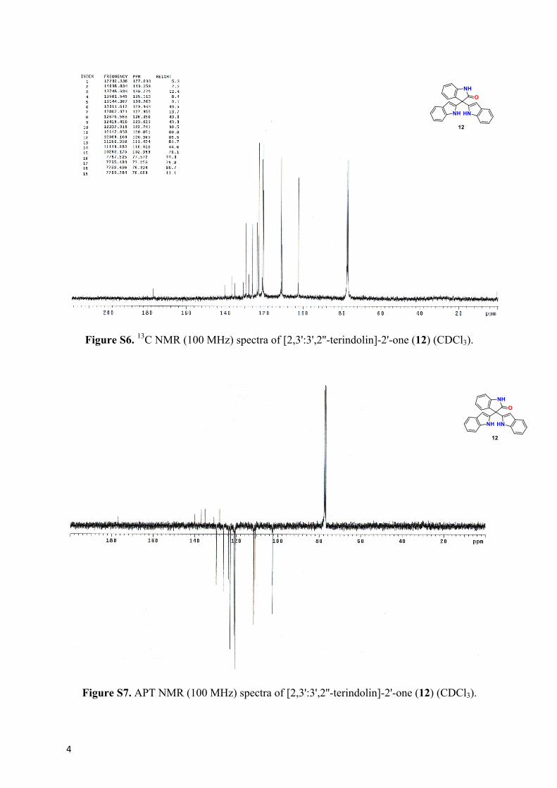
Figure S6. 13C NMR (100 MHz) spectra of [2,3':3',2''-terindolin]-2'-one (12) (CDCl3).
Figure S7. APT NMR (100 MHz) spectra of [2,3':3',2''-terindolin]-2'-one (12) (CDCl3).

5
Figure S8. In vitro effect of 5 (a), 7 (b), and 12 (c) on rat erythrocyte G6PD with
corresponding IC50 graphs.

6
Figure S9. Lineweaver–Burk double reciprocal plot of initial velocity against G6PD and inhibitors 5 (a), 7 (b), and 12 (c) at different concentrations.

7
Figure S10. In vitro effect of compounds 5 (a), 7 (b), and 12 (c) on rat erythrocyte 6PGD with corresponding IC50 (c) and activation graphs (a and b).

8
a)
b)
Figure S11. Representation of full view of a) NADP+ (PDB-ID: 1PGN) and b) 6PGA (PDB-ID: 1PGP) binding sites in 6PGD.
9
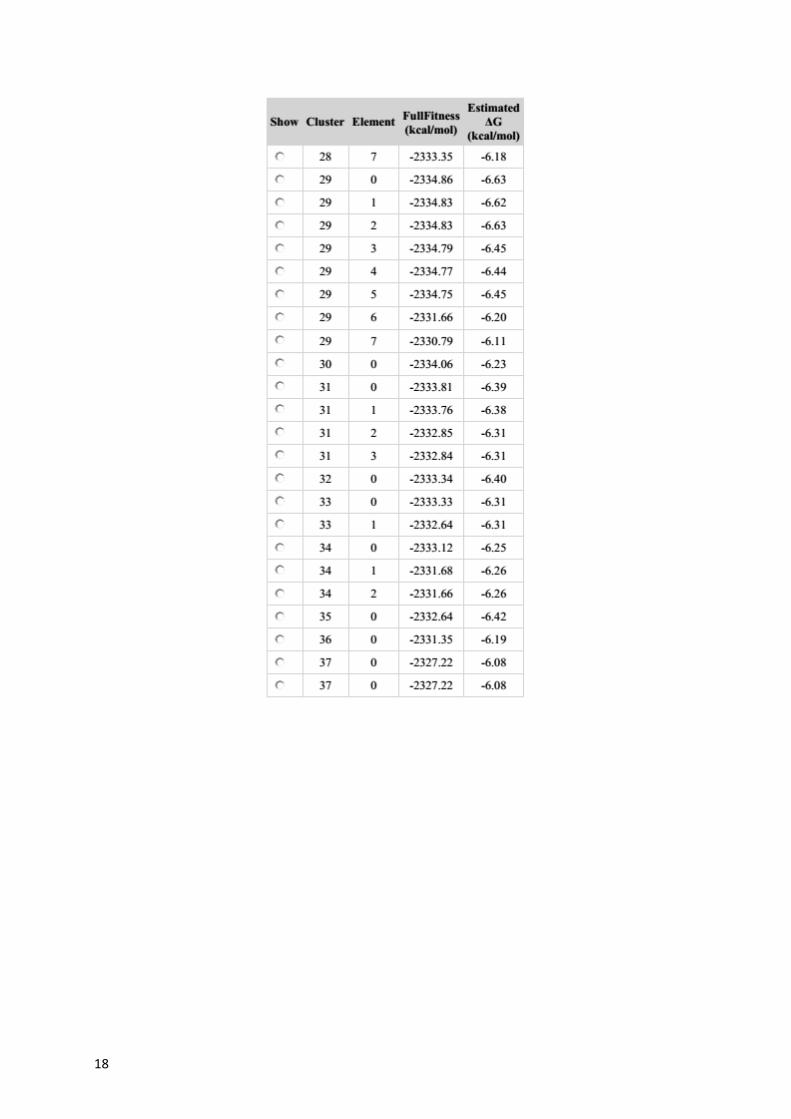
Table S1. Estimated DG values for each docking cluster calculated for G6PD.

10

11

12

13

14
Table S2. Estimated DG values for each docking cluster calculated for 6PGD.

15
16

17
18