the surface chemistry of a nanocellulose drug carrier
TRANSCRIPT

ChemicalScience
EDGE ARTICLE
Ope
n A
cces
s A
rtic
le. P
ublis
hed
on 1
3 M
arch
202
0. D
ownl
oade
d on
11/
22/2
021
2:21
:52
PM.
Thi
s ar
ticle
is li
cens
ed u
nder
a C
reat
ive
Com
mon
s A
ttrib
utio
n 3.
0 U
npor
ted
Lic
ence
.
View Article OnlineView Journal | View Issue
The surface chem
aUniv. Grenoble Alpes, CEA, CNRS, IRIG
[email protected]. Grenoble Alpes, CNRS, Grenoble-INP
[email protected] Technique du Papier (CTP), GrenobdUniv. Grenoble Alpes, CNRS, CERMAV, GreeUniv. Grenoble Alpes, CNRS, DPM, Grenob
† Electronic supplementary information (Eof metronidazole-maleimide, optimizationexperimental conditions and parametertitration, FTIR spectroscopy, elemental aSee DOI: 10.1039/c9sc06312a
‡ These authors contributed equally.
Cite this: Chem. Sci., 2020, 11, 3868
All publication charges for this articlehave been paid for by the Royal Societyof Chemistry
Received 12th December 2019Accepted 12th March 2020
DOI: 10.1039/c9sc06312a
rsc.li/chemical-science
3868 | Chem. Sci., 2020, 11, 3868–38
istry of a nanocellulose drugcarrier unravelled by MAS-DNP†
Akshay Kumar,‡a Hippolyte Durand,‡b Elisa Zeno,c Cyril Balsollier,de Bastien Watbled,e
Cecile Sillard,b Sebastien Fort, d Isabelle Baussanne, e Naceur Belgacem, b
Daniel Lee,a Sabine Hediger, a Martine Demeunynck, e Julien Bras*b and Gael DePaepe *a
Cellulose nanofibrils (CNF) are renewable bio-based materials with high specific area, which makes
them ideal candidates for multiple emerging applications including for instance on-demand drug
release. However, in-depth chemical and structural characterization of the CNF surface chemistry is
still an open challenge, especially for low weight percentage of functionalization. This currently
prevents the development of efficient, cost-effective and reproducible green synthetic routes and
thus the widespread development of targeted and responsive drug-delivery CNF carriers. We show
in this work how we use dynamic nuclear polarization (DNP) to overcome the sensitivity limitation of
conventional solid-state NMR and gain insight into the surface chemistry of drug-functionalized
TEMPO-oxidized cellulose nanofibrils. The DNP enhanced-NMR data can report unambiguously on
the presence of trace amounts of TEMPO moieties and depolymerized cellulosic units in the starting
material, as well as coupling agents on the CNFs surface (used in the heterogeneous reaction). This
enables a precise estimation of the drug loading while differentiating adsorption from covalent
bonding (�1 wt% in our case) as opposed to other analytical techniques such as elemental analysis
and conductometric titration that can neither detect the presence of coupling agents, nor
differentiate unambiguously between adsorption and grafting. The approach, which does not rely on
the use of 13C/15N enriched compounds, will be key to further develop efficient surface chemistry
routes and has direct implication for the development of drug delivery applications both in terms of
safety and dosage.
Introduction
Wood-derived cellulose nds industrial applications in a widerange of elds, from paper products to buildings, cosmetics,foodstuffs or medical industry,1,2 with numerous kinds ofindustrially produced cellulosic materials. An important mile-stone was achieved by Turbak and co-workers in the 1980s withthe introduction of a new type of cellulosic material, called
-MEM, Grenoble, France. E-mail: gael.
, LGP2, Grenoble, France. E-mail: julien.
le, France
noble, France
le, France
SI) available: Liquid-state NMR spectraof the Diels–Alder reaction conditions,s for AFM on CNF-t, conductometrynalysis data, and DNP-enhanced NMR.
77
cellulose nanobrils (CNFs),3–5 which triggered a strong andlasting scientic enthusiasm. Indeed, in addition to the char-acteristics inherited from cellulose, such as widespread avail-ability, biodegradability and biocompatibility, CNFs alsopresent excellent mechanical properties, 5 to 20 nmwidth bersand a high specic area (�100 m2 g�1) that results in extendedtunable surface chemistry.6 CNFs can be organized intodifferent 2D and 3D nano-structures such as lms, membranes,hydrogels or aerogels. At the end of the 2000s, a particular gradeof CNF was designed by pretreating the cellulose bers bymeans of a TEMPO-mediated oxidation.7,8 These TEMPO-oxidized CNFs (CNF-t) bear aldehyde and carboxylic acidgroups at the nanober surface that offer new opportunities forfunctionalization strategies.
Drug delivery is one of the promising elds of application forCNF-t in the biomedical industry.9 For example, equivalentperformances to commercial products were obtained in 2016for CNF-t gel formulations containing ve times less ibuprofen,providing a proof of concept for the efficiency of drug deliveryfor such systems.10 In 2017, the release prole for six activepharmaceutical ingredients (APIs) incorporated in CNF-t
This journal is © The Royal Society of Chemistry 2020

Fig. 1 General multistep immobilization procedure of maleimide-modified metronidazole on CNF-t.
Edge Article Chemical Science
Ope
n A
cces
s A
rtic
le. P
ublis
hed
on 1
3 M
arch
202
0. D
ownl
oade
d on
11/
22/2
021
2:21
:52
PM.
Thi
s ar
ticle
is li
cens
ed u
nder
a C
reat
ive
Com
mon
s A
ttrib
utio
n 3.
0 U
npor
ted
Lic
ence
.View Article Online
hydrogels was also investigated, demonstrating a stablebehavior towards freeze-drying and subsequent successfulrehydration.11 Notably, one of the drugs studied, an antibacte-rial compound metronidazole was further tested in interactionwith different cellulose derivatives in order to better control itsrelease.12 At this stage it is important to note that these studiesonly relied so far on drug adsorption mechanisms. It is thusimportant to investigate the loading of drugs, such as metro-nidazole, onto CNF-t through covalent binding with the aim toprovide a better-controlled drug release on-demand.
Covalent binding of molecules onto cellulosic nanomaterialsis currently an intensive eld of research. Functional linkers canbe covalently graed to the CNF surface through e.g. ether-ication, amidation, esterication, and sialylation.13–16 Theseapproaches mostly rely so far on the use of organic solventssuch as DMF/DMSO and optimized reagents such as pyridine,carbodiimides, etc. to induce graing at the C6 position on theCNF.15 Subsequent steps typically involve click-chemistry reac-tions, as described by Sharpless et al. in 2001, to further attachon to the linker an additional molecule of interest. Within thiscontext, Diels–Alder reactions, which are compatible with theseprinciples, have been recently implemented to produce newmaterials, using in particular the furan–maleimide strategy.17,18
In the biomedical context, the Diels–Alder reaction has alsobeen used to immobilize multicolor uorescent probes ontoCNF for biological imaging16 and to produce enzymaticallyactivated oligosaccharide-prodrugs of doxorubicin.19
Nevertheless, access to unambiguous informationregarding the surface chemistry of this type of system iscurrently lacking. This has recently been discussed in detail byFoster et al.20 Standard techniques (FTIR, solid-state NMR,XPS) do not provide enough sensitivity and resolution todifferentiate between low levels of adsorption and graing orto understand reliably the surface chemistry of these keymaterials.20 Elemental analysis (N, C, and O) and conducto-metric titration are oen the only techniques available toindirectly probe surface modications. In addition, and inline with a sustainable green strategy compatible withecological concerns and medical applications, it is becomingincreasingly important to perform these surface reactions inaqueous conditions employing readily available reagents/catalysts with minimal sensitivity to water. This furtherdecreases the kinetic and activation efficiency of the carbox-ylic groups compared to the use of organic solvent.21 All in all,the lack of techniques able to report on low level of CNFsurface modications impedes the development of efficient,cost-effective and reproducible green synthetic routes andthus the widespread development of targeted and responsivedrug-delivery CNF carriers.
In this work, we show that dynamic nuclear polarization(DNP) enhanced solid-state NMR (ssNMR) can be used tounravel the surface chemistry of an innovative nanocellulosedrug carrier even in the case of a very low level of graing(<1 wt%), currently well beyond reach for all othertechniques.
More precisely we report on the implementation of a Diels–Alder reaction under heterogeneous aqueous conditions
This journal is © The Royal Society of Chemistry 2020
aiming at graing the metronidazole drug onto CNF. CNFs-twere rst amidated with furfurylamine in order to function-alize them with pending furan groups. Metronidazole waschemically modied with a maleimide derived carboxylic acidin order to introduce an ester function between the drug andthe maleimide ring. The Diels–Alder reaction between thefuran functionalized CNF-t (CNF-fur) and metronidazolecontaining maleimide, as depicted in Fig. 1, was triggered byheat. The ester function present on the linking chain has beenchosen for its known sensitivity to enzymatic or chemicalhydrolysis.22–24 Indeed, esterases are ubiquitous enzymes,available in fat tissues and at infection sites.25 This new CNF-tbased complex represents thus a smart drug carrier formu-lation with “on-demand” API release abilities in the presenceof esterases.
Results and discussionHeterogeneous two-step synthesis in water
The starting CNF-t material was prepared by controlled TEMPOoxidation to achieve a high degree of oxidation, as alreadydescribed by Isogai et al. in 2011.7 The rst step of functional-ization corresponds to a catalyzed amidation with furfuryl-amine performed in water using a large excess of both reactantsand reagents. 1-Ethyl-3-(3-dimethylaminopropyl)carbodiimide(EDC) and N-hydroxysuccinimide (NHS) were used as couplingagents, and multiple washing steps were conducted in an effortto remove all the molecules that were not covalently bound,including the by-products issued from the coupling (EDC-urea,NHS) and excess of amine. Metronidazole was esteried withthe 6-maleimido-hexanoic acid in the presence of N,N0-dicyclo-hexylcarbodiimide (DCC) as coupling agent, to give themetronidazole-maleimide precursor that was then used in thenext step (see Fig. S1†).
Even if CNF-fur is supposed to be suitable for Diels–Alderclick chemistry reactions with the prodrug moleculemetronidazole-maleimide, the reaction efficiency is lowconsidering the heterogeneous conditions and sparse furangroups. The reaction was therefore rst implemented, validatedand optimized by reacting furfurylamine with 6-maleimido-hexanoic acid in homogeneous phase (see Section 2 in ESI†)
Chem. Sci., 2020, 11, 3868–3877 | 3869

Fig. 2 (a) AFM image of 7.5 � 10�5 wt% CNF suspension, (b) 13CCPMAS NMR spectra of CNF-metro with and without the applicationof microwave (mw) irradiation suitable for DNP.
Chemical Science Edge Article
Ope
n A
cces
s A
rtic
le. P
ublis
hed
on 1
3 M
arch
202
0. D
ownl
oade
d on
11/
22/2
021
2:21
:52
PM.
Thi
s ar
ticle
is li
cens
ed u
nder
a C
reat
ive
Com
mon
s A
ttrib
utio
n 3.
0 U
npor
ted
Lic
ence
.View Article Online
before being performed in similar conditions with CNF-fur andmetronidazole-maleimide. Several washing steps were againperformed to remove excess of reactants.
Combining conventional characterization spectroscopy
The main goal was then to validate the different reactionsteps, quantify the amount of functionalization present ateach step, and nally to distinguish between adsorbed andcovalently bound drug. Several experimental techniques(FTIR, solid-state NMR, etc.) were employed with the aim togather information regarding the surface chemistry (seeSection 4 in ESI†).
FTIR spectra of CNF-t, CNF-fur, and CNF-metro do notshow noticeable changes (see Fig. S6†). The lack of sensitivityand resolution prevents any conclusion (even qualitative) to bemade regarding the amount of drug loading and the presenceof graing versus adsorption. Solid-state NMR spectroscopywas also implemented to characterize surface species of themodied CNF. However, it was not possible to detect any ofthe reaction products, beyond the typical CNF core signals andthe carbonyl signal resulting from this CNF oxidation. Thissuggests a rather small drug loading level. In the end, noconclusive answer regarding the mode of functionalizationcan be drawn from the combined use of the techniquesmentioned above, which is consistent with previously pub-lished work.16,26 As a consequence, we then investigatewhether dynamic nuclear polarization can be used to solvethis limitation.
DNP-enhanced solid-state NMR
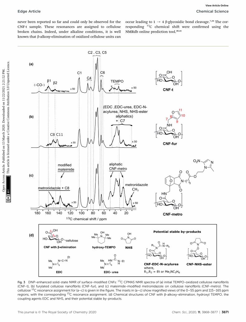
High-eld dynamic nuclear polarization (DNP), an emerginghyperpolarization technique, is currently changing thescope of solid-state NMR spectroscopy since it allowsenhancing the NMR sensitivity by several orders of magni-tude.27–29 This technique relies on the use of optimizedparamagnetic molecules,30–33 called polarizing agents, andsuitable microwave irradiation to induce a signicantincrease of nuclear polarization. Recently, DNP-enhancedNMR has been shown as a powerful tool to understandsurface chemistry of various systems27,34–36 including cellu-lose.37,38 In the experiments reported here, the abundantprotons of the samples are hyperpolarized by DNP and1H–1H spin diffusion at 100 K, typically in several seconds.The 1H hyperpolarization is then transferred to 13C or 15Nusing cross polarization. DNP experiments were performedon CNF-t, CNF-fur, and CNF-metro. In the three cases, largesensitivity gain was achieved compared to conventionalNMR experiments (see Fig. 2b). The DNP-enhanced 13C CrossPolarization (CP) Magic Angle Spinning (MAS) NMR spectraof the different CNF samples are given in Fig. 3. In additionto the large resonances from the cellulose and the surfacecarboxylic groups that were already detected in standardsolid-state NMR spectra, one can now observe additionalsignals in the aromatic and aliphatic regions. Also, it isworth noting that no aldehyde groups are observed in the 13CCPMAS NMR spectra of the CNF-t sample.
3870 | Chem. Sci., 2020, 11, 3868–3877
Insight into the surface chemistry of TEMPO-oxidized CNFs:DNP enables the detection of traces of TEMPOmoieties and b-alkoxy-elimination
Along with cellulose carbons (C1–C6) between 63 ppm to105 ppm, the carbonyl groups resulting from TEMPO-mediatedoxidation pre-treatment of cellulose can be seen at 177 ppm inthe DNP-enhanced 13C CPMAS spectrum of the TEMPO-oxidised CNF (CNF-t) shown in Fig. 3a. The high-shiedcarbonyl signal observed here is due to the alkaline condi-tions used (pH 10, NaOCl/NaOBr), which stabilize the negativecharges on the CNF surface. This is conrmed by the presenceof Na in the sample, as observed in DNP-enhanced 23Na CPMASspectra (see Fig. S7†). This is thus fully consistent with thepresence of sodium carboxylate groups at the CNF-t surface(stabilized due to pH 10). Fig. 3a also displays weak resonancesbetween 10–50 ppm that can be assigned to the CH2 and methylgroups of reduced TEMPO moieties. Remaining resonancesfrom the quaternary carbons in reduced TEMPO are expectedaround 65–68 ppm. Overlap with the much larger signal fromthe cellulose C6 resonance prevents their detection. Finally, the13C DNP-enhanced NMR spectrum from Fig. 3a also shows weakresonances at 160 ppm and 148 ppm. Such resonances have
This journal is © The Royal Society of Chemistry 2020
Edge Article Chemical Science
Ope
n A
cces
s A
rtic
le. P
ublis
hed
on 1
3 M
arch
202
0. D
ownl
oade
d on
11/
22/2
021
2:21
:52
PM.
Thi
s ar
ticle
is li
cens
ed u
nder
a C
reat
ive
Com
mon
s A
ttrib
utio
n 3.
0 U
npor
ted
Lic
ence
.View Article Online
never been reported so far and could only be observed for theCNF-t sample. These resonances are assigned to cellulosebroken chains. Indeed, under alkaline conditions, it is wellknown that b-alkoxy-elimination of oxidized cellulose units can
Fig. 3 DNP-enhanced solid-state NMR of surface-modified CNFs: 13C C(CNF-t), (b) furylated cellulose nanofibrils (CNF-fur), and (c) maleimidecellulose 13C resonance assignment for (a–c) is given in the figure. The inregions, with the corresponding 13C resonance assignment. (d) Chemiccoupling agents EDC and NHS, and their potential stable by-products.
This journal is © The Royal Society of Chemistry 2020
occur leading to 1 / 4 b-glycosidic bond cleavage.7,39 The cor-responding 13C chemical shi were conrmed using theNMRdb online prediction tool.40,41
PMAS NMR spectra of (a) initial TEMPO-oxidized cellulose nanofibrils-modified metronidazole on cellulose nanofibrils (CNF-metro). Thesets in (a–c) showmagnified views of the 0–55 ppm and 115–165 ppmal structures of CNF with b-alkoxy-elimination, hydroxyl TEMPO, the
Chem. Sci., 2020, 11, 3868–3877 | 3871

Chemical Science Edge Article
Ope
n A
cces
s A
rtic
le. P
ublis
hed
on 1
3 M
arch
202
0. D
ownl
oade
d on
11/
22/2
021
2:21
:52
PM.
Thi
s ar
ticle
is li
cens
ed u
nder
a C
reat
ive
Com
mon
s A
ttrib
utio
n 3.
0 U
npor
ted
Lic
ence
.View Article Online
Insight into the surface chemistry of CNF-t modied withfurfurylamine: DNP enables the unambiguous detection ofreacted coupling agents “invisible” with other techniques
In the DNP-enhanced 13C CPMAS NMR spectrum of CNF-furdisplayed in Fig. 3b, two carbon resonances at 145 and 152 ppmare observed, which are assigned to furan C11 and C8, respec-tively. Resonances of the two remaining carbons, C9 and C10,from the furan ring are expected between 105–115 ppm. The C10resonance can be observed as a shoulder on the le of the cellu-lose C1 resonance, as highlighted in Fig. 4b, while the furan C9resonance overlaps with the cellulose C1 resonance and could notbe observed. The C7 resonance is expected near 40 ppm both inthe case of an amine (adsorption on CNF) or an amide function(covalent graing onto CNF). In both cases, observation of thissignal is hindered by the overlap with contributions from residualcoupling agents (EDC, NHS) and/or reaction byproducts (EDC-urea, NHS-ester, etc.).42 It is thus not straightforward to differen-tiate between graing and adsorption using the C7 resonance,especially since the peak intensities from the furan moieties aresimilar to the ones of the remaining coupling agents, suggestinga small amount of graing.
At this point, it is importing to mention that intense puri-cation methods, i.e. centrifugation–dispersion cycles andextensive dialysis, were employed to minimize the amount ofremaining coupling agents. Nevertheless, DNP-enhanced NMR,as opposed to FTIR, UV and solid-state NMR, is still able todetect remaining traces.
Differentiating graing and adsorption
Even though the presence of furan is clearly seen in the DNP-enhanced spectra of CNF-fur, the crucial question of its modeof graing, adsorbed on, or bound to the CNF surface, is not yetaddressed. Answer to that question can be obtained througha detailed analysis of the broad CO resonance around 175 ppm
Fig. 4 (a) Deconvolution of the signal between 170 and 180 ppm fromCNF-fur sample, showing three distinct peaks (in blue) at 175, 172 and170 ppm, which are respectively assigned to carboxylic function,carbonyl from coupling agents, and to amide carbonyl. The red lineshows the result of the deconvolution (sumof the three contributions).(b) Extracted region between 120 and 110 ppm of the different 13CCPMAS NMR spectra of Fig. 3, highlighting the evolution of the C10furan resonance at 111 ppm throughout the different samples.
Fig. 5 (a) DNP-enhanced 13C CPMAS NMR spectra of CNF-t modifiedin the presence of the coupling agents (EDC and NHS) only. Insetsshow magnified views of the 0–55 ppm and 110–165 ppm regions,with the corresponding 13C resonance assignment. Spinning side-bands are marked by asterisks. Note that glycerol was used in the DNPmatrix. (b) Deconvolution of the carboxyl signal of (a), showing twocontributions, at 175 and 172 ppm (in blue), corresponding respectivelyto carboxylic function and to carbonyl from coupling agents. The redline shows the result of the deconvolution (sum of the two con-tributions).The presence of EDC-N-acylurea is interesting as it has notbeen considered as a possible side-reaction of CNF-t functionaliza-tion. Indeed, amidation of CNF carboxyl groups generally proceed bytheir reaction with carbodiimides in the presence of NHS to preventthe formation of stable N-acylureas.44 Here we hypothesize that thekinetics of the NHS reaction is limited in the case of heterogeneousmixtures and can lead to the formation of N-acylureas.
3872 | Chem. Sci., 2020, 11, 3868–3877
in the DNP-enhanced CPMAS spectrum of CNF-fur (see Fig. 4a).Deconvolution of the asymmetric lineshape requires threecomponents, whose chemical shis were found at 175, 172, and
This journal is © The Royal Society of Chemistry 2020

Edge Article Chemical Science
Ope
n A
cces
s A
rtic
le. P
ublis
hed
on 1
3 M
arch
202
0. D
ownl
oade
d on
11/
22/2
021
2:21
:52
PM.
Thi
s ar
ticle
is li
cens
ed u
nder
a C
reat
ive
Com
mon
s A
ttrib
utio
n 3.
0 U
npor
ted
Lic
ence
.View Article Online
170 ppm. The most intense component at 175 ppm is close tothe carboxyl chemical shi of CNF-t (177 ppm). It correspondsto unmodied carboxylic acid groups on the CNF surface. Thedifference in chemical shi (177 vs. 175 ppm) results froma change in pH between the two samples, as described above.The two other resonances at 170 and 172 ppm are more chal-lenging to assign. The chemical shi at 170 ppm (the lessintense component) is consistent with an amide resonanceresulting from the covalent bonding of furfurylamine with thesurface carboxyl. The corresponding signal intensity of thiscomponent matches the intensity of the other furan resonances(C8, C11). Tentative assignment of the last component at172 ppm is less obvious, with possible contributions fromadsorbed NHS, as well as stable graed by-products such asEDC-N-acylurea and/or NHS-ester (see Fig. 3d).42
To clarify this point, a new CNF sample was prepared usingthe same reaction conditions as for CNF-fur, but without addingfurfurylamine. We refer to this sample as “CNF with couplingagents only”. The corresponding 13C DNP-enhancedCPMASNMRspectrum is presented in Fig. 5a. Strikingly, deconvolution of thecarbonyl signal in Fig. 5b reveals only a single additional contri-bution at 172 ppm (with similar relative intensity as in CNF-fur)next to the major resonance at 175 ppm, whereas the resonanceat 170 ppm is totally absent. This conrms the resonanceassignment of the 170 ppm peak to furfurylamide and of theresonance at 172 ppm to residual reacted and/or unreactedcoupling agents on the CNF surface. We suggest here theformation of stable by-products (N-acylurea and NHS-ester) graf-ted on the CNF surface, as well as a potential contribution fromadsorbed NHS.42 Resonances from aliphatic carbons of thesereacted and unreacted coupling agents can be seen between 10–55 ppm, as shown in the inset of Fig. 5a. Furthermore, thepresence of resonances at 156 and 159 ppm conrms the pres-ence of EDC-N-acylurea and EDC-urea, in agreement withprevious studies.42,43 The relative intensity of furan (C8 and C11)and amide (at 170 ppm) resonances (see Fig. 4a) can be used toclaim that the amount of unreacted adsorbed furfurylamine isrelatively low, or under the noise level.
Diels–Alder reaction on furan-graed CNFs
New resonances at 139, 49, and 14 ppm appearing in the DNP-enhanced CPMAS NMR spectrum of CNF-metro in Fig. 3cconrm the presence of maleimide-modied metronidazole.Furthermore, the decrease in intensity of the furan resonance at110 ppm (see Fig. 4b) in the CNF-metro spectrum compared tothe CNF-fur spectrum, and the presence of a new aromaticresonance at 139 ppm conrm the Diels–Alder reaction of themaleimide with the furan. The change in relative intensity ofthe resonance at 110 ppm gives an estimation for the yield of theDiels–Alder reaction of �50%, with respect to the number ofsurface furfuryl groups in CNF-fur.
Quantifying surface species using DNP-enhanced NMR, fromCNF-t to CNF-metro
DNP-enhanced CPMAS NMR spectra as analyzed so far providea detailed qualitative picture of the surface species induced
This journal is © The Royal Society of Chemistry 2020
through the two-step reaction. They can however not be directlyused to quantify these different species as CP spin-dynamicsmay vary for different types of carbon environments, in partic-ular between protonated and quaternary carbon sites. In case oflarger particles, the signal enhancement provided by DNP mayalso not be uniform between surface and bulk sites. This ishowever not the case here, as the size of the CNF used, about10 nm in diameter, is sufficiently small to obtain a uniform DNPenhancement and mono-exponential hyperpolarization build-up times across the different resonances of the spectrum. Toaddress the problem of non-uniform CP spin-dynamics, weimplemented the quantitative MultiCP experiment,45 which wasapplied under DNP conditions to the CNF-t sample. Deconvo-lution of the C1, C4, C6 and CO signals leads to a preciseintegral calculation despite partial overlap of the differentcontributions (see Fig. S8 and Table S2†). For the quantitativeanalysis, the C4 integral was used as a reference (set to 100).
Our analysis reveals that the TEMPO oxidizing treatmentinduces a modication of the C6 carbon, resulting in �24%carboxylate groups in CNF-t (see Table S2†) with 74% of C6carbons remaining unmodied. Carboxylate groups at 160 ppmresulting from b-alkoxy-elimination in the CNF-t sample can beestimated to �1.0% of the number of cellulose glucose units.Overall, the sum of the unmodied and oxidized C6 contribu-tions is thus �99 � 1%, which prove the self-consistency andaccuracy of the analysis. Interestingly, no aldehydes areobserved within our detection limit. Note that these groupscould also appear as hemiacetals and acetals, whose weak 13CNMR signal would overlap with the intense C1 peak. Theamount of remaining reduced TEMPO moieties is estimated to0.5%.
Comparison of the relative integrals of C4 (or C1) andcarbonyl resonances in CPMAS and MultiCP experiments ofCNF-t allows the estimation of a CP-dynamics correctionfactor, which can then be used to further quantitativelyanalyse the DNP-enhanced 13C CPMAS NMR spectra of CNF-fur and CNF-metro. Thus, the degree of furfurylaminesubstitution in CNF-fur can be estimated to �2% compared toa glucose unit of cellulose based on the 170 ppm peak inten-sity. Comparatively, the peak at 172 ppm, assigned to couplingagent derivatives, contributes to about 7%. It is important tonote that most of these correspond to compounds that havereacted with the CNF surface carboxylic groups, formingstable species (that cannot be washed away), such as N-acy-lurea from EDC and NHS-ester from NHS, but also to adsorbedNHS moieties.42,43
Finally, the well-isolated metronidazole methyl resonancepresent in the CNF-metro DNP-enhanced 13C CPMAS NMRspectrum can be used to roughly estimate the degree of druggraing to �1% compared to the glucose unit in cellulose. Thisis consistent with the amount of unreacted furfuryl in CNF-metro (estimated to �1% using the peak at 145 ppm) and theestimated yield of �50% for the Diels–Alder reaction step.
Table 1 summarizes the amount of surface species revealedby DNP-enhanced solid-state NMR spectroscopy, relative to thecellulose C4 peak for CNF-t, CNF-fur, and CNF-metro. Moredetails can be found in ESI Section 5.†
Chem. Sci., 2020, 11, 3868–3877 | 3873
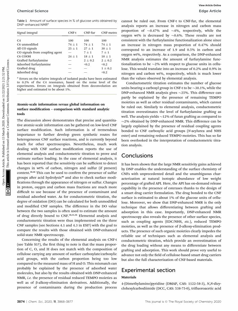
Table 1 Amount of surface species in % of glucose units obtained byDNP-enhanced NMRa
Signal integral CNF-t CNF-fur CNF-metro
C4 100 100 100C6 unmodied 74 � 1 74 � 1 74 � 1All CO signals 25 � 1 27 � 1 30 � 1CO signals from coupling agent — 7 � 1 7 � 1C6 COOH 24 � 1 18 � 1 18 � 1Graed furfurylamine — 2 � 0.2 2 � 0.2Adsorbed furfurylamine — <0.2 <0.2Graed drug — — 1 � 0.2Adsorbed drug — — <0.2
a Errors on the relative integrals of isolated peaks have been estimatedto 0.2% of the C4 resonance, based on the noise level of theexperiments. Errors on integrals obtained from deconvolution arehigher and estimated to be about 1%.
Chemical Science Edge Article
Ope
n A
cces
s A
rtic
le. P
ublis
hed
on 1
3 M
arch
202
0. D
ownl
oade
d on
11/
22/2
021
2:21
:52
PM.
Thi
s ar
ticle
is li
cens
ed u
nder
a C
reat
ive
Com
mon
s A
ttrib
utio
n 3.
0 U
npor
ted
Lic
ence
.View Article Online
Atomic-scale information versus global information onsurface modication – comparison with standard analytictools
The discussion above demonstrates that precise and quantita-tive atomic-scale information can be gathered on low-level CNFsurface modication. Such information is of tremendousimportance to further develop green synthetic routes forheterogeneous CNF surface reactions, and is currently beyondreach for other spectroscopies. Nevertheless, much workdealing with CNF surface modication reports the use ofelemental analysis and conductometric titration to prove andestimate surface loading. In the case of elemental analysis, ithas been reported that the sensitivity can be sufficient to detectchanges in wt% of carbon, nitrogen and sulfur (if present)content.20,46 This can be used to conrm the presence of sulfurgroups aer acid hydrolysis46 and also to check surface modi-cations through the appearance of nitrogen or sulfur. Changesin proton, oxygen and carbon mass fractions are much moredifficult to use because of the presence of contaminant andresidual adsorbed water. As for conductometric titration, thedegree of oxidation (DO) can be calculated for both unmodiedand modied CNF samples. The difference in the DO valuebetween the two samples is oen used to estimate the amountof drug directly bound to CNF.20,21,26 Elemental analysis andconductometric titration were thus implemented on the threeCNF samples (see Sections 4.1 and 4.3 in ESI†) with the goal tocompare the results with those obtained with DNP-enhancedsolid-state NMR spectroscopy.
Concerning the results of the elemental analysis on CNF-t(see Table S1†), the rst thing to note is that the mass propor-tion of C, O, and H does not match with the composition ofcellulose carrying any amount of surface carboxylate/carboxylicacid groups, with the carbon proportion being too lowcompared to themeasuredmass of H and O. This mismatch canprobably be explained by the presence of adsorbed watermolecules, but also by the results obtained with DNP-enhancedNMR, i.e. the presence of residual reduced TEMPO moieties aswell as of b-alkoxy-elimination derivatives. Additionally, thepresence of contaminants during the production process
3874 | Chem. Sci., 2020, 11, 3868–3877
cannot be ruled out. From CNF-t to CNF-fur, the elementalanalysis reports an increase in nitrogen and carbon massproportion of �0.47% and �4%, respectively, while theoxygen wt% is decreased by �0.6%. These results are notconsistent with the furfurylamine functionalization alone sincean increase in nitrogen mass proportion of 0.47% shouldcorrespond to an increase of 1.9 and 0.5% in carbon andoxygen wt%, respectively. As a comparison, the DNP-enhancedNMR analysis estimates the amount of furfurylamine func-tionalization to be �2% with respect to glucose units in cellu-lose. This would translate into an increase of 0.2% and 0.9% innitrogen and carbon wt%, respectively, which is much lowerthan the values observed by elemental analysis.
Conductometric titration estimates the number of glucoseunits bearing a carboxyl group in CNF-t to be �30.1%, while theDNP-enhanced NMR analysis gives �25%. This difference canpartly be explained by the presence of remaining TEMPOmoieties as well as other residual contaminants, which cannotbe ruled out. Similarly to elemental analysis, conductometrictitration overestimates the level of furfurylamine graing aswell. The analysis yields �12% of furan graing as compared to�2% obtained by DNP-enhanced NMR. This difference can belargely explained by the presence of residual coupling agentsbonded to CNF carboxylic acid groups (N-acylurea and NHSester) and remaining reduced TEMPO moieties. This has so farbeen overlooked in the interpretation of conductometric titra-tion analysis.
Conclusions
It has been shown that the large NMR sensitivity gains achievedby DNP enables the understanding of the surface chemistry ofCNFs with unprecedented detail and the unambiguous char-acterization at natural isotopic abundance of low weightpercentage of graed API. Here, the API has on-demand releasecapability in the presence of esterases thanks to the design ofa smart drug carrier formulation. The drug bonded to the CNFsurface is estimated to about 1% of the glucose units of cellu-lose. Moreover, we show that DNP-enhanced NMR is the onlytechnique that allows differentiating between graing andadsorption in this case. Importantly, DNP-enhanced NMRspectroscopy also reveals the presence of other surface species,such as coupling agents (EDC/NHS, etc.), reduced TEMPOmoieties, as well as the presence of b-alkoxy-elimination prod-ucts. The presence of such organic moieties clearly impedes thereliable use of techniques such as elemental analysis andconductometric titration, which provide an overestimation ofthe drug loading without any means to differentiate betweengraing and adsorption. This work should prove very useful toadvance not only the eld of cellulose-based smart drug carriersbut also the full characterization of CNF-based materials.
Experimental sectionMaterials
4-(Dimethylamino)pyridine (DMAP, CAS: 1122-58-3), N,N-dicy-clohexylcarbodiimide (DCC, CAS: 538-75-0), triuoroacetic acid
This journal is © The Royal Society of Chemistry 2020

Edge Article Chemical Science
Ope
n A
cces
s A
rtic
le. P
ublis
hed
on 1
3 M
arch
202
0. D
ownl
oade
d on
11/
22/2
021
2:21
:52
PM.
Thi
s ar
ticle
is li
cens
ed u
nder
a C
reat
ive
Com
mon
s A
ttrib
utio
n 3.
0 U
npor
ted
Lic
ence
.View Article Online
(TFA, CAS: 76-05-1), metronidazole (CAS: 443-48-1), furfuryl-amine (CAS: 617-89-0), N-(3-dimethylaminopropyl)-N0-ethyl-carbodiimide hydrochloride (EDC, CAS: 25952-53-8), N-hydroxysuccinimide (NHS, CAS: 6066-82-6), sodium hydroxide(NaOH, CAS: 1310-73-2), hydrogen chloride (HCl, CAS: 7647-01-0) were purchased from Sigma Aldrich, Alfa Aesar or AcrosOrganics and used without further purication. Solution NMRspectra were recorded at room temperature in 5 mm tubes ona Bruker AC 400 MHz spectrometer (NMR facility, PCN-ICMG,Grenoble). Chemical shis (d) are reported in parts permillion (ppm) from low to high eld and referenced to residualnon-deuterated solvent relative to Me4Si. Standard abbrevia-tions for multiplicity were used as follows: s ¼ singlet; d ¼doublet; t ¼ triplet; m ¼ multiplet. High resolution massspectrometry (HRMS) was carried out on a Waters Xevo G2-S-QTof mass spectrometer using ElectroSpray Ionisation (NMRfacility, PCN-ICMG, Grenoble).
Synthesis of metronidazole-maleimide (Metro-MAL)
6-Maleimido-hexanoic acid47,48 (753 mg, 3.6 mmol), metronida-zole (2, 626 mg, 3.6 mmol) and DMAP (40 mg, 0.36 mmol) weredissolved in CH2Cl2 (25 ml) at 0 �C. DCC (905 mg, 4.4 mmol) wasadded aer 15 minutes. The reacting mixture was stirred for 4 hat room temperature (rt), then ltered and concentrated undervacuum. The crude product was puried by ash chromatog-raphy on silica gel with CH2Cl2/MeOH 98/2 (v/v) as eluent to givethe compound as a yellow amorphous solid (669 mg, 1.83 mmol,51%). 1H NMR (CDCl3, 400 MHz) d 7.96, (s, 1H), 6.69 (s, 2H), 4.59(t, 2H, J ¼ 5.3 Hz), 4.41 (t, 2H, J ¼ 5.3 Hz), 3.50 (t, 2H, J ¼ 7.2 Hz),2.51 (s, 3H), 2.26 (t, 2H, J ¼ 7.5 Hz), 1.63–1.54 (m, 4H), 1.31–1.25(m, 2H); 13C NMR (CDCl3, 100 MHz) d 172.7, 170.8, 150.8, 134.1,133.2, 62.4, 45.1, 37.5, 33.7, 28.2, 26.1, 24.1, 14.4; HRMS (ESI)m/z:calc. for C16H21N4O6 [M + H]+ 365.1461, obs. 365.1468. Moredetails can be found in ESI Section 1.†
TEMPO-oxidation of cellulose nanobrils (CNF-t)
The cellulose nanobril (CNF) suspension was provided by theCentre Technique du Papier (CTP, Grenoble, France). Suspen-sion of TEMPO-oxidized CNF (CNF-t) was produced from a pre-rened (40� SR) bleached bisulte pulp provided by RayonierAdvanced Materials (previously TEMBEC). The pulp concen-tration was adjusted to 1.5 wt% and the oxidation was per-formed at pH 10 for 2 h in the presence of NaBr, NaClO, and theTEMPO reagent. A high pressure homogenizer from GEA (Niro,Soavi, Italy) was used to debrillate the oxidized cellulose bersand produce the CNF-t suspension. The produced CNF-tsuspension is in the form of a thick opaque gel at 1.6 wt%.AFM images of the low concentrated CNF-t suspensionconrmed the nanosized morphology of CNF-t (see Fig. 2a).Using conductometric titration (CT), the degree of oxidation(DO) of the CNF-t suspension was found to be 30.1%, (seeFig. S5†).
Functionalization of CNF-t with furan (CNF-fur)
The CNF-t suspension concentration was decreased from1.5 wt% to 0.4 wt% in order to be easily stirred. Deionized water
This journal is © The Royal Society of Chemistry 2020
was added before homogenization with an IKA Ultra-Turraxhigh shear mixer for 1 min at 10 000 rpm. The pH of thesuspension was then adjusted to 4 under magnetic stirringusing a 0.5 M HCl solution.
A solution of the coupling agents EDC and NHS wasprepared in deionized water and added to the suspension ofCNF-t with a molar ratio of 4 equivalents of EDC and NHS for 1equivalent of CNF-t surface carboxyl group (using the 30.1% DOobserved by CT). The mixture was stirred for 30 min at rt at pH 4and then the pH was increased to 8.5 before amine addition.Furfurylamine, with amolar ratio of 4 equivalents of amine for 1equivalent of carboxylic acid of CNF-t, was mixed with 5 ml ofdeionized water and added to the mixture that was then stirredat rt with the pH maintained at 8.5.
Aer 72 h of reaction, the reaction was quenched bydecreasing the pH to 2–2.5 with 0.5 M HCl solution. To removenon-covalently bound chemicals (EDC, NHS and free amine), 6cycles of washing, centrifugation (10 min at 11 100 rpm) and re-dispersion with a high shear mixer (Ultra-turrax, IKA) for 1 minat rt were repeated. The last dispersion was done in neutralwater to afford the furan modied CNF-t, referred to as CNF-fur.
The last step of purication consisted in a dialysis of theCNF-fur suspension against neutral water with 6–8 kDa MWCOmembranes (Spectra/Por® 1 Standard RC Tubing, SPECTRUM)for at least 5 days under slow magnetic stirring and renewal ofthe medium twice a day.
Diels–Alder reaction of Metro-MAL on CNF-fur
The Diels–Alder reaction was rst optimized under homoge-neous conditions before being implemented on the CNF-fursample (see ESI Section 2†). Metro-MAL (95.2 mg) was dis-solved in 40 ml of 1 : 1 mixture of deionized water and ethanol(v/v) in an ultrasound bath (IKA, USA). The CNF-fur suspensionwas diluted to 0.15 wt% concentration and the Metro-MALsolution (9.42 ml) was added dropwise under magnetic stir-ring in order to reach one molar equivalent of Metro-MAL to thefuran groups available on the CNF substrate. The Diels–Alderreaction was then triggered by heating the system at 40 �C for24 h under continuous stirring. The reaction was followed withUV analysis. The reacted CNF-fur, now referred to as CNF-metro, was washed with several centrifugation/re-dispersioncycles as described above to remove remaining reactants. Therst 3 cycles were done with 1 : 1 (v/v) water/ethanol solution,followed by 2 cycles using deionized water only. Washedsuspension was further puried by dialysis (6–8 kDa MWCOmembranes, Spectra/Por® 1 Standard RC Tubing, SPECTRUM)against deionized water under slow magnetic stirring for 5 dayswith daily renewal of the dialysis medium. CNF-metro suspen-sion was stored at 5 �C.
AFM, FTIR, conductometric titration, and elemental analysisof CNF samples
Characterization of the different CNF (CNF-t, CNF-fur and CNF-metro) were performed on thin lms and/or cryogel powders.Thin lms were produced by solvent casting in Petri dishes of0.1 wt% CNF suspensions in order to obtain 30 g m�2
lms. The
Chem. Sci., 2020, 11, 3868–3877 | 3875

Chemical Science Edge Article
Ope
n A
cces
s A
rtic
le. P
ublis
hed
on 1
3 M
arch
202
0. D
ownl
oade
d on
11/
22/2
021
2:21
:52
PM.
Thi
s ar
ticle
is li
cens
ed u
nder
a C
reat
ive
Com
mon
s A
ttrib
utio
n 3.
0 U
npor
ted
Lic
ence
.View Article Online
lms were dried overnight in an oven at 40 �C. An alternative dryform of the different CNFs was obtained by freeze drying thesuspensions (cryogel). The results obtained with AFM,conductometric titration, FTIR and elemental analysis can befound in ESI Sections 3 and 4.
DNP-enhanced solid-state NMR
DNP-enhanced NMR experiments were performed on the threeCNF samples (CNF-t, CNF-fur and CNF-metro). The samples wereprepared by impregnating the CNF powder with a DNP matrix (1)composed of 10 mMAMUPol30 in D2O. For CNF-t, 28 mg (aerogel)was impregnated with 40 mL of the DNP matrix (1). For CNF-furand CNF-metro, 30 mg of each were impregnated with 80 mL ofDNP matrix (1), an additional CNF sample was preparedfollowing the synthesis used for CNF-fur, except that no furfur-ylamine was added. We refer to this sample as the CNF samplewith coupling agents only. In this case, a DNPmatrix (2) of 10mMAsymPolPOK31 in glycerol/D2O/H2O (60 : 30 : 10; v/v) was used.25 mg of CNF with coupling agents only was impregnated with 50mL of DNP matrix (2). Each sample was then fully packed intoa 3.2 mm outer-diameter sapphire rotor.
All experiments were performed on a Bruker Avance III 400MHz (1H resonance) DNP-NMR spectrometer equipped with 263GHz gyrotron for microwave irradiation, a corrugated trans-mission line, and a low temperature 3.2 mmMAS probe used indouble-resonance mode.49 All experiments were performed ata sample temperature of 100 K, using a magic-angle spinning(MAS) frequency of 13.3 kHz. Cross-polarization under MAS50
(CPMAS) experiments were performed with a radio frequency(rf) eld strength of 50 kHz on the 13C while 1H rf eld strengthswere adjusted accordingly depending upon the ramp51 used tomatch CP conditions. The CP decay time constants weremeasured and were found to be in excess of 25 ms. The CPcontact time was set to 2 ms for CPMAS experiments and 1 msfor the quantitative CP experiment using the Multi-CP45 pulsesequence. A 50-to-100% and 90-to-100% ramp were used for the1H CP spin-lock in the CPMAS and quantitative CP using theMultiCP, respectively.51,52 The inter-scan delay was optimizedaccording to the hyperpolarization build-up time of eachsample, and set to 3.0 s for CNF-t, 2.5 s for CNF-fur, 1.9 s forCNF-metro and 1.9 s for CNF with coupling agents only. For theMultiCP experiment the inter-scan delay and the delay betweeneach CP block were set to 3.9 s and 6 s, respectively. All exper-iments were processed and analyzed using Bruker Topspin 3.2soware, including the peak deconvolution and integrationcarried out to perform the analysis herein. 13C peak intensitiesare discussed relatively to carbon magnetization from theglucose unit of cellulose. The signal-to-noise of DNP-enhanced13C CPMAS NMR spectra is about 500 : 1 for each of the carbonresonances of the glucose unit.
Abbreviation
FTIR
3876 | Chem. Sci., 20
Fourier transform infra-red
NMR Nuclear magnetic resonance20, 11, 3868–3877
DNP
This j
Dynamic nuclear polarization
CNFs Cellulose nanobrilsConflicts of interest
There are no conicts to declare.
Acknowledgements
This work was supported by the French National ResearchAgency (ANR) through (Cellical ANR-15-CE08-0033, ARCANEANR-11-LABX-0003-01, CBH-EUR-GS Grant ANR-17-EURE-0003,and Glyco@Alps ANR-15-IDEX-02) and the European ResearchCouncil (ERC-CoG-2015, No. 682895). Support from the ICMG(FR 2607) Chemistry Nanobio Platforms, Grenoble, on whichmass spectrometry and solution-state NMR have been per-formed is also acknowledged.
References
1 D. Klemm, B. Heublein, H. P. Fink and A. Bohn, Angew.Chem., Int. Ed., 2005, 44, 3358–3393.
2 N. Lin and A. Dufresne, Eur. Polym. J., 2014, 59, 302–325.3 F. W. Herrick, R. L. Casebier, J. K. Hamilton andK. R. Sandberg, J. Appl. Polym. Sci.: Appl. Polym. Symp.,1983, 37, 797–813.
4 A. F. Turbak, F. W. Snyder and K. R. Sandberg, J. Appl. Polym.Sci.: Appl. Polym. Symp., 1983, 37, 815–827.
5 A. F. Turbak, F. W. Snyder and K. R. Sandberg, US pat.,4374702A, 1983.
6 H. Kargarzadeh, M. Mariano, D. Gopakumar, I. Ahmad,S. Thomas, A. Dufresne, J. Huang and N. Lin, Cellulose,2018, 25, 2151–2189.
7 A. Isogai, T. Saito and H. Fukuzumi, Nanoscale, 2011, 3, 71–85.
8 T. Saito, Y. Nishiyama, J. L. Putaux, M. Vignon and A. Isogai,Biomacromolecules, 2006, 7, 1687–1691.
9 D. V. Plackett, K. Letchford, J. K. Jackson and H. M. Burt,Nord. Pulp Pap. Res. J., 2014, 29, 105–118.
10 D. Celebi, R. H. Guy, K. J. Edler and J. L. Scott, Int. J. Pharm.,2016, 514, 238–243.
11 H. Paukkonen, M. Kunnari, P. Lauren, T. Hakkarainen,V. V. Auvinen, T. Oksanen, R. Koivuniemi, M. Yliperttulaand T. Laaksonen, Int. J. Pharm., 2017, 532, 269–280.
12 Y. Dong, H. Paukkonen, W. Fang, E. Kontturi, T. Laaksonenand P. Laaksonen, Int. J. Pharm., 2018, 548, 113–119.
13 K. Missoum, M. N. Belgacem and J. Bras, Materials, 2013, 6,1745–1766.
14 Y. Habibi, Chem. Soc. Rev., 2014, 43, 1519–1542.15 F. Rol, M. N. Belgacem, A. Gandini and J. Bras, Prog. Polym.
Sci., 2019, 88, 241–264.16 J. R. G. Navarro, G. Conzatti, Y. Yu, A. B. Fall, R. Mathew,
M. Eden and L. Bergstrom, Biomacromolecules, 2015, 16,1293–1300.
17 A. Gandini, A. J. F. Carvalho, E. Trovatti, R. K. Kramer andT. M. Lacerda, Eur. J. Lipid Sci. Technol., 2018, 120, 1700091.
ournal is © The Royal Society of Chemistry 2020

Edge Article Chemical Science
Ope
n A
cces
s A
rtic
le. P
ublis
hed
on 1
3 M
arch
202
0. D
ownl
oade
d on
11/
22/2
021
2:21
:52
PM.
Thi
s ar
ticle
is li
cens
ed u
nder
a C
reat
ive
Com
mon
s A
ttrib
utio
n 3.
0 U
npor
ted
Lic
ence
.View Article Online
18 E. Trovatti, A. G. Cunha, A. J. F. Carvalho and A. Gandini, Int.J. Biol. Macromol., 2017, 95, 762–768.
19 D. Bliman, M. Demeunynck, P. Leblond, S. Meignan,I. Baussane and S. Fort, Bioconjugate Chem., 2018, 29,2370–2381.
20 E. J. Foster, R. J. Moon, U. P. Agarwal, M. J. Bortner, J. Bras,S. Camarero-Espinosa, K. J. Chan, M. J. D. Cli,E. D. Cranston, S. J. Eichhorn, et al., Chem. Soc. Rev., 2018,47, 2609–2679.
21 E. Lasseuguette, Cellulose, 2008, 15, 571–580.22 N. M. Mahfouz andM. A. Hassan, J. Pharm. Pharmacol., 2001,
53, 841–848.23 C. Mura, D. Valenti, C. Floris, R. Sanna, M. A. De Luca,
A. M. Fadda and G. Loy, Eur. J. Med. Chem., 2011, 46,4142–4150.
24 M. Johansen, B. Møllgaard, P. K. Wotton, C. Larsen andA. Hoelgaard, Int. J. Pharm., 1986, 32, 199–206.
25 J. M. Schakenraad, M. J. Hardonk, J. Feijen, I. Molenaar andP. Nieuwenhuis, J. Biomed. Mater. Res., 1990, 24, 529–545.
26 E. Gicquel, C. Martin, L. Heux, B. Jean and J. Bras,Carbohydr. Polym., 2019, 210, 100–109.
27 A. J. Rossini, A. Zagdoun, M. Lelli, A. Lesage, C. Coperet andL. Emsley, Acc. Chem. Res., 2013, 46, 1942–1951.
28 A. S. L. Thankamony, J. J. Wittmann, M. Kaushik andB. Corzilius, Prog. Nucl. Magn. Reson. Spectrosc., 2017, 102–103, 120–195.
29 A. N. Smith, K. Marker, S. Hediger and G. De Paepe, J. Phys.Chem. Lett., 2019, 10, 4652–4662.
30 C. Sauvee, M. Rosay, G. Casano, F. Aussenac, R. T. Weber,O. Ouari and P. Tordo, Angew. Chem., Int. Ed., 2013, 52,10858–10861.
31 F. Mentink-Vigier, I. Marin-Montesinos, A. P. Jagtap,T. Halbritter, J. Van Tol, S. Hediger, D. Lee,S. Th. Sigurdsson and G. De Paepe, J. Am. Chem. Soc., 2018,140, 11013–11019.
32 S. Hediger, D. Lee, F. Mentink-Vigier and G. De Paepe,eMagRes, 2018, 7, 105–116.
33 D. J. Kubicki, G. Casano, M. Schwarzwalder, S. Abel,C. Sauvee, K. Ganesan, M. Yulikov, A. J. Rossini,G. Jeschke, C. Coperet, et al., Chem. Sci., 2016, 7, 550–558.
34 A. Lesage, M. Lelli, D. Gajan, M. A. Caporini, V. Vitzthum,P. Mieville, J. Alauzun, A. Roussey, C. Thieuleux, A. Mehdi,
This journal is © The Royal Society of Chemistry 2020
G. Bodenhausen, C. Coperet and L. E. Emsley, J. Am. Chem.Soc., 2010, 132, 15459–15461.
35 D. Lee, G. Monin, N. T. Duong, I. Zamanillo Lopez,M. Bardet, V. Mareau, L. Gonon and G. De Paepe, J. Am.Chem. Soc., 2014, 136, 13781–13788.
36 A. G. M. Rankin, J. Trebosc, F. Pourpoint, J. P. Amoureux andO. Lafon, Solid State Nucl. Magn. Reson., 2019, 101, 116–143.
37 L. Zhao, W. Li, A. Plog, Y. Xu, G. Buntkowsky, T. Gutmannand K. Zhang, Phys. Chem. Chem. Phys., 2014, 16, 26322–26329.
38 L. Zhao, I. Smolarkiewicz, H. H. Limbach, H. Breitzke,K. Pogorzelec-Glaser, R. Pankiewicz, J. Tritt-Goc,T. Gutmann and G. Buntkowsky, J. Phys. Chem. C, 2016,120, 19574–19585.
39 T. Hosoya, M. Bacher, A. Potthast, T. Elder and T. Rosenau,Cellulose, 2018, 25, 3797–3814.
40 C. Steinbeck, S. Krause and S. Kuhn, J. Chem. Inf. Comput.Sci., 2003, 43, 1733–1739.
41 D. Ban and L. Patiny, Chimia, 2008, 62, 280–281.42 S. Fujisawa, Y. Okita, T. Saito, E. Togawa and A. Isogai,
Cellulose, 2011, 18, 1191–1199.43 T. Pouyani, J. W. Kuo, G. S. Harbison and G. D. Prestwich, J.
Am. Chem. Soc., 1992, 114, 5972–5976.44 H. G. Khorana, Chem. Rev., 1953, 53, 145–166.45 R. L. Johnson and K. Schmidt-Rohr, J. Magn. Reson., 2014,
239, 44–49.46 W. P. Flauzino Neto, J. L. Putaux, M. Mariano, Y. Ogawa,
H. Otaguro, D. Pasquini and A. Dufresne, RSC Adv., 2016,6, 76017–76027.
47 J. Han, L. Sun, Y. Chu, Z. Li, D. Huang, X. Zhu, H. Qian andW. Huang, J. Med. Chem., 2013, 56, 9955–9968.
48 Q. A. Bui, T. H. H. Vu, V. K. T. Ngo, I. R. Kennedy, N. A. Leeand R. Allan, Anal. Bioanal. Chem., 2016, 408, 6045–6052.
49 M. Rosay, M. Blank and F. Engelke, J. Magn. Reson., 2016,264, 88–98.
50 E. O. Stejskal, J. Schaefer and J. S. Waugh, J. Magn. Reson.,1977, 28, 105–112.
51 G. Metz, X. L. Wu and S. O. Smith, J. Magn. Reson., Ser. A,1994, 110, 219–227.
52 S. Hediger, B. H. Meier and R. R. Ernst, Chem. Phys. Lett.,1995, 240, 449–456.
Chem. Sci., 2020, 11, 3868–3877 | 3877