the structure of mouse cytomegalovirus m04 protein...
TRANSCRIPT
Please cite this article in press as: Sgourakis et al., The Structure of Mouse Cytomegalovirus m04 Protein Obtained from Sparse NMR Data Reveals aConserved Fold of the m02-m06 Viral Immune Modulator Family, Structure (2014), http://dx.doi.org/10.1016/j.str.2014.05.018
Structure
Article
The Structure of Mouse Cytomegalovirus m04 ProteinObtained fromSparse NMRData Reveals a ConservedFold of the m02-m06 Viral Immune Modulator FamilyNikolaos G. Sgourakis,1 KannanNatarajan,2 Jinfa Ying,1 Beat Vogeli,1,3 Lisa F. Boyd,2 David H.Margulies,2,* and AdBax1,*1Laboratory of Chemical Physics, National Institute of Diabetes and Digestive and Kidney Diseases, National Institutes of Health, Bethesda,
MD 20892, USA2Molecular Biology Section, Laboratory of Immunology, National Institute of Allergy and Infectious Diseases, National Institutes of Health,Bethesda, MD 20892, USA3Present address: Laboratory of Physical Chemistry, Swiss Federal Institute of Technology, 8093 Zurich, Switzerland
*Correspondence: [email protected] (D.H.M.), [email protected] (A.B.)
http://dx.doi.org/10.1016/j.str.2014.05.018
SUMMARY
Immunoevasins are key proteins used by viruses tosubvert host immune responses. Determining theirhigh-resolution structures is key to understandingvirus-host interactions toward the design of vaccinesand other antiviral therapies. Mouse cytomegalo-virus encodes a unique set of immunoevasins, them02-m06 family, that modulates major histocompat-ibility complex class I (MHC-I) antigen presentationto CD8+ T cells and natural killer cells. Notwith-standing the large number of genetic and functionalstudies, the structural biology of immunoevasinsremains incompletely understood, largely becauseof crystallization bottlenecks. Here we implementa technology using sparse nuclear magnetic reso-nance data and integrative Rosetta modeling todetermine the structure of the m04/gp34 immunoe-vasin extracellular domain. The structure reveals ab fold that is representative of the m02-m06 familyof viral proteins, several of which are known to bindMHC-I molecules and interfere with antigen presen-tation, suggesting its role as a diversified immuneregulation module.
INTRODUCTION
Cytomegaloviruses (CMVs) are important models of pathogen-
host interactions, widely recognized for their ability to interfere
with host immune responses to accomplish the multifaceted
task of inhibiting the recognition of infected cells by CD8+
T cells while avoiding destruction by natural killer (NK) cells
according to the ‘‘missing-self’’ hypothesis (i.e., the lack of anti-
gen-presenting major histocompatibility complex class I [MHC-I]
molecules on the cell surface) (Lemmermann et al., 2012). To do
this, the virus maintains a series of genes encoding immune
evasion and regulatory proteins (Lilley and Ploegh, 2005). In
particular, the m02-m16 family is a class of early-expressed
genes of mouse CMV (MCMV) crucial for viral survival and infec-
Structure 22
tivity. However, there is no identifiable homology to any deter-
mined structure in the Protein Data Bank (PDB), nor is there
amino acid sequence similarity to any other protein family. A
member of the m02-m06 class, the m04/gp34 protein, is unique
in its ability to bind MHC-I molecules in the endoplasmic reticu-
lum (ER) and accompany them to the cell surface (Kleijnen et al.,
1997), while the closely related m06 protein binds MHC-I and di-
rects it to the endosome (Tomas et al., 2010). Therefore, it has
been proposed that by countering the MHC-retaining functions
of other viral proteins (m06 and the MHC-I-like m152), m04
helps MCMV evade the NK cell response (Babi�c et al., 2010,
2011; Holtappels et al., 2006). The combined effects of m04,
m06, and m152 on CD8+ T cells and NK responses reveal a
complex and still poorly understood aspect of MCMV immune
evasion (Pinto et al., 2006). Although the structural basis for
the function of m152/gp40 immunoevasin has been previously
revealed by X-ray crystallography (Wang et al., 2012), and struc-
tures of other MCMV MHC-I-like proteins have been similarly
characterized (Adams et al., 2007; Berry et al., 2013; Mans
et al., 2007; Natarajan et al., 2006), for other molecules, a
detailed picture is still lacking because of difficulties in crystalli-
zation and the large sizes of their in vivo functional complexes,
making conventional nuclear magnetic resonance (NMR) studies
challenging. In particular, extensive crystallization trials in our
laboratory using different constructs of m04 have repeatedly
failed to yield well-diffracting protein crystals.
Recent advances in computational methods using the pro-
gram Rosetta (Leaver-Fay et al., 2011) allow accurate modeling
of protein structures from sparse NMR data sets containing
chemical shifts (CSs), residual dipolar couplings (RDCs), and
a minimal subset of the proton-proton distances used by con-
ventional methods (Shen et al., 2008). By combining the use of
CS-Rosetta with extensive deuteration of side-chain protons
to improve 13C relaxation and the use of transverse relaxation
optimized spectroscopy (TROSY) at high magnetic fields to
improve 15N and amide 1H relaxation (Pervushin et al., 1997),
the structure of larger proteins and protein complexes can now
be determined by solution NMR (Raman et al., 2010; Sgourakis
et al., 2011). Methyl protons are reintroduced using site-specific
labeling of Ile, Leu, and Val (ILV) residues (Tugarinov et al., 2006),
a scheme that greatly simplifies the NMR spectra while still
providing a sparse set of long-range methyl-methyl nuclear
, 1–11, September 2, 2014 ª2014 Elsevier Ltd All rights reserved 1

Figure 1. Biophysical Characterization of m04ED for Structural
Studies
(A) Domain organization of full-lengthm04/gp34, indicating the positions of SP,
ED, and TM domain. Disulfide bonds are indicated with connected lines and
glycosylation sites with filled red prisms.
(B) SPR binding sensograms collected using immobilized m04ED (WT) with
increasing concentrations of H2-Dd flowthrough, as outlined in Experimental
Procedures. The start of the injection (association) and washout (dissociation)
phases are indicated with vertical arrows. The data were fit using EVILFIT
(Svitel et al., 2003) (thin red lines: Kd � 395 mM). Residual errors in the fit are
shown in the inset. As a negative control, the MHC-I-like molecule MULT-1
was injected over the same SPR surface (Figure S1).
(C) TROSY-HSQC 1H-15N correlation spectra of m04ED -C7S recorded at 900
MHz, 12�C, and pH 6.5.
Structure
m04 NMR Structure by Integrative Rosetta Modeling
Please cite this article in press as: Sgourakis et al., The Structure of Mouse Cytomegalovirus m04 Protein Obtained from Sparse NMR Data Reveals aConserved Fold of the m02-m06 Viral Immune Modulator Family, Structure (2014), http://dx.doi.org/10.1016/j.str.2014.05.018
Overhauser effects (NOEs) as structural restraints. In addition,
the use of the resolution-adapted structural recombination
(RASREC) algorithm within Rosetta greatly enhances the sam-
pling of nonlocal features such as long-range contacts through
b pairings (Bradley and Baker, 2006) and allows structural
convergence in systems with complicated backbone topologies
and high contact order (Lange and Baker, 2012). RASREC-
Rosetta offers clear advantages over conventional simulated
annealing protocols in terms of both performance and conver-
gence: In a benchmark set of five proteins of sizes 15 to
40 kDa with known crystal structures, RASREC guided by ILV
2 Structure 22, 1–11, September 2, 2014 ª2014 Elsevier Ltd All rights
data was found to outperform conventional protocols in terms
of both precision (convergence) and accuracy relative to the
target structure (Lange et al., 2012). This is due in part to the
use of an empirically optimized all-atom energy function that
defines the local hydrogen-bonding and side-chain-packing
features once a sufficiently converged low-resolution model of
the backbone (within 2–5 A accuracy relative to the native struc-
ture) can be obtained using a sparse network of long-range
experimental restraints. Thus, although experimental data are
still required to define an overall backbone fold, Rosetta allevi-
ates the need for extensive side-chain assignments and a high
density of NOE restraints (in excess of ten restraints per residue)
that is typically required by conventional protocols. For larger,
more challenging systems in which the sparse ILV and amide-
amide NOE data are insufficient to determine conclusively the
target backbone structure, obtaining RDCs from spectra gath-
ered in multiple alignment media is a powerful way to improve
further structural convergence and validate the final Rosetta
models, as shown in recent RASREC applications (Rao et al.,
2014; Warner et al., 2011). Recent TROSY-based methods
allow quantitative RDC measurements for larger proteins at
high accuracy (Fitzkee and Bax, 2010).
Here we combine these advancedNMR technologies to deter-
mine the structure of the m04 extracellular domain (m04ED). The
calculations converge to a well-defined structure showing an
elaborate b sheet topology that is reminiscent of, but highly
divergent from, the canonical immunoglobulin (Ig) fold (Williams
and Barclay, 1988). Sequence alignments with other members
of the viral m02-m06 family show that the structural features of
the m04 core are broadly conserved among members of this
family, suggesting that the interactions involved in MHC-I bind-
ing are likely to be similar as well.
RESULTS
The m04ED Binds MHC-I Moleculesm04 was originally identified as a 34 kDa glycoprotein that
coimmunoprecipitates with MHC-I molecules upon MCMV
infection (Kleijnen et al., 1997). Toward determining the molecu-
lar requirements of m04 recognition by MHC, previous studies
have highlighted the requirement for proper MHC folding and
association with its light-chain b2-microglobulin to accommo-
date m04 binding, suggesting that m04 interacts with properly
conformed, peptide-loaded MHC-I molecules (Lu et al., 2006).
This work further suggested that the transmembrane (TM) region
of m04 is critical for its interaction with the MHC-I molecule H2-
Kb. To test whether this is a general feature of m04-MHC interac-
tions and to identify a minimal, functional m04 construct suitable
for structural studies by NMR, we expressed the ED of the m04
protein (m04ED), excluding the signal peptide (SP), the TM re-
gion, and the intracellular C-terminal tail (Figure 1A). The protein
was expressed in E. coli and therefore also lacked the posttrans-
lational glycosylations present in the wild-type (WT) protein
(WT m04 has five consensus glycosylation sites, of which three
have been confirmed to be used in vivo [Kleijnen et al., 1997]).
To evaluate the biological activity of recombinant m04 protein,
we examined its interaction with the MHC-I molecule H2-Dd
by surface plasmon resonance (SPR). Preliminary SPR experi-
ments (Figure 1B) demonstrate a direct but weak (KD R 100 mM)
reserved

−1.0−0.8−0.6−0.4−0.2
0.00.20.40.60.81.0
20 40 60 80 100 120 140 160 180
0.05.0
10.015.020.025.030.035.040.045.0
20 40 60 80 100 120 140 160 180
R21
H98
G176
0.40
0.60
0.80
1.00
1.20
1.40
1.60
1.80
20 40 60 80 100 120 140 160 180
A
B
C
Figure 2. 15N Amide Relaxation Rates and15N-{1H} NOE Ratios for the Full-Length
m04ED Sequence, Recorded at 600 MHz1H Frequency
(A–C) R2 values were obtained from measured R1r
rates after correction for off-resonance effects
(Massi et al., 2004). Key residues that demarcate
the structural core (21–176) and flexible loop re-
gion (95–102) are highlighted on the plots. The
dotted line in (A) indicates the 0.5 15N-{1H} NOE
cutoff value used to define rigid and dynamic
structural elements within the m04ED molecule.
Structure
m04 NMR Structure by Integrative Rosetta Modeling
Please cite this article in press as: Sgourakis et al., The Structure of Mouse Cytomegalovirus m04 Protein Obtained from Sparse NMR Data Reveals aConserved Fold of the m02-m06 Viral Immune Modulator Family, Structure (2014), http://dx.doi.org/10.1016/j.str.2014.05.018
interaction between m04ED and the MHC-I molecule H2-Dd,
even in the absence of the TM region and posttranslational mod-
ifications. By contrast, the MHC-I-like molecule, MULT-1, used
here as a negative control, did not bind (Figure S1 available
online).
Our results reveal a measurable interaction between MHC
and the m04 ectodomain using a direct biophysical method.
Previous studies demonstrated the requirement of the m04 TM
region for efficient m04-MHC interaction in detergent cell lysates
(Lu et al., 2006). Taken together, these results support amodel of
weak interaction between the lumenal domains when strongly
coupled via their TM domains.
NMR Backbone Relaxation Rates Identifya Well-Ordered Structural CoreThe optimized m04ED construct shows well-dispersed 2D1H-15N heteronuclear single-quantum coherence (HSQC)
spectra indicative of a folded, stable protein core (Figure 1C).
We used backbone relaxation experiments to probe the extent
of backbone conformational dynamics on the fast (picoseconds
to nanoseconds) timescale (Kay et al., 1989). The resulting 15N
R1 and R2 (Figures 2B and 2C) relaxation rates combined with
the heteronuclear 15N-{1H} NOE (Figure 2A) indicate the pres-
ence of a well-ordered structural core spanning residues 21 to
176, which includes a flexible loop at residues 95 to 102. The
N-terminal region (residues 1–20) is highly mobile on the nano-
second timescale, as evidenced by decreased 15N-{1H} NOE
and increased R1 and decreased R2 relaxation rates. This highly
charged segment (11 of 20 charged residues) includes 3.5 turns
of a regular a helix for residues 6 to 19, as indicated by the anal-
ysis of backbone CSs using the program TALOS-N (Shen and
Structure 22, 1–11, September 2, 20
Bax, 2013), and the presence of short-
range daN(i,i+3) and dNN(i,i+1) NOE pat-
terns (Wuthrich, 1986), confirming the
a-helical structure. The elevated dynamic
characteristics and the absence of long-
range HN-HN or HN-CH3 NOEs to core
residues suggest that the N-terminal
helix interacts only transiently with the
core. Similarly, the C-terminal residues
177 to 197 are highly mobile on the
picosecond-to-nanosecond timescale,
as indicated by the 15N relaxation data.
This region is found primarily in a disor-
dered loop/coil conformation that, like
the N terminus, does not participate in strong packing interac-
tions with the core structure. Taken together, these results reveal
the presence of a well-ordered protein fold for residues 21 to
176, flanked by two terminal-capping sequences. We therefore
focus on determining the structure of this m04ED core region
to elucidate its fold and identify structural features that could
serve as possible binding sites for MHC-I molecules.
Rosetta Structure Determination Using RDCsand Sparse ILV NOE DataToward obtaining a converged structure of the m04ED core,
we performed a number of iterative CS-Rosetta calculations by
progressively increasing the number of experimental restraints
supplied (Table 1; Figure 3). First, using only the backbone
CSs and 23 long-range HN-HN NOEs (Table 1, No. 1; Figure 3A)
we obtained a preliminary structure of the m04ED core, in which
the first 130 residues (residues 21–150) converged to a well-
defined fold. This fold consists of a five-stranded antiparallel b
sheet platform (with the b-pairing topology b1–b9–b4–b5–b6), a
shorter antiparallel four-stranded sheet (b2–b3–b8–b7) packing
underneath the upper side of the platform, and connecting loops
of different lengths, with a six-residue helical segment located
in the b8–b9 loop that connects the bottom and top b sheets
(as outlined in Figure 6D). Two disulfide bonds at residues 26
to 147 (b1–b9) and 47 to 142 (b3–b9) stabilize the tertiary fold.
Analysis of the 13Ca and 13Cb CSs of the four Cys residues
confirms their oxidized state. Although the CSs help define the
local backbone conformation, the long-range HN-HN NOEs
constrain the b-pairing topology. Several salient features of the
structure, including a b bulge centered at Asp72 of strand b4and a bend at Leu88 of strand b5, are supported by the NOE
14 ª2014 Elsevier Ltd All rights reserved 3

Table 1. Summary of Iterative CS-Rosetta Structure Calculations
No. Experimental Restraints Used Converged Residuesa Fconverged (%) <ERosetta>b <Qwork> <Qfree>
c Ct-Helixd
1 NOEamide 24–94, 102–117, 123–149 73 �212 ± 4 1.43 1.23 tope
2 NOEamide, RDC1 23–94, 105–170 89 �224 ± 2 0.40 0.68 under
3 NOEamide, RDC1, RDC2 21–95, 103–170 91 �231 ± 4 0.37/0.41 NA under
4 NOEamide, NOEmet-amide, RDC1 21–94, 103–174 94 �225 ± 3 0.38 0.58 under
5 NOEamide, NOEmet-amide, NOEmet-met 21–94, 102–117, 123–172 90 �230 ± 3 0.54 0.80 under
6 NOEamide, NOEmet-amide, NOEmet-met, RDC1 21–94, 103–174 94 �229 ± 1 0.44 0.60 under
7 NOEamide, NOEmet-amide, NOEmet-met, RDC1, RDC2 21–94, 103–175 95 �236 ± 4 0.36/0.40 NA underaBelow 3 A backbone rmsd in the ten lowest energy models.bAverage and SD computed over the ten lowest energy models, in Rosetta energy units.cComputed using an independent RDC data set not used in the structure calculations. NA indicates that all available RDC data were used as restraints.dPlacement of the C-terminal a-helical segment (residues 160–171) relative to the extended b sheet platform.eThis segment is not converged within 3 A backbone rmsd.
Structure
m04 NMR Structure by Integrative Rosetta Modeling
Please cite this article in press as: Sgourakis et al., The Structure of Mouse Cytomegalovirus m04 Protein Obtained from Sparse NMR Data Reveals aConserved Fold of the m02-m06 Viral Immune Modulator Family, Structure (2014), http://dx.doi.org/10.1016/j.str.2014.05.018
and CS data and confirm the power of the Rosetta structure
determination protocol for this highly sparse NOE data set
(1.5 long-range restraints per 10 residues). The remaining 26
C-terminal residues of the core sequence (151–176) were poorly
converged in the first round of models, as no long-range HN-HN
NOEs could be identified for this part of the sequence. Analysis
of the backbone CSs using TALOS-N (Shen and Bax, 2013)
indicates that this segment contains three turns of an a helix
(residues 160–171).
To better define the conformation of the C-terminal helix, we
measured RDCs in two alignment media (Pf1 phage and posi-
tively charged gel) that report on the orientation of N-H vectors
with respect to an overall alignment frame (Bax and Grishaev,
2005). The two data sets are complementary in the sense that
the two alignment tensors show a normalized scalar product
(Sass et al., 2000) of only 0.51 (corresponding to a 60� angle in
five-dimensional alignment tensor space), meaning that the
two measurements are quite independent of each other. Addi-
tionally, the linear correlation between the raw RDC data sets
is 0.36, further suggesting that the two data sets are for all prac-
tical purposes sufficiently independent (Figure S2C) (Tolman and
Ruan, 2006). Inspection of the RDCs for core residues 160 to 171
(Figure S2A and S2B) reveals a kink in the C-terminal helix,
centered at Ser167, in which the helical axis changes orientation
by an angle of �60�. This trend is consistent between the two
RDC data sets and is further supported by the backbone CS
data. We then performed two sets of calculations (Table 1,
Nos. 2 and 3) using the RDCs in addition to the HN-HN NOEs
and backbone CSs. In the first run, we used only RDCs from
the Pf1-aligned sample, while reserving the second RDC data
set, measured in positively charged gel, for validation. The calcu-
lations resulted in a better converged C terminus (backbone
rmsd within 3 A), in which the kinked a helix packs underneath
strands b1 and b9 of the lower end of the extended b sheet
platform, with hydrophobic contacts mediated by the side
chains of Met162, Leu163, Met166, Val169, Leu170, and
Leu172 (Figure 3B). This placement of the C-terminal kinked
helix cross-validates well with respect to the second, unused
RDC data set (Qfree [Cornilescu et al., 1998] of 0.68 versus 1.23
for the first-round models [constructed with no RDC restraints],
corresponding to a Pearson’s linear correlation coefficient RP
of 0.72 [Cornilescu and Bax, 2000]). However, even with the
4 Structure 22, 1–11, September 2, 2014 ª2014 Elsevier Ltd All rights
inclusion of the second RDC data set as weak restraints in the
calculation, the convergence of the C-terminal helix was insuffi-
cient to define an atomic model (i.e., with a backbone rmsd
of <2 A), because of the lack of long-range NOEs for the helical
residues 160-171 (Table 1, No. 3).
We then recorded additional 3D NOE spectra using a sparsely
labeled ILV sample with the aim of improving structural conver-
gence for the C-terminal segment and to allow further validation
of the structure. Careful analysis of the spectra revealed a total
of 19 long-range HN-CH3 NOEs that were included in the sub-
sequent round of calculations (Table 1, No. 4). This set includes
five restraints fromAsp159, Val168, Leu169, and Leu172 that link
the C-terminal helix to the main core of the structure (Figures 4B
and 4D), while the remaining 14 restraints validate the b sheet
topology obtained in the previous models. The resulting models
show improved convergence of the C terminus (backbone rmsd
within 1.5 A), and RDC cross-validation statistics (Qfree = 0.58,
RP = 0.82). Finally, we identified 25 long-range CH3-CH3 NOEs
in the corresponding methyl NOE spectra, 6 of which map to
the C-terminal a helix (Figures 4A and 4C), validating the previ-
ous round of models (Table 1, Nos. 5–7). All NOE cross-peaks
with peak signal-to-noise ratios > 10, which also showed mirror
peaks in the case of HN(N)H, (H)NNH, H(C)CH, and (H)CCH 3D
NOE spectra, were manually assigned and classified as intrare-
sidue, sequential or medium range (which further validated the
backbone and methyl resonance assignments), or long range,
corresponding to a sequence separation greater than five resi-
dues. Although the short- to medium-range NOEs were con-
sistent with the first-round Rosetta models, the inclusion of
such restraints in the calculations does not lead to any gains in
convergence of the local backbone structure, which is already
heavily constrained by the CS data. Including all the available
long-range NOEs, backbone CSs and RDC data (Table 1, No.
7) led to a structural ensemble (Figure 3C) that is converged to
within a backbone rmsd of 0.85 A, excluding the loop region
spanning residues 95 to 102 (Figure S3B). This loop is highly
mobile on the picosecond-to-nanosecond timescale, as evi-
denced by the reduced 15N-{1H} NOE values (Figure S3A) and
further supported by the near-zero RDC values (Figures S2A
and S2B). We find that the use of RDC restraints strongly im-
proves convergence for the backbone core spanning residues
21 to 176 (95%, with the remaining 5% of the sequence
reserved

Figure 3. Convergence of the m04ED Core
Structure at Different Phases of the Struc-
ture Determination Process
(A–C) Ribbon diagrams of the 10 lowest energy
models at selected stages of CS-Rosetta/
RASREC structure calculations outlined in Table 1.
Each ensemble is superimposed on the co-
ordinates of the lowest energy model and colored
blue to red from N to C terminus. The fraction of
residues converged within 3 A backbone rmsd
is indicated in each diagram. The final ensemble of
(C) is converged to an average backbone rmsd of
0.85 A, excluding the flexible loop region spanning
residues 95–102.
Structure
m04 NMR Structure by Integrative Rosetta Modeling
Please cite this article in press as: Sgourakis et al., The Structure of Mouse Cytomegalovirus m04 Protein Obtained from Sparse NMR Data Reveals aConserved Fold of the m02-m06 Viral Immune Modulator Family, Structure (2014), http://dx.doi.org/10.1016/j.str.2014.05.018
corresponding to the flexible loop at residues 95–102), including
a short, ordered loop at residues 118 to 122 (Table 1, compare
No. 5 with Nos. 6 and 7). The open, type-I0 conformation of the
loop (Figure 5B) is supported by analysis of the backbone CSs
(Shen and Bax, 2012) and further validated by the absence of
a long range HN-HN NOE between Leu49 and Asn122 that
would be expected for an alternative closed conformation
sampled in the RDC-free calculations (Figure 5A). The final full-
atom models (Tables 1, No. 7, and 2; Figure 3C) have good
packing and structural statistics (assessed separately using
MOLPROBITY [Chen et al., 2010]) while satisfying all experi-
mental NOE restraints and showing good fits to both RDC data
sets (Figures S4A and S4B).
The m04 Core Structure Exemplifies a Highly DiverseIg-Based Immunoevasin Foldm04 does not show any homology to nonviral proteins by amino
acid sequence comparisons or to any sequence of known struc-
ture in the PDB. At first sight, the m04 core resembles the fold of
the Ig superfamily, which can be further divided into four to nine
main structural classes (Bateman et al., 1996; Halaby et al., 1999)
depending on overall size, strand connectivity, and loop size.
A Dali (Holm and Rosenstrom, 2010) search using the NMR
structure as a target suggests a statistically significant structural
similarity (Z score % 5.0) to a variety of Ig-like molecules (Igs)
such as various antibody chains (e.g., PDB accession number
1AXS-chain B) and human leukocyte antigen class-II Ig-like
domains (e.g., PDB accession number 4H1L-chain H). More-
over, the disulfide bond at C142–C47with Trp62 packing against
it and the b strand arrangement are clearly indicative of an Ig
fold. However, a closer look at the structural alignments reveals
major differences from the canonical Ig superfamily.
Although the Ig fold is typically characterized by seven strands
on two sheets (Structural Classification of Proteins [Andreeva
et al., 2008]), the m04 core structure has a total of nine strands,
four of which (b2–b3–b8–b7) are short (4 or 5 residues each) and
form one layer of the b sandwich, while five strands of 6 to 12
residues in length (b1–b9–b4–b5–b6) form an extended b sheet
platform on the other side of the sandwich (Figure 6D). The
five-stranded platform provides a nucleus beneath which the
smaller four-stranded sheet packs at its upper end. The kinked
C-terminal helix supports the lower end of the larger five-
stranded platform. In addition, the strand connectivity is clearly
different from the closest Ig fold, found in the antibody variable
(V) domains (typically an A[b1], B[b2], E[b5], D[b4] sheet packed
Structure 22
against C[b3], F[b6], G[b7]). Furthermore the spacing between
the Cys residues forming the disulfide bond that links the two b
layers (typically between the B and F strands) (Cys47, Cys142)
is significantly larger than found in Igs (95 versus 55–75 residues)
and connects b3 to b9. Finally, m04 has one additional disulfide
bond connecting Cys26 (b1) and Cys147 (b9). These features,
specifically the secondary structural elements forming the hy-
drophobic core of the fold and the second disulfide, are highly
conserved in the m02-m06 family and among different MCMV
strains (Corbett et al., 2007) (Figure 7). In particular, this family
preserves the NAXWXXE/HW motif (in strand b4) throughout a
large number of laboratory and wild-derived isolates of MCMV.
This segment is a central element of the m04 fold, likely to
play a key role in stabilizing the core structure, which provides
a scaffold for the grafting of various loop sequences among
different family members. Taken together, these results suggest
that the m04 structure is likely a product of convergent evolution
toward an Ig-like structure rather than a true structural homolog
of Igs.
DISCUSSION
This description of the solution structure of the m04/gp34
glycoprotein ED reveals the power of a hybrid methodology
that combines multidimensional NMR with sparse labeling
and data collection techniques and integrative computational
modeling (Ward et al., 2013). The value of progressive addition
of experimental data sets in achieving a converged set of mini-
mum energy structures is documented here and highlights
the importance of including ILV NOEs and RDCs in the proper
placement of structural elements. In particular, these distance
and orientational restraints permitted the placement of the
C-terminal a-helical segment to a defined position underlying a
b sheet platform and precise modeling of a second loop confor-
mation. The dynamic aspects of the m04ED structure revealed
by NMR relaxation experiments, notably the highly mobile amino
and carboxy-terminal segments as well as the central loop
region, may relate to the function of the molecule in its ability
to interact with a number of diverse MHC-I molecules as well
as the difficulty in identifying suitable crystallization conditions.
The overall structure of m04ED reveals a complex b topology,
punctuated with connecting regions containing both coil and
helix, distantly related to the Ig fold. This core b structure permits
variation in the connecting loops, as evidenced by the amino
acid sequence diversity observed in other members of the
, 1–11, September 2, 2014 ª2014 Elsevier Ltd All rights reserved 5

Figure 4. ILV NOE Structural Restraints
Used to Determine the Placement of the
C-Terminal Helix
(A and B) Representative 1H-1H strips from the
CH3-CH3 (A) or HN-CH3 (B) NOESY 3D 1H-sepa-
rated spectra, recorded using an ILV-labeled
sample.
(C and D) Structure diagrams indicating the
position of long-range CH3-CH3 (C) and HN-CH3
NOEs (D) involving at least one residue from the
C-terminal helix, residues 159 to 172.
The same 1H ppm y-scale is used for all NOE
strips within (A) and (B), as indicated in the axis of
the last strip from each group. The strips were
drawn at the ppm values corresponding to the
methyl (A) or amide (B) atom pair assignments
indicated below each panel, with each x axis
centered on the Hm (A) or HN (B) assigned CS
values. In (A), the first and second most intense
peaks correspond to the diagonal (self-peak) and
secondmethyl group within the same spin system,
respectively. The assignments of each additional
NOE cross-peak are further indicated. The spectra
were contoured using ten levels spaced by factors
of 1.4, starting at a height equal to 2 times the
background noise. The lowest energy model in
the final NMR ensemble was used in (C) and (D).
Structure
m04 NMR Structure by Integrative Rosetta Modeling
Please cite this article in press as: Sgourakis et al., The Structure of Mouse Cytomegalovirus m04 Protein Obtained from Sparse NMR Data Reveals aConserved Fold of the m02-m06 Viral Immune Modulator Family, Structure (2014), http://dx.doi.org/10.1016/j.str.2014.05.018
m02-m06 viral protein family, and should prove useful in
designing crystallizable variants toward determining the struc-
tures of other family members. The closely related m06 also
carries out an immunomodulatory function and binds MHC-I
molecules (Reusch et al., 1999). Amino acid sequence analysis
of the extended family that includes m02-m16 has suggested
that all of these genes encode structurally related proteins, and
the structure of m04ED provides a toehold in deriving their struc-
tures. Whether the structural similarities carry over to related
functions or the m04 structure serves as a robust scaffold for
loop variability offering novel function remains to be determined.
m04/gp34 is posttranslationally processed in the ER and con-
tains at least three N-asparaginyl-linked carbohydrate moieties
(Kleijnen et al., 1997). We have determined the NMR structure
of m04 using a bacterially expressed construct, so it is important
to consider the predicted location of the N-asparaginyl sites in
the context of this structure. An analysis of the amino acid
sequence of m04 using the NetNGlyc 1.0 Server (Blom et al.,
2004) identifies seven potential N-X-S/T sites inm04ED, of which
five, at positions 5, 32, 55, 116, and 131, are likely to be used. The
core of m04ED contains four of these sites, and the locations of
the Asn residues on the amino acid sequence alignment and
on the ribbon diagram of the structure are shown in Figures 6
and 7. All of the glycosylation sites are located in accessible
loop regions and are not expected to interfere with the proper
folding of the core structure.
6 Structure 22, 1–11, September 2, 2014 ª2014 Elsevier Ltd All rights reserved
Immunoprecipitation experiments of
MCMV-infected cells demonstrate that
m04 is the major component associated
with MHC-I molecules (Kleijnen et al.,
1997). Biochemical experiments indicate
the importance of a functional peptide-
loading apparatus (including transporter associated with anti-
gen processing and tapasin) as well as the TM region of m04
for the association (Lu et al., 2006). Our experiments, using a
truncated m04 construct lacking the TM and cytoplasmic re-
gions of the protein, indicate that the m04ED is sufficient for a
transient interaction with MHC-I and that the m04ED may
contribute to interaction with the lumenal domain of assembled
MHC-I heterotrimers (MHC-I heavy chain, b2 m, and peptide).
The apparent low affinity of the m04ED interaction with soluble
MHC-I contrasts with the results of immunoprecipitation exper-
iments of virus-infected or transfectant cell lysates, in which
m04 is a predominant MHC-I-associated molecule (Kleijnen
et al., 1997; Lu et al., 2006). These differences may also result
from the clear differences in measurement of association of
molecules constrained by 2D membrane surfaces as compared
with 3D solution measurements. Alternatively, in addition to the
demonstrated role of the TM regions, we cannot rule out the
participation of additional cellular or viral components in this
interaction. Inspection of the structure and consideration of
other molecules that interact with MHC-I and MHC-I-like mole-
cules (T cell receptors, CD8 coreceptor, NK receptors, and
other viral immunoevasins including m152, US2, CV203, and
E3-19K glycoprotein) prompts us to speculate on possible
m04ED sites that might participate in MHC-I interactions.
Examination of the surface electrostatic potential of m04ED
(Figure 6B) suggests that the large basic groove along the

Table 2. NMR Restraints and Structural Statistics
NMR Distance, Orientation, and Dihedral Angle Restraints
Distance restraints
Total NOE (long range) 67
HN-HN 23
HN-CH3a 19
CH3-CH3a 25
Total dihedral angle restraintsb 292
F 146
J 146
Total RDC restraints 231
Data set 1 (Pf1) 119
Data set 2 (gel) 112
Structural Statisticsc,d
RDC Q-factors
Data set 1 (Pf1) 0.36
Data set 2 (gel) 0.40
Average rmsd (A)c
Backbone 0.85
All heavy atom 1.25aNot stereospecifically assigned.bUsed to bias the selection of 3-mer and 9-mer backbone fragments,
as outlined in Experimental Procedures.cComputed over the ten lowest energy structures, excluding the loop
region at residues 95 to 105.dNo NOE violations (distance > 4.0 A between interacting sites) present
in the final ensemble.
Figure 5. Possible Loop Conformations for Residues D115-1N125
(A and B) Closed (A) and open (B) loop conformations sampled in Rosetta
structure calculations using only the sparse NOE distance restraints (A) or
NOEs supplemented by amide RDCs from two alignmentmedia (B). The rms of
the residual c2 in the RDC structure fits (Pf1/gel) is indicated for each
conformation. A strong HN-HN NOE cross-peak between the well-resolved
resonances of Leu49 and Asn122 (indicated with a yellow line in [A]) expected
in the ‘‘closed’’ conformation was not observed in the H(N)NH and (H)NNH
NOESY spectra, further supporting the ‘‘open’’ loop conformation. Although
the NOE data alone are insufficient to define the loop, convergence to the
‘‘open’’ conformation is obtained when RDC restraints are included in addition
to NOEs.
Structure
m04 NMR Structure by Integrative Rosetta Modeling
Please cite this article in press as: Sgourakis et al., The Structure of Mouse Cytomegalovirus m04 Protein Obtained from Sparse NMR Data Reveals aConserved Fold of the m02-m06 Viral Immune Modulator Family, Structure (2014), http://dx.doi.org/10.1016/j.str.2014.05.018
platform of the five-stranded b sheet formed by Arg143 and
Arg145 on b9 and Arg89 on b6 may accommodate an extended
acidic region of the MHC-I, like the conserved region observed
on many MHC-I a3 domains, and particularly notable on H2-Dd
(Achour et al., 1998; Li et al., 1998; Wang et al., 2002). The MHC
footprint on m04 and vice versa can be mapped using high-
resolution NMR, as shown previously for interactions involving
the T cell receptor (Duchardt et al., 2007; Varani et al., 2007),
an approach that is currently pursued in our laboratory using
labeled H2-Dd.
The novelty of the m04 fold, the lack of homologous amino
acid sequences in any genomic database (including genomes
of the closely related rat CMV [Vink et al., 2000]), and the
preservation of a range of related family members in a number
of different MCMV isolates raise the question as to whether
the m02-m06 family has arisen uniquely in the MCMVs and
rapidly expanded because of its utility, or that CMVs of other
species lost members of the family because of lack of selective
advantage. For example, the closely related K181, G4, and
Smith MCMV strains all have functional m02-m06 genes, while
strains G1F and MI6A have substituted m03.5 for m03, and
strains W8211 and W8 have both m03 and m03.5 in addition
to m02, m04-m06 (Corbett et al., 2007). The availability of
an m02 family paradigm structure not only may contribute to
a detailed understanding of the basis of its MHC regulatory
function but may provide insight into the expansion and
contraction of immunomodulatory viral gene families (Smith
et al., 2013).
EXPERIMENTAL PROCEDURES
Sample Preparation for SPR and NMR
DNA encoding m04 was PCR amplified from virus containing cell culture
supernatants and has the sequence of the K181 laboratory strain of MCMV
(Uniprot ID A2Q6L0), with the additional mutations I31V, I44V. m04ED was
expressed in E. coli as insoluble inclusion bodies, denatured in 6M guani-
dine-HCl, refolded in vitro, and purified by size exclusion and ion-exchange
Structure 22
chromatography. The final sample conditions for NMR were 0.5 mM m04ED,
50 mM NaCl in 20 mM NaH2PO4 buffer (pH 6.5). The quality of the 2D1H-15N HSQC correlation spectra was further improved by the single C7S
mutation, leading to well-resolved spectra free of conformational exchange
line broadening. However, protein degradation and aggregation limited
sample life to 3 to 5 weeks, even while keeping the sample at a relatively
low temperature of 12�C during NMR data collection. The ED of H2-Dd and
mouse b2-microglobulin for SPR studies were expressed, refolded with the
synthetic decamer peptide p18-I10 (RGPGRAFVTI), and purified as described
previously (Wang et al., 2009).
SPR Measurements
SPR binding studies were carried out on a Biacore T100 System (GE Health-
care Life Sciences) in which a CM5 Sensor chip was derivatized with 6,300
resonance units (RU) of recombinant m04ED. The purified m04ED protein
was coupled to the dextran surface of a CM5 sensor chip at a density of
1,000 RU, following standard N-hydroxysuccinimide/N-ethyl-N0-(3-dimethy-
laminopropyl) carbodiimide hydrochloride activation and covalent coupling
at pH 5.0 (Corr et al., 1993). Graded concentrations of H2-Dd/p18-I10 (Fig-
ure 1B) or MULT-1, used here as a negative control (Figure S1), prepared
and purified as outlined above, were then injected over the m04 surface in
HBST buffer (HBS-EP, 10 mM HEPES [pH 7.4], 0.15 M NaCl, 3 mM EDTA,
0.05% surfactant P20). For MULT-1, concentrations were 38.76 mM,
19.4 mM, 9.7 mM, 4.85 mM, 2.4 mM, and 1.2 mM; for H2-Dd/p18I10/b2 m,
21.5 mM, 10.75 mM, 5.4 mM, 2.7 uM, 1.35 mM, 0.67 mM, and 0.335 mM. Ana-
lyte proteins were offered to the coupled surfaces at a flow rate of 30 ml/min,
for 60 s, at which time washout was initiated and carried out for 600 s.
Regeneration of the m04ED surface was carried out with a 30 s pulse of
0.5M NaCl followed by reequilibration for 100 s in running buffer. Data
were corrected for background binding to a mock coupled surface and
analyzed with EVILFIT (Svitel et al., 2003).
, 1–11, September 2, 2014 ª2014 Elsevier Ltd All rights reserved 7
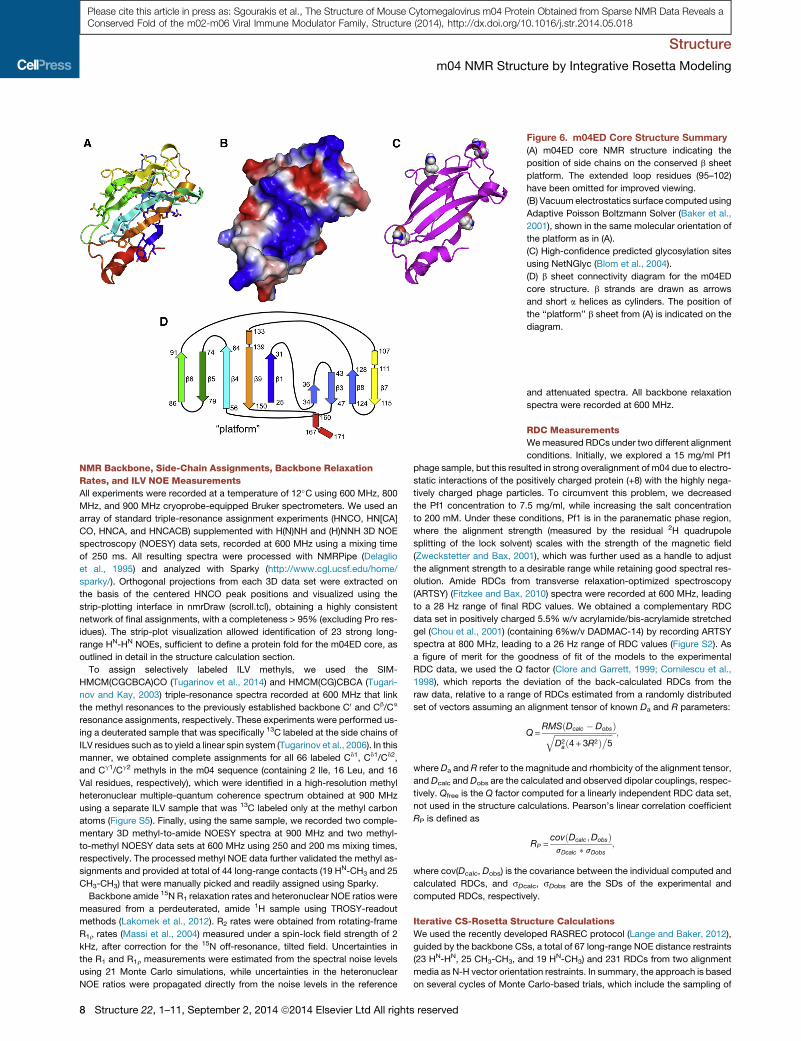
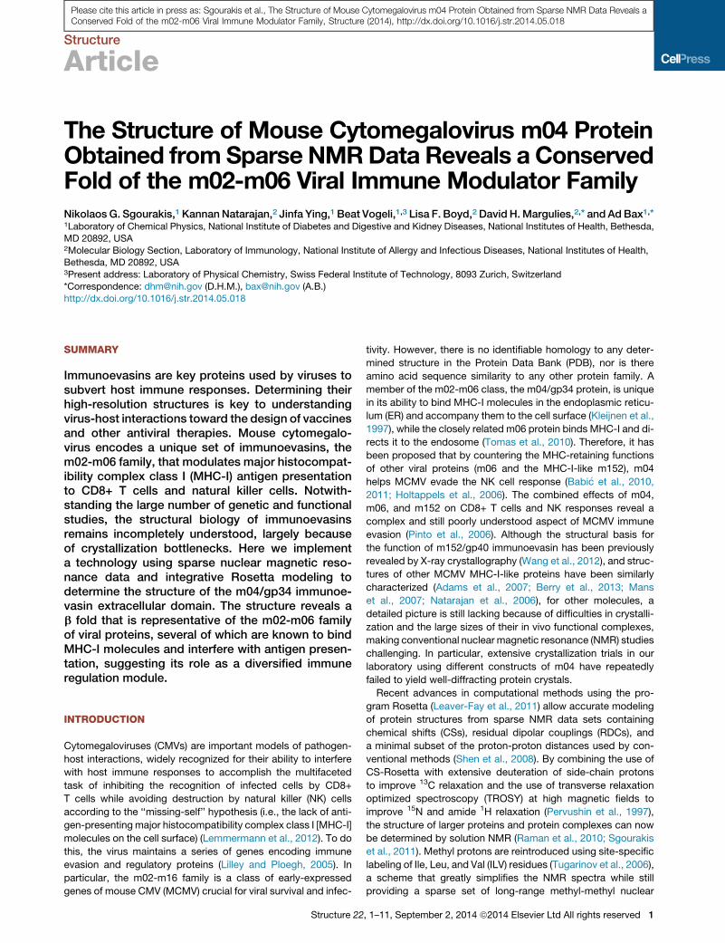
Figure 6. m04ED Core Structure Summary
(A) m04ED core NMR structure indicating the
position of side chains on the conserved b sheet
platform. The extended loop residues (95–102)
have been omitted for improved viewing.
(B) Vacuum electrostatics surface computed using
Adaptive Poisson Boltzmann Solver (Baker et al.,
2001), shown in the same molecular orientation of
the platform as in (A).
(C) High-confidence predicted glycosylation sites
using NetNGlyc (Blom et al., 2004).
(D) b sheet connectivity diagram for the m04ED
core structure. b strands are drawn as arrows
and short a helices as cylinders. The position of
the ‘‘platform’’ b sheet from (A) is indicated on the
diagram.
Structure
m04 NMR Structure by Integrative Rosetta Modeling
Please cite this article in press as: Sgourakis et al., The Structure of Mouse Cytomegalovirus m04 Protein Obtained from Sparse NMR Data Reveals aConserved Fold of the m02-m06 Viral Immune Modulator Family, Structure (2014), http://dx.doi.org/10.1016/j.str.2014.05.018
NMR Backbone, Side-Chain Assignments, Backbone Relaxation
Rates, and ILV NOE Measurements
All experiments were recorded at a temperature of 12�C using 600 MHz, 800
MHz, and 900 MHz cryoprobe-equipped Bruker spectrometers. We used an
array of standard triple-resonance assignment experiments (HNCO, HN[CA]
CO, HNCA, and HNCACB) supplemented with H(N)NH and (H)NNH 3D NOE
spectroscopy (NOESY) data sets, recorded at 600 MHz using a mixing time
of 250 ms. All resulting spectra were processed with NMRPipe (Delaglio
et al., 1995) and analyzed with Sparky (http://www.cgl.ucsf.edu/home/
sparky/). Orthogonal projections from each 3D data set were extracted on
the basis of the centered HNCO peak positions and visualized using the
strip-plotting interface in nmrDraw (scroll.tcl), obtaining a highly consistent
network of final assignments, with a completeness > 95% (excluding Pro res-
idues). The strip-plot visualization allowed identification of 23 strong long-
range HN-HN NOEs, sufficient to define a protein fold for the m04ED core, as
outlined in detail in the structure calculation section.
To assign selectively labeled ILV methyls, we used the SIM-
HMCM(CGCBCA)CO (Tugarinov et al., 2014) and HMCM(CG)CBCA (Tugari-
nov and Kay, 2003) triple-resonance spectra recorded at 600 MHz that link
the methyl resonances to the previously established backbone C0 and Cb/Ca
resonance assignments, respectively. These experiments were performed us-
ing a deuterated sample that was specifically 13C labeled at the side chains of
ILV residues such as to yield a linear spin system (Tugarinov et al., 2006). In this
manner, we obtained complete assignments for all 66 labeled Cd1, Cd1/Cd2,
and Cg1/Cg2 methyls in the m04 sequence (containing 2 Ile, 16 Leu, and 16
Val residues, respectively), which were identified in a high-resolution methyl
heteronuclear multiple-quantum coherence spectrum obtained at 900 MHz
using a separate ILV sample that was 13C labeled only at the methyl carbon
atoms (Figure S5). Finally, using the same sample, we recorded two comple-
mentary 3D methyl-to-amide NOESY spectra at 900 MHz and two methyl-
to-methyl NOESY data sets at 600 MHz using 250 and 200 ms mixing times,
respectively. The processed methyl NOE data further validated the methyl as-
signments and provided at total of 44 long-range contacts (19 HN-CH3 and 25
CH3-CH3) that were manually picked and readily assigned using Sparky.
Backbone amide 15N R1 relaxation rates and heteronuclear NOE ratios were
measured from a perdeuterated, amide 1H sample using TROSY-readout
methods (Lakomek et al., 2012). R2 rates were obtained from rotating-frame
R1r rates (Massi et al., 2004) measured under a spin-lock field strength of 2
kHz, after correction for the 15N off-resonance, tilted field. Uncertainties in
the R1 and R1r measurements were estimated from the spectral noise levels
using 21 Monte Carlo simulations, while uncertainties in the heteronuclear
NOE ratios were propagated directly from the noise levels in the reference
8 Structure 22, 1–11, September 2, 2014 ª2014 Elsevier Ltd All rights reserved
and attenuated spectra. All backbone relaxation
spectra were recorded at 600 MHz.
RDC Measurements
Wemeasured RDCs under two different alignment
conditions. Initially, we explored a 15 mg/ml Pf1
phage sample, but this resulted in strong overalignment of m04 due to electro-
static interactions of the positively charged protein (+8) with the highly nega-
tively charged phage particles. To circumvent this problem, we decreased
the Pf1 concentration to 7.5 mg/ml, while increasing the salt concentration
to 200 mM. Under these conditions, Pf1 is in the paranematic phase region,
where the alignment strength (measured by the residual 2H quadrupole
splitting of the lock solvent) scales with the strength of the magnetic field
(Zweckstetter and Bax, 2001), which was further used as a handle to adjust
the alignment strength to a desirable range while retaining good spectral res-
olution. Amide RDCs from transverse relaxation-optimized spectroscopy
(ARTSY) (Fitzkee and Bax, 2010) spectra were recorded at 600 MHz, leading
to a 28 Hz range of final RDC values. We obtained a complementary RDC
data set in positively charged 5.5% w/v acrylamide/bis-acrylamide stretched
gel (Chou et al., 2001) (containing 6%w/v DADMAC-14) by recording ARTSY
spectra at 800 MHz, leading to a 26 Hz range of RDC values (Figure S2). As
a figure of merit for the goodness of fit of the models to the experimental
RDC data, we used the Q factor (Clore and Garrett, 1999; Cornilescu et al.,
1998), which reports the deviation of the back-calculated RDCs from the
raw data, relative to a range of RDCs estimated from a randomly distributed
set of vectors assuming an alignment tensor of known Da and R parameters:
Q=RMSðDcalc � DobsÞffiffiffiffiffiffiffiffiffiffiffiffiffiffiffiffiffiffiffiffiffiffiffiffiffiffiffiffiffiffi
D2að4+ 3R2Þ�5
q ;
where Da and R refer to the magnitude and rhombicity of the alignment tensor,
and Dcalc and Dobs are the calculated and observed dipolar couplings, respec-
tively. Qfree is the Q factor computed for a linearly independent RDC data set,
not used in the structure calculations. Pearson’s linear correlation coefficient
RP is defined as
RP =covðDcalc;DobsÞsDcalc � sDobs
;
where cov(Dcalc, Dobs) is the covariance between the individual computed and
calculated RDCs, and sDcalc, sDobs are the SDs of the experimental and
computed RDCs, respectively.
Iterative CS-Rosetta Structure Calculations
We used the recently developed RASREC protocol (Lange and Baker, 2012),
guided by the backbone CSs, a total of 67 long-range NOE distance restraints
(23 HN-HN, 25 CH3-CH3, and 19 HN-CH3) and 231 RDCs from two alignment
media as N-H vector orientation restraints. In summary, the approach is based
on several cycles of Monte Carlo-based trials, which include the sampling of
Figure 7. Multiple Sequence Alignment of m04ED with the Predicted EDs of Other m02-m06 Family Members
The approximate positions of conserved secondary structure elements are highlighted on the top of each alignment block (b1–b9: b strands; red ovals: a helices).
The position of the two disulfide bonds on the m04ED structure is indicated with capital C letters and connecting dashed lines, while high-confidence predicted
glycosylation sites on the m04ED sequence are indicated with stars. Protein sequences for m02 (UniProt ID YP_214010), m03 (YP_214011), m05 (YP_214013),
andm06 (YP_214014) were all taken from the SmithMCMVgenome (NC_004065). The sequence ofm03.5 (ABM74010), not present in the Smith strain, was taken
from strain G1F.
To identify the ER-lumenal domains of these proteins from the full-length protein sequence, we first identified the end of the SP using the SignalP (http://www.cbs.
dtu.dk/services/SignalP/) and Phobius (http://phobius.sbc.su.se/) servers, then identified the start of TM domains using TMpred (http://www.ch.embnet.org/
software/TMPRED_form.html). Sequence alignments were constructed using ClustalW2 (http://www.ebi.ac.uk/Tools/msa/clustalw2/) and colored by
sequence similarity using BoxShade (http://www.ch.embnet.org/software/BOX_form.html).
Structure
m04 NMR Structure by Integrative Rosetta Modeling
Please cite this article in press as: Sgourakis et al., The Structure of Mouse Cytomegalovirus m04 Protein Obtained from Sparse NMR Data Reveals aConserved Fold of the m02-m06 Viral Immune Modulator Family, Structure (2014), http://dx.doi.org/10.1016/j.str.2014.05.018
backbone fragments (3-mers and 9-mers) and b strand pairings. Structural
features that consistently lead to optimization of a target function (defined as
the sum of the Rosetta energy and the experimental NOE and RDC score
terms) are recombined during six generations of iterative structure calcula-
tions. In a series of benchmark calculations with targets of known structures,
the protocol showed improved sampling efficiency over both standard CS-
Rosetta structure calculations (Shen et al., 2008) and conventional NMR
structure determination programs (Herrmann et al., 2002) for b-rich proteins
with complicated topologies (Lange et al., 2012).
While the backbone CSs are used to guide the selection of backbone
fragments from high-resolution structures in the PDB (Vernon et al., 2013),
long-range NOEs help define the overall protein fold and b strand connectivity.
RDCs are used as bond vector orientation restraints relative to an overall
alignment frame to better define the local backbone structure, particularly
in loop regions that have few or no long-range NOEs. The calculations are
heavily restrained by the experimental data during the low-resolution back-
bone search. However, the final placement of side-chain rotamers and fine-
tuning of the backbone torsion angles is guided primarily by the Rosetta
Structure 22
energy function (Leaver-Fay et al., 2011), with almost no bias from the exper-
imental score terms.
NOEswere implemented as flat-bottom restraints with an upper limit of 4.0 A
and an exponential penalty function. In the absence of stereospecific methyl
proton assignments, all pairwise combinations of protons within each pair of
interacting methyl sites were averaged as r�6 to compute an effective NOE
distance (Nilges, 1993). In all calculations, the connectivity of disulfide bonds
was also used as an input restraint using an orientation-dependent potential
(Raman et al., 2009). All calculations were carried out in 200 threads of an
SGI UV2000 cluster using Intel Xeon E5-4640 processors at 2.40 GHz with
1,024 GB of memory, customized to accommodate the MPI interface required
by RASREC. Typical calculation runtimes under these conditions were 10
to 12 hr.
ACCESSION NUMBERS
The final 10 lowest energy models were deposited in the PDB (accession
number 2MIZ).
, 1–11, September 2, 2014 ª2014 Elsevier Ltd All rights reserved 9
Structure
m04 NMR Structure by Integrative Rosetta Modeling
Please cite this article in press as: Sgourakis et al., The Structure of Mouse Cytomegalovirus m04 Protein Obtained from Sparse NMR Data Reveals aConserved Fold of the m02-m06 Viral Immune Modulator Family, Structure (2014), http://dx.doi.org/10.1016/j.str.2014.05.018
SUPPLEMENTAL INFORMATION
Supplemental Information includes five figures and can be found with this
article online at http://dx.doi.org/10.1016/j.str.2014.05.018.
ACKNOWLEDGMENTS
We thank Dr. Oliver Lange for providing consultation with setting up the
RASREC-Rosetta protocol and Dr. Vitali Tugarinov for assistance with the
SIM-HMCM(CGCBCA)CO and HMCM(CG)CBCA triple-resonance experi-
ments. We are grateful to Drs. Nathan May for preparing multiple sequence
alignments of the m02 family and to Drs. Yang Shen and Lishan Yao for
preliminary work on determining optimal m04 sample conditions, as well as
to Dr. Manqing Hong for providing the MULT-1 protein sample. This work is
supported by the National Institute of Diabetes and Digestive and Kidney
Diseases and National Institute of Allergy and Infectious Diseases intramural
research programs and the Intramural AIDS-Targeted Antiviral Program of
the Office of the Director, NIH.
Received: March 6, 2014
Revised: May 23, 2014
Accepted: May 27, 2014
Published: August 7, 2014
REFERENCES
Achour, A., Persson, K., Harris, R.A., Sundback, J., Sentman, C.L., Lindqvist,
Y., Schneider, G., and Karre, K. (1998). The crystal structure of H-2Dd MHC
class I complexed with the HIV-1-derived peptide P18-I10 at 2.4 A resolution:
implications for T cell and NK cell recognition. Immunity 9, 199–208.
Adams, E.J., Juo, Z.S., Venook, R.T., Boulanger, M.J., Arase, H., Lanier, L.L.,
and Garcia, K.C. (2007). Structural elucidation of the m157 mouse cytomega-
lovirus ligand for Ly49 natural killer cell receptors. Proc. Natl. Acad. Sci. U S A
104, 10128–10133.
Andreeva, A., Howorth, D., Chandonia, J.M., Brenner, S.E., Hubbard, T.J.,
Chothia, C., and Murzin, A.G. (2008). Data growth and its impact on the
SCOP database: new developments. Nucleic Acids Res. 36, D419–D425.
Babi�c, M., Pyzik, M., Zafirova, B., Mitrovi�c, M., Butorac, V., Lanier, L.L.,
Krmpoti�c, A., Vidal, S.M., and Jonji�c, S. (2010). Cytomegalovirus immunoeva-
sin reveals the physiological role of ‘‘missing self’’ recognition in natural killer
cell dependent virus control in vivo. J. Exp. Med. 207, 2663–2673.
Babi�c, M., Krmpoti�c, A., and Jonji�c, S. (2011). All is fair in virus-host interac-
tions: NK cells and cytomegalovirus. Trends Mol. Med. 17, 677–685.
Baker, N.A., Sept, D., Joseph, S., Holst, M.J., and McCammon, J.A. (2001).
Electrostatics of nanosystems: application to microtubules and the ribosome.
Proc. Natl. Acad. Sci. U S A 98, 10037–10041.
Bateman, A., Eddy, S.R., andChothia, C. (1996). Members of the immunoglob-
ulin superfamily in bacteria. Protein Sci. 5, 1939–1941.
Bax, A., and Grishaev, A. (2005). Weak alignment NMR: a hawk-eyed view of
biomolecular structure. Curr. Opin. Struct. Biol. 15, 563–570.
Berry, R., Ng,N., Saunders, P.M., Vivian, J.P., Lin, J., Deuss, F.A., Corbett, A.J.,
Forbes, C.A., Widjaja, J.M., Sullivan, L.C., et al. (2013). Targeting of a natural
killer cell receptor family by a viral immunoevasin. Nat. Immunol. 14, 699–705.
Blom, N., Sicheritz-Ponten, T., Gupta, R., Gammeltoft, S., and Brunak, S.
(2004). Prediction of post-translational glycosylation and phosphorylation of
proteins from the amino acid sequence. Proteomics 4, 1633–1649.
Bradley, P., and Baker, D. (2006). Improved beta-protein structure prediction
by multilevel optimization of nonlocal strand pairings and local backbone
conformation. Proteins 65, 922–929.
Chen, V.B., Arendall, W.B., 3rd, Headd, J.J., Keedy, D.A., Immormino, R.M.,
Kapral, G.J., Murray, L.W., Richardson, J.S., and Richardson, D.C. (2010).
MolProbity: all-atom structure validation for macromolecular crystallography.
Acta Crystallogr. D Biol. Crystallogr. 66, 12–21.
Chou, J.J., Gaemers, S., Howder, B., Louis, J.M., and Bax, A. (2001). A
simple apparatus for generating stretched polyacrylamide gels, yielding uni-
10 Structure 22, 1–11, September 2, 2014 ª2014 Elsevier Ltd All righ
form alignment of proteins and detergent micelles. J. Biomol. NMR 21,
377–382.
Clore, G.M., and Garrett, D.S. (1999). R-factor, free R, and complete cross-
validation for dipolar coupling refinement of NMR structures. J. Am. Chem.
Soc. 121, 9008–9012.
Corbett, A.J., Forbes, C.A., Moro, D., and Scalzo, A.A. (2007). Extensive
sequence variation exists among isolates of murine cytomegalovirus within
members of the m02 family of genes. J. Gen. Virol. 88, 758–769.
Cornilescu, G., and Bax, A. (2000). Measurement of proton, nitrogen, and
carbonyl chemical shielding anisotropies in a protein dissolved in a dilute liquid
crystalline phase. J. Am. Chem. Soc. 122, 10143–10154.
Cornilescu, G., Marquardt, J.L., Ottiger, M., and Bax, A. (1998). Validation of
protein structure from anisotropic carbonyl chemical shifts in a dilute liquid
crystalline phase. J. Am. Chem. Soc. 120, 6836–6837.
Corr, M., Boyd, L.F., Padlan, E.A., andMargulies, D.H. (1993). H-2Dd exploits a
four residue peptide binding motif. J. Exp. Med. 178, 1877–1892.
Delaglio, F., Grzesiek, S., Vuister, G.W., Zhu, G., Pfeifer, J., and Bax, A. (1995).
NMRPipe: a multidimensional spectral processing system based on UNIX
pipes. J. Biomol. NMR 6, 277–293.
Duchardt, E., Sigalov, A.B., Aivazian, D., Stern, L.J., and Schwalbe, H. (2007).
Structure induction of the T-cell receptor zeta-chain upon lipid binding inves-
tigated by NMR spectroscopy. ChemBioChem 8, 820–827.
Fitzkee, N.C., and Bax, A. (2010). Facile measurement of 1H-15N residual
dipolar couplings in larger perdeuterated proteins. J. Biomol. NMR 48, 65–70.
Halaby, D.M., Poupon, A., andMornon, J. (1999). The immunoglobulin fold fam-
ily: sequenceanalysis and3Dstructure comparisons. ProteinEng.12, 563–571.
Herrmann, T., Guntert, P., and Wuthrich, K. (2002). Protein NMR structure
determination with automated NOE assignment using the new software
CANDID and the torsion angle dynamics algorithm DYANA. J. Mol. Biol. 319,
209–227.
Holm, L., and Rosenstrom, P. (2010). Dali server: conservation mapping in 3D.
Nucleic Acids Res. 38, W545–W549.
Holtappels, R., Gillert-Marien, D., Thomas, D., Podlech, J., Deegen, P., Herter,
S., Oehrlein-Karpi, S.A., Strand, D., Wagner, M., and Reddehase, M.J. (2006).
Cytomegalovirus encodes a positive regulator of antigen presentation. J. Virol.
80, 7613–7624.
Kay, L.E., Torchia, D.A., and Bax, A. (1989). Backbone dynamics of proteins as
studied by 15N inverse detected heteronuclear NMR spectroscopy: applica-
tion to staphylococcal nuclease. Biochemistry 28, 8972–8979.
Kleijnen, M.F., Huppa, J.B., Lucin, P., Mukherjee, S., Farrell, H., Campbell,
A.E., Koszinowski, U.H., Hill, A.B., and Ploegh, H.L. (1997). A mouse cytomeg-
alovirus glycoprotein, gp34, forms a complex with folded class I MHC mole-
cules in the ER which is not retained but is transported to the cell surface.
EMBO J. 16, 685–694.
Lakomek, N.A., Ying, J., and Bax, A. (2012). Measurement of 15N relaxation
rates in perdeuterated proteins by TROSY-based methods. J. Biomol. NMR
53, 209–221.
Lange, O.F., and Baker, D. (2012). Resolution-adapted recombination of struc-
tural features significantly improves sampling in restraint-guided structure
calculation. Proteins 80, 884–895.
Lange, O.F., Rossi, P., Sgourakis, N.G., Song, Y., Lee, H.W., Aramini, J.M.,
Ertekin, A., Xiao, R., Acton, T.B., Montelione, G.T., and Baker, D. (2012).
Determination of solution structures of proteins up to 40 kDa using CS-
Rosetta with sparse NMR data from deuterated samples. Proc. Natl. Acad.
Sci. U S A 109, 10873–10878.
Leaver-Fay, A., Tyka, M., Lewis, S.M., Lange, O.F., Thompson, J., Jacak, R.,
Kaufman, K., Renfrew, P.D., Smith, C.A., Sheffler, W., et al. (2011).
ROSETTA3: an object-oriented software suite for the simulation and design
of macromolecules. Methods Enzymol. 487, 545–574.
Lemmermann, N.A., Fink, A., Podlech, J., Ebert, S., Wilhelmi, V., Bohm, V.,
Holtappels, R., and Reddehase, M.J. (2012). Murine cytomegalovirus immune
evasion proteins operative in the MHC class I pathway of antigen processing
and presentation: state of knowledge, revisions, and questions. Med.
Microbiol. Immunol. (Berl.) 201, 497–512.
ts reserved

Structure
m04 NMR Structure by Integrative Rosetta Modeling
Please cite this article in press as: Sgourakis et al., The Structure of Mouse Cytomegalovirus m04 Protein Obtained from Sparse NMR Data Reveals aConserved Fold of the m02-m06 Viral Immune Modulator Family, Structure (2014), http://dx.doi.org/10.1016/j.str.2014.05.018
Li, H., Natarajan, K., Malchiodi, E.L., Margulies, D.H., and Mariuzza, R.A.
(1998). Three-dimensional structure of H-2Dd complexed with an immunodo-
minant peptide from human immunodeficiency virus envelope glycoprotein
120. J. Mol. Biol. 283, 179–191.
Lilley, B.N., and Ploegh, H.L. (2005). Viral modulation of antigen presentation:
manipulation of cellular targets in the ER and beyond. Immunol. Rev. 207,
126–144.
Lu, X., Kavanagh, D.G., and Hill, A.B. (2006). Cellular and molecular require-
ments for association of the murine cytomegalovirus protein m4/gp34 with
major histocompatibility complex class I molecules. J. Virol. 80, 6048–6055.
Mans, J., Natarajan, K., Balbo, A., Schuck, P., Eikel, D., Hess, S., Robinson, H.,
Simic, H., Jonjic, S., Tiemessen, C.T., and Margulies, D.H. (2007). Cellular
expression and crystal structure of the murine cytomegalovirus major histo-
compatibility complex class I-like glycoprotein, m153. J. Biol. Chem. 282,
35247–35258.
Massi, F., Johnson, E., Wang, C., Rance, M., and Palmer, A.G., 3rd. (2004).
NMR R1 rho rotating-frame relaxation with weak radio frequency fields.
J. Am. Chem. Soc. 126, 2247–2256.
Natarajan, K., Hicks, A., Mans, J., Robinson, H., Guan, R., Mariuzza, R.A., and
Margulies, D.H. (2006). Crystal structure of the murine cytomegalovirus MHC-I
homolog m144. J. Mol. Biol. 358, 157–171.
Nilges, M. (1993). A calculation strategy for the structure determination of
symmetric dimers by 1H NMR. Proteins 17, 297–309.
Pervushin, K., Riek, R., Wider, G., and Wuthrich, K. (1997). Attenuated T2
relaxation by mutual cancellation of dipole-dipole coupling and chemical shift
anisotropy indicates an avenue to NMR structures of very large biological
macromolecules in solution. Proc. Natl. Acad. Sci. U S A 94, 12366–12371.
Pinto, A.K., Munks, M.W., Koszinowski, U.H., and Hill, A.B. (2006).
Coordinated function of murine cytomegalovirus genes completely inhibits
CTL lysis. J. Immunol. 177, 3225–3234.
Raman, S., Vernon, R., Thompson, J., Tyka, M., Sadreyev, R., Pei, J., Kim, D.,
Kellogg, E., DiMaio, F., Lange, O., et al. (2009). Structure prediction for CASP8
with all-atom refinement using Rosetta. Proteins 77 (Suppl 9 ), 89–99.
Raman, S., Lange, O.F., Rossi, P., Tyka, M., Wang, X., Aramini, J., Liu, G.,
Ramelot, T.A., Eletsky, A., Szyperski, T., et al. (2010). NMR structure determi-
nation for larger proteins using backbone-only data. Science 327, 1014–1018.
Rao, T., Lubin, J.W., Armstrong, G.S., Tucey, T.M., Lundblad, V., and Wuttke,
D.S. (2014). Structure of Est3 reveals a bimodal surface with differential roles in
telomere replication. Proc. Natl. Acad. Sci. U S A 111, 214–218.
Reusch, U., Muranyi, W., Lucin, P., Burgert, H.G., Hengel, H., and
Koszinowski, U.H. (1999). A cytomegalovirus glycoprotein re-routes MHC
class I complexes to lysosomes for degradation. EMBO J. 18, 1081–1091.
Sass, H.J., Musco, G., Stahl, S.J., Wingfield, P.T., and Grzesiek, S. (2000).
Solution NMR of proteins within polyacrylamide gels: diffusional properties
and residual alignment by mechanical stress or embedding of oriented purple
membranes. J. Biomol. NMR 18, 303–309.
Sgourakis, N.G., Lange, O.F., DiMaio, F., Andre, I., Fitzkee, N.C., Rossi, P.,
Montelione, G.T., Bax, A., and Baker, D. (2011). Determination of the struc-
tures of symmetric protein oligomers from NMR chemical shifts and residual
dipolar couplings. J. Am. Chem. Soc. 133, 6288–6298.
Shen, Y., and Bax, A. (2012). Identification of helix capping and b-turn motifs
from NMR chemical shifts. J. Biomol. NMR 52, 211–232.
Shen, Y., and Bax, A. (2013). Protein backbone and sidechain torsion
angles predicted from NMR chemical shifts using artificial neural networks.
J. Biomol. NMR 56, 227–241.
Shen, Y., Lange, O., Delaglio, F., Rossi, P., Aramini, J.M., Liu, G., Eletsky, A.,
Wu, Y., Singarapu, K.K., Lemak, A., et al. (2008). Consistent blind protein
structure generation from NMR chemical shift data. Proc. Natl. Acad. Sci.
U S A 105, 4685–4690.
Smith, L.M., McWhorter, A.R., Shellam, G.R., and Redwood, A.J. (2013).
The genome of murine cytomegalovirus is shaped by purifying selection and
extensive recombination. Virology 435, 258–268.
Svitel, J., Balbo, A., Mariuzza, R.A., Gonzales, N.R., and Schuck, P. (2003).
Combined affinity and rate constant distributions of ligand populations from
Structure 22,
experimental surface binding kinetics and equilibria. Biophys. J. 84, 4062–
4077.
Tolman, J.R., and Ruan, K. (2006). NMR residual dipolar couplings as probes
of biomolecular dynamics. Chem. Rev. 106, 1720–1736.
Tomas, M.I., Kuci�c, N., Mahmutefendi�c, H., Blagojevi�c, G., and Lucin, P.
(2010). Murine cytomegalovirus perturbs endosomal trafficking of major histo-
compatibility complex class I molecules in the early phase of infection. J. Virol.
84, 11101–11112.
Tugarinov, V., and Kay, L.E. (2003). Ile, Leu, and Val methyl assignments of
the 723-residue malate synthase G using a new labeling strategy and novel
NMR methods. J. Am. Chem. Soc. 125, 13868–13878.
Tugarinov, V., Kanelis, V., and Kay, L.E. (2006). Isotope labeling strategies for
the study of high-molecular-weight proteins by solution NMR spectroscopy.
Nat. Protoc. 1, 749–754.
Tugarinov, V., Venditti, V., and Marius Clore, G. (2014). A NMR experiment
for simultaneous correlations of valine and leucine/isoleucine methyls with
carbonyl chemical shifts in proteins. J. Biomol. NMR 58, 1–8.
Varani, L., Bankovich, A.J., Liu, C.W., Colf, L.A., Jones, L.L., Kranz, D.M.,
Puglisi, J.D., andGarcia, K.C. (2007). Solutionmapping of T cell receptor dock-
ing footprints on peptide-MHC. Proc. Natl. Acad. Sci. U SA 104, 13080–13085.
Vernon, R., Shen, Y., Baker, D., and Lange, O.F. (2013). Improved chemical
shift based fragment selection for CS-Rosetta using Rosetta3 fragment picker.
J. Biomol. NMR 57, 117–127.
Vink, C., Beuken, E., and Bruggeman, C.A. (2000). Complete DNA sequence of
the rat cytomegalovirus genome. J. Virol. 74, 7656–7665.
Wang, J., Whitman, M.C., Natarajan, K., Tormo, J., Mariuzza, R.A., and
Margulies, D.H. (2002). Binding of the natural killer cell inhibitory receptor
Ly49A to its major histocompatibility complex class I ligand. Crucial contacts
include both H-2Dd ANDbeta 2-microglobulin. J. Biol. Chem. 277, 1433–1442.
Wang, R., Natarajan, K., and Margulies, D.H. (2009). Structural basis of the
CD8 alpha beta/MHC class I interaction: focused recognition orients CD8
beta to a T cell proximal position. J. Immunol. 183, 2554–2564.
Wang, R., Natarajan, K., Revilleza, M.J., Boyd, L.F., Zhi, L., Zhao, H.,
Robinson, H., and Margulies, D.H. (2012). Structural basis of mouse cytomeg-
alovirus m152/gp40 interaction with RAE1g reveals a paradigm for MHC/MHC
interaction in immune evasion. Proc. Natl. Acad. Sci. U S A 109, E3578–E3587.
Ward, A.B., Sali, A., andWilson, I.A. (2013). Biochemistry. Integrative structural
biology. Science 339, 913–915.
Warner, L.R., Varga, K., Lange, O.F., Baker, S.L., Baker, D., Sousa, M.C., and
Pardi, A. (2011). Structure of the BamC two-domain protein obtained by
Rosetta with a limited NMR data set. J. Mol. Biol. 411, 83–95.
Williams, A.F., and Barclay, A.N. (1988). The immunoglobulin superfamily—
domains for cell surface recognition. Annu. Rev. Immunol. 6, 381–405.
Wuthrich,K. (1986).NMRofProteinsandNucleicAcids. (NewYork: JohnWiley).
Zweckstetter, M., and Bax, A. (2001). Characterization of molecular alignment
in aqueous suspensions of Pf1 bacteriophage. J. Biomol. NMR 20, 365–377.
Note Added in Proof
Concurrently with the release of this work, the following article reported the 3.0
A-resolution X-ray structure of the ectodomain of a very similar m04 strain
sequence, including all functional glycosylations (PDB ID 4PN6). The structure
is remarkably similar to our solution NMR model (backbone/heavy atom
RMSD 0.6/0.8 A, excluding the flexible loop at residues 95–102). Notably,
the placement of the C-terminal helix (Figure 4) and loop conformation at res-
idues 115–125 (Figure 5) are virtually identical in the X-ray structure. Given the
unusual b-topology of the structure and lack of structural homologs in the PDB,
this result highlights the power of the combined Rosetta/NMR approach in
delivering highly accurate models at atomic resolution from a limited set of
experimental restraints relative to conventional NMR methods.
Berry, R., Vivian, J.P., Deuss, F.A., Balaji, G.R., Saunders, P.M., lin, J., Littler,
D.R., Brooks, A.G., and Rossjohn, J. (2014). The structure of the cytomegalo-
virus-encoded mo4 glycoprotein, a prototypical member of the mo2 family of
immunoevasins. J Biol Chem. Published online June 30, 2014. http://dx.doi.
org/10.1074/jbc.M114.584128.
1–11, September 2, 2014 ª2014 Elsevier Ltd All rights reserved 11