the staphylococcus aureus methicillin resistance factor ... · the staphylococcus aureus...
TRANSCRIPT
The Staphylococcus aureus Methicillin Resistance Factor FmtA Is aD-Amino Esterase That Acts on Teichoic Acids
Muhammad M. Rahman,a Howard N. Hunter,a Shamina Prova,a Vidhu Verma,a Aneela Qamar,b Dasantila Golemi-Kotraa,b
Department of Chemistrya and Department of Biology,b York University, Toronto, Ontario, Canada
ABSTRACT The methicillin resistance factor encoded by fmtA is a core member of the Staphylococcus aureus cell wall stimulon,but its function has remained elusive for the past two decades. First identified as a factor that affects methicillin resistance inS. aureus strains, FmtA was later shown to interact with teichoic acids and to localize to the cell division septum. We have madea breakthrough in understanding FmtA function. We show that FmtA hydrolyzes the ester bond between D-Ala and the back-bone of teichoic acids, which are polyglycerol-phosphate or polyribitol-phosphate polymers found in the S. aureus cell envelope.FmtA contains two conserved motifs found in serine active-site penicillin-binding proteins (PBPs) and �-lactamases. The con-served SXXK motif was found to be important for the D-amino esterase activity of FmtA. Moreover, we show that deletion offmtA (�fmtA) led to higher levels of D-Ala in teichoic acids, and this effect was reversed by complementation of �fmtA withfmtA. The positive charge on D-Ala partially masks the negative charge of the polyol-phosphate backbone of teichoic acids;hence, a change in the D-Ala content will result in modulation of their charge. Cell division, biofilm formation, autolysis, andcolonization are among the many processes in S. aureus affected by the D-Ala content and overall charge of the cell surfaceteichoic acids. The esterase activity of FmtA and the regulation of fmtA suggest that FmtA functions as a modulator of teichoicacid charge, thus FmtA may be involved in S. aureus cell division, biofilm formation, autolysis, and colonization.
IMPORTANCE Teichoic acids are involved in cell division, cell wall synthesis, biofilm formation, attachment of bacteria to artifi-cial surfaces, and colonization. However, the function of teichoic acids is not fully understood. Modification by glycosylationand/or D-alanylation of the polyol-phosphate backbone of teichoic acids is important in the above cell processes. The intrinsicnegative charge of teichoic acid backbone plays a role in the charge and/or pH of the bacterial surface, and D-alanylation repre-sents a means through which bacteria modulate the charge or the pH of their surfaces. We discovered that FmtA removes D-Alafrom teichoic acids. We propose FmtA may provide a temporal and spatial regulation of the bacterial cell surface charge in twoways, by removing the D-Ala from LTA to make it available to wall teichoic acid (WTA) in response to certain conditions and byremoving it from WTA to allow the cell to reset its surface charge to a previous condition.
Received 2 December 2015 Accepted 8 January 2016 Published 9 February 2016
Citation Rahman MM, Hunter HN, Prova S, Verma V, Qamar A, Golemi-Kotra D. 2016. The Staphylococcus aureus methicillin resistance factor fmta is a D-amino esterase that actson teichoic acids. mBio 7(1):e02070-15. doi:10.1128/mBio.02070-15.
Editor Julian E. Davies, University of British Columbia
Copyright © 2016 Rahman et al. This is an open-access article distributed under the terms of the Creative Commons Attribution-Noncommercial-ShareAlike 3.0 Unportedlicense, which permits unrestricted noncommercial use, distribution, and reproduction in any medium, provided the original author and source are credited.
Address correspondence to Dasantila Golemi-Kotra, [email protected].
Staphylococcus aureus is the leading cause of hospital- andcommunity-acquired infections (1). S. aureus was once inher-
ently susceptible to most antibiotics. However, it is now a patho-gen of great concern due to its intrinsic virulence and its remark-able ability to rapidly adapt to different environmental conditionsby mutation and DNA transfer (2–4). Multiple-drug-resistantS. aureus strains, such as methicillin-resistant S. aureus (MRSA),have become notoriously difficult to treat, with 20 to 40% of casescausing mortality (1). Emergence of MRSA strains resistant tovancomycin (5), an antibiotic reserved for the treatment of severeMRSA infections, has led to limited treatment options for S. au-reus infections (6–9). To make matters worse, infections causedby multiple MRSA strains have reached epidemic proportions(8, 10).
The need for novel antibiotics for the treatment of S. aureus-related infections is as urgent today as it was in 1940, when peni-cillin was introduced to treat S. aureus infections (11, 12). Antibi-
otics that target cell wall biosynthesis (referred to as cell wallinhibitors), such as �-lactams and glycopeptides, are among themost efficient antibacterial agents for treating S. aureus infections;however, their biological activities have been compromised by theemergence of resistance mechanisms (11, 12). Recent reports haveshown that MRSA can be resensitized to �-lactams and vancomy-cin by inhibiting nonessential genes involved in the biosynthesis ofcell envelope components, such as peptidoglycan and teichoic ac-ids, and these reports have rekindled interest in targeting the cellwall for drug discovery and provide evidence that antibiotic po-tency can be rescued (13–19).
Komatsuzawa et al. reported that fmtA was a factor in themethicillin resistance of MRSA strains (20) and that deletion offmtA reduced the methicillin MIC for S. aureus Col (an MRSAstrain) from 1,024 �g/ml to 128 �g/ml (20). In addition, deletionof fmtA was shown to disrupt the homogeneity of methicillin re-sistance (20). Further, fmtA was identified as a core member of the
RESEARCH ARTICLE
crossmark
January/February 2016 Volume 7 Issue 1 e02070-15 ® mbio.asm.org 1
on January 13, 2020 by guesthttp://m
bio.asm.org/
Dow
nloaded from
cell wall stimulon; fmtA expression increased in the presence ofcell wall inhibitors and when genes involved in cell wall biosyn-thesis were deleted (21–24). The primary structure of FmtA sharessimilarities with D,D-carboxypeptidase from Streptomyces R61(D,D-carboxypeptidase R61) and class C �-lactamases (25). FmtAharbors two of the three conserved motifs, SXXK and SND, foundin the serine active site of penicillin-binding proteins (PBPs) and�-lactamases. The third conserved motif, KTG, has not been iden-tified in FmtA. Our previous studies on FmtA revealed that itinteracts covalently with �-lactams via a serine residue located inthe conserved SXXK motif. We also showed that FmtA has veryweak D,D-carboxypeptidase activity and interacts with teichoic ac-ids (25). These observations led to the proposal that FmtA may bea PBP (25, 26). S. aureus has four native PBPs. PBP1 and PBP2 areessential enzymes (27). PBP1 and PBP4 are transpeptidases, andPBP2 is a bifunctional enzyme. The function of PBP3 remainselusive. PBP2 and PBP4 are involved in the synthesis of highlycross-linked peptidoglycan (28).
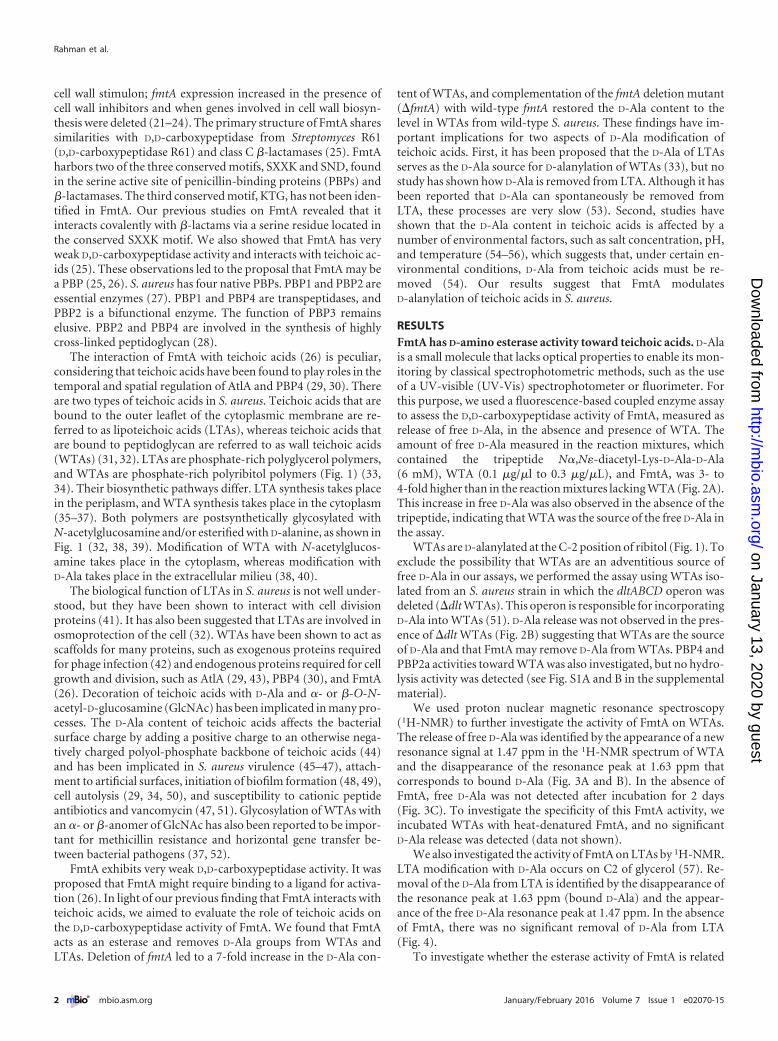
The interaction of FmtA with teichoic acids (26) is peculiar,considering that teichoic acids have been found to play roles in thetemporal and spatial regulation of AtlA and PBP4 (29, 30). Thereare two types of teichoic acids in S. aureus. Teichoic acids that arebound to the outer leaflet of the cytoplasmic membrane are re-ferred to as lipoteichoic acids (LTAs), whereas teichoic acids thatare bound to peptidoglycan are referred to as wall teichoic acids(WTAs) (31, 32). LTAs are phosphate-rich polyglycerol polymers,and WTAs are phosphate-rich polyribitol polymers (Fig. 1) (33,34). Their biosynthetic pathways differ. LTA synthesis takes placein the periplasm, and WTA synthesis takes place in the cytoplasm(35–37). Both polymers are postsynthetically glycosylated withN-acetylglucosamine and/or esterified with D-alanine, as shown inFig. 1 (32, 38, 39). Modification of WTA with N-acetylglucos-amine takes place in the cytoplasm, whereas modification withD-Ala takes place in the extracellular milieu (38, 40).
The biological function of LTAs in S. aureus is not well under-stood, but they have been shown to interact with cell divisionproteins (41). It has also been suggested that LTAs are involved inosmoprotection of the cell (32). WTAs have been shown to act asscaffolds for many proteins, such as exogenous proteins requiredfor phage infection (42) and endogenous proteins required for cellgrowth and division, such as AtlA (29, 43), PBP4 (30), and FmtA(26). Decoration of teichoic acids with D-Ala and �- or �-O-N-acetyl-D-glucosamine (GlcNAc) has been implicated in many pro-cesses. The D-Ala content of teichoic acids affects the bacterialsurface charge by adding a positive charge to an otherwise nega-tively charged polyol-phosphate backbone of teichoic acids (44)and has been implicated in S. aureus virulence (45–47), attach-ment to artificial surfaces, initiation of biofilm formation (48, 49),cell autolysis (29, 34, 50), and susceptibility to cationic peptideantibiotics and vancomycin (47, 51). Glycosylation of WTAs withan �- or �-anomer of GlcNAc has also been reported to be impor-tant for methicillin resistance and horizontal gene transfer be-tween bacterial pathogens (37, 52).
FmtA exhibits very weak D,D-carboxypeptidase activity. It wasproposed that FmtA might require binding to a ligand for activa-tion (26). In light of our previous finding that FmtA interacts withteichoic acids, we aimed to evaluate the role of teichoic acids onthe D,D-carboxypeptidase activity of FmtA. We found that FmtAacts as an esterase and removes D-Ala groups from WTAs andLTAs. Deletion of fmtA led to a 7-fold increase in the D-Ala con-
tent of WTAs, and complementation of the fmtA deletion mutant(�fmtA) with wild-type fmtA restored the D-Ala content to thelevel in WTAs from wild-type S. aureus. These findings have im-portant implications for two aspects of D-Ala modification ofteichoic acids. First, it has been proposed that the D-Ala of LTAsserves as the D-Ala source for D-alanylation of WTAs (33), but nostudy has shown how D-Ala is removed from LTA. Although it hasbeen reported that D-Ala can spontaneously be removed fromLTA, these processes are very slow (53). Second, studies haveshown that the D-Ala content in teichoic acids is affected by anumber of environmental factors, such as salt concentration, pH,and temperature (54–56), which suggests that, under certain en-vironmental conditions, D-Ala from teichoic acids must be re-moved (54). Our results suggest that FmtA modulatesD-alanylation of teichoic acids in S. aureus.
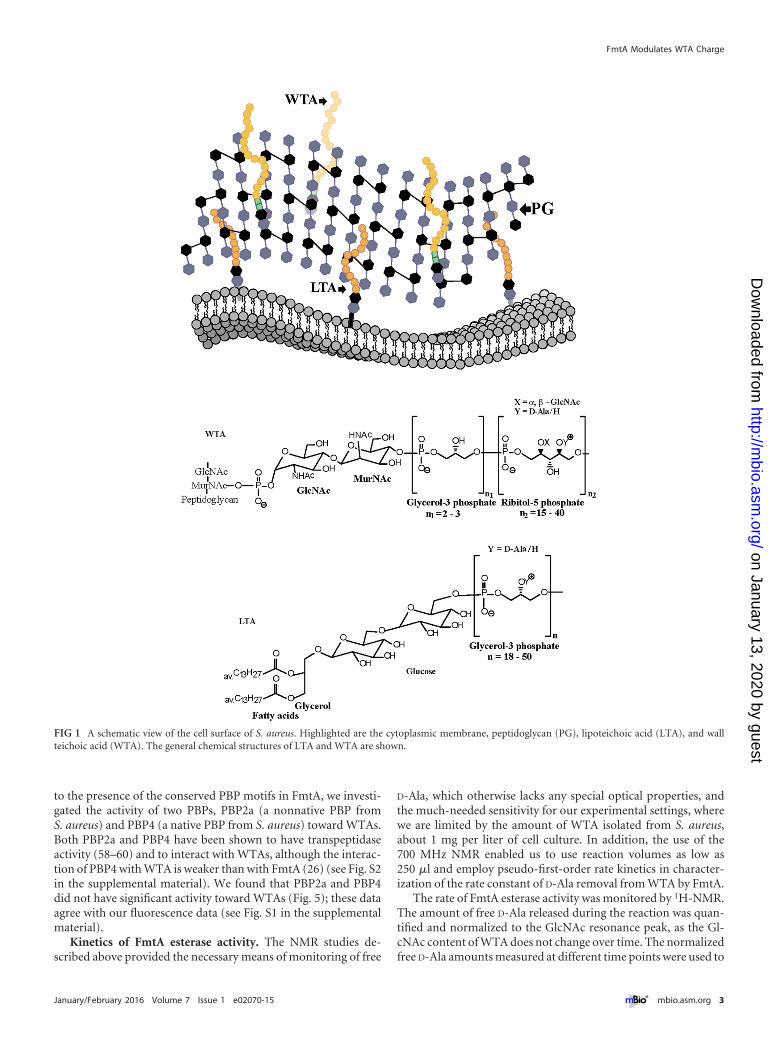
RESULTSFmtA has D-amino esterase activity toward teichoic acids. D-Alais a small molecule that lacks optical properties to enable its mon-itoring by classical spectrophotometric methods, such as the useof a UV-visible (UV-Vis) spectrophotometer or fluorimeter. Forthis purpose, we used a fluorescence-based coupled enzyme assayto assess the D,D-carboxypeptidase activity of FmtA, measured asrelease of free D-Ala, in the absence and presence of WTA. Theamount of free D-Ala measured in the reaction mixtures, whichcontained the tripeptide N�,N�-diacetyl-Lys-D-Ala-D-Ala(6 mM), WTA (0.1 �g/�l to 0.3 �g/�L), and FmtA, was 3- to4-fold higher than in the reaction mixtures lacking WTA (Fig. 2A).This increase in free D-Ala was also observed in the absence of thetripeptide, indicating that WTA was the source of the free D-Ala inthe assay.
WTAs are D-alanylated at the C-2 position of ribitol (Fig. 1). Toexclude the possibility that WTAs are an adventitious source offree D-Ala in our assays, we performed the assay using WTAs iso-lated from an S. aureus strain in which the dltABCD operon wasdeleted (�dlt WTAs). This operon is responsible for incorporatingD-Ala into WTAs (51). D-Ala release was not observed in the pres-ence of �dlt WTAs (Fig. 2B) suggesting that WTAs are the sourceof D-Ala and that FmtA may remove D-Ala from WTAs. PBP4 andPBP2a activities toward WTA was also investigated, but no hydro-lysis activity was detected (see Fig. S1A and B in the supplementalmaterial).
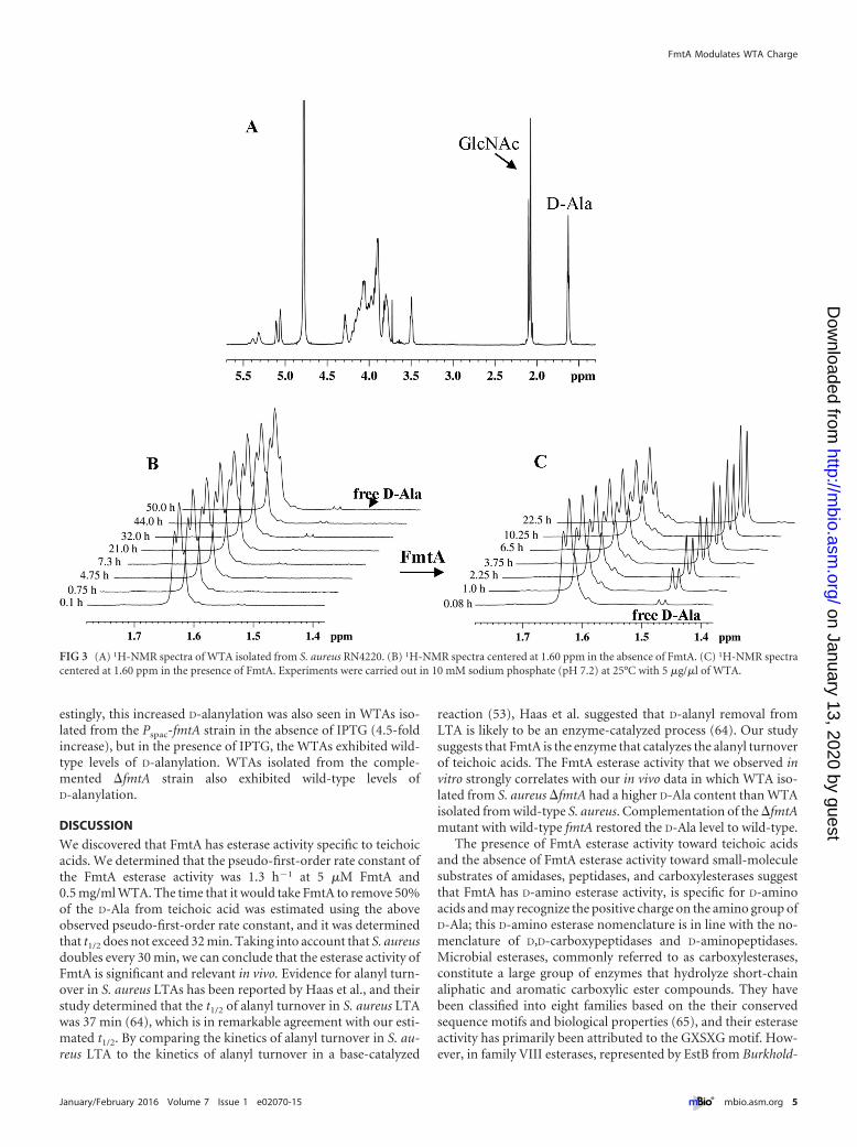
We used proton nuclear magnetic resonance spectroscopy(1H-NMR) to further investigate the activity of FmtA on WTAs.The release of free D-Ala was identified by the appearance of a newresonance signal at 1.47 ppm in the 1H-NMR spectrum of WTAand the disappearance of the resonance peak at 1.63 ppm thatcorresponds to bound D-Ala (Fig. 3A and B). In the absence ofFmtA, free D-Ala was not detected after incubation for 2 days(Fig. 3C). To investigate the specificity of this FmtA activity, weincubated WTAs with heat-denatured FmtA, and no significantD-Ala release was detected (data not shown).
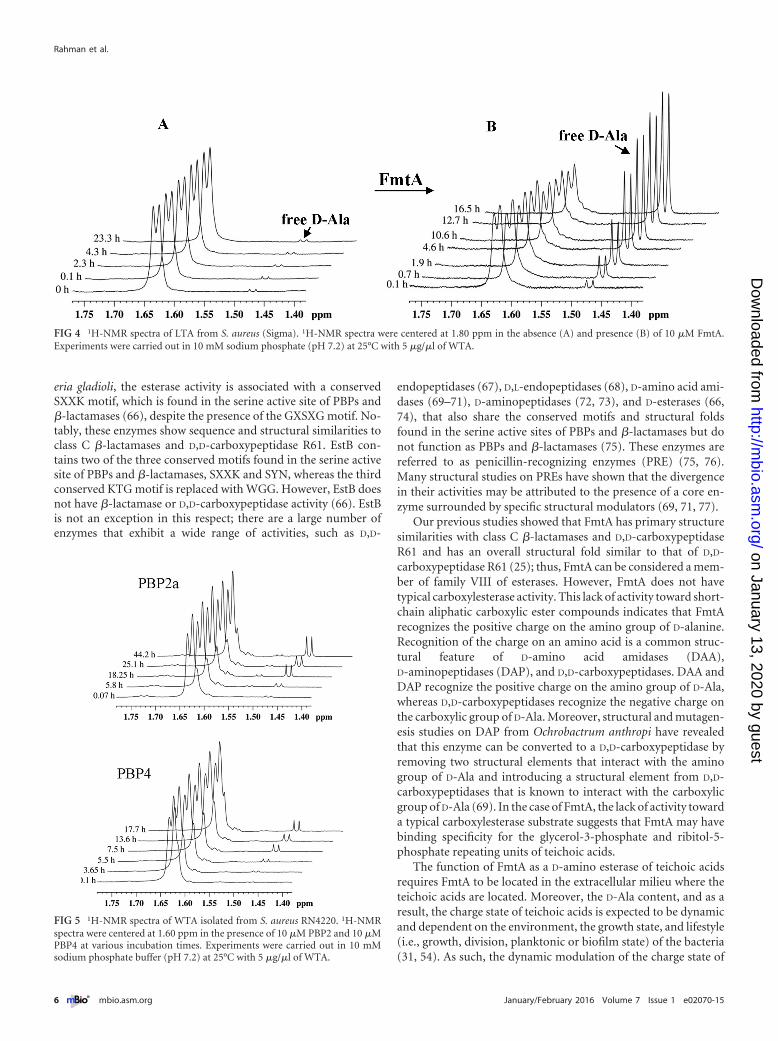
We also investigated the activity of FmtA on LTAs by 1H-NMR.LTA modification with D-Ala occurs on C2 of glycerol (57). Re-moval of the D-Ala from LTA is identified by the disappearance ofthe resonance peak at 1.63 ppm (bound D-Ala) and the appear-ance of the free D-Ala resonance peak at 1.47 ppm. In the absenceof FmtA, there was no significant removal of D-Ala from LTA(Fig. 4).
To investigate whether the esterase activity of FmtA is related
Rahman et al.
2 ® mbio.asm.org January/February 2016 Volume 7 Issue 1 e02070-15
on January 13, 2020 by guesthttp://m
bio.asm.org/
Dow
nloaded from
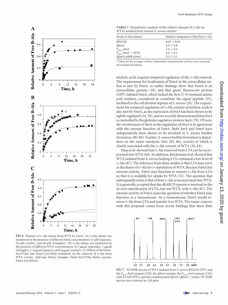
to the presence of the conserved PBP motifs in FmtA, we investi-gated the activity of two PBPs, PBP2a (a nonnative PBP fromS. aureus) and PBP4 (a native PBP from S. aureus) toward WTAs.Both PBP2a and PBP4 have been shown to have transpeptidaseactivity (58–60) and to interact with WTAs, although the interac-tion of PBP4 with WTA is weaker than with FmtA (26) (see Fig. S2in the supplemental material). We found that PBP2a and PBP4did not have significant activity toward WTAs (Fig. 5); these dataagree with our fluorescence data (see Fig. S1 in the supplementalmaterial).
Kinetics of FmtA esterase activity. The NMR studies de-scribed above provided the necessary means of monitoring of free
D-Ala, which otherwise lacks any special optical properties, andthe much-needed sensitivity for our experimental settings, wherewe are limited by the amount of WTA isolated from S. aureus,about 1 mg per liter of cell culture. In addition, the use of the700 MHz NMR enabled us to use reaction volumes as low as250 �l and employ pseudo-first-order rate kinetics in character-ization of the rate constant of D-Ala removal from WTA by FmtA.
The rate of FmtA esterase activity was monitored by 1H-NMR.The amount of free D-Ala released during the reaction was quan-tified and normalized to the GlcNAc resonance peak, as the Gl-cNAc content of WTA does not change over time. The normalizedfree D-Ala amounts measured at different time points were used to
FIG 1 A schematic view of the cell surface of S. aureus. Highlighted are the cytoplasmic membrane, peptidoglycan (PG), lipoteichoic acid (LTA), and wallteichoic acid (WTA). The general chemical structures of LTA and WTA are shown.
FmtA Modulates WTA Charge
January/February 2016 Volume 7 Issue 1 e02070-15 ® mbio.asm.org 3
on January 13, 2020 by guesthttp://m
bio.asm.org/
Dow
nloaded from
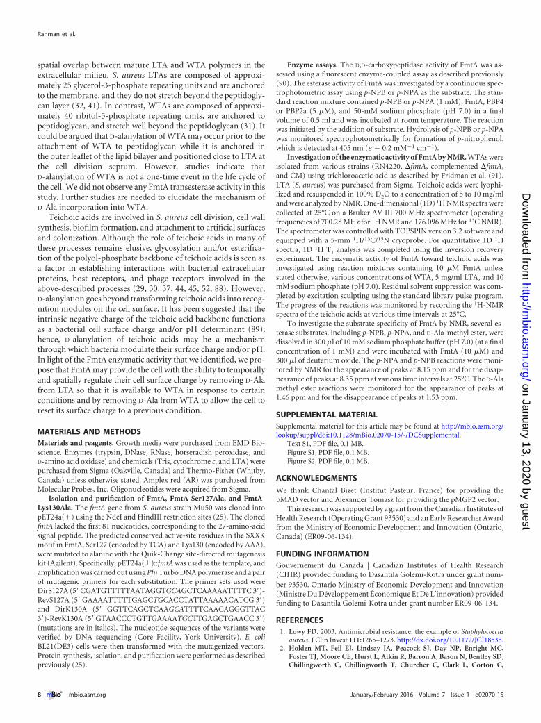
construct progress curves in which the data were fitted to pseudo-first-order rate kinetics. The observed pseudo-first-order rateconstant (kobs) was 0.57 � 0.01 h�1 with 10 �M FmtA and 5mg/ml WTA. The reaction rates increased with increased enzymeconcentrations as indicated from the calculated kobs values, whichincreased from 0.46 h�1 with 5 �M FmtA to 1.11 h�1 with 20 �MFmtA ([WTA] � 5 mg/ml) (Fig. 6A). In contrast, FmtA reactionrates increased with the decrease of WTA concentrations. The kobs
value increased 3-fold, to 1.3 � 0.2 h�1, when the WTA concen-tration was reduced from 5 mg/ml to 0.5 mg/ml (Fig. 6B). The kobs
value measured at 0.5 mg/ml WTA is the highest pseudo-first-order rate constant that we could measure; as such, it can serve asan approximation for the apparent first-order rate constant of theesterase reaction catalyzed by FmtA, although it is an underesti-mation of the true first-order rate constant for this reaction. Fromthis kobs value, we estimated the time required to hydrolyze 50% ofthe D-alanyl ester bonds in WTA (half time [t1/2]) to be 32 min.
The reduction in FmtA esterase activity found with increasedWTA concentrations could be a result of structural changes inFmtA upon WTA binding. Indeed, an earlier investigation of theinteraction of FmtA with WTA by circular dichroism revealed thatthe secondary structural elements of FmtA were altered exten-sively in the presence of high WTA concentrations (26). We haveobserved that high concentrations of WTA (�10 mg/ml) causeaggregation of FmtA. This may be due to a salting-out effect pro-duced by the highly negatively charged WTAs.
To investigate the significance of the conserved PBP motif
SXXK for the esterase activity of FmtA, we constructed two vari-ants of FmtA by replacing Ser127 and Lys130 with Ala. By deter-mining the ratios of the observed pseudo-first-order rate con-stants of the mutants and wild-type FmtA, we found thatFmtA-Ser127Ala and FmtA-Lys130Ala had 16% of the activity ofwild-type FmtA (Fig. 6C). The seemingly high remaining activity ofFmtA mutant could result from relative instability of ester moieties inslight acidic or basic aqueous solutions and binding of the WTA toFmtA; i.e., the topology of the FmtA active site may introduce enoughconstraints to the carbonyl C of the D-Ala bound to WTA to lead to itsincreased electrophilicity and attack by weak nucleophiles such aswater molecules in long incubation times.
To investigate whether FmtA can catalyze esterification ofWTA by D-Ala, we set up several assays. We incubated D-Ala(6 mM) with ribitol (10 mM) or with �dlt WTAs (2.5 mg/ml) andmonitored the reaction by 1H-NMR in the absence and presenceof FmtA (20 �M). In addition, LTA (0.5 mg/ml) and �dlt WTA(2.5 mg/ml) were incubated together in the absence and presenceof FmtA (20 �M) to investigate whether D-Ala released from LTAcan be transferred to WTA by FmtA. Lastly, we incubated D-Alawith ATP (5 mM), MgCl2 (10 mM), dithiothreitol (DTT) (1 mM),and �dlt WTA in the absence and presence of FmtA (20 �M).None of the above conditions led to the incorporation of D-Ala toWTA (data not shown).
Specificity of FmtA esterase activity. To determine the sub-strate specificity of FmtA, we tested several carboxylesterase sub-strates, including p-nitrophenyl butyrate (p-NPB) andp-nitrophenyl acetate (p-NPA). FmtA showed very low esteraseactivity on p-NPB and p-NPA as assessed by continuous spectro-photometric assay (data not shown). We also used 1H-NMR tomonitor hydrolysis of p-NPB and p-NPA and detected very littleactivity on p-NPB and p-NPA (data not shown). In addition, weinvestigated potential �-amino-acid esterase and aminopeptidaseactivities in FmtA with D-alanine methyl ester and L-alaninep-nitroanilide, respectively. However, we did not observe any cat-alytic activity against these substrates. Carboxylesterases havebeen reported to catalyze the hydrolysis of short-chain aliphaticand aromatic carboxylic ester compounds (61) suggesting thatFmtA may be specific for the D-Ala ester attached at the C-2 ofribitol-5-phosphate and glycerol-3-phosphate, which are the re-peating units of teichoic acids.
Assessment of FmtA esterase activity in cells. To investigatethe significance of our findings in S. aureus, we constructed threeS. aureus RN4220 mutant strains (see Text S1 in the supplementalmaterial). The fmtA deletion (�fmtA) strain was constructed us-ing a pMAD vector (62). The complementation strain was createdby cloning fmtA into a pMK4 vector (63) and introducing theconstruct into S. aureus RN4220. We also constructed an fmtAconditional mutant (CM) in which fmtA expression is underthe control of an IPTG (isopropyl-�-D-thiogalactopyranoside)-inducible promoter, Pspac (Pspac-fmtA), using a pMUTIN integra-tion vector (Bacillus Genetic Stock Center, The Ohio University).
We isolated WTAs from S. aureus RN4220 and the �fmtA,complemented �fmtA, and Pspac-fmtA strains in the presence orabsence of 0.5 mM IPTG. WTAs from each strain were analyzedby 1H-NMR. The amount of D-Ala attached to ribitol relative tothe amount of N-acetylglucosamine attached to ribitol was deter-mined by integrating the resonance peaks of N-acetylglucosamineat 2.08 ppm and D-Ala at 1.63 ppm. Deletion of fmtA resulted in a7-fold increase in D-alanylation of WTAs (Table 1; Fig. 7). Inter-
FIG 2 Analysis of D-Ala removal from the tripeptide N�,N�-diacetyl-Lys-D-Ala-D-Ala (TP, 6 mM) by FmtA (10 �M) in the absence or presence of WTA(0.1 �g/�l, 0.2 �g/�l, 0.3 �g/�l) (A) or in the presence of �dlt WTA (B). FreeD-Ala was measured using a fluorescence-based coupled enzymatic assay. Er-ror bars represent standard deviations from three independent experiments.
Rahman et al.
4 ® mbio.asm.org January/February 2016 Volume 7 Issue 1 e02070-15
on January 13, 2020 by guesthttp://m
bio.asm.org/
Dow
nloaded from
estingly, this increased D-alanylation was also seen in WTAs iso-lated from the Pspac-fmtA strain in the absence of IPTG (4.5-foldincrease), but in the presence of IPTG, the WTAs exhibited wild-type levels of D-alanylation. WTAs isolated from the comple-mented �fmtA strain also exhibited wild-type levels ofD-alanylation.
DISCUSSION
We discovered that FmtA has esterase activity specific to teichoicacids. We determined that the pseudo-first-order rate constant ofthe FmtA esterase activity was 1.3 h�1 at 5 �M FmtA and0.5 mg/ml WTA. The time that it would take FmtA to remove 50%of the D-Ala from teichoic acid was estimated using the aboveobserved pseudo-first-order rate constant, and it was determinedthat t1/2 does not exceed 32 min. Taking into account that S. aureusdoubles every 30 min, we can conclude that the esterase activity ofFmtA is significant and relevant in vivo. Evidence for alanyl turn-over in S. aureus LTAs has been reported by Haas et al., and theirstudy determined that the t1/2 of alanyl turnover in S. aureus LTAwas 37 min (64), which is in remarkable agreement with our esti-mated t1/2. By comparing the kinetics of alanyl turnover in S. au-reus LTA to the kinetics of alanyl turnover in a base-catalyzed
reaction (53), Haas et al. suggested that D-alanyl removal fromLTA is likely to be an enzyme-catalyzed process (64). Our studysuggests that FmtA is the enzyme that catalyzes the alanyl turnoverof teichoic acids. The FmtA esterase activity that we observed invitro strongly correlates with our in vivo data in which WTA iso-lated from S. aureus �fmtA had a higher D-Ala content than WTAisolated from wild-type S. aureus. Complementation of the �fmtAmutant with wild-type fmtA restored the D-Ala level to wild-type.
The presence of FmtA esterase activity toward teichoic acidsand the absence of FmtA esterase activity toward small-moleculesubstrates of amidases, peptidases, and carboxylesterases suggestthat FmtA has D-amino esterase activity, is specific for D-aminoacids and may recognize the positive charge on the amino group ofD-Ala; this D-amino esterase nomenclature is in line with the no-menclature of D,D-carboxypeptidases and D-aminopeptidases.Microbial esterases, commonly referred to as carboxylesterases,constitute a large group of enzymes that hydrolyze short-chainaliphatic and aromatic carboxylic ester compounds. They havebeen classified into eight families based on the their conservedsequence motifs and biological properties (65), and their esteraseactivity has primarily been attributed to the GXSXG motif. How-ever, in family VIII esterases, represented by EstB from Burkhold-
FIG 3 (A) 1H-NMR spectra of WTA isolated from S. aureus RN4220. (B) 1H-NMR spectra centered at 1.60 ppm in the absence of FmtA. (C) 1H-NMR spectracentered at 1.60 ppm in the presence of FmtA. Experiments were carried out in 10 mM sodium phosphate (pH 7.2) at 25°C with 5 �g/�l of WTA.
FmtA Modulates WTA Charge
January/February 2016 Volume 7 Issue 1 e02070-15 ® mbio.asm.org 5
on January 13, 2020 by guesthttp://m
bio.asm.org/
Dow
nloaded from
eria gladioli, the esterase activity is associated with a conservedSXXK motif, which is found in the serine active site of PBPs and�-lactamases (66), despite the presence of the GXSXG motif. No-tably, these enzymes show sequence and structural similarities toclass C �-lactamases and D,D-carboxypeptidase R61. EstB con-tains two of the three conserved motifs found in the serine activesite of PBPs and �-lactamases, SXXK and SYN, whereas the thirdconserved KTG motif is replaced with WGG. However, EstB doesnot have �-lactamase or D,D-carboxypeptidase activity (66). EstBis not an exception in this respect; there are a large number ofenzymes that exhibit a wide range of activities, such as D,D-
endopeptidases (67), D,L-endopeptidases (68), D-amino acid ami-dases (69–71), D-aminopeptidases (72, 73), and D-esterases (66,74), that also share the conserved motifs and structural foldsfound in the serine active sites of PBPs and �-lactamases but donot function as PBPs and �-lactamases (75). These enzymes arereferred to as penicillin-recognizing enzymes (PRE) (75, 76).Many structural studies on PREs have shown that the divergencein their activities may be attributed to the presence of a core en-zyme surrounded by specific structural modulators (69, 71, 77).
Our previous studies showed that FmtA has primary structuresimilarities with class C �-lactamases and D,D-carboxypeptidaseR61 and has an overall structural fold similar to that of D,D-carboxypeptidase R61 (25); thus, FmtA can be considered a mem-ber of family VIII of esterases. However, FmtA does not havetypical carboxylesterase activity. This lack of activity toward short-chain aliphatic carboxylic ester compounds indicates that FmtArecognizes the positive charge on the amino group of D-alanine.Recognition of the charge on an amino acid is a common struc-tural feature of D-amino acid amidases (DAA),D-aminopeptidases (DAP), and D,D-carboxypeptidases. DAA andDAP recognize the positive charge on the amino group of D-Ala,whereas D,D-carboxypeptidases recognize the negative charge onthe carboxylic group of D-Ala. Moreover, structural and mutagen-esis studies on DAP from Ochrobactrum anthropi have revealedthat this enzyme can be converted to a D,D-carboxypeptidase byremoving two structural elements that interact with the aminogroup of D-Ala and introducing a structural element from D,D-carboxypeptidases that is known to interact with the carboxylicgroup of D-Ala (69). In the case of FmtA, the lack of activity towarda typical carboxylesterase substrate suggests that FmtA may havebinding specificity for the glycerol-3-phosphate and ribitol-5-phosphate repeating units of teichoic acids.
The function of FmtA as a D-amino esterase of teichoic acidsrequires FmtA to be located in the extracellular milieu where theteichoic acids are located. Moreover, the D-Ala content, and as aresult, the charge state of teichoic acids is expected to be dynamicand dependent on the environment, the growth state, and lifestyle(i.e., growth, division, planktonic or biofilm state) of the bacteria(31, 54). As such, the dynamic modulation of the charge state of
FIG 4 1H-NMR spectra of LTA from S. aureus (Sigma). 1H-NMR spectra were centered at 1.80 ppm in the absence (A) and presence (B) of 10 �M FmtA.Experiments were carried out in 10 mM sodium phosphate (pH 7.2) at 25°C with 5 �g/�l of WTA.
FIG 5 1H-NMR spectra of WTA isolated from S. aureus RN4220. 1H-NMRspectra were centered at 1.60 ppm in the presence of 10 �M PBP2 and 10 �MPBP4 at various incubation times. Experiments were carried out in 10 mMsodium phosphate buffer (pH 7.2) at 25°C with 5 �g/�l of WTA.
Rahman et al.
6 ® mbio.asm.org January/February 2016 Volume 7 Issue 1 e02070-15
on January 13, 2020 by guesthttp://m
bio.asm.org/
Dow
nloaded from
teichoic acids requires temporal regulation of the D-Ala removal.The requirement for localization of FmtA in the extracellular mi-lieu is met by FmtA, as earlier findings show that FmtA is anextracellular protein (20) and that green fluorescent protein(GFP)-labeled FmtA, which lacked the first 27 N-terminal aminoacid residues, considered to constitute the signal peptide (25),localized to the cell division septum of S. aureus (26). The require-ment for temporal regulation of D-Ala content of teichoic acids isalso met by FmtA, as the expression of fmtA has been shown to betightly regulated (24, 78), and we recently demonstrated that fmtAis controlled by the globular regulatory protein SarA (79). Of note,the involvement of SarA in the regulation of fmtA is in agreementwith the esterase function of FmtA. Both SarA and FmtA haveindependently been shown to be involved in S. aureus biofilmformation (80–84). Further, S. aureus biofilm formation is depen-dent on the major autolysin AtlA (85, 86), activity of which isclosely associated with the D-Ala content of WTA (29, 43).
Haas et al. showed that D-Ala removed from LTA can be incor-porated into WTA (64). In addition, Reichmann et al. showed thatWTA isolated from S. aureus lacking LTA contained a low level ofD-Ala (87). The inference from these studies is that LTA may serveas the donor of D-Ala for D-alanylation of WTA. Because FmtA hasesterase activity, FmtA may function to remove D-Ala from LTAso that it is available for uptake by WTA (31). The question thatsubsequently arises is that of how D-Ala is incorporated into WTA.It is generally accepted that the dltABCD operon is involved in thede novo esterification of LTA, but not WTA, with D-Ala (87). Theesterase activity of FmtA raises the question of whether FmtA mayfunction as a transesterase. As a transesterase, FmtA would re-move D-Ala from LTA and transfer it to WTA. The major concernwith this proposal comes from recent findings that show little
FIG 6 Kinetics of D-Ala release from WTA by FmtA. (A) D-Ala release wasmonitored in the presence of different FmtA concentrations (5 �M [squares],10 �M [circles], and 20 �M [triangles]). (B) D-Ala release was monitored inthe presence of different WTA concentrations (0.5 �g/�l [asterisks], 1 �g/�l[triangles], 2.5 �g/�l [squares], and 5 �g/�l [circles]). (C) Effects of the FmtA-Ser127Ala and FmtA-Lys130Ala mutations on the removal of D-Ala fromWTA (circles, wild-type FmtA; triangles, FmtA-Ser127Ala FmtA; squares,FmtA-Lys130Ala).
TABLE 1 Quantitative analysis of the relative amount of D-Ala onWTAs isolated from various S. aureus strainsa
Strain or description Relative integration (GlucNac/D-Ala)
RN4220 0.63 � 0.03�fmtA 4.0 � 0.8Pspac-fmtA 2.6 � 0.5Pspac-fmtA � IPTG 0.6 � 0.1�fmtA::pMK4:fmtA 0.5 � 0.1a Values are the averages of three independent measurements and the errors representthe standard deviations.
FIG 7 1H-NMR spectra of WTA isolated from S. aureus RN4220 (WT) andthe Pspac-fmtA mutant (CM), the �fmtA mutant, the Pspac-fmtA mutant (CM)with 0.5 mM IPTG, and the complemented �fmtA (�fmtA�) strain. 1H-NMRspectra were centered at 1.80 ppm.
FmtA Modulates WTA Charge
January/February 2016 Volume 7 Issue 1 e02070-15 ® mbio.asm.org 7
on January 13, 2020 by guesthttp://m
bio.asm.org/
Dow
nloaded from
spatial overlap between mature LTA and WTA polymers in theextracellular milieu. S. aureus LTAs are composed of approxi-mately 25 glycerol-3-phosphate repeating units and are anchoredto the membrane, and they do not stretch beyond the peptidogly-can layer (32, 41). In contrast, WTAs are composed of approxi-mately 40 ribitol-5-phosphate repeating units, are anchored topeptidoglycan, and stretch well beyond the peptidoglycan (31). Itcould be argued that D-alanylation of WTA may occur prior to theattachment of WTA to peptidoglycan while it is anchored inthe outer leaflet of the lipid bilayer and positioned close to LTA atthe cell division septum. However, studies indicate thatD-alanylation of WTA is not a one-time event in the life cycle ofthe cell. We did not observe any FmtA transesterase activity in thisstudy. Further studies are needed to elucidate the mechanism ofD-Ala incorporation into WTA.
Teichoic acids are involved in S. aureus cell division, cell wallsynthesis, biofilm formation, and attachment to artificial surfacesand colonization. Although the role of teichoic acids in many ofthese processes remains elusive, glycosylation and/or esterifica-tion of the polyol-phosphate backbone of teichoic acids is seen asa factor in establishing interactions with bacterial extracellularproteins, host receptors, and phage receptors involved in theabove-described processes (29, 30, 37, 44, 45, 52, 88). However,D-alanylation goes beyond transforming teichoic acids into recog-nition modules on the cell surface. It has been suggested that theintrinsic negative charge of the teichoic acid backbone functionsas a bacterial cell surface charge and/or pH determinant (89);hence, D-alanylation of teichoic acids may be a mechanismthrough which bacteria modulate their surface charge and/or pH.In light of the FmtA enzymatic activity that we identified, we pro-pose that FmtA may provide the cell with the ability to temporallyand spatially regulate their cell surface charge by removing D-Alafrom LTA so that it is available to WTA in response to certainconditions and by removing D-Ala from WTA to allow the cell toreset its surface charge to a previous condition.
MATERIALS AND METHODSMaterials and reagents. Growth media were purchased from EMD Bio-science. Enzymes (trypsin, DNase, RNase, horseradish peroxidase, andD-amino acid oxidase) and chemicals (Tris, cytochrome c, and LTA) werepurchased from Sigma (Oakville, Canada) and Thermo-Fisher (Whitby,Canada) unless otherwise stated. Amplex red (AR) was purchased fromMolecular Probes, Inc. Oligonucleotides were acquired from Sigma.
Isolation and purification of FmtA, FmtA-Ser127Ala, and FmtA-Lys130Ala. The fmtA gene from S. aureus strain Mu50 was cloned intopET24a(�) using the NdeI and HindIII restriction sites (25). The clonedfmtA lacked the first 81 nucleotides, corresponding to the 27-amino-acidsignal peptide. The predicted conserved active-site residues in the SXXKmotif in FmtA, Ser127 (encoded by TCA) and Lys130 (encoded by AAA),were mutated to alanine with the Quik-Change site-directed mutagenesiskit (Agilent). Specifically, pET24a(�)::fmtA was used as the template, andamplification was carried out using Pfu Turbo DNA polymerase and a pairof mutagenic primers for each substitution. The primer sets used wereDirS127A (5=CGATGTTTTTAATAGGTGCAGCTCAAAAATTTTC 3=)-RevS127A (5= GAAAATTTTTGAGCTGCACCTATTAAAAACATCG 3=)and DirK130A (5= GGTTCAGCTCAAGCATTTTCAACAGGGTTAC3=)-RevK130A (5= GTAACCCTGTTGAAAATGCTTGAGCTGAACC 3=)(mutations are in italics). The nucleotide sequences of the variants wereverified by DNA sequencing (Core Facility, York University). E. coliBL21(DE3) cells were then transformed with the mutagenized vectors.Protein synthesis, isolation, and purification were performed as describedpreviously (25).
Enzyme assays. The D,D-carboxypeptidase activity of FmtA was as-sessed using a fluorescent enzyme-coupled assay as described previously(90). The esterase activity of FmtA was investigated by a continuous spec-trophotometric assay using p-NPB or p-NPA as the substrate. The stan-dard reaction mixture contained p-NPB or p-NPA (1 mM), FmtA, PBP4or PBP2a (5 �M), and 50-mM sodium phosphate (pH 7.0) in a finalvolume of 0.5 ml and was incubated at room temperature. The reactionwas initiated by the addition of substrate. Hydrolysis of p-NPB or p-NPAwas monitored spectrophotometrically for formation of p-nitrophenol,which is detected at 405 nm (� � 0.2 mM�1 cm�1).
Investigation of the enzymatic activity of FmtA by NMR. WTAs wereisolated from various strains (RN4220, �fmtA, complemented �fmtA,and CM) using trichloroacetic acid as described by Fridman et al. (91).LTA (S. aureus) was purchased from Sigma. Teichoic acids were lyophi-lized and resuspended in 100% D2O to a concentration of 5 to 10 mg/mland were analyzed by NMR. One-dimensional (1D) 1H NMR spectra werecollected at 25°C on a Bruker AV III 700 MHz spectrometer (operatingfrequencies of 700.28 MHz for 1H NMR and 176.096 MHz for 13C NMR).The spectrometer was controlled with TOPSPIN version 3.2 software andequipped with a 5-mm 1H/13C/15N cryoprobe. For quantitative 1D 1Hspectra, 1D 1H T1 analysis was completed using the inversion recoveryexperiment. The enzymatic activity of FmtA toward teichoic acids wasinvestigated using reaction mixtures containing 10 �M FmtA unlessstated otherwise, various concentrations of WTA, 5 mg/ml LTA, and 10mM sodium phosphate (pH 7.0). Residual solvent suppression was com-pleted by excitation sculpting using the standard library pulse program.The progress of the reactions was monitored by recording the 1H-NMRspectra of the teichoic acids at various time intervals at 25°C.
To investigate the substrate specificity of FmtA by NMR, several es-terase substrates, including p-NPB, p-NPA, and D-Ala-methyl ester, weredissolved in 300 �l of 10 mM sodium phosphate buffer (pH 7.0) (at a finalconcentration of 1 mM) and were incubated with FmtA (10 �M) and300 �l of deuterium oxide. The p-NPA and p-NPB reactions were moni-tored by NMR for the appearance of peaks at 8.15 ppm and for the disap-pearance of peaks at 8.35 ppm at various time intervals at 25°C. The D-Alamethyl ester reactions were monitored for the appearance of peaks at1.46 ppm and for the disappearance of peaks at 1.53 ppm.
SUPPLEMENTAL MATERIALSupplemental material for this article may be found at http://mbio.asm.org/lookup/suppl/doi:10.1128/mBio.02070-15/-/DCSupplemental.
Text S1, PDF file, 0.1 MB.Figure S1, PDF file, 0.1 MB.Figure S2, PDF file, 0.1 MB.
ACKNOWLEDGMENTS
We thank Chantal Bizet (Institut Pasteur, France) for providing thepMAD vector and Alexander Tomasz for providing the pMGP2 vector.
This research was supported by a grant from the Canadian Institutes ofHealth Research (Operating Grant 93530) and an Early Researcher Awardfrom the Ministry of Economic Development and Innovation (Ontario,Canada) (ER09-06-134).
FUNDING INFORMATIONGouvernement du Canada | Canadian Institutes of Health Research(CIHR) provided funding to Dasantila Golemi-Kotra under grant num-ber 93530. Ontario Ministry of Economic Development and Innovation(Ministre Du Développement Économique Et De L’innovation) providedfunding to Dasantila Golemi-Kotra under grant number ER09-06-134.
REFERENCES1. Lowy FD. 2003. Antimicrobial resistance: the example of Staphylococcus
aureus. J Clin Invest 111:1265–1273. http://dx.doi.org/10.1172/JCI18535.2. Holden MT, Feil EJ, Lindsay JA, Peacock SJ, Day NP, Enright MC,
Foster TJ, Moore CE, Hurst L, Atkin R, Barron A, Bason N, Bentley SD,Chillingworth C, Chillingworth T, Churcher C, Clark L, Corton C,
Rahman et al.
8 ® mbio.asm.org January/February 2016 Volume 7 Issue 1 e02070-15
on January 13, 2020 by guesthttp://m
bio.asm.org/
Dow
nloaded from
Cronin A, Doggett J. 2004. Complete genomes of two clinical Staphylo-coccus aureus strains: evidence for the rapid evolution of virulence anddrug resistance. Proc Natl Acad Sci U S A 101:9786 –9791. http://dx.doi.org/10.1073/pnas.0402521101.
3. Fitzgerald JR, Sturdevant DE, Mackie SM, Gill SR, Musser JM. 2001.Evolutionary genomics of Staphylococcus aureus: insights into the ori-gin of methicillin-resistant strains and the toxic shock syndrome epi-demic. Proc Natl Acad Sci U S A 98:8821– 8826. http://dx.doi.org/10.1073/pnas.161098098.
4. Hiramatsu K, Cui L, Kuroda M, Ito T. 2001. The emergence and evolu-tion of methicillin-resistant Staphylococcus aureus. Trends Microbiol9:486 – 493. http://dx.doi.org/10.1016/S0966-842X(01)02175-8.
5. Bartley J. 2002. First case of VRSA identified in Michigan. Infect ControlHosp Epidemiol 23:480.
6. Brown DFJ, Reynolds PE. 1980. Intrinsic resistance to beta-lactam anti-biotics in Staphylococcus aureus. FEBS Lett 122:275–278. http://dx.doi.org/10.1016/0014-5793(80)80455-8.
7. Bancroft EA. 2007. Antimicrobial resistance—it’s not just for hospitals.JAMA 298:1803–1804.
8. Klevens RM, Morrison MA, Nadle J, Petit S, Gershman K, Ray S,Harrison LH, Lynfield R, Dumyati G, Townes JM, Craig AS, Zell ER,Fosheim GE, McDougal LK, Carey RB, Fridkin SK, Active BacterialCore surveillance (ABCs) MRSA Investigators. 2007. Invasivemethicillin-resistant Staphylococcus aureus infections in the United States.JAMA 298:1763–1771. http://dx.doi.org/10.1001/jama.298.15.1763.
9. Silver LL. 2003. Novel inhibitors of bacterial cell wall synthesis. Curr OpinMicrobiol 6:431– 438. http://dx.doi.org/10.1016/j.mib.2003.08.004.
10. David MZ, Daum RS. 2010. Community-associated methicillin-resistantStaphylococcus aureus: epidemiology and clinical consequences of anemerging epidemic. Clin Microbiol Rev 23:616 – 687. http://dx.doi.org/10.1128/CMR.00081-09.
11. Walsh C. 2003. Where will new antibiotics come from? Nat Rev Microbiol1:65–70. http://dx.doi.org/10.1038/nrmicro727.
12. Bassetti M, Merelli M, Temperoni C, Astilean A. 2013. New antibioticsfor bad bugs: where are we? Ann Clin Microbiol Antimicrob 12:22. http://dx.doi.org/10.1186/1476-0711-12-22.
13. Swoboda JG, Meredith TC, Campbell J, Brown S, Suzuki T, BollenbachT, Malhowski AJ, Kishony R, Gilmore MS, Walker S. 2009. Discovery ofa small molecule that blocks wall teichoic acid biosynthesis in Staphylo-coccus aureus. ACS Chem Biol 4:875– 883. http://dx.doi.org/10.1021/cb900151k.
14. Farha MA, Leung A, Sewell EW, D’Elia MA, Allison SE, Ejim L, PereiraPM, Pinho MG, Wright GD, Brown ED. 2013. Inhibition of WTAsynthesis blocks the cooperative action of PBPs and sensitizes MRSA tobeta-lactams. ACS Chem Biol 8:226 –233. http://dx.doi.org/10.1021/cb300413m.
15. Campbell J, Singh AK, Santa Maria JP, Kim Y, Brown S, Swoboda JG,Mylonakis E, Wilkinson BJ, Walker S. 2011. Synthetic lethal compoundcombinations reveal a fundamental connection between wall teichoic acidand peptidoglycan biosyntheses in Staphylococcus aureus. ACS Chem Biol6:106 –116. http://dx.doi.org/10.1021/cb100269f.
16. Blake KL, O’Neill AJ, Mengin-Lecreulx D, Henderson PJ, Bostock JM,Dunsmore CJ, Simmons KJ, Fishwick CW, Leeds JA, Chopra I. 2009.The nature of Staphylococcus aureus MurA and MurZ and approaches fordetection of peptidoglycan biosynthesis inhibitors. Mol Microbiol 72:335–343. http://dx.doi.org/10.1111/j.1365-2958.2009.06648.x.
17. Gardete S, Ludovice AM, Sobral RG, Filipe SR, de Lencastre H, TomaszA. 2004. Role of murE in the expression of beta-lactam antibiotic resis-tance in Staphylococcus aureus. J Bacteriol 186:1705–1713. http://dx.doi.org/10.1128/JB.186.6.1705-1713.2004.
18. Sobral RG, Ludovice AM, de Lencastre H, Tomasz A. 2006. Role of murFin cell wall biosynthesis: isolation and characterization of a murF condi-tional mutant of Staphylococcus aureus. J Bacteriol 188:2543–2553. http://dx.doi.org/10.1128/JB.188.7.2543-2553.2006.
19. Balibar CJ, Shen X, Tao J. 2009. The mevalonate pathway of Staphylo-coccus aureus. J Bacteriol 191:851– 861. http://dx.doi.org/10.1128/JB.01357-08.
20. Komatsuzawa H, Sugai M, Ohta K, Fujiwara T, Nakashima S, Suzuki J,Lee CY, Suginaka H. 1997. Cloning and characterization of the fmt genewhich affects the methicillin resistance level and autolysis in the presenceof Triton X-100 in methicillin-resistant Staphylococcus aureus. Antimi-crob Agents Chemother 41:2355–2361.
21. Bernal P, Lemaire S, Pinho MG, Mobashery S, Hinds J, Taylor PW.
2010. Insertion of epicatechin gallate into the cytoplasmic membraneof methicillin-resistant Staphylococcus aureus disrupts penicillin-binding protein (PBP) 2a-mediated beta-lactam resistance by delocal-izing PBP2. J Biol Chem 285:24055–24065. http://dx.doi.org/10.1074/jbc.M110.114793.
22. McAleese F, Wu SW, Sieradzki K, Dunman P, Murphy E, Projan S,Tomasz A. 2006. Overexpression of genes of the cell wall stimulon inclinical isolates of Staphylococcus aureus exhibiting vancomycin-intermediate-S. aureus-type resistance to vancomycin. J Bacteriol 188:1120 –1133. http://dx.doi.org/10.1128/JB.188.3.1120-1133.2006.
23. McCallum N, Spehar G, Bischoff M, Berger-Bächi B. 2006. Strain de-pendence of the cell wall-damage induced stimulon in Staphylococcus au-reus. Biochim Biophys Acta 1760:1475–1481. http://dx.doi.org/10.1016/j.bbagen.2006.06.008.
24. Utaida S, Dunman PM, Macapagal D, Murphy E, Projan SJ, Singh VK,Jayaswal RK, Wilkinson BJ. 2003. Genome-wide transcriptional profilingof the response of Staphylococcus aureus to cell-wall-active antibiotics re-veals a cell-wall-stress stimulon. Microbiology 149:2719 –2732. http://dx.doi.org/10.1099/mic.0.26426-0.
25. Fan X, Liu Y, Smith D, Konermann L, Siu KW, Golemi-Kotra D. 2007.Diversity of penicillin-binding proteins. Resistance factor FmtA of Staph-ylococcus aureus. J Biol Chem 282:35143–35152. http://dx.doi.org/10.1074/jbc.M706296200.
26. Qamar A, Golemi-Kotra D. 2012. Dual roles of FmtA in Staphylococcusaureus cell wall biosynthesis and autolysis. Antimicrob Agents Chemother56:3797–3805. http://dx.doi.org/10.1128/AAC.00187-12.
27. Wada A, Watanabe H. 1998. Penicillin-binding protein 1 of Staphylococ-cus aureus is essential for growth. J Bacteriol 180:2759 –2765.
28. Łeski TA, Tomasz A. 2005. Role of penicillin-binding protein 2(PBP2) in the antibiotic susceptibility and cell wall cross-linking ofStaphylococcus aureus: evidence for the cooperative functioning ofPBP2, PBP4, and PBP2A. J Bacteriol 187:1815–1824. http://dx.doi.org/10.1128/JB.187.5.1815-1824.2005.
29. Schlag M, Biswas R, Krismer B, Kohler T, Zoll S, Yu W, Schwarz H,Peschel A, Götz F. 2010 Role of Staphylococcal wall teichoic acid intargeting the major autolysin Atl. Mol Microbiol 75:864 – 873.
30. Atilano ML, Pereira PM, Yates J, Reed P, Veiga H, Pinho MG, Filipe SR.2010. Teichoic acids are temporal and spatial regulators of peptidoglycancross-linking in Staphylococcus aureus. Proc Natl Acad Sci U S A 107:18991–18996. http://dx.doi.org/10.1073/pnas.1004304107.
31. Brown S, Santa Maria JP, Jr., Walker S. 2013. Wall teichoic acids ofGram-positive bacteria. Annu Rev Microbiol 67:313–336. http://dx.doi.org/10.1146/annurev-micro-092412-155620.
32. Percy MG, Gründling A. 2014. Lipoteichoic acid synthesis and functionin Gram-positive bacteria. Annu Rev Microbiol 68:81–100. http://dx.doi.org/10.1146/annurev-micro-091213-112949.
33. Reichmann NT, Gründling A. 2011. Location, synthesis and function ofglycolipids and polyglycerolphosphate lipoteichoic acid in Gram-positivebacteria of the phylum Firmicutes. FEMS Microbiol Lett 319:97–105.http://dx.doi.org/10.1111/j.1574-6968.2011.02260.x.
34. Swoboda JG, Campbell J, Meredith TC, Walker S. 2010. Wall teichoicacid function, biosynthesis, and inhibition. Chembiochem 11:35– 45.http://dx.doi.org/10.1002/cbic.200900557.
35. Koch HU, Haas R, Fischer W. 1984. The role of lipoteichoic acid biosyn-thesis in membrane lipid metabolism of growing Staphylococcus aureus.Eur J Biochem 138:357–363. http://dx.doi.org/10.1111/j.1432-1033.1984.tb07923.x.
36. Allison SE, D’Elia MA, Arar S, Monteiro MA, Brown ED. 2011. Studiesof the genetics, function, and kinetic mechanism of TagE, the wall teichoicacid glycosyltransferase in Bacillus subtilis 168. J Biol Chem 286:23708 –23716. http://dx.doi.org/10.1074/jbc.M111.241265.
37. Brown S, Xia G, Luhachack LG, Campbell J, Meredith TC, Chen C,Winstel V, Gekeler C, Irazoqui JE, Peschel A, Walker S. 2012. Methi-cillin resistance in Staphylococcus aureus requires glycosylated wallteichoic acids. Proc Natl Acad Sci U S A 109:18909 –18914. http://dx.doi.org/10.1073/pnas.1209126109.
38. Perego M, Glaser P, Minutello A, Strauch MA, Leopold K, Fischer W.1995. Incorporation of D-alanine into lipoteichoic acid and wall teichoicacid in Bacillus subtilis. Identification of genes and regulation. J Biol Chem270:15598 –15606. http://dx.doi.org/10.1074/jbc.270.26.15598.
39. Neuhaus FC, Baddiley J. 2003. A continuum of anionic charge: structuresand functions of D-alanyl-teichoic acids in Gram-positive bacteria. Micro-
FmtA Modulates WTA Charge
January/February 2016 Volume 7 Issue 1 e02070-15 ® mbio.asm.org 9
on January 13, 2020 by guesthttp://m
bio.asm.org/
Dow
nloaded from
biol Mol Biol Rev 67:686 –723. http: / /dx.doi .org/10.1128/MMBR.67.4.686-723.2003.
40. Kovács M, Halfmann A, Fedtke I, Heintz M, Peschel A, Vollmer W,Hakenbeck R, Brückner R. 2006. A functional dlt operon, encoding pro-teins required for incorporation of D-alanine in teichoic acids in gram-positive bacteria, confers resistance to cationic antimicrobial peptides inStreptococcus pneumoniae. J Bacteriol 188:5797–5805. http://dx.doi.org/10.1128/JB.00336-06.
41. Reichmann NT, Piçarra Cassona C, Monteiro JM, Bottomley AL, Cor-rigan RM, Foster SJ, Pinho MG, Gründling A. 2014. Differential local-ization of LTA synthesis proteins and their interaction with the cell divi-sion machinery in Staphylococcus aureus. Mol Microbiol 92:273–286.http://dx.doi.org/10.1111/mmi.12551.
42. Chatterjee AN. 1969. Use of bacteriophage-resistant mutants to study thenature of the bacteriophage receptor site of Staphylococcus aureus. J Bac-teriol 98:519 –527.
43. Peschel A, Vuong C, Otto M, Götz F. 2000. The D-alanine residues ofStaphylococcus aureus teichoic acids alter the susceptibility to vancomycinand the activity of autolytic enzymes. Antimicrob Agents Chemother 44:2845–2847. http://dx.doi.org/10.1128/AAC.44.10.2845-2847.2000.
44. Collins LV, Kristian SA, Weidenmaier C, Faigle M, Van Kessel KP, VanStrijp JA, Götz F, Neumeister B, Peschel A. 2002. Staphylococcus aureusstrains lacking D-alanine modifications of teichoic acids are highly suscep-tible to human neutrophil killing and are virulence attenuated in mice. JInfect Dis 186:214 –219. http://dx.doi.org/10.1086/341454.
45. Weidenmaier C, Kokai-Kun JF, Kristian SA, Chanturiya T, KalbacherH, Gross M, Nicholson G, Neumeister B, Mond JJ, Peschel A. 2004. Roleof teichoic acids in Staphylococcus aureus nasal colonization, a major riskfactor in nosocomial infections. Nat Med 10:243–245. http://dx.doi.org/10.1038/nm991.
46. Park KH, Kurokawa K, Zheng L, Jung DJ, Tateishi K, Jin JO, Ha NC,Kang HJ, Matsushita M, Kwak JY, Takahashi K, Lee BL. 2010. Humanserum mannose-binding lectin senses wall teichoic acid glycopolymer ofStaphylococcus aureus, which is restricted in infancy. J Biol Chem 285:27167–27175. http://dx.doi.org/10.1074/jbc.M110.141309.
47. Abachin E, Poyart C, Pellegrini E, Milohanic E, Fiedler F, Berche P,Trieu-Cuot P. 2002. Formation of D-alanyl-lipoteichoic acid is requiredfor adhesion and virulence of Listeria monocytogenes. Mol Microbiol 43:1–14. http://dx.doi.org/10.1046/j.1365-2958.2002.02723.x.
48. Gross M, Cramton SE, Götz F, Peschel A. 2001. Key role of teichoic acidnet charge in Staphylococcus aureus colonization of artificial surfaces. In-fect Immun 69:3423–3426. http://dx.doi.org/10.1128/IAI.69.5.3423-3426.2001.
49. Fabretti F, Theilacker C, Baldassarri L, Kaczynski Z, Kropec A, Holst O,Huebner J. 2006. Alanine esters of enterococcal lipoteichoic acid play arole in biofilm formation and resistance to antimicrobial peptides. InfectImmun 74:4164 – 4171. http://dx.doi.org/10.1128/IAI.00111-06.
50. Steen A, Palumbo E, Deghorain M, Cocconcelli PS, Delcour J, KuipersOP, Kok J, Buist G, Hols P. 2005. Autolysis of Lactococcus lactis isincreased upon D-alanine depletion of peptidoglycan and lipoteichoic ac-ids. J Bacteriol 187:114 –124. http://dx.doi.org/10.1128/JB.187.1.114-124.2005.
51. Peschel A, Otto M, Jack RW, Kalbacher H, Jung G, Götz F. 1999.Inactivation of the dlt operon in Staphylococcus aureus confers sensitivityto defensins, protegrins, and other antimicrobial peptides. J Biol Chem274:8405– 8410. http://dx.doi.org/10.1074/jbc.274.13.8405.
52. Winstel V, Liang C, Sanchez-Carballo P, Steglich M, Munar M, BrökerBM, Penadés JR, Nübel U, Holst O, Dandekar T, Peschel A, Xia G. 2013.Wall teichoic acid structure governs horizontal gene transfer between ma-jor bacterial pathogens. Nat Commun 4:2345. http://dx.doi.org/10.1038/ncomms3345.
53. Childs WC III, Taron DJ, Neuhaus FC. 1985. Biosynthesis of D-alanyl-lipoteichoic acid by Lactobacillus casei: interchain transacylation ofD-alanyl ester residues. J Bacteriol 162:1191–1195.
54. Koprivnjak T, Mlakar V, Swanson L, Fournier B, Peschel A, Weiss JP.2006. Cation-induced transcriptional regulation of the dlt operon ofStaphylococcus aureus. J Bacteriol 188:3622–3630. http://dx.doi.org/10.1128/JB.188.10.3622-3630.2006.
55. Koch HU, Döker R, Fischer W. 1985. Maintenance of D-alanine estersubstitution of lipoteichoic acid by reesterification in Staphylococcus au-reus. J Bacteriol 164:1211–1217.
56. MacArthur AE, Archibald AR. 1984. Effect of culture pH on the D-alanine
ester content of lipoteichoic acid in Staphylococcus aureus. J Bacteriol 160:792–793.
57. Fischer W. 1988. Physiology of lipoteichoic acids in bacteria. Adv MicrobPhysiol 29:233–302. http://dx.doi.org/10.1016/S0065-2911(08)60349-5.
58. Pinho MG, de Lencastre H, Tomasz A. 2001. An acquired and a nativepenicillin-binding protein cooperate in building the cell wall of drug-resistant staphylococci. Proc Natl Acad Sci U S A 98:10886 –10891. http://dx.doi.org/10.1073/pnas.191260798.
59. Pinho MG, Filipe SR, de Lencastre H, Tomasz A. 2001. Complementa-tion of the essential peptidoglycan transpeptidase function of penicillin-binding protein 2 (PBP2) by the drug resistance protein PBP2A in Staph-ylococcus aureus. J Bacteriol 183:6525– 6531. http://dx.doi.org/10.1128/JB.183.22.6525-6531.2001.
60. Qiao Y, Lebar MD, Schirner K, Schaefer K, Tsukamoto H, Kahne D,Walker S. 2014. Detection of lipid-linked peptidoglycan precursors byexploiting an unexpected transpeptidase reaction. J Am Chem Soc 136:14678 –14681. http://dx.doi.org/10.1021/ja508147s.
61. Chahinian H, Sarda L. 2009. Distinction between esterases and lipases:comparative biochemical properties of sequence-related carboxyles-terases. Protein Pept Lett 16:1149 –1161. http://dx.doi.org/10.2174/092986609789071333.
62. Arnaud M, Chastanet A, Débarbouillé M. 2004. New vector for efficientallelic replacement in naturally nontransformable, low-GC-content,Gram-positive bacteria. Appl Environ Microbiol 70:6887– 6891. http://dx.doi.org/10.1128/AEM.70.11.6887-6891.2004.
63. Sullivan MA, Yasbin RE, Young FE. 1984. New shuttle vectors forBacillus subtilis and Escherichia coli which allow rapid detection ofinserted fragments. Gene 29:21–26. http://dx.doi.org/10.1016/0378-1119(84)90161-6.
64. Haas R, Koch HU, Fischer W. 1984. Alanyl turnover from lipoteichoicacid to teichoic-acid in Staphylococcus aureus. FEMS Microbiol Lett 21:27–31. http://dx.doi.org/10.1111/j.1574-6968.1984.tb00180.x.
65. Arpigny JL, Jaeger KE. 1999. Bacterial lipolytic enzymes: classificationand properties. Biochem J 343:177–183. http://dx.doi.org/10.1042/bj3430177.
66. Wagner UG, Petersen EI, Schwab H, Kratky C. 2002. EstB from Burk-holderia gladioli: a novel esterase with a beta-lactamase fold reveals stericfactors to discriminate between esterolytic and beta-lactam cleaving activ-ity. Protein Sci 11:467– 478. http://dx.doi.org/10.1110/ps.33002.
67. Asano Y, Ito H, Dairi T, Kato Y. 1996. An alkaline D-stereospecificendopeptidase with beta-lactamase activity from Bacillus cereus. J BiolChem 271:30256 –30262. http://dx.doi.org/10.1074/jbc.271.47.30256.
68. Bourne DG, Riddles P, Jones GJ, Smith W, Blakeley RL. 2001. Charac-terisation of a gene cluster involved in bacterial degradation of the cyano-bacterial toxin microcystin LR. Environ Toxicol 16:523–534. http://dx.doi.org/10.1002/tox.10013.
69. Delmarcelle M, Boursoit MC, Filée P, Baurin SL, Frère JM, Joris B.2005. Specificity inversion of Ochrobactrum anthropi d-aminopeptidaseto a D,D-carboxypeptidase with new penicillin binding activity by directedmutagenesis. Protein Sci 14:2296 –2303. http://dx.doi.org/10.1110/ps.051475305.
70. Komeda H, Asano Y. 2000. Gene cloning, nucleotide sequencing, andpurification and characterization of the D-stereospecific amino-acid ami-dase from Ochrobactrum anthropi SV3. Eur J Biochem 267:2028 –2035.http://dx.doi.org/10.1046/j.1432-1327.2000.01208.x.
71. Okazaki S, Suzuki A, Komeda H, Yamaguchi S, Asano Y, Yamane T.2007. Crystal structure and functional characterization of aD-stereospecific amino acid amidase from Ochrobactrum anthropi SV3, anew member of the penicillin-recognizing proteins. J Mol Biol 368:79 –91.http://dx.doi.org/10.1016/j.jmb.2006.10.070.
72. Asano Y, Nakazawa A, Kato Y, Kondo K. 1989. Properties of a novelD-stereospecific aminopeptidase from Ochrobactrum anthropi. J BiolChem 264:14233–14239.
73. Fanuel L, Thamm I, Kostanjevecki V, Samyn B, Joris B, Goffin C,Brannigan J, Van Beeumen J, Frère JM. 1999. Two new aminopeptidasesfrom Ochrobactrum anthropi active on D-alanyl-p-nitroanilide. Cell MolLife Sci 55:812– 818. http://dx.doi.org/10.1007/s000180050334.
74. Petersen EI, Valinger G, Sölkner B, Stubenrauch G, Schwab H. 2001. Anovel esterase from Burkholderia gladioli which shows high deacetylationactivity on cephalosporins is related to beta-lactamases and DD-peptidases. J Biotechnol 89:11–25. http://dx.doi.org/10.1016/S0168-1656(01)00284-X.
75. Joris B, Ghuysen JM, Dive G, Renard A, Dideberg O, Charlier P, Frère
Rahman et al.
10 ® mbio.asm.org January/February 2016 Volume 7 Issue 1 e02070-15
on January 13, 2020 by guesthttp://m
bio.asm.org/
Dow
nloaded from

JM, Kelly JA, Boyington JC, Moews PC. 1988. The active-site-serinepenicillin-recognizing enzymes as members of the streptomyces R61 DD-peptidase family. Biochem J 250:313–324. http://dx.doi.org/10.1042/bj2500313.
76. Frère J, Joris B, Dideberg O, Charlier P, Ghuysen J. 1988. Penicillin-recognizing enzymes. Biochem Soc Trans 16:934 –938. http://dx.doi.org/10.1042/bst0160934.
77. Cougnoux A, Gibold L, Robin F, Dubois D, Pradel N, Darfeuille-Michaud A, Dalmasso G, Delmas J, Bonnet R. 2012. Analysis ofstructure-function relationships in the colibactin-maturating enzymeC l b P . J Mol Biol 424:203–214. http: / /dx.doi .org/10.1016/j.jmb.2012.09.017.
78. Komatsuzawa H, Ohta K, Labischinski H, Sugai M, Suginaka H. 1999.Characterization of fmtA, a gene that modulates the expression of methi-cillin resistance in Staphylococcus aureus. Antimicrob Agents Chemother43:2121–2125.
79. Zhao Y, Verma V, Belcheva A, Singh A, Fridman M, Golemi-Kotra D.2012. Staphylococcus aureus methicillin-resistance factor fmtA is regulatedby the global regulator SarA. PLoS One 7:e43998. http://dx.doi.org/10.1371/journal.pone.0043998.
80. Wolz C, Pöhlmann-Dietze P, Steinhuber A, Chien YT, Manna A, vanWamel W, Cheung A. 2000. Agr-independent regulation of fibronectin-binding protein(s) by the regulatory locus sar in Staphylococcus aureus.Mol Microbiol 36:230 –243. http://dx.doi.org/10.1046/j.1365-2958.2000.01853.x.
81. Cheung AL, Bayer AS, Zhang G, Gresham H, Xiong YQ. 2004. Regula-tion of virulence determinants in vitro and in vivo in Staphylococcus au-reus. FEMS Immunol Med Microbiol 40:1–9. http://dx.doi.org/10.1016/S0928-8244(03)00309-2.
82. Trotonda MP, Xiong YQ, Memmi G, Bayer AS, Cheung AL. 2009. Roleof mgrA and sarA in methicillin-resistant Staphylococcus aureus autolysisand resistance to cell wall-active antibiotics. J Infect Dis 199:209 –218.http://dx.doi.org/10.1086/595740.
83. Boles BR, Thoendel M, Roth AJ, Horswill AR. 2010. Identification of
genes involved in polysaccharide-independent Staphylococcus aureus bio-film formation. PLoS One 5:e10146. http://dx.doi.org/10.1371/journal.pone.0010146.
84. Tu Quoc PH, Genevaux P, Pajunen M, Savilahti H, Georgopoulos C,Schrenzel J, Kelley WL. 2007. Isolation and characterization of biofilmformation-defective mutants of Staphylococcus aureus Infect Immun 75:1079 –1088. http://dx.doi.org/10.1128/IAI.01143-06.
85. Bayles KW. 2007. The biological role of death and lysis in biofilm devel-opment. Nat Rev Microbiol 5:721–726. http://dx.doi.org/10.1038/nrmicro1743.
86. Mann EE, Rice KC, Boles BR, Endres JL, Ranjit D, Chandramohan L,Tsang LH, Smeltzer MS, Horswill AR, Bayles KW. 2009. Modulation ofeDNA release and degradation affects Staphylococcus biofilm maturation.PLoS One 4:e5822. http://dx.doi.org/10.1371/journal.pone.0005822.
87. Reichmann NT, Cassona CP, Gründling A. 2013. Revised mechanism ofD-alanine incorporation into cell wall polymers in Gram-positive bacteria.Microbioly 159:1868 –1877. http://dx.doi.org/10.1099/mic.0.069898-0.
88. Xia G, Kohler, T, Peschel A. 2010. The wall teichoic acid and lipoteichoiccaid polymers of Staphylococcus aureus Int J Med Microbiol 300:148 –154.http://dx.doi.org/10.1016/j.ijmm.2009.10.001.
89. Biswas R, Martinez RE, Göhring N, Schlag M, Josten M, Xia G, HeglerF, Gekeler C, Gleske AK, Götz F, Sahl HG, Kappler A, Peschel A. 2012.Proton-binding capacity of Staphylococcus aureus wall teichoic acid and itsrole in controlling autolysin activity. PLoS One 7:e41415. http://dx.doi.org/10.1371/journal.pone.0041415.
90. Gutheil WG, Stefanova ME, Nicholas RA. 2000. Fluorescent coupledenzyme assays for D-alanine: application to penicillin-binding protein andvancomycin activity assays. Anal Biochem 287:196 –202. http://dx.doi.org/10.1006/abio.2000.4835.
91. Fridman M, Williams GD, Muzamal U, Hunter H, Siu KW, Golemi-Kotra D. 2013. Two unique phosphorylation-driven signaling pathwayscrosstalk in Staphylococcus aureus to modulate the cell-wall charge: Stk1/Stp1 meets GraSR. Biochemistry 52:7975–7986. http://dx.doi.org/10.1021/bi401177n.
FmtA Modulates WTA Charge
January/February 2016 Volume 7 Issue 1 e02070-15 ® mbio.asm.org 11
on January 13, 2020 by guesthttp://m
bio.asm.org/
Dow
nloaded from