the stability of monoclonal antibodies (mabs) - health in … stability o… · · 2015-10-27ph...
TRANSCRIPT

+
The Stability of Monoclonal Antibodies (mAbs)
Mark Oldcorne
North Wales Pharmaceutical Quality Assurance
Betsi Cadwaladr University Health Board
Phil Weir
Quality Control North West

+ What are Abs 1
Antibodies are proteins produced by the B lymphocytes of the immune system in response to foreign proteins, called antigens.
Structurally antibodies are proteins consisting of four polypeptide chains. These four chains form a quaternary structure somewhat resembling a Y shape.

+ What are mAbs 2
Monoclonal antibodies are antibodies which have been artificially produced against a specific antigen.
They are extremely specific and bind to their target antigens.
Monoclonal antibodies (mAb or moAb) are monospecific antibodies that are the same because they are made by identical immune cells that are all clones of a unique parent cell
Sources of MABs – mouse and human (chimeric)

+ Source substems: naming Human parts are shown in red, non-human parts are blue
Mouse Chimeric
Humanized
Chimeric/humanized
Human

+ Mode of Action
Bind to specific target antigens
May - inhibit antigens biological actions
May - cause death of target cells
Specificity of action
Used to treat many diseases

+ Protein Structure 1
Primary Structure – the sequence of a chain of amino acid
Secondary Structure – linking of sequences of amino acids by hydrogen bonding (formation of regular sub-structures such as pleated sheets or alpha helices)
Tertiary Structure – occurs when there are certain attractions between pleated sheets and alpha helices (3-d structures)
Quaternary Structures – protein consisting of more than one amino acid chain (complex of protein molecules)

+ Protein Structure 2

+ Methods of mAbs Degradation
Chemical degradation of amino acids
Fragmentation
Denaturation of tertiary structure – disruption of bonds essential for native configuration
Aggregation to form dimers, tetramers or larger aggregates
Adherence - extremely interactive with surfaces of all types. They can potentially bind or interact with containers, filers and tubing

+ Potential MAB instability
Physical
denaturation of tertiary structure – disruption of bonds essential for native configuration
self-association into dimers, tetramers or larger aggregates
Chemical
disulphide formation and exchange
deamidation
isomerisation
oxidation
crosslinking – non-reducible
formation of acidic and basic species - fragmentation

+ Factors affecting degradation
Temperature
Freezing
pH extremes
Surfactants
Pressure
Shaking \ Shearing
Light (uv)
Metals
Oxygen
Presence of water
Absence of water
Denaturants
Excipients
Interfaces
factors influence development, production, preparation, storage & handling

+ Source Guidance 1
INTERNATIONAL CONFERENCE ON HARMONISATION OF TECHNICAL REQUIREMENTS FOR REGISTRATION OF
PHARMACEUTICALS FOR HUMAN USE
ICH HARMONISED TRIPARTITE GUIDELINE
STABILITY TESTING OF NEW DRUG
SUBSTANCES AND PRODUCTS
Q1A(R2)
Current Step 4 version
dated 6 February 2003
Generally applies to small medicine molecules

+ Source Guidance 2
QUALITY OF BIOTECHNOLOGICAL PRODUCTS:
STABILITY TESTING OF BIOTECHNOLOGICAL/BIOLOGICAL PRODUCTS
Q5C
Current Step 4 version
dated 30 November 1995
RQC Yellow Cover document - Stability

+ Scope of Q5C
Q5c - characterised proteins and polypeptides
their derivatives and products of which they are components
and which are isolated from tissues, body fluids, cell cultures, or produced using rDNA technology
cytokines (interferons, interleukins, colony-stimulating factors, tumour necrosis factors)
erythropoietins
plasminogen activators
blood plasma factors
growth hormones and growth factors
insulins
monoclonal antibodies
vaccines consisting of well-characterised proteins or polypeptides
Q5C - does not cover antibiotics, allergenic extracts, heparins, vitamins, whole blood, or cellular blood components.

+ Stability Approach
Primary data to support a requested storage period long-term, real-time, real-condition stability studies
Preferable to not use accelerated \stressed stability testing

+ Biologicals specifically mAbs
active components are typically
proteins and/or polypeptides
Therefore
maintenance of molecular conformation and, hence of biological activity, is dependent on non-covalent as well as covalent forces.
particularly sensitive to
environmental factors such as temperature changes, oxidation, light, ionic content, and shear.
In order to ensure maintenance of biological activity and to avoid degradation, stringent conditions for their storage are usually necessary.
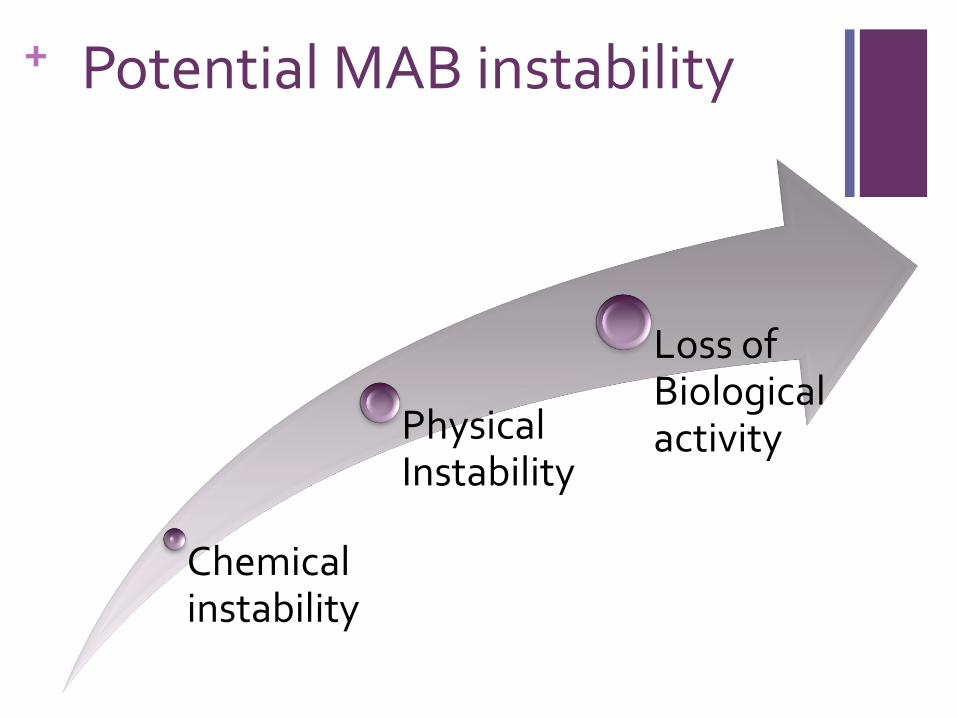
+ Potential MAB instability
Chemical instability
Physical Instability
Loss of Biological activity

+ Evaluation of mAbs stability
• assays for biological activity, where applicable, should be part of the pivotal stability studies
Potency
• appropriate physicochemical, biochemical and immunochemical methods for the analysis of
- the molecular entity
- the quantitative detection of degradation products
Purity and quality

+ Stability-indicating Profile
no single stability-indicating assay or parameter - profiles the stability characteristics of a biotechnological/biological product
the stability-indicating profile should provide assurance that changes in the
Identity
Potency
Purity
Other characteristics
of the product will be detected
the determination of which tests should be included will be product-specific

+ Potency 1 Potency - potency is the specific ability or capacity of a product to
achieve its intended effect
When the intended use of a product is linked to a definable and measurable biological activity, testing for potency should be part of the stability studies
In general, potencies of biotechnological/biological products tested by different laboratories - expressed in relation to an appropriate reference material (linked to national or international reference standard)
Biological characteristics
immunoreactivity and crossreactivity
the determination of relevant functional characteristics
binding studies to determine affinity
How do we measure? What is acceptable?

+ Potency 2
Receptor
Studies
Cell Line
Studies
Clinical
Effect

+ Purity and Molecular Characterisation 1
Purity is a relative term
the absolute purity of biologicals is extremely difficult to determine
the purity of a biotechnological/biological product should be typically assessed by
more than one method
purity value derived is method-dependent
tests for purity should focus on methods for determination of degradation products
The degree of purity, as well as individual and total amounts of degradation products of the biological product
limits of acceptable degradation should be derived from the analytical profiles of batches of the drug substance and drug product used in the preclinical and clinical studies.

+ Purity and Molecular Characterisation 2
The use of relevant physicochemical, biochemical and immunochemical analytical methodologies should permit a comprehensive characterisation
drug substance and/or drug product (e.g., molecular size, charge, hydrophobicity)
accurate detection of degradation changes during storage

+ Purity and Molecular Characterisation 3
Method Information
Visual Appearance of solution – particulate material
pH Optimised conditions for stability
Particle Counting / Microflow Imaging
Presence of aggregates\presence of silicone oil droplets
Size Exclusion Chromatography (SEC HPLC)
Size distribution, degradation products (high molecular weight), dimers and aggregates, quantification (mg/ml) Identification, higher structure (2o and 3o structure), adsorption, physical changes
Capillary electrophoresis (SDS-PAGE)
Size distribution, degradation products (small molecular weight). Information on identification and chemical changes
Peptide Mapping AA sequence – chemical changes
Total Protein Assay (BCA) Loss of protein - adsorption
High-resolution chromatography (e.g., RP, gel filtration, ion exchange, affinity)
Identification and quantification of biological and degradation products

+ Other characteristics
The following product characteristics should be monitored and reported for the drug product in its final container:
Visual appearance of the product
colour and opacity for solutions/suspensions
colour, texture and dissolution time for powders
visible particulates
Aggregates
Silicone oil droplets
pH
moisture level of powders and lyophilised products

+ Extended stability
Maximise
dose banding
production throughput - batching
vial sharing
quality evaluation and assurance
chemical
microbiological
patient experience

+ Examples of extended stability
Rituximab
Genetech – RTX – no change in activity when stored at 5oC during 154 days
Trastuzumab
US direction leaflet – a reconstituted vial with Bacteriostatic Water for Injection – stable for 28 days after reconstitution when stored refrigerated at 2-8oC

+ Examples of extended stability Case 1: Baxter 1
Baxter
Rituximab (MabThera) – 24 hrs 2-8oC - (12 hrs RT)
Trastuzumab (Herceptin) – 24hrs <30oC
Baxter – undertaken following
visual inspection (appearance and particles)
pH (optimal stability)
size exculsion chromatography (SEC HPLC) – size distribution, degradation products (), aggregates, quantification
ID, 2o 3o structure, adsorption, physical changes
Capillary electrophoresis (SDS PAGE) – size distribution, degradation products ()
ID, 2o 3o structure, chemical changes
BCA assay – quantification of total protein content

+ Examples of extended stability Case 1: Baxter 2
Viaflo, Viaflex and Intermate SV
7,14,21,28,35,days 0-8oC + 48hrs at RT
Essentially – mAbs stabile for the parameters described
Genetech – RTX – no change in activity when stored at 5oC
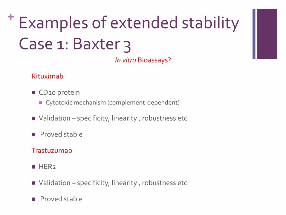
+ Examples of extended stability Case 1: Baxter 3
In vitro Bioassays?
Rituximab
CD20 protein
Cytotoxic mechanism (complement-dependent)
Validation – specificity, linearity , robustness etc
Proved stable
Trastuzumab
HER2
Validation – specificity, linearity , robustness etc
Proved stable

+ Infliximab Stabilite de L’infliximab en solution diluees (Guirao, S et al)
3 months in NaCl 09%; PE bags; 0.7-1.6mg/ml; 4-22oC
Methods
Turbidity
Dynamic light scattering
SEC HPLC
CEX HPLC
NHS studies underway

+ Rituximab
Stabilite d’un aticorps monoclonal d’interet therapeutique apres reconstitution: Le Rituximab (Jaccoullet et al)
3 months in NaCl 09%; 1 and 4 mg/ml; 4oC
Methods
HPLC SE
HPLC CEX
Optical density
Turbidity
Dynamic light scattering
Peptide mapping

+ Cetuximab
Stability of cetuximab and panitumumab in glass vials and PVC bags (Ikesue, H et al)
14 days in NaCl 09%; PVC bags; 2 mg/ml; 4oC
Methods
Enzyme-linked immunosorbent assay (ELISHA)
Cf. Aggregation studies (Astier et al)

+ Bevacizumab 1 Case 2 Six-month stability of bevacizumab (Avastin) binding to vascular
endothelial growthfactor after withdrawal into a syringe and
refrigeration or freezing
Bakri SJ et al 2006 Retina 26:519
Primary reference source for Bevacizumab
Biological effect - binding to vascular endothelial growth factor
No physico-chemical analysis
No visual examination

Six-month stability of bevacizumab (Avastin) binding to vascular endothelial growth factor after withdrawal into a syringe and refrigeration or freezing Bakri S.J et al 2006 Retina 26:519
82
84
86
88
90
92
94
96
98
100
102
0 5 10 15 20 25 30
%
V
E
G
F
B
i
n
d
i
n
g
Time (weeks)
Stability of Bevacizumab % Binding to VEGF
% Bevacizumab 4oC Vial
% Bevacizumab 4oC Syringe
% Bevacizumab -10oC Syringe

+ Bevacizumab 2
Sustained Elevation in Intraocular Pressure Associated With
Intravitreal Bevacizumab Injections
Kahook et al 2009 Ophthalmic Surgery, Lasers & Imaging 40:3
6 cases of patients exhibiting increased Intraocular Pressure (IOP) after single or repeated Bevacizumab
IOP lowering therapy was required

+ Bevacizumab 3.1
High–molecular-weight Aggregates In Repackaged Bevacizumab
Kahook et al 2010 Retina, The Journal of Retinal and Vitreous Diseases 30:6
887-892
Bevacizumab syringes (repackaged) obtained from 3 outside compounding pharmacies vs samples obtained directly from the original vial
Methodology
enzyme-linked immunosorbent assay
size exclusion chromatography
polyacrylamide gel electrophoresis
microflow imaging was used to examine particulate material within samples

+ Bevacizumab 3.2
all syringes contained statistically similar amounts of protein, consisting of immunoglobulin (IgG) heavy and light chains (polyacrylamide gel electrophoresis)
however, two of the three compounding pharmacies’ batches had significantly less functional IgG in the solution (enzyme-linked immunosorbent assay).
additionally, the compounding pharmacies with the lowest IgG (circa 50%) also contained 10-fold the number of micron-sized particulate matter (microflow imaging).
increase in micron-sized protein aggregates with the decrease in IgG concentration
large particulate matter within some samples may lead to obstruction of aqueous outflow and subsequent elevation in intraocular pressure.
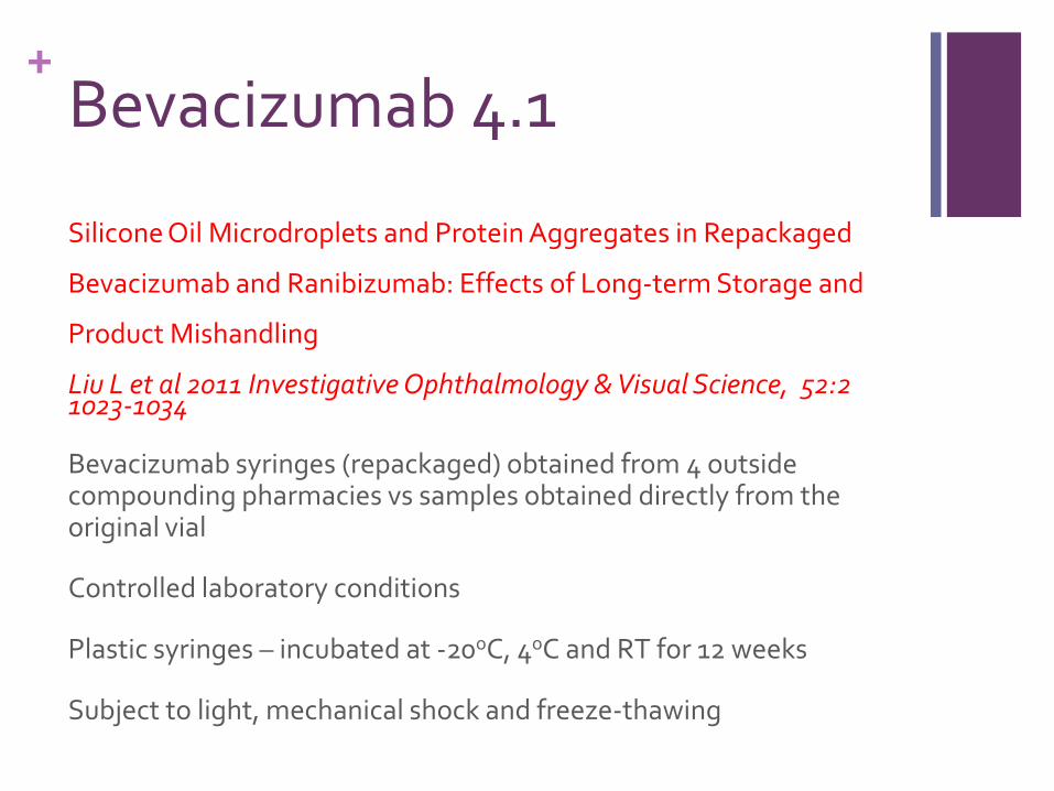
+ Bevacizumab 4.1
Silicone Oil Microdroplets and Protein Aggregates in Repackaged
Bevacizumab and Ranibizumab: Effects of Long-term Storage and
Product Mishandling
Liu L et al 2011 Investigative Ophthalmology & Visual Science, 52:2 1023-1034
Bevacizumab syringes (repackaged) obtained from 4 outside compounding pharmacies vs samples obtained directly from the original vial
Controlled laboratory conditions
Plastic syringes – incubated at -20oC, 4oC and RT for 12 weeks
Subject to light, mechanical shock and freeze-thawing

+ Bevacizumab 4.2
Methodology
Particle counting and size distribution
SE-HPLC
Results
1. Particle Counts (>1um)
1. Syringes 89,006 + 56,406 /mL to 602,062 + 18,349 /mL
2. Glass Vial 63,839 + 349/mL
Intercompany and intra\inter batch variation
Large particles observed >111um
High proportion identified at silicone oil microdroplets

+ Bevacizumab 4.3
2. SE-HPLC
Dimers to tetramers – 2-4%
Octamers to decamers – 0.2-0.4%
Could not always correlate loss of monomers with increase in aggregates – loss on syringes or adsorption on silicone oil droplets
3. Freeze-thawing (single and repeated)
Increased particle levels - >1.2 x 10⁶ particles per ml
Also due primarily to silicone oil droplets
Concern re. freezing during transportation

+ Bevacizumab 4.4
4. Mechanical Shock
Increase in silicone oil droplets
5. Light
Short term exposure caused clogging of the syringe needle
Ranibizumab
Comparable particle counts (5um syringe filter reduces)

+ Bevacizumab Summary
Studies have observed the
formation of aggregates
Physico-chemical considerations
decrease in biological activity (anti VEGF activity)
Biological activity considerations
release of silicone oil droplets
Physical considerations

+ Conclusion
Complex area of stability
Heavy investment by pharmaceutical industry
Stability indicators
Physical
Physico-chemical
Biological activity
Multiple test approach required
Care with preparation, transportation, procurement
Ongoing review of science