the soybean vegetative storage proteins vspa and vspo are acid phosphatases active on
TRANSCRIPT

T H E JOURNAL OF BIOLOGICAL CHEMISTRY 0 1992 by The American Society for Biochemistry and Molecular Biology, Inc.
Vol. 267, No. 22, Issue of August 5, pp. 15958-15964,1992 Printed in U.S.A.
The Soybean Vegetative Storage Proteins VSPa and VSPO Are Acid Phosphatases Active on Polyphosphates*
(Received for publication, January 29, 1992)
Daryll B. DeWald, Hugh S. Mason, and John E. Mullet$ From the Department of Biochemistry and Biophysics, Texas A h M University, College Station, Texas 77843
The soybean vegetative storage protein genes (USPA, and uspB) are regulated in a complex manner devel- opmentally and in response to external stimuli such as wounding and water deficit. The proteins accumulate to almost one-half the amount of soluble leaf protein when soybean plants are continually depodded and have been identified as storage proteins because of their abundance and pattern of expression in plant tissues. We have shown that purified VSP homodimers (VSPa and VSPP) and heterodimers (VSPa//3) possess acid phosphatase activity (a = 0.3-0.4 unitslmg; B = 2-4 unitslmg; a/@ = 7-10 unitslmg). Specific activities were determined by monitoring o-carboxyphenyl phosphate (0.7 mM) cleavage at pH 5.5 (VSPa) or pH 6.0 (VSPa//3 and VSPP) in 0.15 M sodium acetate buffer at 25 “C. These enzymes are active over a broad pH range, maintaining greater than 40% of maximal ac- tivity from pH 4.0 to 6.5 and having maximal activity at pH 5.0-5.5. They are inactivated by sodium fluo- ride, sodium molybdate, and heating at 70 “C for 10 min. These phosphatases can liberate Pi from several different substrates, including napthyl acid phosphate, carboxyphenyl phosphate, sugar-phosphates, glycer- aldehyde 3-phosphate, dihydroxyacetone phosphate, phosphoenolpyruvate, ATP, ADP, PPi, and short chain polyphosphates. VSPa//3 cleaved phosphoenolpyru- vate, ATP, ADP, PPI, and polyphosphates most effi- ciently. Apparent K,,, and V,,, values at 25 “C and pH 5.0 were 42 p~ and 2.0 pmol/min/mg, 150 WM and 4.2 pmol/min/mg, and 420 p~ and 4.1 pmol/min/mg, for tetrapolyphosphate, pyrophosphate, and phosphoenol- pyruvate, respectively.
In 1982 and 1983, Wittenbach (1-3) described proteins that accumulated in leaves of soybean when the plants were con- tinually depodded. The accumulation of these proteins is remarkable because after weeks of depodding, they repre- sented over 45% of the soluble protein in leaves. Franceschi and co-workers (4-6) determined that the proteins are local- ized in vacuoles of bundle sheath and paraveinal mesophyll (PVM)’ cells of leaves. Later studies showed that these pro-
* This work was supported by United States Department of Agri- culture Grant 91-37304-6658 and the Texas Agricultural Experiment Station. The costs of publication of this article were defrayed in part by the payment of page charges. This article must therefore be hereby marked “adoertisernent” in accordance with 18 U.S.C. Section 1734 solely to indicate this fact.
$To whom correspondence should be addressed. Tel.: 409-845- 0722; Fax: 409-845-9724.
The abbreviations used are: PVM, paraveinal mesophyll; SDS, sodium dodecyl sulfate; PAGE, polyacrylamide gel electrophoresis; VSP, vegetative storage proteins; ConA, concanavalin A PEP, phosphoenolpyruvate; MES, 2-(N-morpho1ino)ethanesulfonic acid; MOPS, 3-(N-morpholino)propanesulfonic acid.
teins also accumulate in epidermal cells (7). The PVM cells play an important role in transferring amino acids and carbon between the vascular system and mesophyll cells of the leaf. The accumulation of the proteins in response to removal of reproductive sinks and their localization in vacuoles of trans- fer cells led Wittenbach to name them vegetative storage proteins (VSPa, and VSPP). More recent studies have shown that usp mRNA and protein accumulate in young leaves, stems, flowers, pods, and the cotyledons of germinating seed- lings, but are rare in seeds and roots (8-12). The expression of the usp is decreased when plants are nitrogen-limited (13). In contrast, usp mRNA accumulation is stimulated by carbon sources such as sucrose and by the plant growth regulator jasmonic acid (14). It is noteworthy that other soybean pro- teins (e.g. a lipoxygenase) with similar expression patterns and intracellular localization have been described (15, 16).
VSP accumulation is modulated developmentally and by environmental factors (8-14, 17). Bozarth et al. (17) first reported that VSPa accumulated in cell wall fractions in soybean seedlings exposed to water deficit. Later studies showed that usp mRNA and protein accumulate in seedlings in response to water deficit (8). When water is withheld from older plants, mRNAs accumulate in internodes and interme- diate age leaves (12). The accumulation of VSP under these conditions has been attributed to decreased growth coupled with continued transport of amino acids and carbon to grow- ing regions (12). The VSP are also induced in wounded plants, although their function in this response is unknown (7, 12). The induction of the genes in wounded tissue appears to result from wound-induced accumulation of jasmonic acid or its methyl ester, methyl jasmonate. This idea is consistent with the stimulation of usp expression by addition of jasmonic acid or methyl jasmonate to cell cultures or plants (12-14,18, 19). In addition, inhibitors of lipoxygenase, which should limit jasmonic acid biosynthesis in wounded tissue, block wound- induced accumulation of the VSP (13).
The VSP are glycoproteins that are 80% identical in amino acid sequence (8, 9). However, the isoelectric points of VSPa and VSPP are 8.6 and 5.8, respectively (8). Based on cDNA sequences (8), the predicted protein molecular masses are 25.1 (a ) and 25.3 kDa (p), and based on chemical analysis, the mature proteins are approximately 3% carbohydrate (20). The VSP have been reported to migrate on SDS-PAGE gels with apparent molecular masses of 25-28 kDa for VSPa and 27- 31 kDa for VSPP (1-4,7-11,17-21). The native form of VSP in soybean leaves is az, a@, and PZ dimers (20,21).
Recently, Williamson and Colwell (22), Aarts et al. (23), and Erion et al. (24) reported the isolation of an acid phos- phatase from tomato that is encoded by Aps-1. These authors noted that portions of the acid phosphatase-1 sequence were similar to VSP sequences, suggesting that the vegetative storage proteins might have acid phosphatase activity. We have investigated this possibility and report that VSPa and
15958
Soybean Vegetative Storage Protein Acid Phosphatase Activity 15959
VSPP are acid phosphatases that have substrate preference for polyphosphates, pyrophosphate, and PEP.
EXPERIMENTAL PROCEDURES
Supplies-Ammonium sulfate, concanavalin A-Sepharose (ConA) affinity resin, the Mono Q column, and the fast protein liquid chro- matography system are products of Pharmacia LKB Biotechnology Inc. Electrophoresis equipment and chemicals were purchased from Bio-Rad. Nitrocellulose (0.45 pm) was from Schleicher & Schuell, and all other chemicals were purchased from Sigma.
Plant Material-Soybean (Glycine maz Merr cv Williams 82) seed- lings were grown at 27 "C in the dark a t high humidity for hypocotyl tissue or in constant light (350 microeinsteins m-' s-l) at 25 "C for leaf tissue. Hypocotyl tissue harvested for protein purification was the growing hook and elongating region of 3-day-old seedlings, and young leaves were initial trifoliates from 12-day-old seedlings.
Protein Extraction and Isolation-VSPa and VSPP were purified by a modification of the procedure described by Spilatro and Anderson (20) with all manipulations performed at 4 "C or colder. Hypocotyls or leaves were harvested and immediately frozen in liquid nitrogen. Tissue was stored at -80 "C until extraction. Extractions were carried out by grinding tissue in liquid nitrogen and then adding buffer (50 mM Tris/HCl (pH 7.5), 5 mM EDTA, 30 mM ascorbic acid, 10 mM dithiothreitol, 0.8 mM phenylmethylsulfonyl fluoride) at a ratio of 1:5 w/v. The homogenate was filtered through Miracloth (Calbiochem) and centrifuged at 15,000 X g for 20 min. Finely ground ammonium sulfate was added to the supernatant sequentially to final concentra- tions of 1.7, 2.0, and 2.8 M. The 2.8 M pellet was resuspended in a minimal volume of 20 mM Tris/HCl (pH 7.5), 10 mM dithiothreitol, and dialyzed overnight against 2,000 volumes of the resuspension buffer. The dialysate was centrifuged at 14,000 X g, and the super- natant was applied, collected, and reapplied to a ConA column. The affinity column was washed with five column volumes of 50 mM sodium phosphate (pH 7.5), 200 mM sodium chloride to remove unbound protein, and elution of bound protein was achieved by washing the column with loading buffer containing 200 mM a-methyl- D-mannopyranoside. Fractions containing concentrated VSP were pooled and dialyzed against 20 mM Tris/HCl, 5 mM dithiothreitol. The dialysate was cleared by centrifugation, and VSP was purified with fast protein liquid chromatography using a Mono Q column.
Protein Concentration Determination-Protein concentrations were determined by the dye-binding method of Bradford (25) using bovine serum albumin as a standard.
Polyacrylamide Gel Electrophoresis-Denaturing SDS-PAGE (12% acrylamide, acry1amide:bisacrylamide = 30:0.8) was performed by the method of Laemmli (26). Nondenaturing gel electrophoresis (8%) was performed as above, except that SDS and mercaptoethanol were not used in any buffers. Protein electroblotting and antibody detection of blotted proteins were performed as described (27) using alkaline phosphatase-conjugated secondary antibody (Bio-Rad). Polyacryl- amide acid phosphatase activity gels (8% nondenaturing) were devel- oped using conditions described previously (28).
Antibody Preparation-For preparation of polyclonal antibodies against VSP, fusion proteins were expressed in Escherichia coli. A portion of the open reading frame from cDNAs (8) encoding either VSPa (pKSH2, amino acids 195-254) or VSPP (pKSH3, amino acids 196-254) on a BamHI/ClaI fragment was fused in-frame to the 3' end of a portion of the trpE gene of E. coli in the vector PATH 10 (kindly supplied by C. Dieckman, University of Arizona, Tucson). Transformation of E. coli strain RR1 with the recombinant plasmids and induction of the fusion proteins were performed as described (29). The fusion proteins were extracted and partially purified by precipitation with high salt (30) and further purified by SDS-PAGE on 12% acrylamide gels and electroelution of Coomassie-stained proteins (29). Female New Zealand rabbits were immunized by sub- cutaneous injection of protein solutions emulsified with equal volumes of Freund's adjuvant and boosted monthly before serum was collected after 4 months.
Acid Phosphatase Assays-Acid phosphatase activity of purified protein was quantitated using assay conditions described in the Worthington enzyme manual (Worthington Biochemical Corp., Free- hold, NJ). Briefly, all assays were conducted using 0.15 M sodium acetate buffer at the pH described with 0.7 mM o-carboxyphenyl phosphate as the substrate. Increase in absorbance a t 300 nm corre- sponding to the release of salicylic acid was monitored using a Beckman DU 64 spectrophotometer and the kinetics software com- patible with this instrument. One unit of enzyme hydrolyzes 1 pmol
of o-carboxyphenyl phosphate/min at 25 "C, pH 5.0 under the con- ditions specified above.
Assays to determine pH optimum were done as above, except different buffers were used at higher pH. The activity of VSPa and VSPP homodimers and the VSPa/P heterodimer was determined in acetate (3.5-6.0), MES (5.5-6.5), and MOPS (6.5-7.5) buffers. All assays were duplicated, and activities at overlapping pH in different buffers were averaged. No difference in relative activity greater than 10% was observed at the same pH in different buffers.
Phosphatase Substrate Specificity-Release of inorganic phosphate from substrates was measured by the method of Taussky and Shorr (31). All assays were performed in duplicate at pH 5.0 or 5.5 and 25 "C. Cleavage of substrate was examined using 0.01-0.03 units/ assay enzyme and 5 mM substrate. The release of phosphate in control samples (no enzyme) was measured and subtracted from correspond- ing enzyme-containing samples. Inorganic phosphate measured fell within the linear portion of the standard curve (0.2-40 pg) for this assay. The fraction of consumed substrate was always less than 15% of the total in each sample, indicating that initial velocities were measured.
K, and Ifrnar Determination-We determined apparent K,,, and V,,, values of purified hypocotyl VSPa/P for phosphoenolpyruvate, pyrophosphate, and tetrapolyphosphate using KinetAsyst software (Intellikinetics, State College, PA) created from programs detailed by Cleland (32). The rate of substrate cleavage by 0.01 units of VSPa/P was measured at substrate concentrations from 1 p M to 10 mM. Assays were performed at pH 5.0, 25 "C, for 5 min.
RESULTS
Sequence Comparison of Acid Phosphatase-I, VSPa, and VSPP-VSPa and VSPP have 45% overall amino acid se- quence identity with acid phosphatase-1 (22-24). Several amino acid regions have a high degree of similarity among the three proteins (see Ref. 24). Acid phosphatase activity prob- ably requires participation of histidyl groups (33). We noted that HiP4 (numbering for acid phosphatase-1) and His"' were conserved among the three proteins. In contrast, a Gly- Pro-Gly-Tyr motif located in acid phosphatase-1 (amino acids 75-78), characteristic of alkaline phosphatases (23), is not located in the VSP sequences. Acid phosphatase-1 shows affinity for ConA columns consistent with evidence that it is a glycoprotein (34). Similarly, VSPa and VSPP are glycopro- teins, and each contains a potential site for asparagine-linked glycosylation (amino acids 129-131 and 130-132; NST and NET (see Ref. 8 for numbering)). This site is not conserved in acid phosphatase-1, but a site nearby (amino acids 142- 144; NGT) could represent a glycosylation site. Another dif- ference between these proteins is that their isoelectric points (PI) vary widely (VSPa = 8.6, VSPP = 5.8, acid phosphatase- 1 = 4.7).
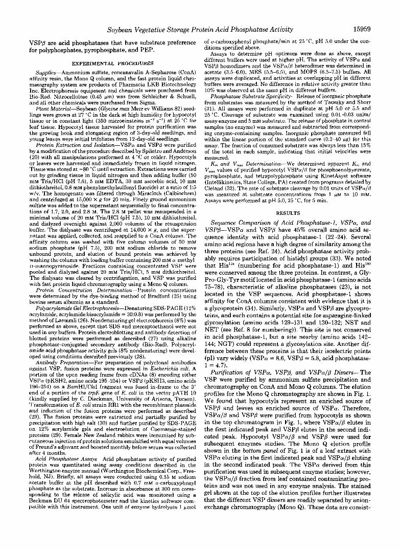
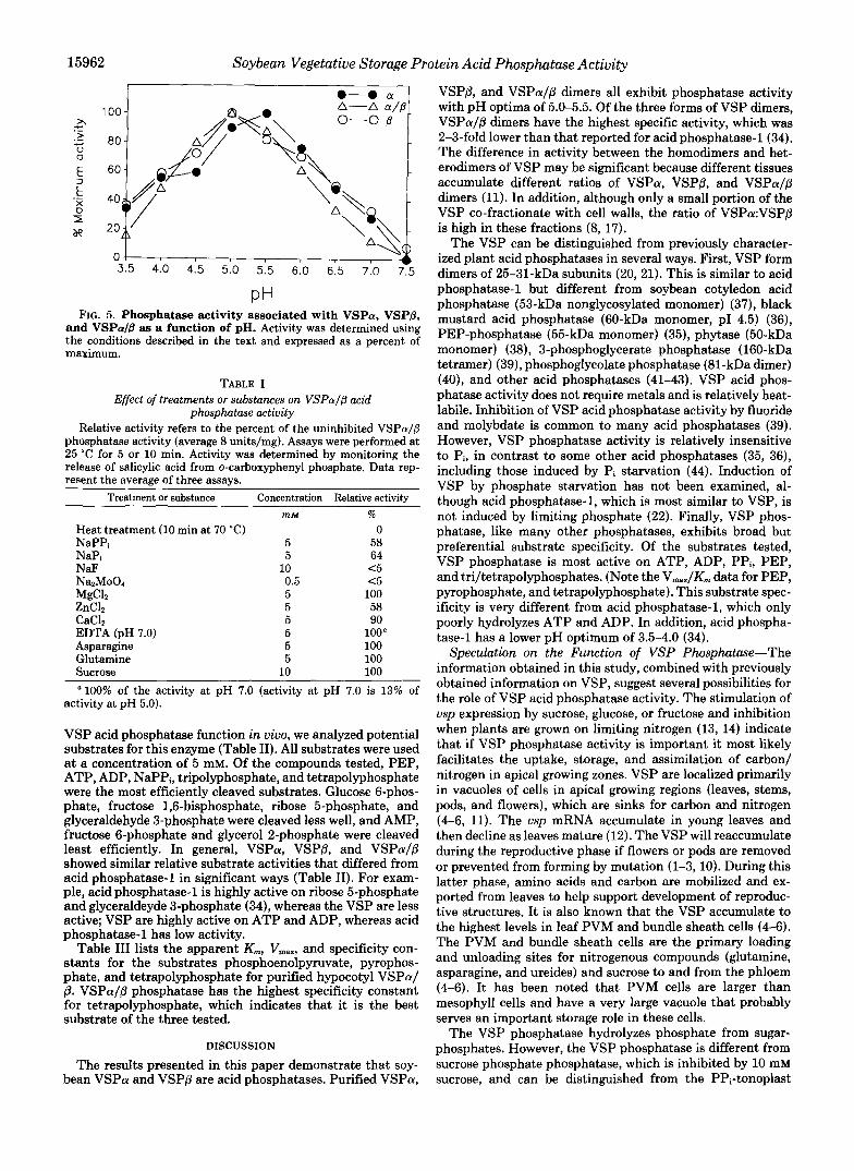
Purification of VSPa, VSPP, and VSPaIP Dimers-The VSP were purified by ammonium sulfate precipitation and chromatography on ConA and Mono Q columns. The elution profiles for the Mono Q chromatography are shown in Fig. 1. We found that hypocotyls represent an enriched source of VSPP and leaves an enriched source of VSPa. Therefore, VSPaIP and VSPP were purified from hypocotyls as shown in the top chromatogram in Fig. 1, where VSPaIP elutes in the first indicated peak and VSPP elutes in the second indi- cated peak. Hypocotyl VSPaIP and VSPP were used for subsequent enzymes studies. The Mono Q elution profile shown in the bottom panel of Fig. 1 is of a leaf extract with VSPa eluting in the first indicated peak and VSPaIP eluting in the second indicated peak. The VSPa derived from this purification was used in subsequent enzyme studies; however, the VSPa/P fraction from leaf contained contaminating pro- teins and was not used in any enzyme analysis. The stained gel shown at the top of the elution profiles further illustrates that the different VSP dimers are readily separated by anion- exchange chromatography (Mono &). These data are consist-
15960 Soybean Vegetative Storage Protein Acid Phosphatase Activity
A
E 0 m % W 0 z a m U
u) 0 m a W 5 -I W U
W
f 5 W U
I " - ~ 1-P
0.6 CI z
0.4 2 E - Y
0.2
0.0 0 20 40 60 80 100
TIME (min)
TIME (min)
FIG. 1. Elution profiles of soybean tissue extracts chromat- ographed on a Mono Q column. Chromatograms of the final step in VSP purification. The upperpanel shows the profile obtained when hypocotyl proteins are eluted from a Mono Q column, and the lower panel shows the profile obtained when leaf proteins are eluted from a Mono Q column. Insets above each chromatogram are photographs of Coomassie Blue-stained gel sections of the three fractions aligned directly with the indicated absorbance peak. VSPaIVSPP heterodi- mers and VSPP homodimers were purified from hypocotyl tissue. VSPa homodimers were purified from leaf tissue. The dotted lines indicate the concentration of NaCl in the elution buffer.
ent with earlier observations (20,21). Anti- VSPAntibodies-In an earlier study, we used antibod-
ies prepared against VSP isolated from soybean (8, 17). As expected, the anti-VSP antibodies were highly reactive against carbohydrate determinants, as well as VSP protein determinants. To circumvent this problem in the present study, we fused portions of the vspA and vspB open reading frames to trpE and overexpressed the fusion proteins in E. coli. Antibodies were prepared against the fusion proteins. Mono Q fractions containing VSPa, VSPP, and VSPaIP dimers and total soluble cell protein were fractionated on SDS-PAGE gels and then stained or blotted for immunode- tection with anti-VSP antibodies (Fig. 2). As shown in Fig.
A
P - a -
1 2 3 4 5 1 2 3 4 5
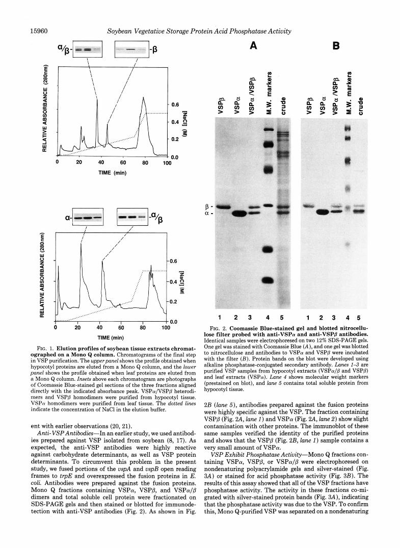
FIG. 2. Coomassie Blue-stained gel and blotted nitrocellu- lose filter probed with anti-VSPa and anti-VSPB antibodies. Identical samples were electrophoresed on two 12% SDS-PAGE gels. One gel was stained with Coomassie Blue ( A ), and one gel was blotted to nitrocellulose and antibodies to VSPa and VSPP were incubated with the filter ( B ) . Protein bands on the blot were developed using alkaline phosphatase-conjugated secondary antibody. Lanes 1-3 are purified VSP samples from hypocotyl extracts (VSPa/P and VSPP) and leaf extracts (VSPa). Lane 4 shows molecular weight markers (prestained on blot), and lune 5 contains total soluble protein from hypocotyl tissue.
2B ( l a n e 5), antibodies prepared against the fusion proteins were highly specific against the VSP. The fraction containing VSPP (Fig. 2 4 , lane 1) and VSPa (Fig. 2 4 , lane 2) show slight contamination with other proteins. The immunoblot of these same samples verified the identity of the purified proteins and shows that the VSPP (Fig. 2B, lane 1) sample contains a very small amount of VSPa.
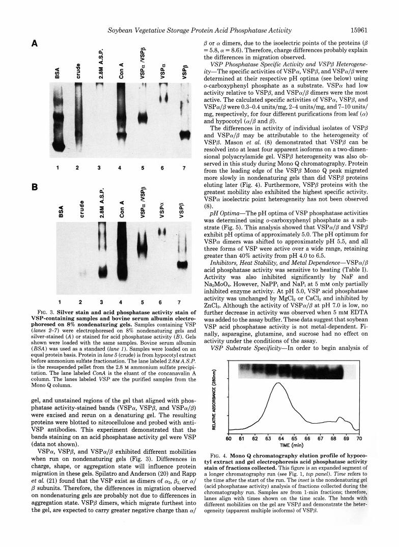
VSP Exhibit Phosphatase Activity-Mono Q fractions con- taining VSPa, VSPP, or VSPaIP were electrophoresed on nondenaturing polyacrylamide gels and silver-stained (Fig. 3A) or stained for acid phosphatase activity (Fig. 3B). The results of this assay showed that all of the VSP fractions have phosphatase activity. The activity in these fractions co-mi- grated with silver-stained protein bands (Fig. 3A), indicating that the phosphatase activity was due to the VSP. To confirm this, Mono Q-purified VSP was separated on a nondenaturing
15962 Soybean Vegetative Storage Protein Acid Phosphatase Activity
1 nn ,. . \ 0-0 /9 A-A a//9
3.5 T 4
4.0 4.5 5.0 5.5 6.0 6.5 7.0 7.5
PH FIG. 5. Phosphatase activity associated with VSPa, VSP&
and VSP& as a function of pH. Activity was determined using the conditions described in the text and expressed as a percent of maximum.
TABLE I Effect of treatments or substances on VSPol/@ acid
phosphatase activity Relative activity refers to the percent of the uninhibited VSPol/p
phosphatase activity (average 8 units/mg). Assays were performed at 25 "C for 5 or 10 min. Activity was determined by monitoring the release of salicylic acid from o-carboxyphenyl phosphate. Data rep- resent the average of three assays.
Treatment or substance Concentration Relative activity mM %
Heat treatment (10 min at 70 "C) 0 NaPPi 5 58 NaPi 5 64 NaF 10 <5 NazMoOl 0.5 <5 MgCL 5 100 ZnClZ 5 58 CaClZ 5 90 EDTA (pH 7.0) 5 100" Asparagine 5 100 Glutamine 5 100 Sucrose 10 100
100% of the activity at pH 7.0 (activity at pH 7.0 is 13% of activity at pH 5.0).
VSP acid phosphatase function in uiuo, we analyzed potential substrates for this enzyme (Table 11). All substrates were used at a concentration of 5 mM. Of the compounds tested, PEP, ATP, ADP, NaPPi, tripolyphosphate, and tetrapolyphosphate were the most efficiently cleaved substrates. Glucose 6-phos- phate, fructose 1,6-bisphosphate, ribose &phosphate, and glyceraldehyde 3-phosphate were cleaved less well, and AMP, fructose 6-phosphate and glycerol 2-phosphate were cleaved least efficiently. In general, VSPa, VSPP, and VSPcu/P showed similar relative substrate activities that differed from acid phosphatase-1 in significant ways (Table 11). For exam- ple, acid phosphatase-1 is highly active on ribose 5-phosphate and glyceraldeyde 3-phosphate (34), whereas the VSP are less active; VSP are highly active on ATP and ADP, whereas acid phosphatase-1 has low activity.
Table I11 lists the apparent K,, Vmax, and specificity con- stants for the substrates phosphoenolpyruvate, pyrophos- phate, and tetrapolyphosphate for purified hypocotyl VSPa/ @. VSPa/(l phosphatase has the highest specificity constant for tetrapolyphosphate, which indicates that it is the best substrate of the three tested.
DISCUSSION
The results presented in this paper demonstrate that soy- bean VSPa and VSPP are acid phosphatases. Purified VSPa,
VSPP, and VSPa/P dimers all exhibit phosphatase activity with pH optima of 5.0-5.5. Of the three forms of VSP dimers, VSPa/P dimers have the highest specific activity, which was 2-3-fold lower than that reported for acid phosphatase-1 (34). The difference in activity between the homodimers and het- erodimers of VSP may be significant because different tissues accumulate different ratios of VSPa, VSPP, and VSPa/p dimers (11). In addition, although only a small portion of the VSP co-fractionate with cell walls, the ratio of VSPa:VSPP is high in these fractions (8, 17).
The VSP can be distinguished from previously character- ized plant acid phosphatases in several ways. First, VSP form dimers of 25-31-kDa subunits (20, 21). This is similar to acid phosphatase-1 but different from soybean cotyledon acid phosphatase (53-kDa nonglycosylated monomer) (37), black mustard acid phosphatase (60-kDa monomer, PI 4.5) (36), PEP-phosphatase (55-kDa monomer) (35), phytase (50-kDa monomer) (38), 3-phosphoglycerate phosphatase (160-kDa tetramer) (39), phosphoglycolate phosphatase (81-kDa dimer) (40), and other acid phosphatases (41-43). VSP acid phos- phatase activity does not require metals and is relatively heat- labile. Inhibition of VSP acid phosphatase activity by fluoride and molybdate is common to many acid phosphatases (39). However, VSP phosphatase activity is relatively insensitive to Pi, in contrast to some other acid phosphatases (35, 36), including those induced by Pi starvation (44). Induction of VSP by phosphate starvation has not been examined, al- though acid phosphatase-l, which is most similar to VSP, is not induced by limiting phosphate (22). Finally, VSP phos- phatase, like many other phosphatases, exhibits broad but preferential substrate specificity. Of the substrates tested, VSP phosphatase is most active on ATP, ADP, PPi, PEP, and tri/tetrapolyphosphates. (Note the V,,,/K, data for PEP, pyrophosphate, and tetrapolyphosphate). This substrate spec- ificity is very different from acid phosphatase-1, which only poorly hydrolyzes ATP and ADP. In addition, acid phospha- tase-1 has a lower pH optimum of 3.5-4.0 (34).
Speculation on the Function of VSP Phosphatase-The information obtained in this study, combined with previously obtained information on VSP, suggest several possibilities for the role of VSP acid phosphatase activity. The stimulation of usp expression by sucrose, glucose, or fructose and inhibition when plants are grown on limiting nitrogen (13, 14) indicate that if VSP phosphatase activity is important it most likely facilitates the uptake, storage, and assimilation of carbon/ nitrogen in apical growing zones. VSP are localized primarily in vacuoles of cells in apical growing regions (leaves, stems, pods, and flowers), which are sinks for carbon and nitrogen (4-6, 11). The usp mRNA accumulate in young leaves and then decline as leaves mature (12). The VSP will reaccumulate during the reproductive phase if flowers or pods are removed or prevented from forming by mutation (1-3,lO). During this latter phase, amino acids and carbon are mobilized and ex- ported from leaves to help support development of reproduc- tive structures. It is also known that the VSP accumulate to the highest levels in leaf PVM and bundle sheath cells (4-6). The PVM and bundle sheath cells are the primary loading and unloading sites for nitrogenous compounds (glutamine, asparagine, and ureides) and sucrose to and from the phloem (4-6). It has been noted that PVM cells are larger than mesophyll cells and have a very large vacuole that probably serves an important storage role in these cells.
The VSP phosphatase hydrolyzes phosphate from sugar- phosphates. However, the VSP phosphatase is different from sucrose phosphate phosphatase, which is inhibited by 10 mM sucrose, and can be distinguished from the PPi-tonoplast

Soybean Vegetative Storage Protein Acid Phosphatase Activity 15963
TABLE I1 Comparison of VSP phosphatase activity on different substrates
Data represent efficiency of liberation of Pi (measured spectrophotometrically) from different substrates, as compared to P-napthyl acid phosphate (100%). Substrate concentrations are 5 mM, and digestions were for 60 min. Average activities of enzyme on napthyl acid phosphate were: VSPa/R = 10 units/mz VSPa = 0.4 units/me. and VSPR = 3 units/ma.
~
Relative activity Substrate
VSPa/VSP@ VSPn VSPO Acid phosphatase"
P-Napthyl acid phosphate 100% 100% 100% 100% o-Carboxyphenyl phosphate 70 D-Fructose 6-phosphate 5 15 18 D-Glucose 6-phosphate 20 13 22 15 Glycerol 2-phosphate 8 Fructose 1,6-bisphosphate 30 Ribose 5-phosphate 17 69 Glyceraldehyde 3-phosphate 28 12 73 Dihydroxyacetone phosphate 15 Phosphoenolpyruvate 90 53 100 ATP 80 28 60 0 ADP 98 22 75 3 AMP 9 5 9 5 NaPPi 106 19 20 Tripolyphosphate 110 37 63 Tetrapolyphosphate 118 68 Sodium Dhvtate 0
a Values were calculated from Paul and Williamson (34).
TABLE I11 Substrate specificity of VSPaIP
The rate of substrate cleavage by 0.01 units of enzyme was meas- ured at substrate concentrations from 1 pM to 10 mM by the following increments: 1 p ~ , 5 p ~ , 10 p ~ , 50 p ~ , 100 p ~ , 500 p ~ , 1 mM, 5 mM, and 10 mM. Assays were performed a t pH 5.0, 25 "C, for 5 min.
Substrate
m M pmol/min/mg Phosphoenolpyruvate 0.420 f 0.09 4.1 k 0.2 10 f 1.8 Pyrophosphate 0.150 f 0.02 4.2 f 0.2 28 f 3.7 Tetrapolyphosphate 0.042 f 0.008 2.0 k 0.1 49 f 8.6
phosphatase (with a broad alkaline pH optimum), which is involved in proton pumping (45). In addition, sugar-phos- phates do not readily pass into the vacuole (46). Further, the preference of VSP for PPi, ADP, ATP, PEP, and polyphos- phates suggests that the VSP probably play a different role. Among these substrates, ADP and ATP appear less likely substrates due to their low levels in vacuoles (47).
VSP could be a PEP-phosphatase that in theory could accelerate the synthesis of pyruvate from glucose. The pyru- vate produced in this way could be utilized in nitrogen assim- ilation in PVM cells or to produce other carbon compounds that are transported to mesophyll cells. Hydrolysis of PEP also generates Pi that could be used in the formation of hexose-phosphates from sucrose. A PEP-phosphatase (35) has been isolated and characterized that is different from VSP phosphatase. PEP-phosphatase is a monomer of 55 kDa, whereas VSP form dimers of 25-31-kDa subunits (20, 21). PEP-phosphatase requires metals and is inhibited by 0.5 mM P, (35). In contrast, VSPa/P phosphatase activity is relatively insensitive to Pi, does not require metal ions, and has a relatively high K,,, for PEP (420 uersus 50 pM, VSP phospha- tase uersus PEP-phosphatase) (35). Moreover, the previously characterized PEP-phosphatase is induced by Pi starvation, apparently to bypass a step in glycolysis that requires aden- ylates (44). Phosphate starvation results in depressed aden- ylate pools and the induction of pyrophosphate: fructose 6-phosphate 1-phosphotransferase, nonphosphorylating NADP-glyceraldehyde-3-phosphate dehydrogenase, and PEP-phosphatase. Each of these enzymes bypasses a step in
glycolysis requiring adenylates. In contrast, addition of su- crose to soybean cells, a condition required for VSP accumu- lation, does not depress ATP levels (48). Therefore, if VSP functions as a PEP-phosphatase i n uiuo, it seems likely it functions to supplement the action of pyruvate kinase rather than to bypass this step.
VSP phosphatase showed the highest substrate specificity for tetrapolyphosphate. This was surprising because we were unable to locate information documenting polyphosphates in higher plants. However, polyphosphates are localized in vac- uoles of many organisms (49). In Dunaliella, hydrolysis of polyphosphates has been implicated in osmotic adjustment in response to salt stress (50). Furthermore, polyphosphate hy- drolysis in Dunaliella is an important response to amine uptake because it contributes to the maintenance of vacuolar pH needed for ammonium/basic amino acid sequestration in vacuoles (51). As noted earlier, the VSP are localized in vacuoles of PVM cells that play a primary role in amino acid transfer between mesophyll cells and vascular tissue. There- fore, it is reasonable to speculate that the VSP may be polyphosphate exophosphatases that play a role in amino acid uptake and temporary sequestration in PVM cells.
The VSP were so named because they accumulate in PVM vacuoles during periods of high amino acid and carbon flux to and from leaf mesophyll cells (1-6). The finding that VSP has phosphatase activity does not rule out a protein storage function because other vegetative storage proteins, such as patatin, retain enzyme activity (lipid acyl hydrolase) (52). Furthermore, the VSP, especially VSPa, exhibit relatively low activity as compared with other phosphatases. Moreover, VSP phosphatase activity could be regulated through changes in VSPaIVSPP ratios and as yet undefined modification of VSPP that results in greater activity in the more negatively charged forms of VSPP.
Acknowledgments-We would like to thank Drs. Don Pettigrew and Paul Fitzpatrick for helpful discussions and John Denu for assistance with the kinetic analysis.
REFERENCES
1. Wittenbach, V. A. (1982) Plant Physiol. 70, 1544-1548 2. Wittenbach, V. A. (1983) Plant Physiol. 73, 121-124 3. Wittenbach, V. A. (1983) Plant Physiol. 73, 125-129
15964 Soybean Vegetative Storage Protein Acid Phosphatase Activity 4. Franceschi, V. R., Wittenbach, V. A., and Giaquinta, R. T. (1983) Plant
5. Franceschi, V. R., and Giaquinta, R. T. (1983) Planta 167,411-421 6. Franceschi, V. R., and Giaquinta, R. T. (1983) Planta 157,422-431 7. Staswsk. P. E. (1990) Plant Cell 2. 1-6
Physiol. 72,586-589
8.
9. 10.
12. 11.
13. 14.
15.
Mason, H . S., Guerrero, F. D., Boyer, J. S., and Mullet, J. E.
Staswick, P. E. (1988) Plant Physiol. 87,250-254 Staswick, P. E. (1989) Plant Physiol. 89,309-315 Staswick, P. E. (1989) Plant Physiol. 9 0 , 1252-1255 Mason, H. S., and Mullet, J. E. (1990) Plant Cell 2,569-579 Staswick, P. E., Huang, J., and Rhee, Y. (1991) Plant Physiol. Mason, H. S., DeWald, D. B., Creelman, R. A,, and Mullet,
Francescni. V. R.. and Grimes. H. D. (1991) Proc. Natl. Acad.
Mol. Biol. 11,845-856
Plant Physiol. 92,859-867
(1988) Plant
96,130-136 J. E. (1992)
Sci. U. S. A. 88,6745-6749'
(1991) Plant Cell 3,973-987
. ,
16. Tranbarger, T. J., Franceschi, V. R., Hildebrand, D. F., and Grimes, H. D.
17. Bozarth. C. S.. Mullet. J. E.. and Bover. J. S. (1987) Plant Phvsiol. 85, , , - . 261-267 '
18. Anderson, J. M. (1988) J. Plant Growth Regul. 7,203-211
20. Spilatro, S. R., and Anderson, J. M. (1989) Plant Physiol. 90,1387-1393 19. Anderson, J. M. (1991) J. Plant Growth Regul. 10, 5-10
21. Rapp, W. D., Lilley, G. G., and Nielsen, N. C. (1990) Theor. Appl. Genet.
22. Williamson, V. M., and Colwell, G. (1991) Plant Physiol. 97, 139-146 23. Aarts, J. M. M. J. G., Hontelez, J. G. J., Fisher, P., Verkerk, R., Kammen,
24. Erion, J. L., Ballo, B., May, L., Bussell, J., Fox, T., and Thomas, S. R. A. V., and Zabel, P. (1991) Plant Mol. Biol. 16,647-661
25. Bradford, M. M. (1976) Anal. Biochem. 72,248-254 (1991) Plant Physiol. 9 7 , 1462-1469
26. Laemmli, U. K. (1970) Nature 227,680-685 27. Towbin, H., Staehelin, T., and Gordon, J. (1979) Proc. Natl. Acad. Sci.
28. Vallejos, C. E. (1983) in Isozymes in Plant Genetics and Breeding (Tanksley,
79,785-792
U. S. A. 76,4350-4354
29. 30.
31. 32. 33. 34. 35.
36.
37.
S. D.. and Orton. T. J.. eds) Vol. 1A. DO. 469-516. Elsevier Science Publishers B. V., Amsterdam '
I ..
Kleid, D. G., Yansura, D., Small, B., Dowbenko, D., Moore, D. M., Grub- Gamble, P. E., and Mullet, J. E. (1989) J. Biol. Chem. 264, 7236-7243
man, M. J., McKercher, P. D., Morgan, D. O., Robertson, B. H., and Bachrach, H. L. (1981) Science 214, 1125-1129
Taussk , H H and Sborr, E. (1953) J. BWL Chem. 202,675-685 Clelanc? W: W:'(1979) Methods Enzymol. 63,103-138 Tabatadai, M. A., and Juma, N. G. (1988) Plant and Soil 107,39-48 Paul, E. M., and Williamson, V. M. (1987) Plant Physiol. 84,399-403 Duff, S. M. G., Lefebvre, D. D., and Plaxton, W. C. (1989) Plant Physiol.
Duff, S. M. G., Lefebvre, D. D., and Plaxton, W. C. (1991) Arch. Biochem.
Ullah, A. H. J., and Gibson, D. M. (1988) Arch Biochem. Biophys. 260,
90,734-741
Biophys. 286, 226-232
51 4-570 38. Gibson, D. M., and Ullah, A. H. J. (1988) Arch. Biochem. Biophys. 260,
40. Christelier, J. *., and Tolbert, N. E. (1978) J. Biol. Chem. 263,1780-1785 39. Randall D. D. and Tolbert, N. E. (1971) J. Biol. Chem. 246,5510-5517
41. Beck, J. L., McConachie, L. A. Summors, A. C., Arnold, W. N., DeJersey,
42. Fujimoto, S., Nakagqawa, T., and Ohara, L?. 11977) Agric. Bwl. Chem. 41, J., and Zerner, B. (1986) Bi&him. Bioph s Acta 869 ,61+3
44. Duff, S. M. G., Moorhead, G. B. G., Lefebvre, D. D., and Plaxton, W. C. 43. Park, H. S., and Van Etten, R. L. (1986) Phytochemistry 26,351-357
46. Echeverria, E., and Salvucci, M. E. (1991) Plant Physiol. 96, 1014-1017 45. Blumwald, E. (1987) Physiol. Plant. 69, 731-734
47. Steingraber, M., and Hampp, R., (1987) in Plant Vacuoles (Marin, B., ed)
48. Spiktro, S. R., and Anderson, J. M., (1988) Plant Physiol. 88,862-868 49. Wood, H. G., and Clark, J. E: (1988) Annu. Reu. Biochqn. 67,235-260 50. Weiss, M., Benta!, M., and Plck, U. (1991),Plant Physwl. 97, 1244-1248 51. Plck, U., and Welss, M. (1991) Plant Physwl. 97,1234-1240 52. Andrews, D. L., Beames, B., Summers, M. D., and Park, W. D. (1988)
"" "1
503-513
599-600
(1989) Plant Physiol. 90, 1275-1278
p 417-423, Plenum Press, New York
Biochem. J. 252, 199-206