the seco-iridoid pathway from catharanthus roseus · the seco-iridoid pathway from catharanthus...
TRANSCRIPT
ARTICLE
Received 23 Aug 2013 | Accepted 10 Mar 2014 | Published 7 Apr 2014
The seco-iridoid pathway from Catharanthus roseusKarel Miettinen1,*, Lemeng Dong2,*, Nicolas Navrot3,*, Thomas Schneider4,w, Vincent Burlat5,
Jacob Pollier6, Lotte Woittiez4,w, Sander van der Krol2, Raphael Lugan3, Tina Ilc3, Robert Verpoorte1,
Kirsi-Marja Oksman-Caldentey7, Enrico Martinoia4, Harro Bouwmeester2, Alain Goossens6,
Johan Memelink1 & Daniele Werck-Reichhart3
The (seco)iridoids and their derivatives, the monoterpenoid indole alkaloids (MIAs), form two
large families of plant-derived bioactive compounds with a wide spectrum of high-value
pharmacological and insect-repellent activities. Vinblastine and vincristine, MIAs used as
anticancer drugs, are produced by Catharanthus roseus in extremely low levels, leading to high
market prices and poor availability. Their biotechnological production is hampered by the
fragmentary knowledge of their biosynthesis. Here we report the discovery of the last four
missing steps of the (seco)iridoid biosynthesis pathway. Expression of the eight genes
encoding this pathway, together with two genes boosting precursor formation and two
downstream alkaloid biosynthesis genes, in an alternative plant host, allows the heterologous
production of the complex MIA strictosidine. This confirms the functionality of all enzymes
of the pathway and highlights their utility for synthetic biology programmes towards a
sustainable biotechnological production of valuable (seco)iridoids and alkaloids with
pharmaceutical and agricultural applications.
DOI: 10.1038/ncomms4606 OPEN
1 Sylvius Laboratory, Institute of Biology Leiden, Leiden University, Sylviusweg 72, PO Box 9505, Leiden 2300 RA, The Netherlands. 2 Laboratory of PlantPhysiology, Wageningen University, Droevendaalsesteeg 1, Wageningen 6708 PB, The Netherlands. 3 Institut de Biologie Moleculaire des Plantes, UnitePropre de Recherche 2357 du Centre National de la Recherche Scientifique, Universite de Strasbourg, 28 rue Goethe, Strasbourg 67000, France. 4 Institute ofPlant Biology, University Zurich, Zollikerstrasse 107, Zurich CH-8008, Switzerland. 5 CNRS; UMR 5546, Universite de Toulouse; UPS; UMR 5546, Laboratoirede Recherche en Sciences Vegetales, BP 42617 Auzeville, Castanet-Tolosan F-31326, France. 6 Department of Plant Systems Biology, VIB and Department ofPlant Biotechnology and Bioinformatics, Ghent University, Technologiepark 927, Gent B-9052, Belgium. 7 Industrial Biotechnology, VTT Technical ResearchCentre of Finland, P.O. Box 1000, FI-02044 VTT (Espoo), Finland. * These authors contributed equally to this work. w Present addresses: Philip MorrisInternational R&D, Philip Morris Products S.A., Quai Jeanrenaud 5, Neuchatel 2000, Switzerland (T.S.); Plant Production Systems Group, WageningenUniversity, P.O. Box 430, Wageningen 6700 AK, The Netherlands (L.W.). Correspondence and requests for materials should be addressed to D.W.-R.(email: [email protected]) or to J.M. (email: [email protected]).
NATURE COMMUNICATIONS | 5:3606 | DOI: 10.1038/ncomms4606 | www.nature.com/naturecommunications 1
& 2014 Macmillan Publishers Limited. All rights reserved.
Monoterpenoid indole alkaloids (MIAs) are a largegroup of plant-derived natural products with a rangeof pharmacological properties. Examples of MIAs are
camptothecin—used to treat cancer—and quinine—the antima-larial drug of choice till the mid of the last century. Madagascarperiwinkle, Catharanthus roseus, the best-characterizedMIA-producing plant species, is the source of the valuable MIAsvincristine and vinblastine, which are used directly or asderivatives for the treatment of several cancer types. Owing tothe extremely low concentrations (0.0002% fresh weight),production of vincristine and vinblastine is expensive andavailability of the drug is sensitive to environmental and politicalinstability in the production countries. Therefore, biotechnology-based production of MIAs in microorganisms or alternative planthosts has been proposed as a sustainable substitute; however,progress has been hampered by the lack of knowledge of theenzymes responsible for MIA biosynthesis, particularly in thesecologanin pathway (Fig. 1). Secologanin is the monoterpenoid(also called iridoid or secoiridoid) branch end point and iscoupled to tryptamine by strictosidine synthase (STR) to formstrictosidine, the universal MIA precursor in plants. Thesecologanin pathway has broad importance as many plant speciesaccumulate iridoids and secoiridoids (including secologanin) asend products without incorporating them into complex alkaloids.Many (seco)iridoids are bioactive themselves, with among othersanticancer, antimicrobial and anti-inflammatory activities1–4.Iridoids are also pheromones in some insect species, and assuch can be employed for pest management in agriculture and thecontrol of insect-related disease vectors5,6.
The (seco)iridoid pathway is still largely unresolved. It startswith geraniol and is thought to comprise B10 enzymes catalysingsuccessive oxidation, reduction, glycosylation and methylationreactions (Fig. 1). Although the pathway has been investigated fordecades7,8, only the first step (geraniol 10-hydroxylase/8-oxidase,G8O)9 and the two last steps (loganic acid O-methyltransferase,LAMT; and secologanin synthase, SLS)10,11 are well established.Only recently, two additional enzymes were identified, iridoidsynthase (IS) responsible for the reductive cyclization step12 andgeraniol synthase (GES)13. A complicating factor for genediscovery as well as biotechnological production is that theMIA pathway in C. roseus is organized in a complex manner, withthe enzymes localized in different cell types and subcellularcompartments (Supplementary Fig. 1)14,15.
Here we report the characterization of the last missing steps ofthe C. roseus secoiridoid pathway. We use an integratedtranscriptomics and proteomics approach for gene discovery,followed by biochemical characterization of the isolated candi-dates. Furthermore, we reconstitute the entire MIA pathway up tostrictosidine in the plant host Nicotiana benthamiana, byheterologous expression of the newly identified genes incombination with the previously known biosynthesis genes. Thiswork provides essential tools that will allow development ofsynthetic biology platforms for the production of bioactiveiridoids, secoiridoids and complex MIAs with a wide range ofagricultural and pharmaceutical applications, including thetreatment of cancer.
ResultsIridoid gene discovery. We previously reported the assembly ofCathaCyc, a C. roseus metabolic pathway database based onIllumina HiSeq2000 RNA sequencing data16. The data set wasderived from C. roseus suspension cells and shoots treated withthe plant hormone methyl jasmonate16. Here we complementedthis data set with RNA-Seq data from cell suspensionsoverexpressing either ORCA2 or ORCA3, transcription factors
that regulate the expression of LAMT, SLS and several other genesin the MIA biosynthesis pathway17, but not GES and G8O(Fig. 2a). These four and all other known MIA genes are inducedby MeJA both in cell suspension cultures and whole C. roseusplants, although GES/G8O and LAMT/SLS show differentinduction characteristics (Fig. 2b). Furthermore, GES, G8O andIS are expressed in the internal phloem-associated parenchyma(IPAP) cells, whereas LAMT and SLS are expressed in the leafepidermis10–13,18,19. The differential induction and in situexpression data suggested that the first part of the pathway(possibly up to 7-deoxyloganic acid) corresponds to onetranscriptional regulon, whereas the subsequent steps up to thesynthesis of secologanin would comprise a second regulon.
On the basis of the hypothetical pathway and predicted enzymeactivities catalysing the hitherto missing steps (Fig. 1), wescreened our data set for genes encoding NAD(P)-bindingRossmann fold domain-type oxidoreductases, cytochrome P450monooxygenases (P450s) and UDP-glycosyltransferases (UGTs)that display co-expression with GES/G8O, and for P450s thatshow co-expression with LAMT/SLS. Three genes encodingputative oxidoreductases showed a high degree of co-expressionwith GES/G8O (Fig. 2a). The first (accession numberCaros008267 in the ORCAE database: http://bioinformatics.psb.ugent.be/orcae16) was annotated as a progesterone reductase, thesecond (Caros003452) as an aldehyde dehydrogenase and thethird (Caros009903) as a 12-oxophytodienoate reductase. We alsoidentified four P450s (Caros020058, Caros001222, Caros018961and Caros005234) and one UGT (Caros009839) that showedclose co-expression with GES/G8O (Fig. 2a). The firstoxidoreductase (Caros008267) was found to encode the recentlydescribed iridoid synthase12, thus confirming the validity of ourscreening strategy. The others were selected for further functionalanalysis. We also verified the co-expression of these candidategenes in the publicly available data set from the Medicinal PlantGenomics Resource consortium (http://medicinalplantgenomics.msu.edu), which has been integrated into the ORCAE website(http://bioinformatics.psb.ugent.be/orcae16). This analysis stronglysupported our selection of candidate genes (Fig. 2b).
The tissue localization of the enzymes in the leaf was usedas a second criterion to pinpoint the most promising candidategenes. This was investigated using proteomics on epidermaland mesophyll protoplasts isolated from C. roseus leaves.The proteomics analysis resulted in the identification of2,200 proteins. Three P450s (Caros005234, Caros006766 andCaros020058) and one UGT (Caros020739) were enriched in themesophyll fraction, whereas one P450 (Caros007986), oneUGT (Caros004449) and nine oxidoreductases (Caros022489,Caros002459, Caros017236, Caros002170, Caros012730,Caros006689, Caros007544, Caros021570 and Caros003491) wereenriched in the epidermal fraction. One P450 (Caros003164) waspresent in both tissues (Fig. 2c and Supplementary Table 1).
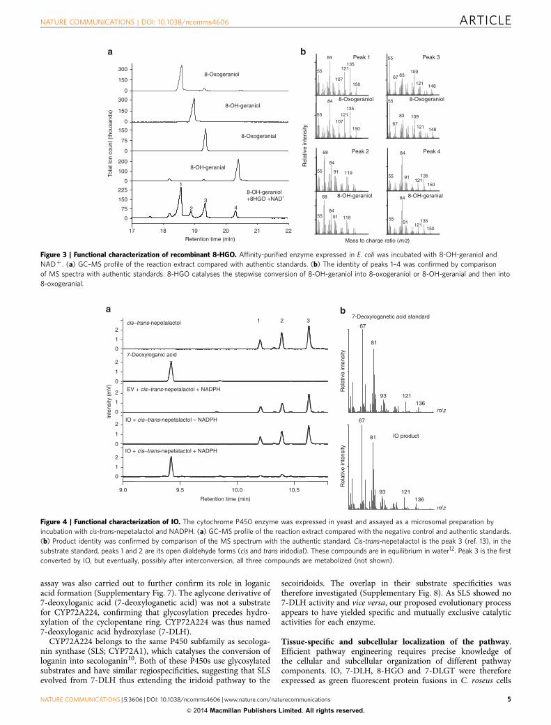
Caros003452 is the missing 8-hydroxygeraniol oxidoreductase.The three oxidoreductases (Caros008267, Caros003452 andCaros009903) were produced in Escherichia coli and purified forin vitro enzyme assays. Confirming a previous report12, theputative progesterone reductase (Caros008267) was shown topossess IS activity in the presence of 8-oxogeranial, yieldinga mixture of cis-trans-iridodial and cis-trans-nepetalactol.Caros003452 was active with the substrates 8-OH-geraniol,8-OH-geranial and 8-oxogeraniol in the presence of NADþ ,yielding mixtures of the three compounds and 8-oxogeranial invarying relative amounts depending on the combination and theincubation time (Fig. 3). The enzyme was therefore coined8-hydroxygeraniol oxidoreductase (8-HGO). With the cofactor
ARTICLE NATURE COMMUNICATIONS | DOI: 10.1038/ncomms4606
2 NATURE COMMUNICATIONS | 5:3606 | DOI: 10.1038/ncomms4606 | www.nature.com/naturecommunications
& 2014 Macmillan Publishers Limited. All rights reserved.
NADþ it did not convert 8-oxogeranial (Fig. 3), and it was notactive with any of the substrates listed above in the presence ofNADPþ /NADPH. Relatively high activity was observed withsome other primary alcohols such as geraniol (Supplementary
Table 2). Given the complex kinetics, with four interconvertiblecompounds and eight possible reactions, the reaction constantscould not be determined.
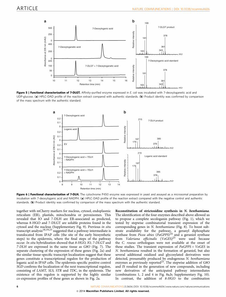
CYP76A26 is the missing iridoid oxidase. Six candidateP450 genes (CYP76A26, CYP81Z1, CYP81Q32, CYP72A224,CYP71AY1 and CYP71AY2) were transferred to a yeast expres-sion vector and co-expressed in Saccharomyces cerevisiae togetherwith the P450 reductase ATR1 from Arabidopsis thaliana20
(Supplementary Fig. 2). Functional screening was carried out asdescribed21 with geraniol, 8-hydroxygeraniol, 8-hydroxygeranial,8-oxogeraniol, 8-oxogeranial, iridodial, iridotrial, 7-deoxyloganicacid and 7-deoxyloganetic acid as potential substrates. CYP76A26converted both iridodial and iridotrial into 7-deoxyloganetic acid.The cis-iridodial and trans-iridodial freely interconverted withcis-trans-nepetalactol12, and although CYP76A26 seemed to usethe bicyclic nepetalactol as the preferred substrate, themonocyclic cis- and trans-iridodials were also utilized, possiblyafter spontaneous conversion into nepetalactol (Fig. 4). Theinterconversion and sequential metabolism of nepetalactol andthe iridodials in aqueous solution prevented reliable evaluationof the catalytic parameters with these substrates. Iridotrialwas previously proposed as an intermediate of the secologaninpathway8. Whereas we never detected iridotrial as anintermediate in the iridodial to 7-deoxyloganetic acidconversion under conditions more likely to capture earlyreaction products such as low substrate concentrations, shortincubations or low temperature, the latter was very efficientlyconverted into 7-deoxyloganetic acid with a Kmapp of 25 mMand kcat of 5.2 s� 1 (Supplementary Fig. 3). This suggests thatCYP76A26 is a multifunctional P450 enzyme catalysingsuccessive hydroxylation/dehydration steps via a mechanismsimilar to that recently described for CYP701A3, which catalysesthe conversion of kaurene to kaurenoic acid in the biosynthesis ofgibberellins22.
As the related G8O (CYP76B6) was previously shown tooxidize several (other) monoterpene alcohols in addition togeraniol9, these compounds were also tested as potentialsubstrates for CYP76A26. The enzyme converted 8-oxogeraniolinto an unidentified product (Supplementary Fig. 4), albeit with alow efficiency, and catalysed the hydroxylation of linalool, nerol,citronellol and lavandulol, but not geraniol (SupplementaryFig. 4; Supplementary Table 3). CYP76A26 thus catalyses theconversion of iridodial into 7-deoxyloganetic acid and wasconsequently named iridoid oxidase (IO; Fig. 1).
UGT709C2 is 7-deoxyloganetic acid glucosyl transferase. TheUGT (Caros009839 or UGT709C2), produced in E. coli, catalysed
TDC
STR
SLS
LAMT
7-DLH
7-DLGT
IO
IO
IS
G8O
GES
GPPS
IPP
OH
Geranyl diphosphate
Geraniol
+
OH
OH8-Hydroxygeraniol
CHOOH 8-Hydroxygeranial
CHO
OH
8-Oxogeraniol
CHO
CHO8-Oxogeranial
CHO
OH
OIridotrial
OH
Ocis–trans-iridodial
CHO
CHO
COOH
OH
O
COOH
OGlc
O
7-Deoxyloganetic acid
7-Deoxyloganic acid
COOH
OGlc
OOH
COOCH3
OGlc
OOH
COOCH3
OGlc
O
OHC
Loganic acid
Loganin
Secologanin
NH
NH2
L-tryptophan
Tryptamine
NH
NH
O
COOCH3
OGlcStrictosidine
H
H
H
H
H
H
H
H
H
H
H
H
H
H
H
H
cis–trans-nepetalactol
G8O
1 2
1 2
8-HGO 1 2
8-HGO 1 2
H
DMAPP
3
Figure 1 | The secologanin–strictosidine pathway. Genes indicated in
boxes were published before (black background) or during (white
background) the present study, or are reported here (yellow background).
Frames indicate mRNA localization in the leaf IPAP (pink) or epidermis
(blue). Numbers indicate predicted enzyme classes in the initial gene
discovery strategy. 1: oxidoreductase, 2: cytochrome P450, 3: UGT. IPP,
isopentenyl pyrophosphate; DMAPP, dimethylallyl pyrophosphate; Glc,
glucose; GPPS, geranyl diphosphate synthase, GES, geraniol synthase; G8O,
geraniol 8-oxidase; 8-HGO, 8-hydroxygeraniol oxidoreductase; IS, iridoid
synthase; IO, iridoid oxidase; 7-DLGT, 7-deoxyloganetic acid glucosyl
transferase; 7-DLH, 7-deoxyloganic acid hydroxylase; LAMT, loganic acid
O-methyltransferase; SLS, secologanin synthase; STR, strictosidine
synthase; TDC, tryptophan decarboxylase. Iridotrial indicated in brackets
was a previously proposed intermediate that we did not detect in vitro or
in vivo.
NATURE COMMUNICATIONS | DOI: 10.1038/ncomms4606 ARTICLE
NATURE COMMUNICATIONS | 5:3606 | DOI: 10.1038/ncomms4606 | www.nature.com/naturecommunications 3
& 2014 Macmillan Publishers Limited. All rights reserved.
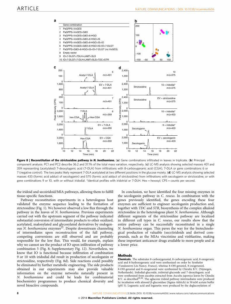
the glucosylation of 7-deoxyloganetic acid to form 7-deoxyloganicacid using UDP-glucose as the sugar donor (Fig. 5). The enzymehad a Kmapp of 9.8 mM and a kcat of 1.25 s� 1 (SupplementaryFig. 5). The enzyme was inactive with loganetic acid, loganetin,iridodial, iridotrial, 8-OH-geraniol, jasmonic acid, gibberellicacid, indole acetic acid, salicylic acid, abscisic acid, zeatin andluteolin. It thus behaved as a selective 7-deoxyloganetic acidglucosyl transferase (7-DLGT; Fig. 1).
CYP72A224 is the 7-deoxyloganic acid hydroxylase. Despite itspoor expression in yeast, CYP72A224 catalysed the conversion of7-deoxyloganic acid into loganic acid in yeast microsomes (Fig. 6)with a Kmapp of 400 mM and a Vmax of 0.01 pmol s� 1 per mgmicrosomal protein (Supplementary Fig. 6). kcat could not bedetermined due to low expression in yeast preventing precisequantification of the enzyme concentration. Owing to the lowexpression of CYP72A224 in yeast, a N. benthamiana leaf-disc
Fl
mL
St
Ro
iL MeJ
AC
on
Not
Sdlg
MeJ
A6
Con
24N
ot
MeJ
A12
MeJ
A24
YE
6Y
E12
YE
24
CellSus
WtM
eJA
WtC
on
TdM
eJA
TdC
on
WtN
ot
TdN
ot
Reb
H
HairRt
Caros020166-STR3Caros010005-STR1Caros003452-ADH10Caros003727-GESCaros020058-CYP76A26 (IO)Caros006766-G8OCaros008267-ISCaros018961-CYP71AY1Caros014930-TDCCaros009426-SGDCaros002904-LAMTCaros020659-SLSCaros011578-STR2Caros005234-CYP72A224 (7-DLH)Caros009839-UGT709C2Caros009903-OPR-likeCaros001222-CYP81Z1
Caros010005-STR1 Caros003452-ADH10Caros006766-G8O Caros003727-GESCaros005234-CYP72A224 (7-DLH)Caros001222-CYP81Z1Caros009839-UGT709C2 (7-DLGT)Caros009903-OPR-likeCaros020058-CYP76A26 (IO)Caros018961-CYP71AY1 Caros008267-ISCaros020659-SLSCaros011578-STR2 Caros002904-LAMT Caros020166-STR3Caros009426-SGDCaros014930-TDC
MeJ
A6
Sdlg
Not
Not
MeJ
A24
MeJ
A
O3
O2
CellSus
CYP81Q32
CYP71AY2
CYP76A26 (IO)
CYP76B6 (G8O)
CYP72A224 (7-DLH)
1.0±1.0
0.6±1.2 11.0±8.4
0.0±0.0 2.3±2.5
Epider
mis
Mes
ophy
ll
8.0±2.6 3.0±2.6
6.3±4.2 7.7±7.2
14.7±9.9Caros006766
Caros005234
Caros003676
Caros007686
Caros003164
Hit numbers
Figure 2 | Gene discovery strategy. (a,b) Complete-linkage hierarchical clustering of early MIA pathway gene expression in C. roseus based on our data
(a) or the Medicinal Plant Genomics Resource consortium (http://medicinalplantgenomics.msu.edu) (b). Colours indicate transcriptional activation (blue)
or repression (yellow) relative to untreated samples. Tissues: Fl, flower; mL, mature leaves; iL, immature leaves; St, stem; Ro, root; Sdlg, seedling.
Suspension cells (CellSus): Wt, wild-type; O2, ORCA2; O3, ORCA3. Hairy roots (HairRt): Wt, wild-type; Td, TDCi; RebH, RebH_F. Treatments: Not, no
treatment; MeJA, methyl jasmonate (6, 12 or 24 h); Con, mock; YE, yeast extract. (c) Candidate P450 protein hits in the epidermis and mesophyll
samples from proteomics analysis±s.d. (n¼ 3). GES, geraniol synthase; G8O, geraniol 8-oxidase; 8-HGO, 8-hydroxygeraniol oxidoreductase; IS, iridoid
synthase; IO, iridoid oxidase; 7-DLGT, 7-deoxyloganetic acid glucosyl transferase; 7-DLH, 7-deoxyloganic acid hydroxylase; LAMT, loganic acid
O-methyltransferase; SGD, strictosidine b-D-glucosidase; SLS, secologanin synthase; STR, strictosidine synthase (1–3: three related genes); TDC,
tryptophan decarboxylase.
ARTICLE NATURE COMMUNICATIONS | DOI: 10.1038/ncomms4606
4 NATURE COMMUNICATIONS | 5:3606 | DOI: 10.1038/ncomms4606 | www.nature.com/naturecommunications
& 2014 Macmillan Publishers Limited. All rights reserved.
assay was also carried out to further confirm its role in loganicacid formation (Supplementary Fig. 7). The aglycone derivative of7-deoxyloganic acid (7-deoxyloganetic acid) was not a substratefor CYP72A224, confirming that glycosylation precedes hydro-xylation of the cyclopentane ring. CYP72A224 was thus named7-deoxyloganic acid hydroxylase (7-DLH).
CYP72A224 belongs to the same P450 subfamily as secologa-nin synthase (SLS; CYP72A1), which catalyses the conversion ofloganin into secologanin10. Both of these P450s use glycosylatedsubstrates and have similar regiospecificities, suggesting that SLSevolved from 7-DLH thus extending the iridoid pathway to the
secoiridoids. The overlap in their substrate specificities wastherefore investigated (Supplementary Fig. 8). As SLS showed no7-DLH activity and vice versa, our proposed evolutionary processappears to have yielded specific and mutually exclusive catalyticactivities for each enzyme.
Tissue-specific and subcellular localization of the pathway.Efficient pathway engineering requires precise knowledge ofthe cellular and subcellular organization of different pathwaycomponents. IO, 7-DLH, 8-HGO and 7-DLGT were thereforeexpressed as green fluorescent protein fusions in C. roseus cells
0
150
300
150
300
0
75
150
0
100
200
75
150
225
17 18 19 20 21 22
Retention time (min)
8-Oxogeraniol
8-OH-geraniol
8-Oxogeranial
8-OH-geranial
1
2
34
8-OH-geraniol+8HGO +NAD+
0
Tota
l Ion
cou
nt (
thou
sand
s)
a
Rel
ativ
e in
tens
ity
b
0
84
55
55135
121
107150
67 83109
121 148
Peak 1 Peak 3
84 55
67
135121
10783 109
121 148150
55
8-Oxogeraniol 8-Oxogeraniol
55
84
91121
135
150
Peak 4
55
8-OH-geranial84
91121
135
150
8455
68
91 119
Mass to charge ratio (m/z)
8-OH-geraniol
84
55
68
91 119
Peak 2
Figure 3 | Functional characterization of recombinant 8-HGO. Affinity-purified enzyme expressed in E. coli was incubated with 8-OH-geraniol and
NADþ . (a) GC–MS profile of the reaction extract compared with authentic standards. (b) The identity of peaks 1–4 was confirmed by comparison
of MS spectra with authentic standards. 8-HGO catalyses the stepwise conversion of 8-OH-geraniol into 8-oxogeraniol or 8-OH-geranial and then into
8-oxogeranial.
9.0 9.5 10.0 10.5
Retention time (min)
EV + cis–trans-nepetalactol + NADPH
IO + cis–trans-nepetalactol + NADPH
IO + cis–trans-nepetalactol – NADPH
7-Deoxyloganic acid
cis–trans-nepetalactol
Inte
nsity
(m
V)
7-Deoxyloganetic acid standard
67
13612193
81
m/z
Rel
ativ
e in
tens
ity
IO product
67
13612193
81
m/z
Rel
ativ
e in
tens
ity
0
1
321
0
1
0
1
0
1
0
1
2
2
2
2
2
Figure 4 | Functional characterization of IO. The cytochrome P450 enzyme was expressed in yeast and assayed as a microsomal preparation by
incubation with cis-trans-nepetalactol and NADPH. (a) GC–MS profile of the reaction extract compared with the negative control and authentic standards.
(b) Product identity was confirmed by comparison of the MS spectrum with the authentic standard. Cis-trans-nepetalactol is the peak 3 (ref. 13), in the
substrate standard, peaks 1 and 2 are its open dialdehyde forms (cis and trans iridodial). These compounds are in equilibrium in water12. Peak 3 is the first
converted by IO, but eventually, possibly after interconversion, all three compounds are metabolized (not shown).
NATURE COMMUNICATIONS | DOI: 10.1038/ncomms4606 ARTICLE
NATURE COMMUNICATIONS | 5:3606 | DOI: 10.1038/ncomms4606 | www.nature.com/naturecommunications 5
& 2014 Macmillan Publishers Limited. All rights reserved.
together with mCherry markers for nucleus, cytosol, endoplasmicreticulum (ER), plastids, mitochondria or peroxisomes. Thisrevealed that IO and 7-DLH are ER-associated as predicted,whereas 8-HGO and 7-DLGT are soluble proteins found in thecytosol and the nucleus (Supplementary Fig. 9). Previous in situtranscript analysis18,19,23 suggested that a pathway intermediate istranslocated from IPAP cells (the site of the early biosyntheticsteps) to the epidermis, where the final steps of the pathwayoccur. In situ hybridization showed that 8-HGO, IO, 7-DLGT and7-DLH are expressed in the same tissue as G8O (Fig. 7). Theseparate clustering of the expression of these genes (Fig. 2a) andthe similar tissue-specific transcript localization suggest that thesegenes constitute a transcriptional regulon for the production ofloganic acid in IPAP cells. The epidermis-specific positive controlSGD confirms the localization of the next transcriptional regulon,consisting of LAMT, SLS, STR and TDC, in the epidermis. Theexistence of this regulon is supported by the highly similarco-expression profiles of these genes as shown in Fig. 2a.
Reconstitution of strictosidine synthesis in N. benthamiana.The identification of the four enzymes described above allowed usto propose a complete secologanin pathway (Fig. 1), which wetested by stepwise combinatorial transient expression of thecorresponding genes in N. benthamiana (Fig. 8). To boost sub-strate availability for the pathway, a geranyl diphosphatesynthase from Picea abies (PaGPPS)24 and a geraniol synthasefrom Valeriana officinalis (VoGES)25 were used becausethe C. roseus orthologues were not available at the onset ofthese studies. The transient expression of PaGPPSþVoGES inN. benthamiana resulted in the formation of geraniol, but alsoseveral additional oxidized and glycosylated derivatives weredetected, presumably produced by endogenous N. benthamianaenzymes as previously reported25. The stepwise addition of G8Oand IS resulted in the generation of new compounds, includingnew derivatives of the anticipated pathway intermediates(combinations 1, 2 and 4 in Fig. 8a,b; Supplementary Fig. 10).In contrast, the addition of 8-HGO to the combination
7-Deoxyloganic acid
a b
7-Deoxyloganetic acid
Retention time (min)
10 1514131211 16
Abs
orba
nce
at 2
35 n
m (
mA
U)
7-DLGT + 7-Deoxyloganetic acid
7-Deoxyloganic acid standard
199
181
163
378
361
343
7-DLGT product
Rel
ativ
e in
tens
ityR
elat
ive
inte
nsity
m/z
m/z
199
181
163
378
361
343
0
225
150
50
0
500
250
0
400
200
Figure 5 | Functional characterization of 7-DLGT. Affinity-purified enzyme expressed in E. coli was incubated with 7-deoxyloganetic acid and
UDP-glucose. (a) HPLC-DAD profile of the reaction extract compared with authentic standards. (b) Product identity was confirmed by comparison
of the mass spectrum with the authentic standard.
7-Deoxyloganic acid + 7DLH+ NADPH
Loganic acid
Abs
orba
nce
at 2
36.5
nm
(A
U)
EV
7-Deoxyloganic acid + 7DLH– NADPH
8 9 10 11 12 13 14
Retention time (min)
7-Deoxyloganic acida b
7-DLH product
m/z
Rel
ativ
e in
tens
ity
Loganic acid standard
m/z
215
179
395
378
197
Rel
ativ
e in
tens
ity
215
179395
378
197
0
0.1
0
0.1
0
0.1
0
0.1
0
0.1
0.2
0.2
0.2
0.2
0.2
Figure 6 | Functional characterization of 7-DLH. The cytochrome P450 enzyme was expressed in yeast and assayed as a microsomal preparation by
incubation with 7-deoxyloganic acid and NADPH. (a) HPLC-DAD profile of the reaction extract compared with the negative control and authentic
standards. (b) Product identity was confirmed by comparison of the mass spectrum with the authentic standard.
ARTICLE NATURE COMMUNICATIONS | DOI: 10.1038/ncomms4606
6 NATURE COMMUNICATIONS | 5:3606 | DOI: 10.1038/ncomms4606 | www.nature.com/naturecommunications
& 2014 Macmillan Publishers Limited. All rights reserved.

PaGPPSþVoGESþG8O only modified the existing productprofile quantitatively but did not generate any new compounds(combination 3 in Fig. 8a,b; Supplementary Fig. 10). This isprobably due to the fact that endogenous N. benthamianaenzymes have similar activity as 8-HGO (Supplementary Fig. 11).When IO was co-expressed with PaGPPSþVoGESþG8Oþ 8-HGOþ IS (combination 5), the metabolic profile did not change,and 7-deoxyloganetic acid and its derivatives were not detected(Fig. 8a,b). However, reconstitution of the pathway up to 7-DLGT(combination 6) was successful and resulted in the production of
7-deoxyloganic acid and putative acetylated 7-deoxyloganic acid(Fig 8a,c). Without IO, these products were not detected(Supplementary Fig. 12), indicating that IO is functional inN. benthamiana and is an essential part of the biosynthesispathway. These findings also illustrate the importance of fullpathway coverage for functional analysis of individual enzymes.
Subsequently, the entire postulated secologanin pathway(PaGPPS to SLS) was introduced by agroinfiltration; however,this only yielded products up to 7-deoxyloganic acid. Therefore,we increased the input halfway into the pathway by infiltratingthe intermediates iridodial, iridotrial or 7-deoxyloganic acid incombination with the pathway genes. In all cases this resulted inthe production of secologanin, indicating that the second half ofthe pathway is also functional (Fig. 8d). When 7-DLH, LAMT orSLS were omitted from combination 9—with infiltration of theintermediates iridodial, iridotrial or 7-deoxyloganic acid—nosecologanin was detected showing that all conversions require theinfiltrated C. roseus genes (Supplementary Fig. 13). Finally, wetested whether the biosynthesis pathway up to secologanin can befunctionally combined with the tryptamine branch of the MIApathway. When the secologanin biosynthesis pathway genes wereco-infiltrated with the tryptophan decarboxylase (TDC) andSTR genes, and the flux through the pathway was boosted byco-infiltration of the intermediates iridodial, iridotrial or7-deoxyloganic acid, strictosidine was indeed produced (combi-nation 10 in Fig. 8d).
DiscussionWhereas feeding experiments clearly indicated that secologanin isderived from geraniol26, the exact sequence of intermediates andenzymes leading to the formation of its penultimate precursorloganic acid was still obscure. We report here four novel enzymesthat together with two previously reported enzymes12,13 fill theexisting gaps and thereby provide a full description of the core(seco)iridoid pathway. Geraniol is converted to secologanin bythe sequential action of four different cytochrome P450 enzymes,two different oxidoreductases, one glucosyltransferase and onemethyltransferase. The missing enzymes and correspondinggenes were identified by a combination of transcriptomic andproteomic approaches exploiting the current knowledge of thespatiotemporal regulation of the secoiridoid pathway.
Our results also address longstanding questions in the field.First, as previously proposed for oxidoreductase proteins purifiedfrom Rauwolfia serpentina and Nepeta racemosa27,28, 8-HGOcatalyses two successive and reversible oxidation steps forthe formation of 8-oxogeranial. Therefore, two enzymes cancontribute to the formation of the intermediate 8-oxogeraniol.G8O was recently shown to also produce 8-oxogeraniol fromgeraniol21, thus G8O and 8-HGO appear to catalyse partiallyoverlapping (and in the case of G8O, monodirectional) oxidationreactions that result in the production of 8-oxogeraniol from8-hydroxygeraniol (Fig. 1). Second, a single cytochrome P450enzyme IO/CYP76A26 can convert cis-trans-iridodial andcis-trans-nepetalactol into 7-deoxyloganetic acid without therelease of an iridotrial intermediate. Third, our datademonstrate that 7-deoxyloganetic acid glycosylation precedesits further oxygenation by 7-DLH. This answers the longstandingquestion which intermediate is transferred from IPAP cells to theepidermis. The expression of 7-DLH in IPAP cells indicates thatloganic acid is the mobile intermediate transferred to theepidermis, and hence that glycosylation by 7-DLGT is notsufficient for mobility but that further hydroxylation by 7-DLH isalso required. The tissue-specific expression of pathway sectionsmay increase the flux through the pathway by alleviating feedbackinhibition by intermediates and products, and/or it may segregate
ipap
ipap
ipap
ipap
ipap
ep
G8O AS
8-HGO AS
IO AS
7-DLGT AS
7-DLH AS 7-DLH S
7-DLGT S
IO S
8-HGO S
SGD AS
Figure 7 | Expression of the transcriptional regulon required for loganic
acid biosynthesis in the IPAP cells. In situ hybridization on serial
longitudinal sections of young developing leaves was carried out with
antisense (AS) probes and sense (S) probes as controls. G8O and
strictosidine b-D-glucosidase (SGD) AS probes were used as IPAP and
epidermis markers, respectively. Sense probe controls gave no signals (not
shown). 8-HGO, 8-hydroxygeraniol oxidoreductase; IO, iridoid oxidase;
7-DLGT, 7-deoxyloganetic acid glucosyl transferase; 7-DLH, 7-deoxyloganic
acid hydroxylase; IPAP, internal phloem-associated parenchyma;
ep, epidermis. Scale bar¼ 100 mm.
NATURE COMMUNICATIONS | DOI: 10.1038/ncomms4606 ARTICLE
NATURE COMMUNICATIONS | 5:3606 | DOI: 10.1038/ncomms4606 | www.nature.com/naturecommunications 7
& 2014 Macmillan Publishers Limited. All rights reserved.
the iridoid and secoiridoid/MIA pathways, allowing them to fulfilltissue-specific functions.
Pathway reconstitution experiments in a heterologous hostvalidated the enzyme sequence leading to the formation ofstrictosidine (Fig. 1). We however observed a low flux through thepathway in the leaves of N. benthamiana. Previous experimentscarried out with the upstream segment of the pathway indicatedsubstantial conversion of intermediate products to other oxidized,acetylated, malonylated and glycosylated derivatives by endogen-ous N. benthamiana enzymes21. Despite downstream channelingof intermediates upon reconstruction of the full pathway,competing conversions are still observed and are probablyresponsible for the low flux. This would, for example, explainwhy we cannot see the product of IO upon infiltration of pathwaycombination 5 (Fig. 8; Supplementary Fig. 12). Nevertheless, weknow that IO is functional because infiltration of combination9 or 10 with iridodial did result in production of secologanin orstrictosidine, respectively (Fig. 8d). Side reactions could possiblybe eliminated by further metabolic engineering. The side productsobtained in our experiments may also provide valuableinformation on the enzyme networks naturally present inN. benthamiana and could be leads for combinatorialbiochemistry programmes to produce chemical diversity andnovel bioactive compounds.
In conclusion, we have identified the four missing enzymes inthe secologanin pathway in C. roseus. In combination with thegenes previously identified, the genes encoding these fourenzymes are sufficient to engineer secologanin production and,together with TDC and STR, biosynthesis of the complex alkaloidstrictosidine in the heterologous plant N. benthamiana. Althoughdifferent segments of the strictosidine pathway are localizedin different cell types in C. roseus, our results show that theentire pathway can be successfully reconstituted in a singleN. benthamiana organ. This paves the way for the biotechnolo-gical production of valuable (seco)iridoids and derived com-pounds, such as the MIAs vincristine and vinblastine, makingthese important anticancer drugs available to more people and ata lower price.
MethodsChemicals. The substrates 8-carboxygeranial, 8-carboxygeranic acid, 8-oxogeranicacid and 8-hydroxygeranic acid were synthesized on order by Synthelor(Vandoeuvre-Les-Nancy, France), whereas 8-OH-geraniol, 8-oxogeraniol,8-OH-geranial and 8-oxogeranial were synthesized by Chiralix B.V. (Nijmegen,Netherlands). Iridodial-glucoside, iridotrial-glucoside and 7-deoxyloganic acidwere synthesized from aucubin extracted from Aucuba japonica leaves by ChiralixB.V. as described29,30. The aglucone iridoid pathway intermediates were producedby incubation with almond b-glucosidase (Sigma-Aldrich) in 50 mM acetate buffer(pH 5). Loganetic acid and loganetin were produced by the deglucosylation of
–150 150500–50
200
100
–100
064
5
32
17
8
Gene combination
1 PaGPPS+VoGES
2 PaGPPS+VoGES+G8O
3 PaGPPS+VoGES+G8O+8-HGO
4 PaGPPS+VoGES+G8O+8-HGO+IS
5 PaGPPS+VoGES+G8O+8-HGO+IS+IO
6 PaGPPS+VoGES+G8O+8-HGO+IS+IO+7-DLGT
7 PaGPPS+G8O+8-HGO+IS+IO+7-DLGT (no VoGES)
8 Empty vector
9 IO+7-DLGT+7DLH+LAMT+SLS
10 IO+7-DLGT+7-DLH+LAMT+SLS+TDC+STR
6 m/z=401
EV + 7-DLAm/z=401
7 m/z=401
6 m/z=359
EV + 7-DLAm/z=359
EV + CGAm/z=359
Pea
k in
tens
ity (
CP
S)
Pea
k in
tens
ity (
CP
S)
Acetyl-7-DLA
Acetyl-7-DLA
Hex-CGA
Hex-CGAhex-CGA
7-DLA
7-DLA
10 18161412 2220Time (min)
10 18161412 2220Time (min)
Secologanin
Secologanin
Strictosidine
Strictosidine
10m/z=575
10 + iridodial*m/z=575
EV + strictosidinem/z=575
EV + secologaninm/z=433
10 + iridodial*m/z=433
9 + iridodial*m/z=433
200
100
01,600
800
0200
100
03,200
1,600
03,200
1,600
03,200
1,600
0
2,400
1,200
02,400
1,200
02,400
1,200
02,000
1,000
02,000
1,000
02,000
1,000
0
Figure 8 | Reconstitution of the strictosidine pathway in N. benthamiana. (a) Gene combinations infiltrated in leaves in triplicate. (b) Principal
component analysis. PC1 and PC2 describe 36.2 and 31.1% of the total mass variation, respectively. (c) LC-MS analysis showing selected masses 401 and
359 representing (acetylated) 7-deoxyloganic acid (7-DLA) from infiltrations with 8-carboxygeranic acid (CGA), 7-DLA or gene combinations 6 or
7 (negative control). The two peaks likely represent 7-DLA acetylated at two different positions in the glucose moiety. (d) LC-MS analysis showing selected
masses 433 (formic acid adduct of secologanin) and 575 (formic acid adduct of strictosidine) from infiltrations with secologanin or strictosidine, or with
gene combinations 9 or 10, with or without iridodial. *Identical profiles with iridotrial or 7-DLH. Hex¼ hexosyl; CPS¼ counts per second.
ARTICLE NATURE COMMUNICATIONS | DOI: 10.1038/ncomms4606
8 NATURE COMMUNICATIONS | 5:3606 | DOI: 10.1038/ncomms4606 | www.nature.com/naturecommunications
& 2014 Macmillan Publishers Limited. All rights reserved.

loganic acid and loganin (Extrasynthese). Aglycones were extracted with diethylether, evaporated under N2 and quantified by 1H-NMR (1H-nuclear magneticresonance).
Transcriptomic analysis. Transgenic derivatives of C. roseus cell line MP183L(overexpressing the ORCA transcription factors) were generated by particlebombardment31 with derivatives of the pER8 plasmid32 carrying either the ORCA2or ORCA3 open reading frames (ORFs). Selected transgenic lines were treated for24 h with 10mM estradiol, and RNA was isolated as described16. IlluminaHiSeq2000 RNA sequencing, assembly, annotation and mapping of the RNA-Seqreads on the reference transcriptome was carried out as described16. Completelinkage hierarchical cluster analysis was achieved using the CLUSTER andTREEVIEW software33 and the log10 transformed values of the normalized FPKMvalues were used as input for CLUSTER.
Proteomic analysis. For the plant material of C. roseus var. little Bright Eyes seedswere sown in sterilized soil and covered with transparent plastic until germination.The soil was kept humid. The plants were fertilized weekly with 0.2% liquid Wuxal(29 g l� 1 nitrate-N; 46 g l� 1 ammonium-N; 25 g l� 1 carbamide-N; 100 g l� 1 P2O5
total phosphate; 75 g l� 1 K2O; 124 mg l� 1 B; 50 mg l� 1 Cu; 248 mg l� 1 Fe; Cu, Fe,Mn and Zn as EDTA-chelate). The plants were re-potted twice during furthergrowth. For the isolation of epidermal protoplasts, young, light-green C. roseusleaves (length 4–7 cm) from side branches (without buds or flowers) of 8- to11-week-old plants were harvested for protoplast isolation. The mid-vein wasremoved and the leaves were cut into 1- to 2-mm strips. Protoplasts were isolatedas described34. To collect epidermal protoplasts, a layer of 1-ml betaine solution(0.5 M betaine, 1 mM CaCl2, 10 mM MES, pH 5.6 with KOH) was added on topof the tubes containing the protoplast mix. After centrifugation for 7 min at1,500 r.p.m. and 4 �C, epidermal protoplasts were collected from the upperinterphase. The suspension was mixed with 4-ml protoplast solution and 25%Percoll (pH 6), and overlaid with 1.5 ml of betaine solution for a secondpurification step. The tubes were centrifuged at 700 r.p.m. for 30 min and theepidermal protoplasts were again collected from the upper interphase. Protoplastswere pelleted in the betaine solution. Isolation of mesophyll protoplasts wasperformed as described for the epidermal protoplasts with replacement of the MCPsolution with TEX solution (3.2 g l� 1 Gamborg’s B5 medium, 3.1 mM NH4NO3,5.1 mM CaCl2, 2.6 mM MES, 0.4 M sucrose, pH 5.6–5.8 with 0.5 M KOH), and thebetaine solution was replaced with mannitol/W5 (1 mM D-glucose, 30 mM NaCl,25 mM CaCl2, 1 mM KCl, 0.3 mM MES, 320 mM a-mannitol, pH 5.6–5.8 withKOH). For the first gradient, 5–10% Percoll (pH 6) was added instead of 3–5%. Theprotoplast pellet was resuspended in 2–4 ml buffer A (20 mM HEPES pH 7.2,1 tablet per 10 ml Roche Protease Inhibitor, 1 mM PMSF). The mixture was pressed10–20 times through a syringe with needle and centrifuged for 10 min at3,000 r.p.m. and 4 �C. The samples were ultra-centrifuged for 1 h at 30,000 r.p.m.The membrane pellets were dissolved in 1 ml washing solution (0.3 M NaCI,20 mM HEPES-KOH pH 7.2, 1 tablet per 10 ml Roche Protease Inhibitor, 1 mMPMSF) by stirring with a small brush, and then vortexed for 30 s. The microsomeswere frozen at –80 �C for at least 1 h, thawed and then centrifuged for 1 h at30,000 r.p.m. and 4 �C. The pellets were dissolved in 50–100 ml buffer A for furtheranalysis. The protein concentration was determined using the Bio-Rad DC proteinassay according to the manufacturer’s instructions.
For mass spectrometry analysis, B50 mg of protein was separated by one-dimensional gel electrophoresis35 in 10% polyacrylamide gels to reduce samplecomplexity. The gels were stained with Coomassie Brilliant Blue, the lanes were cutinto 10 slices, the proteins were reduced with tris(carboxyethyl)phosphinehydrochloride and sulhydryl groups were blocked with iodoacetamide. In-geldigestion with sequencing-grade-modified trypsin (Promega V5111) was carriedout overnight at 37 �C36. The resulting peptides were recovered by adding 40 mMTris-HCl (pH 8.0) and 50% acetonitrile/1% formic acid. The peptide mixtures weredesalted by solid-phase extraction on C18 reversed-phase columns and analysed onan LTQ Orbitrap mass spectrometer (Thermo Fischer Scientific, Bremen,Germany) coupled to an Eksigent Nano HPLC system (Eksigent Technologies,Dublin, CA, USA) as previously described37. For protein identification, databaseswere searched using Mascot v2.3. Raw data were searched against a compositedatabase consisting of all entries in the NCBI Viridiplantae database (released inNovember 2010), all publicly available C. roseus-expressed sequence tags(downloaded from NCBI in November 2010) and the reference transcriptomereleased on CathaCyc and ORCAE16 (database contained forward and reverseprotein entries, total number of protein entries 1,166,013). The parameters forprecursor and fragment ion mass tolerance were set to 5 p.p.m. and 0.8 Da,respectively. One missed trypsin cleavage was allowed. Carboxyamidomethylationof cysteine was specified as fixed modification, and oxidation of methionine andpyroglutamate formation from glutamine were selected as variable modifications.Scaffold v3.0 (Proteome Software, Portland, OR, USA) was used to validate andquantify MS/MS-based peptide and protein identifications. Peptide identificationswere accepted if they were established at 495% probability as specified by thePeptide Prophet algorithm38. Protein identifications were accepted if they wereestablished at 490% probability and at least one peptide was uniquely assigned toa corresponding protein in a minimum of two of our samples. Protein probabilitywas assigned by the Protein Prophet algorithm39. Proteins that were identified with
the same set of peptides and could not be differentiated by the MS/MS analysiswere grouped to protein clusters to satisfy the principles of parsimony. Peptide andprotein false-discovery rates (FDR) were determined by the Scaffold software.A peptide FDR of 0.01% and a protein FDR of 0.2% were computed. The Scaffoldsoftware was also used to determine protein abundance in the mesophyll andepidermal factions based on the number of spectra assigned to each protein.An F-test was applied to assess significant differences in protein abundances.
Gene isolation. ORFs were amplified by PCR from a pACT2 cDNA library of aC. roseus cell culture elicited with yeast extract40 using the primers listed inSupplementary Table 4. For expression in plants, the ORFs were transferred tovector pRT101 (ref. 41) to bring them under the control of the Cauliflower MosaicVirus 35S promoter, and the expression cassettes were then transferred to thebinary vector pCAMBIA1300 (Cambia). For expression in E. coli, the ORFs weretransferred to vector pASK-IBA45plus (IBA) and/or pET16-H (Novagen pET-16bderivative). Probes for in situ hybridization were prepared from the same ORFscloned in pBluescript II SKþ . For localization analysis, the ORFs were transferredto vector pTH2 (refs 42,43) and/or pTH2BN (a derivative of pTH2). The markerfor nucleocytosolic localization (pRT101-mCherry) was prepared by amplifying themCherry ORF from plasmid ER-rk44 (The Arabidopsis Information Resources,TAIR, clone CD3-959).
Isolation of His-tagged recombinant proteins. Recombinant proteins carrying aHis6 tag were expressed using plasmid pASK-IBA45plus and/or pET16-H in E. colistrain BL21 (DE3) pLysS and were purified using Ni-NTA agarose chromatography(Qiagen). For quality analysis, the recombinant proteins were separated by 12.5%(w/v) SDS–PAGE, transferred to Protran nitrocellulose membranes (Whatman) bysemidry electroblotting, and western blots were probed with mouse monoclonalanti-His horseradish peroxidase-conjugated antibodies (5Prime). Antibody bindingwas detected by incubation in 250mM sodium luminol, 0.1 M Tris–HCl (pH 8.6),3 mM H2O2 and 67mM p-coumaric acid, followed by exposure to an X-ray film.
Enzymatic assays of UGT and oxidoreductases. UGT activity was detected in0.1-ml reaction buffer containing 50 mM potassium phosphate (pH 7.5), 2 mMUDP-glucose, 5–1,000 mM 7-deoxyloganetic acid or 2 mM of other testedcompounds and 50–1,000 ng of purified enzyme. Reactions were incubated at 32 �Cfor 15 min and stopped by adding 1 volume of methanol, mixed by vortexing andkept on ice for 10 min. The tubes were centrifuged at 4,000 g for 10 min, and thesupernatants were passed through 0.22-mm nylon filters.
Oxidoreductase activity was detected in 1-ml reaction buffer containing 50 mMbis-tris propane (pH 9 for oxidation and pH 7.5 for reduction), 2–1,000 mM8-OH-geraniol, 8-oxogeraniol, 8-OH-geranial, 8-oxogeranial or other testedcompounds, 2–2,000 mM NADþ or NADH and 50–1,000 ng of purified enzyme.Reactions were incubated for 15 min at 32 �C, stopped by adding 0.2 volumes of1 M sodium citrate (pH 3) and centrifuged and filtered as above. Quantitativeassays were carried out by measuring NADH production at 340 nm in aNanodrop2000c (Thermoscientific)
Chromatographic analysis of 8-HGO and 7-DLGT products. Identification of8-HGO enzyme products was performed using capillary gas chromatography massspectrometry. The ethyl acetate extract of the reaction mixture was separated on aAgilent GC 7890A series equipped with a 5975C MSD and DB-5 capillary column(30 m� 0.25 mm, film thickness of 0.25 mm; JpW Scientific). Helium gas was usedas a carrier at a flow rate of 1.2 ml min� 1. The separation conditions were asfollows: split mode 1:5, injection volume 5 ml, injector temperature 230 �C, initialoven temperature 60 �C, and then linear gradient to 100 �C at a rate of20 �C min� 1 followed by a linear gradient to 160 �C at a rate of 2 �C min� 1
(run time 32 min). Analysis of 7-DLGT products was carried out using an Agilentseries 1200 HPLC with a diode array detector and a Polymer Laboratories PL-ELS2100 ICE evaporative light scattering detector and a Phenomenex Luna 5 micron150� 4.6-mm C18 column. The injection volume was 10 or 100 ml. The binarysolvent system consisted of acetonitrile and 0.1% trifluoroacetic acid in water. Theelution program was as follows: 5 min isocratic 10% acetonitrile and then 25 mingradient until 95% acetonitrile. Peak areas were calculated using AgilentChemStation.
NMR. Structures of enzyme products were analysed by NMR spectroscopy in750 ml of acetone-d6 or methanol-d4 in a 5-mm NMR glass tube45.
Subcellular localization studies. C. roseus MP183L cell suspension cultures weremaintained by weekly 10-fold dilution in 50 ml Linsmaier & Skoog (LS) mediumcontaining 88 mM sucrose, 2.7 mM 1-NAA and 0.23 mM kinetin (LS-13). The cellswere grown at 25 �C with an 18/6-h light–dark cycle. The cells were bombarded28
using the plasmids pTH2-IO, pTH2-7-DLH, pTH2-7-DLGT, pTH2BN-7-DLGT,pTH2-8-HGO and pTH2BN-8-HGO. The first two were combined with equalamounts of the ER marker ER-rk, and the others with the nucleocytosolic markermCherry44. Bombarded cells were placed on Petri dishes with LS-13 medium andviewed after 24 h using a Zeiss Observer laser scanning microscope.
NATURE COMMUNICATIONS | DOI: 10.1038/ncomms4606 ARTICLE
NATURE COMMUNICATIONS | 5:3606 | DOI: 10.1038/ncomms4606 | www.nature.com/naturecommunications 9
& 2014 Macmillan Publishers Limited. All rights reserved.

In situ hybridization. pBluescript plasmid derivatives containing the cDNAs for8-HGO, IO, 7-DLGT and 7-DLH were used for the synthesis of antisense and sensedigoxigenin-labelled riboprobes as previously described19. G8O antisense probes19
and SGD antisense probes46 were used as internal phloem-associated parenchymaand epidermis markers, respectively. Paraffin-embedded serial longitudinal sectionsof young developing leaves were hybridized with digoxigenin-labelled riboprobesand localized with antidigoxigenin-alkaline phosphatase-conjugated antibodies47.
P450 expression in yeast and enzyme assays. P450-coding sequences wereamplified from pRT101 source vectors using specific primers to introduce USERsites at the ends of each sequence. The genes were subsequently transferred to theplasmid pYeDP60 using the USER cloning technique (New England Biolabs,Ipswich, UK)48. The resulting recombinant plasmids were introduced intoS. cerevisiae strain WAT11, cultivated at 28 �C and P450 expression was inducedas described21. Cells were harvested by centrifugation and manually broken with0.45-mm glass beads in 50 mM Tris–HCl buffer (pH 7.5) containing 1 mM EDTAand 600 mM sorbitol. The homogenate was centrifuged for 10 min at 10,000 g andthe supernatant was centrifuged for 1 h at 30,000 g. The pellet, comprisingmicrosomal membranes, was resuspended in 50 mM Tris–HCl (pH 7.4), 1 mMEDTA and 30% (v/v) glycerol with a Potter-Elvehjem homogenizer and storedat –20 �C. All procedures for microsomal preparation were carried out at 0–4 �C.P450 expression was evaluated as described49 and enzymatic activities weredetermined in a standard 0.1 ml assay comprising for IO 2.3 nmol cytochromeP450, 0.6 mM NADPH and substrate in 20 mM sodium phosphate (pH 7.4). Thereaction was initiated by the addition of NADPH and was stopped after 5 min bythe addition of 10ml 1 M HCl. Iridoids were extracted in 1 ml ethyl acetate, and theorganic phase was concentrated to 200 ml before GC-FID analysis on a Varian 3900gas chromatograph (Agilent Technologies) equipped with a flame ionizationdetector and a DB-5 column (30 m, 0.25 mm, 0.25 mm; Agilent Technologies)with splitless injection, at 250 �C injector temperature, and with a temperatureprogramme of 0.5 min at 50 �C, 10 �C min� 1 to 320 �C, and 5 min at 320 �C.For 7-DLH, 10ml of yeast microsomes expressing 7-DLH (130 mg of microsomalprotein) were incubated for 20 min at 27 �C, in 0.1 ml of 20 mM Na-phosphate(pH 7.4) containing 0.6 mM NADPH and substrate. The reaction was initiated bythe addition of NADPH and was stopped after 20 min on ice. After addition of50ml of 50% acetic acid, tubes were vortexed and centrifuged. The supernatant wasrun on reverse-phase HPLC (Alliance 2695 Waters system, NOVA-PAK C184.6� 250 mm column) with photo-diode array detection at 236.5 nm. Peak areas ofthe product(s) were used to calculate the catalytic parameters of each enzyme.
Leaf disc assays. Five-week-old N. benthamiana leaves were infiltrated withA. tumefaciens transformed with vector pCAMBIA1300 containing the relevantcandidate genes, plus the helper plasmid p19. Five days post infiltration, leaveswere used in a leaf disc assay as previously described21.
Pathway reconstruction in N. benthamiana. The pathway genes were transientlyexpressed in the leaves of five-week-old N. benthamiana plants by agroinfiltrationas previously described25. Briefly, bacteria carrying the relevant expressionconstructs (PaGPPS, VoGES, G8O, 8-HGO, IS, IO, 7-DLGT, 7-DLH, LAMT, SLS,TDC, STR, empty vector or TBSV p19 (ref. 50)) were grown individually at 28 �Cfor 24 h. Cells were harvested by centrifugation and then resuspended in infiltrationbuffer containing 10 mM MES (Duchefa Biochemie), 10 mM MgCl2 and 100 mMacetosyringone (40-hydroxy-30 ,50-dimethoxyacetophenone, Sigma) to a final OD600
of B0.5. For all gene combinations that compared subsequent steps in thepathway, the amounts of cell suspension for each expression construct were keptconstant by adding the corresponding amount of A. tumefaciens carrying an emptyvector. All infiltrations were performed in three replicates. In several experiments,pathway intermediates were injected into the same leaves 3 days afteragroinfiltration. Compounds used for infiltration were diluted to a finalconcentration of 400 mM in methanol/water (1:19), with the same ratio ofmethanol/water alone as a negative control and 400 mM 7-deoxyloganic acid,8-carboxygeranic acid, secologanin or strictosidine as positive controls. Leaves wereharvested for metabolite analysis 5 days after agroinfiltration. Frozen, powderedN. benthamiana leaves (200 mg aliquots) were extracted in 0.6 ml 99.867%methanol, 0.133% formic acid and 5 ml of the extract was analysed using a WatersAlliance 2795 HPLC connected to a QTOF Ultima V4.00.00 mass spectrometer(Waters, MS Technologies, UK). Measurements were taken in negative ionizationmode. LC-MS data were acquired using MassLynx 4.0 (Waters) and processedusing MetAlign version 1.0. The normalized and log-transformed data matrixwas used for principal component analysis implemented in GeneMath XT v 2.1.ANOVA was used to evaluate the statistical significance of differences in metabolitelevels between all treatments.
References1. Dinda, B., Debnath, S. & Harigaya, Y. Naturally occurring iridoids. A review,
part 1. Chem. Pharm. Bull. (Tokyo) 55, 159–222 (2007).
2. Dinda, B., Debnath, S. & Harigaya, Y. Naturally occurring secoiridoids andbioactivity of naturally occurring iridoids and secoiridoids. A review, part 2.Chem. Pharm. Bull. (Tokyo) 55, 689–728 (2007).
3. Tundis, R., Loizzo, M. R., Menichini, F., Statti, G. A. & Menichini, F. Biologicaland pharmacological activities of iridoids: Recent developments. Mini Rev.Med. Chem. 8, 399–420 (2008).
4. Viljoen, A., Mncwangi, N. & Vermaak, I. Anti-inflammatory iridoids ofbotanical origin. Curr. Med. Chem. 19, 2104–2127 (2012).
5. Birkett, M. A. & Pickett, J. A. Aphid sex pheromones: from discovery tocommercial production. Phytochemistry 62, 651–656 (2003).
6. Birkett, M. A., Hassanali, A., Hoglund, S., Pettersson, J. & Pickett, J. A.Repellent activity of catmint, Nepeta cataria, and iridoid nepetalactone isomersagainst Afro-tropical mosquitoes, ixodid ticks and red poultry mites.Phytochemistry 72, 109–114 (2011).
7. Oudin, A. et al. Spatial distribution and hormonal regulation of gene productsfrom methyl erythritol phosphate and monoterpene-secoiridoid pathways inCatharanthus roseus. Plant Mol. Biol. 65, 13–30 (2007).
8. Loyola-Vargas, V. M., Galaz-Avalos, R. M. & Ku-Cauich, R. Catharanthusbiosynthetic enzymes: the road ahead. Phytochem. Rev. 6, 307–339 (2007).
9. Collu, G. et al. Geraniol 10-hydroxylase, a cytochrome P450 enzyme involvedin terpenoid indole alkaloid biosynthesis. FEBS Lett. 508, 215–220 (2001).
10. Irmler, S. et al. Indole alkaloid biosynthesis in Catharanthus roseus: newenzyme activities and identification of cytochrome P450CYP72A1 assecologanin synthase. Plant J. 24, 797–804 (2000).
11. Murata, J., Roepke, J., Gordon, H. & De Luca, V. The leaf epidermome ofCatharanthus roseus reveals its biochemical specialization. Plant Cell 20,524–542 (2008).
12. Geu-Flores, F. et al. An alternative route to cyclic terpenes by reductivecyclization in iridoid biosynthesis. Nature 492, 138–142 (2012).
13. Simkin, A. J. et al. Characterization of the plastidial geraniol synthase fromMadagascar periwinkle which initiates the monoterpenoid branch of thealkaloid pathway in internal phloem associated parenchyma. Phytochemistry85, 36–43 (2013).
14. Mahroug, S., Burlat, V. & Saint-Pierre, B. Cellular and sub-cellular organisationof the monoterpenoid indole alkaloid pathway in Catharanthus roseus.Phytochem. Rev. 6, 363–381 (2007).
15. Verma, P., Mathur, A. K., Srivastava, A. & Mathur, A. Emerging trends inresearch on spatial and temporal organization of terpenoid indole alkaloidpathway in Catharanthus roseus: a literature update. Protoplasma 249, 255–268(2012).
16. Van Moerkercke, A. et al. CathaCyc, a metabolic pathway database builtfrom Catharanthus roseus RNA-seq data. Plant Cell Physiol. 54, 673–685(2013).
17. van der Fits, L. & Memelink, J. ORCA3, a jasmonate-responsive transcriptionalregulator of plant primary and secondary metabolism. Science 289, 295–297(2000).
18. Guirimand, G. et al. The subcellular organization of strictosidine biosynthesisin Catharanthus roseus epidermis highlights several trans-tonoplasttranslocations of intermediate metabolites. FEBS J. 278, 749–763 (2011).
19. Burlat, V., Oudin, A., Courtois, M., Rideau, M. & St-Pierre, B. Co-expression ofthree MEP pathway genes and geraniol 10-hydroxylase in internal phloemparenchyma of Catharanthus roseus implicates multicellular translocation ofintermediates during the biosynthesis of monoterpene indole alkaloids andisoprenoid-derived primary metabolites. Plant J. 38, 131–141 (2004).
20. Pompon, D., Louerat, B., Bronine, A. & Urban, P. Yeast expression of animaland plant P450s in optimized redox environments. Cytochrome P450, Pt B 272,51–64 (1996).
21. Hofer, R. et al. Geraniol hydroxylase and hydroxygeraniol oxidase activities ofthe CYP76 family of cytochrome P450 enzymes and potential for engineeringthe early (seco)iridoid pathway. Metab. Eng. 08, 001 (2013).
22. Morrone, D., Chen, X., Coates, R. M. & Peters, R. J. Characterization of thekaurene oxidase CYP701A3, a multifunctional cytochrome P450 fromgibberellin biosynthesis. Biochem. J. 431, 337–344 (2010).
23. Guirimand, G. et al. Spatial organization of the vindoline biosynthetic pathwayin Catharanthus roseus. J. Plant Physiol. 168, 549–557 (2011).
24. Schmidt, A. et al. A bifunctional geranyl and geranylgeranyl diphosphatesynthase is involved in terpene oleoresin formation in Picea abies. Plant Physiol.152, 639–655 (2010).
25. Dong, L. et al. Characterization of two geraniol synthases from Valerianaofficinalis and Lippia dulcis: similar activity but difference in subcellularlocalization. Metab. Eng. 20, 198–211 (2013).
26. Uesato, S., Kanomi, S., Iida, A., Inouye, H. & Zenk, M. H. Mechanism foriridane skeleton formation in the biosynthesis of secologanin and indolealkaloids in Lonicera tatarica, Catharanthus roseus and suspension cultures ofRauwolfia serpentina. Phytochemistry 25, 839–842 (1986).
27. Ikeda, H. et al. Acyclic monoterpene primary alcohol:NADPþ oxidoreductaseof Rauwolfia serpentina cells: the key enzyme in biosynthesis of monoterpenealcohols. J. Biochem. 109, 341–347 (1991).
ARTICLE NATURE COMMUNICATIONS | DOI: 10.1038/ncomms4606
10 NATURE COMMUNICATIONS | 5:3606 | DOI: 10.1038/ncomms4606 | www.nature.com/naturecommunications
& 2014 Macmillan Publishers Limited. All rights reserved.

28. Hallahan, D. L. et al. Purification and characterization of an acyclicmonoterpene primary alcohol:NADPþ oxidoreductase from catmint (Nepetaracemosa). Arch. Biochem. Biophys. 318, 105–112 (1995).
29. Jensen, S. R., Kirk, O. & Nielsen, B. J. Application of the vilsmeierformylation in the synthesis of 11-C-13 labeled iridoids. Tetrahedron 43,1949–1954 (1987).
30. Jensen, S. R., Kirk, O. & Nielsen, B. J. Biosynthesis of the iridoid glucosidecornin in Verbena officinalis. Phytochemistry 28, 97–105 (1989).
31. van der Fits, L. & Memelink, J. Comparison of the activities of CaMV 35S andFMV 34S promoter derivatives in Catharanthus roseus cells transiently andstably transformed by particle bombardment. Plant Mol. Biol. 33, 943–946(1997).
32. Zuo, J., Niu, Q. W. & Chua, N. H. Technical advance: an estrogenreceptor-based transactivator XVE mediates highly inducible gene expressionin transgenic plants. Plant J. 24, 265–273 (2000).
33. Eisen, M. B., Spellman, P. T., Brown, P. O. & Botstein, D. Cluster analysis anddisplay of genome-wide expression patterns. Proc. Natl Acad. Sci. USA 95,14863–14868 (1998).
34. Tohge, T. et al. Toward the storage metabolome: profiling the barley vacuole.Plant Physiol. 157, 1469–1482 (2011).
35. Laemmli, U. K. Cleavage of structural proteins during the assembly of the headof bacteriophage T4. Nature 227, 680–685 (1970).
36. Shevchenko, A., Wilm, M., Vorm, O. & Mann, M. Mass spectrometricsequencing of proteins silver-stained polyacrylamide gels. Anal. Chem. 8,850–858 (1996).
37. Schneider, T. et al. A proteomics approach to investigate the process ofZn hyperaccumulation in Noccaea caerulescens (J & C. Presl) F.K. Meyer.Plant J 73, 131–142 (2013).
38. Keller, A., Nesvizhskii, A. I., Kolker, E. & Aebersold, R. Empirical statisticalmodel to estimate the accuracy of peptide identifications made by MS/MS anddatabase search. Anal. Chem. 74, 5383–5392 (2002).
39. Nesvizhskii, A. I., Keller, A., Kolker, E. & Aebersold, R. A statistical model foridentifying proteins by tandem mass spectrometry. Anal. Chem. 75, 4646–4658(2003).
40. Menke, F. L. H., Champion, A., Kijne, J. W. & Memelink, J. A noveljasmonate- and elicitor-responsive element in the periwinkle secondarymetabolite biosynthetic gene Str interacts with a jasmonate- andelicitor-inducible AP2-domain transcription factor, ORCA2. EMBO J. 18,4455–4463 (1999).
41. Topfer, R., Matzeit, V., Gronenborn, B., Schell, J. & Steinbiss, H. H. A set ofplant expression vectors for transcriptional and translational fusions. NucleicAcids Res. 15, 5890 (1987).
42. Chiu, W. et al. Engineered GFP as a vital reporter in plants. Curr. Biol. 6,325–330 (1996).
43. Niwa, Y., Hirano, T., Yoshimoto, K., Shimizu, M. & Kobayashi, H.Non-invasive quantitative detection and applications of non-toxic, S65T-typegreen fluorescent protein in living plants. Plant J. 18, 455–463 (1999).
44. Nelson, B. K., Cai, X. & Nebenfuhr, A. A multicolored set of in vivo organellemarkers for co-localization studies in Arabidopsis and other plants. Plant J. 51,1126–1136 (2007).
45. Kim, H. K., Choi, Y. H. & Verpoorte, R. NMR-based metabolomic analysis ofplants. Nat. Protoc. 5, 536–549 (2010).
46. Guirimand, G. et al. Strictosidine activation in Apocynaceae: towards a ‘nucleartime bomb?’. BMC. Plant Biol. 10, 182 (2010).
47. Mahroug, S., Courdavault, V., Thiersault, M., St-Pierre, B. & Burlat, V.Epidermis is a pivotal site of at least four secondary metabolic pathways inCatharanthus roseus aerial organs. Planta 223, 1191–1200 (2006).
48. Nour-Eldin, H. H., Hansen, B. G., Norholm, M. H., Jensen, J. K. &Halkier, B. A. Advancing uracil-excision based cloning towards
an ideal technique for cloning PCR fragments. Nucleic Acids Res. 34, e122(2006).
49. Omura, T. & Sato, R. The carbon monoxide-binding pigment of livermicrosomes. i. evidence for its hemoprotein nature. J. Biol. Chem. 239,2370–2378 (1964).
50. Voinnet, O., Rivas, S., Mestre, P. & Baulcombe, D. An enhanced transientexpression system in plants based on suppression of gene silencing by the p19protein of tomato bushy stunt virus. Plant J. 33, 949–956 (2003).
AcknowledgementsWe are grateful to Richard Twyman for critical reading of the manuscript and to SørenRosendal Jensen for advice on iridoid substrate synthesis. David Nelson is acknowledgedfor naming P450 enzymes. The research leading to these results has received fundingfrom the European Union Seventh Framework Programme FP7/2007-2013 under grantagreement number 222716–SMARTCELL. T.I. received funding from the People Pro-gramme (Marie Curie Actions) of the European Union’s 7th Framework Programme(FP7/2007–2013) under REA Grant Agreement 289217.
Author contributionsJ.M., A.G., K.-M.O.-C., R.V., E.M., H.B., N.N. and D.W. conceived the project andgeneral strategy; A.G. and J.P. performed the expression analysis and gene selection.K.M. isolated the candidate genes and prepared most vectors, and expressed andfunctionally characterized 8-HGO and 7DLGT, performed subcellular localizationexperiments and parts of the synthesis of iridoid substrates; L.D. did and analysedthe pathway reconstruction in N. benthamiana; N.N. assisted by T.I. expressed andfunctionally characterized the candidate P450 genes; T.S. assisted by L.W. did theproteomics analysis; V.B. carried out the in situ hybridizations; N.N. and R.L. contributedto metabolite analysis; D.W.-R., J.M., A.G., S.vd K. and H.B. wrote the manuscript;D.W.-R. acted as coordinator.
Additional informationAccession Codes: Transcriptomic data have been deposited in the GenBank SequenceRead Archive (SRA) under the accession code SRP026417. Gene sequences have beendeposited in GenBank nucleotide core database under the accession codes KF302066(IO), KF302067 (7DLH), KF302068 (7DLGT), KF302069 (8-HGO), KF302070(CYP81Q32), KF302071 (CYP81C13), KF302072 (CYP71AY2), KF302073 (CYP71BT1),KF302074 (CYP72A225), KF302075 (CYP76T24), KF302076 (CYP71D2), KF302077(ADH9), KF302078 (ADH13), KF302079 (ADH14), KF302080 (UGT9) and KF309243(CYP71AY1).
Supplementary Information accompanies this paper at http://www.nature.com/naturecommunications
Competing financial interests: The authors declare no competing financial interests.
Reprints and permission information is available online at http://npg.nature.com/reprintsandpermissions/
How to cite this article: Miettinen, K. et al. The seco-iridoid pathwayfrom Catharanthus roseus. Nat. Commun. 5:3606 doi: 10.1038/ncomms4606(2014).
This work is licensed under a Creative Commons Attribution-NonCommercial-NoDerivs 3.0 Unported License. The images or other
third party material in this article are included in the article’s Creative Commons license,unless indicated otherwise in the credit line; if the material is not included under theCreative Commons license, users will need to obtain permission from the license holderto reproduce the material. To view a copy of this license, visit http://creativecommon-s.org/licenses/by-nc-nd/3.0/
NATURE COMMUNICATIONS | DOI: 10.1038/ncomms4606 ARTICLE
NATURE COMMUNICATIONS | 5:3606 | DOI: 10.1038/ncomms4606 | www.nature.com/naturecommunications 11
& 2014 Macmillan Publishers Limited. All rights reserved.