the science and lore of the plant cell wall · the science and lore of the plant cell wall...
TRANSCRIPT

The Science and Lore of the
Plant Cell Wall Biosynthesis, Structure and
Function
Edited by Takahisa Hayashi
BrownWalker Press Boca Raton • 2006

The Science and Lore of the Plant Cell Wall: Biosynthesis, Structure and Function
Copyright © 2006 Takahisa Hayashi All rights reserved.
BrownWalker Press Boca Raton , Florida
USA • 2006
ISBN: 1-58112-445-7 (paperback) ISBN: 1-58112-446-5 (ebook)
BrownWalker.com
vi
vii
1
3 1123
30
41
48
55
63
71
78
87
97
106114
123
Contents
Foreword
Preface Deborah Delmer
Introduction Takahisa Hayashi
Cell Wall StructureModels of plant cell walls. Kei’ichi BabaImaging the primary cell wall. Tobias I. BaskinExcursions in cell-wall biophysics. Mike C. JarvisFrom cell wall architecture to wall modeling: a systems
biology approach ‘avant la lettre’. Anne Mie C. EmonsImplications of emergence, degeneracy and redundancy for
the modeling of the plant cell wall. Marcos S. BuckeridgeChemistry in the determination of cell wall structure.
Andrew J. MortRhamnogalacturonan II-borate complex in plant cell
walls. Tadashi IshiiScreening for cell-wall phenotypes by infrared spectroscopy.
Maureen McCann and Nicholas Carpita
BiosynthesisThe biochemistry of plant polysaccharide biosynthesis: four
decades and counting. Kenneth KeegstraStarting a life in science with Debby Delmer: exploring the
synthesis of cellulose and the maize mixed-linkage(1→3),(1→4)-β-glucan. Nicholas C. Carpita
Unraveling the secrets of cellulose biosynthesis in plants usingbiochemical approaches – Prospects for the future. VincentBulone
Establishing the cellular and biophysical context of cellulosebiogenesis. Candace H. Haigler
Thermodynamics in cellulose biosynthesis: Entropy-drivensynthesis of cellulose microfibrils. Yasushi Kawagoe
Golgi glucan synthases. Kanwarpal S. DhuggaA novel method to analyze the biosynthesis of xyloglucan.
David M. Cavalier, Mazz Marry, and Alan R. White
iii
Cell Walls in DevelopmentExplorations on the cell walls of Grasses and cereals. Bruce
StoneSecondary wall formation in an in vitro system. Maureen
McCannA brief history of the XTH gene family. Kazuhiko NishitaniUsing cotton fiber development to discover how plant cells
grow. Barbara A. Triplett and Hee Jin KimProline-Rich Cell Wall Proteins – Building Blocks for an
Expanding Cell Wall? Christine Bernhardt and Mary L.Tierney
Wall Assembly and LooseningWall-to-wall biochemistry: a personal perspective. Stephen
C. FryTopography of polysaccharide biosyntheses in the Golgi
apparatus. Ariel OrellanaXyloglucan and xyloglucan endotransglucosylase (XET)
during fruit cell wall degradation. Eva Miedes and EsterP. Lorences
Thoughts on the molecular basis of plant cell wallextensibility. Simon J. McQueen-Mason
Does xyloglucan endotransglucosylase promote cellelongation? Takahisa Hayashi and Takumi Takeda
A cell wall, autolytic ß-galactosidase that degrades pectinsduring cell wall loosening and elongation. EmiliaLabrador and Berta Dopico
Cell Wall Genomics, Proteomics and GlycomicsGenetic analysis of cell walls. Chris SomervilleCell wall formation and xylem integrity. Simon TurnerFucose-deficiency of Arabidopsis thaliana mutant mur1
decreases cell wall porosity. María Teresa Herrera,Beatriz Barral, Gloria Revilla and Ignacio Zarra
Molecular and functional analysis of pectins in intercellularattachment by T-DNA tagging using a haploid tobaccotissue culture system. Hiroaki Iwai Tadashi Ishii, ShingoSakai and Shinobu Satoh
132
140148
155
164
171
182
189
196
207
214
222229
235
242
iv
Early insights into the plant cell wall proteome. JocelynK.C. Rose and Sang-Jik Lee
Functional wall glycomics through oligosaccharide massprofiling. Nicolai Obel, Veronika Erben and Markus Pauly
Woody Wall FormationXylem cell expansion – lessons from poplar. Ewa J.
MellerowiczCellulose synthase genes in conifers: what we know and what
we need to learn. Anita Sherrie Klein and Josquin TibbitsWhat makes a good monolignol substitute? John RalphNew insight on the occurrence and role of lignin in the
secondary cell wall assembly. Jean-Paul Joseleau andKatia Ruel
DefenseHydroxyproline-rich glycoproteins in plant-microorganism
interactions. Marie-Thérèse Esquerré-TugayéCell wall-associated hydroxyproline-rich glycopeptide
defense signals. Clarence A. Ryan, Gregory Pearceand Javier Narvaez-Vasquez
BiotechnologiesCell wall proteins as a tool for plant cell modification. Ziv
Shani and Oded ShoseyovThe biotechnology of plant cell walls – Established
technology and new trends. Tuula T. Teeri
Glossary
Index
249
258
267
276285
294
303
310
317
326
335
350
v
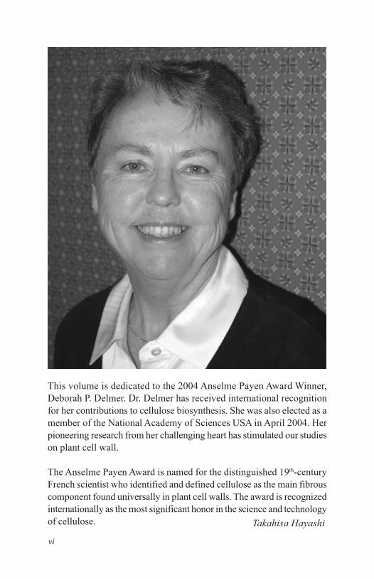
This volume is dedicated to the 2004 Anselme Payen Award Winner,Deborah P. Delmer. Dr. Delmer has received international recognitionfor her contributions to cellulose biosynthesis. She was also elected as amember of the National Academy of Sciences USA in April 2004. Herpioneering research from her challenging heart has stimulated our studieson plant cell wall.
The Anselme Payen Award is named for the distinguished 19th-centuryFrench scientist who identified and defined cellulose as the main fibrouscomponent found universally in plant cell walls. The award is recognizedinternationally as the most significant honor in the science and technologyof cellulose. Takahisa Hayashi
vi
Preface
My thanks go out to Takahisa Hayashi, the Editor of this book, fororganizing such an interesting effort. Taka also asked ME to contributean article, but, as I’ve moved on to a whole new life dealing withissues of agriculture in the developing world, I declined. I did sobecause of my feeling that the field of plant cell walls now has somany talented individuals, it was definitely time for me to step asideand let you all speak for yourselves. But I did agree to write thispreface and reminisce very briefly about my own research with cellwalls and with the topic of cellulose synthesis in particular..
I thought that one interesting way to “reminisce” might be to use myaccumulated experiences in a different way and instead to ask thequestion: “If I were young and starting over again to work on plantcell walls and cellulose synthesis, where would I like to focus myefforts?” Obviously, we tend to focus on projects that suit our ownparticular set of skills, which in my case, are primarily as a biochemistand a Johnny-Come-Lately molecular biologist. But I’ll cheat a littleand just imagine I could learn all the new skills I would need in orderfor me to extend the range of possibilities I see emerging as potentialtopics for research.
Just to touch briefly on the non-cellulosic part of the wall, I am a bithesitant to suggest new directions, knowing that others will have amuch better perspective than I. But I can say that I think that, interms of biosynthesis, the powers of both genomics and moleculargenetics are beginning to reveal themselves as being huge assets. It isclear that, as for cellulose synthesis for so many years, it has provenexceedingly difficult to demonstrate synthesis of complex non-cellulosic polymers using isolated membrane preparations. Onenotable exception has been ability to synthesize galactomannans, butwhen it comes to pectins and other non-cellulosic polysaccharides,the complexity of the systems, the need for suitable primers and/orcooperative interactions among a large number of enzymes, and thelow levels of such enzymes in membranes, has made this taskexceedingly challenging. Clearly the ability to isolate mutants withaltered polysaccharide composition provides one avenue for the
vii

identification of key genes encoding biosynthetic enzymes. Yet,possible redundancy of genes and/or inability to detect alteredphenotypes for whatever reason can still limit this approach. This iswhere genomics can play an important role, and we are finallybeginning to see how searching for important genes based upon theirhomology with other related glycosyltransferases is beginning to payoff for pathways involved in synthesis of polymers such as the pectins,and xyloglucan. And, when it comes to structure, well, I think itwon’t be long before we’ll understand just how all those variousstructures that we lump as together as pectins really do associatewith each other and perhaps also with other non-pectic polysaccharidesand/or proteins. One hopes we will also finally get clarification ofhow expansins and XETs modulate extensibility, and maybe evenone day we’ll really understand that great white whale of plantbiology—how does auxin really control cell elongation?
Discussion of these possibilities also helps me make one veryimportant point—it is no longer possible just to be one kind of scientistas many of us used to be. Each group now needs to have memberswho are either skilled themselves in many fields, or find partnerswho can complement their own skills in order to really make progress.Certainly each group needs to be able to use model systems effectivelybecause they offer ease of manipulation, complete genome sequences,and extensive mutant collections. Yet model systems also havelimitations. I think most of us really do hope that our research willone day lead to greater benefits to agriculture. Thus, we hope that, atsome point, the results of model systems can to be translated toenhancing our understanding and ultimate improvement of crops suchas cotton, trees, and major cereal or legume crops. Model systemsalso may not be the best for hard biochemistry where large amountsof tissue may be required, and they may lack specialized tissues (likethe cotton fiber for cellulose synthesis) where a particular processpredominates at one particular stage of development in one distinctand easily-isolated cell type. In short, we need to be flexible and notwedded just to one approach for our entire careers. Certainly myown career took a turn for the better when I was able to make theleap beyond just biochemistry.
viii

Turning to cellulose synthesis, I have to say that I am REALLY pleasedto see how many new faces have joined the field and the new insightsyou have added to our understanding of this process. Throughgenomics and mutant studies, we now know that there are a numberof CesA genes that indeed can be predicted to catalyze glucan chainelongation in some way, and that some of these distinct CESA proteinsmysteriously seem to be required to work in combination to synthesizemicrofibrils. But why this is true still eludes us. It is clear that geneticscan only suggest that this is true and may also provide indications ofother new partner proteins that may be necessary for the process.But hard work involving good, serious biochemistry will still beimportant to provide a final confirmation of the structure of the rosette,the function of each CESA within the rosette, and the role of otheraccessory proteins. Questions also remain as to what distinguishesprimary from secondary wall cellulose synthesis. Steryl glycosidesseem to function as primers in vitro in membranes of cotton fibersengaged in secondary wall synthesis. Is this some in vitro artefact, isit unique to secondary wall synthesis in vivo, or is it a generalphenomenon for all of cellulose synthesis? Is one CESA responsiblefor elongating primers while others may function in further elongation?What is different about the CESA’s required for primary vs. secondarywall synthesis? Why do rosettes/ CESAs appear to have such shorthalf lives? Only time will tell. Why should cellulases like Korriganbe needed? Perhaps to cleave the primers, to edit mistakes in growingmicrofibrils, or to facilitate chain termination? How do rosettesassemble? What are the domains that associate to form rosettes—e.g., do the zinc finger domains interact in a redox-dependent fashion?(In fact, if there were two topics I’m sure I’d address more seriouslyif I were to continue would be the general role of redox regulationand the role of protein turnover in the process of cell wall assembly.)Are there scaffolding proteins and/or other accessory proteinsembedded within the rosettes? Do microtubules really interact directlywith rosettes to guide their movement as suggested by some recentdata? The answers to some of these questions are beginning and willcontinue to be answered with the additional help of the fantastic newtools for imaging that are improving at a very rapid pace. Will weone day really be able to see clearly the rosette moving within themembrane and show with such imaging techniques exactly what
ix

proteins are within the rosette and how its movement is guided? (SimonTurner’s lab seems to be getting close to doing just that!) And whatabout proteins that may be involved but may not directly interactwith the rosette, such as Korrigan or sucrose synthase? And, well,have we really solved the issue of how synthesis of cellulose andcallose may or may not be connected? And how did these biosyntheticpathways evolve?
Finally, what may be the practical benefits that could arise from suchwork? One prediction is that work with enzymes like sucrose synthasemay provide new insights into how carbon is partitioned in plantcells. Clearly the cell wall is a major sink for carbon, but, unless youare a ruminant, it’s not exactly the world’s best source of calories.And lignin is even less pleasant than cellulose—clearly of benefit tothe plant, but with major impacts on digestibility for both humansand animals, and on the efficiency and safety of processing of planttissues for production of paper, starch, and ethanol. So understandinghow carbon may be partitioned between the digestible and non-digestible polymers may offer opportunities to alter the value of manyplant products. As one who invariably seems to choose in the marketthe toughest old radishes, turnips, rutabagas, and carrots and thestringiest celery and green beans, I’ve also wondered if there may beopportunities to alter cellulose (and lignin) content in such veggies toenhance texture as well as digestibility. With suitable tissue-specificpromoters and the right genes, it surely seems quite possible. Finally,I’ve been most gratified to see that some of our research may alsoprove valuable for the kind of work I’m now doing with the RockefellerFoundation. Lodging of cereals is a problem throughout the world,not only for maize in the fields of commercial farmers, but also forcrops such as rice, millet, sorghum and tef in the developing world.While the green revolution genes have played an important role inhelping solve this problem, it now appears there may be opportunitiesto enhance resistance to lodging by over-expression of CesA genes inspecific tissues.
So I leave this fascinating field just when the opportunities seem thegreatest. But it makes leaving so much easier to see how many talentedyoung (and older!) scientists have taken up the cause. Taka has asked
x
you all to speculate as you write the articles for this book. This issurely fitting since I’ve not ever been shy to speculate throughout mycareer. (How I remember Bruce Stone moaning “Oh Dear! Not anotherDelmer theory!!!”). But it’s a valuable exercise even when we get itwrong, as it does challenge us and our colleagues to extend our wayof thinking. Yet, as valuable as speculation can be, it is no substitutefor good hard facts. And that is what’s so gratifying now—you nolonger have to do SO MUCH speculating, as the answers to many ofthe questions posed above are now well within your reach.Enjoy the journey!
Deborah Delmer
xi
Introduction
Plant cell walls are composed of complex carbohydrates, proteins,phenolic compounds, and inorganic ions, all of which play functionalroles. Several models for plant cell walls help explain their structure,which consists of primary and secondary walls. The primary wall isthe outer wall of a growing plant cell, and elongates and/or expandsover the life of the cell. The secondary wall exists inside the primarywall in elongated and expanded cells. Structural analysis has primarilyemployed microscopy of walls, because “seeing is believing.” Sincecellulose microfibrils are skeletal and architectural components, theirorientation determines the physical properties of plant cell walls. Thefine structure of the wall has been determined by chemical analysis.
Cellulose (1,4-β-glucan) and callose (1,3-β-glucan) are synthesizedin the plasma membrane, while other polysaccharides are synthesizedin the Golgi. Although cellulose is the most abundant biopolymer onthe earth, the details of its biosynthesis have been difficult to elucidate,despite the ongoing work of many scientists for many years. Thesuccess story of cellulose biosynthesis, by V. Bulone, serves as anexcellent plenary chapter in this book.
Plant cell growth occurs with the loosening of the walls, which maybe caused by several enzymatic actions. Many polysaccharidessynthesized in the Golgi are exported to the cell wall, where hydrogenbonding occurs between xyloglucan and cellulose, together withchemical cross-linking between polymers. By measuring wallextensibility, tethers between xyloglucan and cellulose have beenproven to play important roles. Xyloglucan metabolism is undoubtedlyinvolved in the loosening.
Plant development is related to the morphological changes of cellsand tissue, which is caused by structural changes of the walls: thegeneration of walls from grasses to seeds, induction of secondarywall formation, and cotton fiber development. The key action mightbe caused by XTHs and proline-rich wall proteins.
1

A recent genome project has revealed about 248 glycan hydrolasesand 214 glycan transferase genes, including 10 cellulose-synthase(CesA) and 30 cellulose-synthase-like (Csl) genes in Arabidopsisthaliana. Genetic analysis of gene knockout mutants in Arabidopsisthaliana has revealed the function of these genes. Proteomics andglycomics also provide new insights into the studies on plant cellwalls.
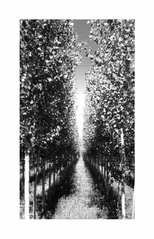
Woody plants make up 80% of total plant biomass, which is thecarbon source for living organisms and also the biological sink ofCO2 on the Earth. Cell walls control plant cell growth and define thestructure of plants, both in guiding the development of plants and inproviding a role in their defense against pathogens. Other benefits ofplant cell walls are their uses as food, various materials, and energyfor human beings.
Takahisa Hayashi
2
Models of Plant Cell Walls
Kei’ichi Baba
The primary cell wall is composed of complex carbohydrates andproteins. Many models have been proposed to understand its structure,beginning with the first complete model by Keegstra et al. (1973),which was based on enzymatic and chemical analyses of the walls ofgrowing sycamore cells (Figure 1). Their primary theory was thatrhamnogalacturonan, arabinogalactan, xyloglucan, andhydroxyproline-rich proteins are interconnected by covalent bonds,and the connection between cellulose and xyloglucan is hydrogenbonds. The walls of growing plant cells were thought to act as amacromolecular complex. Based on this model, numerous othermodels were formed using additional findings (Figures 2-5). Figure2 (Fry 1986) shows the interactions between the molecules in the cellwalls, and Figure 3 shows the matrix polysaccharides arranged nearlyparallel to the cellulose microfibrils.
The models can be divided into two approximate categories. Onetype stresses chemistry and the interactions and bonds betweenmolecules, while the other attempts to express an artistic architecturalconstruction of the molecules. Figure 4 (Roberts 1994) illustratesthe most popular model of the latter type. It appears in many generalbiology textbooks; though the drawing is simple, it is a beautifuldescription of cell wall construction. Since some of thepolysaccharides and the cell wall structure of some monocots aredifferent from those of dicots and other monocots, they were drawnseparately as Types I and II in Figure 5. Most dicots andnoncommelinoid monocots have Type I walls, while commelinoidmonocots have Type II walls.
A completely different model has been proposed for secondary wallsin woody plants (e.g., Esau 1977; Higuchi 1997). Figure 6 (Liese1970) shows a gymnosperm tracheid and the alteration of microfibrilorientation in the layered secondary wall. Later, the fiber wall ofangiosperms was revealed to have the same structure as this model.However, bamboo has cell walls with more layers (Figure 7:Parameswaran and Liese 1981). The secondary wall is known to
Cell Wall Structure
3

Fig. 1 Tentative structure of sycamore cell walls.This model is not intended to be quantitative, but is instead an effort topresent the wall components in approximately correct proportions. Thedistance between cellulose elementary fibrils is expanded to allowpresentation of the interconnecting structure. The circled areas arerepresentative wall fractions released by the degrading enzymes. They arefractions PG-1B and PG-2 released by endopolygalacturonase, fractions C-1 and C-2 released by endoglucanase, and fraction PR-2 released by pronase.The symbols shown at right represent the various components of the cellwall (Keegstra et al. 1973).
consist of cellulose microfibrils, hemicelluloses, and lignin. A model(Figure 8) has been proposed for their localizations, but no details ofthe molecular bonding level have been found. How they interact andconstruct the secondary wall remains unknown.
We can clarify the story by gathering the models together, focusingon each stage of wall material production, and so on. Because oflimited space, only two figures for wall formation are shown here.Cellulose microfibrils are produced on the membrane along the corticalmicrotubules (Figure 9: Gunning and Steer 1996). New cellulose andxyloglucan meet just outside the membrane and immediately bondtightly to each other (Figure 10: Hayashi 1989).
4
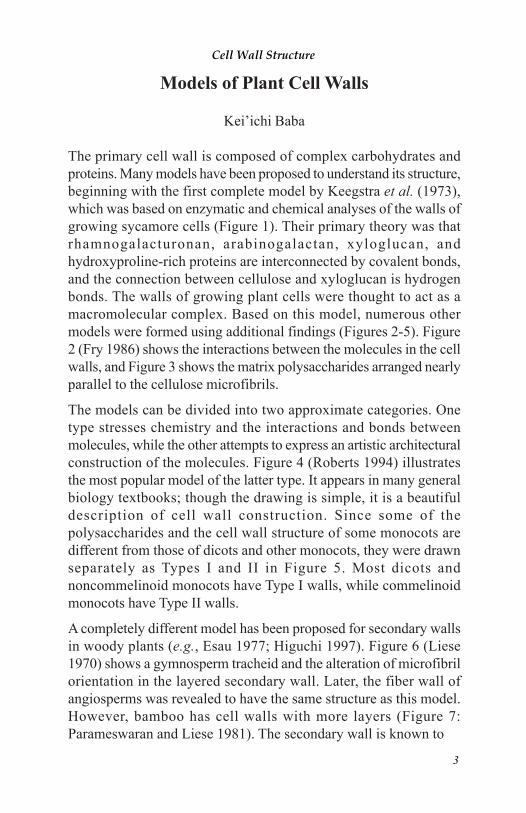
A, arabinose; F, fucose; G, glucose; L, galactose; R, rhamnose; U, galacturonicacid; U^, galacturonic acid methyl ester; a, amino acid other than tyrosine;y, tyrosine; y:y, isodityrosine; φ, φ, φ, φ, φ, ferulic acid; φ:φ,φ:φ,φ:φ,φ:φ,φ:φ, diferulic acid.
Fig. 3 Schematic of suggested polymer organization in peaprimary walls, depicted in the plane of the cellulose microfibrils.
Fig. 2 Representative primary structures and possible cross-linking of wall polymers.This is not a model of the plant cell wall, and no significance is placed on thechain length, orientation, conformation, or spacing of the molecules (Fry1986).(•) hydrogen bonds:
1. cellulose-cellulose2. xyloglucan-cellulose3. xylan-cellulose
( o ) calcium bridges:4. homogalacturonan-
homogalacturonan( + ) other ionic bonds:
5. extensin-pectin( : ) coupled phenols:
6. extensin-extensin
7. pectin-pectin8. arabinoxylan-
arabinoxylan( = ) ester bonds:
9. pectin-cellulose( - ) glycosidic bonds:
10. arabinogalactan-rhamnogalacturonan
( ) entanglement:11. pectin-in-extensin
Cell Wall Structure
5

Fig. 4 Scale model of a portion of primary cell wall showing thetwo major polysaccharide networks.The orthogonally arranged layers of cellulose microfibrils are cross-linkedinto a network by hydrogen-bonded hemicellulose. This network iscoextensive with a network of pectin polysaccharides. The cellulose andhemicellulose network provides tensile strength, while the pectin networkresists compression. Cellulose, hemicellulose, and pectin are typically presentin roughly equal quantities in a primary cell wall. The middle lamella ispectin-rich and cements adjacent cells together (Roberts 1994).
The xyloglucan and arabinogalactan layers form a hemicellulosic sheath(“cortex”) around each microfibril, the non-crystalline portions of whichcontain intercalated xyloglucan chains. Interstices between the hemicellulose-coated microfibrils are occupied by pectin. Where microfibrils approachone another more closely, their hemicellulose sheaths may overlap, and somexyloglucan chains may extend from one microfibril to another. The differentlylabeled polymer types are depicted in the relative portions in which theyoccur in pea cell walls, according to this study and previous reports. Smoothportions of polyuronide backbones donate homogalacturonan, and side chain-bearing portions are rhamnogalacturonan blocks. The longer blocksrepresent lengthy runs of substituted galacturonosyl-rhamnose, such as RG-I. Except for extensin, minor components (e.g., mannan and xylan) are notshown (Talbot and Ray 1992).
6
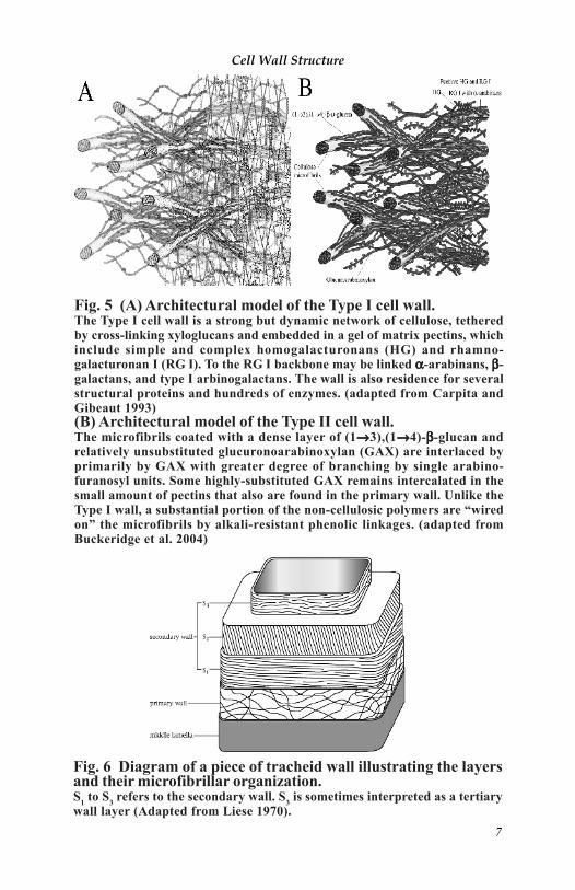
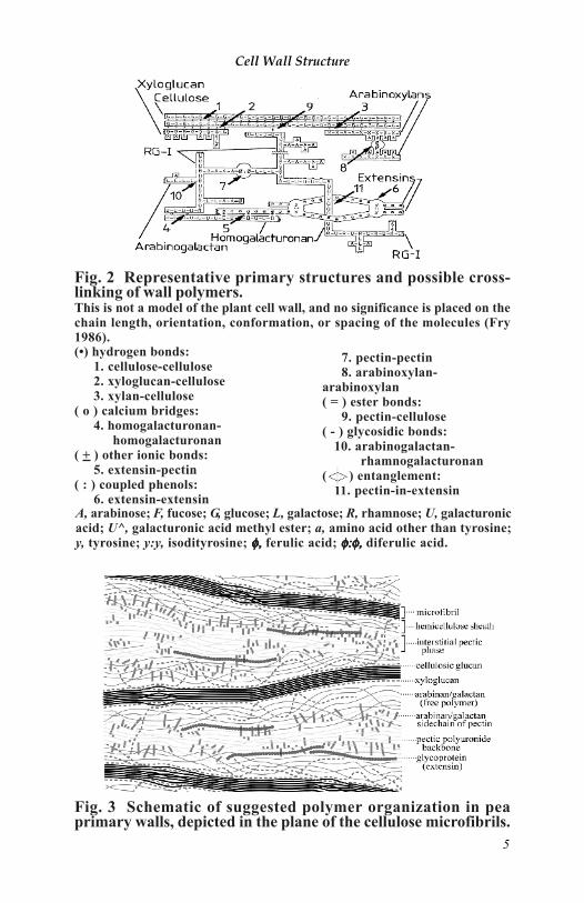
Fig. 6 Diagram of a piece of tracheid wall illustrating the layersand their microfibrillar organization.S1 to S3 refers to the secondary wall. S3 is sometimes interpreted as a tertiarywall layer (Adapted from Liese 1970).
Fig. 5 (A) Architectural model of the Type I cell wall.The Type I cell wall is a strong but dynamic network of cellulose, tetheredby cross-linking xyloglucans and embedded in a gel of matrix pectins, whichinclude simple and complex homogalacturonans (HG) and rhamno-galacturonan I (RG I). To the RG I backbone may be linked ααααα-arabinans, βββββ-galactans, and type I arbinogalactans. The wall is also residence for severalstructural proteins and hundreds of enzymes. (adapted from Carpita andGibeaut 1993)(B) Architectural model of the Type II cell wall.The microfibrils coated with a dense layer of (1→→→→→3),(1→→→→→4)-βββββ-glucan andrelatively unsubstituted glucuronoarabinoxylan (GAX) are interlaced byprimarily by GAX with greater degree of branching by single arabino-furanosyl units. Some highly-substituted GAX remains intercalated in thesmall amount of pectins that also are found in the primary wall. Unlike theType I wall, a substantial portion of the non-cellulosic polymers are “wiredon” the microfibrils by alkali-resistant phenolic linkages. (adapted fromBuckeridge et al. 2004)
Cell Wall Structure
7

Fig. 8 Localization of and the relationships between cellulose,hemicelluloses, and lignin in the S2 layer (Ruel et al. 1978).
Fig. 7 Polylamellar wall structureof a bamboo culm fiber.ML, Middle lamella; P, primary wall;s , secondary wall; l and t denotelongitudinal and transverse orientationso f m i c r o f i b r i l s , r e s p e c t i v e l y(Parameswaran and Liese 1981).
8
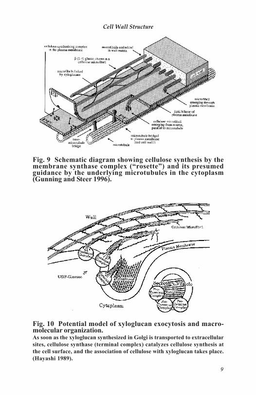
Fig. 10 Potential model of xyloglucan exocytosis and macro-molecular organization.As soon as the xyloglucan synthesized in Golgi is transported to extracellularsites, cellulose synthase (terminal complex) catalyzes cellulose synthesis atthe cell surface, and the association of cellulose with xyloglucan takes place.(Hayashi 1989).
Fig. 9 Schematic diagram showing cellulose synthesis by themembrane synthase complex (“rosette”) and its presumedguidance by the underlying microtubules in the cytoplasm(Gunning and Steer 1996).
Cell Wall Structure
9
AcknowledgementThis work was supported in part by Program for Promotion of Basic Research Activities forInnovation Biosiences (PROBRAIN).
ReferencesBuckeridge MS, Rayon C, Urbanowicz B, Tiné MAS, Carpita NC. (2004) Mixed-linkage
(1→3),(1→4)-β-D-glucans of grasses.Cereal Chem 81: 115-127.Carpita NC, Gibeaut DM (1993) Structural models of primary cell walls in flowering plants:
consistency of molecular structure with the physical properties of the walls during growth.Plant J 3: 1-30.
Esau K (1977) Anatomy of Seed Plants 2nd ed. John Wiley & Sons, New York.Fry SC (1986) Cross-linking of matrix polymers in the growing cell walls of angiosperms.
Annu Rev Plant Physiol 37: 165-186.Gunning BS, Steer MW (1996) Plant Cell Biology: Structure and function. Jones and Bartlett
Publishers, Boston.Hayashi T (1989) Xyloglucan in the primary cell wall. Annu Rev Plant Physiol Mol Biol 40:
139-168.Higuchi T (1997) Biochemistry and molecular biology of wood. Springer, Berlin.Keegstra K,
Talmadge KW, Bauer WD, Albersheim P (1973) The structure of plant cell walls. PlantPhysiol 51: 188-196.
Liese W (1970) Elektronmikroskopie des Holzes. In: Handbuch der Mikroskopie in der Technik.Band V, Teil 1. Umschau, Frankfurt, pp 109-170.
Parameswaran N, Liese W (1981) The fine structure of bamboo. In: T Higuchi (ed) Bambooproduction and utilization. Wood Res Inst, Kyoto Univ, 178-183.
Roberts K (1994) The plant cell wall. In: Molecular Biology of the Cell 3rd ed. Garland, NewYork, pp 1000-1009.
Ruel K, Barnoud F, Goring DAI (1978) Lamellation in S2 layer of softwood tracheid asdemonstrated by scanning transmission electron microscopy. Wood Sci Technology 12: 287-291.
Taiz L, Zeiger E (2002) Plant Physiology 3rd ed. Sinauer, Sunderland.Talbot LD, Ray PM (1992) Molecular size and separability features of pea cell wall
polysaccharides. Plant Physiol 98: 357-368.
10

Imaging the Primary Cell Wall
Tobias I. Baskin
IntroductionAs the son of an artist, I grew up surrounded by drawings, paintings,and sculpture. Perhaps for that reason, making images is central tomy work as a scientist. The problem I study, organ morphogenesis,requires analysis of the cell wall. Fertile approaches stretch out inmany directions (see the rest of this book) but I have been drawn toarchitectural problems, revealing how components are integrated intoa whole. For structural appreciation, imaging is paramount.
For imaging the cell wall, my laboratory has developed or enhancedseveral methods. I will describe some of these here, and discussadvantages and limitations, without attempting to review cell wallimaging comprehensively. The first section is on immunocyto-chemistry at the light-microscope level. The second and third sectionsare on high-resolution imaging based on field-emission scanningelectron microscopy. The first is convenient and allows many samplesto be examined and a relatively large area of tissue to be viewed; thesecond and third are technically more demanding but resolve structureat virtually the level of macromolecules.
Butyl-methyl-methacrylate embedding for immunocytochemistryCell wall researchers will be familiar with the use of antibody probesto localize polysaccharide as well as protein epitopes in the cell wall(Knox 1997), but are probably less familiar with the use of butyl-methyl-methacrylate as an embedding matrix. I first encountered thisresin while attempting to localize microtubules (Baskin et al. 1992).This methacrylate is easy to section, dry or wet; however, its primaryadvantage for immunocytochemistry is that following sectioning, themajority of the embedding matrix can be removed with a briefincubation in acetone, in contrast to nearly all other plasticembedments, which can be removed partially if at all only with harshtreatments. By removing the embedment, access to the antigen forthe antibody is enlarged. Removability is shared by paraffin and waxbut these preserve most samples poorly compared to plastic. At theelectron-microscope level, adequate access for antibody to antigen is
Cell Wall Structure
11
provided by various plastics that are sufficiently porous to allowantibodies to penetrate 10 nm or so into the section, thus sampling anappreciable proportion of the volume of an ultra-thin section (60 nmthickness). However, for the semi-thin sections (1 to 2 µm thick)typically used in light microscopy, a 10 nm penetration depth amountsto a negligible proportion of the section volume, and the greaterpenetration gained by removing the embedment becomes a significantadvantage.
Butyl-methyl-methacrylate, like most methacrylates, polymerizes viaa free radical-based mechanism. This is useful because it means thatpolymerization can be catalyzed by ultraviolet light, thus avoidingdenaturation caused by high-temperature polymerization. However,early efforts to use this methacrylate mix were frustrated by oxidativedamage to the sample that lowered antigenicity, damage presumablymediated by free radicals. I found that adding the free-radicalscavenger dithiothreitol to the resin allowed polymerization butblocked the attack on the sample (Baskin et al. 1992). Subsequently,this resin has been used to localize tubulin and other antigens in avariety of samples (e.g., Herman et al. 1994; Stadler et al. 1995;Hoffman et al. 1998; Palmer et al. 2001).
In general, embedding in butyl-methyl-methacrylate is straightforward(Baskin and Wilson, 1997). However, the small size of arabidopsisroots (ca 0.15 mm diameter) makes them easy to lose while changingsolutions. To retain the roots, I use a method that is not only convenientbut also turns out to be beneficial for sample preservation. Originally,I encased each root in a small droplet of low-gelling-temperatureagarose (Baskin et al. 1992), but this is messy and exposes the sampleto heat, albeit briefly. Then, I modified a method from cryofixationwhere samples are mounted on a Formvar film (Baskin et al. 1996).A chemically fixed root tip is placed on a Formvar-coated wire loop,a second Formvar film secures the root tip on the loop. The Formvarfilms are readily permeated by solvents and small molecules. BetweenFormvar films, the thin arabidopsis root tip is prevented from bendingor twisting. I call this “mechanical fixation” and beyond beingconvenient, it seems to enhance sample preservation.
Loops are made in advance and coated by casting Formvar rectangles(measuring a little more than the loop diameter on one side and a
12
little more than twice the loop diameter on the other) and plungingthe loop into the water over the rectangle so that the plane of the loopbisects the long axis of the rectangle. The Formvar rectangle wrapsaround the wire loop and the coated loop is removed at once from thewater. Such loops remain stable for months. To secure a sample, theprocedure is repeated: After the sample has been fixed and rinsed, aloop (already Formvar coated) is placed horizontally on a drop ofwater (or buffer) and the sample placed on the Formvar. Excess sampleis trimmed if needed, and the loop (with sample) is plunged onto anew Formvar rectangle, thus encasing the sample between Formvarlayers, held by the loop. Several loops can be placed in a vial andsolutions exchanged without losing the sample. The loop is embeddedwith the sample, and removed during trimming. I use fine copperwire (36 gauge), which can be trimmed along with the block.
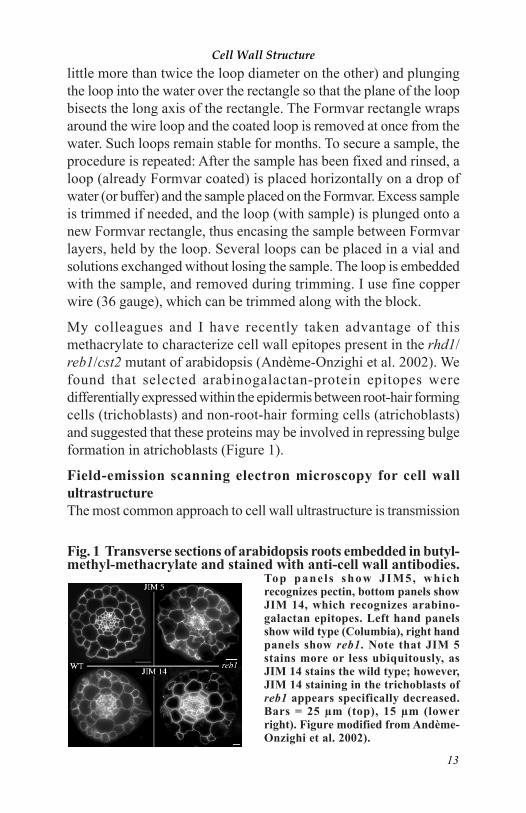
My colleagues and I have recently taken advantage of thismethacrylate to characterize cell wall epitopes present in the rhd1/reb1/cst2 mutant of arabidopsis (Andème-Onzighi et al. 2002). Wefound that selected arabinogalactan-protein epitopes weredifferentially expressed within the epidermis between root-hair formingcells (trichoblasts) and non-root-hair forming cells (atrichoblasts)and suggested that these proteins may be involved in repressing bulgeformation in atrichoblasts (Figure 1).
Field-emission scanning electron microscopy for cell wallultrastructureThe most common approach to cell wall ultrastructure is transmission
Fig. 1 Transverse sections of arabidopsis roots embedded in butyl-methyl-methacrylate and stained with anti-cell wall antibodies.
Top panels show JIM5, whichrecognizes pectin, bottom panels showJIM 14, which recognizes arabino-galactan epitopes. Left hand panelsshow wild type (Columbia), right handpanels show reb1. Note that JIM 5stains more or less ubiquitously, asJIM 14 stains the wild type; however,JIM 14 staining in the trichoblasts ofreb1 appears specifically decreased.Bars = 25 µm (top), 15 µm (lowerright). Figure modified from Andème-Onzighi et al. 2002).
Cell Wall Structure
13