the roles of vitamin c in skin health - mindbodygreen
TRANSCRIPT

nutrients
Review
The Roles of Vitamin C in Skin Health
Juliet M. Pullar, Anitra C. Carr and Margreet C. M. Vissers *
Department of Pathology, University of Otago, Christchurch, P.O. Box 4345, Christchurch 8140, New Zealand;[email protected] (J.M.P.); [email protected] (A.C.C.)* Correspondence: [email protected]; Tel.: +64-3364-1524
Received: 10 July 2017; Accepted: 9 August 2017; Published: 12 August 2017
Abstract: The primary function of the skin is to act as a barrier against insults from the environment,and its unique structure reflects this. The skin is composed of two layers: the epidermal outer layer ishighly cellular and provides the barrier function, and the inner dermal layer ensures strength andelasticity and gives nutritional support to the epidermis. Normal skin contains high concentrations ofvitamin C, which supports important and well-known functions, stimulating collagen synthesis andassisting in antioxidant protection against UV-induced photodamage. This knowledge is often usedas a rationale for the addition of vitamin C to topical applications, but the efficacy of such treatment,as opposed to optimising dietary vitamin C intake, is poorly understood. This review discussesthe potential roles for vitamin C in skin health and summarises the in vitro and in vivo research todate. We compare the efficacy of nutritional intake of vitamin C versus topical application, identifythe areas where lack of evidence limits our understanding of the potential benefits of vitamin C onskin health, and suggest which skin properties are most likely to benefit from improved nutritionalvitamin C intake.
Keywords: ascorbate; dermis; epidermis; skin barrier function; vitamin C status; skin aging; woundhealing; collagen; UV protection
1. Introduction
The skin is a multi-functional organ, the largest in the body, and its appearance generally reflectsthe health and efficacy of its underlying structures. It has many functions, but its fundamental roleis to provide a protective interface between the external environment and an individual’s tissues,providing shielding from mechanical and chemical threats, pathogens, ultraviolet radiation and evendehydration (functions reviewed in [1]). Being in constant contact with the external environment,the skin is subject to more insults than most of our other organs, and is where the first visible signs ofaging occur.
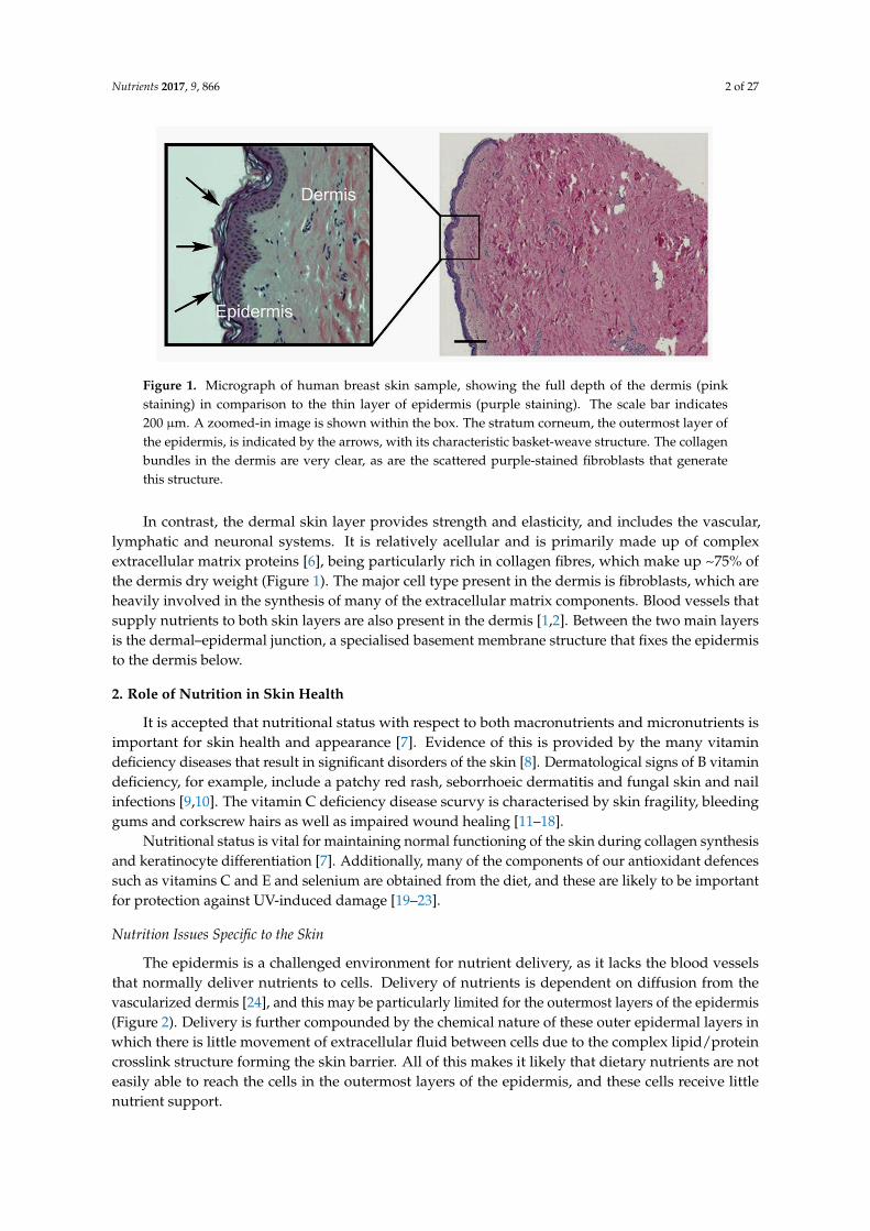
The skin is composed of two main layers with quite different underlying structures—theoutermost epidermis and the deeper dermis (Figure 1). The epidermis fulfils most of the barrierfunctions of the skin and is predominantly made up of cells, mostly keratinocytes [2]. The keratinocytesare arranged in layers throughout the epidermis; as these cells divide and proliferate away fromthe basal layer, which is closest to the dermis, they begin to differentiate. This process is calledkeratinization, and involves the production of specialized structural proteins, secretion of lipids, andthe formation of a cellular envelope of cross-linked proteins. During differentiation, virtually all ofthe subcellular organelles disappear, including the nucleus [3,4]. The cytoplasm is also removed,although there is evidence that some enzymes remain [4]. Thus, the uppermost layer of the epidermisthat interacts with the outside environment is composed of flattened metabolically ‘dead’ cells (theterminally differentiated keratinocytes). These cells are sealed together with lipid-rich domains,forming a water-impermeable barrier. This layer is known as the stratum corneum (Figure 1) and fulfilsthe primary barrier function of the epidermis, although the lower epidermal layers also contribute [5].
Nutrients 2017, 9, 866; doi:10.3390/nu9080866 www.mdpi.com/journal/nutrients
Nutrients 2017, 9, 866 2 of 27Nutrients 2017, 9, 866 2 of 27
Figure 1. Micrograph of human breast skin sample, showing the full depth of the dermis (pink staining) in comparison to the thin layer of epidermis (purple staining). The scale bar indicates 200 µm. A zoomed-in image is shown within the box. The stratum corneum, the outermost layer of the epidermis, is indicated by the arrows, with its characteristic basket-weave structure. The collagen bundles in the dermis are very clear, as are the scattered purple-stained fibroblasts that generate this structure.
In contrast, the dermal skin layer provides strength and elasticity, and includes the vascular, lymphatic and neuronal systems. It is relatively acellular and is primarily made up of complex extracellular matrix proteins [6], being particularly rich in collagen fibres, which make up ~75% of the dermis dry weight (Figure 1). The major cell type present in the dermis is fibroblasts, which are heavily involved in the synthesis of many of the extracellular matrix components. Blood vessels that supply nutrients to both skin layers are also present in the dermis [1,2]. Between the two main layers is the dermal–epidermal junction, a specialised basement membrane structure that fixes the epidermis to the dermis below.
2. Role of Nutrition in Skin Health
It is accepted that nutritional status with respect to both macronutrients and micronutrients is important for skin health and appearance [7]. Evidence of this is provided by the many vitamin deficiency diseases that result in significant disorders of the skin [8]. Dermatological signs of B vitamin deficiency, for example, include a patchy red rash, seborrhoeic dermatitis and fungal skin and nail infections [9,10]. The vitamin C deficiency disease scurvy is characterised by skin fragility, bleeding gums and corkscrew hairs as well as impaired wound healing [11–18].
Nutritional status is vital for maintaining normal functioning of the skin during collagen synthesis and keratinocyte differentiation [7]. Additionally, many of the components of our antioxidant defences such as vitamins C and E and selenium are obtained from the diet, and these are likely to be important for protection against UV-induced damage [19–23].
2.1. Nutrition Issues Specific to the Skin
The epidermis is a challenged environment for nutrient delivery, as it lacks the blood vessels that normally deliver nutrients to cells. Delivery of nutrients is dependent on diffusion from the vascularized dermis [24], and this may be particularly limited for the outermost layers of the epidermis (Figure 2). Delivery is further compounded by the chemical nature of these outer epidermal layers in which there is little movement of extracellular fluid between cells due to the complex lipid/protein crosslink structure forming the skin barrier. All of this makes it likely that dietary nutrients are not easily able to reach the cells in the outermost layers of the epidermis, and these cells receive little nutrient support.
Figure 1. Micrograph of human breast skin sample, showing the full depth of the dermis (pinkstaining) in comparison to the thin layer of epidermis (purple staining). The scale bar indicates200 µm. A zoomed-in image is shown within the box. The stratum corneum, the outermost layer ofthe epidermis, is indicated by the arrows, with its characteristic basket-weave structure. The collagenbundles in the dermis are very clear, as are the scattered purple-stained fibroblasts that generatethis structure.
In contrast, the dermal skin layer provides strength and elasticity, and includes the vascular,lymphatic and neuronal systems. It is relatively acellular and is primarily made up of complexextracellular matrix proteins [6], being particularly rich in collagen fibres, which make up ~75% ofthe dermis dry weight (Figure 1). The major cell type present in the dermis is fibroblasts, which areheavily involved in the synthesis of many of the extracellular matrix components. Blood vessels thatsupply nutrients to both skin layers are also present in the dermis [1,2]. Between the two main layersis the dermal–epidermal junction, a specialised basement membrane structure that fixes the epidermisto the dermis below.
2. Role of Nutrition in Skin Health
It is accepted that nutritional status with respect to both macronutrients and micronutrients isimportant for skin health and appearance [7]. Evidence of this is provided by the many vitamindeficiency diseases that result in significant disorders of the skin [8]. Dermatological signs of B vitamindeficiency, for example, include a patchy red rash, seborrhoeic dermatitis and fungal skin and nailinfections [9,10]. The vitamin C deficiency disease scurvy is characterised by skin fragility, bleedinggums and corkscrew hairs as well as impaired wound healing [11–18].
Nutritional status is vital for maintaining normal functioning of the skin during collagen synthesisand keratinocyte differentiation [7]. Additionally, many of the components of our antioxidant defencessuch as vitamins C and E and selenium are obtained from the diet, and these are likely to be importantfor protection against UV-induced damage [19–23].
Nutrition Issues Specific to the Skin
The epidermis is a challenged environment for nutrient delivery, as it lacks the blood vesselsthat normally deliver nutrients to cells. Delivery of nutrients is dependent on diffusion from thevascularized dermis [24], and this may be particularly limited for the outermost layers of the epidermis(Figure 2). Delivery is further compounded by the chemical nature of these outer epidermal layers inwhich there is little movement of extracellular fluid between cells due to the complex lipid/proteincrosslink structure forming the skin barrier. All of this makes it likely that dietary nutrients are noteasily able to reach the cells in the outermost layers of the epidermis, and these cells receive littlenutrient support.

Nutrients 2017, 9, 866 3 of 27Nutrients 2017, 9, 866 3 of 27
Figure 2. Delivery of nutrients to the skin. The location of the vitamin C transport proteins SVCT1 and SVCT2 are indicated. Red arrows depict nutrient flow from the blood vessels in the dermis to the epidermal layer. Nutrients delivered by topical application would need to penetrate the barrier formed by the stratum corneum.
The skin can be targeted for nutrient delivery through topical application (Figure 2). However, in this case the delivery vehicle is influential, as the stratum corneum functions as an effective aqueous barrier and prevents the passage of many substances [1]. Although some uncharged and lipid-soluble molecules can pass through the surface layer, it is unlikely that nutrients delivered via topical application would easily penetrate into the lower layers of the dermis [22]. The dermal layer functions are therefore best supported by nutrients delivered through the bloodstream.
3. Vitamin C Content of Skin
Normal skin contains high concentrations of vitamin C, with levels comparable to other body tissues and well above plasma concentrations, suggesting active accumulation from the circulation. Most of the vitamin C in the skin appears to be in intracellular compartments, with concentrations likely to be in the millimolar range [25–27]. It is transported into cells from the blood vessels present in the dermal layer. Skin vitamin C levels have not often been reported and there is considerable variation in the published levels, with a 10-fold range across a number of independent studies (Table 1). Levels are similar to that found in numerous other body organs. The variation in reported levels most likely reflects the difficulty in handling skin tissue, which is very resilient to degradation and solubilisation, but may also be due to the location of the skin sample and the age of the donor.
Table 1. Vitamin C content of human skin and a comparison with other tissues.
Tissue Vitamin C Content (mg/100 g Wet Weight) References Adrenal glands 30–40 [28] Pituitary glands 40–50 [29]
Liver 10–16 [28,30] Spleen 10–15 [28,31] Lungs 7 [28]
Kidneys 5–15 [30] Heart muscle 5–15 [28,29,31]
Skeletal muscle 3–4 [29,32] Brain 13–15 [28]
Skin-epidermis 6–64 [25–27] Skin-dermis 3–13 [25–27]
Figure 2. Delivery of nutrients to the skin. The location of the vitamin C transport proteins SVCT1and SVCT2 are indicated. Red arrows depict nutrient flow from the blood vessels in the dermis to theepidermal layer. Nutrients delivered by topical application would need to penetrate the barrier formedby the stratum corneum.
The skin can be targeted for nutrient delivery through topical application (Figure 2). However,in this case the delivery vehicle is influential, as the stratum corneum functions as an effectiveaqueous barrier and prevents the passage of many substances [1]. Although some uncharged andlipid-soluble molecules can pass through the surface layer, it is unlikely that nutrients delivered viatopical application would easily penetrate into the lower layers of the dermis [22]. The dermal layerfunctions are therefore best supported by nutrients delivered through the bloodstream.
3. Vitamin C Content of Skin
Normal skin contains high concentrations of vitamin C, with levels comparable to other bodytissues and well above plasma concentrations, suggesting active accumulation from the circulation.Most of the vitamin C in the skin appears to be in intracellular compartments, with concentrationslikely to be in the millimolar range [25–27]. It is transported into cells from the blood vessels present inthe dermal layer. Skin vitamin C levels have not often been reported and there is considerable variationin the published levels, with a 10-fold range across a number of independent studies (Table 1). Levelsare similar to that found in numerous other body organs. The variation in reported levels most likelyreflects the difficulty in handling skin tissue, which is very resilient to degradation and solubilisation,but may also be due to the location of the skin sample and the age of the donor.
Table 1. Vitamin C content of human skin and a comparison with other tissues.
Tissue Vitamin C Content (mg/100 g Wet Weight) References
Adrenal glands 30–40 [28]Pituitary glands 40–50 [29]
Liver 10–16 [28,30]Spleen 10–15 [28,31]Lungs 7 [28]
Kidneys 5–15 [30]Heart muscle 5–15 [28,29,31]
Skeletal muscle 3–4 [29,32]Brain 13–15 [28]
Skin-epidermis 6–64 [25–27]Skin-dermis 3–13 [25–27]

Nutrients 2017, 9, 866 4 of 27
Several reports have indicated that vitamin C levels are lower in aged or photodamagedskin [25–27]. Whether this association reflects cause or effect is unknown, but it has also beenreported that excessive exposure to oxidant stress via pollutants or UV irradiation is associatedwith depleted vitamin C levels in the epidermal layer [33,34]. Indeed, more vitamin C is found inthe epidermal layer than in the dermis, with differences of 2–5-fold between the two layers beingconsistently reported (Table 1 and [25,26]). Levels of vitamin C in skin are similar to the levels of otherwater soluble antioxidants such as glutathione [25–27,35]. There is a suggestion that vitamin C in thestratum corneum layer of the epidermis exists in a concentration gradient [36]. The lowest vitamin Cconcentration was present at the outer surface of the epidermis of the SKH-1 hairless mouse, a modelof human skin, with a sharp increase in concentration in the deeper layers of the stratum corneum,possibly reflecting depletion in the outer cells due to chronic exposure to the environment [36].
3.1. The Bioavailability and Uptake of Vitamin C into the Skin
3.1.1. The Sodium-Dependent Vitamin C Transporters
Vitamin C uptake from the plasma and transport across the skin layers is mediated by specificsodium-dependent vitamin C transporters (SVCTs) that are present throughout the body and are alsoresponsible for transport into other tissues. Interestingly, cells in the epidermis express both typesof vitamin C transporter, SVCT1 and SVCT2 (Figure 2) [37]. This contrasts with most other tissues,which express SVCT2 only [37–39]. SVCT1 expression in the body is largely confined to the epithelialcells in the small intestine and the kidney and is associated with active inter-cellular transport of thevitamin [40,41]. The specific localisation of SVCT1 in the epidermis is of interest due to the lack ofvasculature in this tissue, and suggests that the combined expression of both transporters 1 and 2ensures effective uptake and intracellular accumulation of the vitamin. Together with the high levelsof vitamin C measured in the epidermal layer, the dual expression of the SVCTs suggests a highdependency on vitamin C in this tissue.
Both transporters are hydrophobic membrane proteins that co-transport sodium, driving theuptake of vitamin C into cells. Replacement of sodium with other positively charged ions completelyabolishes transport [42]. SVCT1 and SVCT2 have quite different uptake kinetics reflecting theirdifferent physiological functions. SVCT1 transports vitamin C with a low affinity but with a highcapacity (Km of 65–237 µmol/L) mediating uptake of vitamin C from the diet and re-uptake in thetubule cells in the kidney [41]. SVCT2, which is present in almost every cell in the body, is thoughtto be a high-affinity, low capacity transporter, with a Km of ~20 µM meaning it can function at lowconcentrations of vitamin C [41]. As well as transporter affinity, vitamin C transport is regulated bythe availability of the SVCT proteins on the plasma membrane.
3.1.2. Bioavailability and Uptake
Most tissues of the body respond to plasma availability of vitamin C and concentrations varyaccordingly, with lower tissue levels being reported when plasma levels are below saturation [43–47].The kinetics of uptake varies between tissues, with vitamin C levels in some organs (e.g., the brain)reaching a plateau at lower plasma vitamin C status, whereas other tissue levels (e.g., skeletal muscle)continue to increase in close association with increasing plasma supply [32,44,45,48].
Very little is known about vitamin C accumulation in the skin and there are no studies that haveinvestigated the relationship between skin vitamin C content and nutrient intake or plasma supply.Two human studies have shown an increase in skin vitamin C content following supplementation withvitamin C, but neither contained adequate measures of plasma vitamin C levels in the participantsbefore or after supplementation [27,49]. In one other study, vitamin C content was measured inbuccal keratinocytes, as these cells are proposed to be a good model for skin keratinocytes [50].The keratinocyte vitamin C concentration doubled upon supplementation of the participants with

Nutrients 2017, 9, 866 5 of 27
3 g/day vitamin C for six weeks, a dosage that is significantly higher than the recommended dailyintake and would achieve plasma saturation and likely also tissue saturation [44].
Thus it appears likely that, as with many other tissues, skin vitamin C levels respond to increasesin plasma supply [27,50]. A paper by Nusgens and co-workers suggests that skin levels do not increasefurther once plasma saturation is reached [51]. Dietary supplementation is therefore only expected tobe effective in elevating skin vitamin C in individuals who have below-saturation plasma levels priorto intervention.
3.1.3. Topical Application of Vitamin C
When plasma levels are low, some vitamin C can be delivered to the epidermal layer by topicalapplication, although the efficacy of this is dependent on the formulation of the cream or serumused on the skin [51–55]. Vitamin C, as a water-soluble and charged molecule, is repelled by thephysical barrier of the terminally differentiated epidermal cells. It is only when pH levels are below4 and vitamin C is present as ascorbic acid that some penetration occurs [56], but whether this resultsin increased levels in the metabolically compromised stratum corneum is unknown. A great dealof effort has been put into the development of ascorbic acid derivatives for the purpose of topicalapplication. Such derivatives need to ensure stabilization of the molecule from oxidation and alsoovercome the significant challenge of skin penetration. In addition, they must be converted to ascorbicacid in vivo in order to be effective. Whether there is a single solution to all these challenges isunclear [57]. The addition of a phosphate group confers greater stability and these derivatives may beconverted to ascorbic acid in vivo, albeit at a slow rate [58], but they are poorly absorbed through theskin [56,59,60]. Ascorbyl glucoside also exhibits superior stability and can penetrate, but the rate ofits in vivo conversion is not known [57,61–63]. Derivatives containing lipid-soluble moieties such aspalmitate are designed to assist with delivery, and although increased uptake has been demonstratedin animals [64], they do not necessarily show improved stability and there is some doubt as to whetherthese derivatives are efficiently converted in vivo [57]. Recent studies suggest that encapsulation intoa lipospheric form may assist with transport into the lower layers of the epidermis and could resultin increased uptake [65–67]. However, the most pertinent issue for the efficacy of topical applicationis likely to be the plasma status of the individual: if plasma levels are saturated, then it appears thattopical application does not increase skin vitamin C content [51].
3.1.4. Vitamin C Deficiency
One of the most compelling arguments for a vital role for vitamin C in skin health is the associationbetween vitamin C deficiency and the loss of a number of important skin functions. In particular,poor wound healing (associated with collagen formation), thickening of the stratum corneum andsubcutaneous bleeding (due to fragility and loss of connective tissue morphology) are extreme andrapid in onset in vitamin-C-deficient individuals [11,15–18]. It is thought that similar processes occurwhen body stores are below optimal, although to a lesser extent [46,68].
4. Potential Functions of Vitamin C in the Skin
The high concentration of vitamin C in the skin indicates that it has a number of importantbiological functions that are relevant to skin health. Based on what we know about vitamin C function,attention has been focused on collagen formation and antioxidant protection; however, evidence isemerging for other activities.
4.1. The Promotion of Collagen Formation
Vitamin C acts as a co-factor for the proline and lysine hydroxylases that stabilise the collagenmolecule tertiary structure, and it also promotes collagen gene expression [69–77]. In the skin, collagenformation is carried out mostly by the fibroblasts in the dermis, resulting in the generation of thebasement membrane and dermal collagen matrix (Figure 3) [75,78]. The dependence of the collagen

Nutrients 2017, 9, 866 6 of 27
hydroxylase enzymes on vitamin C has been demonstrated in a number of studies with fibroblastcells in vitro [69,73,79], with both decreased total synthesis and decreased crosslinking when vitaminC is absent [80–82]. The activity of the hydroxylases is much more difficult to measure in vivo,as the amount of collagen synthesised may vary only a little [51,52]. Rather, animal studies withthe vitamin-C-deficient GULO mouse indicate that the stability of the synthesised collagen varieswith vitamin C availability, reflecting the stabilising function of the collagen crosslinks formed by thehydroxylases [76]. In addition to stabilising the collagen molecule by hydroxylation, vitamin C alsostimulates collagen mRNA production by fibroblasts [78,83].
Nutrients 2017, 9, 866 6 of 27
in vivo, as the amount of collagen synthesised may vary only a little [51,52]. Rather, animal studies with the vitamin-C-deficient GULO mouse indicate that the stability of the synthesised collagen varies with vitamin C availability, reflecting the stabilising function of the collagen crosslinks formed by the hydroxylases [76]. In addition to stabilising the collagen molecule by hydroxylation, vitamin C also stimulates collagen mRNA production by fibroblasts [78,83].
Figure 3. Structure of the dermis. Higher magnification of H&E-stained dermis, showing the irregular nature of the bundled collagen fibres (pink stained) and sparse presence of the fibroblasts (blue nuclear staining). Vitamin C present in the fibroblasts supports the synthesis of the collagen fibres.
4.2. The Ability to Scavenge Free Radicals and Dispose of Toxic Oxidants
Vitamin C is a potent antioxidant that can neutralise and remove oxidants, such as those found in environmental pollutants and after exposure to ultraviolet radiation. This activity appears to be of particular importance in the epidermis, where vitamin C is concentrated in the skin. However, vitamin C is only one player in the antioxidant arsenal that includes enzymatic defences (catalase, glutathione peroxidase and superoxide dismutase) as well as other non-enzymatic defences (vitamin E, glutathione, uric acid and other putative antioxidants such as carotenoids) [19,21,33,34,84–88]. Most intervention studies carried out to determine the capacity of antioxidants to prevent oxidative damage to skin have used a cocktail of these compounds [21,88–90]. Vitamin C is particularly effective at reducing oxidative damage to the skin when it is used in conjunction with vitamin E [21,54,89,91,92]. This is in accord with its known function as a regenerator of oxidised vitamin E, thereby effectively recycling this important lipid-soluble radical scavenger and limiting oxidative damage to cell membrane structures [92,93] (Figure 4).
Figure 3. Structure of the dermis. Higher magnification of H&E-stained dermis, showing the irregularnature of the bundled collagen fibres (pink stained) and sparse presence of the fibroblasts (blue nuclearstaining). Vitamin C present in the fibroblasts supports the synthesis of the collagen fibres.
4.2. The Ability to Scavenge Free Radicals and Dispose of Toxic Oxidants
Vitamin C is a potent antioxidant that can neutralise and remove oxidants, such as those found inenvironmental pollutants and after exposure to ultraviolet radiation. This activity appears to be ofparticular importance in the epidermis, where vitamin C is concentrated in the skin. However, vitaminC is only one player in the antioxidant arsenal that includes enzymatic defences (catalase, glutathioneperoxidase and superoxide dismutase) as well as other non-enzymatic defences (vitamin E, glutathione,uric acid and other putative antioxidants such as carotenoids) [19,21,33,34,84–88]. Most interventionstudies carried out to determine the capacity of antioxidants to prevent oxidative damage to skin haveused a cocktail of these compounds [21,88–90]. Vitamin C is particularly effective at reducing oxidativedamage to the skin when it is used in conjunction with vitamin E [21,54,89,91,92]. This is in accord withits known function as a regenerator of oxidised vitamin E, thereby effectively recycling this importantlipid-soluble radical scavenger and limiting oxidative damage to cell membrane structures [92,93](Figure 4).

Nutrients 2017, 9, 866 7 of 27Nutrients 2017, 9, 866 7 of 27
Figure 4. The central role for vitamin C and other antioxidants pertinent to the skin. The interdependence of vitamins E and C, and glutathione, in the scavenging of free radicals and regeneration of the reduced antioxidants, is shown. Vitamin E is in the lipid fraction of the cell, whereas vitamin C and glutathione are water-soluble and present in the cytosol.
4.3. Inhibition of Melanogenesis
Vitamin C derivatives, including the magnesium phosophate ascorbyl derivative, have been shown to decrease melanin synthesis both in cultured melanocytes and in vivo [94,95]. This activity has been proposed to be due to its ability to interfere with the action of tyrosinase, the rate-limiting enzyme in melanogenesis. Tyrosinase catalyses the hydroxylation of tyrosine to dihydroxyphenylalanine (DOPA), and the oxidation of DOPA to its corresponding ortho-quinone. The inhibition in melanin production by vitamin C is thought to be due to the vitamin’s ability to reduce the ortho-quinones generated by tyrosinase [94], although other mechanisms are also possible [96]. Agents that decrease melanogenesis are used to treat skin hyperpigmentation in conditions such as melisma or age spots.
4.4. Interaction with Cell Signalling Pathways
In vitro studies clearly show that vitamin C can play a role in the differentiation of keratinocytes (Table 2). For example, vitamin C enhanced the differentiation of rat epidermal keratinocytes cells in an organotypic culture model [97], with markedly improved ultrastructural organisation of the stratum corneum, accompanied by enhanced barrier function. Vitamin C also increased numbers of keratohyalin granules and levels of the late differentiation marker filaggrin, which appeared to be due to altered gene expression [97]. Others have also shown that vitamin C promotes synthesis and organization of barrier lipids and increased cornified envelope formation during differentiation [98–102]. The mechanism(s) by which vitamin C modulates keratinocyte differentiation is not yet elucidated; however, it has been hypothesized to be under the control of protein kinase C and AP-1 [99].
In addition to vitamin C’s ability to promote collagen synthesis [73,79], there is evidence to suggest that vitamin C increases proliferation and migration of dermal fibroblasts [78,82,102], functions vital for effective wound healing, although the underlying mechanisms driving this activity are not yet known [78]. Through the stimulation of regulatory hydroxylases, vitamin C also regulates the stabilization and activation of the hypoxia-inducible factor (HIF)-1, a metabolic sensor that controls the expression of hundreds of genes involved with cell survival and tissue remodelling, including collagenases [103–105]. Vitamin C has been shown to both stimulate [69] and inhibit elastin synthesis in cultured fibroblasts [81]. Glycosaminoglycan synthesis as part of extracellular matrix formation is also increased by vitamin C treatment [106], and it may also influence gene expression of antioxidant enzymes, including those involved in DNA repair [78]. As such, vitamin C has been shown to increase the repair of oxidatively damaged bases. [78]. The modulation of gene expression
Figure 4. The central role for vitamin C and other antioxidants pertinent to the skin. The interdependenceof vitamins E and C, and glutathione, in the scavenging of free radicals and regeneration of the reducedantioxidants, is shown. Vitamin E is in the lipid fraction of the cell, whereas vitamin C and glutathioneare water-soluble and present in the cytosol.
4.3. Inhibition of Melanogenesis
Vitamin C derivatives, including the magnesium phosophate ascorbyl derivative, have beenshown to decrease melanin synthesis both in cultured melanocytes and in vivo [94,95]. This activity hasbeen proposed to be due to its ability to interfere with the action of tyrosinase, the rate-limiting enzymein melanogenesis. Tyrosinase catalyses the hydroxylation of tyrosine to dihydroxyphenylalanine(DOPA), and the oxidation of DOPA to its corresponding ortho-quinone. The inhibition in melaninproduction by vitamin C is thought to be due to the vitamin’s ability to reduce the ortho-quinonesgenerated by tyrosinase [94], although other mechanisms are also possible [96]. Agents that decreasemelanogenesis are used to treat skin hyperpigmentation in conditions such as melisma or age spots.
4.4. Interaction with Cell Signalling Pathways
In vitro studies clearly show that vitamin C can play a role in the differentiation of keratinocytes(Table 2). For example, vitamin C enhanced the differentiation of rat epidermal keratinocytes cellsin an organotypic culture model [97], with markedly improved ultrastructural organisation of thestratum corneum, accompanied by enhanced barrier function. Vitamin C also increased numbersof keratohyalin granules and levels of the late differentiation marker filaggrin, which appearedto be due to altered gene expression [97]. Others have also shown that vitamin C promotessynthesis and organization of barrier lipids and increased cornified envelope formation duringdifferentiation [98–102]. The mechanism(s) by which vitamin C modulates keratinocyte differentiationis not yet elucidated; however, it has been hypothesized to be under the control of protein kinase Cand AP-1 [99].
In addition to vitamin C’s ability to promote collagen synthesis [73,79], there is evidence tosuggest that vitamin C increases proliferation and migration of dermal fibroblasts [78,82,102], functionsvital for effective wound healing, although the underlying mechanisms driving this activity are notyet known [78]. Through the stimulation of regulatory hydroxylases, vitamin C also regulates thestabilization and activation of the hypoxia-inducible factor (HIF)-1, a metabolic sensor that controlsthe expression of hundreds of genes involved with cell survival and tissue remodelling, includingcollagenases [103–105]. Vitamin C has been shown to both stimulate [69] and inhibit elastin synthesisin cultured fibroblasts [81]. Glycosaminoglycan synthesis as part of extracellular matrix formation isalso increased by vitamin C treatment [106], and it may also influence gene expression of antioxidantenzymes, including those involved in DNA repair [78]. As such, vitamin C has been shown to increasethe repair of oxidatively damaged bases. [78]. The modulation of gene expression may be important

Nutrients 2017, 9, 866 8 of 27
for its ability to protect during UV exposure via its inhibition of pro-inflammatory cytokine secretionand apoptosis [107–109].
4.5. Modulation of Epigenetic Pathways
In addition to the gene regulatory activities listed above, vitamin C has a role in epigeneticregulation of gene expression by functioning as a co-factor for the ten-eleven translocation (TET)family of enzymes, which catalyse the removal of methylated cytosine through its hydroxylation to5-hydroxymethylcytosine (5 hmC) [110–112]. As well as being a DNA demethylation intermediate, itappears that 5 hmC is an epigenetic mark in its own right, with transcriptional regulatory activity [113].Aberrant epigenetic alterations are thought to have a role in cancer progression, and there is data tosuggest that a loss of 5 hmC occurs during the early development and progression of melanoma [114].Interestingly, vitamin C treatment has been shown to increase 5 hmC content in melanoma cell lines,also causing a consequent alteration in the transcriptome and a decrease in malignant phenotype [115].Because TETs have a specific requirement for vitamin C to maintain enzyme activity [116], this providesa further mechanism by which the vitamin may affect gene expression and cell function. For example,Lin and co-workers showed that vitamin C protected against UV-induced apoptosis of an epidermal cellline via a TET-dependent mechanism, which involved increases in p21 and p16 gene expression [117].
Table 2. Summary of key in vitro studies investigating potential effects of vitamin C on the skin.
Study Description Measured Parameters Outcome and Comment Reference
Effects on collagen and elastin synthesis
Vit. C effects on collagen andelastin synthesis in human skinfibroblasts and vascular smoothmuscle cells.
Monitored vit. C time of exposureand dose on collagen synthesisand gene expression, and elastinsynthesis and gene regulation.
Vit. C exposure increased collagen,decreased elastin. Stabilization ofcollagen mRNA, lesser stability ofelastin mRNA, and repression ofelastin gene transcription.
[81]
Effect of vit. C on collagensynthesis and SVCT2 expressionin human skin fibroblasts. Vit. Cadded to culture medium for5 days.
Vit. C uptake measured into cells,collagen I and IV measured withRT-PCR and ELISA, and RT-PCRfor SVCT2.
Vit. C increased collagen I and IV,and increased SVCT2 expression. [73]
Effect of vit. C on elastingeneration by fibroblasts fromnormal human skin,stretch-marked skin, keloids anddermal fat.
Immunohistochemistry andwestern blotting for detection ofelastin and precursors.
50 and 200 µM vit. C increasedelastin production, 800 µMinhibited. No measures of vit. Cuptake into cells.
[69]
Effects on morphology, differentiation and gene expression
Vit. C addition to cultures of ratkeratinocytes (REK).
Effect on differentiation andstratum corneum formation.
Morphology showed enhancedstratum corneum structure,increased keratohyalin granules andorganization of intercellular lipidlamellae in the interstices of thestratum corneum. Increasedprofilaggrin and filaggrin.
[97]
Effect of vit. C on humankeratinocyte (HaCaT) cell linedifferentiation in vitro.
Measured development ofcornified envelope (CE),gene expression.
CE formation and keratinocytedifferentiation induced by vit. C,suggesting a role in formation ofstratum corneum and barrierformation in vivo.
[99]
Effect of vit. C supplementationon gene expression in humanskin fibroblasts.
Total RNA nano assay, for geneticprofiling, with and without vit. Cin culture medium.
Increased gene expression for DNAreplication and repair and cell cycleprogression. Increased mitogenicstimulation and cell motility in thecontext of wound healing. Fasterrepair of damaged DNA bases.
[78]

Nutrients 2017, 9, 866 9 of 27
Table 2. Cont.
Study Description Measured Parameters Outcome and Comment Reference
Effect of vit. C on dermalepidermal junction in skin model(keratinocytes and fibroblasts).
Keratinocyte organisation,fibroblast number, basementmembrane protein deposition andmRNA expression.
Vit. C improved keratinocyte andbasement membrane organisation.Increased fibroblast number, sawdeposition of basementmembrane proteins.
[102]
Effect of vit. C on cultured skinmodels—combined humanepidermal keratinocytes anddermal fibroblasts.
Monitored morphology, lipidcomposition.
Vit. C, but not vit. E, improvedepidermal morphology, ceramideproduction and phospholipidlayer formation.
[98]
Protective effects against UV irradiation
Effect of vit. C on UVA irradiationof primary cultures of humankeratinocytes.
Vit. C added in lowconcentrations, monitored MDA,TBA, GSH, cell viability, IL-1,IL-6 generation.
Vit. C improved resistance to UVA,decreased MDA and TBA levels,increased GSH levels, decreasedIL-1 and IL-6 levels.
[109]
Effect of vit. C uptake into humankeratinocyte (HaCaT) cell line onoutcome to UV irradiation.
Accumulation of vit. C inkeratinocytes, antioxidantcapacity by DHDCF and apoptosisinduction by UV irradiation.
Keratinocytes accumulated mMlevels of vit. C, increasingantioxidant status and protectingagainst apoptosis.
[108]
Effect of UVB on vit. C uptakeinto human keratinocyte cell line(HaCaT) and effects oninflammatory gene expression.
Cellular vit. C measured byHPLC, mRNA expression forchemokines, western blotting forSVCT localisation.
Vit. C uptake was increased withUVB irradiation, chemokineexpression decreased withvit. C uptake.
[107]
Protective effects against ozone exposure
Effect of antioxidant mixtures ofvit. C, vit. E and ferulic acid onexposure of cultured normalhuman keratinocytes to ozone.
Cell viability, proliferation, HNE,protein carbonyls, Nrf2,NFkappaB activation,IL-8 generation.
Vit. C-containing mixtures inhibitedtoxicity. The presence of vit. Eprovided additional protectionagainst HNE and protein carbonyls.
[118]
Protection of cultured skin cellsagainst ozone exposure with vit.C, vit. E, and resveratrol. 3-Dculture of humandermis—fibroblasts with collagenI + III.
Cell death, HNE levels, expressionof transcription factors Nrf-2 andNfkappaB
Extensive protection against celldamage with mixtures containingvit. C. Increased expression ofantioxidant proteins. Additionaleffect of vit. E + C. No effect withVit. E alone.
[119]
5. Challenges to the Maintenance of Skin Health and Potential Protection by Vitamin C
During the course of a normal lifetime, the skin is exposed to a number of challenges that canaffect structure, function and appearance, including:
• Deterioration due to normal aging, contributing to loss of elasticity and wrinkle formation.• Exposure to the elements, leading to discolouration, dryness and accelerated wrinkling.• Chemical insults including exposure to oxidising beauty and cleansing products (hair dyes, soaps,
detergents, bleaches).• Direct injury, as in wounding and burning.
Vitamin C may provide significant protection against these changes and regeneration of healthyskin following insult and injury is a goal for most of us. The following sections, and the summary inTables 3 and 4, review the available evidence of a role for vitamin C in the maintenance of healthy skinand the prevention of damage.
5.1. Skin Aging
Like the rest of the human body, the skin is subject to changes caused by the process of naturalaging. All skin layers show age-related changes in structure and functional capacity [6,120] and,as occurs in other body systems, this may result in increased susceptibility to a variety of disordersand diseases, such as the development of dermatoses and skin cancer [6,22,121,122]. As well as this,

Nutrients 2017, 9, 866 10 of 27
changes in the appearance of skin are often the first visible signs of aging and this can have implicationsfor our emotional and mental wellbeing.
Aging of skin can be thought of as two distinct processes—natural or ‘intrinsic’ aging, causedsimply by the passage of time, and environmental aging [121,123,124]. Lifestyle factors such assmoking and exposure to environmental pollutants increase the rate of environmental aging, andcan have a marked impact on the function and appearance of skin [22,121–124]. Exposure to chronicultraviolet radiation from sunlight is also a major environmental factor that prematurely damages ourskin (effects are detailed in the photoaging section below) [125]. The changes due to environmentalaging are usually superimposed on those that occur naturally, often making it difficult to distinguishbetween the two [22].
Intrinsic aging is a slow process and, in the absence of environmental aging, changes are notusually apparent until advanced age, when smooth skin with fine wrinkles, pale skin tone, reducedelasticity, and occasional exaggerated expression lines are evident [6,22,24]. There is a reduction in thethickness of the dermal layer [22], along with fewer fibroblasts and mast cells, less collagen productionand reduced vascularisation [24]. Specifically, during intrinsic aging there is gradual degradation ofthe extracellular matrix components, particularly elastin and collagen [124,126]. The loss of elastinresults in the reduction in elasticity and capacity for recoil that is observed in aging skin.
Dry skin is very common in older adults [127], largely due to a loss of glycosaminoglycans andaccompanied reduction in the ability to maintain moisture levels [126,128]. The dermal-epidermaljunction may also become flattened, losing surface area and leading to increased skin fragility [22],and potentially causing reduced nutrient transfer between the two layers. In general, the dermissuffers from greater age-related changes than the epidermis [1]. However, the aged epidermis shows areduced barrier function and also reduced repair following insult [6]. Antioxidant capacity, immunefunction and melanin production may also be impaired in aged skin [22].
Intrinsic aging is largely unavoidable and may be largely dependent on our genetic backgroundand other factors [129,130]. Some mitigation of these effects may be achieved by:
• Limiting exposure to environmental risk factors such as smoking, poor nutrition and chronicexposure to sunlight, which cause premature skin aging.
• Using treatments to potentially reverse skin damage, including topical or systemic treatmentsthat help regenerate the elastic fibre system and collagen [126].
5.1.1. The Role of Vitamin C in the Prevention of Skin Aging
The ability of vitamin C to limit natural aging is difficult to distinguish from its ability to preventthe additional insults due to excessive sun exposure, smoking or environmental stress and there is verylimited information available concerning a relationship between vitamin C levels and general skindeterioration. The most compelling argument for a role of vitamin C in protecting skin function comesfrom observations that deficiency causes obvious skin problems—early signs of scurvy, for example,include skin fragility, corkscrew hairs and poor wound healing [11–17].
Because vitamin C deficiency results in impaired function, it is assumed that increasing intakewill be beneficial. However, there are no studies that have measured vitamin C levels or intake andassociated aging changes [130]. Vitamin C is almost never measured in the skin and this information isneeded before we can improve our understanding of what level of intake might be beneficial for skinhealth and protection against aging-related changes.
5.1.2. Nutritional Studies Linking Vitamin C with Skin Health
Although there is no information specific to vitamin C and aging in the skin, many studies haveattempted to determine the role of nutrition more generally [85,131–133]. A recent systematic review ofstudies involving nutrition and appearance identified 27 studies that were either dietary interventionstudies or reported dietary intakes [134]. The analysis indicated that, in the most reliable studies,

Nutrients 2017, 9, 866 11 of 27
intervention with a nutrient supplement (15 studies) or general foods (one study) was associatedwith improved measures of skin elasticity, facial wrinkling, roughness and colour [134]. Many of thenutrient interventions that showed a benefit included a high intake of fruit and vegetables, whichcontribute significant levels of vitamin C to the diet.
A double-blind nutrition intervention study has evaluated the effects of dietary supplementationwith a fermented papaya extract, thought to have antioxidant activity [135], and an antioxidantcocktail containing 10 mg trans resveratrol, 60 µg selenium, 10 mg vitamin E and 50 mg vitamin Cin a population of healthy individuals aged between 40 and 65, all with visible signs of skin aging.Following a 90-day supplementation period, skin surface, brown spots, evenness, moisture, elasticity(face), lipid peroxidation markers, superoxide dismutase activity, nitric oxide (NO) concentration, andthe expression levels of key genes were measured. Notably, the intervention resulted in a measureableimprovement in skin physical parameters, with a generally enhanced response from the fermentedpapaya extract compared with the antioxidant cocktail. Gene expression, measured by RNA extractionand RT-PCR, indicated that the papaya extract increased expression of aquaporin-3, and decreasedexpression of cyclophilin A and CD147. Aquaporin 3 regulates water transport across the lipidbilayer in keratinocytes and fibroblasts and therefore improves skin health [136]; cyclophilin A andthe transmembrane glycoprotein CD147 negatively impact on skin DNA repair mechanisms andaffect the inflammatory response, therefore negatively impacting skin health. This is an interestingstudy and suggests that antioxidant supplementation, including vitamin C, could benefit skin healthgenerally. The antioxidant cocktail did not affect gene expression, and this may reflect the lowconcentrations of each component in the supplement, which is unlikely to influence levels in a healthypopulation. Although there were no direct measures to determine whether antioxidant status wasactually improved in the participants, antioxidant activity was improved in the skin following intake ofthe papaya extract, as evidenced by decreased markers of lipid peroxidation and increased superoxidedismutase activity.
5.2. UV Radiation and Photoaging
There is mounting evidence to suggest that the most significant environmental challenge to theskin is chronic exposure to ultraviolet radiation from the sun or from tanning beds [22,90,123,137].UV radiation damages skin through the production of reactive oxygen species, which can damage theextracellular matrix components and affect both the structure and function of cells. While the skincontains endogenous antioxidant defences, vitamins E and C and antioxidant enzymes to quench theseoxidants and repair the resultant damage, these antioxidants will be consumed by repeated exposureand the skin’s defences can thereby be overwhelmed [25,138–141].
Acute exposure of skin to UV radiation can cause sunburn, resulting in a large inflammatoryresponse causing characteristic redness, swelling and heat. In addition, altered pigmentation,immune suppression and damage to the dermal extracellular matrix can occur [24,25,56,142,143].By comparison, chronic long-term exposure to UV radiation causes premature aging of the skin,with dramatic and significant disruption to skin structure, and leads to the development of skincancer [6,24]. Termed photoaging, the most obvious features are wrinkles, hyperpigmentation andmarked changes in skin elasticity that cause skin sagging, with the skin also becoming sallow androugher with age [123,144]. Photoaged skin is most likely to be found on the face, chest and uppersurface of the arms.
Both the epidermal and dermal layers of skin are susceptible to chronic UV exposure; however,the most profound changes occur in the extracellular matrix of the dermis [24]. Changes includea significant loss of collagen fibrils within the dermis, but also specific loss of collagen anchoringfibrils at the dermal–epidermal junction [126]. Dermal glycosaminoglycan content is increased in whatappear to be disorganised aggregates [126]. Elastic fibres throughout the dermis are also susceptible toUV radiation, with accumulation of disorganised elastic fibre proteins evident in severely photoagedskin. Indeed, this accumulation, termed ‘solar elastosis’, is considered to be a defining characteristic

Nutrients 2017, 9, 866 12 of 27
of severely photoaged skin [6,22,24,126]. There is also evidence of epidermal atrophy or ‘wastingaway’ during photoaging, and of a reduction in the barrier function [6]. In addition, the epidermiscan become hyperpigmented from chronic UV exposure; these lesions are known as age spots orliver spots.
Preventing exposure to UV radiation is the best means of protecting the skin from the detrimentaleffects of photoaging. However, avoidance is not always possible, so sunscreen is commonly usedto block or reduce the amount of UV reaching the skin. However, sunscreens expose the skin tochemicals that may cause other problems such as disruption of the skin barrier function or inductionof inflammation [56].
Vitamin-C-Mediated Protection against Photoaging and UV Damage
Changes to the skin due to UV exposure have much in common with the rather slower process of‘natural’ aging, with one major difference being a more acute onset. It is established that vitamin Climits the damage induced by UV exposure [27,54,89,145,146]. This type of injury is directly mediatedby a radical-generating process, and protection is primarily related to its antioxidant activity. Thishas been demonstrated with cells in vitro and in vivo, using both topical and dietary intake ofvitamin C [54,139,147,148]. It appears that UV light depletes vitamin C content in the epidermis,which also indicates that it is targeted by the oxidants induced by such exposure [138,149]. VitaminC prevents lipid peroxidation in cultured keratinocytes following UV exposure and also protects thekeratinocyte from apoptosis and increases cell survival [21,99,107].
Sunburn is measured as the minimal erythemal dose (MED) in response to acute UV exposure.A number of studies have shown that supplementation with vitamin C increases the resistanceof the skin to UV exposure. However, vitamin C in isolation is only minimally effective, andmost studies showing a benefit use a multi-component intervention [21,50,86,90,107,150–152]. Inparticular, a synergy exists between vitamin C and vitamin E, with the combination being particularlyeffective [50]. These results indicate the need for complete oxidant scavenging and recycling asindicated in Figure 4, in order to provide effective protection from UV irradiation. This combinationalso decreases the inflammation induced by excessive UV exposure.
Topical application of vitamin C, in combination with vitamin E and other compounds, has alsobeen shown to reduce injury due to UV irradiation [50,54,65,89,150,152,153]. However, the efficacy oftopical vitamin C and other nutrients may depend on the pre-existing status of the skin. One studysuggests that when health status is already optimal there is no absorption of vitamin C followingtopical application. Hence, “beauty from the inside”, via nutrition, may be more effective than topicalapplication [132].
Vitamin-C-mediated prevention of radiation injury from acute UV exposure is relatively easilydemonstrated, and these studies are highlighted above. However, reversal of photoaging due toprior, chronic sun damage is much more problematic. Although there are a number of studiesthat claim a significant benefit from an antioxidant supplement or topical cream, interpretation ofthe data is confounded by the complex formulation of the interventions, with most studies usinga cocktail of compounds and with the formulation of topical creams providing a moisturising effect initself [23,61,88,154].
5.3. Dry Skin
Dry skin is a common condition typically experienced by most people at some stage in their lives.It can occur in response to a particular skin care regime, illness, medications, or due to environmentalchanges in temperature, air flow and humidity. The prevalence of dry skin also increases with age [127];this was originally believed to be due to decreased water content or sebum production in the skin aswe get older. However, it is now considered likely to be due to alterations in the keratinisation processand lipid content of the stratum corneum [127].

Nutrients 2017, 9, 866 13 of 27
The pathogenesis of dry skin is becoming clearer and three contributing deficiencies havebeen identified.
• A deficiency in the skin barrier lipids, the ceramides, has been identified. These lipids are the mainintercellular lipids in the stratum corneum, accounting for 40 to 50 percent of total lipids [155].
• A reduction in substances known as the natural moisturising factor (NMF) [156,157] is alsothought to be involved in dry skin. These substances are found in the stratum corneum withinthe corneocytes, where they bind water, allowing the corneocyte to remain hydrated despite thedrying effects of the environment.
• More recently, a deficiency of the skin’s own moisture network in the epidermis, mediated by thenewly discovered aquaporin water channels, has been suggested to play a role [131].
Treatment of dry skin involves maintenance of the lipid barrier and the natural moisturising factorcomponents of the stratum corneum, generally through topical application (91), although nutritionalsupport of the dermis may also be useful [135,156].
Potential for Vitamin C to Prevent Dry Skin Conditions
Cell culture studies have shown that the addition of vitamin C enhances the production of barrierlipids and induces differentiation of keratinocytes, and from these observations it has been proposedthat vitamin C may be instrumental in the formation of the stratum corneum and may thereby influencethe ability of the skin to protect itself from water loss [99,157]. Some studies have indicated that topicalapplication of vitamin C may result in decreased roughness, although this may depend more onthe formulation of the cream than on the vitamin C content [52,55]. Because most studies in thisarea involve topical application, the complex and variable effects (pH and additional compounds) oftopical formulations make it difficult to come to any firm conclusion as to whether vitamin C affectsskin dryness.
5.4. Wrinkles
Wrinkles are formed during chronological aging and the process is markedly accelerated byexternal factors such as exposure to UV radiation or smoking. The formation of wrinkles is thoughtto be due to changes in the lower, dermal layer of the skin [22] but little is known about the specificmolecular mechanisms responsible. It is thought that loss of collagen, deterioration of collagen andelastic fibres and changes to the dermal–epidermal junction may contribute [22,120,158–160]. Onehypothesis is that UV light induces cytokine production, which triggers fibroblast elastase expressioncausing degradation of elastic fibres, loss of elasticity and consequent wrinkle formation.
The Effect of Vitamin C on Wrinkle Formation and Reversal
The appearance of wrinkles, or fine lines in the skin, has a major impact on appearance andis therefore often a focus of intervention studies. Most have used topical applications, generallycontaining a mixture of vitamin C and other antioxidants or natural compounds, with variedefficacy [51,52,161]. Generally the demonstration of wrinkle decrease in these studies is less thanconvincing, and the technology to measure these changes is limited. More recently, improved andimpartial imaging technologies such as ultrasound have been used to determine the thickness of thevarious skin layers [135,149]. Once again, the efficacy of topical vitamin C creams on wrinkled skinmay depend on the vitamin C status of the person involved. An indication that improved vitaminC status could protect against wrinkle formation through improved collagen synthesis comes fromthe measured differences in wound healing and collagen synthesis in smokers, abstinent smokersand non-smokers with associated variances in plasma vitamin C status [162]. Smokers had depletedvitamin C levels compared with non-smokers; these levels could be improved by smoking cessation,with an associated improvement in wound healing and collagen formation [162].

Nutrients 2017, 9, 866 14 of 27
5.5. Wound Healing
Wound healing is a complex process with three main consecutive and overlapping stages;inflammation, new tissue formation and remodelling [163]. Following vasoconstriction and fibrin clotformation to stem bleeding, inflammatory cells are recruited to the wound site. The first of these cellsis the neutrophil, which clears the wound of any damaged tissue and infectious material and signalsthe recruitment of tissue macrophages [164]. Macrophages continue clearing damaged material andbacteria, including spent neutrophils. Crucially, they are thought to be involved in orchestrating thehealing process, signalling fibroblasts to remodel tissue at the wound site and providing vital signalsfor re-epithelialisation and dermal repair [163,164].
Re-epithelisation restores the skin’s barrier function, and occurs by a combination of migrationand proliferation of the epidermal keratinocytes that reside close to the damaged area. Epidermal stemcells may also be involved in re-epithelisation [163]. In addition to the epidermal layer, the underlyingdermis must also be restored. Fibroblasts from a number of sources also proliferate and move intothe wound area [165], where they synthesise extracellular matrix components. These cells remove thefibrin clot from the wound area, replacing it with a more stable collagen matrix. They are also involvedin wound contraction, and the reordering of collagen fibres. Proliferation of blood vessels is initiatedby growth factor production by macrophages, keratinocytes and fibroblasts.
Typically, the final result of the healing process is the formation of a scar. This is an area offibrous tissue generally made up of collagen arranged in unidirectional layers, rather than the normalbasket-weave pattern. As such, the strength of skin at the repair site is never as great as the uninjuredskin [163]. Variations in scar formation can occur, resulting in keloids—raised and fibrous scars—orweak thin scar tissue. At this stage no intervention has been able to prevent the formation of scar tissuealthough the extent of scarring may be ameliorated [166]. It is thought that nutritional support forregeneration of the skin layers is important for development of strong healthy skin [167].
Vitamin C and the Benefits for Wound Healing
Of all effects of vitamin C on skin health, its beneficial effect on wound healing is the mostdramatic and reproducible. This is directly related to its co-factor activity for the synthesis of collagen,with impaired wound healing an early indicator of hypovitaminosis C [68,168]. Vitamin C turnover atwound sites, due to both local inflammation and the demands of increased collagen production, meansthat supplementation is useful, and both topical application and increased nutrient intake have beenshown to be beneficial [166,169,170]. Supplementation with both vitamin C and vitamin E improvedthe rate of wound healing in children with extensive burns [171], and plasma vitamin C levels insmokers, abstaining smokers and non-smokers were positively associated with the rate of woundhealing [162]. However, it would appear that the extent of the benefits of supplemented vitamin Cintake is, once again, dependent upon the status of the individual at baseline, with any benefit beingless apparent if nutritional intake is already adequate [167,168]. However, the complexity and poorselection of study population has often made it difficult to come to firm conclusions about the efficacyof nutritional interventions, as summarised in a meta-analysis of the effects of varied treatments onulcer healing [172]. In a recent study, topical application of vitamin C in a silicone gel resulted ina significant reduction in permanent scar formation in an Asian population [166].
5.6. Skin Inflammatory Conditions
Inflammation in the skin underlies a number of debilitating conditions such as atopic dermatitis,psoriasis and acne, with symptoms including pain, dryness and itching. The pathology underlyingthese conditions is complex and involves activation of auto-immune or allergic inflammation withassociated generation of cytokines and cellular dysfunction, and consequent breakdown of theskin epidermal lipid barrier [173,174]. Treatments are therefore targeted at both the underlyinginflammation and the repair and maintenance of the epidermal structures. Nutrition plays an

Nutrients 2017, 9, 866 15 of 27
integral part in both these aspects and numerous studies have investigated the impact of dietarymanipulation for alleviation of acute and chronic skin pathologies, although firm conclusionsas to efficacy remain elusive [175–177]. Treatments involving supplementation with essentialomega-fatty acids, lipid-soluble vitamins E and A are often employed in an attempt to assist thegeneration of the lipid barriers and to retain moisture in the skin [177]. Vitamin C is often used inanti-inflammatory formulations or as a component of nutrition studies but its individual efficacy hasnot been investigated [175–177].
Vitamin C and Skin Inflammation
Vitamin C status has been reported to be compromised in individuals with skin inflammation,with lower levels measured compared with unaffected individuals [178,179]. This may reflect increasedturnover of the redox-labile vitamin C, as is seen in many inflammatory conditions [180–182], anddecreased vitamin C status could be expected to impact on the numerous essential functions for whichit is essential as detailed in the sections above. Recent studies have begun to provide more detailedinformation as to specific functional implications for suboptimal vitamin C status in inflamed skinlesions. One notable study [179] has reported significantly compromised vitamin C status in patientswith atopic dermatitis, with plasma levels ranging between 6 and 31 µmol/L (optimal healthy levels> 60 µM), and an inverse relationship between plasma vitamin C and total ceramide levels in theepidermis of the affected individuals. As indicated in the sections above, ceramide is the main lipid ofthe stratum corneum and its synthesis involves an essential hydroxylation step catalysed by ceramidesynthase, an enzyme with a co-factor requirement for vitamin C [100]. Hence the potential impact ofvitamin C extends far beyond its capacity as an inflammatory antioxidant in a pathological setting.
Table 3. Skin ailments, their causes and evidence from in vitro and in vivo studies for association withvitamin C levels.
Type of SkinDamage Cause Skin Structure Affected Evidence of Protection
by Vitamin C References
Sunburn Acute and excessiveUV exposure.
Cell death of all skin cells,with associatedinflammation.
Improving skin vitamin C andvitamin E levels can improveresistance to UV exposure.
[21,50,86,90,107,150–152]
Photoaging,oxidant-induced
damage
Chronic UVoverexposure,cigarette smoking.
Damaged collagen andelastin matrix, thinning ofthe epidermal layer.
Decreased signs of aging withhigher fruit and vegetableintake. Protection inferredfrom studies with acute UVexposure.
[27,54,89,139,145–148]
HyperpigmentationChronic UV exposureand environmentalstresses.
Excessive pigmentformation and propagationof melanocytes in theepidermis.
Nutrition studies showingimproved skin colour withhigher fruit and vegetableintake.
Reviewed in[134,135]
Wrinkle formation
Natural aging,oxidative stress, UVexposure, smoking,medical treatments.
Dermal layer changes,deterioration of collagen andelastic fibres.
Lessening of wrinkle depthfollowing vitamin Csupplementation. Increasedcollagen formation byfibroblasts in cell culture.
[69,73,79–82,135,149]
Skin sagging
Natural aging,oxidative stressdamage, extremeweight loss.
Loss of elastin and collagenfibres, thinning of skinlayers, loss of muscle tone.
Improved skin tightness inindividuals with higher fruitand vegetable intake.
Reviewed in[134,135]
Loss of colour Natural aging, UVexposure, illness.
Thinning of skin layers, lossof melanocytes or decreasedmelanin formation, loss ofvasculature in dermis.
Improved skin tone with highfruit and vegetable intake.
Reviewed in[94,95,134,135]
Surface roughness
Chemical and UVexposure, physicalabrasion, allergy andinflammation.
Stratum corneum, loss ofskin moisture barrierfunction.
Vitamin C enhancesproduction of barrier lipids incell culture.
[98–102,157]

Nutrients 2017, 9, 866 16 of 27
Table 3. Cont.
Type of SkinDamage Cause Skin Structure Affected Evidence of Protection
by Vitamin C References
Dry skin
Medications, illness,extreme temperature,low humidity andwind exposure.
Stratum corneum, loss ofskin barrier lipids andnatural moisturising factor.
Vitamin C enhancesproduction of barrier lipids incell culture.
[98–102,157]
Excessive scarformation,
generation of keloids
Ineffective woundhealing.
Fibroblast function, collagenand elastin formation.
Supplementation improveswound healing, preventskeloid formation in vivo,enhances collagen formationby fibroblasts in vitro.
[73,79–82,166,167]
Poor wound healing,thickening rough
skinVitamin C deficiency. All skin cell functions,
collagen formation.
Direct association Vitamin Cdeficiency prevents woundhealing.
[162,166,169]
Inflammatory skinlesions
Allergic andauto-inflammation.
Skin barrier integrity,underlying inflammationand swelling.
Nutrition support, decreasedlevels associated with loss ofbarrier lipid ceramide.
[179]
Table 4. Summary of key and recent in vivo studies providing evidence of vitamin C effects in the skin.
Study Description Measured Parameters Outcome and Comment References
Animal Studies
Oral Supplementation
Dietary supplementation ofpregnant female rats. Addition of1.25 mg/mL vitamin C to drinkingwater for duration of gestation.
Monitored collagen and elastincontent of uterosacral ligamentsby histology staining andsubjective assessment.
Increased collagen production in vit.-C-supplemented rats, decreased elastinloss. Implied prevention of pelvic organprolapse and stress urinary incontinence.
[183]
Wound healing in guinea pigsfollowing supplementation withmoderate and high-dose vit. C.
Dorsal wound healing rate andstrength of repair monitored.
Increased vit. C associated with fasterwound recovery and strength of skinintegrity. Small sample size limited stats.
[184]
Topical application
Topical application of vit. C andvit. E-containing cream to nudemice, followed by UV irradiation.
Measured melanocytedifferentiation post-irradiation.Change of skin colour—tanning,inflammation.
UVR-induced proliferation andmelanogenesis of melanocytes werereduced by vit. C and E. Melanocytepopulation and confluence reducedwhen vit. C present.
[185]
Cultured skin—humankeratinocytes and fibroblastsattached tocollagen-glycosamino-glycansubstrates, incubated for fiveweeks ± 0.1 mM vit. C, and thengrafted to athymic mice.
Collagen IV, collagen VII andlaminin 5 synthesis, epidermalbarrier formation and skin grafttake in athymic nude mice.
Increased cell viability and basementmembrane development in vitro, bettergraft ability in vivo.
[157]
Human Studies
Oral supplementation
90-day oral supplementation witha fermented papaya preparationor an antioxidant cocktail (10 mgtrans-resveratrol, 60 µg selenium,10 mg vitamin E, 50 mg vitamin C)in 60 healthy non-smoker malesand females aged 40–65 years, allwith clinical signs of skin aging.
Skin surface, brown spots, skinevenness, skin moisture,elasticity (face), lipidperoxidation, superoxidedismutase levels, nitric oxide(NO) generation, and theexpression levels of key genes(outer forearm sample).
Improved skin elasticity, moisture andantioxidant capacity with bothfermented papaya and antioxidantcocktail. Increased effect of papayaextract and on gene expression. Nobaseline measures in study population.Antioxidant components of thefermented papaya unknown and directlink with vit. C not available.
[135]
Intervention with 47 men aged30–45 given oral supplement of 54mg or 22 mg of vit. C, 28 mgtomato extract, 27 mg grape seedextract, 210 mg of marine complex,4 mg zinc gluconate for 180 days.
Subjective assessment ofappearance and objectivemeasures of collagen and elastin(histology and measurement inbiopsy material).
Improvement in erythema, hydration,radiance, and overall appearance.Decreased intensity of general skin spots,UV spots, and brown spots, improvedskin texture and appearance of pores.Increased collagen (43%–57%) andelastin (20%–31%).
[49]

Nutrients 2017, 9, 866 17 of 27
Table 4. Cont.
Study Description Measured Parameters Outcome and Comment References
Supplementation of 33 healthymen and women (aged 22–50),with placebo, 100 mg vit. C or 180mg vit. C daily for four weeks.
EPR measurement of TEMPOscavenging in skin on arm.Raman resonance spectroscopyfor skin carotenoids.
Improved oxygen radical scavengingwith vit. C supplementation, dosedependency indicated and rapidresponse (obvious within two weeks).
[38]
Three month supplementation of12 males and six females (21–77 y)with 2 g vit. C and 1000 IUD-alpha-tocopherol.
Measured blood vitamin levelsbefore and after, skin resilienceto UVB, detection of DNAcrosslinks in skin biopsy.
Serum vit. C and vit. E doubled duringintervention (implies sub-saturation atbaseline). Minimal erythema doseincreased with supplementation, DNAdamage halved.
[20]
Investigation of antioxidantcapacity in human skin before andafter UV irradiation; effect ofsupplementation with 500 mg vit.C per day.
Measurement of erythema andantioxidant levels followingUVB irradiation.
Vit. C and E levels increased, but levelsnot realistic (plasma vit. C 21 µM beforeand 26 µM after 500 mg daily). SkinMDA and glutathione content lowered,no effect on MED.
[27]
Topical application
Topical application of vit. C creamin advance of application of hairdye product p-phenylenediamine.
Visual assessment of allergicreaction following patchapplication on volunteer skin(on back).
Decreased or ablation of dermatitis andallergic response due to local antioxidantaction of vit. C in cream.
[170]
Clinical study applying vit. C inliposomes to human skin(abdomen), then exposure to UVirradiation.
Measured penetration throughskin layers, delivery of vit. C,loss of Trolox, TNFalpha andIl-1beta.
Increased vit. C levels in epidermis anddermis with liposomes. Protectionagainst UV increased over liposomesalone.
[67]
Microneedle skin patches todeliver vit. C into the skinassessed on areas of slight wrinkleformation (around eyes).
Global Photodamage Score byvisual inspection. Skin replicaanalysis and skin assessment byvisiometer.
Slightly improved photodamage scoreand lessening of wrinkles after 12 weeksof treatment with vit. C-loaded patches.
[186]
Vit. C-based solution containingRosa moschata oil rich in vitaminsA, C, E, essential fatty acids/placebo moisturizer creamapplied to facial skin of 60 healthyfemale subjects for 40–60 days.
Ultrasound monitoringthickness of the epidermis anddermis, and low (LEP), medium(MEP), high echogenic pixels(HEP), reflecting hydration,inflammatory processes, elastinand collagen degeneration(LEP), and structure of collagen,elastin and microfibrils (MEPand LEP).
Data suggest epidermis but not thedermis increased in thickness. Increasein MEP and HEP (collagen and elastinsynthesis) and decreased LEP(inflammation and collagendegeneration). No vit. C statusmeasurements in skin of individuals.
[149]
In vivo study with 30 healthyadults. Protective effect of SPF30sunscreen with and withoutanti-oxidants (vit. E, grape seedextract, ubiquinone and vit. C)against Infra-Red A irradiation onpreviously unexposed skin(buttock).
Skin biopsy analysis; mRNAand RT-PCR for matrixmetalloprotein-1 (MMP-1)expression 24 h post irradiation.
Sunscreen plus antioxidants protectedskin against MMP-1 increase, sunscreenalone did not. No indication of levels ofantioxidants, or whether they were ableto penetrate into skin layers.Multi-component antioxidant mix.
[153]
In vivo study of 15 healthy adults.Protective effect of vitamin Cmixtures (vit. C, vit. E, ferulic acidOR vitamin C, phoretin, ferulicacid) on ozone exposure onforearms.
Skin biopsy analysis; 4-HNEand 8-iso prostaglandin levels,immunofluorescence for NF-kBp65, cyclooxygenase-2, matrixmetalloprotein-9 (MMP-9), typeIII collagen. After 5 days of 0.8ppm ozone for 3h/d.
Vitamin C mixture reduced ozoneinduced elevation in lipid peroxidationproducts, NF-kB p65, cyclooxygenase-2expression and completely preventedMMP-9 induction by ozone. Noindication of levels of antioxidants, orwhether they were able to penetrate intoskin layers. Multi-componentantioxidant mix.
[187]
Test of topical silicone gel with vit.C on scar formation in apopulation of 80 Asian people.Gel applied for six months afteroperation.
Scar formation monitored bymodified Vancouver Scar Scale(VSS) as well as erythema andmelanin indices byspectrophotometer.
Vit. C decreased scar elevation anderythema, decreased melanin index.Improved wound healing (stitchremoval).
[166]
6. Conclusions
The role of vitamin C in skin health has been under discussion since its discovery in the 1930s asthe remedy for scurvy. The co-factor role for collagen hydroxylases was the first vitamin C function

Nutrients 2017, 9, 866 18 of 27
that was closely tied to the symptoms of scurvy and the realisation of the importance of this functionfor the maintenance of skin health throughout the human lifespan led to the hypothesised skin healthbenefit of vitamin C. In addition, the antioxidant activity of vitamin C made it an excellent candidateas a protective factor against UV irradiation. These two hypotheses have driven most of the researchinto the role of vitamin C and skin health to date.
The following information is available as a result of research into the role of vitamin C in skinhealth, and Tables 2 and 4 list a sample of key studies:
• Skin fibroblasts have an absolute dependence on vitamin C for the synthesis of collagen, and forthe regulation of the collagen/elastin balance in the dermis. There is ample in vitro data withcultured cells demonstrating this dependency. In addition, vitamin C supplementation of animalshas shown improved collagen synthesis in vivo.
• Skin keratinocytes have the capacity to accumulate high concentrations of vitamin C, and thisin association with vitamin E affords protection against UV irradiation. This information isavailable from in vitro studies with cultured cells, with supportive information from animal andhuman studies.
• Analysis of keratinocytes in culture has shown that vitamin C influences gene expression ofantioxidant enzymes, the organisation and accumulation of phospholipids, and promotes theformation of the stratum corneum and the differentiation of the epithelium in general.
• Delivery of vitamin C into the skin via topical application remains challenging. Although somehuman studies have suggested a beneficial effect with respect to UV irradiation protection, mosteffective formulations contain both vitamins C and E, plus a delivery vehicle.
• Good skin health is positively associated with fruit and vegetable intake in a number ofwell-executed intervention studies. The active component in the fruit and vegetables responsiblefor the observed benefit is unidentified, and the effect is likely to be multi-factorial, althoughvitamin C status is closely aligned with fruit and vegetable intake.
• Signs of aging in human skin can be ameliorated through the provision of vitamin C. A numberof studies support this, although measurement of skin changes is difficult. Some studies includeobjective measures of collagen deposition and wrinkle depth.
• The provision of vitamin C to the skin greatly assists wound healing and minimises raised scarformation. This has been demonstrated in numerous clinical studies in humans and animals.
Acknowledgments: The writing of this review was funded by the University of Otago and Zespri International.No additional costs were obtained to publish in open access. Anitra Carr is the recipient of a Health ResearchCouncil of New Zealand Sir Charles Hercus Health Research Fellowship.
Author Contributions: Juliet Pullar and Margreet Vissers wrote the bulk of the review, with additional input andediting from Anitra Carr.
Conflicts of Interest: The authors declare no conflict of interest. Zespri International, a partial funder, had noinfluence on the selection of material to cover, nor on the focus and interpretation of the studies reviewed.
References
1. Weller, R.H.; John, A.; Savin, J.; Dahl, M. The Function and Structure of Skin, 5th ed.; Wiley-Blackwell:Massachusetts, MA, USA, 2008.
2. Patton, K.T.; Thibodeau, G.A. Anthony’s Textbook of Anatomy & Physiology; Elsevier: Amsterdam,The Netherlands, 2012.
3. Wickett, R.R.; Visscher, M.O. Structure and function of the epidermal barrier. Am. J. Infect. Control 2006, 34,15. [CrossRef]
4. Marks, R. The stratum corneum barrier: The final frontier. J. Nutr. 2004, 134, 2017–2021.5. Proksch, E.; Brandner, J.M.; Jensen, J.M. The skin: An indispensable barrier. Exp. Dermatol. 2008, 17,
1063–1072. [CrossRef] [PubMed]
Nutrients 2017, 9, 866 19 of 27
6. Blume-Peytavi, U.; Kottner, J.; Sterry, W.; Hodin, M.W.; Griffiths, T.W.; Watson, R.E.; Hay, R.J.; Griffiths, C.E.Age-associated skin conditions and diseases: Current perspectives and future options. Gerontologist 2016, 56,230–242. [CrossRef] [PubMed]
7. Park, K. Role of micronutrients in skin health and function. Biomol. Ther. 2015, 23, 207–217. [CrossRef][PubMed]
8. Boelsma, E.; Van de Vijver, L.P.; Goldbohm, R.A.; Klopping-Ketelaars, I.A.; Hendriks, H.F.; Roza, L. Humanskin condition and its associations with nutrient concentrations in serum and diet. Am. J. Clin. Nutr. 2003,77, 348–355. [PubMed]
9. Brescoll, J.; Daveluy, S. A review of vitamin B12 in dermatology. Am. J. Clin. Dermatol. 2015, 16, 27–33.[CrossRef] [PubMed]
10. Fedeles, F.; Murphy, M.; Rothe, M.J.; Grant-Kels, J.M. Nutrition and bullous skin diseases. Clin. Dermatol.2010, 28, 627–643. [CrossRef] [PubMed]
11. Sauberlich, H.E. A History of Scurvy and Vitamin C. In Vitamin C in Health and Disease, 1st ed.; Packer, L.,Fuchs, J., Eds.; Marcel Dekker, Inc.: New York, NY, USA, 1997; pp. 1–24.
12. Talarico, V.; Aloe, M.; Barreca, M.; Galati, M.C.; Raiola, G. Do you remember scurvy? Clin. Ther. 2014, 165,253–256. [PubMed]
13. Alqanatish, J.T.; Alqahtani, F.; Alsewairi, W.M.; Al-kenaizan, S. Childhood scurvy: An unusual cause ofrefusal to walk in a child. Pediatr. Rheumatol. 2015, 13, 23. [CrossRef] [PubMed]
14. Peterkofsky, B. Ascorbate requirement for hydroxylation and secretion of procollagen: Relationship toinhibition of collagen synthesis in scurvy. Am. J. Clin. Nutr. 1991, 54, 1135–1140.
15. Ellinger, S.; Stehle, P. Efficacy of vitamin supplementation in situations with wound healing disorders:Results from clinical intervention studies. Curr. Opin. Clin. Nutr. Metab. Care 2009, 12, 588–595. [CrossRef][PubMed]
16. Ross, R.; Benditt, E.P. Wound healing and collagen formation: II. Fine structure in experimental scurvy.J. Cell Biol. 1962, 12, 533–551. [CrossRef] [PubMed]
17. Hodges, R.E.; Baker, E.M.; Hood, J.; Sauberlich, H.E.; March, S.C. Experimental scurvy in man. Am. J.Clin. Nutr. 1969, 22, 535–548. [PubMed]
18. Hodges, R.E.; Hood, J.; Canham, J.E.; Sauberlich, H.E.; Baker, E.M. Clinical manifestations of ascorbic aciddeficiency in man. Am. J. Clin. Nutr. 1971, 24, 432–443. [PubMed]
19. Evans, J.R.; Lawrenson, J.G. Antioxidant vitamin and mineral supplements for slowing the progression ofage-related macular degeneration. Cochrane Database Syst. Rev. 2017. [CrossRef]
20. Placzek, M.; Gaube, S.; Kerkmann, U.; Gilbertz, K.P.; Herzinger, T.; Haen, E.; Przybilla, B. UltravioletB-induced DNA damage in human epidermis is modified by the antioxidants ascorbic acid andD-alpha-tocopherol. J. Investig. Dermatol 2005, 124, 304–307. [CrossRef] [PubMed]
21. Stewart, M.S.; Cameron, G.S.; Pence, B.C. Antioxidant nutrients protect against UVB-induced oxidativedamage to DNA of mouse keratinocytes in culture. J. Investig. Dermatol. 1996, 106, 1086–1089. [CrossRef][PubMed]
22. Baumann, L. Skin ageing and its treatment. J. Pathol. 2007, 211, 241–251. [CrossRef] [PubMed]23. Zussman, J.; Ahdout, J.; Kim, J. Vitamins and photoaging: Do scientific data support their use? J. Am.
Acad. Dermatol. 2010, 63, 507–525. [CrossRef] [PubMed]24. Langton, A.K.; Sherratt, M.J.; Griffiths, C.E.; Watson, R.E. A new wrinkle on old skin: The role of elastic
fibres in skin ageing. Int. J. Cosmet. Sci. 2010, 32, 330–339. [CrossRef] [PubMed]25. Rhie, G.; Shin, M.H.; Seo, J.Y.; Choi, W.W.; Cho, K.H.; Kim, K.H.; Park, K.C.; Eun, H.C.; Chung, J.H.
Aging- and photoaging-dependent changes of enzymic and nonenzymic antioxidants in the epidermis anddermis of human skin in vivo. J. Investig. Dermatol. 2001, 117, 1212–1217. [CrossRef] [PubMed]
26. Shindo, Y.; Witt, E.; Han, D.; Epstein, W.; Packer, L. Enzymic and non-enzymic antioxidants in epidermisand dermis of human skin. J. Investig. Dermatol. 1994, 102, 122–124. [CrossRef] [PubMed]
27. McArdle, F.; Rhodes, L.E.; Parslew, R.; Jack, C.I.; Friedmann, P.S.; Jackson, M.J. UVR-induced oxidative stressin human skin in vivo: Effects of oral vitamin C supplementation. Free Radic. Biol. Med. 2002, 33, 1355–1362.[CrossRef]
28. Kirk, J.E. Vitamins and Hormones; Academic Press: New York, NY, USA, 1962; pp. 83–92.29. Schaus, R. The vitamin C content of human pituitary, cerebral cortex, heart, and skeletal muscle and its
relation to age. Am. J. Clin. Nutr. 1957, 5, 3.
Nutrients 2017, 9, 866 20 of 27
30. Yavorsky, M.; Almaden, P.; King, C.G. The vitamin C content of human tissues. J. Biol. Chem. 1934, 106,525–529.
31. Lloyd, B.B.; Sinclair, H.M. Chapter 1, pp. 369–471. In Biochemistry and Physiology of Nutrition; Bourne, G.H.,Kidder, G.W., Eds.; Academic Press: New York, NY, USA, 1953.
32. Carr, A.C.; Bozonet, S.M.; Pullar, J.M.; Simcock, J.W.; Vissers, M.C. Human skeletal muscle ascorbate is highlyresponsive to changes in vitamin C intake and plasma concentrations. Am. J. Clin. Nutr. 2013, 97, 800–807.[CrossRef] [PubMed]
33. Shindo, Y.; Witt, E.; Han, D.; Packer, L. Dose-response effects of acute ultraviolet irradiation on antioxidantsand molecular markers of oxidation in murine epidermis and dermis. J. Investig. Dermatol. 1994, 102, 470–475.[CrossRef] [PubMed]
34. Shindo, Y.; Witt, E.; Packer, L. Antioxidant defense mechanisms in murine epidermis and dermis and theirresponses to ultraviolet light. J. Investig. Dermatol. 1993, 100, 260–265. [CrossRef] [PubMed]
35. Wheeler, L.A.; Aswad, A.; Connor, M.J.; Lowe, N. Depletion of cutaneous glutathione and the induction ofinflammation by 8-methoxypsoralen plus UVA radiation. J. Investig. Dermatol. 1986, 87, 658–662. [CrossRef][PubMed]
36. Weber, S.U.; Thiele, J.J.; Cross, C.E.; Packer, L. Vitamin C, uric acid, and glutathione gradients in murinestratum corneum and their susceptibility to ozone exposure. J. Investig. Dermatol. 1999, 113, 1128–1132.[CrossRef] [PubMed]
37. Steiling, H.; Longet, K.; Moodycliffe, A.; Mansourian, R.; Bertschy, E.; Smola, H.; Mauch, C.; Williamson, G.Sodium-dependent vitamin C transporter isoforms in skin: Distribution, kinetics, and effect of UVB-inducedoxidative stress. Free Radic. Biol. Med. 2007, 43, 752–762. [CrossRef] [PubMed]
38. Lauer, A.C.; Groth, N.; Haag, S.F.; Darvin, M.E.; Lademann, J.; Meinke, M.C. Dose-dependent vitaminC uptake and radical scavenging activity in human skin measured with in vivo electron paramagneticresonance spectroscopy. Skin Pharmacol. Physiol. 2013, 26, 147–154. [CrossRef] [PubMed]
39. Mandl, J.; Szarka, A.; Banhegyi, G. Vitamin C: Update on physiology and pharmacology. Br. J. Pharmacol.2009, 157, 1097–1110. [CrossRef] [PubMed]
40. May, J.M. The SLC23 family of ascorbate transporters: Ensuring that you get and keep your daily dose ofvitamin C. Br. J. Pharmacol. 2011, 164, 1793–1801. [CrossRef] [PubMed]
41. Savini, I.; Rossi, A.; Pierro, C.; Avigliano, L.; Catani, M.V. SVCT1 and SVCT2: Key proteins for vitamin Cuptake. Amino Acids 2008, 34, 347–355. [CrossRef] [PubMed]
42. Rajan, D.P.; Huang, W.; Dutta, B.; Devoe, L.D.; Leibach, F.H.; Ganapathy, V.; Prasad, P.D. Humanplacental sodium-dependent vitamin C transporter (SVCT2): Molecular cloning and transport function.Biochem. Biophys. Res. Commun. 1999, 262, 762–768. [CrossRef] [PubMed]
43. Levine, M.; Cantilena, C.C.; Dhariwal, K.R. In situ kinetics and ascorbic acid requirements. World Rev.Nutr. Diet 1993, 72, 114–127. [PubMed]
44. Levine, M.; Conry-Cantilena, C.; Wang, Y.; Welch, R.W.; Washko, P.W.; Dhariwal, K.R.; Park, J.B.;Lazarev, A.; Graumlich, J.F.; King, J.; et al. Vitamin C pharmacokinetics in healthy volunteers: Evidence fora recommended dietary allowance. Proc. Natl. Acad. Sci. USA 1996, 93, 3704–3709. [CrossRef] [PubMed]
45. Levine, M.; Dhariwal, K.R.; Washko, P.; Welch, R.; Wang, Y.H.; Cantilena, C.C.; Yu, R. Ascorbic acid andreaction kinetics in situ: A new approach to vitamin requirements. J. Nutr. Sci. Vitaminol. 1992, 38, 169–172.[CrossRef]
46. Levine, M.; Dhariwal, K.R.; Welch, R.W.; Wang, Y.; Park, J.B. Determination of optimal vitamin Crequirements in humans. Am. J. Clin. Nutr. 1995, 62, 1347–1356.
47. Carr, A.C.; Frei, B. Toward a new recommended dietary allowance for vitamin C based on antioxidant andhealth effects in humans. Am. J. Clin. Nutr. 1999, 69, 1086–1107. [PubMed]
48. Carr, A.C.; Bozonet, S.M.; Pullar, J.M.; Simcock, J.W.; Vissers, M.C. A randomized steady-state bioavailabilitystudy of synthetic versus natural (kiwifruit-derived) vitamin C. Nutrients 2013, 5, 3684–3695. [CrossRef][PubMed]
49. Costa, A.; Pereira, E.S.P.; Assumpção, E.C.; Dos Santos, F.B.C.; Ota, F.S.; De Oliveira Pereira, M.; Fidelis, M.C.;Fávaro, R.; Langen, S.S.B.; De Arruda, L.H.F.; et al. Assessment of clinical effects and safety of an oralsupplement based on marine protein, vitamin C, grape seed extract, zinc, and tomato extract in theimprovement of visible signs of skin aging in men. Clin. Cosmet. Investig. Dermatol. 2015, 8, 319–328.[CrossRef] [PubMed]

Nutrients 2017, 9, 866 21 of 27
50. Fuchs, J.; Kern, H. Modulation of UV-light-induced skin inflammation by D-alpha-tocopherol and L-ascorbicacid: A clinical study using solar simulated radiation. Free Radic. Biol. Med. 1998, 25, 1006–1012. [CrossRef]
51. Nusgens, B.V.; Humbert, P.; Rougier, A.; Colige, A.C.; Haftek, M.; Lambert, C.A.; Richard, A.; Creidi, P.;Lapiere, C.M. Topically applied vitamin C enhances the mRNA level of collagens I and III, their processingenzymes and tissue inhibitor of matrix metalloproteinase 1 in the human dermis. J. Investig. Dermatol. 2001,116, 853–859. [CrossRef] [PubMed]
52. Humbert, P.G.; Haftek, M.; Creidi, P.; Lapiere, C.; Nusgens, B.; Richard, A.; Schmitt, D.; Rougier, A.;Zahouani, H. Topical ascorbic acid on photoaged skin. Clinical, topographical and ultrastructural evaluation:Double-blind study vs. placebo. Exp. Dermatol. 2003, 12, 237–244. [CrossRef] [PubMed]
53. Lee, W.R.; Shen, S.C.; Kuo-Hsien, W.; Hu, C.H.; Fang, J.Y. Lasers and microdermabrasion enhance andcontrol topical delivery of vitamin C. J. Investig. Dermatol. 2003, 121, 1118–1125. [CrossRef] [PubMed]
54. Lin, J.Y.; Selim, M.A.; Shea, C.R.; Grichnik, J.M.; Omar, M.M.; Monteiro-Riviere, N.A.; Pinnell, S.R. UVphotoprotection by combination topical antioxidants vitamin C and vitamin E. J. Am. Acad. Dermatol. 2003,48, 866–874. [CrossRef] [PubMed]
55. Sauermann, K.; Jaspers, S.; Koop, U.; Wenck, H. Topically applied vitamin C increases the density of dermalpapillae in aged human skin. BMC Dermatol. 2004, 4, 13. [CrossRef] [PubMed]
56. Pinnell, S.R. Cutaneous photodamage, oxidative stress, and topical antioxidant protection. J. Am.Acad. Dermatol. 2003, 48, 1–22. [CrossRef] [PubMed]
57. Stamford, N.P.J. Stability, transdermal penetration, and cutaneous effects of ascorbic acid and its derivatives.J. Cosmet. Dermatol. 2012, 11, 310–317. [CrossRef] [PubMed]
58. Nayama, S.; Takehana, M.; Kanke, M.; Itoh, S.; Ogata, E.; Kobayashi, S. Protective effects ofsodium-L-ascorbyl-2 phosphate on the development of UVB-induced damage in cultured mouse skin.Biol. Pharm. Bull. 1999, 22, 1301–1305. [CrossRef] [PubMed]
59. Kobayashi, S.; Takehana, M.; Itoh, S.; Ogata, E. Protective effect of magnesium-L-ascorbyl-2 phosphateagainst skin damage induced by UVB irradiation. Photochem. Photobiol. 1996, 64, 224–228. [CrossRef][PubMed]
60. Maia Campos, P.M.; Gaspar, L.R.; Goncalves, G.M.; Pereira, L.H.; Semprini, M.; Lopes, R.A. Comparativeeffects of retinoic acid or glycolic acid vehiculated in different topical formulations. Biomed. Res. Int. 2015,2015, 650316. [CrossRef] [PubMed]
61. Pinnell, S.R.; Yang, H.; Omar, M.; Monteiro-Riviere, N.; DeBuys, H.V.; Walker, L.C.; Wang, Y.; Levine, M.Topical L-ascorbic acid: Percutaneous absorption studies. Dermatol. Surg. 2001, 27, 137–142. [CrossRef][PubMed]
62. Yamamoto, I.; Muto, N.; Murakami, K.; Akiyama, J. Collagen synthesis in human skin fibroblasts is stimulatedby a stable form of ascorbate, 2-O-alpha-D-glucopyranosyl-L-ascorbic acid. J. Nutr. 1992, 122, 871–877.[PubMed]
63. Yamamoto, I.; Suga, S.; Mitoh, Y.; Tanaka, M.; Muto, N. Antiscorbutic activity of L-ascorbic acid 2-glucosideand its availability as a vitamin C supplement in normal rats and guinea pigs. J. Pharmacobio-Dyn. 1990, 13,688–695. [CrossRef] [PubMed]
64. Jurkovic, P.; Sentjurc, M.; Gasperlin, M.; Kristl, J.; Pecar, S. Skin protection against ultraviolet induced freeradicals with ascorbyl palmitate in microemulsions. Eur. J. Pharm. Biopharm. 2003, 56, 59–66. [CrossRef]
65. Wu, Y.; Zheng, X.; Xu, X.G.; Li, Y.H.; Wang, B.; Gao, X.H.; Chen, H.D.; Yatskayer, M.; Oresajo, C. Protectiveeffects of a topical antioxidant complex containing vitamins C and E and ferulic acid against ultravioletirradiation-induced photodamage in Chinese women. J. Drugs Dermatol. 2013, 12, 464–468. [PubMed]
66. Xu, T.H.; Chen, J.Z.; Li, Y.H.; Wu, Y.; Luo, Y.J.; Gao, X.H.; Chen, H.D. Split-face study of topical 23.8%L-ascorbic acid serum in treating photo-aged skin. J. Drugs Dermatol. 2012, 11, 51–56. [PubMed]
67. Serrano, G.; Almudever, P.; Serrano, J.M.; Milara, J.; Torrens, A.; Exposito, I.; Cortijo, J. Phosphatidylcholineliposomes as carriers to improve topical ascorbic acid treatment of skin disorders. Clin. Cosmet.Investig. Dermatol. 2015, 8, 591–599. [PubMed]
68. Carr, A.C.; Vissers, M.C. Good nutrition matters: Hypovitaminosis C associated with depressed mood andpoor wound healing. N. Z. Med. J. 2012, 125, 107–109.
69. Hinek, A.; Kim, H.J.; Wang, Y.; Wang, A.; Mitts, T.F. Sodium L-ascorbate enhances elastic fibers deposition byfibroblasts from normal and pathologic human skin. J. Dermatol. Sci. 2014, 75, 173–182. [CrossRef] [PubMed]

Nutrients 2017, 9, 866 22 of 27
70. Ivanov, V.; Ivanova, S.; Kalinovsky, T.; Niedzwiecki, A.; Rath, M. Inhibition of collagen synthesis by selectcalcium and sodium channel blockers can be mitigated by ascorbic acid and ascorbyl palmitate. Am. J.Cardiovasc. Dis. 2016, 6, 26–35. [PubMed]
71. Kivirikko, K.I.; Myllyla, R.; Pihlajaniemi, T. Protein hydroxylation: Prolyl 4-hydroxylase, an enzyme withfour cosubstrates and a multifunctional subunit. FASEB. J. 1989, 3, 1609–1617. [PubMed]
72. May, J.M.; Harrison, F.E. Role of vitamin C in the function of the vascular endothelium. Antioxid. RedoxSignal. 2013, 19, 2068–2083. [CrossRef] [PubMed]
73. Kishimoto, Y.; Saito, N.; Kurita, K.; Shimokado, K.; Maruyama, N.; Ishigami, A. Ascorbic acid enhances theexpression of type 1 and type 4 collagen and SVCT2 in cultured human skin fibroblasts. Biochem. Biophys.Res. Commun. 2013, 430, 579–584. [CrossRef] [PubMed]
74. May, J.M.; Qu, Z.C. Transport and intracellular accumulation of vitamin C in endothelial cells: Relevance tocollagen synthesis. Arch. Biochem. Biophys. 2005, 434, 178–186. [CrossRef] [PubMed]
75. Miller, R.L.; Elsas, L.J.; Priest, R.E. Ascorbate action on normal and mutant human lysyl hydroxylases fromcultured dermal fibroblasts. J. Investig. Dermatol. 1979, 72, 241–247. [CrossRef] [PubMed]
76. Parsons, K.K.; Maeda, N.; Yamauchi, M.; Banes, A.J.; Koller, B.H. Ascorbic acid-independent synthesis ofcollagen in mice. Am. J. Physiol. Endocrinol. Metab. 2006, 290, 1131–1139. [CrossRef] [PubMed]
77. Pihlajaniemi, T.; Myllyla, R.; Kivirikko, K.I. Prolyl 4-hydroxylase and its role in collagen synthesis. J. Hepatol.1991, 13, 2–7. [CrossRef]
78. Duarte, T.L.; Cooke, M.S.; Jones, G.D. Gene expression profiling reveals new protective roles for vitamin C inhuman skin cells. Free Radic. Biol. Med. 2009, 46, 78–87. [CrossRef] [PubMed]
79. Takahashi, Y.; Takahashi, S.; Shiga, Y.; Yoshimi, T.; Miura, T. Hypoxic induction of prolyl 4-hydroxylase alpha(I) in cultured cells. J. Biol. Chem. 2000, 275, 14139–14146. [CrossRef] [PubMed]
80. Geesin, J.C.; Darr, D.; Kaufman, R.; Murad, S.; Pinnell, S.R. Ascorbic acid specifically increases type I andtype III procollagen messenger RNA levels in human skin fibroblast. J. Investig. Dermatol. 1988, 90, 420–424.[CrossRef] [PubMed]
81. Davidson, J.M.; LuValle, P.A.; Zoia, O.; Quaglino, D., Jr.; Giro, M. Ascorbate differentially regulates elastin andcollagen biosynthesis in vascular smooth muscle cells and skin fibroblasts by pretranslational mechanisms.J. Biol. Chem. 1997, 272, 345–352. [CrossRef] [PubMed]
82. Phillips, C.L.; Combs, S.B.; Pinnell, S.R. Effects of ascorbic acid on proliferation and collagen synthesis inrelation to the donor age of human dermal fibroblasts. J. Investig. Dermatol. 1994, 103, 228–232. [CrossRef][PubMed]
83. Tajima, S.; Pinnell, S.R. Ascorbic acid preferentially enhances type I and III collagen gene transcription inhuman skin fibroblasts. J. Dermatol. Sci. 1996, 11, 250–253. [CrossRef]
84. Agrawal, S.; Kumar, A.; Dhali, T.K.; Majhi, S.K. Comparison of oxidant-antioxidant status in patients withvitiligo and healthy population. Kathmandu Univ. Med. J. 2014, 12, 132–136. [CrossRef]
85. Nagata, C.; Nakamura, K.; Wada, K.; Oba, S.; Hayashi, M.; Takeda, N.; Yasuda, K. Association of dietaryfat, vegetables and antioxidant micronutrients with skin ageing in Japanese women. Br. J. Nutr. 2010, 103,1493–1498. [CrossRef] [PubMed]
86. Bissett, D.L.; Chatterjee, R.; Hannon, D.P. Photoprotective effect of superoxide-scavenging antioxidantsagainst ultraviolet radiation-induced chronic skin damage in the hairless mouse. Photodermatol. Photoimmunol.Photomed. 1990, 7, 56–62. [PubMed]
87. Shukla, A.; Rasik, A.M.; Patnaik, G.K. Depletion of reduced glutathione, ascorbic acid, vitamin E andantioxidant defence enzymes in a healing cutaneous wound. Free Radic. Res. 1997, 26, 93–101. [CrossRef][PubMed]
88. Steenvoorden, D.P.; Van Henegouwen, G.M. The use of endogenous antioxidants to improve photoprotection.J. Photochem. Photobiol. B 1997, 41, 1–10. [CrossRef]
89. Darr, D.; Dunston, S.; Faust, H.; Pinnell, S. Effectiveness of antioxidants (vitamin C and E) with and withoutsunscreens as topical photoprotectants. Acta Derm Venereol. 1996, 76, 264–268. [PubMed]
90. DeBuys, H.V.; Levy, S.B.; Murray, J.C.; Madey, D.L.; Pinnell, S.R. Modern approaches to photoprotection.Dermatol. Clin. 2000, 18, 577–590. [CrossRef]
91. Dreher, F.; Gabard, B.; Schwindt, D.A.; Maibach, H.I. Topical melatonin in combination with vitamins Eand C protects skin from ultraviolet-induced erythema: A human study in vivo. Br. J. Dermatol. 1998, 139,332–339. [CrossRef] [PubMed]

Nutrients 2017, 9, 866 23 of 27
92. Mukai, K. Kinetic study of the reaction of vitamin C derivatives with tocopheroxyl (vitamin E radical) andsubstituted phenoxyl radicals in solution. Biochim. Biophys. Acta 1989, 993, 168–173. [CrossRef]
93. Tanaka, K.; Hashimoto, T.; Tokumaru, S.; Iguchi, H.; Kojo, S. Interactions between vitamin C and vitamin Eare observed in tissues of inherently scorbutic rats. J. Nutr. 1997, 127, 2060–2064. [PubMed]
94. Kameyama, K.; Sakai, C.; Kondoh, S.; Yonemoto, K.; Nishiyama, S.; Tagawa, M.; Murata, T.; Ohnuma, T.;Quigley, J.; Dorsky, A.; et al. Inhibitory effect of magnesium L-ascorbyl-2-phosphate (VC-PMG) onmelanogenesis in vitro and in vivo. J. Am. Acad. Dermatol. 1996, 34, 29–33. [CrossRef]
95. Matsuda, S.; Shibayama, H.; Hisama, M.; Ohtsuki, M.; Iwaki, M. Inhibitory effects of a novel ascorbicderivative, disodium isostearyl 2-O-L-ascorbyl phosphate on melanogenesis. Chem. Pharm. Bull. 2008, 56,292–297. [CrossRef] [PubMed]
96. Ebanks, J.P.; Wickett, R.R.; Boissy, R.E. Mechanisms regulating skin pigmentation: The rise and fall ofcomplexion coloration. Int. J. Mol. Sci. 2009, 10, 4066–4087. [CrossRef] [PubMed]
97. Pasonen-Seppanen, S.; Suhonen, T.M.; Kirjavainen, M.; Suihko, E.; Urtti, A.; Miettinen, M.; Hyttinen, M.;Tammi, M.; Tammi, R. Vitamin C enhances differentiation of a continuous keratinocyte cell line (REK) intoepidermis with normal stratum corneum ultrastructure and functional permeability barrier. Histochem. Cell.Biol. 2001, 116, 287–297. [CrossRef] [PubMed]
98. Ponec, M.; Weerheim, A.; Kempenaar, J.; Mulder, A.; Gooris, G.S.; Bouwstra, J.; Mommaas, A.M. Theformation of competent barrier lipids in reconstructed human epidermis requires the presence of vitamin C.J. Investig. Dermatol. 1997, 109, 348–355. [CrossRef] [PubMed]
99. Savini, I.; Catani, M.V.; Rossi, A.; Duranti, G.; Melino, G.; Avigliano, L. Characterization of keratinocytedifferentiation induced by ascorbic acid: Protein kinase C involvement and vitamin C homeostasis. J. Investig.Dermatol. 2002, 118, 372–379. [CrossRef] [PubMed]
100. Uchida, Y.; Behne, M.; Quiec, D.; Elias, P.M.; Holleran, W.M. Vitamin C stimulates sphingolipid productionand markers of barrier formation in submerged human keratinocyte cultures. J. Investig. Dermatol. 2001, 117,1307–1313. [CrossRef] [PubMed]
101. Kim, K.P.; Shin, K.O.; Park, K.; Yun, H.J.; Mann, S.; Lee, Y.M.; Cho, Y. Vitamin C stimulates epidermalceramide production by regulating its metabolic enzymes. Biomol. Ther. 2015, 23, 525–530. [CrossRef][PubMed]
102. Marionnet, C.; Vioux-Chagnoleau, C.; Pierrard, C.; Sok, J.; Asselineau, D.; Bernerd, F. Morphogenesis ofdermal-epidermal junction in a model of reconstructed skin: Beneficial effects of vitamin C. Exp. Dermatol.2006, 15, 625–633. [CrossRef] [PubMed]
103. Vissers, M.C.; Gunningham, S.P.; Morrison, M.J.; Dachs, G.U.; Currie, M.J. Modulation of hypoxia-induciblefactor-1 alpha in cultured primary cells by intracellular ascorbate. Free Radic. Biol. Med. 2007, 42, 765–772.[CrossRef] [PubMed]
104. Vissers, M.C.; Kuiper, C.; Dachs, G.U. Regulation of the 2-oxoglutarate-dependent dioxygenases andimplications for cancer. Biochem. Soc. Trans. 2014, 42, 945–951. [CrossRef] [PubMed]
105. Vissers, M.C.; Lee, W.G.; Hampton, M.B. Regulation of apoptosis by vitamin C. Specific protection ofthe apoptotic machinery against exposure to chlorinated oxidants. J. Biol. Chem. 2001, 276, 46835–46840.[CrossRef] [PubMed]
106. Kao, J.; Huey, G.; Kao, R.; Stern, R. Ascorbic acid stimulates production of glycosaminoglycans in culturedfibroblasts. Exp. Mol. Pathol. 1990, 53, 1–10. [CrossRef]
107. Kang, J.S.; Kim, H.N.; Jung, D.J.; Kim, J.E.; Mun, G.H.; Kim, Y.S.; Cho, D.; Shin, D.H.; Hwang, Y.I.; Lee, W.J.Regulation of UVB-induced IL-8 and MCP-1 production in skin keratinocytes by increasing vitamin C uptakevia the redistribution of SVCT-1 from the cytosol to the membrane. J. Investig. Dermatol. 2007, 127, 698–706.[CrossRef] [PubMed]
108. Savini, I.; D’Angelo, I.; Ranalli, M.; Melino, G.; Avigliano, L. Ascorbic acid maintenance in HaCaT cellsprevents radical formation and apoptosis by UV-B. Free Radic. Biol. Med. 1999, 26, 1172–1180. [CrossRef]
109. Tebbe, B.; Wu, S.; Geilen, C.C.; Eberle, J.; Kodelja, V.; Orfanos, C.E. L-Ascorbic acid inhibits UVA-inducedlipid peroxidation and secretion of IL-1alpha and IL-6 in cultured human keratinocytes in vitro.J. Investig. Dermatol. 1997, 108, 302–306. [CrossRef] [PubMed]
110. Minor, E.A.; Court, B.L.; Young, J.I.; Wang, G. Ascorbate induces ten-eleven translocation (Tet) methylcytosinedioxygenase-mediated generation of 5-hydroxymethylcytosine. J. Biol. Chem. 2013, 288, 13669–13674.[CrossRef] [PubMed]

Nutrients 2017, 9, 866 24 of 27
111. Blaschke, K.; Ebata, K.T.; Karimi, M.M.; Zepeda-Martinez, J.A.; Goyal, P.; Mahapatra, S.; Tam, A.; Laird, D.J.;Hirst, M.; Rao, A.; et al. Vitamin C induces Tet-dependent DNA demethylation and a blastocyst-like state inES cells. Nature 2013, 500, 222–226. [CrossRef] [PubMed]
112. Yin, R.; Mao, S.Q.; Zhao, B.; Chong, Z.; Yang, Y.; Zhao, C.; Zhang, D.; Huang, H.; Gao, J.; Li, Z.; et al. Ascorbicacid enhances Tet-mediated 5-methylcytosine oxidation and promotes DNA demethylation in mammals.J. Am. Chem. Soc. 2013, 135, 10396–10403. [CrossRef] [PubMed]
113. Song, C.X.; He, C. Potential functional roles of DNA demethylation intermediates. Trends Biochem. Sci. 2013,38, 480–484. [CrossRef] [PubMed]
114. Lian, C.G.; Xu, Y.; Ceol, C.; Wu, F.; Larson, A.; Dresser, K.; Xu, W.; Tan, L.; Hu, Y.; Zhan, Q.; et al. Loss of5-hydroxymethylcytosine is an epigenetic hallmark of melanoma. Cell 2012, 150, 1135–1146. [CrossRef][PubMed]
115. Gustafson, C.B.; Yang, C.; Dickson, K.M.; Shao, H.; Van Booven, D.; Harbour, J.W.; Liu, Z.J.; Wang, G.Epigenetic reprogramming of melanoma cells by vitamin C treatment. Clin. Epigenet. 2015, 7, 51. [CrossRef][PubMed]
116. Kuiper, C.; Vissers, M.C. Ascorbate as a co-factor for Fe- and 2-oxoglutarate dependent dioxygenases:Physiological activity in tumor growth and progression. Front. Oncol. 2014, 4, 359. [CrossRef] [PubMed]
117. Lin, J.R.; Qin, H.H.; Wu, W.Y.; He, S.J.; Xu, J.H. Vitamin C protects against UV irradiation-induced apoptosisthrough reactivating silenced tumor suppressor genes p21 and p16 in a Tet-dependent DNA demethylationmanner in human skin cancer cells. Cancer Biother. Radiopharm. 2014, 29, 257–264. [CrossRef] [PubMed]
118. Valacchi, G.; Sticozzi, C.; Belmonte, G.; Cervellati, F.; Demaude, J.; Chen, N.; Krol, Y.; Oresajo, C. Vitamin Ccompound mixtures prevent ozone-induced oxidative damage in human keratinocytes as initial assessmentof pollution protection. PLoS ONE 2015, 10, e0131097. [CrossRef] [PubMed]
119. Valacchi, G.; Muresan, X.M.; Sticozzi, C.; Belmonte, G.; Pecorelli, A.; Cervellati, F.; Demaude, J.; Krol, Y.;Oresajo, C. Ozone-induced damage in 3D-kkin model is prevented by topical vitamin C and vitamin Ecompound mixtures application. J. Dermatol. Sci. 2016, 82, 209–212. [CrossRef] [PubMed]
120. Puizina-Ivic, N. Skin aging. Acta Dermatovenerol. Alp. Pannonica Adriat. 2008, 17, 47–54. [PubMed]121. Farage, M.A.; Miller, K.W.; Elsner, P.; Maibach, H.I. Intrinsic and extrinsic factors in skin ageing: A review.
Int. J. Cosmet. Sci. 2008, 30, 87–95. [CrossRef] [PubMed]122. Fenske, N.A.; Lober, C.W. Structural and functional changes of normal aging skin. J. Am. Acad. Dermatol.
1986, 15, 571–585. [CrossRef]123. Kang, S.; Fisher, G.J.; Voorhees, J.J. Photoaging: Pathogenesis, prevention, and treatment. Clin. Geriatr. Med.
2001, 17, 643–659. [CrossRef]124. El-Domyati, M.; Attia, S.; Saleh, F.; Brown, D.; Birk, D.E.; Gasparro, F.; Ahmad, H.; Uitto, J. Intrinsic aging
vs. photoaging: A comparative histopathological, immunohistochemical, and ultrastructural study of skin.Exp. Dermatol. 2002, 11, 398–405. [CrossRef] [PubMed]
125. Lopez-Torres, M.; Shindo, Y.; Packer, L. Effect of age on antioxidants and molecular markers of oxidativedamage in murine epidermis and dermis. J. Investig. Dermatol. 1994, 102, 476–480. [CrossRef] [PubMed]
126. Naylor, E.C.; Watson, R.E.; Sherratt, M.J. Molecular aspects of skin ageing. Maturitas 2011, 69, 249–256.[CrossRef] [PubMed]
127. White-Chu, E.F.; Reddy, M. Dry skin in the elderly: Complexities of a common problem. Clin. Dermatol.2011, 29, 37–42. [CrossRef] [PubMed]
128. Papakonstantinou, E.; Roth, M.; Karakiulakis, G. Hyaluronic acid: A key molecule in skin aging.Derm.-Endocrinol. 2012, 4, 253–258. [CrossRef] [PubMed]
129. Monnat, R.J., Jr. “...Rewritten in the skin”: Clues to skin biology and aging from inherited disease. J. Investig.Dermatol. 2015, 135, 1484–1490. [CrossRef] [PubMed]
130. Rinnerthaler, M.; Bischof, J.; Streubel, M.K.; Trost, A.; Richter, K. Oxidative stress in aging human skin.Biomolecules 2015, 5, 545–589. [CrossRef] [PubMed]
131. Draelos, Z.D. Aging skin: The role of diet: Facts and controversies. Clin. Dermatol. 2013, 31, 701–706.[CrossRef] [PubMed]
132. Marini, A. Beauty from the inside. Does it really work? Hautarzt 2011, 62, 614–617. [CrossRef] [PubMed]133. Cosgrove, M.C.; Franco, O.H.; Granger, S.P.; Murray, P.G.; Mayes, A.E. Dietary nutrient intakes and
skin-aging appearance among middle-aged American women. Am. J. Clin. Nutr. 2007, 86, 1225–1231.[PubMed]

Nutrients 2017, 9, 866 25 of 27
134. Pezdirc, K.; Hutchesson, M.; Whitehead, R.; Ozakinci, G.; Perrett, D.; Collins, C.E. Can dietary intakeinfluence perception of and measured appearance? A systematic review. Nutr. Res. 2015, 35, 175–197.[CrossRef] [PubMed]
135. Bertuccelli, G.; Zerbinati, N.; Marcellino, M.; Nanda Kumar, N.S.; He, F.; Tsepakolenko, V.; Cervi, J.;Lorenzetti, A.; Marotta, F. Effect of a quality-controlled fermented nutraceutical on skin aging markers:An antioxidant-control, double-blind study. Exp. Ther. Med. 2016, 11, 909–916. [CrossRef] [PubMed]
136. Qin, H.; Zheng, X.; Zhong, X.; Shetty, A.K.; Elias, P.M.; Bollag, W.B. Aquaporin-3 in keratinocytes and skin:Its role and interaction with phospholipase D2. Arch. Biochem. Biophys. 2011, 508, 138–143. [CrossRef][PubMed]
137. Podda, M.; Traber, M.G.; Weber, C.; Yan, L.J.; Packer, L. UV-irradiation depletes antioxidants and causesoxidative damage in a model of human skin. Free Radic. Biol. Med. 1998, 24, 55–65. [CrossRef]
138. Buettner, G.R.; Motten, A.G.; Chignell, C.E. ESR detection of endogenous ascorbyl free radical in mouse skin:Enhancement of radical production during UV irradiation following topical application of chlorpromazine.Photochem. Photobiol. 1987, 46, 161–162. [CrossRef] [PubMed]
139. Miura, K.; Green, A.C. Dietary antioxidants and melanoma: Evidence from cohort and intervention studies.Nutr. Cancer 2015, 67, 867–876. [CrossRef] [PubMed]
140. Vile, G.F.; Tyrrell, R.M. UVA radiation-induced oxidative damage to lipids and proteins in vitro and inhuman skin fibroblasts is dependent on iron and singlet oxygen. Free Radic. Biol. Med. 1995, 18, 721–730.[CrossRef]
141. Sander, C.S.; Chang, H.; Salzmann, S.; Muller, C.S.; Ekanayake-Mudiyanselage, S.; Elsner, P.; Thiele, J.J.Photoaging is associated with protein oxidation in human skin in vivo. J. Investig. Dermatol. 2002, 118,618–625. [CrossRef] [PubMed]
142. Berneburg, M.; Plettenberg, H.; Krutmann, J. Photoaging of human skin. Photodermatol. Photoimmunol.Photomed. 2000, 16, 239–244. [CrossRef] [PubMed]
143. Kligman, L.H.; Kligman, A.M. The nature of photoaging: Its prevention and repair. Photo-Dermatology 1986,3, 215–227. [PubMed]
144. Trojahn, C.; Dobos, G.; Blume-Peytavi, U.; Kottner, J. The skin barrier function: Differences between intrinsicand extrinsic aging. G. Ital. Dermatol. Venereol. 2015, 150, 687–692. [PubMed]
145. Darr, D.; Combs, S.; Dunston, S.; Manning, T.; Pinnell, S. Topical vitamin C protects porcine skin fromultraviolet radiation-induced damage. Br. J. Dermatol. 1992, 127, 247–253. [CrossRef] [PubMed]
146. Mikirova, N.A.; Ichim, T.E.; Riordan, N.H. Anti-angiogenic effect of high doses of ascorbic acid. J. Transl. Med.2008, 6, 50. [CrossRef] [PubMed]
147. Nakamura, T.; Pinnell, S.R.; Darr, D.; Kurimoto, I.; Itami, S.; Yoshikawa, K.; Streilein, J.W. Vitamin C abrogatesthe deleterious effects of UVB radiation on cutaneous immunity by a mechanism that does not depend onTNF-alpha. J. Investig. Dermatol. 1997, 109, 20–24. [CrossRef] [PubMed]
148. Eberlein-Konig, B.; Placzek, M.; Przybilla, B. Protective effect against sunburn of combined systemic ascorbicacid (vitamin C) and d-alpha-tocopherol (vitamin E). J. Am. Acad. Dermatol. 1998, 38, 45–48. [CrossRef]
149. Crisan, D.; Roman, I.; Crisan, M.; Scharffetter-Kochanek, K.; Badea, R. The role of vitamin C in pushingback the boundaries of skin aging: An ultrasonographic approach. Clin. Cosmet. Investig. Dermatol. 2015, 8,463–470. [CrossRef] [PubMed]
150. Murray, J.C.; Burch, J.A.; Streilein, R.D.; Iannacchione, M.A.; Hall, R.P.; Pinnell, S.R. A topical antioxidantsolution containing vitamins C and E stabilized by ferulic acid provides protection for human skin againstdamage caused by ultraviolet irradiation. J. Am. Acad. Dermatol. 2008, 59, 418–425. [CrossRef] [PubMed]
151. Amber, K.T.; Shiman, M.I.; Badiavas, E.V. The use of antioxidants in radiotherapy-induced skin toxicity.Integr. Cancer Ther. 2014, 13, 38–45. [CrossRef] [PubMed]
152. Lin, F.H.; Lin, J.Y.; Gupta, R.D.; Tournas, J.A.; Burch, J.A.; Selim, M.A.; Monteiro-Riviere, N.A.; Grichnik, J.M.;Zielinski, J.; Pinnell, S.R. Ferulic acid stabilizes a solution of vitamins C and E and doubles its photoprotectionof skin. J. Investig. Dermatol. 2005, 125, 826–832. [CrossRef] [PubMed]
153. Grether-Beck, S.; Marini, A.; Jaenicke, T.; Krutmann, J. Effective photoprotection of human skin againstinfrared A radiation by topically applied antioxidants: Results from a vehicle controlled, double-blind,randomized study. Photochem. Photobiol. 2015, 91, 248–250. [CrossRef] [PubMed]
154. Traikovich, S.S. Use of topical ascorbic acid and its effects on photodamaged skin topography.Arch. Otolaryngol. Head Neck Surg. 1999, 125, 1091–1098. [CrossRef] [PubMed]

Nutrients 2017, 9, 866 26 of 27
155. Jungersted, J.M.; Hellgren, L.I.; Jemec, G.B.; Agner, T. Lipids and skin barrier function–A clinical perspective.Contact Dermat. 2008, 58, 255–262. [CrossRef] [PubMed]
156. Rawlings, A.V.; Scott, I.R.; Harding, C.R.; Bowser, P.A. Stratum corneum moisturization at the molecularlevel. J. Investig. Dermatol. 1994, 103, 731–741. [CrossRef] [PubMed]
157. Boyce, S.T.; Supp, A.P.; Swope, V.B.; Warden, G.D. Vitamin C regulates keratinocyte viability, epidermalbarrier, and basement membrane in vitro, and reduces wound contraction after grafting of cultured skinsubstitutes. J. Investig. Dermatol. 2002, 118, 565–572. [CrossRef] [PubMed]
158. Craven, N.M.; Watson, R.E.; Jones, C.J.; Shuttleworth, C.A.; Kielty, C.M.; Griffiths, C.E. Clinical featuresof photodamaged human skin are associated with a reduction in collagen VII. Br. J. Dermatol. 1997, 137,344–350. [CrossRef] [PubMed]
159. Sachs, D.L.; Rittie, L.; Chubb, H.A.; Orringer, J.; Fisher, G.; Voorhees, J.J. Hypo-collagenesis in photoaged skinpredicts response to anti-aging cosmeceuticals. J. Cosmet. Dermatol. 2013, 12, 108–115. [CrossRef] [PubMed]
160. Contet-Audonneau, J.L.; Jeanmaire, C.; Pauly, G. A histological study of human wrinkle structures:Comparison between sun-exposed areas of the face, with or without wrinkles, and sun-protected areas.Br. J. Dermatol. 1999, 140, 1038–1047. [CrossRef] [PubMed]
161. Thomas, J.R.; Dixon, T.K.; Bhattacharyya, T.K. Effects of topicals on the aging skin process. Facial Plast. Surg.Clin. North Am. 2013, 21, 55–60. [CrossRef] [PubMed]
162. Sorensen, L.T.; Toft, B.G.; Rygaard, J.; Ladelund, S.; Paddon, M.; James, T.; Taylor, R.; Gottrup, F. Effect ofsmoking, smoking cessation, and nicotine patch on wound dimension, vitamin C, and systemic markers ofcollagen metabolism. Surgery 2010, 148, 982–990. [CrossRef] [PubMed]
163. Gurtner, G.C.; Werner, S.; Barrandon, Y.; Longaker, M.T. Wound repair and regeneration. Nature 2008, 453,314–321. [CrossRef] [PubMed]
164. Rodero, M.P.; Khosrotehrani, K. Skin wound healing modulation by macrophages. Int. J. Clin. Exp. Pathol.2010, 3, 643–653. [PubMed]
165. Ilina, O.; Friedl, P. Mechanisms of collective cell migration at a glance. J. Cell Sci. 2009, 122, 3203–3208.[CrossRef] [PubMed]
166. Yun, I.S.; Yoo, H.S.; Kim, Y.O.; Rah, D.K. Improved scar appearance with combined use of silicone gel andvitamin C for Asian patients: A comparative case series. Aesthet. Plast. Surg. 2013, 37, 1176–1181. [CrossRef][PubMed]
167. Thompson, C.; Fuhrman, M.P. Nutrients and wound healing: Still searching for the magic bullet.Nutr. Clin. Pract. 2005, 20, 331–347. [CrossRef] [PubMed]
168. Young, M.E. Malnutrition and wound healing. Heart Lung 1988, 17, 60–67. [PubMed]169. Lund, C.C.; Crandon, J.H. Ascorbic acid and human wound healing. Ann. Surg. 1941, 114, 776–790.
[CrossRef] [PubMed]170. Basketter, D.A.; White, I.R.; Kullavanijaya, P.; Tresukosol, P.; Wichaidit, M.; McFadden, J.P. Influence of
vitamin C on the elicitation of allergic contact dermatitis to p-phenylenediamine. Contact Dermat. 2016, 74,368–372. [CrossRef] [PubMed]
171. Barbosa, E.; Faintuch, J.; Machado Moreira, E.A.; Goncalves da Silva, V.R.; Lopes Pereima, M.J.; MartinsFagundes, R.L.; Filho, D.W. Supplementation of vitamin E, vitamin C, and zinc attenuates oxidative stressin burned children: A randomized, double-blind, placebo-controlled pilot study. J. Burn Care Res. 2009, 30,859–866. [CrossRef] [PubMed]
172. Ubbink, D.T.; Santema, T.B.; Stoekenbroek, R.M. Systemic wound care: A meta-review of cochrane systematicreviews. Surg. Technol. Int. 2014, 24, 99–111. [PubMed]
173. Furue, M.; Kadono, T. “Inflammatory skin march” in atopic dermatitis and psoriasis. Inflamm. Res. 2017.[CrossRef]
174. Han, H.; Roan, F.; Ziegler, S.F. The atopic march: Current insights into skin barrier dysfunction and epithelialcell-derived cytokines. Immunol. Rev. 2017, 278, 116–130. [CrossRef] [PubMed]
175. Liakou, A.I.; Theodorakis, M.J.; Melnik, B.C.; Pappas, A.; Zouboulis, C.C. Nutritional clinical studies indermatology. J. Drugs Dermatol. 2013, 12, 1104–1109. [PubMed]
176. Rackett, S.C.; Rothe, M.J.; Grant-Kels, J.M. Diet and dermatology. The role of dietary manipulation in theprevention and treatment of cutaneous disorders. J. Am. Acad. Dermatol. 1993, 29, 447–461. [CrossRef]
177. Pappas, A.; Liakou, A.; Zouboulis, C.C. Nutrition and skin. Rev. Endocr. Metab. Disord. 2016, 17, 443–448.[CrossRef] [PubMed]

Nutrients 2017, 9, 866 27 of 27
178. Leveque, N.; Robin, S.; Muret, P.; Mac-Mary, S.; Makki, S.; Berthelot, A.; Kantelip, J.P.; Humbert, P. In vivoassessment of iron and ascorbic acid in psoriatic dermis. Acta Derm. Venereol. 2004, 84, 2–5. [CrossRef][PubMed]
179. Shin, J.; Kim, Y.J.; Kwon, O.; Kim, N.I.; Cho, Y. Associations among plasma vitamin C, epidermal ceramideand clinical severity of atopic dermatitis. Nutr. Res. Pract. 2016, 10, 398–403. [CrossRef] [PubMed]
180. Kallner, A.B.; Hartmann, D.; Hornig, D.H. On the requirements of ascorbic acid in man: Steady-state turnoverand body pool in smokers. Am. J. Clin. Nutr. 1981, 34, 1347–1355. [PubMed]
181. Evans-Olders, R.; Eintracht, S.; Hoffer, L.J. Metabolic origin of hypovitaminosis C in acutely hospitalizedpatients. Nutrition 2010, 26, 1070–1074. [CrossRef] [PubMed]
182. Gan, R.; Eintracht, S.; Hoffer, L.J. Vitamin C deficiency in a university teaching hospital. J. Am. Coll. Nutr.2008, 27, 428–433. [CrossRef] [PubMed]
183. Findik, R.B.; Ilkaya, F.; Guresci, S.; Guzel, H.; Karabulut, S.; Karakaya, J. Effect of vitamin C on collagenstructure of cardinal and uterosacral ligaments during pregnancy. Eur. J. Obstet. Gynecol. Reprod. Biol. 2016,201, 31–35. [CrossRef] [PubMed]
184. Silverstein, R.J.; Landsman, A.S. The effects of a moderate and high dose of vitamin C on wound healing in acontrolled guinea pig model. J. Foot Ankle Surg. 1999, 38, 333–338. [CrossRef]
185. Quevedo, W.C., Jr.; Holstein, T.J.; Dyckman, J.; McDonald, C.J.; Isaacson, E.L. Inhibition of UVR-inducedtanning and immunosuppression by topical applications of vitamins C and E to the skin of hairless (hr/hr)mice. Pigment Cell Res. 2000, 13, 89–98. [CrossRef] [PubMed]
186. Lee, C.; Yang, H.; Kim, S.; Kim, M.; Kang, H.; Kim, N.; An, S.; Koh, J.; Jung, H. Evaluation of the anti-wrinkleeffect of an ascorbic acid-loaded dissolving microneedle patch via a double-blind, placebo-controlled clinicalstudy. Int. J. Cosmet. Sci. 2016, 38, 375–381. [CrossRef] [PubMed]
187. Valacchi, G.; Pecorelli, A.; Belmonte, G.; Pambianchi, E.; Cervellati, F.; Lynch, S.; Krol, Y.; Oresajo, C.Protective Effects of Topical Vitamin C Compound Mixtures against Ozone-Induced Damage in HumanSkin. J. Investig. Dermatol. 2017, 137, 1373–1375. [CrossRef] [PubMed]
© 2017 by the authors. Licensee MDPI, Basel, Switzerland. This article is an open accessarticle distributed under the terms and conditions of the Creative Commons Attribution(CC BY) license (http://creativecommons.org/licenses/by/4.0/).