the roles and expression of cationic host defence peptides...
TRANSCRIPT
The Roles and Expression of Cationic Host
Defence Peptides in Normal and
Compromised Pregnancies
Mr Christopher Coyle
BSc (Hons) PG Dip
Masters by Research
October 2014
A thesis submitted in partial fulfilment of the requirements of
Edinburgh Napier University, for the award of Masters by Research
1
Contents
Acknowledgements ....................................................................................................... 3
Abstract ........................................................................................................................ 4
Chapter 1 – Introduction ............................................................................................... 6
1.1 General Introduction & Overview ......................................................................... 6
1.2 Cationic Host Defence Peptides .......................................................................... 8
1.2.1 Cathelicidins ................................................................................................. 8
1.2.1.1 Cathelicidin response to Vitamin D3 ...................................................... 10
1.2.2 Defensins .................................................................................................... 11
1.3 The Steroid Environment during Pregnancy ...................................................... 12
1.3.1 Dexamethasone .......................................................................................... 13
1.3.2 Testosterone ............................................................................................... 14
1.4 Chlamydial pathogens ....................................................................................... 15
1.4.1 Chlamydia abortus ...................................................................................... 15
1.4.2 Waddlia chondrophilia ................................................................................. 17
1.5 Study purpose and outline ................................................................................. 17
1.5.1 Tissues analysed ........................................................................................ 18
1.5.1.1 Fetal Lung ............................................................................................ 18
1.5.1.2 Fetal Thymus ....................................................................................... 18
1.5.1.3 Placenta (Placentome/Cotyledon) ........................................................ 19
Chapter 2 – CHDP and Steroids during Pregnancy .................................................... 21
2.1 Methods ............................................................................................................ 21
2.1.1 Tissues ....................................................................................................... 21
2
2.1.2 In Vitro Steroid Treatments ......................................................................... 21
2.1.3 RNA Extraction, DNase treatment and cDNA Synthesis ............................. 22
2.1.4 Quantitative Real Time – Polymerase Chain Reaction (qRT-PCR) ............. 23
2.1.5 Statistical analysis....................................................................................... 24
2.2 Results .............................................................................................................. 25
2.2.1 qRT-PCR results from in utero dexamethasone exposures ......................... 25
2.2.2 qRT-PCR results from in utero testosterone propionate exposure .............. 27
2.2.3 AH-1 qPCR results after 24h steroid exposure ............................................ 29
2.3 Discussion ......................................................................................................... 33
Chapter 3 – Expression and Activity of CHDP and Chlamydial Pathogens ................. 37
3.1 Methods ............................................................................................................ 37
3.1.1 C.abortus and W.chondrophilia CHDP exposure experiments .................... 37
3.1.2 DNA Extraction ........................................................................................... 38
3.1.3 RNA Extraction, DNase treatment and cDNA Synthesis ............................. 38
3.1.4 Quantitative Real Time – Polymerase Chain Reaction (qRT-PCR) ............. 38
3.1.5 Statistical analysis....................................................................................... 39
3.2 Results .............................................................................................................. 41
3.2.1 C.abortus and W.chondrophilia growth after incubation with CHDP ............ 41
3.2.2 CHDP and cytokine expression after W.chondrophilia exposure ................. 45
3.3 Discussion ......................................................................................................... 48
Final Conclusions and Further Work ........................................................................... 52
Appended data from Chapter 2 ............................................................................... 53
A549 qPCR results after 24h steroid exposure .................................................... 53
Melt Curve analysis ............................................................................................. 55
3
Reference List ............................................................................................................ 57
Acknowledgements
I’d firstly like to kindly thank my supervisors, Drs. Peter Barlow, Mick Rae and Nick
Wheelhouse for their outstanding support, patience and guidance. I’d also like to thank
the University for awarding the Strategic Funding Award to cover tuition fees and
consumables in the lab, and the Moredun Research Institute for the use of their facilities
and the support whilst there. Finally I’d also like to thank Mr Maxime Jacques who carried
out some of this work as part of his placement project in the lab, Dr David Longbottom
for their support and Dr Douglas Fraser-Pitt for provision of primers.

4
Abstract
Cationic Host Defense Peptides (CHDP), also known as antimicrobial peptides, are key
components of the host innate immune response that have a wide range of direct
microbicidal activities against a range of bacterial and viral pathogens, as well as a
variety of immunomodulatory effects. CHDP are primarily expressed in tissues exposed
to the external environment, and by cells of the innate immune system such as epithelial
cells, macrophages, neutrophils and monocytes.
During pregnancy it is critical that the steroid milieu and the sterility of the reproductive
tract are maintained for a successful pregnancy. In this study a mixture of ovine in vivo
and in vitro steroid manipulations studies and an in vitro model of ovine placental
infection are used. Using a fetal ovine animal model of dexamethasone and testosterone
excess as models of compromised steroid milieu, we assessed the mRNA expression of
the sheep cathelicidin SMAP-29 (Sheep Myeloid Antimicrobial Peptide-29) and sheep β-
defensin 1 & 2 (sBD-1 and sBD-2) in the placenta, fetal thymus and fetal lung. These in
vivo observations were followed up by in vitro steroid manipulation studies using the
ovine trophoblast cell line AH-1 and the human lung epithelial cell line A549. Chlamydia
abortus and Waddlia chondrophilia are both pathogens known to cause abortion in both
livestock and humans. These pathogens were incubated with CHDP to determine their
efficacy against such pathogens using an in vitro model of placental infection.
In vivo steroid manipulations resulted in significantly increased SMAP-29 levels in the
fetal thymus of female sheep that were administered testosterone in utero. In vitro
experiments found that the levels of SMAP-29, sBD1 and sBD2 were significantly altered
by dexamethasone and only SMAP-29 was significantly altered by testosterone in the
trophoblast cell line, AH-1.
The pre-incubation of C.abortus and W.chondrophilia with CHDP appears to aid
organism growth, which is a novel observation for CHDP contributing to a potential role
in pathogenicity. The proinflammatory profile of W.chondrophilia infection in the AH-1

5
cell line shows that the organism initiates an aggressive inflammatory response indicated
by IL-8, IL-1β and TNFα expression. Waddlia infection also stimulates expression of
sBD1 and sBD2 in the AH-1 cell line but, interestingly, not SMAP-29.
These data show how the steroid milieu and sterility, in the context of infection, of the
reproductive tract can regulate the expression of CHDP, which could ultimately have an
impact on the success of a pregnancy.
6
Chapter 1 – Introduction
1.1 General Introduction & Overview
Cationic Host Defence Peptides (CHDP), also known as antimicrobial peptides, have a
wide range of direct microbicidal activities against a range of bacterial and viral pathogens
(Zasloff, 2002; Ramanathan et al., 2002; Brown & Hancock, 2006). One of the major
CHDP families are cathelicidins, which are primarily expressed in tissues exposed to the
external environment, and by cells of the innate immune system such as epithelial cells,
macrophages, neutrophils and monocytes (Bowdish, Davidson & Hancock, 2005). A
CHDP with some of the best-characterised immunomodulatory activities is the sole
human cathelicidin, LL-37, a pluripotent peptide with functions that include
immunomodulation, angiogenesis, chemotaxis, LPS binding and neutralisation, and the
modulation of cell death pathways (Barlow et al., 2011; Bowdish, Davidson & Hancock,
2005). The other major family of CHDP are the defensins, which are divided into three
structural groups; α, β, and θ (Ganz, 2003). The molecules of interest in this study are
the β-defensins as they have a significant role in defence against infection. β-defensins
have similar antimicrobial properties to cathelicidins but fewer immunomodulatory
properties (Semple & Dorin, 2012). CHDP are known to be present on mucosal surfaces,
in the reproductive tract, and within the amnion during pregnancy (Frew & Stock, 2011;
Horne et al., 2008).
Steroids are frequently used as therapeutics in a wide range of pathophysiological
conditions and are also thought to underlie the progress of some developmental diseases
(Barnes, 1998; Tegethoff et al., 2009; Walker, 2007). Glucocorticoids (GC’s) are
administered therapeutically in pregnancies at risk of preterm delivery in order to increase
surfactant protein production in the lungs to prepare the fetus for the extra-uterine
environment and as potent anti-inflammatory agents (Reynolds & Seckl, 2012; Lee et al.,
2006; Newnham et al., 1999). Male sex steroids (androgens) are required for
masculinisation of the fetus and program the development of the primary and secondary
7
male sex organs (Scott et al., 2009). In utero exposure to excess androgens is thought
to “program” the development of polycystic ovary syndrome (PCOS) in females
(Padmanabhan & Veiga-Lopez, 2013; Rae et al., 2013). Another potent secosteroid,
which is crucial during the development of a healthy skeletal system, is Vitamin D3 (VitD)
(Lagishetty et al., 2011; Luk et al., 2012). There is an increasing prevalence for the role
of VitD in relation to efficacy of the host innate immune response (Lagishetty et al., 2011).
This is primarily thought to be due to the ability of VitD to stimulate production of
cathelicidin due to a Vitamin D response element upstream of the cathelicidin
antimicrobial peptide gene (CAMP) (Gombart et al., 2005). The wide-ranging effects of
steroids, either as part of normal development, a disease process, or as a therapeutic,
have resulted in a significant amount of research into the effects of steroids, pre- and
post-natally, on the metabolic and reproductive systems. The effect of steroids on the
developing immune system, specifically in the context of antimicrobial effector molecules
such as CHDP remains undetermined.
The clinically relevant livestock pathogen Chlamydia abortus is a pathogen of ruminant
and porcine pregnancy primarily (Wheelhouse et al., 2008; Kerr et al., 2005). C.abortus
does have the ability to infect humans but can only infect women during pregnancy. The
organism is an abortifacient agent in human pregnancy (Pospischil et al., 2002).
C.abortus is an obligate intracellular pathogen and infects its host and lays dormant within
host cells until the animal is pregnant, once pregnancy is detected through some
unknown mechanism the bacteria become pathologic and cause destruction of the
trophoblast of the placenta (Sammin et al., 2006; Sammin et al., 2009). It has been
hypothesised that the bacterium lays dormant within the lymphatic tissue (namely the
tonsils) before being released into the blood and lymph to further infection (Papp et al.,
1993). Additional to this, the emerging abortifacient pathogen, Waddlia chondrophilia is
a chlamydia-like organism that has been isolated from bovine abortion tissues
(Rurangirwa et al., 1999). Less is known about Waddlia’s route of infection and
pathogenesis, although it has been shown that Waddlia is less discriminate about which
8
species it will inhabit meaning it has higher potential to cause human miscarriage (Baud
et al., 2007; Baud et al., 2014).
This project aims to discover if either pharmacological or pathological changes during
pregnancy such as manipulated steroid status of the fetus or chlamydial infection will alter
or modulate CHDP expression in tissues of the fetus (lung and thymus) and the placenta.
Using these tissues, the presence of CHDP will initially be confirmed and subsequent
analysis will determine if there are any changes in CHDP expression from treatment with
steroids. In addition, infection with the clinically significant reproductive pathogens
Chlamydia abortus and Waddlia chondrophilia will also be examined to assess the role
of CHDP in the pathophysiological effects of the organism. CHDP are known to play key
antimicrobial and immunomodulatory roles for both the mother and fetus during
pregnancy, and perturbation of peptide expression or activity could have adverse effects
in the successful completion of a pregnancy.
1.2 Cationic Host Defence Peptides
CHDP are antimicrobial effector peptides of the innate immune system. These molecules
are highly conserved not only in mammals but also across animals and plants, where
they have wide roles in antimicrobial defence and immunomodulation (Zasloff, 2002). In
humans, there are two main families of CHDP, cathelicidins and defensins. CHDP have
multiple actions within the innate immune system ranging from inflammatory cell
recruitment to modulation of cell death (Bowdish et al., 2006).
1.2.1 Cathelicidins
Cathelicidins are characterised by a highly conserved cathelin region that does not
possess any measureable antimicrobial action. In humans the only known cathelicidin is
hCAP18/LL-37, hCAP18 being the inactive pro-protein and LL-37 the active antimicrobial
cleavage product (Ramanathan et al., 2002; Dürr et al., 2006). The pro-protein is
predominantly stored in specific granules of the neutrophils, but is also expressed by
other cells and tissues such as epithelial cells, various immune cells (NK cells, B cells,
9
monocytes), keratinocytes, and at sites of chronic inflammation (psoriasis and
atherosclerotic plaques) (Bals et al., 1998; Vandamme et al., 2012; Sørensen et al.,
1997). Following release from inflammatory cell granules, this pro-protein is cleaved by
extracellular proteinase-3 to release the mature variable active LL-37 peptide (Sørensen
et al., 1997). In ovine species, the comparable analogue of LL-37 is sheep myeloid
antimicrobial peptide–29 (SMAP-29) (Mahoney et al., 1995). Notably, sheep possess
several other cathelicidins such as the ovine bactenecins (OaBac) OaBac-5, 6, 7.5 and
11, which are similar peptides of differing molecular weights (Huttner et al., 1998).
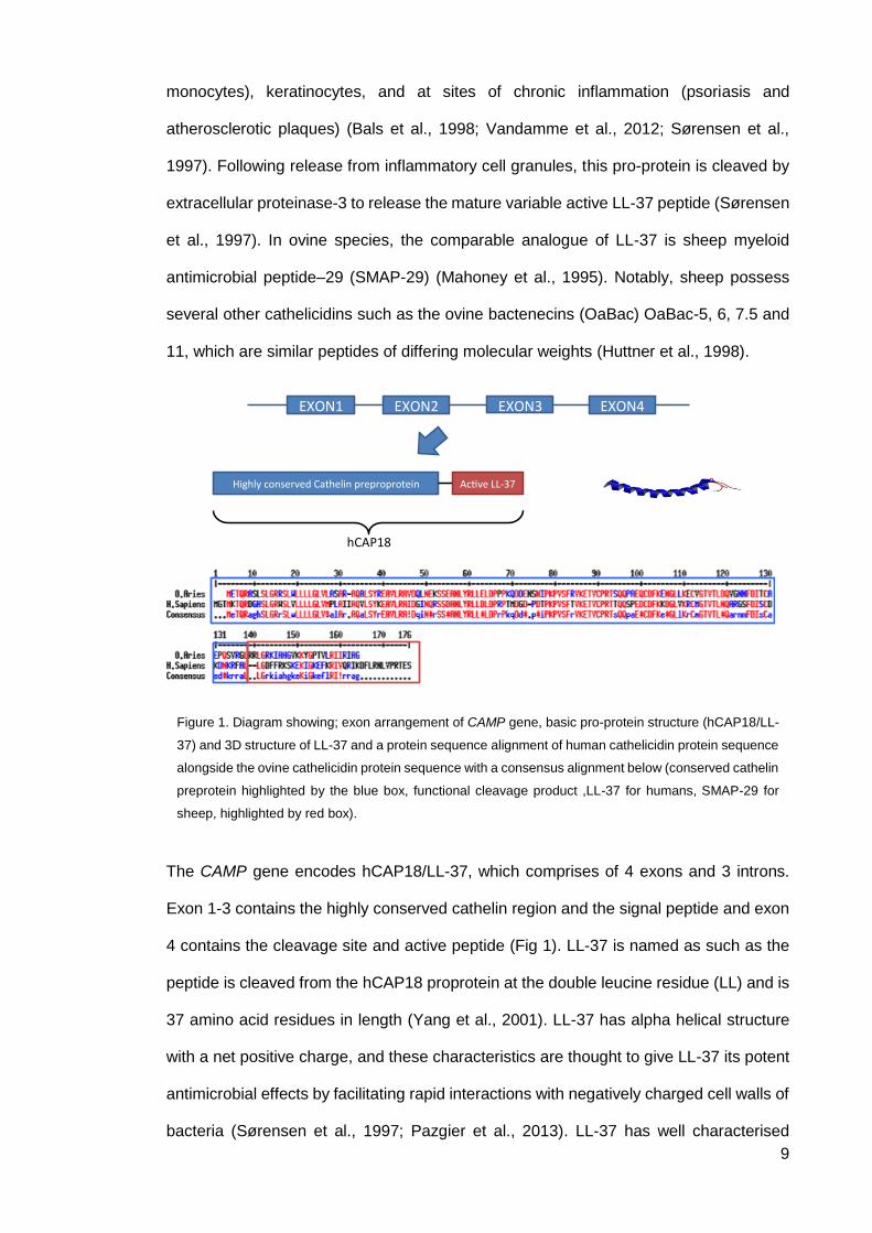
The CAMP gene encodes hCAP18/LL-37, which comprises of 4 exons and 3 introns.
Exon 1-3 contains the highly conserved cathelin region and the signal peptide and exon
4 contains the cleavage site and active peptide (Fig 1). LL-37 is named as such as the
peptide is cleaved from the hCAP18 proprotein at the double leucine residue (LL) and is
37 amino acid residues in length (Yang et al., 2001). LL-37 has alpha helical structure
with a net positive charge, and these characteristics are thought to give LL-37 its potent
antimicrobial effects by facilitating rapid interactions with negatively charged cell walls of
bacteria (Sørensen et al., 1997; Pazgier et al., 2013). LL-37 has well characterised
HighlyconservedCathelinpreproprotein Ac veLL-37
hCAP18
EXON1 EXON4EXON2 EXON3
Figure 1. Diagram showing; exon arrangement of CAMP gene, basic pro-protein structure (hCAP18/LL-
37) and 3D structure of LL-37 and a protein sequence alignment of human cathelicidin protein sequence
alongside the ovine cathelicidin protein sequence with a consensus alignment below (conserved cathelin
preprotein highlighted by the blue box, functional cleavage product ,LL-37 for humans, SMAP-29 for
sheep, highlighted by red box).
10
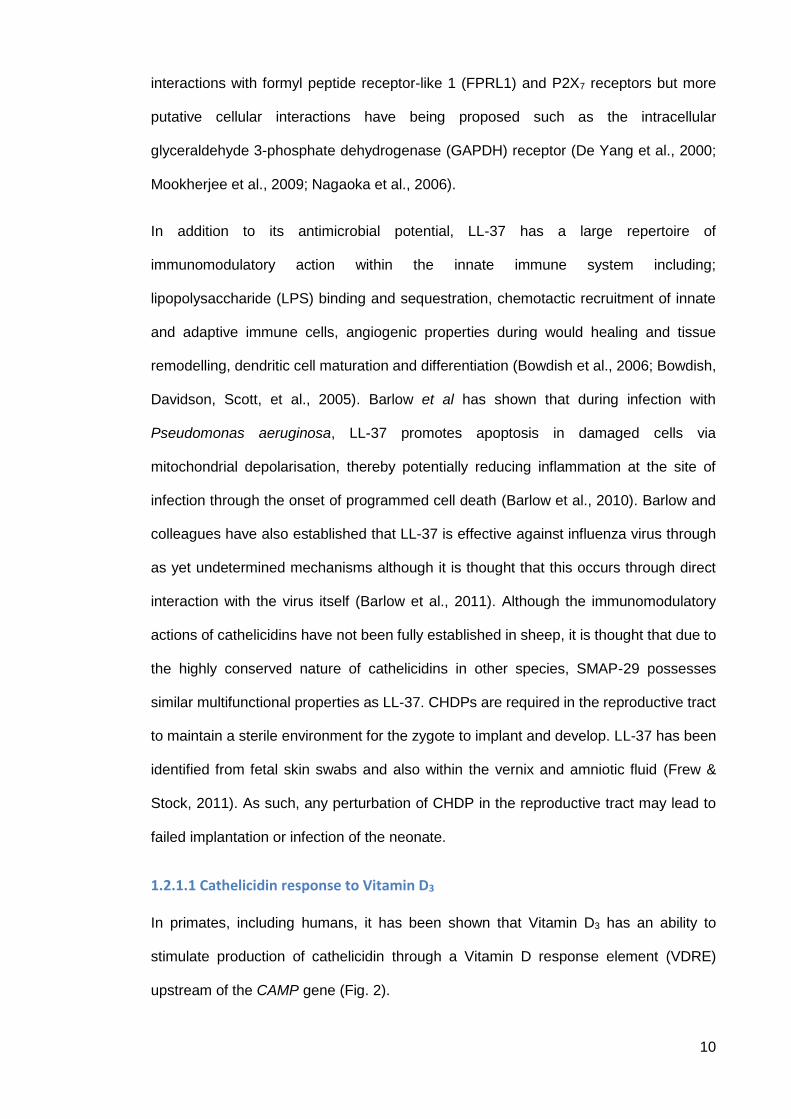
interactions with formyl peptide receptor-like 1 (FPRL1) and P2X7 receptors but more
putative cellular interactions have being proposed such as the intracellular
glyceraldehyde 3-phosphate dehydrogenase (GAPDH) receptor (De Yang et al., 2000;
Mookherjee et al., 2009; Nagaoka et al., 2006).
In addition to its antimicrobial potential, LL-37 has a large repertoire of
immunomodulatory action within the innate immune system including;
lipopolysaccharide (LPS) binding and sequestration, chemotactic recruitment of innate
and adaptive immune cells, angiogenic properties during would healing and tissue
remodelling, dendritic cell maturation and differentiation (Bowdish et al., 2006; Bowdish,
Davidson, Scott, et al., 2005). Barlow et al has shown that during infection with
Pseudomonas aeruginosa, LL-37 promotes apoptosis in damaged cells via
mitochondrial depolarisation, thereby potentially reducing inflammation at the site of
infection through the onset of programmed cell death (Barlow et al., 2010). Barlow and
colleagues have also established that LL-37 is effective against influenza virus through
as yet undetermined mechanisms although it is thought that this occurs through direct
interaction with the virus itself (Barlow et al., 2011). Although the immunomodulatory
actions of cathelicidins have not been fully established in sheep, it is thought that due to
the highly conserved nature of cathelicidins in other species, SMAP-29 possesses
similar multifunctional properties as LL-37. CHDPs are required in the reproductive tract
to maintain a sterile environment for the zygote to implant and develop. LL-37 has been
identified from fetal skin swabs and also within the vernix and amniotic fluid (Frew &
Stock, 2011). As such, any perturbation of CHDP in the reproductive tract may lead to
failed implantation or infection of the neonate.
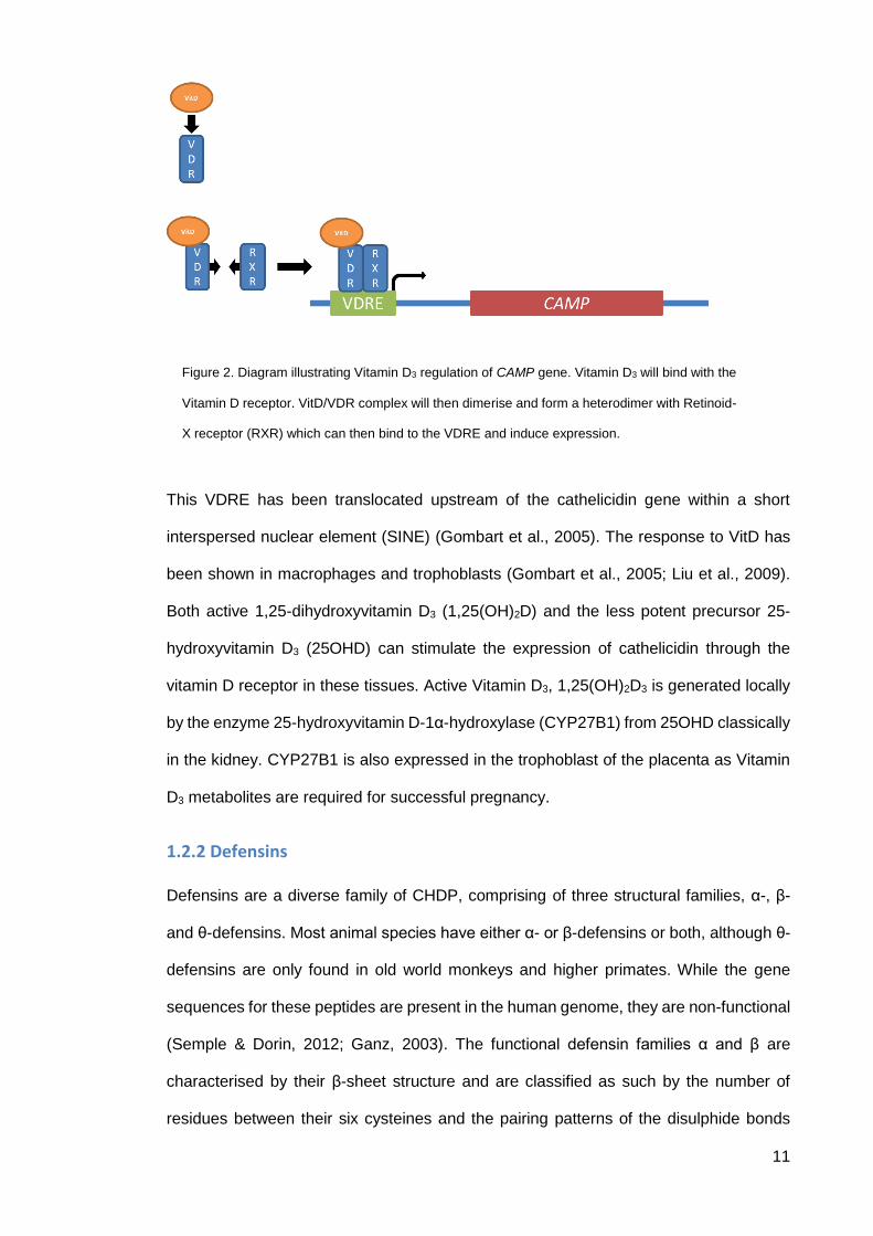
1.2.1.1 Cathelicidin response to Vitamin D3
In primates, including humans, it has been shown that Vitamin D3 has an ability to
stimulate production of cathelicidin through a Vitamin D response element (VDRE)
upstream of the CAMP gene (Fig. 2).
11
This VDRE has been translocated upstream of the cathelicidin gene within a short
interspersed nuclear element (SINE) (Gombart et al., 2005). The response to VitD has
been shown in macrophages and trophoblasts (Gombart et al., 2005; Liu et al., 2009).
Both active 1,25-dihydroxyvitamin D3 (1,25(OH)2D) and the less potent precursor 25-
hydroxyvitamin D3 (25OHD) can stimulate the expression of cathelicidin through the
vitamin D receptor in these tissues. Active Vitamin D3, 1,25(OH)2D3 is generated locally
by the enzyme 25-hydroxyvitamin D-1α-hydroxylase (CYP27B1) from 25OHD classically
in the kidney. CYP27B1 is also expressed in the trophoblast of the placenta as Vitamin
D3 metabolites are required for successful pregnancy.
1.2.2 Defensins
Defensins are a diverse family of CHDP, comprising of three structural families, α-, β-
and θ-defensins. Most animal species have either α- or β-defensins or both, although θ-
defensins are only found in old world monkeys and higher primates. While the gene
sequences for these peptides are present in the human genome, they are non-functional
(Semple & Dorin, 2012; Ganz, 2003). The functional defensin families α and β are
characterised by their β-sheet structure and are classified as such by the number of
residues between their six cysteines and the pairing patterns of the disulphide bonds
Figure 2. Diagram illustrating Vitamin D3 regulation of CAMP gene. Vitamin D3 will bind with the
Vitamin D receptor. VitD/VDR complex will then dimerise and form a heterodimer with Retinoid-
X receptor (RXR) which can then bind to the VDRE and induce expression.
12
between the cysteine residues. In sheep only β-defensins have thus far been identified
and are of focus in this study (Ganz, 2003; Semple & Dorin, 2012; Bowdish et al., 2006).
In similarity with cathelicidins, β-defensins are also positively charged, although there
are thirty-three different β-defensins in humans that are very similar in their core
structure, but with slight variances in amino acid sequence between the cysteine
residues. The genes which encode the β-defensins are all denoted as DEFB(number)
i.e. DEFB1 encodes β-defensin 1, and β-defensin genes are found in two clusters upon
chromosome 8 and 6 respectively (Pazgier et al., 2006). Defensins are constitutively
expressed in the early stages of neutrophil development in the bone marrow reservoir of
progenitor myeloid cells and stored in azurophilic granules until required as constitutive
expression tails off during neutrophil maturation (Semple & Dorin, 2012). The azurophilic
granules are destined to fuse with phago-lysosomes indicating that β-defensins are used
by neutrophils predominantly to aid in the phagocytic destruction of bacteria and other
pathogens. β-defensins produced via pathogen recognition in epithelial tissues are
expressed and released to aid in host defence and not as part of phagocytic attack. β-
defensins have also been attributed with immunomodulatory functions similar to
cathelicidins including potent antimicrobial and antiviral effects. In addition, the
immunomodulatory capacity of defensins has been shown to include the recruitment of
adaptive immune cells such as CD4+ Memory T-cells and immature dendritic cells via
interaction with the CCR6 receptor (Bowdish et al., 2006; Ganz, 2003; Semple & Dorin,
2012; Pazgier et al., 2006).
1.3 The Steroid Environment during Pregnancy
Pregnancy and parturition are two processes that are intricately linked to the steroid state
of the mother in order for a healthy pregnancy and birth of the child. Recent studies have
associated changes in the maternal/fetal steroid environment to predisposition of ‘first
world diseases’ such as type 2 diabetes, cardiovascular disease and obesity through a
phenomenon called fetal programming (Lee et al., 2006). Fetal programming, is a
13
phenomenon whereby the maternal environment can affect the developing fetus during
pregnancy (Fowden & Forhead, 2004; Harris & Seckl, 2011; Roseboom et al., 2001). As
several aspects of pregnancy are dependent on tightly regulated specific steroid profiles,
any changes in steroid concentrations may potentially predispose to altered health in the
offspring
Therapeutic administration of exogenous synthetic GC can be used as a prophylactic
treatment in pregnancies where the mother is at risk of premature delivery. Steroid
treatment can encourage lung development and surfactant production, and ready the
fetus for the ex utero environment in the event of an early delivery (Reynolds & Seckl,
2012). In these instances GCs, such as dexamethasone (Dex) or betamethasone, are
given post- or ante-partum to promote pulmonary maturation and surfactant protein
production in the neonate (Tegethoff et al., 2009).
Interestingly, excess male steroids during pregnancy are thought to be a cause for PCOS
(Veiga-Lopez et al., 2011). This hypothesis came about from psychosexual studies
carried out in the 70’s that discovered by injecting pregnant rhesus macaque monkeys
with testosterone had effects on the metabolic system of female offspring showing a
PCOS as well as effecting the behaviour of the monkeys. This research was later
revisited by Abbot et al and has since become the gold-standard model of investigating
PCOS in terms of generation of animal models (Abbott et al., 1998).
1.3.1 Dexamethasone
Dex is a synthetic GC and is used in a clinical setting as a potent, long-lasting, stable
surrogate of the corticosteroid cortisol. Unlike cortisol, Dex only interacts with the GC
receptor, whereas the more promiscuous cortisol can also interact with the
mineralocorticoid receptor. Dex is used primarily for the replacement of GCs in
conditions where GC production has been lost or diminished (e.g. Addisons disease),
although it is also used as a promoter of fetal lung surfactant protein production in
pregnancies at risk of premature delivery (Tegethoff et al., 2009). Dex is approximately

14
thirty fold more potent than the native cortisol and this is attributed to the fact that Dex
cannot be metabolised by the cortisol inactivation enzyme 11 β-hydroxysteroid
dehydrogenase type 2 (11β-HSD2). In a physiological context, 11βHSD2 is used as an
active barrier in the placenta preventing fetal exposure to maternal GCs (Kapoor et al.,
2008).
Given that Dex cannot be metabolised by 11βHSD2, it is an ideal candidate to be used
to model fetal exposure to excess GCs. In experimental models of maternal stress, Dex
is used to simulate an increase in cortisol secretion from the maternal adrenals, which is
thought to cross the placental barrier and affect the developing fetus. These studies so
far have concluded that fetal exposure to GCs can result in rodent and ovine
hypertension, altered HPA axis, impaired glucose homeostasis and obesity (reviewed in
Fowden & Forhead, 2004). It should be noted that the steroid manipulation in these
studies described here occurs at early/mid gestation and not completely translatable to
effect that an antenatal treatment regime would have as they are only administered in
the last few weeks of pregnancy or diagnosis of a condition where the neonate is at risk
of delivering early. Large cohort studies on the risks of using Dex/beta-methasone as a
antenatal intervention have been carried out to assess the long-term side effects of these
treatments (Tegethoff et al., 2009). The immediate benefits are clear to be seen with a
reduction in infant mortality and morbidity but from animal studies the consequences of
excess GCs on development and in later life are well known (Harris & Seckl, 2011). The
large scale cohort studies to date haven’t concluded whether the long-term effects are
adequately balanced from a risk/reward standpoint but these studies have certainly
identified areas of further study which will help answer if in utero GCs are beneficial or
not (Tegethoff et al., 2009; Reynolds & Seckl, 2012).
1.3.2 Testosterone
Testosterone is the male sex hormone that is responsible for development of male sexual
organs and production of spermatozoa in the testis. It is produced primarily in the testis,
but also in the ovary and adrenal, by a suite of steroidogenic enzymes in which 17β-
15
hydroxysteroid dehydrogenase (17βHSD) is the final step. Testosterone can be
metabolised by P450 aromatase to oestradiol. Aromatase is present in the placenta to
prevent fetal exposure to maternal androgens as the timing of sexual characteristics
development is controlled by fetal production of androgens (Padmanabhan & Veiga-
Lopez, 2013; Padmanabhan & Veiga-Lopez, 2011; Veiga-Lopez et al., 2011; Abbott et
al., 1998). Male sexual development is a delicately timed process where deviations
thereof can cause disorders of sexual development such as, testis dysgenesis syndrome
(cryptorchidism, hypospadias and increased incidence of male germ cell tumors) (Scott
et al., 2007; Welsh et al., 2008; Sharpe, 2006). Testosterone propionate (TP) is the
injectable formula of testosterone and has negligible differences in activity compared to
testosterone. Testosterone is primarily used as a treatment in gender reassignment
patients and men suffering from hypogonadism (small or underdeveloped testes) where
lowered serum testosterone levels require supplementation (Scott et al., 2009).
1.4 Chlamydial pathogens
1.4.1 Chlamydia abortus
Chlamydia abortus is an obligate intracellular pathogen that primarily infects ruminants
and pigs and causes enzootic abortion of ewes (EAE), which has a large economic
impact on the livestock industry (Kerr et al., 2005; Wheelhouse et al., 2008; Wheelhouse
et al., 2009). A Chlamydia infection has a biphasic development cycle where the microbe
inhabits two states that facilitate intra and extracellular existence of this pathogen. The
elementary body (EB) state enables Chlamydia spp to survive in the extracellular
environment. In this phase there is no metabolic activity and in this state the microbe is
infective and able to interact with the mucosal membranes and invade host cells using
as yet undetermined mechanisms. The EB is taken into an intracytoplasmic inclusion
that is prevented from binding to phagolysosomes and at this point the Chlamydia
switches to its other state, the reticular body (RB). The RB is metabolically active and
during this phase the Chlamydia reproduces using binary fission within the inclusion.
16
Before the inclusion becomes large enough to lyse the cell the Chlamydia switches back
to the infective EB state to enable further infection (Longbottom & Coulter, 2003). In
C.abortus infection this process wipes out specifically the trophoblast within the placenta,
which eventually prevents the transfer of nutrients and O2 to the fetus (Sammin et al.,
2009; Sammin et al., 2006). In the context of this project, infection with Chlamydia spp
provides an appropriate model of infected pregnancy where CHDP modulation and
activity can be assessed.
To date most major studies using CHPD against chlamydia spp have been directed
towards the human pathogens C.trachomatis and C.pneumoniae (Donati & Leo, 2005).
Both cathelicidins and defensins have been tested against these Chlamydia spp. and
have been found to be effective as antimicrobial agents against these species. One study
tested the efficacy of different cathelicidins from different host species against clinical
strains of C.trachomatis and C.pneumoniae. Additional to these test groups there was
also an ‘animal chlamydia strain’ group which were assessed as a group for susceptibility
but the group comprised of a mixture of C.psittaci (parakeet), C.felis (cats), C.abortus
(livestock, sheep, cows and pigs) and C.pecorum (koala and livestock). Cathelicidins
were shown to have an antimicrobial effect on these species of chlamydia but as they
were assessed as a group we cannot infer too much on specific reactions from specific
species. One interesting finding from the paper was that the sheep cathelicidin SMAP-
29 had differing efficacies being more potent against the human species as compared
to the animal strains (Donati & Leo, 2005). This therefore lends support to using
cathelicidins from other animals as therapies against human pathogens. Evolutionarily
human pathogens have co-evolved with human cathelicidins and thusly have developed
some form of tolerance or resistance but as can be seen in Donati & Leo (2005),
cathelicidins from another host, ovine cathelicidins for example, are shown to be effective
at lower concentrations indicating a higher potency and therefore potential therapeutic
benefit.
17
1.4.2 Waddlia chondrophilia
Waddlia chondrophilia is an emerging intracellular pathogen of the order chlamydiales
(Rurangirwa et al., 1999). Like other members of this order Waddlia has a biphasic
reproductive cycle, multiplying via binary fission in the inclusion body of infected cells in
the metabolically active reticulate stage. Then whilst in the metabolically inactive EB
stage, is infective (Goy et al., 2008; de Barsy & Greub, 2013). Waddlia was originally
isolated from an aborted bovine fetus in 1986 but has since been identified in other
bovine abortions as well as being implicated in human birth complications and
spontaneous abortions/miscarriages (Baud et al., 2014; Goy et al., 2008). Recent studies
have shown that Waddlia is capable of infecting several cell lines of varying species as
well as primary human macrophages (Kebbi-Beghdadi et al., 2011 a,b). To date there
has been no attempts to test the efficacy of CHDP against this emerging zoonotic
pathogen.
1.5 Study purpose and outline
The sterility and the steroid milieu of the reproductive tract are both of key importance to
ensure a successful pregnancy and birth of the newborn. An aim of this study is to assess
the impact of an altered steroid environment (excess androgens or GCs) during
pregnancy on the expression of CHDP in the functional subunit of the placenta, the
trophoblast. In addition to this, fetal lung and thymus (sites of regular CHDP production)
will be assessed to determine if altered steroid concentrations in utero will program any
lasting effects in the CHDP expression of the offspring.
Sterility of the reproductive tract during pregnancy is paramount for a successful
pregnancy. Some reproductive pathogens, such as the C.abortus, and W.chondrophilia
have specifically evolved in livestock species (sheep, pigs and goats) to only become
pathogenic during pregnancy. The immunomodulatory roles of CHDP, together with their
antimicrobial and LPS neutralising properties are if significant interest, and designing
novel antimicrobial agents based on naturally occurring peptide is now a popular
18
strategy. A number of new therapeutic agents based upon naturally occurring
antimicrobial peptides are currently undergoing clinical trails (Baltzer & Brown, 2011;
Padhi et al., 2014; Yeung et al., 2011). Here, CHDP (human, ovine and porcine) was
tested to assess whether it could be used a viable novel antimicrobial agent against this
economically relevant pathogen.
1.5.1 Tissues analysed
1.5.1.1 Fetal Lung
The lung is a well-known and well characterized site of CHDP expression due to its
constant exposure to the external environment (Tecle et al., 2010; Seaborn et al., 2010;
Collie et al., 2013; Scott et al., 2002). The lungs develop throughout pregnancy but aren’t
fully matured and able to facilitate oxygen exchange until the latter stages of pregnancy
(Grenache & Gronowski, 2006). Lung development is a tightly regulated and sequential
process beginning around week 3 of embryological development and not fully completing
until postnatal life (Seaborn et al., 2010; Grenache & Gronowski, 2006; Schaller-Bals et
al., 2002). Surfactant protein, which is vital for gas exchange within the alveoli, begins
production around mid-late gestation in preparation for the extrauterine environment. In
human neonates the expression of certain CHDP are developmentally regulated
throughout pregnancy showing that they are key to a healthy epithelial environment for
when the lungs are exposed to the extrauterine environment for the first time (Starner et
al., 2005).
1.5.1.2 Fetal Thymus
The thymus is the organ where immature T-cells migrate to, from the lymphoid progenitor
cells in the bone marrow, to undergo maturation (Blackburn & Manley, 2004). The
thymus can be spilt into two distinct zones, the outer cortical area (thymic cortex) and
the inner medullary zone (thymic medulla) that are demarcated by a cortico-medullary
junction. Immature T-cells enter the thymus and migrate to the outer cortical region to
undergo the first steps of maturation (thymopoiesis), β-selection and lineage
19
commitment (Blackburn & Manley, 2004). Thymocytes are now either CD4 or CD8
positive with a functional T-cell receptor (TCR) they move to the cortico-medullary
junction where they encounter plasmacytoid dendritic cells, which present ‘self’ antigens
to test the T-cells for autoreactivity (Lee et al., 2010; Blackburn & Manley, 2004). T-cells
that recognise ‘self’ antigens are negatively selected and programmed into apoptosis, T-
cells that don’t react with self-antigens are transported to the periphery and matured
further into specific lineages of T-cell (e.g. T-helper, T-reg and T-suppressor) (Lee et al.,
2010; Blackburn & Manley, 2004). There is a body of literature showing LL-37 has a role
in constitutively expressing interferon-α stimulated genes which confer a resistance to
viral infection within the medulla of the thymus (where T-cells that have undergone
positive-selection are found) (Colantonio et al., 2011; Lee et al., 2010).
1.5.1.3 Placenta (Placentome/Cotyledon)
The ovine placenta is different to the human placenta in a number of aspects. There is
not one single placental unit like a human placenta, the ovine placenta consists of ~90
discreet individual placentomes interconnected by the intercotyledonary membrane
(Sammin et al., 2006). The ovine system consists of an intercotyledonary membrane with
individual cotyledons interspersed throughout the membrane (Entrican, 2002; Entrican
et al., 2010). When these cotyledons (which are the fetal side of the placentome)
interface with the maternal caruncles (cup-like features) of the endometrium the fetal and
maternal villi interdigitate to form a functional placentome (Sammin et al., 2009). Each
of these placentome structures has a common blood supply and coalesces to form the
umbilical cord that supports the fetal lamb. The ovine placenta is syneptheliochorial,
which means that the fetal chorion and maternal uterine epithelium come into direct
contact with one another and transfer nutrients directly between each other. Whereas
the human placenta is haemochorial, where the trophoblasts of the placenta are bathed
in maternal blood and transfer nutrient to the fetus from the maternal circulation directly
(Sammin et al., 2009; Sammin et al., 2006; Entrican et al., 2010). It has been shown in

20
human trophoblasts and choriocarcinoma cell lines (BeWo) that LL-37 can be expressed
in response to VitD in these tissues in humans (Liu et al., 2009; Gombart et al., 2005).
21
Chapter 2 – CHDP and Steroids during Pregnancy
2.1 Methods
2.1.1 Tissues
All tissues used were collected from ovine models of steroid manipulation during
pregnancy which were conducted under a Home Office Project Licence as regulated by
the Animals (Scientific Procedures) Act, 1986. Briefly, 20mg of testosterone propionate
(TP) or 100 µg dexamethasone (Dex) dissolved in vegetable oil or oil vehicle alone were
the treatments. Injections were carried out using an ultrasound-guided needle into the
rump of the fetus under maternal anaesthesia at days 62 and 82 of a 147 day of
pregnancy. Fetuses from both singleton and multiple pregnancies where injected directly
with either TP, Dex or vegetable oil. Female Scottish Greyface fetuses were injected with
TP, male Scottish Greyface fetuses were injected with Dex and both male and female
fetuses injected with vegetable oil as a vehicle control. Fetal lung and thymus were
gathered from fetuses sacrificed on day 90 of gestation and from the same fetus a
cotyledon/placentome structure from the placenta was also collected. These tissues
were snap frozen and stored at -800C for mRNA expression analysis.
2.1.2 In Vitro Steroid Treatments
The ovine trophoblast cell line, AH-1 and human lung epithelial cell line A549 were used
to model the response of the placenta and lungs to different steroid exposures in vitro.
AH-1 cells were kindly provided by Drs Nicholas Wheelhouse and David Longbottom
from Moredun Research Institute, Edinburgh, UK, and were grown and maintained in
Iscove’s modified Dulbecco’s medium (IMDM) supplemented with 5% fetal bovine serum
(PAA Laboratories Ltd, Yeovil, Somerset, UK) at 370C with 5% CO2. A549 cells were
obtained from European Collection of Cell Cultures (ECACC, Salisbury, UK) and were
grown and maintained in Dulbeccoo’s modified eagles medium with high glucose
(4500mg/L) and GlutaMAX (L-alanyl-Glutamine, 862mg/L) (DMEM High glucose with
22
GlutaMAX, PAA Laboratories Ltd, Yeovil, Somerset, UK) supplemented with
penicillin/streptomycin (50U/mL) and 10% fetal bovine serum (Sigma, UK). Treatment
medias containing TP, Dex and VitD were created in serum free media (IMDM for AH-1
and DMEM for A549). All steroids were dissolved in the absolute alcohol at a stock
concentration of 1mM and stored at -800C. In all steroid treatments an ethanol (EtOH)
vehicle control was run (1μl/mL) to ensure EtOH wasn’t having an independent effect. A
1:10 serial dilution from 1M to 1nM (1μM, 100nM, 10nM and 1nM) was run for both Dex
and TP. AH-1 cells were exposed to VitD at 100nM, 10nM and 1nM to accommodate
LPS stimulation (500ng/mL, lipopolysaccharide from Escherichia coli O111:B4, Sigma,
L2630-10mg). AH-1 cells were serum starved for 24h and steroid treatments were
carried out in respective serum-free medias for 24hrs and were maintained in a heated,
humidified incubator at 37oC with 5% CO2.
2.1.3 RNA Extraction, DNase treatment and cDNA Synthesis
For all RNA extractions RLT buffer (with 2-mercaptoenthanol, 1% v/v) from Qiagen
RNeasy mini kits (Qiagen, Crawley, UK) were used for lysis of both in vitro and ex vivo
samples. For frozen tissues, 30mg tissue was weighed and lysed in RLT buffer using a
magnetic bead and the Qiagen TissueLyser (Qiagen, Crawley, UK). In vitro samples
were lysed in situ and collected into sterile DNase/RNase free eppendorfs and stored at
-800C until ready for extraction. Once samples were lysed, RNA was extracted from the
lysates using the RNeasy mini kit system. All samples were analysed using a NanoDrop
Spectrophotometer (Thermo Fisher Scientific, Loughborough, UK) to ascertain RNA
quantity. Representative samples from each treatment group were assayed for RNA
quality using the Agilent Bioanalyser (Agilent, UK). Genomic DNA (gDNA) was removed
from the extracted RNA using PrimerDesign Precision DNase Kits (PrimerDesign,
Southampton, UK). DNase treatments were carried out on all samples as per
manufacturer’s protocol. Complimentary DNA (cDNA) was synthesised from the
extracted RNA using Precision nanoScript reverse transcription kits as per the
manufacturers protocol (PrimerDesign, Southampton, UK) and stored at -200C until

23
ready for downstream uses. Negative controls with no reverse transcriptase (no RT) in
the reaction were ran for every treatment.
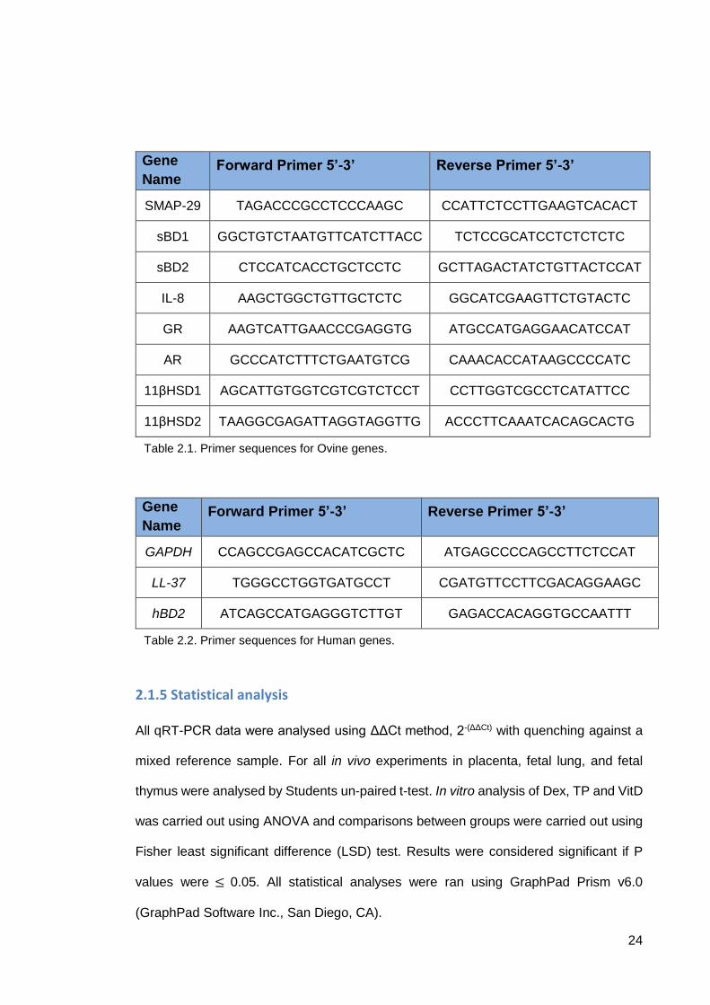
2.1.4 Quantitative Real Time – Polymerase Chain Reaction (qRT-PCR)
Quantitative real-time polymerase chain reaction (qRT-PCR) was performed using
Applied Biosystems StepOne Real-Time PCR machine with SYBR green detection and
a 96-well format setup. qRT-PCR primers for the ovine genes; SMAP-29, sBD1, and
sBD2, were designed and optimised by PrimerDesign Ltd (PrimerDesign Ltd,
Southampton, UK). Primers for androgen receptor (AR), glucocorticoid receptor (GR),
11β-hydroxysteroid dehydrogenase type 1 and 2 (11βHSD1/2) and interleukin-8 (IL-8)
were obtained from MGW Eurofins with primer sequences optimised and previously
published (Rae et al., 2013; Fach et al., 2007). Human primers for LL-37 were obtained
from a previous publication (Li et al., 2013), hBD2 and GAPDH (sequences in Fig 2.2)
were kindly provided by Dr Douglas Fraser-Pitt (Edinburgh Napier University). Melt curve
analysis was carried out with every GOI (available in Appendix) to determine specificity
of the primers alongside a no-template H2O control. Housekeeper genes were identified
using a GeNorm Kit to establish the most stable housekeeping genes (PrimerDesign
GeNorm Ovine 12 reference gene kit, PrimerDesign, Southampton, UK). All samples
were analysed using ΔΔCt method, 2-(ΔΔCt) with quenching against a mixed reference
sample. Each 10μl reaction consisted of 1μl (40ng) cDNA template, 0.5μl (300nM) gene
specific primers, 5μl SYBR Green (Precision 2X qMasterMix with SYBR Green,
PrimerDesign, Southampton, UK) and the reaction was brought to 10μl with
DNase/RNase free H2O. No RT and no template H2O controls were performed on each
plate as negative controls.
24
Gene
Name Forward Primer 5’-3’ Reverse Primer 5’-3’
SMAP-29 TAGACCCGCCTCCCAAGC CCATTCTCCTTGAAGTCACACT
sBD1 GGCTGTCTAATGTTCATCTTACC TCTCCGCATCCTCTCTCTC
sBD2 CTCCATCACCTGCTCCTC GCTTAGACTATCTGTTACTCCAT
IL-8 AAGCTGGCTGTTGCTCTC GGCATCGAAGTTCTGTACTC
GR AAGTCATTGAACCCGAGGTG ATGCCATGAGGAACATCCAT
AR GCCCATCTTTCTGAATGTCG CAAACACCATAAGCCCCATC
11βHSD1 AGCATTGTGGTCGTCGTCTCCT CCTTGGTCGCCTCATATTCC
11βHSD2 TAAGGCGAGATTAGGTAGGTTG ACCCTTCAAATCACAGCACTG
Gene
Name Forward Primer 5’-3’ Reverse Primer 5’-3’
GAPDH CCAGCCGAGCCACATCGCTC ATGAGCCCCAGCCTTCTCCAT
LL-37 TGGGCCTGGTGATGCCT CGATGTTCCTTCGACAGGAAGC
hBD2 ATCAGCCATGAGGGTCTTGT GAGACCACAGGTGCCAATTT
2.1.5 Statistical analysis
All qRT-PCR data were analysed using ΔΔCt method, 2-(ΔΔCt) with quenching against a
mixed reference sample. For all in vivo experiments in placenta, fetal lung, and fetal
thymus were analysed by Students un-paired t-test. In vitro analysis of Dex, TP and VitD
was carried out using ANOVA and comparisons between groups were carried out using
Fisher least significant difference (LSD) test. Results were considered significant if P
values were ≤ 0.05. All statistical analyses were ran using GraphPad Prism v6.0
(GraphPad Software Inc., San Diego, CA).
Table 2.1. Primer sequences for Ovine genes.
Table 2.2. Primer sequences for Human genes.
25
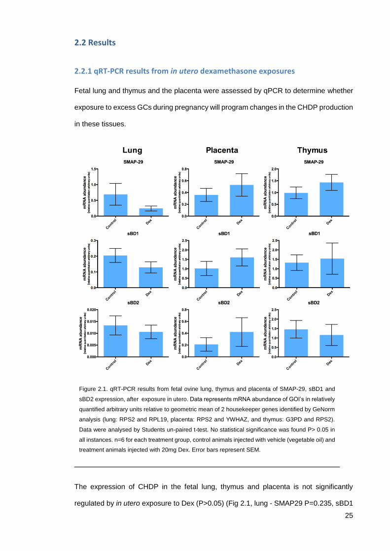
2.2 Results
2.2.1 qRT-PCR results from in utero dexamethasone exposures
Fetal lung and thymus and the placenta were assessed by qPCR to determine whether
exposure to excess GCs during pregnancy will program changes in the CHDP production
in these tissues.
The expression of CHDP in the fetal lung, thymus and placenta is not significantly
regulated by in utero exposure to Dex (P>0.05) (Fig 2.1, lung - SMAP29 P=0.235, sBD1
Figure 2.1. qRT-PCR results from fetal ovine lung, thymus and placenta of SMAP-29, sBD1 and
sBD2 expression, after exposure in utero. Data represents mRNA abundance of GOI’s in relatively
quantified arbitrary units relative to geometric mean of 2 housekeeper genes identified by GeNorm
analysis (lung: RPS2 and RPL19, placenta: RPS2 and YWHAZ, and thymus: G3PD and RPS2).
Data were analysed by Students un-paired t-test. No statistical significance was found P> 0.05 in
all instances. n=6 for each treatment group, control animals injected with vehicle (vegetable oil) and
treatment animals injected with 20mg Dex. Error bars represent SEM.

26
P=0.211 and sBD2 P=0.591, thymus - SMAP29 P=0.315, sBD1 P=0.509 and sBD2
0.689, placenta SMAP29 P=0.460, sBD1 P=0.341 and sBD2 P=0.452).
27
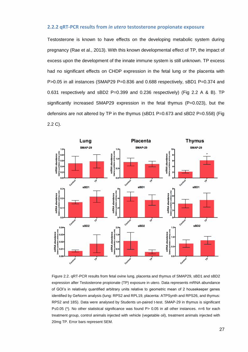
2.2.2 qRT-PCR results from in utero testosterone propionate exposure
Testosterone is known to have effects on the developing metabolic system during
pregnancy (Rae et al., 2013). With this known developmental effect of TP, the impact of
excess upon the development of the innate immune system is still unknown. TP excess
had no significant effects on CHDP expression in the fetal lung or the placenta with
P>0.05 in all instances (SMAP29 P=0.836 and 0.688 respectively, sBD1 P=0.374 and
0.631 respectively and sBD2 P=0.399 and 0.236 respectively) (Fig 2.2 A & B). TP
significantly increased SMAP29 expression in the fetal thymus (P=0.023), but the
defensins are not altered by TP in the thymus (sBD1 P=0.673 and sBD2 P=0.558) (Fig
2.2 C).
Figure 2.2. qRT-PCR results from fetal ovine lung, placenta and thymus of SMAP29, sBD1 and sBD2
expression after Testosterone propionate (TP) exposure in utero. Data represents mRNA abundance
of GOI’s in relatively quantified arbitrary units relative to geometric mean of 2 housekeeper genes
identified by GeNorm analysis (lung: RPS2 and RPL19, placenta: ATPSynth and RPS26, and thymus:
RPS2 and 18S). Data were analysed by Students un-paired t-test. SMAP-29 in thymus is significant
P≤0.05 (*). No other statistical significance was found P> 0.05 in all other instances. n=6 for each
treatment group, control animals injected with vehicle (vegetable oil), treatment animals injected with
20mg TP. Error bars represent SEM.
28
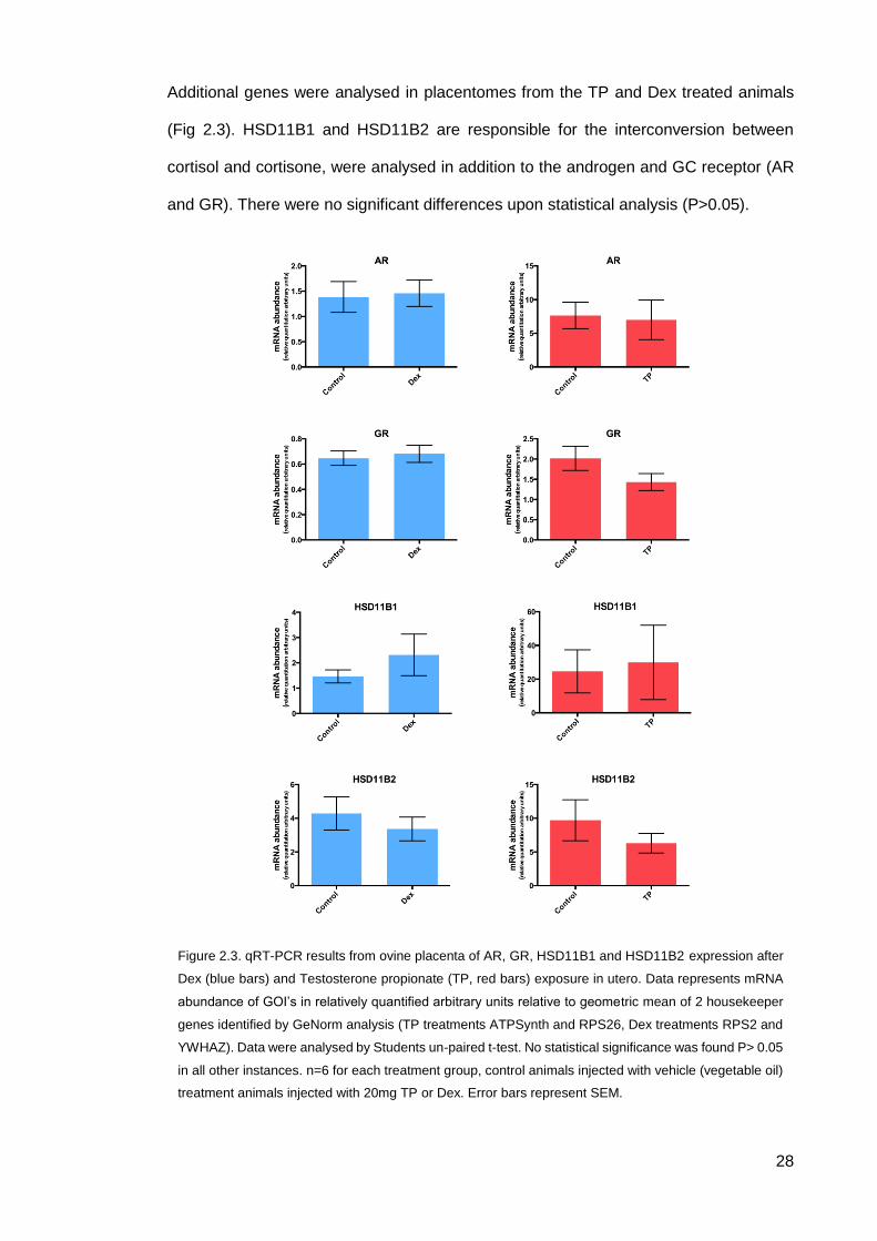
Additional genes were analysed in placentomes from the TP and Dex treated animals
(Fig 2.3). HSD11B1 and HSD11B2 are responsible for the interconversion between
cortisol and cortisone, were analysed in addition to the androgen and GC receptor (AR
and GR). There were no significant differences upon statistical analysis (P>0.05).
Figure 2.3. qRT-PCR results from ovine placenta of AR, GR, HSD11B1 and HSD11B2 expression after
Dex (blue bars) and Testosterone propionate (TP, red bars) exposure in utero. Data represents mRNA
abundance of GOI’s in relatively quantified arbitrary units relative to geometric mean of 2 housekeeper
genes identified by GeNorm analysis (TP treatments ATPSynth and RPS26, Dex treatments RPS2 and
YWHAZ). Data were analysed by Students un-paired t-test. No statistical significance was found P> 0.05
in all other instances. n=6 for each treatment group, control animals injected with vehicle (vegetable oil)
treatment animals injected with 20mg TP or Dex. Error bars represent SEM.
29
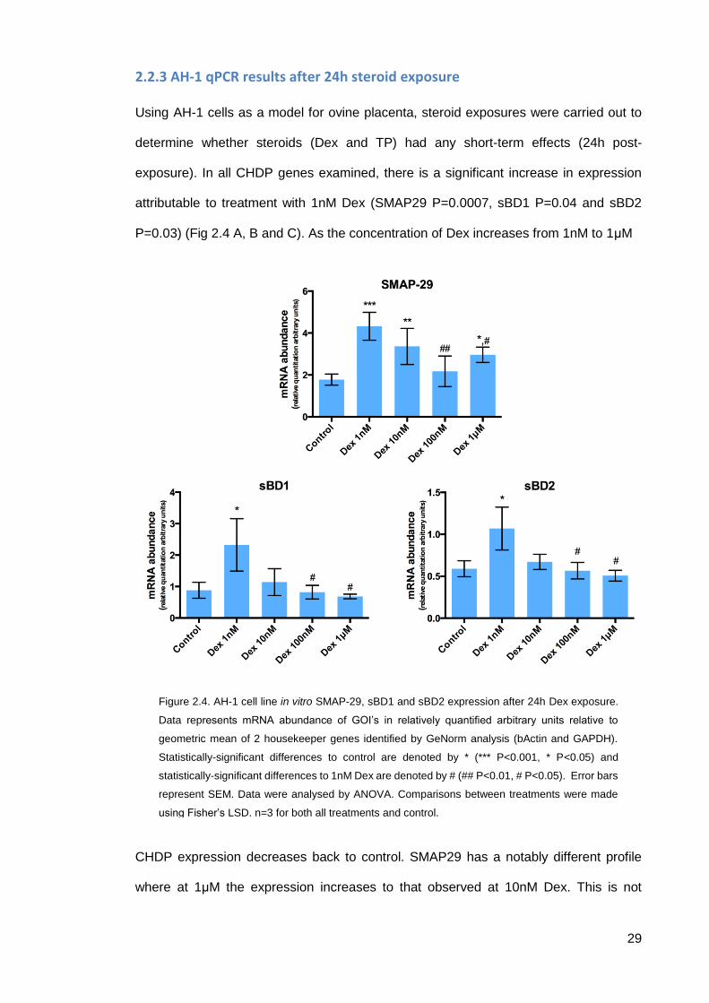
2.2.3 AH-1 qPCR results after 24h steroid exposure
Using AH-1 cells as a model for ovine placenta, steroid exposures were carried out to
determine whether steroids (Dex and TP) had any short-term effects (24h post-
exposure). In all CHDP genes examined, there is a significant increase in expression
attributable to treatment with 1nM Dex (SMAP29 P=0.0007, sBD1 P=0.04 and sBD2
P=0.03) (Fig 2.4 A, B and C). As the concentration of Dex increases from 1nM to 1μM
CHDP expression decreases back to control. SMAP29 has a notably different profile
where at 1μM the expression increases to that observed at 10nM Dex. This is not
Figure 2.4. AH-1 cell line in vitro SMAP-29, sBD1 and sBD2 expression after 24h Dex exposure.
Data represents mRNA abundance of GOI’s in relatively quantified arbitrary units relative to
geometric mean of 2 housekeeper genes identified by GeNorm analysis (bActin and GAPDH).
Statistically-significant differences to control are denoted by * (*** P<0.001, * P<0.05) and
statistically-significant differences to 1nM Dex are denoted by # (## P<0.01, # P<0.05). Error bars
represent SEM. Data were analysed by ANOVA. Comparisons between treatments were made
using Fisher’s LSD. n=3 for both all treatments and control.
30
significantly greater than 100nM so does not fit a classical non-monotonic response (Fig
2.4 A).
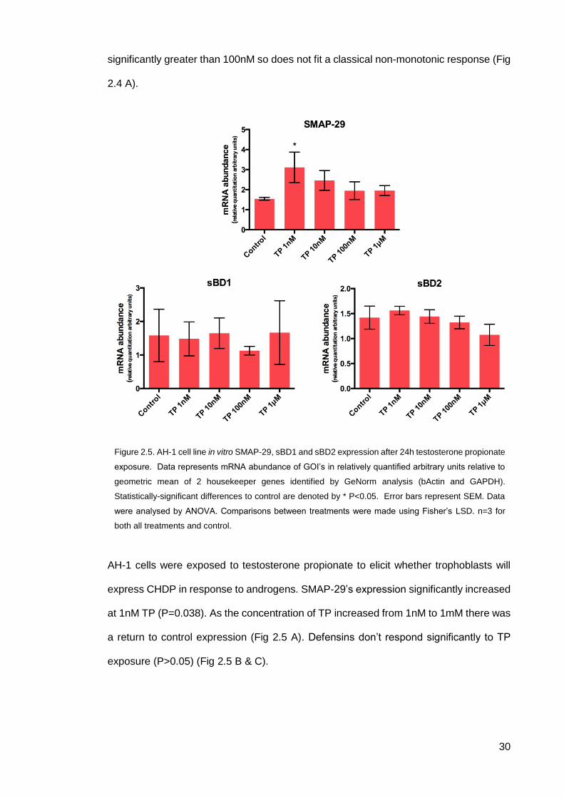
AH-1 cells were exposed to testosterone propionate to elicit whether trophoblasts will
express CHDP in response to androgens. SMAP-29’s expression significantly increased
at 1nM TP (P=0.038). As the concentration of TP increased from 1nM to 1mM there was
a return to control expression (Fig 2.5 A). Defensins don’t respond significantly to TP
exposure (P>0.05) (Fig 2.5 B & C).
Figure 2.5. AH-1 cell line in vitro SMAP-29, sBD1 and sBD2 expression after 24h testosterone propionate
exposure. Data represents mRNA abundance of GOI’s in relatively quantified arbitrary units relative to
geometric mean of 2 housekeeper genes identified by GeNorm analysis (bActin and GAPDH).
Statistically-significant differences to control are denoted by * P<0.05. Error bars represent SEM. Data
were analysed by ANOVA. Comparisons between treatments were made using Fisher’s LSD. n=3 for
both all treatments and control.
31
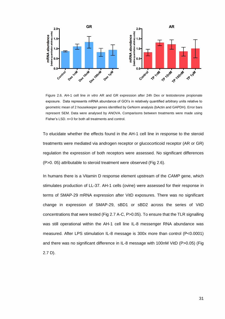
To elucidate whether the effects found in the AH-1 cell line in response to the steroid
treatments were mediated via androgen receptor or glucocorticoid receptor (AR or GR)
regulation the expression of both receptors were assessed. No significant differences
(P>0. 05) attributable to steroid treatment were observed (Fig 2.6).
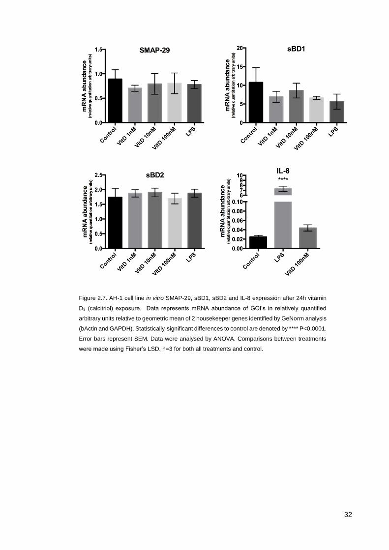
In humans there is a Vitamin D response element upstream of the CAMP gene, which
stimulates production of LL-37. AH-1 cells (ovine) were assessed for their response in
terms of SMAP-29 mRNA expression after VitD exposures. There was no significant
change in expression of SMAP-29, sBD1 or sBD2 across the series of VitD
concentrations that were tested (Fig 2.7 A-C, P>0.05). To ensure that the TLR signalling
was still operational within the AH-1 cell line IL-8 messenger RNA abundance was
measured. After LPS stimulation IL-8 message is 300x more than control (P<0.0001)
and there was no significant difference in IL-8 message with 100nM VitD (P>0.05) (Fig
2.7 D).
Figure 2.6. AH-1 cell line in vitro AR and GR expression after 24h Dex or testosterone propionate
exposure. Data represents mRNA abundance of GOI’s in relatively quantified arbitrary units relative to
geometric mean of 2 housekeeper genes identified by GeNorm analysis (bActin and GAPDH). Error bars
represent SEM. Data were analysed by ANOVA. Comparisons between treatments were made using
Fisher’s LSD. n=3 for both all treatments and control.
32
Figure 2.7. AH-1 cell line in vitro SMAP-29, sBD1, sBD2 and IL-8 expression after 24h vitamin
D3 (calcitriol) exposure. Data represents mRNA abundance of GOI’s in relatively quantified
arbitrary units relative to geometric mean of 2 housekeeper genes identified by GeNorm analysis
(bActin and GAPDH). Statistically-significant differences to control are denoted by **** P<0.0001.
Error bars represent SEM. Data were analysed by ANOVA. Comparisons between treatments
were made using Fisher’s LSD. n=3 for both all treatments and control.

33
2.3 Discussion
In vivo study results demonstrate that administration of Dex during pregnancy has no
significant effects upon fetal lung, thymus and placenta in terms of SMAP-29, sBD1 and
sBD2 expression (Fig 2.1). TP administration similarly has no effect in the fetal lung and
the placenta on SMAP-29, sBD1 and sBD2 expression (Fig 2.2A and B); however there
was a significant increase in expression in SMAP29 in response to in utero direct fetal
administration of TP in the thymus (Fig 2.2C). The relevance that this has in terms of the
development of the thymus and thymopoiesis (T-cell maturation), given the importance
of these systems throughout life, requires further investigation.
The thymus in humans and rodents constitutively expresses LL-37/mCRAMP (mouse
cathelicidin-related antimicrobial peptide) during fetal and postnatal life (Colantonio et
al., 2011). This expression is part of a mechanism responsible for constitutive expression
of interferon stimulated genes (ISG’s) in the thymus to increase resistance to viral
infection. LL-37’s role in this mechanism is to complex with DNA or RNA from apoptotic
T-cells undergoing negative selection and to stimulate IFN-α expression though TLR7/9
signaling which will then cause downstream expression of ISG’s via IFN-α receptor
(IFNαR) (Lande et al., 2007; Ganguly et al., 2009). This constitutive expression of IFN-α
is localised to the medulla of the thymus where mature T-cells, pDCs, and, macrophages
reside. This localised expression is thought to confer a host defence advantage against
pathogens such as CCR-5 tropic HIV that targets mature thymocytes as compared to
CXCR-4 tropic strains (Colantonio et al., 2011).
Classically, androgens are known to be suppressive of thymopoiesis via AR signalling in
the thymic epithelial cells, as upon withdrawal or suppression of androgens, there is an
enlargement of the thymus and increase in T-cell production (Olsen et al., 2001). This
increase in SMAP-29 could serve two functions in response to androgens; first to help
co-ordinate the suppression of thymopoiesis seen in response to androgens (Olsen et
al., 2001; Olsen & Kovacs, 2001). Secondly, with the known ability of cathelicidins to
34
mediate the inflammatory response (Barlow et al., 2006; Li et al., 2009; Nagaoka et al.,
2006), the increase in expression of SMAP-29 could control the inflammatory state of
the thymus in a similar manner to how LL-37 acts as an anti-inflammatory mediator
during secondary necrosis of apoptotic neutrophils (Li et al., 2009). Thus , this would
ensure that regulated induction of apoptosis, due to negative selection during T-cell
maturation, carries on without a large inflammatory response.
A final theory, which is difficult to link to the effects of androgens on the female thymus
in the context of SMAP-29, is that the expression of SMAP-29 in response to androgens
could allude to a mechanism that confers the sexual dimorphism found in the
development of autoimmune diseases (Hince et al., 2008; Gubbels Bupp et al., 2008;
Gui et al., 2011). It has been shown, in a mouse model of systemic lupus erythematosus,
that there is a male specific delay in the onset of symptoms; although after castration the
males mimic the progression of the females (Gubbels Bupp et al., 2008; Gui et al., 2011).
There is no direct evidence linking SMAP-29 or any other cathelicidin to the stringency
of negative selection during T-cell maturation, but with the discovery here that there is a
sex difference in expression due to androgens, this is a result that warrants further
investigation.
The key organ in successful mammalian pregnancy is the placenta, which is responsible
for the transport of nutrients and O2 to the developing fetus (Igwebuike, 2006; Sammin
et al., 2009; Burton, 2009). The function of this organ is intricately regulated by the
maternal and fetal steroid milieu. The placenta also acts as an immunological barrier to
the fetus We have shown here using the ovine trophoblast cell line AH-1 to model the
placenta in vitro, that contradictory to the in vivo results, there is a short-term effect (24h
post exposure) on the placental expression of CHDP after exposure to excess GC’s. In
these experiments, the levels of CHDP mRNA expression at 1nM Dex are double that of
control. As the concentration of Dex increases towards 1μM the expression of sDB1 and
sBD2 returns to control levels showing an inverse dose response. SMAP-29 also has
this apparent inverse dose response reducing towards control levels at 10nM and
35
100nM, but unlike the defensins at 1µM the mRNA abundance of SMAP-29 significantly
increases above control but not to levels (Fig. 2.4). These data suggests that there could
be some type of non-monotonic dose relationship with SMAP-29 and Dex but a wider
range of concentrations would have to be tested to elicit whether this response exists or
not. Inverse dose responses are common in the field of endocrine disrupting chemicals
(reviewed in; Lagarde et al., 2015).
In an attempt to explain this response, the expression of GR mRNA was measured to
assess whether the reduced CHDP responses at the higher Dex concentrations were
receptor mediated. The data indicates that there is no significant change in the
expression of GR mRNA after Dex treatment (Fig. 2.6) but this does not rule out GR-
mediated actions such as receptor ubiquitination and subsequent degradation (Wallace
& Cidlowski, 2001).
TP has a similar effect on SMAP29 expression only in AH-1 cells, while defensin
expression remains unchanged in response to androgens (Fig. 2.5). Again, to determine
whether this inverse dose response is receptor mediated, AR receptor mRNA
abundance was assayed and similarly did not show any change in expression in
response to TP.
The stimulation of CHDP production in response to Dex in the placenta is interesting as
Dex is a potent anti-inflammatory drug used to ameliorate inflammation in autoimmune
conditions and severe inflammatory disorders and here it is paradoxically being shown
to stimulate production of a proinflammatory peptide.
Similarly with the increase in SMAP-29 expression in response to TP is also interesting
as TP is generally a suppressant on the immune system (Olsen et al., 2001; Sakiani et
al., 2013). These findings require further investigation as they could have an impact in
neonatal medicine (Dex) and also our current understanding of fetal programming of
innate immunity (Dex or TP), which currently is very scarce. Whether this stimulation of
CHDP are a direct action of steroid signaling remains to be seen as these increases in

36
expression are only found in vitro after 24h exposure but in vivo there are no prolonged
effects programmed in the placenta.
In humans and higher primates there has been a translocation of a short interspersed
nuclear element (SINE) containing a VDRE upstream of the CAMP gene (Gombart et
al., 2005). This translocated response element has allowed humans and primates to
increase expression of cathelicidin in response to Vitamin D3. Here we show that sheep
do not have this VDRE upstream of their cathelicidin, as they do not increase expression
of SMAP-29 in response to VitD (Fig 2.7 A). Interestingly we also show a possible
tolerance in the AH-1 cell line within the LPS stimulation pathway (Toll-like receptor 4)
as when challenged with LPS, SMAP-29, sBD1 and sBD2 expression is not altered.
However, IL-8 message after LPS stimulation is drastically increased compared to
control and VitD stimulation (Fig 2.7 D). Further experiments challenging AH-1 with
peptidoglycan or poly I:C would assess the involvement of TLR4 ,or if other TLR
pathways or NOD-like receptors are altered (Lappas, 2013). However, the lack of VitD
stimulation is not unexpected as Gombert et al, shown that the VitD response is not
present in the rat, mouse and dog but is present in the higher primates. This highlights
that this translocation event must have happened in primate and human evolution and
has not occurred earlier in evolutionary terms (Gombart et al., 2005).
37
Chapter 3 – Expression and Activity of CHDP and Chlamydial
Pathogens
3.1 Methods
3.1.1 Chlamydia abortus and Waddlia chondrophilia CHDP exposure
experiments
Chlamydia abortus (C.abortus) strain S26/3 and Waddlia chondrophilia strain ATCC VR-
1470 were exposed to LL-37 (LLGDFFRKSKEKIGKEFKRIVQRIKDFLRN-LVPRTES),
scrambled LL-37 (sLL-37, RSLEGTDRFPFVRLKNSRKLEFKDI-KGIKREQFVKIL),
SMAP-29 (RGLRRLGRKIAHGVKKYGPTVLRIIRIAG) and PG-1
(RGGRLCYCRRRFCVCVGR), the porcine cathelicidin, to assess the ability of the
antimicrobial peptides to kill or inhibit the infectivity of the organisms. Both organisms
are obligate intracellular and were grown in McCoy cells (murine fibroblast cell line
regularly used to propagate Chlamydia spp.) in RMPI 1640 supplemented with 5% fetal
calf serum (PAA Laboratories, Peterborough, UK). Frozen stocks of C.abortus and
W.chondrophilia that had been previously propagated and stored were used in our
experiments. Placental infection was modelled by seeding AH-1 cells in 48-well plates
(Corning) and grown to 80% confluency overnight in IMDM growth medium (w/ 5% FCS).
Organisms were then incubated with CHDP in serum-free media at 100μg/ml, 50μg/ml,
25μg/ml, 10μg/ml and serum-free media as a control (0μg/ml). For LL-37, a scrambled
peptide, sLL-37, was available and was used as a negative control (equivalent of
100μg/ml) as well as a serum free media control. Approx. 400000 organisms, equal to
MOI 1 (multiplicity of infection, MOI 1 = 1 intracellular organism per cell) were pre-
incubated with the CHDP enriched mediums for 1h at 4oC. Exposure mediums were
diluted 1 in 10 in 2% FCS IMDM media, then transferred to AH-1 cells and incubated for
1h at 37oC to allow infection of cells and then replaced with normal growth medium and
38
grown for a further 48h. Infected cells were then lysed in vitro with AL buffer from DNeasy
kit (Qiagen, Crawley, UK) and lysates collected and stored at -20oC for DNA extraction.
3.1.2 DNA Extraction
DNA was extracted from AH-1 cells infected with C.abortus and W.chondrophilia using
DNeasy Blood and Tissue kit (Qiagen, Crawley, UK) as per manufacturer’s instructions
with no amendment to protocol. Extracted DNA was stored at -20oC for PCR analysis.
3.1.3 RNA Extraction, DNase treatment and cDNA Synthesis
All RNA extraction, DNase treatments and cDNA synthesis was carried out as per
protocol explained in Chapter 2 section 3 (2.3).
3.1.4 Quantitative Real Time – Polymerase Chain Reaction (qRT-PCR)
3.1.4.1 Measurement of CHDP and inflammatory response to W.chondrophilia
qRT-PCR was performed using Applied Biosystems StepOne Reat-Time PCR machine
with SYBR green detection and a 96-well format setup. PCR conditions were as follows;
10 mins at 95°C, then 40 cycles of 15 secs at 95°C, and 1min at 60°C, with a melt curve
analysis included in each run. Primers for IL-1α, IL-1β, IL-8 and TNFα were obtained
from MGW Eurofins with primer sequences optimised and previously published (Fach et
al., 2007; Smeed et al., 2007) and SMAP-29, sBD1 and sBD2 primers from PrimerDesign
Ltd (PrimerDesign Ltd, UK). Housekeeper genes were identified using a GeNorm Kit to
establish the most stable housekeeping genes (PrimerDesign GeNorm Ovine 12
reference gene kit, PrimerDesign, Southampton, UK). All samples were analysed using
ΔΔCt method, 2-(ΔΔCt) with quenching against a mixed reference sample. Each 10μl
reaction consisted of 1μl (40ng) cDNA template, 0.5μl (300nM) gene specific primers,
5μl SYBR Green (Precision 2X MasterMix, PrimerDesign) and the reaction was brought
to 10μl with DNase/RNase free H2O. No RT and no template H2O controls were run on
each plate as negative controls.
39
3.1.4.2 qRT-PCR determination of bacterial growth
Chlamydiales 16S gene copy number was used to quantify the number of organisms and
indirectly bacterial growth in the CHDP exposure experiments. Taqman pan-
chlamydiales 16S gene PCRs were carried out in 20μl reactions consisting of, 10μl
Quanta PCR MasterMix (Quanta Toughmix Low ROX PCR MasterMix) 0.2μl forward
primer (100nM), 0.2μl reverse primer (100nM) and 0.2μl probe (100nM) (FAM/BlackHole
Quencher) 1μl of DNA template and 8.4μl of DNase/RNase free H2O. Using Lienard’s
method (Lienard et al., 2011), a standard curve using a plasmid with Chlamydiales 16S
gene from Parachlamydia acanthamoeba inserted, at predetermined concentrations was
run on each plate to determine copy number. This was used as a marker of organism
growth.
Gene Name
Forward Primer 5’-3’ Reverse Primer 5’-3’
IL-1α TTGGTGCACATGGCAAGTG GCACAGTCAAGGCTATTTTTCC
IL-1β ATGGGTGTTCTGCATGAG AAGGCCACAGGAATCTTG
IL-8 AAGCTGGCTGTTGCTCTC GGCATCGAAGTTCTGTACTC
TNFα CTCATCTACTCGCAGGTCCTC ACTGCAATGCGGCTGATGG
Primer Name Primer sequence 5’-3’
panCh16F2 CCGCCAACACTGGGACT
panCh16R2 GGAGTTAGCCGGTGCTTCTTTAC
pan16S probe FAM-CTACGGGAGGCTGCAGTCGAGAATC-BHQ1
3.1.5 Statistical analysis
All CHDP exposures were run in duplicate so statistical analysis was not performed. All
qRT-PCR results from 24hr Waddlia infections were analysed using ΔΔCt method, 2-
Table 3.1: qPCR primers for genes of interest
Table 3.2: Taqman qPCR primers for pan-chlamydiales 16S gene

40
(ΔΔCt) with quenching against a mixed reference sample. Statistical analyses were carried
out using ANOVA and comparisons between groups were carried out using Fishers LSD
test. Results were considered significant if P≤0.05. Statistical analyses were run using
GraphPad Prism v6.0 (GraphPad Software Inc., San Diego, CA).
41
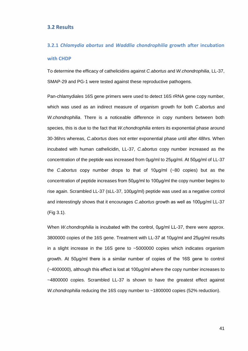
3.2 Results
3.2.1 Chlamydia abortus and Waddlia chondrophilia growth after incubation
with CHDP
To determine the efficacy of cathelicidins against C.abortus and W.chondrophilia, LL-37,
SMAP-29 and PG-1 were tested against these reproductive pathogens.
Pan-chlamydiales 16S gene primers were used to detect 16S rRNA gene copy number,
which was used as an indirect measure of organism growth for both C.abortus and
W.chondrophilia. There is a noticeable difference in copy numbers between both
species, this is due to the fact that W.chondrophilia enters its exponential phase around
30-36hrs whereas, C.abortus does not enter exponential phase until after 48hrs. When
incubated with human cathelicidin, LL-37, C.abortus copy number increased as the
concentration of the peptide was increased from 0µg/ml to 25µg/ml. At 50μg/ml of LL-37
the C.abortus copy number drops to that of 10μg/ml (~80 copies) but as the
concentration of peptide increases from 50μg/ml to 100μg/ml the copy number begins to
rise again. Scrambled LL-37 (sLL-37, 100µg/ml) peptide was used as a negative control
and interestingly shows that it encourages C.abortus growth as well as 100μg/ml LL-37
(Fig 3.1).
When W.chondrophilia is incubated with the control, 0μg/ml LL-37, there were approx.
3800000 copies of the 16S gene. Treatment with LL-37 at 10μg/ml and 25μg/ml results
in a slight increase in the 16S gene to ~5000000 copies which indicates organism
growth. At 50μg/ml there is a similar number of copies of the 16S gene to control
(~4000000), although this effect is lost at 100μg/ml where the copy number increases to
~4800000 copies. Scrambled LL-37 is shown to have the greatest effect against
W.chondrophilia reducing the 16S copy number to ~1800000 copies (52% reduction).
42
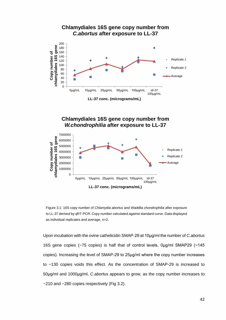
Upon incubation with the ovine cathelicidin SMAP-29 at 10μg/ml the number of C.abortus
16S gene copies (~75 copies) is half that of control levels, 0μg/ml SMAP29 (~145
copies). Increasing the level of SMAP-29 to 25μg/ml where the copy number increases
to ~130 copies voids this effect. As the concentration of SMAP-29 is increased to
50μg/ml and 1000μg/ml, C.abortus appears to grow, as the copy number increases to
~210 and ~280 copies respectively (Fig 3.2).
0
20
40
60
80
100
120
140
160
180
200
0μg/mL 10μg/mL 25μg/mL 50μg/mL 100μg/mL sll-37 100μg/mL
Co
py n
um
be
r o
f ch
lam
yd
iale
s 1
6S
gen
e
LL-37 conc. (micrograms/mL)
Chlamydiales 16S gene copy number from C.abortus after exposure to LL-37
Replicate 1
Replicate 2
Average
0
1000000
2000000
3000000
4000000
5000000
6000000
7000000
0μg/mL 10μg/mL 25μg/mL 50μg/mL 100μg/mL sll-37 100μg/mL
Co
py n
um
ber
of
ch
lam
yd
iale
s 1
6S
gen
e
LL-37 conc. (micrograms/mL)
Chlamydiales 16S gene copy number from W.chondrophilia after exposure to LL-37
Replicate 1
Replicate 2
Average
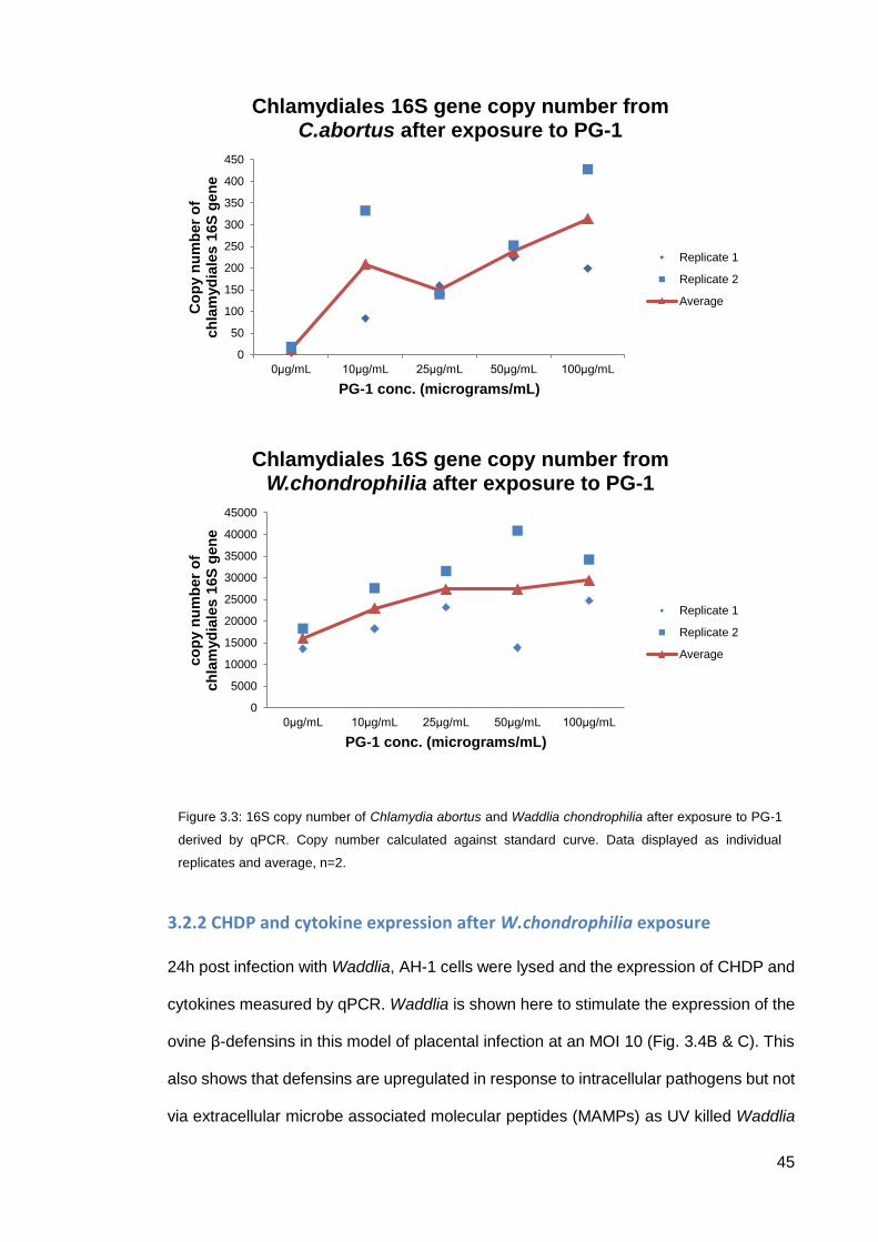
Figure 3.1: 16S copy number of Chlamydia abortus and Waddlia chondrophilia after exposure
to LL-37 derived by qRT-PCR. Copy number calculated against standard curve. Data displayed
as individual replicates and average, n=2.
43
SMAP-29 had a similar effect on W.chondrophilia as it does with C.abortus reducing the
copy number at 10μg/ml (~210000 copies at control down to ~120000 copies at
10μg/ml). Additionally to this SMAP-29 at, 25μg/ml and 50μg/ml both seem to have an
antimicrobial effect reducing the 16S copy number by ~100000 copies. At 100μg/ml
SMAP-29 the copy number returns to control levels of ~210000 copies.
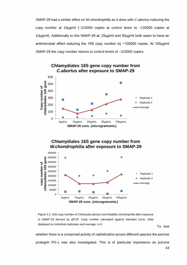
To test
whether there is a conserved activity of cathelicidins across different species the porcine
protegrin PG-1 was also investigated. This is of particular importance as porcine
0
100
200
300
400
500
600
0μg/mL 10μg/mL 25μg/mL 50μg/mL 100μg/mL
Co
py n
um
ber
of
ch
lam
yd
iale
s 1
6S
gen
e
SMAP-29 conc. (micrograms/mL)
Chlamydiales 16S gene copy number from C.abortus after exposure to SMAP-29
Replicate 1
Replicate 2
Average
0
50000
100000
150000
200000
250000
300000
350000
400000
450000
0μg/mL 10μg/mL 25μg/mL 50μg/mL 100μg/mL
co
py n
um
ber
of
ch
lam
yd
iale
s 1
6S
gen
e
SMAP-29 conc. (micrograms/mL)
Chlamydiales 16S gene copy number from W.chondrophilia after exposure to SMAP-29
Replicate 1
Replicate 2
Average
Figure 3.2: 16S copy number of Chlamydia abortus and Waddlia chondrophilia after exposure
to SMAP-29 derived by qPCR. Copy number calculated against standard curve. Data
displayed as individual replicates and average, n=2.
44
livestock can suffer from Chlamydial infection, albeit a different Chlamydia strain (spp.
suis). PG-1 also has less cytotoxic effects against mammalian cells so would be an ideal
candidate as a blueprint for a novel therapeutic. At control levels (0µg/ml) there was a
very low C.abortus 16S copy number present (~10 copies). Upon incubation with PG-1
at 10μg/ml this increased to over 200 copies. Further addition of PG-1 (25μg/ml) reduced
the number of 16S copies to ~150. As the dose of PG-1 is increased to 50μg/ml and
100μg/ml, the copy number of C.abortus increases to ~250 and ~320 respectively (Fig
3.3). PG-1 seemed to promote W.chondrophilia growth as the copy number increased
from control at ~16000 copies to ~23000 copies at 10μg/ml, and increases again to
~27000 copies at 25μg/ml where this effect seems to plateau at 50μg/ml. There is only
a slight increase at 100μg/ml (~29000) relative to the previous increases.
45
3.2.2 CHDP and cytokine expression after W.chondrophilia exposure
24h post infection with Waddlia, AH-1 cells were lysed and the expression of CHDP and
cytokines measured by qPCR. Waddlia is shown here to stimulate the expression of the
ovine β-defensins in this model of placental infection at an MOI 10 (Fig. 3.4B & C). This
also shows that defensins are upregulated in response to intracellular pathogens but not
via extracellular microbe associated molecular peptides (MAMPs) as UV killed Waddlia
0
50
100
150
200
250
300
350
400
450
0μg/mL 10μg/mL 25μg/mL 50μg/mL 100μg/mL
Co
py n
um
ber
of
ch
lam
yd
iale
s 1
6S
gen
e
PG-1 conc. (micrograms/mL)
Chlamydiales 16S gene copy number from C.abortus after exposure to PG-1
Replicate 1
Replicate 2
Average
0
5000
10000
15000
20000
25000
30000
35000
40000
45000
0μg/mL 10μg/mL 25μg/mL 50μg/mL 100μg/mL
co
py n
um
ber
of
ch
lam
yd
iale
s 1
6S
gen
e
PG-1 conc. (micrograms/mL)
Chlamydiales 16S gene copy number from W.chondrophilia after exposure to PG-1
Replicate 1
Replicate 2
Average
Figure 3.3: 16S copy number of Chlamydia abortus and Waddlia chondrophilia after exposure to PG-1
derived by qPCR. Copy number calculated against standard curve. Data displayed as individual
replicates and average, n=2.
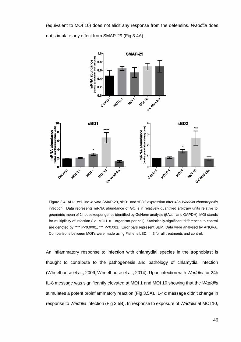
46
(equivalent to MOI 10) does not elicit any response from the defensins. Waddlia does
not stimulate any effect from SMAP-29 (Fig 3.4A).
An inflammatory response to infection with chlamydial species in the trophoblast is
thought to contribute to the pathogenesis and pathology of chlamydial infection
(Wheelhouse et al., 2009; Wheelhouse et al., 2014). Upon infection with Waddlia for 24h
IL-8 message was significantly elevated at MOI 1 and MOI 10 showing that the Waddlia
stimulates a potent proinflammatory reaction (Fig 3.5A). IL-1α message didn’t change in
response to Waddlia infection (Fig 3.5B). In response to exposure of Waddlia at MOI 10,
Figure 3.4. AH-1 cell line in vitro SMAP-29, sBD1 and sBD2 expression after 48h Waddlia chondrophilia
infection. Data represents mRNA abundance of GOI’s in relatively quantified arbitrary units relative to
geometric mean of 2 housekeeper genes identified by GeNorm analysis (βActin and GAPDH). MOI stands
for multiplicity of infection (i.e. MOI1 = 1 organism per cell). Statistically-significant differences to control
are denoted by **** P<0.0001, *** P<0.001. Error bars represent SEM. Data were analysed by ANOVA.
Comparisons between MOI’s were made using Fisher’s LSD. n=3 for all treatments and control.
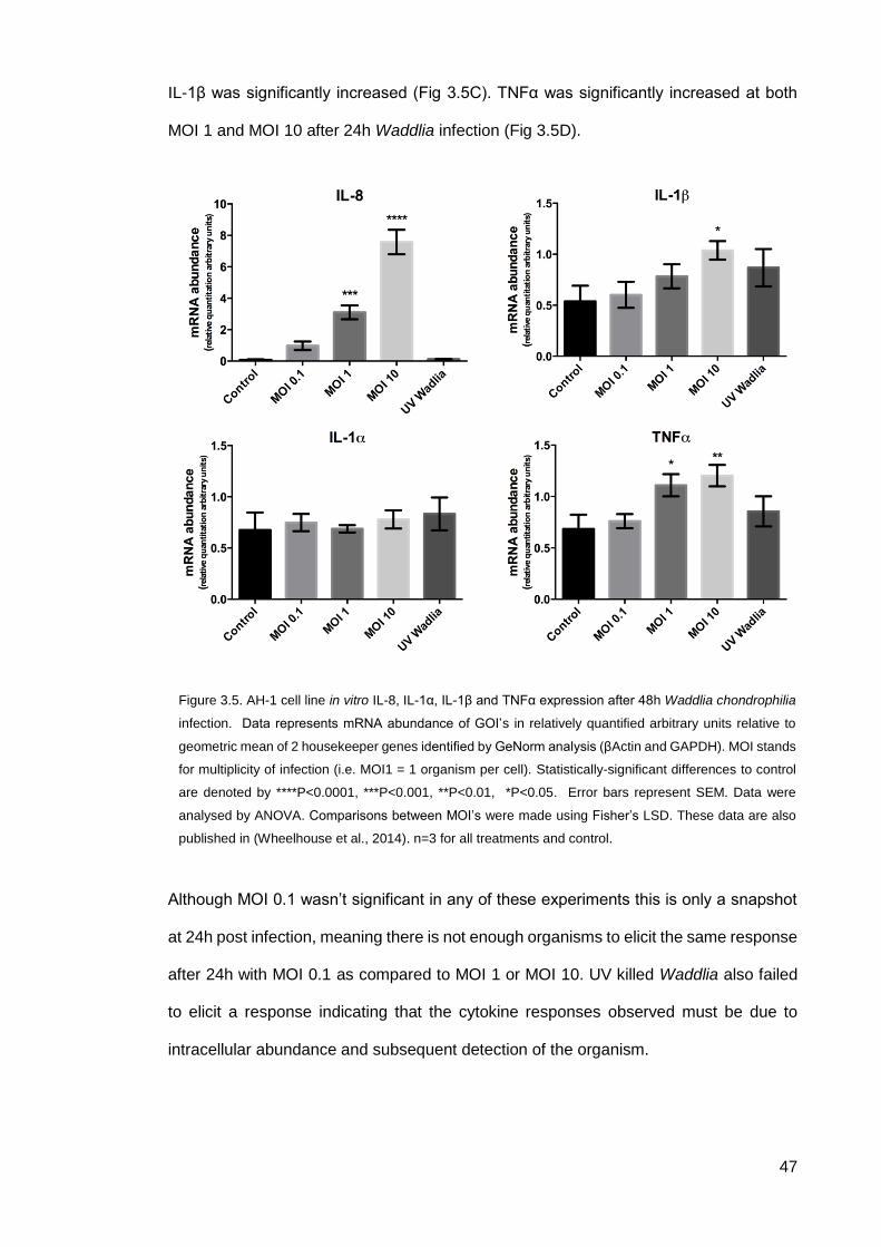
47
IL-1β was significantly increased (Fig 3.5C). TNFα was significantly increased at both
MOI 1 and MOI 10 after 24h Waddlia infection (Fig 3.5D).
Although MOI 0.1 wasn’t significant in any of these experiments this is only a snapshot
at 24h post infection, meaning there is not enough organisms to elicit the same response
after 24h with MOI 0.1 as compared to MOI 1 or MOI 10. UV killed Waddlia also failed
to elicit a response indicating that the cytokine responses observed must be due to
intracellular abundance and subsequent detection of the organism.
Figure 3.5. AH-1 cell line in vitro IL-8, IL-1α, IL-1β and TNFα expression after 48h Waddlia chondrophilia
infection. Data represents mRNA abundance of GOI’s in relatively quantified arbitrary units relative to
geometric mean of 2 housekeeper genes identified by GeNorm analysis (βActin and GAPDH). MOI stands
for multiplicity of infection (i.e. MOI1 = 1 organism per cell). Statistically-significant differences to control
are denoted by ****P<0.0001, ***P<0.001, **P<0.01, *P<0.05. Error bars represent SEM. Data were
analysed by ANOVA. Comparisons between MOI’s were made using Fisher’s LSD. These data are also
published in (Wheelhouse et al., 2014). n=3 for all treatments and control.

48
3.3 Discussion
CHDP are an attractive novel class of antimicrobial therapeutics. Their resilience to
microbial resistance and efficacy against a diverse range of targets including bacteria,
fungi and viruses makes them very attractive as potential future therapies (Padhi et al.,
2014; Tsai et al., 2014; Bucki et al., 2010; Barlow et al., 2014; Yeung et al., 2011). It has
been shown previously that LL-37 can act against intracellular pathogens such as
Mycobacteria spp. (Padhi et al., 2014; Sato et al., 2013).
With previous studies showing that these cathelicidins have the ability to act upon
intracellular pathogens we have chosen three cathelicidins; human LL-37, ovine SMAP-
29 and porcine PG-1, and tested their antimicrobial properties against the common
intracellular pathogen Chlamydia abortus and a new emerging pathogen Waddlia
chondrophilia. Both of C.abortus and W.chondrophilia are intracellular pathogens that
can cause abortion during pregnancy in livestock and humans (Dilbeck-Robertson et al.,
2003; Baud et al., 2007; Baud et al., 2008; Pospischil et al., 2002). Here, using 16S gene
copy number as a proxy for microbial growth, we have shown that LL-37 and PG-1 are
ineffective against C.abortus and W.chondrophilia as paradoxically they do not inhibit
the growth of the organisms; they promote growth of the organisms. As the concentration
of cathelicidin is increased to supraphysiological concentrations (>25-30µg/mL) (Nijnik &
Hancock, 2009), the copy number of the 16S gene in both organisms increases in the
presence of all CHDP tested (Fig. 3.1 and 3.3). SMAP-29 has a slightly different effect
on both C.abortus and W.chondrophilia; showing that the 16S copy number is reduced
at 10g/mL, indicating a reduction in bacterial cell growth. Against C.abortus this effect
of SMAP-29 is only found at 10g/mL and the paradoxical increase in 16S gene copy
number is found as the SMAP-29 concentration increases towards 100g/mL. SMAP-29
seems to have a greater ‘therapeutic window’ and is able to maintain an bacteriostatic
hold on 16S gene copy number at concentrations between 10g/mL to 50g/mL before
bacterial growth resumes.
49
A classical antimicrobial function of CHDP has been the ability to interfere with the
microbial cell wall and create perforations due to the net positive charge of the molecule
and negative net charge of the cell wall of bacteria (Ramanathan et al., 2002). As
indicated by our data, LL-37 and PG-1 in particular do not affect C.abortus and
W.chondrophilia as we would expect them too by reducing bacterial growth. We have
shown that the organisms exhibit greater growth when there is more cathelicidin present.
One hypothesis that may explain this phenomenon is that at supra-physiological
concentrations, LL-37 (Henzler-Wildman et al., 2003; Henzler-Wildman et al., 2004) and
PG-1 (Heller et al., 2000) will disrupt the host cell membrane and this inadvertent
property of these cathelicidins may make the host cells more permissive to infection. As
the sLL-37 control has a similar number of copies of the 16S gene to 100μg/ml this must
be due to some other mechanism that requires further investigation.
The approach we utilized here tested the effect of these cathelicidins against the
microbes directly before they were able to establish an infection (Fig 3.6). By using this
approach, we may have stressed the bacteria, which could explain our findings of an
increase in bacterial growth as the concentration of LL-37 and PG-1 increase. Chlamydia
trachomatis can secrete the serine protease chlamydial proteasome/protease-like
activity factor (CPAF) which can cleave LL-37 and eliminate its antimicrobial actions so
a similar mechanism cannot be ruled out here (Tang et al., 2015). In order to fully assess
the efficacy of cathelicidins against C.abortus and W.chondrophilia infection further work
is required. In the first instance, AH-1 cells could be pre-incubated with differing
concentrations of cathelicidin to elucidate firstly whether there is an effect of the peptides
on the cells themselves before the microbes are introduced, secondly if after introduction
of the microbes, whether these cells are more or less permissive to infection (Fig. 3.6).
We also did not test the ability of the cathelicidins to effect an established infection, which
would be a more realistic scenario (Fig. 3.6).
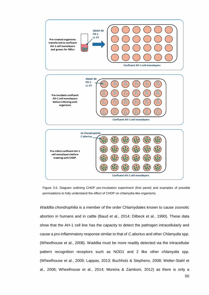
50
Waddlia chondrophilia is a member of the order Chlamydiales known to cause zoonotic
abortion in humans and in cattle (Baud et al., 2014; Dilbeck et al., 1990). These data
show that the AH-1 cell line has the capacity to detect the pathogen intracellularly and
cause a pro-inflammatory response similar to that of C.abortus and other Chlamydia spp.
(Wheelhouse et al., 2008). Waddlia must be more readily detected via the intracellular
pattern recognition receptors such as NOD1 and 2 like other chlamydia spp.
(Wheelhouse et al., 2009; Lappas, 2013; Buchholz & Stephens, 2008; Welter-Stahl et
al., 2006; Wheelhouse et al., 2014; Moreira & Zamboni, 2012) as there is only a
Figure 3.6. Diagram outlining CHDP pre-incubation experiment (first panel) and examples of possible
permutations to fully understand the effect of CHDP on chlamydia-like organisms.

51
noticeable response to live infection and not to the UV killed organisms. For the first time
this data demonstrates a change in CHDP production during Waddlia infection. The
increase in ovine β-defensins could be alluding to a mechanism were the defensins can
be simulated by intracellular pathogen detection potentially via NOD1/2 (Moreira &
Zamboni, 2012).
52
Final Conclusions and Further Work
Within this body of work the importance of the steroid milieu and consequences of
chlamydial infection during pregnancy in the context of cationic host defense peptides
and innate immunity have been outlined, but a significant body of work remains to be
undertaken.
These data show that there is a potential mechanism for manipulating immunological
tolerance within the thymus via excess exposure to androgens in utero. This mechanism
could also be underpinning the sexual dimorphism found in autoimmunity but a whole
suite of studies would be required to fully investigate this hypothesis.
Through the LPS exposures and the chlamydial infection studies in the AH-1 cell line, a
better understanding of pathogen recognition and innate immune response has been
started, Further work using inhibitors of TLR4/2 and NOD1/2 signal transduction is
required to fully understand the mechanism by which W.chondrophilia is detected and
reported to the rest of the immune system. Furthermore, to assess whether there is some
form of dampening or modulation of the classical TLR4 pathway in the AH-1 cell line and
subsequently the placenta as this could be a unique phenomenon, as macrophages
exposed to LPS will produce LL-37 via TLR4 signaling (Bucki et al., 2010).
The expression of cathelicidin in the uterus remains underinvestigated, and it is unclear
whether the ability of cathelicidins to modulate the adaptive immune system is
contributory to the tolerance of heterogeneous fetus during implantation and
subsequently the duration of pregnancy.
These data show how the steroid milieu and sterility, in the context of infection, of the
reproductive tract can regulate the expression of CHDP, which could ultimately have an
impact on the success of pregnancy.
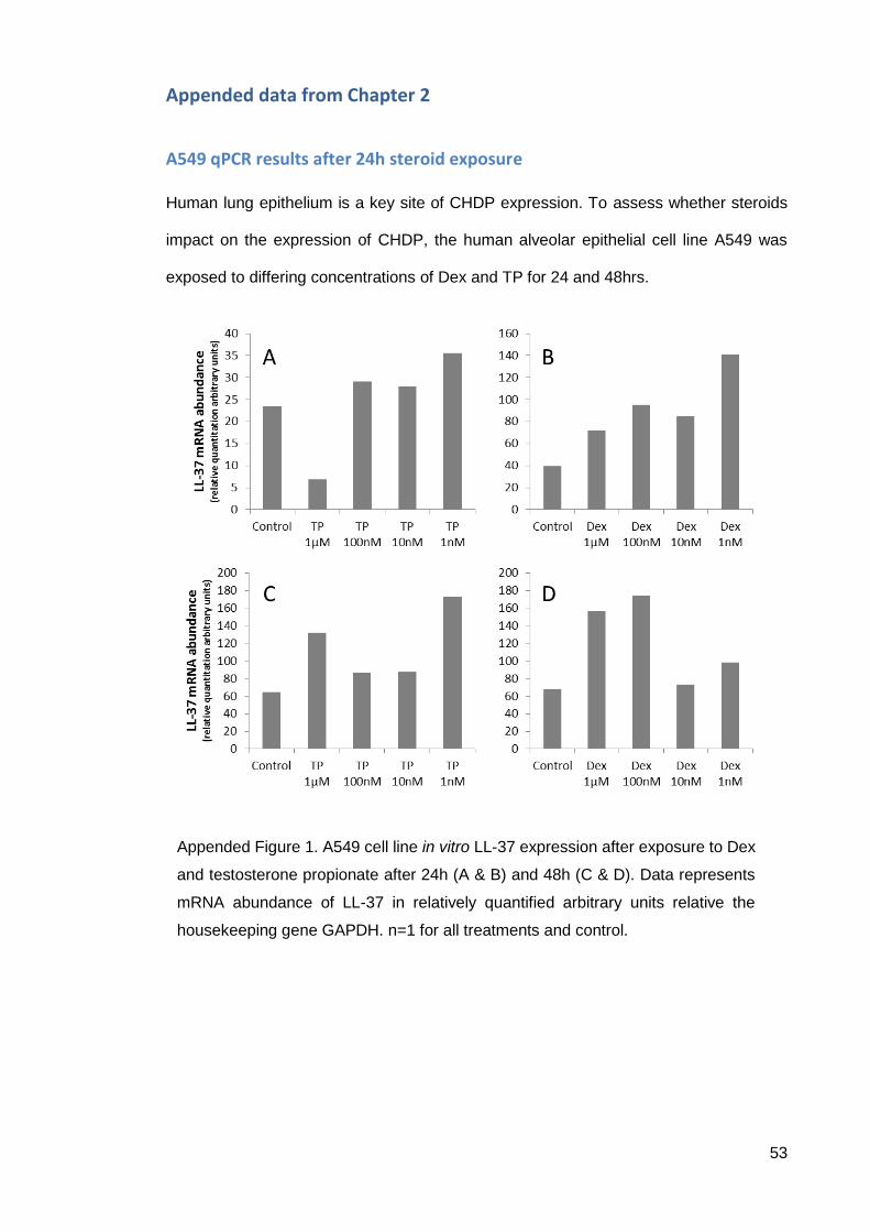
53
Appended data from Chapter 2
A549 qPCR results after 24h steroid exposure
Human lung epithelium is a key site of CHDP expression. To assess whether steroids
impact on the expression of CHDP, the human alveolar epithelial cell line A549 was
exposed to differing concentrations of Dex and TP for 24 and 48hrs.
Appended Figure 1. A549 cell line in vitro LL-37 expression after exposure to Dex
and testosterone propionate after 24h (A & B) and 48h (C & D). Data represents
mRNA abundance of LL-37 in relatively quantified arbitrary units relative the
housekeeping gene GAPDH. n=1 for all treatments and control.
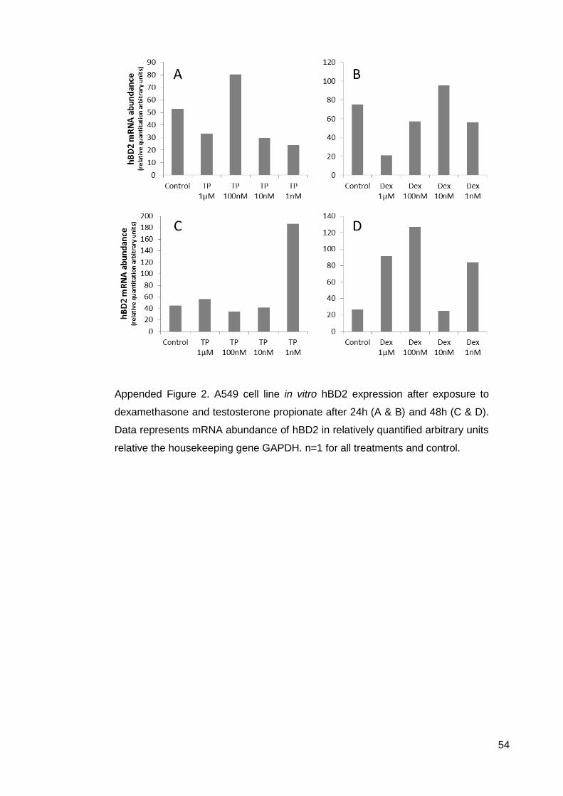
54
Appended Figure 2. A549 cell line in vitro hBD2 expression after exposure to
dexamethasone and testosterone propionate after 24h (A & B) and 48h (C & D).
Data represents mRNA abundance of hBD2 in relatively quantified arbitrary units
relative the housekeeping gene GAPDH. n=1 for all treatments and control.
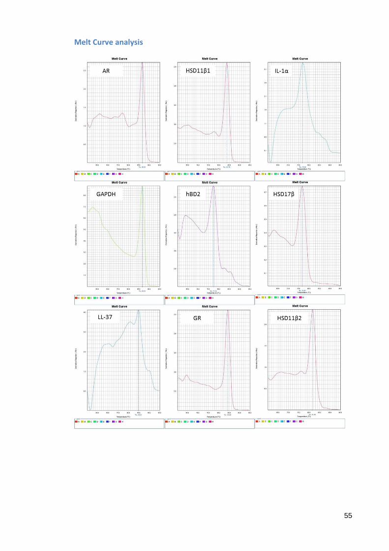
55
Melt Curve analysis
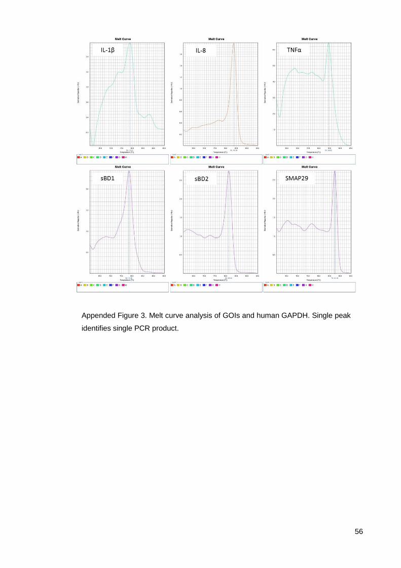
56
Appended Figure 3. Melt curve analysis of GOIs and human GAPDH. Single peak
identifies single PCR product.
57
Reference List
Abbott, D.H., Dumesic, D. a, Eisner, J.R., Colman, R.J. & Kemnitz, J.W., 1998. Insights into the development of polycystic ovary syndrome (PCOS) from studies of prenatally androgenized female rhesus monkeys. Trends in endocrinology and metabolism: TEM, 9(2), pp.62–7.
Bals, R., Wang, X., Zasloff, M. & Wilson, J.M., 1998. The peptide antibiotic LL-37/hCAP-18 is expressed in epithelia of the human lung where it has broad antimicrobial activity at the airway surface. Proceedings of the National Academy of Sciences of the United States of America, 95(16), pp.9541–6.
Baltzer, S. a & Brown, M.H., 2011. Antimicrobial peptides: promising alternatives to conventional antibiotics. Journal of molecular microbiology and biotechnology, 20(4), pp.228–35.
Barlow, P.G., Beaumont, P.E., Cosseau, C., Mackellar, A., Wilkinson, T.S., Hancock, R.E.W., Haslett, C., Govan, J.R.W., Simpson, A.J. & Davidson, D.J., 2010. The human cathelicidin LL-37 preferentially promotes apoptosis of infected airway epithelium. American journal of respiratory cell and molecular biology, 43(6), pp.692–702.
Barlow, P.G., Findlay, E.G., Currie, S.M. & Davidson, D.J., 2014. Antiviral potential of cathelicidins. Future microbiology, 9(1), pp.55–73.
Barlow, P.G., Li, Y., Wilkinson, T.S., Bowdish, D.M.E., Lau, Y.E., Cosseau, C., Haslett, C., Simpson, A.J., Hancock, R.E.W. & Davidson, D.J., 2006. The human cationic host defense peptide LL-37 mediates contrasting effects on apoptotic pathways in different primary cells of the innate immune system. Journal of leukocyte biology, 80(3), pp.509–20.
Barlow, P.G., Svoboda, P., Mackellar, A., Nash, A. a, York, I. a, Pohl, J., Davidson, D.J. & Donis, R.O., 2011. Antiviral activity and increased host defense against influenza infection elicited by the human cathelicidin LL-37. PloS one, 6(10), p.e25333.
Barnes, P.J., 1998. Anti-inflammatory actions of glucocorticoids: molecular mechanisms. Clinical science (London, England : 1979), 94, pp.557–572.
de Barsy, M. & Greub, G., 2013. Waddlia chondrophila: from biology to pathogenicity. Microbes and infection / Institut Pasteur, 15(14-15), pp.1033–41.
Baud, D., Goy, G., Osterheld, M.-C., Croxatto, A., Borel, N., Vial, Y., Pospischil, A. & Greub, G., 2014. Role of Waddlia chondrophila Placental Infection in Miscarriage. Emerging infectious diseases, 20, pp.460–4.
Baud, D., Regan, L. & Greub, G., 2008. Emerging role of Chlamydia and Chlamydia-like organisms in adverse pregnancy outcomes. Current opinion in infectious diseases, 21, pp.70–76.
Baud, D., Thomas, V., Arafa, A., Regan, L. & Greub, G., 2007. Waddlia chondrophila, a potential agent of human fetal death. Emerging infectious diseases, 13, pp.1239–1243.
Blackburn, C.C. & Manley, N.R., 2004. Developing a new paradigm for thymus organogenesis. Nature reviews. Immunology, 4(4), pp.278–89.
Bowdish, D.M.E., Davidson, D.J. & Hancock, R.E.W., 2005. A re-evaluation of the role of host defence peptides in mammalian immunity. Current protein & peptide science, 6(1), pp.35–51.
Bowdish, D.M.E., Davidson, D.J. & Hancock, R.E.W., 2006. Immunomodulatory properties of defensins and cathelicidins. Current topics in microbiology and immunology, 306, pp.27–66.
58
Bowdish, D.M.E., Davidson, D.J., Scott, M.G. & Hancock, R.E.W., 2005. Immunomodulatory activities of small host defense peptides. Antimicrobial agents and chemotherapy, 49, pp.1727–1732.
Brown, K.L. & Hancock, R.E.W., 2006. Cationic host defense (antimicrobial) peptides. Current opinion in immunology, 18(1), pp.24–30.
Buchholz, K.R. & Stephens, R.S., 2008. The cytosolic pattern recognition receptor NOD1 induces inflammatory interleukin-8 during Chlamydia trachomatis infection. Infection and immunity, 76(7), pp.3150–5.
Bucki, R., Leszczyńska, K., Namiot, A. & Sokołowski, W., 2010. Cathelicidin LL-37: a multitask antimicrobial peptide. Archivum immunologiae et therapiae experimentalis, 58(1), pp.15–25.
Burton, G.J., 2009. Oxygen, the Janus gas; its effects on human placental development and function. Journal of anatomy, 215(1), pp.27–35.
Colantonio, A.D., Epeldegui, M., Jesiak, M., Jachimowski, L., Blom, B. & Uittenbogaart, C.H., 2011. IFN-α is constitutively expressed in the human thymus, but not in peripheral lymphoid organs. PloS one, 6(8), p.e24252.
Collie, D., Govan, J., Wright, S., Thornton, E., Tennant, P., Smith, S., Doherty, C. & McLachlan, G., 2013. A lung segmental model of chronic Pseudomonas infection in sheep. PloS one, 8(7), p.e67677.
Dilbeck, P.M., Evermann, J.F., Crawford, T.B., Ward, A.C., Leathers, C.W., Holland, C.J., Mebus, C.A., Logan, L.L., Rurangirwa, F.R. & McGuire, T.C., 1990. Isolation of a previously undescribed rickettsia from an aborted bovine fetus. Journal of clinical microbiology, 28, pp.814–816.
Dilbeck-Robertson, P., McAllister, M.M., Bradway, D. & Evermann, J.F., 2003. Results of a new serologic test suggest an association of Waddlia chondrophila with bovine abortion. Journal of veterinary diagnostic investigation : official publication of the American Association of Veterinary Laboratory Diagnosticians, Inc, 15, pp.568–569.
Donati, M. & Leo, K. Di, 2005. Activity of cathelicidin peptides against Chlamydia spp. Antimicrobial agents …, 49(3), pp.1201–1202.
Dürr, U.H.N., Sudheendra, U.S. & Ramamoorthy, A., 2006. LL-37, the only human member of the cathelicidin family of antimicrobial peptides. Biochimica et biophysica acta, 1758(9), pp.1408–25.
Entrican, G., 2002. Immune regulation during pregnancy and host-pathogen interactions in infectious abortion. Journal of comparative pathology, 126(2-3), pp.79–94.
Entrican, G., Wattegedera, S., Wheelhouse, N., Allan, A. & Rocchi, M., 2010. Immunological paradigms and the pathogenesis of ovine chlamydial abortion. American journal of reproductive immunology (New York, N.Y. : 1989), 64(4), pp.287–94.
Fach, S.J., Meyerholz, D.K., Gallup, J.M., Ackermann, M.R., Lehmkuhl, H.D. & Sacco, R.E., 2007. Neonatal ovine pulmonary dendritic cells support bovine respiratory syncytial virus replication with enhanced interleukin (IL)-4 And IL-10 gene transcripts. Viral immunology, 20(1), pp.119–30.
Fowden, a L. & Forhead, a J., 2004. Endocrine mechanisms of intrauterine programming. Reproduction (Cambridge, England), 127(5), pp.515–26.
Frew, L. & Stock, S.J., 2011. Antimicrobial peptides and pregnancy. Reproduction (Cambridge, England), 141(6), pp.725–35.
Ganguly, D., Chamilos, G., Lande, R., Gregorio, J., Meller, S., Facchinetti, V., Homey, B., Barrat, F.J., Zal, T. & Gilliet, M., 2009. Self-RNA-antimicrobial peptide complexes activate human dendritic cells through TLR7 and TLR8. The Journal of
59
experimental medicine, 206(9), pp.1983–94.
Ganz, T., 2003. Defensins: antimicrobial peptides of innate immunity. Nature reviews. Immunology, 3(9), pp.710–20.
Gombart, A.F., Borregaard, N. & Koeffler, H.P., 2005. Human cathelicidin antimicrobial peptide (CAMP) gene is a direct target of the vitamin D receptor and is strongly up-regulated in myeloid cells by 1,25-dihydroxyvitamin D3. FASEB journal : official publication of the Federation of American Societies for Experimental Biology, 19(9), pp.1067–77.
Goy, G., Croxatto, A. & Greub, G., 2008. Waddlia chondrophila enters and multiplies within human macrophages. Microbes and Infection, 10, pp.556–562.
Grenache, D.G. & Gronowski, A.M., 2006. Fetal lung maturity. Clinical biochemistry, 39(1), pp.1–10.
Gubbels Bupp, M.R., Jørgensen, T.N. & Kotzin, B.L., 2008. Identification of candidate genes that influence sex hormone-dependent disease phenotypes in mouse lupus. Genes and immunity, 9(1), pp.47–56.
Gui, J., Morales, A.J., Maxey, S.E., Bessette, K.A., Ratcliffe, N.R., Kelly, J.A. & Craig, R.W., 2011. MCL1 increases primitive thymocyte viability in female mice and promotes thymic expansion into adulthood. International immunology, 23(10), pp.647–59.
Harris, A. & Seckl, J., 2011. Glucocorticoids, prenatal stress and the programming of disease. Hormones and Behavior, 59, pp.279–289.
Heller, W.T., Waring, A.J., Lehrer, R.I., Harroun, T.A., Weiss, T.M., Yang, L. & Huang, H.W., 2000. Membrane Thinning Effect of the β-Sheet Antimicrobial Protegrin †. Biochemistry, 39(1), pp.139–145.
Henzler Wildman, K.A., Lee, D.-K. & Ramamoorthy, A., 2003. Mechanism of lipid bilayer disruption by the human antimicrobial peptide, LL-37. Biochemistry, 42(21), pp.6545–58.
Henzler-Wildman, K.A., Martinez, G. V, Brown, M.F. & Ramamoorthy, A., 2004. Perturbation of the hydrophobic core of lipid bilayers by the human antimicrobial peptide LL-37. Biochemistry, 43(26), pp.8459–69.
Hince, M., Sakkal, S., Vlahos, K., Dudakov, J., Boyd, R. & Chidgey, A., 2008. The role of sex steroids and gonadectomy in the control of thymic involution. Cellular immunology, 252(1-2), pp.122–38.
Horne, A.W., Stock, S.J. & King, A.E., 2008. Innate immunity and disorders of the female reproductive tract. Reproduction (Cambridge, England), 135(6), pp.739–49.
Huttner, K.M., Lambeth, M.R., Burkin, H.R., Burkin, D.J. & Broad, T.E., 1998. Localization and genomic organization of sheep antimicrobial peptide. Gene, 206, pp.85–91.
Igwebuike, U.M., 2006. Trophoblast cells of ruminant placentas--A minireview. Animal reproduction science, 93(3-4), pp.185–98.
Kapoor, A., Petropoulos, S. & Matthews, S.G., 2008. Fetal programming of hypothalamic-pituitary-adrenal (HPA) axis function and behavior by synthetic glucocorticoids. Brain research reviews, 57(2), pp.586–95.
Kebbi-Beghdadi, C., Batista, C. & Greub, G., 2011. Permissivity of fish cell lines to three Chlamydia-related bacteria: Waddlia chondrophila, Estrella lausannensis and Parachlamydia acanthamoebae. FEMS Immunology and Medical Microbiology, 63, pp.339–345.
Kebbi-Beghdadi, C., Cisse, O. & Greub, G., 2011. Permissivity of Vero cells, human pneumocytes and human endometrial cells to Waddlia chondrophila. Microbes and Infection, 13, pp.566–574.
60
Kerr, K., Entrican, G., McKeever, D. & Longbottom, D., 2005. Immunopathology of Chlamydophila abortus infection in sheep and mice. Research in veterinary science, 78(1), pp.1–7.
Lagarde, F., Beausoleil, C., Belcher, S.M., Belzunces, L.P., Emond, C., Guerbet, M. & Rousselle, C., 2015. Non-monotonic dose-response relationships and endocrine disruptors: a qualitative method of assessment. Environmental health : a global access science source, 14(1), p.13.
Lagishetty, V., Liu, N.Q. & Hewison, M., 2011. Vitamin D metabolism and innate immunity. Molecular and cellular endocrinology, 347(1-2), pp.97–105.
Lande, R., Gregorio, J., Facchinetti, V., Chatterjee, B., Wang, Y.-H., Homey, B., Cao, W., Wang, Y.-H., Su, B., Nestle, F.O., Zal, T., Mellman, I., Schröder, J.-M., Liu, Y.-J. & Gilliet, M., 2007. Plasmacytoid dendritic cells sense self-DNA coupled with antimicrobial peptide. Nature, 449, pp.564–569.
Lappas, M., 2013. NOD1 and NOD2 regulate proinflammatory and prolabor mediators in human fetal membranes and myometrium via nuclear factor-kappa B. Biology of reproduction, 89(1), p.14.
Lee, B.H., Stoll, B.J., McDonald, S. a & Higgins, R.D., 2006. Adverse neonatal outcomes associated with antenatal dexamethasone versus antenatal betamethasone. Pediatrics, 117(5), pp.1503–10.
Lee, D.K., Hakim, F.T. & Gress, R.E., 2010. The thymus and the immune system: layered levels of control. Journal of thoracic oncology : official publication of the International Association for the Study of Lung Cancer, 5, pp.S273–S276.
Li, D., Wang, X., Wu, J.-L.L., Quan, W.-Q.Q., Ma, L., Yang, F., Wu, K.-Y.Y. & Wan, H.-Y.Y., 2013. Tumor-Produced Versican V1 Enhances hCAP18/LL-37 Expression in Macrophages through Activation of TLR2 and Vitamin D3 Signaling to Promote Ovarian Cancer Progression In Vitro. PLoS ONE, 8(2), p.e56616.
Li, H.-N., Barlow, P.G., Bylund, J., Mackellar, A., Björstad, A., Conlon, J., Hiemstra, P.S., Haslett, C., Gray, M., Simpson, A.J., Rossi, A.G. & Davidson, D.J., 2009. Secondary necrosis of apoptotic neutrophils induced by the human cathelicidin LL-37 is not proinflammatory to phagocytosing macrophages. Journal of leukocyte biology, 86, pp.891–902.
Lienard, J., Croxatto, A., Aeby, S., Jaton, K., Posfay-Barbe, K., Gervaix, A. & Greub, G., 2011. Development of a new chlamydiales-specific real-time PCR and its application to respiratory clinical samples. Journal of clinical microbiology, 49(7), pp.2637–42.
Liu, N., Kaplan, a T., Low, J., Nguyen, L., Liu, G.Y., Equils, O. & Hewison, M., 2009. Vitamin D induces innate antibacterial responses in human trophoblasts via an intracrine pathway. Biology of reproduction, 80(3), pp.398–406.
Longbottom, D. & Coulter, L.J., 2003. Animal Chlamydioses and Zoonotic Implications. Journal of Comparative Pathology, 128(4), pp.217–244.
Luk, J., Torrealday, S., Neal Perry, G. & Pal, L., 2012. Relevance of vitamin D in reproduction. Human reproduction (Oxford, England), 27(10), pp.3015–27.
Mahoney, M.M., Lee, A.Y., Brezinski-Caliguri, D.J. & Huttner, K.M., 1995. Molecular analysis of the sheep cathelin family reveals a novel antimicrobial peptide. FEBS letters, 377(3), pp.519–22.
Mookherjee, N., Lippert, D.N.D., Hamill, P., Falsafi, R., Nijnik, A., Kindrachuk, J., Pistolic, J., Gardy, J., Miri, P., Naseer, M., Foster, L.J. & Hancock, R.E.W., 2009. Intracellular receptor for human host defense peptide LL-37 in monocytes. Journal of immunology (Baltimore, Md. : 1950), 183(4), pp.2688–96.
Moreira, L.O. & Zamboni, D.S., 2012. NOD1 and NOD2 Signaling in Infection and Inflammation. Frontiers in immunology, 3, p.328.
61
Nagaoka, I., Tamura, H. & Hirata, M., 2006. An antimicrobial cathelicidin peptide, human CAP18/LL-37, suppresses neutrophil apoptosis via the activation of formyl-peptide receptor-like 1 and P2X7. Journal of immunology (Baltimore, Md. : 1950), 176, pp.3044–3052.
Newnham, J.P., Evans, S.F., Godfrey, M., Huang, W., Ikegami, M. & Jobe, a, 1999. Maternal, but not fetal, administration of corticosteroids restricts fetal growth. The Journal of maternal-fetal medicine, 8(3), pp.81–7.
Nijnik, A. & Hancock, R.E., 2009. The roles of cathelicidin LL-37 in immune defences and novel clinical applications. Current Opinion in Hematology, 16(1), pp.41–47.
Olsen, N.J. & Kovacs, W.J., 2001. Effects of androgens on T and B lymphocyte development. Immunologic research, 23(2-3), pp.281–8.
Olsen, N.J., Olson, G., Viselli, S.M., Gu, X. & Kovacs, W.J., 2001. Androgen receptors in thymic epithelium modulate thymus size and thymocyte development. Endocrinology, 142(3), pp.1278–83.
Padhi, A., Sengupta, M., Sengupta, S., Roehm, K.H. & Sonawane, A., 2014. Antimicrobial peptides and proteins in mycobacterial therapy: Current status and future prospects. Tuberculosis (Edinburgh, Scotland), 94(4), pp.363–73.
Padmanabhan, V. & Veiga-Lopez, A., 2011. Developmental origin of reproductive and metabolic dysfunctions: Androgenic versus estrogenic reprogramming. Seminars in Reproductive Medicine, 29, pp.173–186.
Padmanabhan, V. & Veiga-Lopez, A., 2013. Sheep models of polycystic ovary syndrome phenotype. Molecular and cellular endocrinology, 373(1-2), pp.8–20.
Papp, J.R., Shewen, P.E. & Gartley, C.J., 1993. Chlamydia psittaci infection and associated infertility in sheep. Canadian journal of veterinary research = Revue canadienne de recherche veterinaire, 57, pp.185–189.
Pazgier, M., Ericksen, B., Ling, M., Toth, E., Shi, J., Li, X., Galliher-Beckley, A., Lan, L., Zou, G., Zhan, C., Yuan, W., Pozharski, E. & Lu, W., 2013. Structural and functional analysis of the pro-domain of human cathelicidin, LL-37. Biochemistry, 52(9), pp.1547–58.
Pazgier, M., Hoover, D.M., Yang, D., Lu, W. & Lubkowski, J., 2006. Human beta-defensins. Cellular and molecular life sciences : CMLS, 63(11), pp.1294–313.
Pospischil, A., Thoma, R., Hilbe, M., Grest, P. & Gebbers, F.O., 2002. Abortion in woman caused by caprine Chlamydophila abortus (Chlamydia psittaci serovar 1). Swiss Medical Weekly, 132, pp.64–66.
Rae, M., Grace, C., Hogg, K., Wilson, L.M., McHaffie, S.L., Ramaswamy, S., MacCallum, J., Connolly, F., McNeilly, A.S. & Duncan, C., 2013. The pancreas is altered by in utero androgen exposure: implications for clinical conditions such as polycystic ovary syndrome (PCOS). PloS one, 8(2), p.e56263.
Ramanathan, B., Davis, E.G., Ross, C.R. & Blecha, F., 2002. Cathelicidins: microbicidal activity, mechanisms of action, and roles in innate immunity. Microbes and infection / Institut Pasteur, 4(3), pp.361–72.
Reynolds, R.M. & Seckl, J.R., 2012. Antenatal glucocorticoid treatment: are we doing harm to term babies? The Journal of clinical endocrinology and metabolism, 97(10), pp.3457–9.
Roseboom, T.J., van der Meulen, J.H., Ravelli, a C., Osmond, C., Barker, D.J. & Bleker, O.P., 2001. Effects of prenatal exposure to the Dutch famine on adult disease in later life: an overview. Twin research : the official journal of the International Society for Twin Studies, 4(5), pp.293–8.
Rurangirwa, F.R., Timothy, M.D., Mcguirel, T.C. & Mcelwaing, T.F., 1999. Analysis of the 16s rRNA gene of micro- organism WSU 86-1044 from an aborted bovine foetus
62
reveals that it is a member of the order Chlamydiales : proposal of Waddliaceae fam . nov ., Waddlia chondrophila gen . nov ., sp . nova. Internaional Journal of Systemic Bacteriology, (49), pp.577–581.
Sakiani, S., Olsen, N.J. & Kovacs, W.J., 2013. Gonadal steroids and humoral immunity. Nature reviews. Endocrinology, 9(1), pp.56–62.
Sammin, D., Markey, B., Bassett, H. & Buxton, D., 2009. The ovine placenta and placentitis-A review. Veterinary microbiology, 135(1-2), pp.90–7.
Sammin, D.J., Markey, B.K., Quinn, P.J., McElroy, M.C. & Bassett, H.F., 2006. Comparison of fetal and maternal inflammatory responses in the ovine placenta after experimental infection with Chlamydophila abortus. Journal of comparative pathology, 135(2-3), pp.83–92.
Sato, E., Imafuku, S., Ishii, K., Itoh, R., Chou, B., Soejima, T., Nakayama, J. & Hiromatsu, K., 2013. Vitamin D-dependent cathelicidin inhibits Mycobacterium marinum infection in human monocytic cells. Journal of dermatological science, 70(3), pp.166–72.
Schaller-Bals, S., Schulze, A. & Bals, R., 2002. Increased levels of antimicrobial peptides in tracheal aspirates of newborn infants during infection. American journal of respiratory and critical care medicine, 165(7), pp.992–5.
Scott, H.M., Hutchison, G.R., Mahood, I.K., Hallmark, N., Welsh, M., De Gendt, K., Verhoeven, G., O’Shaughnessy, P. & Sharpe, R.M., 2007. Role of androgens in fetal testis development and dysgenesis. Endocrinology, 148(5), pp.2027–36.
Scott, H.M., Mason, J.I. & Sharpe, R.M., 2009. Steroidogenesis in the fetal testis and its susceptibility to disruption by exogenous compounds. Endocrine reviews, 30(7), pp.883–925.
Scott, M.G., Davidson, D.J., Gold, M.R., Bowdish, D. & Hancock, R.E.W., 2002. The human antimicrobial peptide LL-37 is a multifunctional modulator of innate immune responses. Journal of immunology (Baltimore, Md. : 1950), 169(7), pp.3883–91.
Seaborn, T., Simard, M., Provost, P.R., Piedboeuf, B. & Tremblay, Y., 2010. Sex hormone metabolism in lung development and maturation. Trends in endocrinology and metabolism: TEM, 21(12), pp.729–38.
Semple, F. & Dorin, J.R., 2012. β-Defensins: multifunctional modulators of infection, inflammation and more? Journal of innate immunity, 4(4), pp.337–48.
Sharpe, R.M., 2006. Pathways of endocrine disruption during male sexual differentiation and masculinization. Best practice & research. Clinical endocrinology & metabolism, 20(1), pp.91–110.
Smeed, J.A., Watkins, C.A., Rhind, S.M. & Hopkins, J., 2007. Differential cytokine gene expression profiles in the three pathological forms of sheep paratuberculosis. BMC veterinary research, 3, p.18.
Sørensen, O., Arnljots, K., Cowland, J.B., Bainton, D.F. & Borregaard, N., 1997. The human antibacterial cathelicidin, hCAP-18, is synthesized in myelocytes and metamyelocytes and localized to specific granules in neutrophils. Blood, 90, pp.2796–2803.
Starner, T.D., Agerberth, B., Gudmundsson, G.H. & McCray, P.B., 2005. Expression and activity of beta-defensins and LL-37 in the developing human lung. Journal of immunology (Baltimore, Md. : 1950), 174(3), pp.1608–15.
Tang, L., Chen, J., Zhou, Z., Yu, P., Yang, Z. & Zhong, G., 2015. Chlamydia-secreted protease CPAF degrades host antimicrobial peptides. Microbes and Infection, (March), pp.1–7.
Tecle, T., Tripathi, S. & Hartshorn, K.L., 2010. Review: Defensins and cathelicidins in lung immunity. Innate immunity, 16(3), pp.151–9.
63
Tegethoff, M., Pryce, C. & Meinlschmidt, G., 2009. Effects of intrauterine exposure to synthetic glucocorticoids on fetal, newborn, and infant hypothalamic-pituitary-adrenal axis function in humans: a systematic review. Endocrine reviews, 30(7), pp.753–89.
Tsai, P.-W., Cheng, Y.-L., Hsieh, W.-P. & Lan, C.-Y., 2014. Responses of Candida albicans to the human antimicrobial peptide LL-37. Journal of microbiology (Seoul, Korea), 52(8).
Vandamme, D., Landuyt, B., Luyten, W. & Schoofs, L., 2012. A comprehensive summary of LL-37, the factotum human cathelicidin peptide. Cellular immunology, 280(1), pp.22–35.
Veiga-Lopez, A., Steckler, T.L., Abbott, D.H., Welch, K.B., MohanKumar, P.S., Phillips, D.J., Refsal, K. & Padmanabhan, V., 2011. Developmental programming: impact of excess prenatal testosterone on intrauterine fetal endocrine milieu and growth in sheep. Biology of reproduction, 84(1), pp.87–96.
Walker, B.R., 2007. Glucocorticoids and cardiovascular disease. European journal of endocrinology / European Federation of Endocrine Societies, 157, pp.545–559.
Wallace, A.D. & Cidlowski, J.A., 2001. Proteasome-mediated glucocorticoid receptor degradation restricts transcriptional signaling by glucocorticoids. The Journal of biological chemistry, 276(46), pp.42714–21.
Welsh, M., Saunders, P.T.K., Fisken, M., Scott, H.M., Hutchison, G.R., Smith, L.B. & Sharpe, R.M., 2008. Identification in rats of a programming window for reproductive tract masculinization, disruption of which leads to hypospadias and cryptorchidism. Journal of Clinical Investigation, 118(4), pp.1479–1490.
Welter-Stahl, L., Ojcius, D.M., Viala, J., Girardin, S., Liu, W., Delarbre, C., Philpott, D., Kelly, K.A. & Darville, T., 2006. Stimulation of the cytosolic receptor for peptidoglycan, Nod1, by infection with Chlamydia trachomatis or Chlamydia muridarum. Cellular microbiology, 8(6), pp.1047–57.
Wheelhouse, N., Coyle, C., Barlow, P.G., Mitchell, S., Greub, G., Baszler, T., Rae, M.T. & Longbottom, D., 2014. Waddlia chondrophila Infects and Multiplies in Ovine Trophoblast Cells Stimulating an Inflammatory Immune Response. PloS one, 9(7), p.e102386.
Wheelhouse, N., Wattegedera, S., Fleming, D., Fitch, P., Kelly, R. & Entrican, G., 2008. Chlamydia trachomatis and Chlamydophila abortus induce the expression of secretory leukocyte protease inhibitor in cells of the human female reproductive tract. Microbiology and Immunology, 52(9), pp.465–468.
Wheelhouse, N., Wattegedera, S., Stanton, J., Maley, S., Watson, D., Jepson, C., Deane, D., Buxton, D., Longbottom, D., Baszler, T. & Entrican, G., 2009. Ovine trophoblast is a primary source of TNFalpha during Chlamydophila abortus infection. Journal of reproductive immunology, 80(1-2), pp.49–56.
Yang, D., Chertov, O. & Oppenheim, J.J., 2001. The role of mammalian antimicrobial peptides and proteins in awakening of innate host defenses and adaptive immunity. Cellular and molecular life sciences : CMLS, 58, pp.978–989.
De Yang, Chen, Q., Schmidt, A.P., Anderson, G.M., Wang, J.M., Wooters, J., Oppenheim, J.J. & Chertov, O., 2000. LL-37, the neutrophil granule- and epithelial cell-derived cathelicidin, utilizes formyl peptide receptor-like 1 (FPRL1) as a receptor to chemoattract human peripheral blood neutrophils, monocytes, and T cells. The Journal of experimental medicine, 192, pp.1069–1074.
Yeung, A.T.Y., Gellatly, S.L. & Hancock, R.E.W., 2011. Multifunctional cationic host defence peptides and their clinical applications. Cellular and molecular life sciences : CMLS, 68(13), pp.2161–76.
Zasloff, M., 2002. Antimicrobial peptides of multicellular organisms. Nature,

64
415(January), pp.389–395.