the role of xenopus brg1, a conserved subunit of swi/snf class of
TRANSCRIPT
Dissertation der Fakultät für Biologie
der Ludwig-Maximilians-Universität München
The role of Xenopus BRG1, a conserved
subunit of SWI/SNF class of remodeling
complexes, during early frog development.
Vorgelegt von
Nishant Singhal
Aus Kalagarh, India.
2005

Dissertation eingereicht: 08. März 2005 Erster Berichterstatter: Prof. Dr. Peter Becker Zweiter Berichterstatter: Prof. Dr. Thomas Cremer Sonderberichterstatter: Prof. Ralph Rupp Tag der mündlichen Prüfung: 07. Juni 2005

To my parents……………

Acknowledgements
I am very grateful to Prof. Ralph A.W. Rupp for giving me an opportunity to
work on this project. His valuable guidance in this project from the day when I
just started to pick up developmental biology makes me immensely indebted to
him.
I am very much thankful to Prof. Peter Becker for his guidance and for the
excellent scientific atmosphere in the department. I am also very much indebted
to his painstaking advises, which helped me immensely to correct this
dissertation.
I would also like to extend my acknowledgments to Prof. Klobeck for
providing equipments required during this work and as well as being a source of
all time available scientific advises.
I take this opportunity to extend my acknowledgments to Neil Armstrong
and Dr. Xaio Lei for helping me to learn frog techniques and discussions in late
hours of lab work. I wish to express gratitude to Drs. Ryan Cabot, Maria Kuppner
and Gregor Gilfillan for critically reading parts of my thesis and helping me to
correct the language. I extend my sincere thanks to Prof. Anthony Imbalzano, Dr.
Alex Brehm and Dr. Paul Wade for providing me with initial reagents required in
this project. The acknowledgement will remain incomplete without acknowledging
the support of Prof Elisabeth Kremmer, who helped me to generate monoclonal
antibodies.
Hereby, I would also like to acknowledge all my colleagues who helped
me in this project directly or indirectly.
At last, I would like to extend my thanks to my wife for all those delicious
lunches and for moral support during this project. Indeed, credit goes to my
daughter who made me fresh every evening with her great smile.

I
TABLE OF CONTENTS
1 SUMMARY 1
2 INTRODUCTION 4
2.1 Advantage of Xenopus as a model system 4
2.2 Early development of Xenopus 6
2.2.1 Fertilization and cleavage 6
2.2.2 Gastrulation 7
2.2.3 Neurulation and organogenesis 9
2.3 Role of signaling events in establishment of early pattern
formation 10
2.3.1 Organizer formation 10
2.3.2 Morphogens and signaling thresholds 14
2.4 Evidence for regulation of embryonic patterning by chromatin
environment 15
2.5 Chromatin structure and chromatin remodeling complexes 16
2.5.1 Chromatin structure 16
2.5.2 Chromatin remodeling 19
2.5.2.1 Histone modifications 19
2.5.2.1.1 Acetylation 20
2.5.2.1.2 Deacetylation 20
2.5.2.1.3 Methylation 21
2.5.2.1.4 Phosphorylation 21
2.5.2.1.5 Ubiquitination 21
2.5.2.1.6 ADP-ribosylation and other modification 22
2.5.2.2 ATP dependent chromatin remodeling 22
2.5.2.2.1 ISWI, a SANT-like domain-containing member of the
SNF2 family 24
2.5.2.2.2 The CHD class of remodelers are characterized by
chromodomain 24
2.5.2.2.3 The SWI/SNF complexes 25
II
2.5.2.2.3.1 SWI/SNF complexes 25
2.5.2.2.3.2 Interaction motifs in SWI/SNF class of remodelers 28
2.5.2.2.3.3 Differential targeting of SWI/SNF remodelers 30
2.5.2.2.3.4 Nucleosomal remodeling by SWI/SNF complexes 30
2.5.2.2.3.5 Function of RSC class of remodelers 32
2.5.2.2.3.6 Function of mammalian SWI/SNF complexes 32
2.5.2.2.3.7 SWI/SNF complexes in disease 33
2.6 Objective of this work 35
3 MATERIALS AND METHODS 36
3.1 Reagents 36
3.2 Devices 36
3.3 Nucleic acids 37
3.3.1 Size standards 37
3.3.2 Oligonucleotides 37
3.3.3 Plasmids 39
3.3.3.1 Plasmids for in-vitro transcription 39
3.3.3.2 Plasmids for dig-labeled RNA in-situ hybridization probes 39
3.4 Bacterial manipulation 40
3.5 Embryological methods 40
3.5.1 Solutions 40
3.5.2 Experimental animals 41
3.5.3 Superovulation of the female Xenopus laevis 41
3.5.4 Preparation of testis 41
3.5.5 In-vitro fertilization of eggs and culture of the embryos 41
3.5.6 Jelly coat removal 41
3.5.7 Injection of embryos 42
3.5.8 Preparation of explants 42
3.6 Histological methods 42
3.6.1 Solution 42
3.6.2 Fixation of embryos 43

III
3.6.3 Immunocytochemistry 43
3.7 Protein methods 44
3.7.1 SDS page and western blotting 44
3.7.2 Immunoprecipitation 44
3.8 Molecular biological methods 46
3.8.1 Isolation of nucleic acids 46
3.8.1.1 Mini-preparation with Qiagen kit 46
3.8.1.2 Isolation of RNA 46
3.8.2 Analysis and manipulation of nucleic acids 47
3.8.2.1 Gel electrophoresis of nucleic acids 47
3.8.2.2 Isolation of DNA fragments from agarose gel 47
3.8.2.3 Cloning methods 47
3.8.3 Polymerase chain reaction (PCR) 47
3.8.3.1 PCR amplification of xbrg1 cDNA fragments for cloning 47
3.8.3.2 RT-PCR 48
3.8.3.3 Northern blotting 49
3.8.4 In-vitro transcription 49
3.8.4.1 In-vitro reverse transcription 49
3.8.4.2 In-vitro transcription for microinjection 49
3.8.4.3 In-vitro transcription of dig-labeled RNA probes 50
3.8.5 Site-directed mutagenesis 51
3.8.6 Design and synthesis of antisense morpholino oligonucleotides 52
3.8.7 Expression and purification of GST-xBRG1 fusion protein
for the generation of monoclonal antibody 53
3.8.8 RNA in-situ hybridization 54
4 RESULTS 58
4.1 Dominant negative human BRG1 causes head and eye
defects in Xenopus embryos 58
4.2 Phenotypes produced by dn hBRG1 are specific 61
4.3 Cloning of Xenopus brg1 62
IV
4.4 Xbrg1 is maternally expressed and has a tissue specific
expression pattern 74
4.5 Generation and characterization of monoclonal
antibodies for xBRG1 76
4.6 Optimization of in-vitro transcription for xbrg1 78
4.7 Xenopus BRG1 is required for anterior-posterior axis
formation 80
4.8 Ventral overexpression of wild type xBRG1 produces
partial secondary axis 81
4.9 Reduction of endogenous xBRG1 causes severe head and
axial abnormalities 83
4.10 Phenotypes produced by xBrg1 antisense morpholino
oligonucleotides are rescuable 86
4.11 BRG1 knock-down affects expression of various differentiation
markers 88
4.12 BRG1 knock-down causes down regulation of genes required
for early patterning of the dorsal mesoderm 91
4.13 BRG1 knock-down affect the expression levels of Myod and
Myf-5 94
4.14 Functional interdependence of xBRG1 and -CATENIN 94
4.15 Gene - and signal specific roles of xBRG1 97
4.16 Modulation of WNT-dependent gene-activation by xBRG1 101
4.17 Cloning of Xenopus ini1 104
4.18 Expression pattern of Xini1 107
5 DISCUSSION 108
5.1 Cloning and expression pattern 108
5.2 Role of BRG1 in dorso-ventral patterning 110
5.2.1 Methodological considerations 110
5.2.2 Specific role of BRG1 in anterio-posterior axis formation 112
5.3 BRG1 is required for normal expression of WNT target genes 114
V
5.4 BRG1 containing complexes have gene and signal specific
functions 116
5.5 Effect of BRG1 knock-down on organogenesis 117
5.6 Signaling thresholds at the nucleosomal level 119
5.7 Specific recruitment of BRG1 on its target genes 120
5.8 Conclusions and outlook 121
6 LITERATURE 123
ABBREVIATIONS 149
CURRICULUM VITAE 151
1 Summary
1
1 SUMMARY
BRG1 is a conserved subunit of the SWI/SNF family of ATP dependent
chromatin remodeling complexes. These complexes play an important role in the
transcription of various genes by making promoters accessible to the
transcription machinery. Mutations in BRG1 have been connected to various
cancers. In addition, a BRG1 knock-out in mice is lethal at the periimplantation
stage, while BRG1 heterozygote mice are predisposed to exencephaly and
tumors of epithelial origin, showing the importance of BRG1 in normal
development and disease.
In this study, I used Xenopus laevis to study the role of BRG1 because
this system allows manipulation of endogenous protein levels by the use of
antisense oligonucleotide mediated knock-down as well as interference analysis
at early stages of development by overexpression of wild type and dominant
negative protein variants. Since BRG1 is conserved among all vertebrates, I
initially studied the role of BRG1 in Xenopus development by overexpression of
wild type and dominant negative human BRG1. Overexpression of dominant
negative human BRG1 gave a ventralized phenotype suggesting a role of BRG1
in dorsal-ventral patterning. The specificity of phenotypes was confirmed by
using wild type human BRG1. On the other hand, overexpression of wild type
and dominant negative variants of human BRM showed no developmental
phenotypes.
Prompted by these results, a frog brg1 cDNA was cloned by searching the
Xenopus laevis EST database, using human BRG1 as a query. In addition,
monoclonal antibodies specific to xBRG1 were raised and characterized. The
expression pattern of Xbrg1 was found to be ubiquitous until gastrula stage and
is tissue specific from neurula stage onwards. A Xenopus homologue of INI1, a
subunit of SWI/SNF chromatin-remodeling complex, was cloned using database
search. The expression pattern of Xini1 was found to be similar to Xbrg1.
1 Summary
2
Using site directed mutagenesis, a dominant negative construct of xBRG1
was made by mutating the conserved lysine into arginine (K770R). Loss and gain
of function studies showed that BRG1 is involved in AP axis formation during
Xenopus development. The gain of function studies were done by overex-
pressing wild type and dominant negative xBRG1, while loss of function studies
were done using highly specific antisense morpholino oligos. Specificity of
morpholino treatment was further proven by the rescue of ventralized phenotypes
of morphant embryos by overexpression of human BRG1. It was found that
BRG1 knock-down affects several tissues as assessed by in-situ hybridization
using tissue specific markers.
To determine the molecular explanation for these pleiotropic effects,
several genes involved in early patterning of Xenopus embryo during organizer
formation were analyzed. The analysis was done using whole mount in-situ
hybridization, revealing the spatial gene expression pattern. This analysis
revealed that BRG1 mostly affects WNT signaling dependent genes required for
dorsal mesoderm formation while leaving pan-mesodermal genes unaffected.
Furthermore the genetic interaction of BRG1 with the WNT pathway was
confirmed by epistasis experiments showing that overexpression of -CATENIN
can rescue the xBrg1 antisense morpholino oligos dependent ventralized
phenotypes as well as formation of secondary axis by overexpression of -
CATENIN could be prevented by BRG1 knock-down.
Since the whole embryo represents a complex situation whereby many
signaling pathways interact with each other and influence the outcome, the
animal cap system was used to analyze the effect of BRG1 on various signaling
pathways by analyzing corresponding direct target genes. Animal cap assays
showed that the effect of BRG1 is signal specific. Moreover, among the affected
signaling pathways, BRG1 knock-down affected only specific genes. These
results showed that the BRG1 effect is gene and signal specific.
1 Summary
3
The importance of WNT signaling has also been shown in cancer as well
as in haematopoietic and embryonic stem cell self renewal. Given the importance
of the WNT signaling, the role of BRG1 on the WNT signaling pathway was
further investigated. Treatment of animal cap cells with various doses of Wnt8
mRNA showed the differential requirement of the WNT signal for maximal
stimulation of direct target genes. The direct target genes of the WNT pathway
showed various degrees of reduction in their maximal stimulation upon BRG1
protein knock-down. The requirement of BRG1 for proper stimulation of the WNT
target genes was further confirmed by overexpression of xBRG1 under sub-
optimal conditions of WNT stimulation.
A major conclusion from these experiments is that BRG1 protein defines
signaling thresholds for WNT-mediated activation of target genes. This implies
that chromatin remodeling complexes are part of the machinery, which translates
inductive signals into spatial gene expression domains.

2 Introduction
4
2 INTRODUCTION
An animal starts its life as a single cell, i.e. a fertilized egg. Repeated
division of this cell gives rise to a multicellular organism in a complex but precise
manner. The development of a multicellular organism involves complex series of
genetic, cellular and physiological events. These events occur in a correct order
in specific cells, and at the appropriate times to orchestrate the proper
development of an organism. Selective gene expression controls the four
essential processes to develop an embryo; a) cell proliferation, producing many
cells from one, b) cell differentiation, creating cells with different characteristics at
different positions, c) cell interaction, coordinating the behavior of one cell with
that of its neighbors, and d) cell movement, rearranging the cell to form
structured tissues and organs toward establishing the body plan.
Transient and stable gene expression patterns are governed by various
external stimuli, causing epigenetic changes in the genome via direct or indirect
mechanisms. The external stimuli are relayed to the genome of the cells via
various signaling pathways. The major signaling pathways, which control
embryonic patterning and cell specification, are conserved throughout evolution.
Thus, the study of these pathways using one model system would allow making
predictions in other systems.
2.1 Advantage of Xenopus as a model system
Over several years, the African clawed frog, Xenopus laevis has been one
of the most successful vertebrate model systems to study the various signaling
pathways involved in the patterning and cell specification that give rise to a
normal embryo.
The advantages of the Xenopus model system have been well
appreciated over time. It has been used extensively to study events in early
embryogenesis. The embryonic development of Xenopus is rapid. From
fertilization to feeding stage tadpole, it takes only about 48 hours. The eggs of
2 Introduction
5
Xenopus are about 1.5 mm in diameter and so are easy to handle. Moreover, a
single female lays up to 1500 eggs per day, thus providing sufficient material for
study. Other then these features, eggs can be laid at the desire by hormone
induction. The eggs are easy to culture in semi-sterile conditions and do not
require external nutrients aside from the yolk present in the embryo.
In Xenopus, fate maps were created, showing the statistical contribution of
each blastomeres of the 32-cell stage embryo, which can be used to trace the
origin of the cells in various tissues (Dale and Slack, 1987).
Maternal mRNAs deposited in the egg control the early developmental
events of all embryos, which include the patterning of the basic body plan, the
determination of cell fate, and the early patterning of the major organs and body
musculature. These maternal mRNAs are the source of early patterning events
occurring prior to the start of transcription. These events can be studied easily in
Xenopus embryos because of the long duration taken to complete these events
and in addition, the embryos are easily accessible due to external development.
These features allow to study and learn about the cellular and molecular
mechanisms of early patterning including the role of specific extracellular growth
factors, cell surface receptors and intracellular signaling pathway components.
Many of the factors originally identified in Xenopus were later shown to be
involved in other critical biological processes and oncogenesis. Other then these
features, Xenopus has given an excellent contribution to our understanding of
cell biological and biochemical processes, including chromosome replication,
chromatin, cytoskeleton, and nuclear assembly and cell cycle progression.
Another experimentally important aspect of the Xenopus model system is
the availability of the animal cap assay system. The animal cap is derived from
the roof of the blastocoel, which is made up of prospective ectodermal cells. The
animal cap system allows the study of various signaling pathways in an isolated,
but still endogenous tissue. Animal caps have been used mostly for induction
assays. Animal cap cells consist of multipotential embryonic stem cells, which

2 Introduction
6
can be differentiated into various organs upon differential treatments (Ariizumi
and Asashima, 2001; Fukui et al., 2003). This property of animal cap cells
provides an ideal system to study the complex mechanisms involved in organ
differentiation outside the embryo.
2.2 Early development of Xenopus
2.2.1 Fertilization and cleavage
The mature Xenopus egg has a dark, pigmented animal region and a pale,
yolk-rich vegetal region. The egg is enclosed in a protective vitelline membrane,
which is embedded in a gelatinous coat. The unfertilized egg of Xenopus is
radially symmetrical about the animal-vegetal axis and this symmetry is broken
with the fertilization. At fertilization, one sperm enters the egg in the animal
region. The egg and sperm nuclei fuse to form the diploid zygote nucleus. The
vitelline membrane lifts off the egg surface and in about 15 minute the egg has
rotated within it under the influence of gravity so that the heavier yolky, vegetal
region is now downward. Within 90 minute of fertilization, changes in the egg
become distinguishable opposite to the site of sperm entry. The plasma
membrane and the cortex-, a gel-like layer of actin filaments and associated
material about 5 μm thick beneath the membrane, rotate about 30o relative to the
rest of the cytoplasm, which remains stationary. This cortical rotation is towards
the site of sperm entry, the opposite vegetal cortex move towards the animal pole
(Gerhart et al., 1989).
The first cleavage occurs along the animal-vegetal axis in 90 minute of
fertilization, and divides the embryo into equal left and right halves. The second
cleavage is also along the animal vegetal axis but at right angle to the first and
divides the embryo in the 4-cells at which stage dorsal and ventral sides could be
easily distinguished by the pigmentation and the size of the blastomeres. The
third cleavage is equatorial, at right angle to the first two, and divides the embryo
into four animal cells and four vegetal cells. After about 12 cell divisions a
2 Introduction
7
spherical mass of cells with a fluid filled cavity, known as the blastocoel, is
formed, although it can first be seen at the 4-8-cell stage of the embryo. This
spherical mass of the cells is called as blastula (Gerhart and Keller, 1986; Keller,
1991).
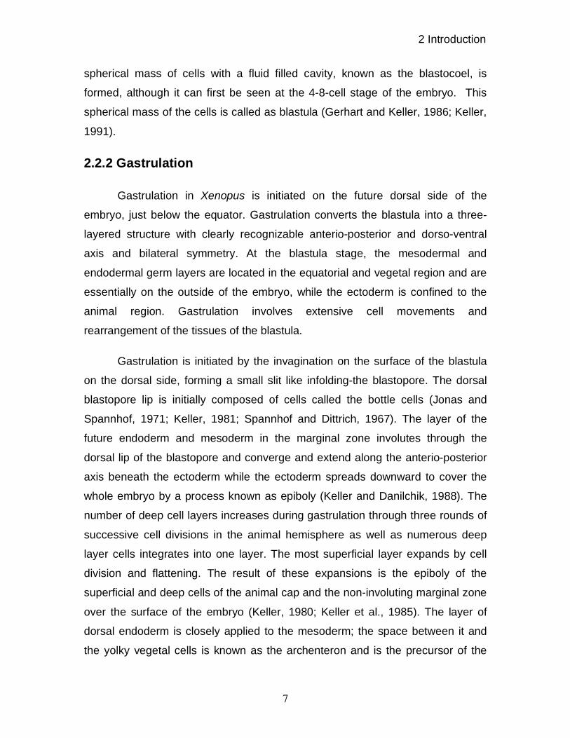
2.2.2 Gastrulation
Gastrulation in Xenopus is initiated on the future dorsal side of the
embryo, just below the equator. Gastrulation converts the blastula into a three-
layered structure with clearly recognizable anterio-posterior and dorso-ventral
axis and bilateral symmetry. At the blastula stage, the mesodermal and
endodermal germ layers are located in the equatorial and vegetal region and are
essentially on the outside of the embryo, while the ectoderm is confined to the
animal region. Gastrulation involves extensive cell movements and
rearrangement of the tissues of the blastula.
Gastrulation is initiated by the invagination on the surface of the blastula
on the dorsal side, forming a small slit like infolding-the blastopore. The dorsal
blastopore lip is initially composed of cells called the bottle cells (Jonas and
Spannhof, 1971; Keller, 1981; Spannhof and Dittrich, 1967). The layer of the
future endoderm and mesoderm in the marginal zone involutes through the
dorsal lip of the blastopore and converge and extend along the anterio-posterior
axis beneath the ectoderm while the ectoderm spreads downward to cover the
whole embryo by a process known as epiboly (Keller and Danilchik, 1988). The
number of deep cell layers increases during gastrulation through three rounds of
successive cell divisions in the animal hemisphere as well as numerous deep
layer cells integrates into one layer. The most superficial layer expands by cell
division and flattening. The result of these expansions is the epiboly of the
superficial and deep cells of the animal cap and the non-involuting marginal zone
over the surface of the embryo (Keller, 1980; Keller et al., 1985). The layer of
dorsal endoderm is closely applied to the mesoderm; the space between it and
the yolky vegetal cells is known as the archenteron and is the precursor of the
2 Introduction
8
gut cavity. The inward movement of the endoderm and mesoderm eventually
spreads to form a complete circle around the blastopore. By the end of
gastrulation, the blastopore has closed. The dorsal mesoderm lies beneath the
dorsal ectoderm, and the lateral mesoderm begins to spread in a ventral direction
on either side. The inner surface of the archenteron becomes completely covered
by a layer of endoderm, forming the gut. At the same time, the ectoderm spread
to cover the whole embryo. During gastrulation, dorsal mesoderm develops into
two main structures, the notochord and the somites. The notochord is a stiff, rod
like structure that forms along the dorsal midline and eventually becomes
incorporated into the vertebrae. The somites form by segmentation of the
paraxial mesoderm lying immediately at either side of the notochord. Somites are
formed in pairs, and segmentation proceeds in an anterio-posterior direction.
Figure 2.1 Schematic representation of early development of Xenopus laevis (adapted from De
Robertis et al., 2000).
2 Introduction
9
2.2.3 Neurulation and organogenesis
Gastrulation is succeeded by neurulation, the formation of the neural tube,
and the early embryonic precursor of the central nervous system. While
notochord and somites are developing, the neural plate ectoderm above them
begins to develop into the neural tube. At this stage, the embryo is called a
Neurula. The early signs of neural development are the formation of the neural
folds, which form on the edges of the neural plate. These rise up fold towards the
midline and fuse together to form the neural tube, which sinks beneath the
epidermis (Keller et al., 1992a; Keller et al., 1992b; Keller, 1980; Keller et al.,
1985). The anterior neural tube gives rise to the brain; further back, the neural
tube overlying the notochord will develop into the spinal cord. The main
structures that can be recognized at this stage are the neural tube, the
notochord, the somites, the lateral plate mesoderm, and the endoderm lining the
gut. The unsegmented lateral plate mesoderm, lying lateral and ventral to the
somites, gives rise to tissues of the heart and kidney, as well as to the gonads
and gut muscle, while the most ventral mesoderm gives rise to the blood islands.
The endoderm lining the gut will bud off organs such as the liver and lungs. At
the tail bud stage, the brain is already divided up into a number of regions while
the eye and ear have begun to develop at the anterior end. There are three
branchial arches, of which the anterior most will form the lower jaw. More
posteriorly, the somites and notochord are well developed. The post-anal tail of
the tadpole is formed last. It develops from the tail bud, which, at the dorsal lip of
the blastopore, gives rise to the continuation of notochord, somites and neural
tube. Nieuwkoop and Faber have divided the early development of Xenopus into
various stages (Nieuwkoop and Faber, 1967).
After organogenesis is completed, the mature tadpole hatches out of its
jelly covering and begins to swim and feed. Later, the tadpole larva undergoes
metamorphosis to give rise to the adult frog.

2 Introduction
10
2.3 Role of signaling events in establishment of early pattern
formation
2.3.1 Organizer formation
At the gastrula stage, the dorsal side of the embryo can be recognized by
the presence of the dorsal blastopore lip. The importance of this dorsal lip was
shown in a transplantation experiment carried out by Spemann and Mangold in
1924, using salamander gastrulae. They isolated dorsal lips from the embryos
and transplanted it in the host gastrula stage embryo on the ventral side opposite
to dorsal lip of the host embryo. This transplantation of dorsal lip resulted in an
embryo with a complete secondary axis (Spemann, H., and Mangold. 1924).
These experiments were also later repeated in Xenopus and were found to have
the same effect. These experiments revealed the importance of dorsal lip cells in
axis formation. In another complementary experiment, embryos were dissected
into dorsal and vegetal halves at gastrula stage. The dorsal half of the gastrula
gave rise to all dorsal structures while the ventral half of the gastrula embryo
remained undifferentiated tissue showing that the differentiation of the embryo is
determined by the dorsal side of the gastrula embryo (De Robertis et al., 2000).
The group of Nieuwkoop in 1969 carried out another important set of
experiments. These experiments utilized the property of Xenopus embryo
explants to be cultured in isolation in normal buffered saline. They showed that
animal caps develop into epidermis, while vegetal explants neither develop into
recognizable tissues nor develop posterior endodermal character (Nieuwkoop,
1963). When animal caps were grafted onto vegetal explants, mesoderm and
pharyngeal endoderm developed. Using pigmentation and 3H-thymidine labeled
cells as markers, it was concluded that mesoderm and head endoderm develop
exclusively from the animal cap tissue and were therefore induced by vegetal
cells. Explants of dorsal vegetal cells induce dorsal mesoderm, giving rise to
notochord and muscles, as well as head endoderm. Explants from the ventral
vegetal cells induce ventral mesoderm and gives rise to blood and mesenchyme
2 Introduction
11
(Nieuwkoop, P.D. 1973). These experiments gave rise to the three signal model
(Heasman, 1997) for mesoderm patterning, consisting of an early pair of signals
differing qualitatively between dorsal and ventral vegetal blastomeres, that acted
in the blastula stage to divide the early marginal zone of mesoderm into two
distinct territories: the dorsal and ventral mesoderm. The third signal, a
dorsalizing inductive signal from the dorsal mesoderm, would then impose dorsal
(paraxial) and intermediate fates on neighboring ventral mesoderm in the
gastrula stage (Heasman, 1997). In subsequent experiments it was shown that
vegetal cells start inducing signals as soon as 16-32 cell stage, arguing for
maternal proteins for meso-endoderm induction since zygotic transcription starts
only after mid blastula transition. Later it was shown that induction of both dorsal
and ventral mesoderm is induced by gradients of several nodal related signals
released by endoderm at the blastula stage and thus modifying the three-signal
model into a two-signal model (Agius et al., 2000). These experiments also
showed a gradient of Xnrs from the dorsal to the ventral side of the embryo.
Xenopus nodal related genes (xnrs) are potent mesoderm inducers (Jones
et al., 1995) and the events leading to the generation of the gradient of Xnrs can
be traced back to the fertilization stage. Fertilization of an egg with sperm starts a
rotation in the cortex of the egg with respect to the yolky cytoplasm leading to the
asymmetry in the egg. This event of cortical rotation brings about stabilization of
-CATENIN on the dorsal side of the embryo (Rowning et al., 1997). Increased
stabilization of -CATENIN leads to activation of the canonical WNT signaling
pathway on the dorsal side of the embryo (Schneider et al., 1996). When the
embryos were UV irradiated, dorsal structures of the embryo were abolished
giving rise to a ventralized embryo. UV treatment of the embryo causes
disruption of microtubules and thus prevents the accumulation of -CATENIN,
which mediates WNT signaling on the dorsal side of the embryo (Moon and
Kimelman, 1998).
Vg1, a TGF- factor was the first known asymmetrically localized RNA in
the egg. Vg1 is localized to the vegetal pole of the embryo (Melton, 1987; Weaks

2 Introduction
12
and Melton, 1987). VG1 precursor protein is abundant in vegetal cells, but the
processed mature form has not been readily detected and no activity has been
demonstrated for the putative VG1 mature protein. By using an engineered VG1
fusion (Bvg1) that promotes formation of mature VG1 protein in vivo, it was
shown that VG1 could be involved in mesoderm induction (Dale et al., 1993;
Thomsen and Melton, 1993). VegT, a T-box transcription factor is localized to the
vegetal pole of the Xenopus oocytes (Zhang and King, 1996). VG1 and VegT are
both potent inducers of endoderm. It was shown that depletion of maternal VegT
leads to the absence of endoderm (Cui et al., 1996; Joseph and Melton, 1998;
Zhang et al., 1998). Besides Vg1, endoderm expresses Xnrs. It was found that in
wild type embryos, microinjection of VegT and Vg1 induces only low levels of
xnrs transcription. However, when -CATENIN is also provided, a high level of
Xnr expression is achieved (Agius et al., 2000). It has been shown that the
expression of Xnrs in the endoderm occurs in a gradient, having higher
expressions in dorsal endoderm and lower levels of expression in ventral
endoderm. This gradient is probably established due to higher levels of Wnt
signaling on the dorsal side of the embryo. The dorsal endoderm thus
establishes a signaling center called “Nieuwkoop center” (De Robertis et al.,
2000). In the dorsal-animal cap marginal region, the -CATENIN signal induces
the expression of Chordin and Noggin. Chordin and Noggin are BMP
antagonists. This region of embryo has been named as the “BCNE center” for
Blastula Chordin and Noggin Expression center (Kuroda et al., 2004). The
Nieuwkoop center and BCNE center show only a limited overlap. Both blastula
centers are formed simultaneously, as soon as zygotic transcription starts and
require the beta-catenin signal on the dorsal side of the embryo, but the
Nieuwkoop center also requires Vg1 and VegT mRNAs localized in the vegetal
pole of the fertilized egg.
A high level of Xnr expression in the Nieuwkoop center induces the
formation of dorsal mesoderm and low level of Xnrs induces the formation of
ventral mesoderm at the gastrula stage of the embryo (Agius et al., 2000). The

2 Introduction
13
dorsal mesoderm is known as “Organizer” (De Robertis et al., 2000). BMP-2, a
maternal component, is expressed in the ventral mesoderm of the embryo and
has been shown to induce ventral mesoderm in animal cap explants. It has also
been shown that dorsal injections of BMP-2 could ventralize the embryo
(Clement et al., 1995; Nishimatsu and Thomsen, 1998; Plessow et al., 1991),
showing it to be a potential inducer of ventral mesoderm.
Figure 2.2 Signaling events during the formation of Organizer (adapted from the De
Robertis et al., 2004).
Nieuwkoop center cells form anterior endoderm at gastrula and BCNE
center cells give rise to prospective brain and floor plate, as well as the
notochord region of the Spemann organizer at gastrula. Both signaling centers
are required for brain formation as Nieuwkoop center cells involute to come into
intimate contact with the future brain to provide a “double assurance” mechanism
for brain formation (De Robertis and Kuroda, 2004).
The organizer acts as an inhibitory center for early maternal signals.
Molecules secreted by the organizer can be divided based on their inhibitory
properties. Among the molecules secreted by the organizer, frzb-1 (Leyns et al.,
1997; Wang et al., 1997), dickkopf-1 (Glinka et al., 1998) and crescent (Pera and
De Robertis, 2000) act as zygotic WNT signaling inhibitors. cerberus (Piccolo et
al., 1999) acts to inhibit WNT, NODAL as well as BMP signaling, while chordin
(Sasai et al., 1994) and noggin (Smith and Harland, 1992) that are first

2 Introduction
14
expressed in the BCNE center and later in the organizer, inhibit BMP signaling.
follistatin also inhibits BMP signaling (Fainsod et al., 1997), while lefty/antivin are
antagonists to the TGF- /NODAL receptor (Cheng et al., 2000; Meno et al.,
1999; Meno et al., 1996; Thisse and Thisse, 1999). The inhibition of BMP
signaling by chordin and noggin sets up a gradient of BMP signaling in
mesodermal tissue that is required for the differentiation of head and tail
structures. Similar to WNT signaling inhibition, inhibition of NODAL signaling by
organizer-secreted molecules is also required for head formation. Thus, the
organizer plays a central role in the patterning of the embryo.
On one hand maternal WNT signaling is required for early patterning as
described above, zygotic WNT signaling is involved in ventral mesoderm
formation in combination with BMP signaling (Hoppler and Moon, 1998).
2.3.2 Morphogens and signaling thresholds
The dynamic gradients of NODAL and WNT/ -CATENIN activity revealed
detailed steps in early patterning. The idea of morphogen gradients and
thresholds has long been an important one in developmental biology (Green,
2002). Morphogens are defined as substances, whose nonuniform distribution in
a field of cells differentially determines the fate and phenotype of those cells. A
graded morphogen provides polarity and a scalar value that can be interpreted
by cells according to threshold values to provide a coordinate system. The
morphogen gradients for Activin (Green and Smith, 1990) and FGF (Green et al.,
1992; Slack, 1987; Slack et al., 1987) have been investigated in the animal cap
assay system. To produce distinct cell types, morphogen interpretation relies on
the sharpening of threshold responses to cellular stimuli. This suggests some
kind of cooperation leading to a steep sigmoid response in the interpreting cell
(Lewis et al., 1977; Slack, 1993). In Xenopus, the detailed analysis of xbra
regulation has shown that the initial dose window of xbra induction by increasing
the dose of ACTIVIN is bounded by an initially relatively “fuzzy” dose threshold,
but it progressively sharpened with time over a few hours (Green et al., 1994;
2 Introduction
15
Gurdon et al., 1999). One of the mechanisms of sharpening of a threshold is cell
contact dependent and consists of a positive feed back loop as manifested by
“community effect” (Gurdon et al., 1993; Standley et al., 2001). The other
mechanism involves inhibition as in the case of xbra by Goosecoid, which is
induced at higher concentrations of ACTIVIN (Artinger et al., 1997). However
other than these mechanisms, chromatin environment may also play an
important role in setting up fine activation thresholds as pointed out by some in-
vitro studies (Laybourn and Kadonaga, 1992).
2.4. Evidence for regulation of embryonic patterning in Xenopus
by chromatin environment
The animal cap cells are able to form the mesodermal tissue from the
morula through to the blastula stage (Woodland and Jones, 1987). At early
gastrula, animal cap cells loose the ability to form mesodermal tissue (Green et
al., 1990). The mechanism of mesodermal competence has been studied in
detail using ACTIVIN to induce the mesoderm in the animal cap cells. It has been
shown that loss of mesodermal competence is programmed cell autonomously
and occurs even in the single cell (Grainger and Gurdon, 1989). One of the
factors required for loss of mesodermal competence of animal cells was shown
to be somatic linker histones (Steinbach et al., 1997). In this report it was shown
that accumulation of somatic linker histone H1, which is required for forming
higher order chromatin structures and acts as a general repressor of transcription
(Paranjape et al., 1994), causes the loss of mesoderm forming capacity of animal
cells. In other words, the repressive chromatin environment blocks the
conversion of ectodermal cells into mesodermal cells. In a recently published
report, the authors showed that BRG1 overexpression could induce xbra
induction even in the gastrula stage animal caps after bFGF induction (Hansis et
al., 2004). It has been also shown that treatment of embryos with TSA, an
inhibitor of HDAC, before gastrulation, results in the loss of muscles in the
embryos (Rupp et al., 2002; Steinbach et al., 2000). These experiments showed
2 Introduction
16
the role of chromatin environment in the correct expression of various genes
required for normal development of the embryo. Below is a brief overview of
chromatin structure and chromatin remodeling mechanisms required for
regulation of gene transcription.
2.5 Chromatin structure and chromatin remodeling complexes
Based on his light microscopic observation of nuclear material the German
anatomist Walter Flemming in 1882 (Fleming et al., 1882) first established the
term chromatin, which is derived from the Greek word “Khorma” which means
color. The details of chromatin were studied using biochemical and electron
microscopic techniques. When chromatin structures were digested with
micrococal nuclease that breaks down unprotected DNA, about 200 bp DNA
fragments were obtained (Clark and Felsenfeld, 1971). When interphase nuclei
are broken open very gently and their contents were examined under the
electron microscope, most of the chromatin is in the form of a fiber with a
diameter of about 30 nm. When this chromatin is subjected to treatments that
cause it to unfold partially, it can be seen under the electron microscope as a
beaded structure termed “beads on a string” (Olins and Olins, 1974). In this, the
string is DNA, and each bead is a nucleosome core particle. The term
“nucleosome” was given by the group of Chambon (Oudet et al., 1975), while a
proposal was made about 31 years ago by Roger Kornberg that the structure of
chromatin is based on a repeating unit of eight histone molecules and about 200
bp, laid the basis for subsequent chromatin research (Kornberg, 1974).
2.5.1 Chromatin structure
The nucleosome represents the basic building block of chromatin. A
nucleosome is composed of DNA and histone proteins. The histone proteins are
present as core histones and as linker histones. In each nucleosome, 147 base
pairs of DNA are wrapped around a core of histone proteins.
2 Introduction
17
Albert Kossel first isolated histones, the basic proteins from nuclei and
termed them “histon” in 1884. Histones represent the major class of DNA binding
proteins. As a universal component of the chromosomes, histones were thought
to be roughly equal to that of DNA; In fact, they were long viewed as the genetic
material itself. Upon extraction in acid, which prevents proteolysis, five types of
histones, designated as H1, H2A, H2B, H3 and H4 were found (Phillips and
Johns, 1965). Among them H2A, H2B, H3 and H4 form the core of the
nucleosome and H1 serves as the linker histone.
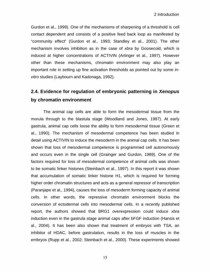
Figure 2.3 Diagrammatic representation of DNA Packaging into chromatin (adapted from Hansen,
2002).
The histone core consists of a (H3)2(H4)2 tetramer flanked by two H2A-
H2B dimer thus forming an octamer. Each core histone contains two functional
domains: a signature “ histone fold” motif sufficient for both histone-histone
interaction as well as DNA-histone interaction within the nucleosome and second
an amino terminal and carboxy terminal “tail domain” containing sites for post-
translational modifications (Arents et al., 1991). Linker histone H1 is unrelated in
sequence to core histones. H1 consists of a globular domain flanked by amino

2 Introduction
18
and carboxyl terminal tail domains while globular domain interacts with the
nucleosome, the H1 tail domains are believed to be required for chromatin
folding (Parseghian and Hamkalo, 2001; Ramakrishnan, 1997).
The low-resolution (7-Å) structure of the nucleosome was determined in
1984, and it revealed that the histone octamer forms a helical ramp around which
1.7 turns of a left handed DNA super helix is wrapped. The high-resolution
structure at 2.8 Å was resolved in the year 1997 (Luger et al., 1997). It shows
that where the DNA enters and leaves the nucleosome, it is bound by N-terminal
extensions of the H3 histone fold. Each of the heterodimers binds about 30 bp of
DNA. The DNA is contacted at 10 bp intervals as the minor groove faces protein.
The average number of base pairs per helical turn of DNA–the helical periodicity
of DNA, was found to be 10.2 bp.
Figure 2.4 High-resolution Nucleosome structure at 2.8 Å (adapted from Luger et al., 1997).
A value of 10.2 bp allows the minor and major grooves from neighboring turns of
the DNA super helix to line up, forming channels through which the histone tail
can pass. This also leaves the major grooves accessible, enabling them to
2 Introduction
19
participate in cellular processes, acting, for example, as DNA integration-
hotspots. In conjunction with the linker histone, H1, which is present in
stoichiometric amounts with histone octamer, nucleosomal arrays fold into
higher-order structures.
2.5.2 Chromatin remodeling
This packaging of the DNA provides on one hand a means to prevent the
DNA from becoming an unmanageable tangle; it also serves as a means to
regulate various processes like DNA replication, repair, recombination and
transcription. The regulation of these processes are mainly governed either by
covalent modifications of histone tails or by energy dependent nucleosomal
structural alterations that may change the nucleosome position with respect to
the DNA sequence, or may displace histone subunits. Regulation of nucleosomal
structure requires enzymatic proteins.
There are two classes of nucleosome remodeling enzymes, also referred
to as chromatin remodeling enzymes. The first class of enzyme is responsible for
covalent modification of histone tails such as histone acetyltransferase and
deacetylases. It was proposed that distinct histone modifications on one or more
tails act sequentially or in combination to form a ‘histone code’ that is read by
other proteins to bring about distinct downstream events (Jenuwein and Allis,
2001; Strahl and Allis, 2000). The second class of enzymes utilize the energy of
ATP hydrolysis to alter or disrupt the nucleosomal structure by affecting DNA-
histone interactions (Becker and Horz, 2002; Kadam and Emerson, 2002;
Katsani et al., 2003; Narlikar et al., 2002; Tsukiyama, 2002).
2.5.2.1 Histone modifications
The known post-synthetic modifications of histones that cause a change in
the state of chromatin are phosphorylation, methylation, ubiquitination, ADP-
ribosylation along with acetylation and deacetylation.

2 Introduction
20
2.5.2.1.1 Acetylation
The discovery of HAT provided a link between histone acetylation and
gene activation (Brownell and Allis, 1995; Brownell and Allis, 1996; Brownell et
al., 1996). Based on their protein sequence and functional conservation, HATs
can be grouped into three main families: The GNATs, the MYST and the
hormone receptor co activator p160 (SRC) family. (Gregory et al., 2001; Vaquero
et al., 2003). Other HATs such as CBP/p300, TAFII250, TAFIIC and NUT1 do not
belong to any family. Even though most of them have been shown to be involved
in transcriptional activation, SAS (some thing about silencing) has been
implicated in transcriptional repression (Reifsnyder et al., 1996) and has been
proposed to be in the MYST family of HATs.
The effect of acetylation is partly explained by a decrease in the positive
charge of the histone while other hypothesis propose that the acetylated lysine’s
are recognized by bromodomain containing proteins, which then affect local
chromatin structure. Many biological processes such as chromatin assembly,
DNA repair and apoptosis, dosage compensation or cell cycle progression are
affected by histone acetylation (Carrozza et al., 2003; Hassan et al., 2001;
Kristeleit et al., 2004; Neely and Workman, 2002).
2.5.2.1.2 Deacetylation
The connection between acetylation and transcription is further shown by
the fact that deacetylation can cause repression. The isolation of a human
histone deacetylase, HDAC1, which was homologous to RPD3 (Furukawa et al.,
1996) demonstrated the connection of deacetylation and repression. The HDACs
have been divided into three groups. Class I enzymes includes HDACs 1-3 and 8
whereas class II includes HDAC 4-7, 9 and 10. The class III enzymes are related
to the silencing regulator SIR2 (Imai et al., 2000).
All of the known deacetylases occur in multi-protein complexes with
important functions. The complexes are able to deacetylate histones in
2 Introduction
21
nucleosomes, whereas the isolated deacetylase subunits cannot deacetylate
histones. Other members of the deacetylase complex include chromodomain
proteins, retinoblastoma protein-associated proteins, and SIN3 (Wang et al.,
2004a).
2.5.2.1.3 Methylation
Histone methylation was first described by Murray in 1964 (Murray,1964).
Arginine residues can be mono- or dimethylated by PRMTs and lysine residues
can be mono- di- or tri-methylated by SET-domain containing histone
methyltransferases (HMTs). While tri-methylation of histone is associated with
silencing of gene transcription, di-methylation of histone has been shown to be
associated with transcriptionally active gene transcription. The group of Thomas
Jenuwein discovered SUV39H1, a homologue of Drosophila Su (var) 3-9
(Aagaard et al., 2000; Zhang and Reinberg, 2001), which supported a direct
connection between heterochromatin formation, gene silencing and specific
histone lysine methylation. The histone methylation is generally related to gene
silencing with some exceptions (Lachner and Jenuwein, 2002; Lachner et al.,
2003).
2.5.2.1.4 Phosphorylation
This modification occurs on serine or threonine residues. Labile forms of
phosphorylation involving P-N linkage of lysines or arginine have also been
described (Smith et al., 1978). Histone phosphorylation has also been observed
on metaphase chromosomes during condensation. (Green, 2001; Mahadevan et
al., 2004; Nowak and Corces, 2004).
2.5.2.1.5 Ubiquitination
This modification of protein is primarily a signal required for protein
turnover and has also been involved in various physiological processes such as
spermiogenesis, DNA repair, and transcription. Ubiquitinated H2A and H2B were

2 Introduction
22
preferentially found in transcriptionally active chromatin, supporting a positive
role of this modification in gene expression (Zhang, Y., 2003).
2.5.2.1.6 ADP-ribosylation and other modification
Nishizuka and colleagues first described Poly-ADP ribosylation (Nishizuka
et al., 1968). It can be catalyzed on arginine or glutamine residues by the poly
(ADP-ribose) polymerase. All four core histones and linker histone H1 can be
used as a substrate for this reaction (Rouleau et al., 2004). Other than this,
biotinylation (Camporeale et al., 2004) as well as SUMOlation (Shiio and
Eisenman, 2003) of histones has also been reported.
Figure 2.5 The known histone post translational modifications (adapted from Khorasanizadeh,
2004).
2.5.2.2 ATP dependent chromatin remodeling
The complexes involved in energy dependent chromatin remodeling are
multi-protein complexes, containing 2-12 subunits (Becker and Horz, 2002). Each
complex has a catalytic subunit, carrying the ATPase activity. These ATPases
are highly conserved throughout evolution. In addition to the ATPase motif,
proteins in the SNF2 family also contain sequence motifs similar to those found

2 Introduction
23
in DNA and RNA helicase protein families. Proteins with these helicase motifs
have been divided into multiple superfamilies based upon amino acid sequences
found within the motifs. By this method, the SNF2 family has been assigned to
the helicase superfamily 2, which also includes the ERCC3, RAD3, PRIA,
ELF4A, and PRP16 protein families (Eisen et al., 1995).
Our lab has established a family of SNF2-like nuclear ATPases by
browsing the annotated human genome database. Furthermore, the homologues
of these ATPases have also been established in Xenopus showing that
vertebrates share a common family of ATPase chromatin remodeling complexes
(Linder et al., 2004). The enzymes in the SNF2 family can be subdivided into
several subfamilies according to the sequence motifs outside of their ATPase
domain.
Figure 2.6 Sequence similarity tree of the human SNF2-domain containing proteins (adapted from
Linder et al., 2004).
Based on this analysis, seven subfamilies have been assigned, out of
which nucleosome-remodeling activity has been shown only for SWI2/SNF2-
related enzymes (Sudarsanam and Winston, 2000), ISWI type enzymes (Langst
and Becker, 2004), and for CHD family members (Brehm et al., 2000). In addition
the recently identified INO80 also shows nucleosome remodeling activity (Shen
et al., 2000). Other members are known to be involved in DNA repair,
recombination, as well as in transcription (Becker and Horz, 2002).
2 Introduction
24
2.5.2.2.1 ISWI, a SANT-like domain-containing member of the
SNF2 family
The ATPase ISWI was first discovered in Drosophila melanogaster
because of the similarity of its ATPase domain to that of BRM (Elfring et al.,
1994). NURF (Gdula et al., 1998; Martinez-Balbas et al., 1998; Xiao et al., 2001),
CHRAC (Varga-Weisz et al., 1997) and ACF (Ito et al., 1999) are various
chromatin-remodeling complexes, which contain the ISWI ATPase. Later ISWI
containing complexes were identified in human (Barak et al., 2003; Poot et al.,
2000), mouse (Lazzaro and Picketts, 2001) and Xenopus (Guschin et al., 2000).
Two SANT-like domains in the C-terminus of the protein distinguish them from
the other members of SNF2 family (Aasland et al., 1996). Homozygous null
mutation of ISWI is lethal in Drosophila (Deuring et al., 2000). The homozygous
deletion of SNF2H, a murine homologue of ISWI, is lethal, but mice with
heterozygous deletion of SNF2H were normal (Stopka and Skoultchi, 2003).
2.5.2.2.2 The CHD class of remodelers are characterized by the
presence of a chromodomain
Among this class of remodelers, CHD3 (MI-2 ) and CHD4 (MI-2 ) are
mostly studied members of this family. The members of this class contain two
PHD fingers in addition to the characteristic chromodomain. MI-2 was identified
as a dermatomyositis-specific autoantigen. It has been shown to reside in the
NURD complex (for nucleosome remodeling and deacetylation)(Knoepfler and
Eisenman, 1999).
Like ISWI, the MI-2 ATPase is an active enzyme for nucleosome
remodeling, able to disrupt histone-DNA interactions and to induce nucleosome
sliding on DNA fragments (Brehm et al., 2000). Interestingly, MI-2 has also
been shown to combine deacetylation and ATP dependent remodeling (Wade et
al., 1999).

2 Introduction
25
2.5.2.2.3 The SWI/SNF complexes
2.5.2.2.3.1 SWI/SNF complexes
The yeast SWI/SNF complex was the first chromatin- remodeling complex
to be described (Stern et al., 1984). The genes encoding its various subunits
were originally identified in two independent screens for mutants affecting either
mating type switching or growth on sucrose (Sudarsanam and Winston, 2000;
Workman and Kingston, 1998) and hence were named “switching defective” and
“Sucrose non-fermenting”. The biochemical evidence for a direct connection
between chromatin and SWI/SNF function was provided by the findings that the
SWI/SNF complex could alter nucleosome structure in an ATP dependent
manner (Vignali et al., 2000; Workman and Kingston, 1998).
A closely related yeast chromatin-remodeling complex is called RSC for
“Remodel the Structure of Chromatin” (Cairns et al., 1994; Cairns et al., 1999).
This complex contains about 15 subunits, sharing two identical and at least four
homologous subunits with the ySWI/SNF complex (Cairns et al., 1998; Wang,
2003). STH1 is the paralogue of the SWI2/SNF2 ATPase subunit in the RSC
complex. Furthermore, RSC8, RSC6 and SFH1 in RSC correspond to SWI3,
SWP73 and SNF5 in ySWI/SNF, respectively. RSC complexes are about 10
times more abundant than SWI/SNF complexes.
Despite these structural similarities, there are several important functional
differences between ySWI/SNF and RSC. In contrast to ySWI/SNF, RSC
functions are required for yeast viability. A Genome wide gene expression
analysis revealed that ySWI/SNF and RSC regulate different, largely non-
overlapping sets of target genes. RSC complexes have also been shown to be
involved in sister chromatid cohesion and chromosome segregation, which
indicates the broader role of these complexes in chromatin dynamics (Baetz et
al., 2004; Hsu et al., 2003; Huang et al., 2004; Wong et al., 2002).

2 Introduction
26
Homologous ATPase complexes have also been isolated in Drosophila
and mammals. Human cells contain two distinct SWI2/SNF2 like ATPase
subunits, named hBRM and BRG1, which are equally similar to yeast
SWI2/SNF2 and STH1. In contrast, Drosophila contains only a single protein
corresponding to yeast SWI2/SNF2 or STH1, called BRM (Papoulas et al., 1998;
Tamkun et al., 1992). In higher eukaryotes, the remodeling complexes of
SWI/SNF class are referred to as BAP (BRM associated proteins) in Drosophila
and hSWI/SNF-BAF (BRG1/hBRM- Associated factors) in mammals, whereas
the RSC orthologue are referred to as PBAP or hSWI/SNF-PBAF in Drosophila
or mammals, respectively. BRM was originally discovered as a suppressor of
polycomb and therefore was classified as a trithorax-group protein. Two BRM
associated proteins, the common subunit Moira (MOR) and the BAP selective
subunit OSA, are also encoded by trxG genes (Brizuela and Kennison, 1997;
Collins et al., 1999; Collins and Treisman, 2000; Crosby et al., 1999; Kennison
and Tamkun, 1988; Vazquez et al., 1999).
Most of the Drosophila and mammalian subunits are equally similar to
their counterparts in ySWI/SNF and RSC. The two exceptions are OSA/BAF250
and Polybromo/BAF180, which are the signature subunits of the ySWI/SNF or
RSC type subfamilies respectively. OSA/BAF250 is related to the ySWI/SNF
subunit Swi1, whereas there is no homologue in RSC (Collins et al., 1999;
Collins and Treisman, 2000; Dallas et al., 2000). Conversely, Polybromo/BAF180
is structurally related to the RSC1, RSC2 and RSC4 proteins, but lacks a
counterpart in ySWI/SNF. In addition, BAF can contain either BRG1 or BRM as
the core motor subunit, whereas PBAF contains only BRG1 (Mohrmann et al.,
2004; Xue et al., 2000). In mammals there appear to be additional tissue specific
subunits of SWI/SNF remodelers. A number of studies have reported additional
sub-complexes in which the SWI/SNF type remodelers are associated with other
factors such as BRCA1 (Bochar et al., 2000) or components of the histone
deacetylating SIN3 complex (Sif et al., 2001). It has been shown that BAF53b is
2 Introduction
27
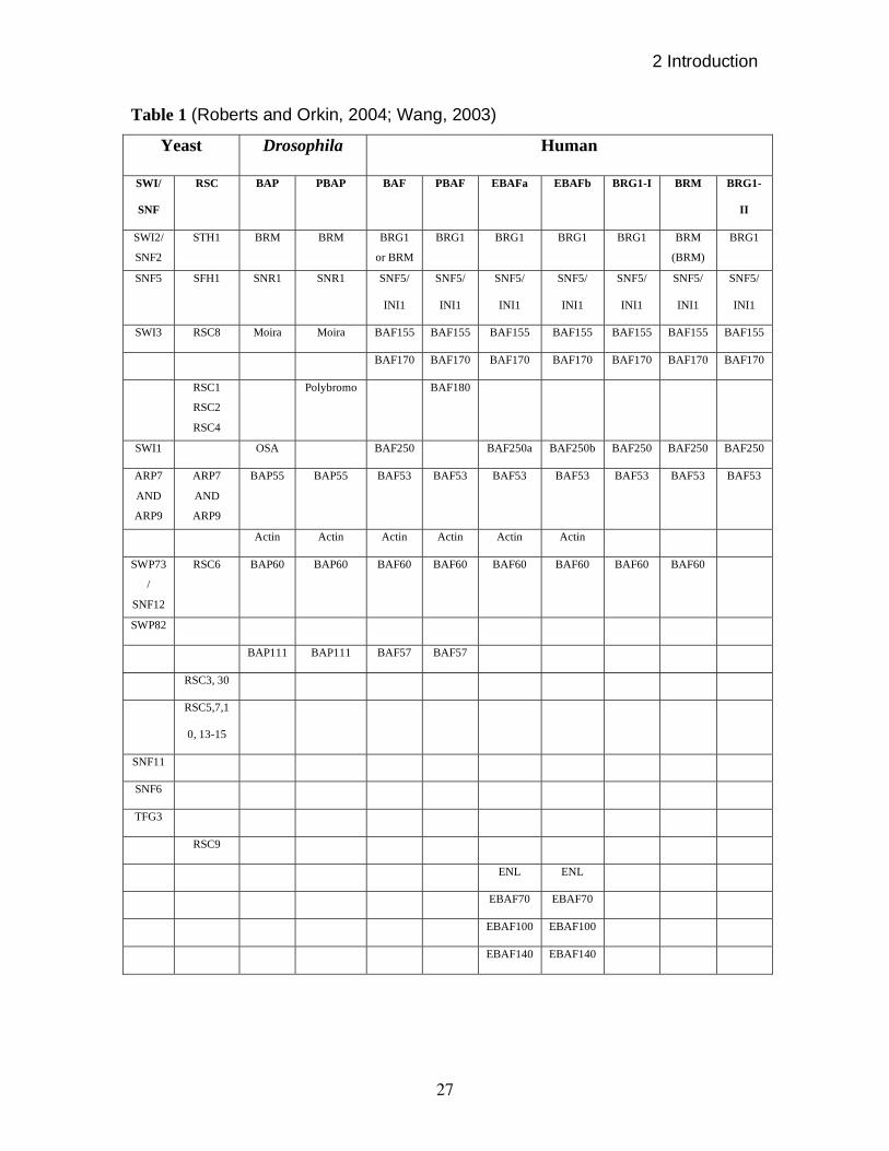
Table 1 (Roberts and Orkin, 2004; Wang, 2003)
Yeast Drosophila Human
SWI/
SNF
RSC BAP PBAP BAF PBAF EBAFa EBAFb BRG1-I BRM BRG1-
II
SWI2/
SNF2
STH1 BRM BRM BRG1
or BRM
BRG1 BRG1 BRG1 BRG1 BRM
(BRM)
BRG1
SNF5 SFH1 SNR1 SNR1 SNF5/
INI1
SNF5/
INI1
SNF5/
INI1
SNF5/
INI1
SNF5/
INI1
SNF5/
INI1
SNF5/
INI1
SWI3 RSC8 Moira Moira BAF155 BAF155 BAF155 BAF155 BAF155 BAF155 BAF155
BAF170 BAF170 BAF170 BAF170 BAF170 BAF170 BAF170
RSC1
RSC2
RSC4
Polybromo BAF180
SWI1 OSA BAF250 BAF250a BAF250b BAF250 BAF250 BAF250
ARP7
AND
ARP9
ARP7
AND
ARP9
BAP55 BAP55 BAF53 BAF53 BAF53 BAF53 BAF53 BAF53 BAF53
Actin Actin Actin Actin Actin Actin
SWP73
/
SNF12
RSC6 BAP60 BAP60 BAF60 BAF60 BAF60 BAF60 BAF60 BAF60
SWP82
BAP111 BAP111 BAF57 BAF57
RSC3, 30
RSC5,7,1
0, 13-15
SNF11
SNF6
TFG3
RSC9
ENL ENL
EBAF70 EBAF70
EBAF100 EBAF100
EBAF140 EBAF140
2 Introduction
28
part of a brain specific complex (Olave et al., 2002a), while BAF60c could be a
part of heart specific complex since it shows a heart specific expression pattern
in early embryonic stage; later it is also expressed in somites (Lickert et al.,
2004). Various SWI/SNF complexes have been summarized in table 1.
2.5.2.2.3.2 Interaction motifs in the SWI/SNF class of remodelers
The distinguishing feature of the SWI/SNF class of chromatin remodelers
is the presence of a bromodomain in the ATPase subunit, which is absent in
ISWI, CHD/MI-2 and INO80 type remodelers (Eberharter and Becker, 2004).
Bromodomains are 90 amino acid long modules. Bromodomains
recognize acetylated lysines in histone tails (Dhalluin et al., 1999; Hassan et al.,
2002; Hudson et al., 2000; Jacobson et al., 2000; Marmorstein and Berger, 2001;
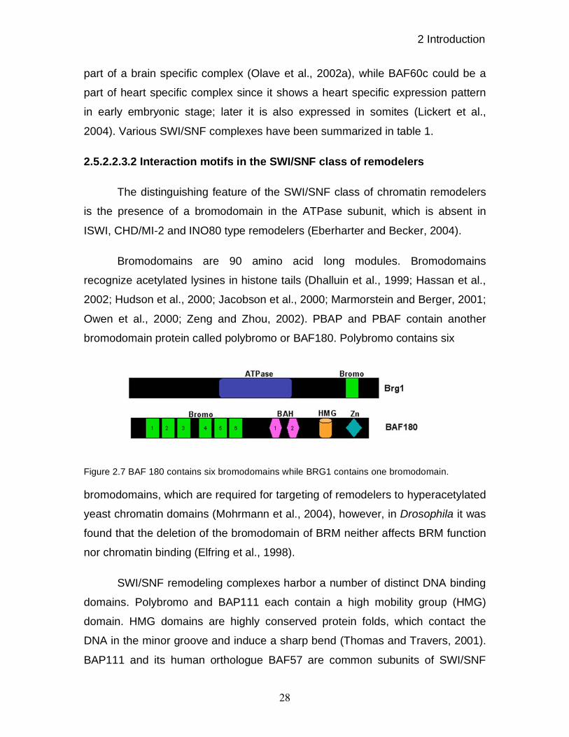
Owen et al., 2000; Zeng and Zhou, 2002). PBAP and PBAF contain another
bromodomain protein called polybromo or BAF180. Polybromo contains six
Figure 2.7 BAF 180 contains six bromodomains while BRG1 contains one bromodomain.
bromodomains, which are required for targeting of remodelers to hyperacetylated
yeast chromatin domains (Mohrmann et al., 2004), however, in Drosophila it was
found that the deletion of the bromodomain of BRM neither affects BRM function
nor chromatin binding (Elfring et al., 1998).
SWI/SNF remodeling complexes harbor a number of distinct DNA binding
domains. Polybromo and BAP111 each contain a high mobility group (HMG)
domain. HMG domains are highly conserved protein folds, which contact the
DNA in the minor groove and induce a sharp bend (Thomas and Travers, 2001).
BAP111 and its human orthologue BAF57 are common subunits of SWI/SNF

2 Introduction
29
remodelers in higher eukaryotes, but are absent in yeast. Studies on Drosophila
established that BAP111 is required for normal BRM complex function in vivo
(Papoulas et al., 2001).
In addition to its highly conserved HMG domain, Polybromo harbors two,
less well conserved, C2H2-type zinc fingers. The putative double C2H2 zinc
finger motif is present in Drosophila and C.elegans polybromo, but absent in
chicken and human. The C-terminus of BAP170 also contains a highly
conserved, double zinc finger motif comprising a canonical C2H2 zinc finger, and
a second one in which the spacing between the two cysteine residue is
somewhat larger. The strict conservation of the zinc finger motifs in BAP170
suggests that they might be functionally important (Mohrmann et al., 2004).
BAP 170 contains a second DNA binding motif in its N-terminus, an AT
rich interaction domain (ARID). ARID domain proteins are also present in yeast
Swi1, Drosophila OSA and mammalian BAF250, which define the ySWI/SNF,
BAP and BAF subclass, respectively (Collins et al., 1999). ARID domains are
sometimes referred to as BRIGHT domains and have also been implicated in
sequence-specific as well as sequence-independent DNA binding (Gregory et al.,
1996; Herrscher et al., 1995; Wilsker et al., 2002). The ARID harbors a helix-turn-
helix region and, as reflected by its name, preferentially binds AT-rich
sequences. ARID domain in OSA binds DNA without sequence specificity while
dead ringer is an example of a sequence-specific DNA binding ARID containing
protein. The ARID of BAF 250 has been implicated in transcriptional co-
activation of hormone receptors (Inoue et al., 2002), suggesting that it acquires
specificity through interactions with other cofactors.
Actin is known for many cellular functions in the cytoplasm of eukaryotic
cells, including processes like muscle contraction, cell motility, or cytokinesis.
Actin is highly expressed and is in fact the most abundant protein in many cell
types. The purification of actin by biochemical means led to the striking finding
that it was a tightly bound subunit of SWI/SNF family of remodeling complexes
2 Introduction
30
(Olave et al., 2002b; Papoulas et al., 1998; Zhao et al., 1998), which was for a
long time considered as a contaminant. Actin Related Proteins (ARPS), consist
of a large and diverse group of proteins that share between 10% to 80%
sequence similarities with actin (Schafer and Schroer, 1999). The actin fold is
conserved in the Arps, but there is much less conservation in flanking regions
(Robinson et al., 2001). The presence of stoichiometric amounts of actin and
Arps in diverse chromatin remodeling complexes has been firmly established
(Olave et al., 2002b). Yeast SWI/SNF and RSC contain ARP7 and ARP9,
whereas Drosophila BAP and PBAP and human BAF and PBAF contain one
ARP, BAP55 and BAF53 respectively and actin (Cairns et al., 1998; Peterson et
al., 1998; Wang et al., 1996a; Wang et al., 1996b). It has been proposed that
Actin and ARPs can modulate binding of the remodeling complex to chromatin or
to the nuclear matrix, stimulate the DNA-dependent ATPase activity, promote
complex assembly and stability, histone binding, or remodeling and translocation
(Boyer and Peterson, 2000; Rando et al., 2002; Shen et al., 2003; Szerlong et
al., 2003).
2.5.2.2.3.3 Differential targeting of SWI/SNF remodelers
Results in Drosophila suggest that BRM is involved in transcription of most
genes (Armstrong et al., 2002), however, one way in which variation in subunit
composition can direct functional differentiation is through unique gene targeting.
Immunolocalization on larval salivary gland polytene chromosomes revealed that
OSA and Polybromo, the defining subunits of BAP and PBAP, each display
distinct, albeit overlapping genome-wide distributions (Mohrmann et al., 2004).
Genome wide expression studies in yeast revealed that ySWI/SNF and RSC
each regulate different sets of target genes (Angus-Hill et al., 2001; Holstege et
al., 1998; Sudarsanam et al., 2000).
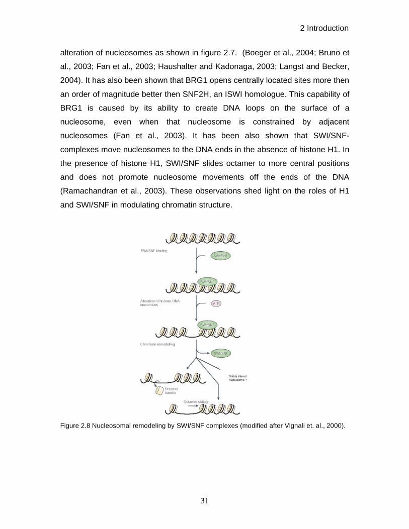
2.5.2.2.3.4 Nucleosomal remodeling by SWI/SNF complexes
Nucleosomal remodeling by SWI/SNF complexes is either by
displacement of histone octamers, sliding of histone octamers or by stable
2 Introduction
31
alteration of nucleosomes as shown in figure 2.7. (Boeger et al., 2004; Bruno et
al., 2003; Fan et al., 2003; Haushalter and Kadonaga, 2003; Langst and Becker,
2004). It has also been shown that BRG1 opens centrally located sites more then
an order of magnitude better then SNF2H, an ISWI homologue. This capability of
BRG1 is caused by its ability to create DNA loops on the surface of a
nucleosome, even when that nucleosome is constrained by adjacent
nucleosomes (Fan et al., 2003). It has been also shown that SWI/SNF-
complexes move nucleosomes to the DNA ends in the absence of histone H1. In
the presence of histone H1, SWI/SNF slides octamer to more central positions
and does not promote nucleosome movements off the ends of the DNA
(Ramachandran et al., 2003). These observations shed light on the roles of H1
and SWI/SNF in modulating chromatin structure.
Figure 2.8 Nucleosomal remodeling by SWI/SNF complexes (modified after Vignali et. al., 2000).

2 Introduction
32
2.5.2.2.3.5 Function of RSC class of remodelers
RSC complexes have been shown to be involved in sister chromatid
cohesion and segregation (Baetz et al., 2004; Hsu et al., 2003; Huang et al.,
2004; Wong et al., 2002). The accurate transmission of the genome during cell
division requires chromatin restructuring. Various studies revealed that several
RSC subunits, namely STH1, SFH1, RSC3 and RSC9 are essential for cell cycle
progression through G2 and mitosis (Angus-Hill et al., 2001; Damelin et al.,
2002). Current evidence suggests that cell cycle failure in RSC mutant is not due
to defective transcription of cell cycle control genes (Ng et al., 2002) rather it
appears that the spindle checkpoints might play a critical role in the G2/M arrest
caused by RSC mutants (Tsuchiya et al., 1992). An independent genetic screen
for haploinsufficient modifiers of chromosome segregation fidelity further
supported this notion (Baetz et al., 2004). Thus, RSC mediates chromatin
restructuring, independent of transcription regulation and appears to be directly
required for chromosome segregation.
Although the molecular mechanisms remain unclear, recent studies
suggest that RSC is required for the loading of cohesin on chromosome arms,
and for kinetochore function (Baetz et al., 2004; Hsu et al., 2003; Huang et al.,
2004). The cohesin complex is the molecular machine responsible for the
controlled pairing of sister chromatids prior to their segregation (Nasmyth et al.,
2000; Yokomori, 2003). PBAF complexes have been shown to be localized at the
kinetochore (Xue et al., 2000).
2.5.2.2.3.6 Function of mammalian SWI/SNF complexes
Biochemical studies revealed important functional differences between
BRM and BRG1. BRG1 binds to Zn-finger proteins through a unique N-terminal
domain, which is not present in BRM. Conversely, BRM interacts with two
ankyrin repeat proteins that are critical components of notch signal transduction
(Kadam and Emerson, 2003). Thus, BRG1 and BRM complexes may direct
distinct cellular processes through recruitment to specific promoters mediated by
2 Introduction
33
protein-protein interaction that is unique to each ATPase. Moreover, PBAF, but
not BAF, is necessary for ligand-dependent transactivation by several nuclear
hormone receptors, such as VDR and PPAR (Lemon et al., 2001). However,
other researchers reported that the BAF complex, but not PBAF complex, is
required for glucocorticoid receptor dependent transcription (Trotter and Archer,
2004).
Studies on mammalian SWI/SNF remodelers also uncovered functional
differences. Gene inactivation experiments in mice revealed that the in-vivo
importance of the very similar mammalian paralogs, BRM and BRG1, is quite
different. BRM knock-out mice are viable and display only a subtly altered control
of cell proliferation (Reyes et al., 1998), while in contrast, BRG1 knock-out mice
die already during early embryogenesis and animals with mono-allelic BRG1
expression are predisposed to tumors, suggesting a role in neoplasias (Bultman
et al., 2000). These differences might be the results of distinct timing of
expression of BRM and BRG1 (LeGouy et al., 1998) or perhaps BRG1 may
compensate for loss within the BAF complex, but conversely, BRM cannot
replace BRG1 in the PBAF complex. Such a scenario would implicate that
inactivation of BRM will only lead to a partial loss of BAF function, whereas loss
of BRG1 will completely abrogate PBAF. Alternatively, the difference between
inactivation of either BRM or BRG1 might reflect the functional difference
between BAF and PBAF.
2.5.2.2.3.7 SWI/SNF complexes in disease
A very important development has been the realization that inactivation of
SWI/SNF complexes plays a critical causal role in the development of human
cancers. Mice with mono-allelic BRG1 expression are also predisposed to
tumors, albeit at a low incidence (Bultman et al., 2000). Tumors are of epithelial
origin, localize in subcutaneous tissues and display glandular structures. Loss of
BRG1 or mutations in BRG1 have been identified in human tumor cell lines and
in some primary tumors (Wong et al., 2000). In non-small cell lung cancers, the
2 Introduction
34
loss of BRG1 expression correlates with a poorer prognosis (Reisman et al.,
2003). BRG1 has been implicated further in tumorogenesis by its association
with proteins with an established role in lung cancers, including pRb, BRCA1,
MLL (Klochendler-Yeivin et al., 2002; Roberts and Orkin, 2004). BRG1 is not only
an anti-proliferation factor but some cells also require its function for cell growth
or differentiation. Thus, the BRG1 containing SWI/SNF complexes act in a cell
type specific manner. Whereas, some cells cannot survive without its activity, in
others it activates the senescence program.
The human snf5 gene, a universal component of SWI/SNF complexes,
was deleted or mutated in atypical teratoid and malignant rhabdoid tumors
(ATRTs and MRTs), very aggressive cancers of early childhood. ATRTs and
MRTs typically occur in the kidneys and the brain. hSNF5/INI1 mutations were
also found associated with chronic myeloid leukemia, chronoid plexus carcinoma,
medulloblastoma and central primitive neuroectodermal tumors (Biegel et al.,
2002; Versteege et al., 2002; Versteege et al., 1998). In addition to somatic
mutations, germ line mutations have been reported, which predispose carriers to
various cancers, including MRTs. Typically, the wild type allele is either lost or
mutated in the resulting tumor, consistent with a typical tumor suppressor
phenotype. Indeed, gene inactivation studies in mice revealed that heterozygous
mice survive but are prone to soft tissue tumors, resembling MRTs (Roberts et
al., 2000; Roberts et al., 2002). These results pointed out the role of SNF5 in
tumor inhibition. Re-expression of hSNF5 in MRT cells causes an accumulation
of cells in G0/G1, cellular senescence and apoptosis through direct
transcriptional activation of the tumor suppressor p16INK4a.
(Ae et al., 2002; Baetz
et al., 2004; Versteege et al., 2002; Zhang et al., 2002). It was also shown that
hSNF5 is critical for the recruitment of BRG1 to the p16INK4a
promoter and
transcriptional activation (Oruetxebarria et al., 2004). Authors also showed that
the induction of cellular senescence by hSNF5 was strictly dependent upon the
p16INK4a
pRb tumor suppressor pathway by using siRNA knock-down
experiments.
2 Introduction
35
To circumvent the periimplantation lethality seen in case of INI1 knock-out
mice (Guidi et al., 2001), a reversibly inactivating conditional allele was used in
mice to study hSNF5 function (Roberts et al., 2000). This experiment
demonstrated that most normal, nonmalignant cells require SNF5 for their
survival. They also showed that loss of SNF5 function resulted in a highly
penetrant and extremely short latency development of lymphomas and rhabdoid
tumors. These results indicate that oncogenic transformation due to a loss of
SNF5 functions might be limited to certain cell types or to cells that contain
additional mutations.
2.6 Objectives of this work
BRG1 is essential in normal development of mice as shown by
homozygous deletion of brg1. BRG1 has also been found to be associated with
various human cancers. Furthermore, BRG1 heterozygous mice display tumors
of epithelial origin. These observations indicate important roles of BRG1 in
normal development and disease.
The periimplantation lethality of mice by homozygous deletion of brg1
precludes the investigation of the role of BRG1 in early development of mice.
Thus, the function of BRG1 will be studied during early development of Xenopus.
Furthermore the role of BRG1 will be addressed to ascertain its role in
global versus selective transcriptional regulation. The role of BRG1 will also be
addressed in embryonic induction and patterning.
3 Materials and methods
36
3 MATERIALS AND METHODS
3.1 Reagents
Fine chemicals: Fluka, Merck, Sigma, USB.
Bio-chemicals
Agar (Difco); Agarose (Gibco/BRL); Ampicillin, Streptomycin, Bacto trypton,
Yeast extract (Difco); Chicken serum, lamb serum (Gibco/BRL); Human
choriongonadotrophin (Sigma); Levamisol (Vector Laboratories).
Enzymes and proteins
Alkaline phosphatase (Roche), BSA fraction V, Chymostatin, Leupeptin,
Pepstatin (Sigma); DNase1 (Stratagene); Klenow enzyme (Roche); MMTV
reverse transcriptase (Gibco/BRL); Restriction endonuclease with 10x restriction
buffer system (New England Bio Labs, Roche, Fermentas); RNaseA (Sigma);
RNAsin (Promega); T3, T7 and SP6 RNA polymerase with 5x incubation buffer
(Promega); Taq DNA polymerase with 10x PCR buffer (Perkin Elmer); Pfu
polymerase with 10x PCR buffer (Stratagene); Proteinase K (Sigma);RNase free
DNase I (Promega);Pre-standard protein molecular weight standard low and high
range (Gibco/BRL).
Immunochemical
Sheep anti-mouse IgG coupled with alkaline peroxidase (1:5000,Roche); Sheep
anti-Digoxygenin Fab fragment coupled with alkaline phosphatase (Roche); BM
Purple solution (Roche).
3.2 Devices
Branso Digital Sonifier; 250-D
FRENCH Pressure Cell Press
Gel filtration columns QuickSpin G-50 (Roche).
Glass injection needles: Glass 1BBL W/FIL 1.0 mm (World Precision Instrument).
Injector Pli-100 (Digitimer Ltd.).
3 Materials and methods
37
Incubator: Driblock DB1 and DB20 (Teche).
Micro needle Puller P-87 (Sutter Instrument).
Micromanipulator: Mm-33 (Science Products).
Microscopes: Stereomicroscopes Stemi SV6 and Stemi SV11 (Zeiss).
Microsurgery: Gastromaster (Xenotek Engineering).
Nylon membrane: Hybond TM N (Amersham).
PVDF membrane: Millipore
Software: Adobe Photoshop 6.0;Illustrator 9.0 (Adobe); McVector 6.0 (Oxford
Molecular Group): Microsoft Office 98 (Microsoft).
Spectrophotometer: GeneQuant II (Pharmacia Biotech).
Thermocycler: Primus 96 plus (MWG).
Centrifuges: Eppendorf centrifuge 5417C (Eppendorf); centrifuge 2.0 RS
(Haereus);Sorvall RC-5B (Dupont).
3.3 Nucleic acids
3.3.1 Size standard
DNA standard: Gene Ruler™ 1kb DNA ladder (Fermentas). The DNA ladder
yields the following 14 discrete fragments (in base pairs): 10000, 8000, 6000,
5000, 4000, 3500,3000, 2500, 2000, 1500, 1000, 750, 500, 250.
DNA standard: Gene Ruler™ 100bp DNA ladder plus (Fermentas). The DNA
ladder yields the following 14 discrete fragments (in base pairs): 3000, 2000,
1500, 1200, 1031, 900, 800, 700, 600, 500, 400, 300, 200, 100.
3.3.2 Oligonucleotides
All oligonucleotides were synthesized from MWG Biotech or by Biomer.
Random Hexamer
RR13: 5’- NNNNNC -3’ (N =G, A, T or C)
Primers used in RT-PCR analyses
3 Materials and methods
38
Xenopus nodal related 3: (Armstrong et al., 1998)
Forward: 5’- TGAATCCACTTGTGCAGTTCC -3’
Reverse: 5’- GACAGTCTGTGTTACATGTCC -3’
Xenopus siamois: (Armstrong et al., 1998)
Forward: 5’- CCATGATATTCATCCAACTGTGG -3’
Reverse: 5’- GTTCTGTTCCTAGATCTGGTAC -3’
Xenopus brachyury: (Sun et al., 1999)
Forward: 5’- TTCTGAAGGTGAGCATGTCG -3’
Reverse: 5’- GTTTGACTTTGCTAAAAGAGACAGG -3’
Xenopus fd-1’: (Friedle et al., 1998)
Forward: 5’- AGGAGATGAAACTGGAGGGAGCTTAA -3’
Reverse: 5’- GCCAAGGTAGCCATCATTAGAGAGAC -3’
Xenopus goosecoid: (Prothmann et al., 2000)
Forward: 5’- ACAACTGGAAGCACTGGA -3’
Reverse: 5’- TCTTATTCCAGAGGAACC -3’
Xenopus nodal related 6: (Yang et al., 2002)
Forward: 5’- TCCAGTATGATCCATCTGTTGC -3’
Reverse: 5’- TTCTCGTTCCTCTTGTGCCTT -3’
Xenopus myf-5: (Shi et al., 2002)
Forward: 5’- CTATTCAGAATGGAGATGGT -3’
Reverse: 5’- GTCTTGGAGACTCTCAATA -3’
Xenopus myodb: (Steinbach et al., 1998)
Forward: 5’- TGACAGCCCAAATGACTCGAGACTT -3’
Reverse: 5’- AGCAACCAGCAGGAGGCAGGCTGAA -3’
Xenopus histone h4: (Niehrs et al., 1994)
Forward: 5’- CGGGATAACATTCAGGGTATCACT -3’
Reverse: 5’- ATCCATGGCGGTAACTGTCTTCCT -3’
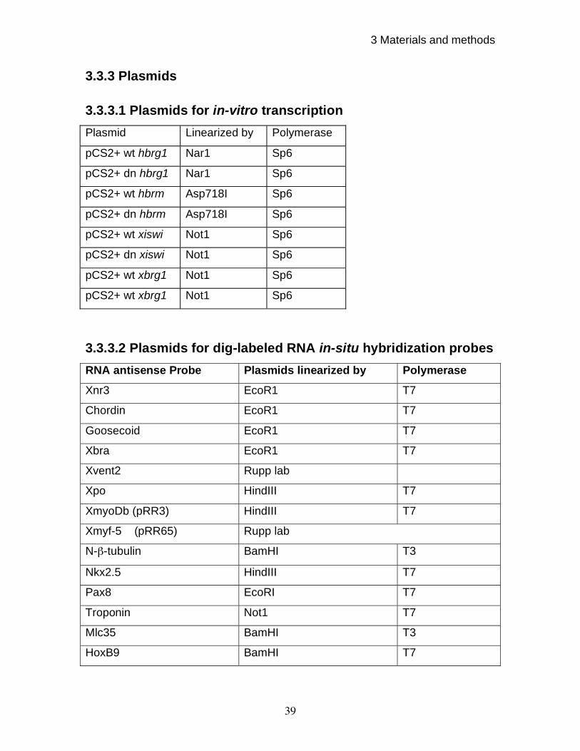
3 Materials and methods
39
3.3.3 Plasmids
3.3.3.1 Plasmids for in-vitro transcription
Plasmid Linearized by Polymerase
pCS2+ wt hbrg1 Nar1 Sp6
pCS2+ dn hbrg1 Nar1 Sp6
pCS2+ wt hbrm Asp718I Sp6
pCS2+ dn hbrm Asp718I Sp6
pCS2+ wt xiswi Not1 Sp6
pCS2+ dn xiswi Not1 Sp6
pCS2+ wt xbrg1 Not1 Sp6
pCS2+ wt xbrg1 Not1 Sp6
3.3.3.2 Plasmids for dig-labeled RNA in-situ hybridization probes
RNA antisense Probe Plasmids linearized by Polymerase
Xnr3 EcoR1 T7
Chordin EcoR1 T7
Goosecoid EcoR1 T7
Xbra EcoR1 T7
Xvent2 Rupp lab
Xpo HindIII T7
XmyoDb (pRR3) HindIII T7
Xmyf-5 (pRR65) Rupp lab
N- -tubulin BamHI T3
Nkx2.5 HindIII T7
Pax8 EcoRI T7
Troponin Not1 T7
Mlc35 BamHI T3
HoxB9 BamHI T7
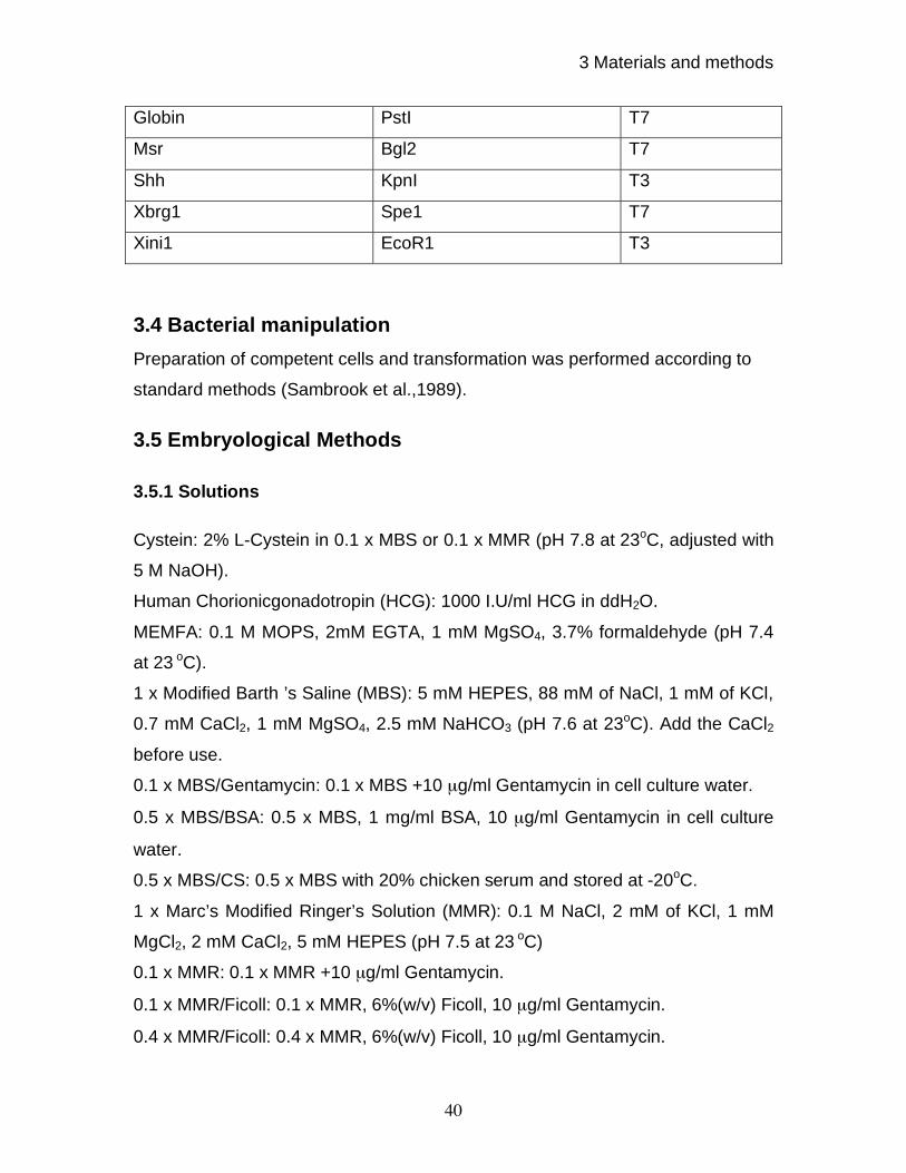
3 Materials and methods
40
Globin PstI T7
Msr Bgl2 T7
Shh KpnI T3
Xbrg1 Spe1 T7
Xini1 EcoR1 T3
3.4 Bacterial manipulation
Preparation of competent cells and transformation was performed according to
standard methods (Sambrook et al.,1989).
3.5 Embryological Methods
3.5.1 Solutions
Cystein: 2% L-Cystein in 0.1 x MBS or 0.1 x MMR (pH 7.8 at 23oC, adjusted with
5 M NaOH).
Human Chorionicgonadotropin (HCG): 1000 I.U/ml HCG in ddH2O.
MEMFA: 0.1 M MOPS, 2mM EGTA, 1 mM MgSO4, 3.7% formaldehyde (pH 7.4
at 23 oC).
1 x Modified Barth ’s Saline (MBS): 5 mM HEPES, 88 mM of NaCl, 1 mM of KCl,
0.7 mM CaCl2, 1 mM MgSO4, 2.5 mM NaHCO3 (pH 7.6 at 23oC). Add the CaCl2
before use.
0.1 x MBS/Gentamycin: 0.1 x MBS +10 μg/ml Gentamycin in cell culture water.
0.5 x MBS/BSA: 0.5 x MBS, 1 mg/ml BSA, 10 μg/ml Gentamycin in cell culture
water.
0.5 x MBS/CS: 0.5 x MBS with 20% chicken serum and stored at -20oC.
1 x Marc’s Modified Ringer’s Solution (MMR): 0.1 M NaCl, 2 mM of KCl, 1 mM
MgCl2, 2 mM CaCl2, 5 mM HEPES (pH 7.5 at 23 oC)
0.1 x MMR: 0.1 x MMR +10 μg/ml Gentamycin.
0.1 x MMR/Ficoll: 0.1 x MMR, 6%(w/v) Ficoll, 10 μg/ml Gentamycin.
0.4 x MMR/Ficoll: 0.4 x MMR, 6%(w/v) Ficoll, 10 μg/ml Gentamycin.
3 Materials and methods
41
3.5.2 Experimental animals
Adult wild type (Nasco) Xenopus laevis frogs were used. The frogs were
kept in water at 16-19oC and at a population density of 5 L water per frog. Frogs
were fed three times weakly.
3.5.3 Superovulation of the female Xenopus laevis
Xenopus laevis females were stimulated for laying eggs by injecting 500
units of human chorionic gonadotropin into the dorsal lymph sac and incubated at
15-23 oC overnight. Frogs layed eggs about 12 hours after injection.
3.5.4 Preparation of testis
The male frog was anaesthetized by 0.1 g 3-Aminobenzoesaeure-
ethylester per 100 ml ddH2O for 20 min, cooled down in ice and killed by
decapitation. The two testes were taken from the abdominal cavity by opening
and pulling out the yellow fat body, to which they are connected by connective
tissue. The testis were either used immediately or stored in MBS/CS plus
antibiotics for maximum of 7 days.
3.5.5 In-vitro fertilization of eggs and culture of the embryos
Freshly laid eggs were in-vitro fertilized either by piercing testis with a
forceps or by mincing a portion of testis to release sperms. Afterwards they were
cultured in 0.1 x MBS or 0.1 x MMR at 15-23oC in 110 mm petriplates.
3.5.6 Jelly coat removal
In order to prepare embryos for injection with mRNA (A) or morpholino
oligos, after approx.1 hpf, the jelly coat was removed in a 2% cystein solution for
5 min under agitation. Embryos were then washed three times with 0.1 x MBS
and were cultured in 0.1 x MBS or 0.1 x MMR at 15-23oC.

3 Materials and methods
42
3.5.7 Injection of embryos
The glass injection needles were pulled with Microneedle Puller (Sutter
Instrument, model P-87). They were filled with 1-2 μl nucleotide acid containing
solution just before the injection. The needles were placed into the holder of the
injection equipment (Medical System, model Pi-100). The injection volume was
adjusted by manual breaking of the needle tip and choice of the injection
pressure and/or the injection duration. Totally, 2.5-10 nanoliter solution was
injected per embryo. Embryos were usually injected at 2-4-cell stage. After
injection, embryos were incubated in 0.1 x MMR at 15-23oC until the desired
developmental stages. The medium was changed every day to increase survival.
3.5.8 Preparation of explants
For the preparation of explants, injected embryos were transferred in 3 cm
cell culture dishes covered with 1% agarose. The culture medium for explants
was 0.5 x MMR. The animal caps were removed with Gastromaster (Xenotek
Engineering) after seven hrs post fertilization. The caps were transferred
individually into a 96-well plate covered with 50 μl 1% agarose and filled with 0.5
x MMR.
3.6 Histological methods
3.6.1 Solution
AP buffer: 100 mM Tris HCl (pH 9.5), 50 mM MgCl2, 100 mM NaCl, 0.1%Tween
20, 5mM Levamisol.
AP staining solution: 4.5 μl NBT, 3.5 μl BCIP in 1 ml AP buffer or BM purple from
Roche.
MEMFA: 0.1 M MOPS, 2 mM EGTA, 1 mM MgSO4, 3.7% formaldehyde (pH 7.4
at 23 oC).
PBS: 137 mM of NaCl, 2.7 mM of KCl, 8 mM Na2HPO4, 1.7 mM KH2PO4 (pH 7.2
at 23 oC).
PBT: PBS plus 2 mg/ml BSA plus 0.1%(v/v) triton-X-100.
3 Materials and methods
43
3.6.2 Fixation of embryos
Embryos were fixed in MEMFA for 2 h under rotation on a vertical wheel.
The embryos were washed thrice with PBS. The vitelline membrane was
manually removed with a pair of sharp forceps from the embryos used for
immunohistochemistry. These embryos were finally washed once with methanol
and then stored in methanol at -20oC
until use. Embryos for in-situ hybridization
were incubated for several hours in 100% ethanol. Rinsed once more with 100 %
ethanol and then stored in 100% ethanol at -20oC
until use.
3.6.3 Immunocytochemistry
1. Fix embryos in MEMFA for two hours at room temperature with rotation. Rinse
in PBS, remove vitelline membrane manually, and store the embryos in 100 %
methanol.
2. Rehydrate by 75%, 50%, 25% methanol in PBS with each wash of 5 minutes.
3. Incubate in PBT for 15 min.
4. Block protein-binding sites by incubating embryos in PBT plus 10% heat
inactivated goat serum at room temperature for one hour.
5. Remove blocking solution and add primary antibody; incubate overnight at
4oC.
6. Wash vials by filling them for at least 5 times with each wash for one hour by
PBT.
7. Blocked as above (step 4) for one hour.
8. Add secondary antibody coupled with AP in blocking solution and incubate at
4oC
overnight.
9. Washing was done with at least five changes, 4 ml of PBT per wash for one
hour each.
10. Wash twice in AP buffer for 5-20 minutes.
11. Staining was performed in 1 ml staining solution in dark.
12. After staining, embryos were washed in PBS and fixed overnight. The
embryos were stored in PBS at 4oC.
3 Materials and methods
44
3.7 Protein Methods
3.7.1 SDS-PAGE and Western blotting
SDS-PAGE (SDS-polyacrylamide gel electrophoresis) and Western
blotting were carried out according to standard protocols (Sambrook et al., 1989).
The signals were detected by ECL kit (Amersham) as per manufacturer’s
instruction. In short, 7.5% gels were used to resolve the proteins with a constant
voltage of 120V and 400mA for one and a half hour. The protein was transferred
on to the PVDF membrane by the wet transfer system from BioRad for 90
minutes at 120V and at 4oC. The membrane was soaked into 5% milk in TBST to
block nonspecific binding sites. The membrane was washed once with TBST for
15 minutes and twice in TBST for 5 minutes each. The membrane was incubated
with primary antibody against xBRG1 antibody XB3F1 at a dilution of 1:20 for
overnight at 4oC. The membrane was washed as described above and then
incubated with anti-Rat IgG and IgM Peroxidase conjugated antibody at a dilution
of 1:5000 for one hour at room temperature. The membrane was washed once in
TBST for 15 minutes and then for four times in TBST each for 5 minutes. The
signal was detected by ECL solution. To detect -CATENIN signal, membrane
was stripped in stripping buffer for 30 minutes at 60oC, blocked and proceeded
as described above with antibody p14L (Schneider et al., 1993), specific for X -
CATENIN.
3.7.2 Immunoprecipitation
Xenopus embryos extracts was prepared in RIPA lysis buffer containing
protease inhibitors by incubating them for 1hour on ice. Protein G beads were
blocked in equal volume of 20% BSA for two hours at 4oC. Blocked beads were
incubated with anti-BRG1 antibody XB3F1 rat monoclonal antibody supernatant
for another two hrs at 4oC. Antibody coated beads were used for
Immunoprecipitation of endogenous or in-vitro translated xBRG1 by incubating
them with whole embryo lysate or in-vitro translated protein for overnight at 4oC.
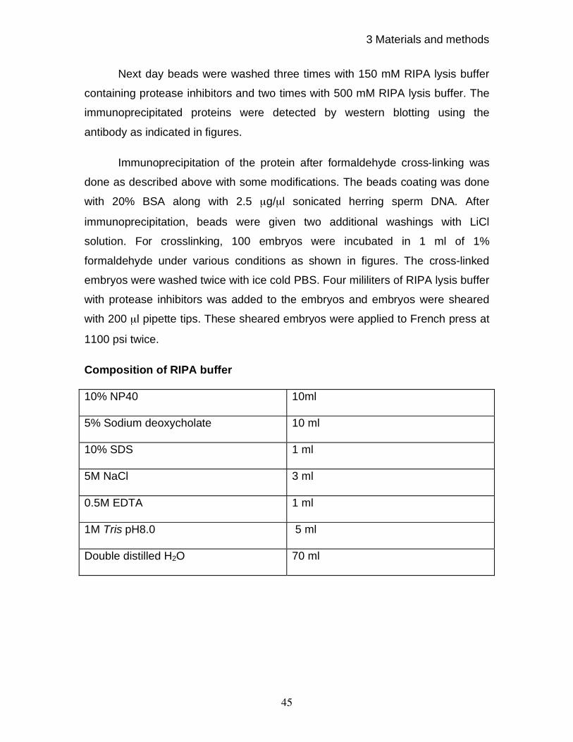
3 Materials and methods
45
Next day beads were washed three times with 150 mM RIPA lysis buffer
containing protease inhibitors and two times with 500 mM RIPA lysis buffer. The
immunoprecipitated proteins were detected by western blotting using the
antibody as indicated in figures.
Immunoprecipitation of the protein after formaldehyde cross-linking was
done as described above with some modifications. The beads coating was done
with 20% BSA along with 2.5 μg/μl sonicated herring sperm DNA. After
immunoprecipitation, beads were given two additional washings with LiCl
solution. For crosslinking, 100 embryos were incubated in 1 ml of 1%
formaldehyde under various conditions as shown in figures. The cross-linked
embryos were washed twice with ice cold PBS. Four mililiters of RIPA lysis buffer
with protease inhibitors was added to the embryos and embryos were sheared
with 200 μl pipette tips. These sheared embryos were applied to French press at
1100 psi twice.
Composition of RIPA buffer
10% NP40 10ml
5% Sodium deoxycholate 10 ml
10% SDS 1 ml
5M NaCl 3 ml
0.5M EDTA 1 ml
1M Tris pH8.0 5 ml
Double distilled H2O 70 ml
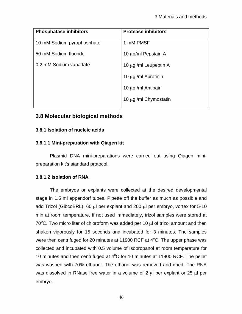
3 Materials and methods
46
Phosphatase inhibitors Protease inhibitors
10 mM Sodium pyrophosphate
50 mM Sodium fluoride
0.2 mM Sodium vanadate
1 mM PMSF
10 μg/ml Pepstain A
10 μg /ml Leupeptin A
10 μg /ml Aprotinin
10 μg /ml Antipain
10 μg /ml Chymostatin
3.8 Molecular biological methods
3.8.1 Isolation of nucleic acids
3.8.1.1 Mini-preparation with Qiagen kit
Plasmid DNA mini-preparations were carried out using Qiagen mini-
preparation kit’s standard protocol.
3.8.1.2 Isolation of RNA
The embryos or explants were collected at the desired developmental
stage in 1.5 ml eppendorf tubes. Pipette off the buffer as much as possible and
add Trizol (GibcoBRL), 60 μl per explant and 200 μl per embryo, vortex for 5-10
min at room temperature. If not used immediately, trizol samples were stored at
70oC. Two micro liter of chloroform was added per 10 μl of trizol amount and then
shaken vigorously for 15 seconds and incubated for 3 minutes. The samples
were then centrifuged for 20 minutes at 11900 RCF at 4oC. The upper phase was
collected and incubated with 0.5 volume of Isopropanol at room temperature for
10 minutes and then centrifuged at 4oC for 10 minutes at 11900 RCF. The pellet
was washed with 70% ethanol. The ethanol was removed and dried. The RNA
was dissolved in RNase free water in a volume of 2 μl per explant or 25 μl per
embryo.
3 Materials and methods
47
3.8.2 Analysis and manipulation of nucleic acids
3.8.2.1 Gel electrophoresis of nucleic acids
DNA or in-vitro synthesized RNA was isolated in horizontal agarose gel.
Depending upon fragment size, 1% or 1.7% agarose gel was used. After
electrophoresis, the gels were photographed. 100bp or 1kb DNA ladder was
used as size standard.
3.8.2.2 Isolation of DNA fragments from agarose gel
In order to isolate DNA fragments after electrophoresis from agarose gel,
the appropriate band was cut out under long-wave UV light. The DNA was
extracted from the gel with Qiagen gel-extraction kit.
3.8.2.3 Cloning methods
The cloning of DNA has been performed according to standard methods
(Sambrook et al., 1989).
3.8.3 Polymerase chain reaction (PCR)
3.8.3.1 PCR amplification of xbrg1 c-DNA fragment for cloning
The reaction was accomplished in a total volume of 50 l. The reaction
mixture contained 1 μl of the cDNA template, 25 pmol each primer, 0.5 mm
dNTPs, 1 U advantage taq polymerase and 1x buffer.
Following Primers were used:
Primers for xbrg1
Forward Primer
5’ TGTCGGTGATAGTTGGTGGAAAAC 3’
Reverse Primer
5’ AGGGGGTAAAGGAATGTGATG 3’
Since normal PCR gave a smear in case of xbrg1, Touch down PCR was
carried out to obtain full-length c-DNA for xbrg1 as following:

3 Materials and methods
48
Step 1. 95oC 1 Min.
Step 2. 95oC 15 Seconds
60oC 30 Seconds
68oC 6 Min
for 5 cycles
Step3. 95oC 15 Seconds
60oC 30 Seconds (temp decrease @ 0.5
oC per cycle)
68oC 6 Min
for 20 cycles
Step 4. 95oC 15 Seconds
50oC 30 Seconds
68oC 6 Min
for 10 cycles
Step 5. 72oC 10 Min
The PCR obtained a band of about 5Kb. The band was excised from the
gel and cloned into PCRII TOPO vector. The clones were checked by EcoR1
restriction digestion. The ORF only of xbrg1 was further sub cloned into pCS2+
vector with a flag tag at the C-terminus. The primers used were having EcorV
and Xho1 restriction sites, which were used to clone xbrg1 into Stu1 and Xho1
restriction sites of pCS2+ vector. The primers were following:
Forward primer:
5’ GAGATATCCACGATGTCCACCCCAGACCCT 3’
Reverse Primer:
5’ CCGCTCGAGTTATTTATCGTCATCGTCTTTGTAGTCGTCTTCTTCACTTCC
ACTTCC 3’
The pCS2+ vector containing full-length xbrg1 was sequenced by MWG.
3.8.3.2 RT-PCR
In RT-PCR assay, RNA was initially reverse-transcribed to yield cDNA.
The cDNA samples were normalized by PCR amplification of H4 (histone4) in the
linear range. The desired target cDNA species were amplified using specific
3 Materials and methods
49
primers. PCR was carried out in the exponential phase of amplification and PCR
samples were loaded side by side in the agarose gel to compare their intensity.
3.8.3.3 Northern blotting
Northern blotting was carried out using standard protocol (Sambrook et al,
1989). For the detection of Xbrg1 transcripts, Northern hybridization was done
using a RNA probe made from N-terminal sequence of xbrg1. The probe was
about 2 kb long. The blots were exposed for three days.
3.8.4 In-vitro transcription
3.8.4.1 In-vitro reverse transcription
A cDNA pool was generated from total cellular RNA by using random
oligonucleotides and reverse transcriptase (RTase) (Steinbach and Rupp,
1999).The reaction was accomplished in a total volume of 20 μl. The reaction
contains 2μl of total RNA, 4μl RT buffer, 1μl RTase, 2μl dNTPs, 1μl RNAsin and
100 pmol random hexamer primers as well as 200 U reverse transcriptase. The
reaction was as following: total RNA was mixed with random hexamers and
RNase free water and incubated for 5 minutes at 70oC. The reaction was cooled
down in ice and rest of the components were mixed. The samples were
incubated at 25oC for 10 min and then transferred to 42
oC for 60 minutes. The
samples were incubated at 70oC for 10 min to inactivate reverse transcriptase.
The sample was cooled down to 4oC. The synthesized cDNA were stored at -
20oC.
3.8.4.2 In-vitro transcription for microinjection
Capped mRNA used for microinjection was in-vitro transcribed with RNA
polymerase. The reaction was setup in a total volume of 50 μl containing 2.5 μg
linearized plasmid DNA, 1 x transcription buffer, 0.5 mM dNTPs, 2.5 mM RNA
cap structure analogue, 10mM DTT, 20U RNAsin and 40U Sp6 or 60U T3 or T7
RNA Polymerase and incubated for at least 2.5 hours at 37oC except xbrg1,
3 Materials and methods
50
which were incubated at 20oC. The template was digested with 10 U of RNase
free DNase I by incubating the samples for 30 min at 37oC. The volume was filled
up with RNase free water to 90 μl, extracted and centrifuged with one volume of
PCI (13500 x g, eppendorf centrifuge 5415C, 14000 rpm, 23oC, 5 min). About 80
μl of the supernatant was applied on the Quick Spin column and eluted by
centrifugation (1100 x g, 23oC, 4 min). It was again centrifuged (13500 x g, 23
oC,
5 min), and supernatant was transferred into a new 1.5 ml reaction tube. The
RNA was precipitated with 0.1 volume of 3 M sodium acetate and 2.5 volume of
ice-cold 100% ethanol for 30 min at -20oC. After centrifugation (13500 x g, 4
oC,
20 min), the pellet was washed with 0.5 ml of 70% ethanol, air-dried and
dissolved in 10 μl RNase free water. The concentration was determined by
GeneQuant II (Pharmacia Biotech).
3.8.4.3 In-vitro transcription of dig labeled RNA probes
Plasmid was linearized and used to generate antisense RNA by using
RNA polymerases. The reactions were set up as following: in a total volume of 20
μl; 1 μg linearized plasmid DNA, 1 x transcription buffer, 0.1 mM Dig NTPs, 20 U
RNAsin and 20 U SP6 or T3 or T7 RNA Polymerase, incubated at 37oC for 2 h.
To obtain the antisense probe for xbrg1, -36 to+ 618 fragment of xbrg1 was
cloned into the pCRII-TOPO (Invitrogen Life Technologies) vector. It was
linearized with Spe1 and transcribed using T7 promoter to obtain antisense
probe.
PCR primers for the xbrg1 (-36 to +618 fragment)
Forward Primer
5’ TGTCGGTGATAGTTGGTGGAAAAC 3’
Reverse Primer
5’ TGGTTCTGGTTGAAGGGAGTAGGG 3’
3 Materials and methods
51
3.8.5 Site-directed mutagenesis
The dominant negative BRG1 was produced by doing a point mutation at
position 770 and dominant negative ISWI was obtained by doing a point mutation
at position 204, converting lysine into arginine.
Mutagenesis Primers for xBRG1 (K770-R)
Forward Primer
5’ GAGATGGGCCTGGGAAGGACTATTCAGACCATT 3’
Reverse Primer
5’ AATGGTCTGAATAGTCCTTCCCAGGCCCATCTC 3’
Mutagenesis Primers for xISWI (K204 - R)
Forward Primer
5’ GAAATGGGTCTAGGAAGGACTTTGCAGACCATC 3’
Reverse Primer
5’ GATGGTCTGCAAAGTCCTTCCTAGACCCATTTC 3’
For BRG1, mutagenesis was done on EST 234591 cDNA clone, which
spans the location of aa 770. Successful mutation was confirmed by sequencing.
This mutated fragment was replaced with the wild type fragment in the wild type
xbrg1 clone into the XmaC1 sites and orientation was checked by restriction
enzyme analysis and by sequencing.
Site-directed mutagenesis was done with the Quick Change site-directed
mutagenesis kit (Stratagene, Cat.200518) according to the instruction manual. In
brief, the Quick Change site-directed mutagenesis method was performed using
Pfu DNA polymerase and a temperature cycler. The basic procedure utilizes a
super coiled double-stranded DNA (dsDNA) template (50 ng each). The
oligonucleotide primers, each complementary to opposite strands of the vector,
are extended during temperature cycling by Pfu DNA polymerase. Incorporation
of the oligonucleotide primers generates a mutated plasmid containing staggered
nicks. Following temperature cycling (96oC 1 min, 55
oC, 1 min, 72
oC 2 min/kb, 20
3 Materials and methods
52
cycles), the product was treated with DpnI. The DpnI endonuclease (target
sequence: 5’-GmATC-3’) is specific for methylated and hemimethylated DNA and
is used to digest the parental DNA template and to select for mutation-containing
synthesized DNA. DNA isolated from almost all E.coli strains is dam methylated
and therefore susceptible to DpnI digestion. The nicked vector DNA containing
the desired mutations was then transformed into XL1-Blue super competent
cells. After the transformation, the competent cells repair the nicks in the mutated
plasmid. The small amount of starting DNA template required to perform this
method, the high fidelity of the Pfu turbo DNA polymerase, and the low number of
thermal cycles all contribute to the high mutation efficiency and decreased
potential for generating random mutations during the reaction. The mutated
plasmids were confirmed by sequencing.
3.8.6. Designing and synthesis of antisense morpholino
oligonucleotides
Following Morpholino antisense oligo was designed against xBrg1
5’ CCATTGGAGGGTCTGGGGTGGACAT 3’
The antisense morpholino oligo were designed against Xbrg1 on the basis of
following guidelines:
1) An optimal target sequence chosen was 25-base pair long to obtain maximum
efficiency.
2) were within in the region from the 5´cap through the first 25 bases of coding
sequence.
3) had approximately 50% GC content.
4) had little or no self-complementarity.
5) having less then seven total guanine and less then three contiguous guanines
in a 25- mer oligo.
Based on above criteria following oligo was synthesized from Gene-Tool, LLC.
Following is the sequence of standard control morpholino oligos supplied by
company:
5' CCTCTTACCTCAGTTACAATTTATA 3'
3 Materials and methods
53
3.8.7. Expression and Purification of GST-xBRG1 fusion protein
for the generation of monoclonal antibody
For the generation of monoclonal antibodies against xBRG1 a fragment
covering aa 202 to aa 282 was sub-cloned into pGEX4T3 vector to generate a
GST fusion protein. The primers used were following:
Primers for GST- xBRG1 fusion protein construction in PGEX: aa202-282
Forward Primers:
5’ CGGGATCCCCCATGCCTGGGATGCAACAA 3’
Reverse Primers:
5’ GGAATTCGGTCAAGTGGCAGGAGCAGCCGCATT 3’
The gst-xbrg1 construct was transformed in to BL21 E.Coli host strain.
Five clones were tested for optimization of the fusion protein production. The
clones were grown until the cell density reached at O.D. 0.7. The cells were
induced by 1mM IPTG for 2hrs. This initial screening yielded two clones, giving
good yield of fusion protein as determined by SDS page analysis. These two
clones were further tested. The clone yielding better amount of fusion protein
was chosen for large-scale production of fusion protein.
This clone was grown in 2.5 liter of culture volume until the cell density of
equivalent to O.D; 0.7 was reached. The bacterial cells were induced as
described. After induction, bacterial pellet was collected by centrifugation at 6000
x g for 10 minutes in aliquots of 500 ml culture volume. The pellet was frozen at
-20oC. Afterwards, pellet from 1 liter culture was resuspended in ice cold PBS.
This 50ml culture pellet was divided into two parts and then each aliquot was
sonicated at 60% amplitude on Branso Digital Sonifier 250-D of 1’ cycle for 3’ on
ice to keep sample cool. Sonicated samples were then incubated with 20%
Triton-X at final concentration of 1% for 30’ at 4oC. The samples were centrifuged
at 12000 x g for 15’ and supernatant was collected. The supernatant was
incubated with 50% glutathione sepharose 4b beads for overnight at 4oC. On
3 Materials and methods
54
next day, the slurry was centrifuged at 500 x g for 5’. The Glutathione sepharose
beads were washed three times with ice cold PBS. The slurry was incubated with
1mL of 8 M urea buffer for two hrs at 20oC. The beads were removed by
centrifugation of the samples at 2000 x g for 10’. The protein amount was
measured in supernatant by Bradford method as well as by running BSA
standards on SDS-PAGE. The protein was about 8 mg/mL by comparing with
BSA standards on gel as well as by Bradford method.
The GST-xBRG1 fusion protein was used to immunize rats for the
production of xBRG1 specific antibodies, which was carried out in collaboration
with Prof. Elisabeth Kremmer.
3.8.8 RNA in-situ hybridization
The RNA in-situ hybridization was performed as described previously
(Harland, 1991) with slight modifications. The method used is described below:
Day 1:
1. The fixed embryos were rehydrated by serial changes of 75%, 50% and 25%
ethanol series.
2. Washed 3 times in PBSw.
3. Treated with 10 μg/ml Proteinase K in PBSw (1 ml per vial) for 30 min at room
temperature.
4. Rinsed with PBSw.
5. Washed 2 times in PBSw, 5 min each
6. Fixed with 4% PFA in PBSw (0.5ml per vial). Stand for 5 min and then rock for
15 min.
7. Short rinsed with PBSw.
8. Washed 5 times in PBSw.
9. Incubation in 50% PBSw / 50% hybridization solution, followed by 100%
hybridization solution for about 3 min each step without agitation at RT.
10. Added 0.5 ml of fresh hybridization solution.

3 Materials and methods
55
11. Embryos were incubated at 65oC for 1 hour to inactivate endogenous
phophatases.
12. Embryos were pre-hybridized at 60oC for 2-6 hours.
13. Heated 30 ng of probe to 95oC for 2-5 min. Add to 100 μl of pre-warmed
hybridization solution.
14. Add the probe to the embryos in pre-hybridization solution.
15. Incubate at 60oC overnight (hybridization).
Day 2:
16. Pre-warmed 2 x SSC / 0.1%CHAPS to 37oC, and 0.2 x SSC / 0.1% CHAPS
to 60oC.
17. Short sequential washes of embryos at 60oC with:
a) Hybridization solution
b) 50% hybridization solution / 50% 2 x SSC / 0.1% CHAPS
c) 2 x SSC / 0.1% CHAPS
18. Washed 2 times in two x SSC / 0.1%CHAPS for 30 min at 37oC.
19. Short rinse with 0.2 x SSC / 0.1% CHAPS
20. Washed 2 times in 0.2 x SSC / 0.1% CHAPS for 30 min at 60oC.
21. Rinsed in 50% TBS / 50% 0.2 x SSC / 0.1% CHAPS.
22. Washed once in TBS.
23. Rinsed in TBS / 0.1%Triton-X (TBSX).
24. Incubated in antibody buffer (0.5 ml per vial) for 2 hours at 4oC.
25. In parallel, pre-absorb AP-conjugated anti-DIG antibodies (1/5000 diluted)
against Xenopus proteins present in antibody solution.
26. Added 0.5 ml of pre absorbed antibody solution to embryos (i.e. final dilution
of antibodies was10-4
).
27. Incubate overnight at 4oC.
Day 3:
28. Rinsed with TBSX
29. Washed 6 times in TBSX for 1 hour each.
30. Rinsed with AP-buffer.

3 Materials and methods
56
31. Equilibrated in AP-buffer for 15 minutes.
32. Replaced AP-buffer with 0.5 ml BM-Purple staining solution.
33. Incubate overnight at room temperature in dark with slight rocking (color
reaction).
Day 4:
34. Stop reaction by washing twice in PBS for 10 min at room temperature with
rocking.
35. Refix embryos in MEMFA for 2 hours, short rinse with H2O or PBS, store in
H2O or PBS at 4oC.
Solutions:
Antibody-buffer: 80% (v/v) TBSX, 15% (v/v) heat-inactivated lamb serum, 5%
(v/v) Xenopus egg extract.
DEPC-H2O: dd H2O with 0.1% (v/v) Diethylpyrocarbonate (DEPC) agitated at
room temperature and incubated overnight and then autoclaved twice.
10 mM DIG NTP mixture: 10 mM CTP, GTP, ATP, 6.5 mM UTP and 3.5 mM Dig-
11- UTP.
Hybridizing solution: 5 x SSC, 50% (v/v) formamide, 1% (w/v) Boehringer block,
0.1 % (w/v) Torula RNA, 0.01% Heparin, 0.1% Tween-20, 0.1 %CHAPS, 5 mM
EDTA.
Lamb Serum: Heat-inactivated lamb serum (30 min with 56oC), stored at -20
oC.
MEMFA: 0.1 M MOPS, 2 mM EGTA,1 mM MgSO4, 3.7% formaldehyde (pH 7.4
at 23oC).
PBS: 137 mM of NaCl, 2.7 mM of KCl, 8 mM Na2HPO4, 1.7 mM KH2PO4 (pH 7.2
at 23oC).
PBSw: 1 x PBS, 0.1 % Tween-20 (pH 7.5 at 23oC).
PCI: 50% (v/v) phenol, 48% (v/v) chloroform, 2% (v/v) isoamyl alcohol.
PCR buffer (Taq): 10 mM of trichloroethylene HCl, 50 mM of KCl, 15 mM MgCl2
(pH 8.4 at 23oC).

3 Materials and methods
57
PCR buffer (Pfu): 10 mM of trichloroethylene HCl, 10 mM of KCl, 2 mM MgCl2, 10
mM (NH4)2SO4, 0.1% (v/v) triton X-100, 100 μg/ml nuclease free BSA in DEPC
H2O (pH 8.4 at 23oC).
5 x RNA polymerase transcriptions buffer: 200 mM of trichloroethylene HCl, 30
mM MgCl2, 10 mM of spermidin, 50 mM of NaCl (pH 7.5 at 23oC).
20 x SSC: 3 M NaCl, 0.3 M sodium citrate (pH 7.0 at 23oC).
TBS: 50 mM of trichloroethylene HCl, 150 mM of NaCl (pH 7.5 at 23oC).
TBSX: 1 x TBS, 0.1% triton-X (pH 7.5 at 23oC).
TE: 1 mM EDTA, 10 mM of Tris HCl (pH 8.0 at 23oC).
TBE: 100 mM of trichloroethylene HCl, 83 mM borate, 0.1 mM EDTA (pH 8.6 at
23oC).
Transcription buffer: 20 mM of Tris HCl, 10 mM of Spermidin, 3 mM MgCl2, 50
mM of NaCl (pH 7.5 at 23oC) in DEPC H2O.
AP-buffer: 100mM Tris HCl, 50mM MgCl2, 100mM NaCl.
Xenopus egg extract for in-situ hybridization
De-jelly unfertilized eggs with 2% Cystein, wash 3 times, add 1 volume of PBS
then lysed by 10 strokes of a dounce homogenizations, and centrifuged (7500 x
g, Sorvall RC-5b, rotors SS-34, 4oC, 10 min). The supernatant was transferred
into a fresh centrifuge tube and centrifuged twice under the same conditions. The
supernatant was aliquoted and stored at -20oC.
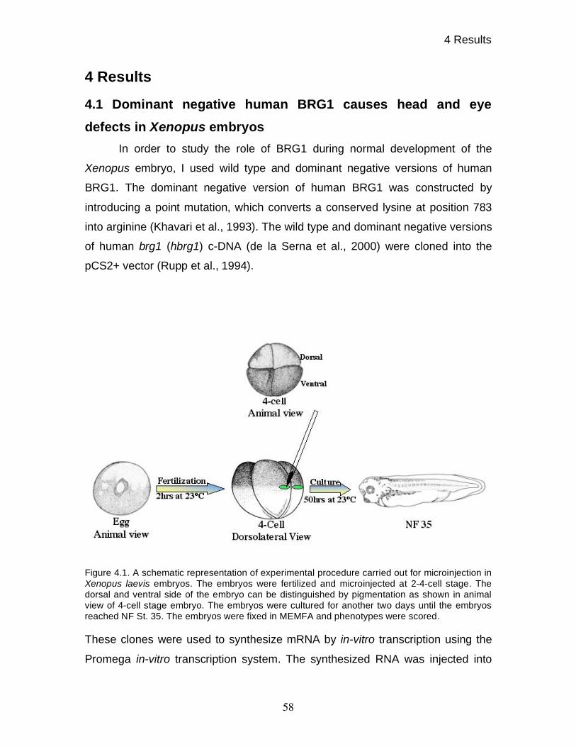
4 Results
58
4 Results
4.1 Dominant negative human BRG1 causes head and eye
defects in Xenopus embryos
In order to study the role of BRG1 during normal development of the
Xenopus embryo, I used wild type and dominant negative versions of human
BRG1. The dominant negative version of human BRG1 was constructed by
introducing a point mutation, which converts a conserved lysine at position 783
into arginine (Khavari et al., 1993). The wild type and dominant negative versions
of human brg1 (hbrg1) c-DNA (de la Serna et al., 2000) were cloned into the
pCS2+ vector (Rupp et al., 1994).
Figure 4.1. A schematic representation of experimental procedure carried out for microinjection in
Xenopus laevis embryos. The embryos were fertilized and microinjected at 2-4-cell stage. The
dorsal and ventral side of the embryo can be distinguished by pigmentation as shown in animal
view of 4-cell stage embryo. The embryos were cultured for another two days until the embryos
reached NF St. 35. The embryos were fixed in MEMFA and phenotypes were scored.
These clones were used to synthesize mRNA by in-vitro transcription using the
Promega in-vitro transcription system. The synthesized RNA was injected into

4 Results
59
either dorsal or ventral marginal of Xenopus embryo at the four-cell stage. The
injections were targeted to the equatorial zone at the boundary of pigmented
animal half and un-pigmented vegetal half as represented in figure 4.1. The
injected embryos were cultured for 50 hrs post-fertilization at 23oC until the
embryos reached NF stage 35 (Nieuwkoop and Faber, 1967).
Interestingly overexpression of dominant negative hBRG1 (dn hBRG1)
into dorsal marginal zone produced embryos with a head and eye defect as well
as a shorter anterio-posterior axis. As shown in figure 4.2, overexpression of dn
hBRG1 gave a spread of phenotypes. The eye was always reduced, even in the
least affected embryo, while more strongly affected embryos had severely
reduced head as well as reduced anterio-posterior axes. Some embryos also
displayed reduced pigmentation. Other than these defects, affected embryos
were non-motile in response to mechanical as well as chemical stimuli. Finally,
no heart beating was observed. The defects produced by overexpression of dn
hBRG1 were dose dependent; about 63% embryos were affected with 250 pg of
dominant negative Hbrg1 mRNA while about 81% embryos were affected by 500
pg of dn Hbrg1 injections (Table 4.1.). The overexpression of wt hBRG1 and
GFP alone did not produce any abnormalities. The embryos injected with wild
type and dominant negative variants of Hbrg1 mRNA into the ventral marginal
zone of four-cell stage embryos, also did not produce any phenotype and were
normal when compared to Gfp alone-injected embryos or with un-manipulated
embryos. These results showed that the overexpression of dn hBRG1 interferes
with the dorsal program of the embryos while the ventral program remains
unaffected.
BRM is a closely related ATPase to BRG1. In order to test if
overexpression of human BRM also interferes with normal development of
Xenopus embryos, the wild type human brm and dominant negative human brm
(de la Serna et al., 2000) were cloned into pCS2+ vector and used to in-vitro
synthesize mRNA.
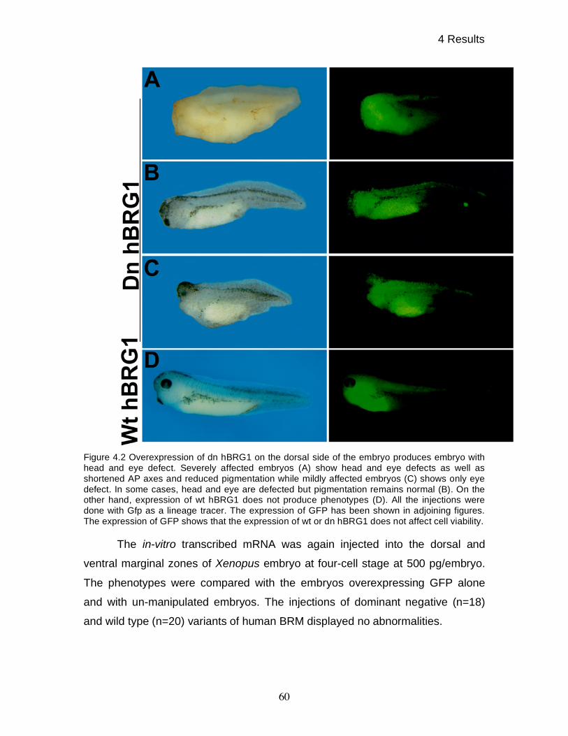
4 Results
60
Figure 4.2 Overexpression of dn hBRG1 on the dorsal side of the embryo produces embryo with
head and eye defect. Severely affected embryos (A) show head and eye defects as well as
shortened AP axes and reduced pigmentation while mildly affected embryos (C) shows only eye
defect. In some cases, head and eye are defected but pigmentation remains normal (B). On the
other hand, expression of wt hBRG1 does not produce phenotypes (D). All the injections were
done with Gfp as a lineage tracer. The expression of GFP has been shown in adjoining figures.
The expression of GFP shows that the expression of wt or dn hBRG1 does not affect cell viability.
The in-vitro transcribed mRNA was again injected into the dorsal and
ventral marginal zones of Xenopus embryo at four-cell stage at 500 pg/embryo.
The phenotypes were compared with the embryos overexpressing GFP alone
and with un-manipulated embryos. The injections of dominant negative (n=18)
and wild type (n=20) variants of human BRM displayed no abnormalities.
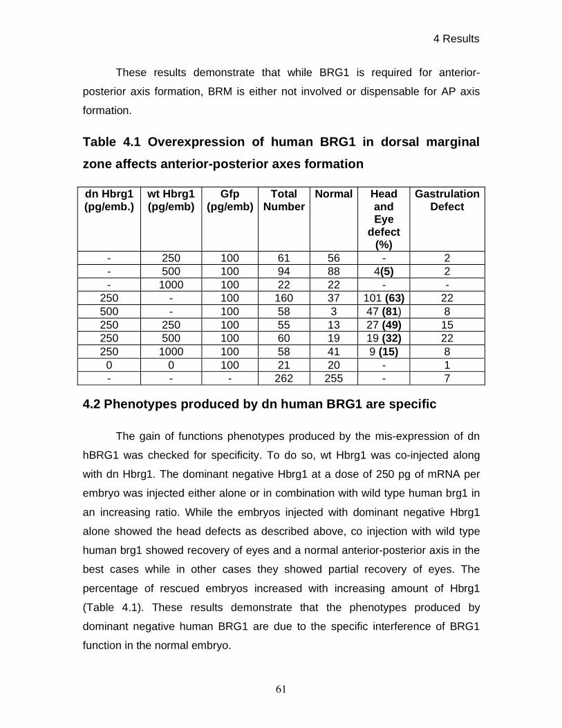
4 Results
61
These results demonstrate that while BRG1 is required for anterior-
posterior axis formation, BRM is either not involved or dispensable for AP axis
formation.
Table 4.1 Overexpression of human BRG1 in dorsal marginal
zone affects anterior-posterior axes formation
4.2 Phenotypes produced by dn human BRG1 are specific
The gain of functions phenotypes produced by the mis-expression of dn
hBRG1 was checked for specificity. To do so, wt Hbrg1 was co-injected along
with dn Hbrg1. The dominant negative Hbrg1 at a dose of 250 pg of mRNA per
embryo was injected either alone or in combination with wild type human brg1 in
an increasing ratio. While the embryos injected with dominant negative Hbrg1
alone showed the head defects as described above, co injection with wild type
human brg1 showed recovery of eyes and a normal anterior-posterior axis in the
best cases while in other cases they showed partial recovery of eyes. The
percentage of rescued embryos increased with increasing amount of Hbrg1
(Table 4.1). These results demonstrate that the phenotypes produced by
dominant negative human BRG1 are due to the specific interference of BRG1
function in the normal embryo.
dn Hbrg1 (pg/emb.)
wt Hbrg1 (pg/emb)
Gfp (pg/emb)
Total Number
Normal Head and Eye
defect (%)
Gastrulation Defect
- 250 100 61 56 - 2
- 500 100 94 88 4(5) 2
- 1000 100 22 22 - -
250 - 100 160 37 101 (63) 22
500 - 100 58 3 47 (81) 8
250 250 100 55 13 27 (49) 15
250 500 100 60 19 19 (32) 22
250 1000 100 58 41 9 (15) 8
0 0 100 21 20 - 1
- - - 262 255 - 7
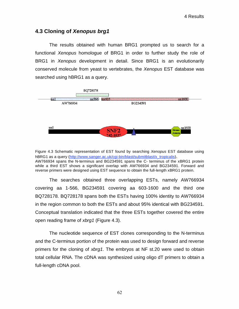
4 Results
62
4.3 Cloning of Xenopus brg1
The results obtained with human BRG1 prompted us to search for a
functional Xenopus homologue of BRG1 in order to further study the role of
BRG1 in Xenopus development in detail. Since BRG1 is an evolutionarily
conserved molecule from yeast to vertebrates, the Xenopus EST database was
searched using hBRG1 as a query.
Figure 4.3 Schematic representation of EST found by searching Xenopus EST database using
hBRG1 as a query (http://www.sanger.ac.uk/cgi-bin/blast/submitblast/x_tropicalis).
AW766934 spans the N-terminus and BG234591 spans the C- terminus of the xBRG1 protein
while a third EST shows a significant overlap with AW766934 and BG234591. Forward and
reverse primers were designed using EST sequence to obtain the full-length xBRG1 protein.
The searches obtained three overlapping ESTs, namely AW766934
covering aa 1-566, BG234591 covering aa 603-1600 and the third one
BQ728178. BQ728178 spans both the ESTs having 100% identity to AW766934
in the region common to both the ESTs and about 95% identical with BG234591.
Conceptual translation indicated that the three ESTs together covered the entire
open reading frame of xbrg1 (Figure 4.3).
The nucleotide sequence of EST clones corresponding to the N-terminus
and the C-terminus portion of the protein was used to design forward and reverse
primers for the cloning of xbrg1. The embryos at NF st.20 were used to obtain
total cellular RNA. The cDNA was synthesized using oligo dT primers to obtain a
full-length cDNA pool.
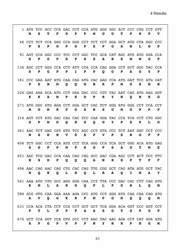
4 Results
63
1 ATG TCC ACC CCA GAC CCT CCA ATG GGG GGC ACT CCC CGG CCT GTT
M S T P D P P M G G T P R P V 46 CCT TCT CCA GGG CCA GGG CCT TCT CCT GGA GCT ATG CTA GGC CCC
P S P G P G P S P G A M L G P 91 AGT CCA GGC CCC TCC CCT GGC TCC GCA CAT AGC ATG ATG GGA CCA
S P G P S P G S A H S M M G P 136 AGC CCT GGG CCA CCT ATT CCA CCA CAG GGA CCT GCT GGC TAC CCA
S P G P P I P P Q G P A G Y P 181 CCC GAA AAT ATG CAA CAG ATG CAC AAG CCA ATG GAT TCC ATG CAT P E N M Q Q M H K P M D S M H 226 GAG AAA GCA ATG CCT GAA GAC CCC CGT TAC AAT CAG ATG AAG GGT
E K A M P E D P R Y N Q M K G 271 ATG GGC ATG AGA CCT GGA GCT CAC TCT GGG ATG GGG CCT CCA CCT
M G M R P G A H S G M G P P P 316 AGT CCT ATG GAC CAG CAC TCC CAA GGA TAC CCA TCA CCT CTG GGC
S P M D Q H S Q G Y P S P L G 361 AAC TCT GAG CAT GTG TCC AGC CCT GTA CCC TCT AAT GGC CCT CCC
N S E H V S S P V P S N G P P 406 TCT GGC CCT CCA ATG CCT TCA GGG CCA TCA GCT GGG ACA ATG GAG
S G P P M P S G P S A G T M E 451 AAC TCG GAC CCA CAA CAG CAG GCC AAC CGA AGC CCT ACT CCC TTC
N S D P Q Q Q A N R S P T P F 496 AAC CAG AAC CAG CTG CAC CAG TTG CGG GCT CAG ATA ATG GCT TAC
N Q N Q L H Q L R A Q I M A Y 541 AAA ATG CTG GCC AGG GGG CAA CCT TTA CCC GAC CAC CTT CAG ATG
K M L A R G Q P L P D H L Q M 586 GCG GTG CAA GGA AAA AGA CCC ATG CCT GGG ATG CAA CAA CAG ATG
A V Q G K R P M P G M Q Q Q M 631 CCA ACA CTA CCT CCA CCT GCT GCT TCA GGG ACA GGT CCC GGT CCT
P T L P P P A A S G T G P G P 676 GCT CCA GGT CCA GTG CCC CCT AAC TAC AAC AGA CCT CAT GGA ATG
A P G P V P P N Y N R P H G M
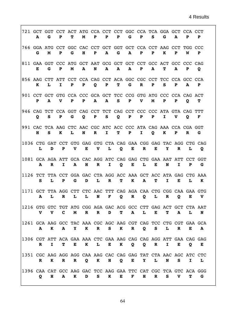
4 Results
64
721 GCT GGT CCT ACT ATG CCA CCT CCT GGC CCA TCA GGA GCT CCA CCT A G P T M P P P G P S G A P P
766 GGA ATG CCT GGC CAC CCT GCT GGT GCT CCA CCT AAG CCT TGG CCC
G M P G H P A G A P P K P W P 811 GAA GGT CCC ATG GCT AAT GCG GCT GCT CCT GCC ACT GCC CCC CAG
E G P M A N A A A P A T A P Q 856 AAG CTT ATT CCT CCA CAG CCT ACA GGC CGC CCT TCC CCA GCC CCA
K L I P P Q P T G R P S P A P 901 CCT GCT GTG CCA CCC GCA GCT TCC CCG GTG ATG CCC CCA CAG ACT
P A V P P A A S P V M P P Q T 946 CAG TCT CCA GGT CAG CCT TCT CAG CCT CCC CCC ATA GTA CAG TTT
Q S P G Q P S Q P P P I V Q F 991 CAC TCA AAG CTC AAC CGC ATC ACC CCC ATA CAG AAA CCA CGA GGT
H S K L N R I T P I Q K P R G 1036 CTG GAT CCT GTG GAG GTG CTA CAG GAA CGG GAG TAC AGG CTG CAG
L D P V E V L Q E R E Y R L Q 1081 GCA AGA ATT GCA CAC AGG ATC CAG GAG CTG GAA AAT ATT CCT GGT
A R I A H R I Q E L E N I P G 1126 TCT TTA CCT GGA GAC CTA AGG ACC AAA GCT ACC ATA GAG CTG AAA
S L P G D L R T K A T I E L K 1171 GCT TTA AGG CTT CTC AAC TTT CAG AGA CAA CTG CGG CAA GAA GTG
A L R L L N F Q R Q L R Q E V 1216 GTG GTC TGT ATG CGG AGA GAC ACG GCC CTT GAG ACT GCT CTA AAT
V V C M R R D T A L E T A L N 1261 GCA AAG GCC TAC AAA CGC AGC AAG CGT CAG TCC CTG CGT GAA GCA
A K A Y K R S K R Q S L R E A 1306 CGT ATT ACA GAA AAA CTC GAA AAG CAG CAG AGG ATT GAA CAG GAG
R I T E K L E K Q Q R I E Q E 1351 CGC AAG AGG AGG CAA AAG CAC CAG GAG TAT CTA AAC AGC ATC CTC
R K R R Q K H Q E Y L N S I L 1396 CAA CAT GCC AAG GAC TCC AAG GAA TTC CAT CGC TCA GTC ACA GGG
Q H A K D S K E F H R S V T G
4 Results
65
1441 AAG ATC CAA AAA CTG ACC AAG GCT GTA TCC ACT TAC CAC GCC AAC K I Q K L T K A V S T Y H A N
1486 ACA GAA CGA GAG CAG AAG AAG GAG AAT GAA AGA ATT GAG AAG GAG
T E R E Q K K E N E R I E K E 1531 AGA ATG CGC AGG CTC ATG GCT GAG GAT GAA GAG GGC TAT AGA AAG
R M R R L M A E D E E G Y R K 1576 CTG ATC GAT CAG AAG AAA GAC AAG CGT CTG GCC TAT CTC CTG CAA
L I D Q K K D K R L A Y L L Q 1621 CAA ACG GAT GAG TAT GTA GCA AAT CTG ACA GAT TTG GTG CGA CAA
Q T D E Y V A N L T D L V R Q 1666 CAC AAG GCG GTC CAA GCC CTC AAA GAA AAG AAG AGA AGA AAA AAG
H K A V Q A L K E K K R R K K 1711 AAA AAG GTG CAA GAG AAC ACA GAA GGA CAA CAG ACT GCA CTT GGG
K K V Q E N T E G Q Q T A L G 1756 CCA GAT GGA GAG CCA CTG GAT GAG ACC AGC CAG ATG AGT GAC CTC
P D G E P L D E T S Q M S D L 1801 CCA GTC AAA GTG ATC CAC GTG GAA AGT GGA AAG ATC TTG ATC GGT
P V K V I H V E S G K I L I G 1846 GCT GAT GCA CCA AAG GCT GGA CAG CTG GAG GCG TGG CTT GAA ATG
A D A P K A G Q L E A W L E M 1891 AAC CCT GGG TAT GAA GTT GCA CCA CGG TCT GAT AGT GAG GAG AGT
N P G Y E V A P R S D S E E S 1936 GGA TCA GAG GAG GAA GAA GAG GAA GAA GAG GAA GAA CAA CAG CAG
G S E E E E E E E E E E Q Q Q 1981 CCT CCA GTC TCT GCC TTG CTG GCT GTG GAT GAG ACC AAA AAA ATC
P P V S A L L A V D E T K K I 2026 ACT GAC CCA GAC AGC TAT GAT GTG CCT GAG GGT TAT GCC CGG GAA
T D P D S Y D V P E G Y A R E 2071 TTA ATA GAT AAT GCC CGG CAG GAT GTG GAT GAT GAG TAT GGT ATA
L I D N A R Q D V D D E Y G I 2116 TCC CAA ATG GAA ATC CGT GGT CTC CAG TCT TAC TAT GCA GTG GCG
S Q M E I R G L Q S Y Y A V A
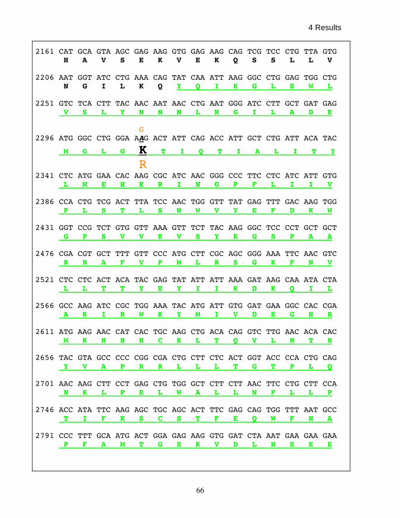
4 Results
66
2161 CAT GCA GTA AGC GAG AAG GTG GAG AAG CAG TCG TCC CTG TTA GTG H A V S E K V E K Q S S L L V
2206 AAT GGT ATC CTG AAA CAG TAT CAA ATT AAG GGC CTG GAG TGG CTG
N G I L K Q Y Q I K G L E W L 2251 GTC TCA CTT TAC AAC AAT AAC CTG AAT GGG ATC CTT GCT GAT GAG
V S L Y N N N L N G I L A D E G 2296 ATG GGC CTG GGA AAG ACT ATT CAG ACC ATT GCT CTG ATT ACA TAC
M G L G K T I Q T I A L I T Y
R 2341 CTC ATG GAA CAC AAG CGC ATC AAC GGG CCC TTC CTC ATC ATT GTG
L M E H K R I N G P F L I I V 2386 CCA CTG TCG ACT TTA TCC AAC TGG GTT TAT GAG TTT GAC AAG TGG
P L S T L S N W V Y E F D K W 2431 GGT CCG TCT GTG GTT AAA GTT TCT TAC AAG GGC TCC CCT GCT GCT
G P S V V K V S Y K G S P A A 2476 CGA CGT GCT TTT GTT CCC ATG CTT CGC AGC GGG AAA TTC AAC GTC
R R A F V P M L R S G K F N V 2521 CTC CTC ACT ACA TAC GAG TAT ATT ATT AAA GAT AAG CAA ATA CTA
L L T T Y E Y I I K D K Q I L 2566 GCC AAG ATC CGC TGG AAA TAC ATG ATT GTG GAT GAA GGC CAC CGA
A K I R W K Y M I V D E G H R 2611 ATG AAG AAC CAT CAC TGC AAG CTG ACA CAG GTC TTG AAC ACA CAC
M K N H H C K L T Q V L N T H 2656 TAC GTA GCC CCC CGG CGA CTG CTT CTC ACT GGT ACC CCA CTG CAG
Y V A P R R L L L T G T P L Q 2701 AAC AAG CTT CCT GAG CTG TGG GCT CTT CTT AAC TTC CTG CTT CCA
N K L P E L W A L L N F L L P 2746 ACC ATA TTC AAG AGC TGC AGC ACT TTC GAG CAG TGG TTT AAT GCC
T I F K S C S T F E Q W F N A 2791 CCC TTT GCA ATG ACT GGA GAG AAG GTG GAT CTA AAT GAA GAA GAA
P F A M T G E K V D L N E E E
4 Results
67
2836 ACA ATT CTA ATT ATC CGC CGT CTC CAT AAA GTC CTT CGT CCT TTC T I L I I R R L H K V L R P F
2881 TTG CTT CGT AGG CTT AAA AAA GAG GTG GAA GCT CAG CTG CCA GAA
L L R R L K K E V E A Q L P E 2926 AAG GTG GAA TAT GTC ATC AAG TGC GAC ATG TCT GCA TTA CAG CGT
K V E Y V I K C D M S A L Q R 2971 GTT CTG TAC AGA CAC ATG CAA GGC AAA GGA GTG CTG CTC ACA GAT
V L Y R H M Q G K G V L L T D 3016 GGA TCT GAG AAG GAT AAA AAG GGT AAG GGA GGC ACC AAG ACC CTC
G S E K D K K G K G G T K T L
3061 ATG AAC ACT ATC ATG CAG CTA AGG AAG ATC AGT AAC CAT CCG TAC M N T I M Q L R K I S N H P Y
3106 ATG TTC CAG CAA ATA GAG GAG TCG TTT TCA GAA CAC TTG GGA TTC
M F Q Q I E E S F S E H L G F 3151 ACA GGA GGA ATT GTA CAG GGG CAG GAT GTC TAC CGG GCA TCT GGG
T G G I V Q G Q D V Y R A S G 3196 AAG TTC GAG CTC TTG GAT CGG ATT CTG CCC AAG TTG AGA GCC ACC
K F E L L D R I L P K L R A T 3241 AAT CAC AAG GTC CTG CTC TTT TGC CAG ATG ACC ACA CTG ATG ACG
N H K V L L F C Q M T T L M T 3286 ATT ATG GAA GAT TAC TTT GCT TAT CGT GGC TTT AAA TAC CTT CGA
I M E D Y F A Y R G F K Y L R 3331 TTG GAC GGC ACC ACC AAA GCA GAT GAC CGT GGA ATG TTA TTG AAA
L D G T T K A D D R G M L L K 3376 ACC TTT AAT GAG CCT GGA TCT GAA TAC TTC ATC TTC CTT CTC AGT
T F N E P G S E Y F I F L L S 3421 ACA CGA GCT GGG GGT CTG GGC TTA AAC CTT CAG TCT GCA GAC ACA
T R A G G L G L N L Q S A D T 3466 GTT GTT ATA TTT GAT AGT GAC TGG AAT CCA CAT CAG GAT TTG CAG
V V I F D S D W N P H Q D L Q 3511 GCT CAG GAC CGA GCT CAC AGA ATC GGG CCA CAG AAT GAA GTG CGT
A Q D R A H R I G P Q N E V R
4 Results
68
3556 GTA CTA CGC TTA TGC ACC GTG AAC AGC GTG GAA GAA AAG ATC CTG
V L R L C T V N S V E E K I L 3601 GCA GCA GCA AAG TAT AAG CTA AAT GTC GAT CAG AAA GTT ATC CAG
A A A K Y K L N V D Q K V I Q 3646 GCC GGA ATG TTT GAC CAG AAA TCC TCC AGC CAT GAG CGC AAG GCC
A G M F D Q K S S S H E R K A 3691 TTC CTG CAG GCC ATT CTG GAA CAT GAA GAG CAA GAT GAG GAG GAA
F L Q A I L E H E E Q D E E E 3736 GAT GAG GTG CCG GAT GAC GAA ACT GTG AAC CAG ATG ATT GCA CGT
D E V P D D E T V N Q M I A R 3781 AAT GAG GAA GAG TTT GAC CTT TTT ATG CGT ATG GAT CTG GAC AGG
N E E E F D L F M R M D L D R 3826 CGA AGA GAA GAA GCC CGT AAT CCC AAA CGC AAA CCT CGC CTC ATG
R R E E A R N P K R K P R L M 3871 GAA GAA GAT GAG CTG CCA TCC TGG ATC ATT AAA GAT GAT GCT GAG
E E D E L P S W I I K D D A E 3916 GTG GAG CGC CTT ACT TGT GAA GAA GAA GAG GAA AAG ATG TTT GGC
V E R L T C E E E E E K M F G 3961 CGT GGC TCT CGT AGC CGA AAG GAG GTG GAT TAT AGT GAC TCG CTC
R G S R S R K E V D Y S D S L 4006 ACA GAA AAG CAG TGG CTG AAG GCC ATA GAA GAG GGA ACT CTA GAA
T E K Q W L K A I E E G T L E 4051 GAG ATT GAG GAG GAA GTG CGC CAG AAG AAA ACC ACT CGC AAG AGG
E I E E E V R Q K K T T R K R 4096 AAG AGG GAT ATA GAC CCT GGG ATG GTA ACG CCA ACC ACA AGC ACC
K R D I D P G M V T P T T S T 4141 CGG GGC AGG GAG AGG GAC GAT GAG GGG AAG AAA CAG AAA AAG AGG
R G R E R D D E G K K Q K K R 4186 GGA CGA CCC CCT GCT GAG AAA CTT TCG CCA AAC CCC CCA AAA CTT
G R P P A E K L S P N P P K L 4231 ACA AAG AAG ATG AAG AAG ATT GTG GAT GCA GTC ATA AAA TAC AAA
T K K M K K I V D A V I K Y K

4 Results
69
4276 GAA AGC TCT GAC GGC CGC CAG CTG AGT GAA GTT TTT ATT CAG TTG
E S S D G R Q L S E V F I Q L 4321 CCA TCC CGT AAG GAA CTC CCA GAG TAC TAC GAG CTC ATT CGC AAG
P S R K E L P E Y Y E L I R K 4366 CCA GTG GAC TTT AGA AAG ATC AAG GAG AGG ATC CGA AAT CAT AAG
P V D F R K I K E R I R N H K 4411 TAC CGA AGC CTG AAC GAT CTG GAG AAA GAT GTC ATG CTG TTG TGT
Y R S L N D L E K D V M L L C 4456 CAG AAT GCT CAG ACC TTC AAC CTG GAG GGG TCC TTG ATC TAC GAA
Q N A Q T F N L E G S L I Y E 4501 GAC TCT ATT GTG CTC CAG TCT GTG TTC ACC AGC GTA AGG CAG AAA
D S I V L Q S V F T S V R Q K 4546 ATA GAG AAG GAA GAA GAG AGC GAA GGA GAT GAG AGC GAG GAA GAG
I E K E E E S E G D E S E E E 4591 GAG GAA GTG GAA GAA GAA GGC TCA GAG TCA GAG TCA CGC TCT GTG
E E V E E E G S E S E S R S V 4636 AAG GTC AAG ATT AAG CTG GGT CGC AAG GAG AAA GGT CAG GAA AGG
K V K I K L G R K E K G Q E R 4681 ATG AAG GGG CGC AGG AGG ACC AGC CGA GGG TGC GGA GCC AAG CCA
M K G R R R T S R G C G A K P
4726 GTG ATT AGT GAT GAT GAT AGC GAG GAA GAA CAA GAA GAG GCC AAT V I S D D D S E E E Q E E A N
4771 GAG CGC TCA GGA AGT GGA AGT GAA GAA GAC TGA E R S G S G S E E D * Figure 4.4 Full-length sequence of the xbrg1 open reading frame. The 4803 nucleotides of the
xbrg1 c-DNA are shown. The amino acid sequence is shown in the single letter amino acid code.
Letters shown in Green represent SNF2 domain, which spans amino acid residues 742 to 1,037
while letters shown in blue represent bromodomain, which spans from amino acid residues 1,414
to 1,504. In the ATPase domain, lysine (K) shown in black at position 770 was mutated to
arginine (R) to obtain the dominant negative xBRG1.

4 Results
70
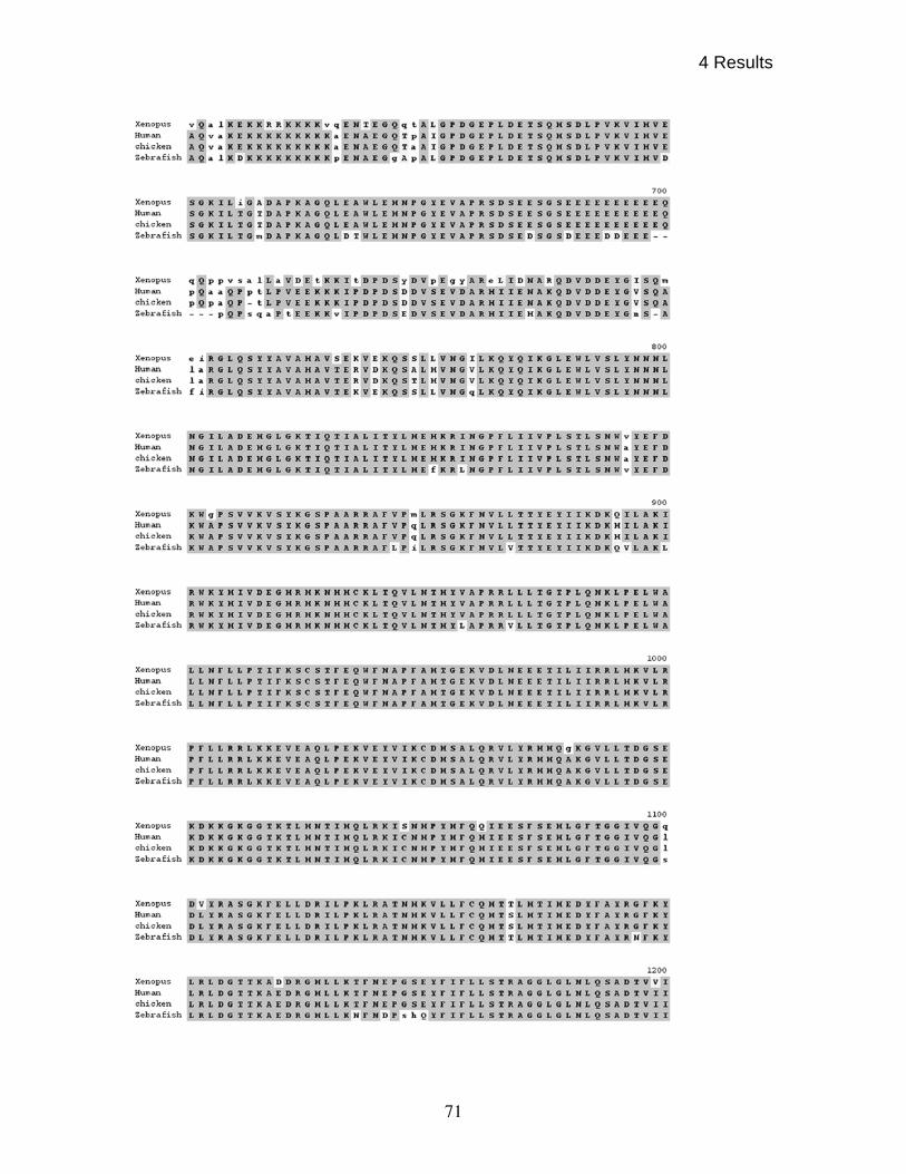
4 Results
71

4 Results
72
Figure 4.5 (A) Clustlaw alignment of Xenopus BRG1 amino acid sequence with human (AAB40977),
chicken (CAA62831), Zebrafish (AAP22968) BRG1. The alignment shows the significant homology with
the BRG1 protein of other vertebrates and shows that the conservation of the amino acid sequence is just
not in the ATPase domain but is through out the entire length of the protein.
Touch down PCR was carried out to obtain the predicted full-length gene
of 5kb length as described in the materials and method section of this report. The
sequencing revealed an open reading frame of 4803 bp for xbrg1 (Fig4.4.).
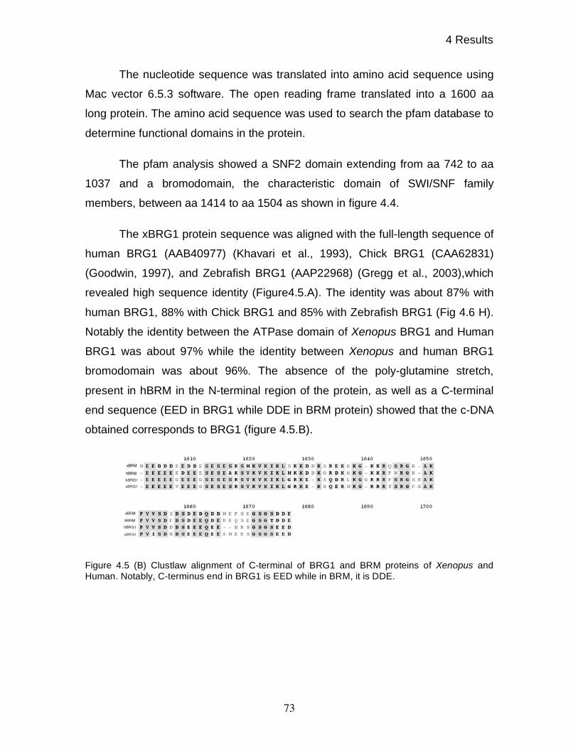
4 Results
73
The nucleotide sequence was translated into amino acid sequence using
Mac vector 6.5.3 software. The open reading frame translated into a 1600 aa
long protein. The amino acid sequence was used to search the pfam database to
determine functional domains in the protein.
The pfam analysis showed a SNF2 domain extending from aa 742 to aa
1037 and a bromodomain, the characteristic domain of SWI/SNF family
members, between aa 1414 to aa 1504 as shown in figure 4.4.
The xBRG1 protein sequence was aligned with the full-length sequence of
human BRG1 (AAB40977) (Khavari et al., 1993), Chick BRG1 (CAA62831)
(Goodwin, 1997), and Zebrafish BRG1 (AAP22968) (Gregg et al., 2003),which
revealed high sequence identity (Figure4.5.A). The identity was about 87% with
human BRG1, 88% with Chick BRG1 and 85% with Zebrafish BRG1 (Fig 4.6 H).
Notably the identity between the ATPase domain of Xenopus BRG1 and Human
BRG1 was about 97% while the identity between Xenopus and human BRG1
bromodomain was about 96%. The absence of the poly-glutamine stretch,
present in hBRM in the N-terminal region of the protein, as well as a C-terminal
end sequence (EED in BRG1 while DDE in BRM protein) showed that the c-DNA
obtained corresponds to BRG1 (figure 4.5.B).
Figure 4.5 (B) Clustlaw alignment of C-terminal of BRG1 and BRM proteins of Xenopus and
Human. Notably, C-terminus end in BRG1 is EED while in BRM, it is DDE.

4 Results
74
4.4 Xbrg1 is maternally expressed and has a tissue-specific
expression pattern
The Northern blot showed that Xbrg1 is expressed as a single transcript of
about 8 kb and is present both before and after the start of zygotic transcription
(figure 4.6.I).
Figure 4.6 Normal expression pattern of Xbrg1 at 4-cell, a time before zygotic transcription
begins, shows that Xbrg1 transcript are provided maternally (A), while at gastrula stage Xbrg1 is
expressed ubiquitously (B). At Neurula, it starts expressing specifically in prospective neural
tissues and shows strong expression in eye anlage (C). At st.28 Xbrg1 transcript are present in
neural tube, eye, otic vesicles, pronephros, branchial arches (D) and at st.35 expression become
more concentrated to the head region with expression in retina (E), at st45 its expression is
detected only in the heart (F). Head section of a st. 35 embryo passing through the eye shows
expression of Xbrg1 in retina(r) and neural tube (nt) but not in iris (i). (G) Degree of amino acid
identity between BRG1 homolog of human (AAB40977), chicken (CAA62831), Zebrafish
(AAP22968) and Xenopus (H). Northern blot analysis of Xbrg1 shows that there is a single
transcript of approximately 8kb in size (I).
In order to determine the spatial pattern of Xbrg1 expression in embryonic
tissues, RNA in-situ hybridization was performed. NF st. 4, NF st. 11, NF st. 14,
4 Results
75
NF st. 28, NF st. 35 and NF st. 45 of embryonic development were probed with a
Xbrg1 specific antisense probe. The –36 to +618 fragment was cloned into the
pCR II-TOPO (Invitrogen Life Technologies) vector to obtain the antisense RNA
probe as described in materials and methods section of this report. A sense
strand probe was used as a negative control. Specific in-situ staining was
observed with the antisense Xbrg1 probe from 2-4 cell stages onwards. At 2-4
cell stage, only maternal transcript are present since in Xenopus embryos the
zygotic transcription starts at the mid blastula transition (MBT) (Newport and
Kirschner, 1982). Up to the gastrula stage, Xbrg1 is expressed ubiquitously in the
embryo. From neurula stage of embryos, Xbrg1 has a restricted expression
pattern. As shown in figure 4.6, the expression of Xbrg1 was detected in the
prospective eye field and prospective neural tube showing a region of high-level
expression of Xbrg1 at Neurula stage. NF st. 28 embryos show that Xbrg1 is
expressed strongly in eye, brain tissue and branchial arches, while it has a
weaker staining in pronephros and in neural tube. Interestingly, its expression
could not be detected in somites, which express Xbrm (Linder et al., 2004).
By NF stage 35, expression could be seen in retina, but not in the lens
tissue, as also seen in embryonic cross-sectioning of head of NF stage 35
embryos passing through the eyes. The expression of Xbrg1 shows an increase
in pronephros and in brain tissue while reduces in neural tube as compared to
NF st. 28 embryos. The expression was also present in the tail region. At NF
stage 45 expression of Xbrg1 has disappeared in head, eye and neural tissue,
and was maintained exclusively in heart.
Thus, Xbrg1 is a maternally expressed gene, and is present ubiquitously
at gastrula stage and later shows a tissue specific expression pointing towards a
role of xBRG1 in tissue specific transcription.
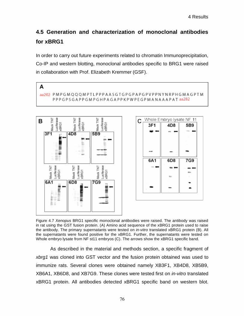
4 Results
76
4.5 Generation and characterization of monoclonal antibodies
for xBRG1
In order to carry out future experiments related to chromatin Immunoprecipitation,
Co-IP and western blotting, monoclonal antibodies specific to BRG1 were raised
in collaboration with Prof. Elizabeth Kremmer (GSF).
Figure 4.7 Xenopus BRG1 specific monoclonal antibodies were raised. The antibody was raised
in rat using the GST fusion protein. (A) Amino acid sequence of the xBRG1 protein used to raise
the antibody. The primary supernatants were tested on in-vitro translated xBRG1 protein (B). All
the supernatants were found positive for the xBRG1. Further, the supernatants were tested on
Whole embryo lysate from NF st11 embryos (C). The arrows show the xBRG1 specific band.
As described in the material and methods section, a specific fragment of
xbrg1 was cloned into GST vector and the fusion protein obtained was used to
immunize rats. Several clones were obtained namely XB3F1, XB4D8, XB5B9,
XB6A1, XB6D8, and XB7G9. These clones were tested first on in-vitro translated
xBRG1 protein. All antibodies detected xBRG1 specific band on western blot.
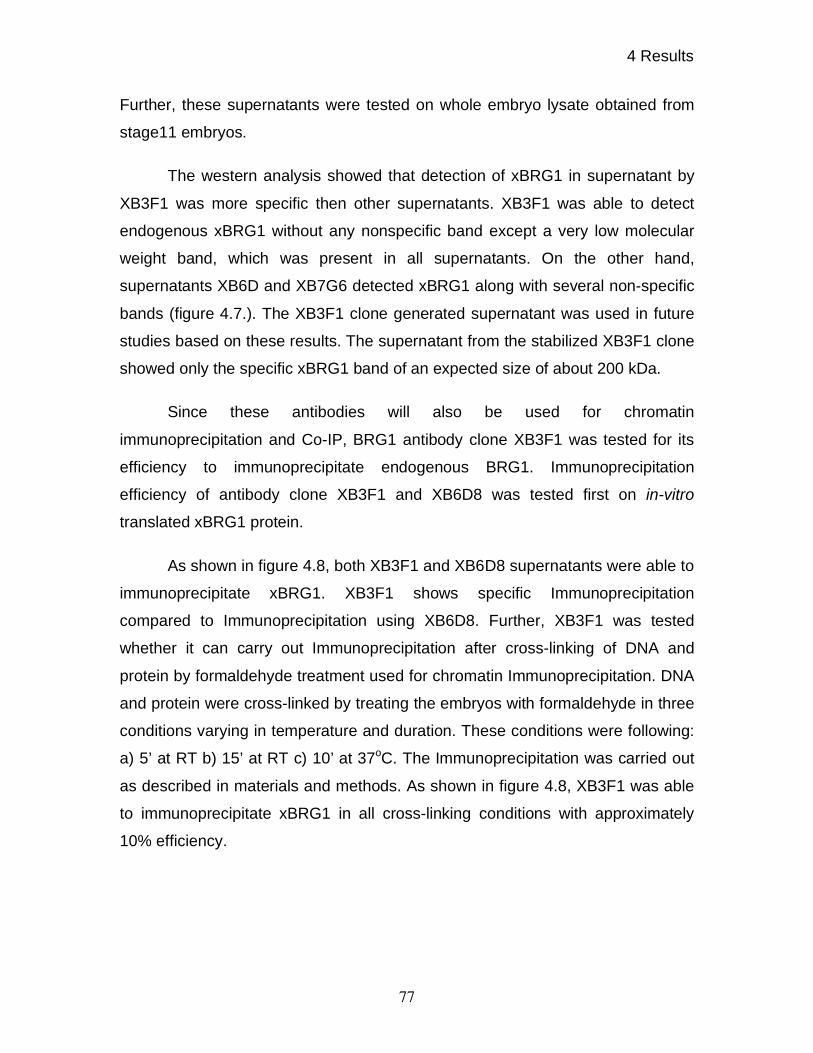
4 Results
77
Further, these supernatants were tested on whole embryo lysate obtained from
stage11 embryos.
The western analysis showed that detection of xBRG1 in supernatant by
XB3F1 was more specific then other supernatants. XB3F1 was able to detect
endogenous xBRG1 without any nonspecific band except a very low molecular
weight band, which was present in all supernatants. On the other hand,
supernatants XB6D and XB7G6 detected xBRG1 along with several non-specific
bands (figure 4.7.). The XB3F1 clone generated supernatant was used in future
studies based on these results. The supernatant from the stabilized XB3F1 clone
showed only the specific xBRG1 band of an expected size of about 200 kDa.
Since these antibodies will also be used for chromatin
immunoprecipitation and Co-IP, BRG1 antibody clone XB3F1 was tested for its
efficiency to immunoprecipitate endogenous BRG1. Immunoprecipitation
efficiency of antibody clone XB3F1 and XB6D8 was tested first on in-vitro
translated xBRG1 protein.
As shown in figure 4.8, both XB3F1 and XB6D8 supernatants were able to
immunoprecipitate xBRG1. XB3F1 shows specific Immunoprecipitation
compared to Immunoprecipitation using XB6D8. Further, XB3F1 was tested
whether it can carry out Immunoprecipitation after cross-linking of DNA and
protein by formaldehyde treatment used for chromatin Immunoprecipitation. DNA
and protein were cross-linked by treating the embryos with formaldehyde in three
conditions varying in temperature and duration. These conditions were following:
a) 5’ at RT b) 15’ at RT c) 10’ at 37oC. The Immunoprecipitation was carried out
as described in materials and methods. As shown in figure 4.8, XB3F1 was able
to immunoprecipitate xBRG1 in all cross-linking conditions with approximately
10% efficiency.
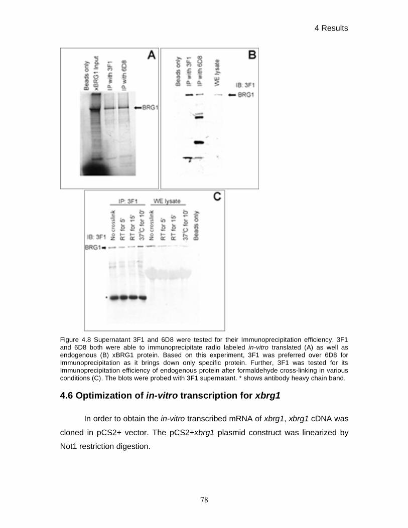
4 Results
78
Figure 4.8 Supernatant 3F1 and 6D8 were tested for their Immunoprecipitation efficiency. 3F1
and 6D8 both were able to immunoprecipitate radio labeled in-vitro translated (A) as well as
endogenous (B) xBRG1 protein. Based on this experiment, 3F1 was preferred over 6D8 for
Immunoprecipitation as it brings down only specific protein. Further, 3F1 was tested for its
Immunoprecipitation efficiency of endogenous protein after formaldehyde cross-linking in various
conditions (C). The blots were probed with 3F1 supernatant. * shows antibody heavy chain band.
4.6 Optimization of in-vitro transcription for xbrg1
In order to obtain the in-vitro transcribed mRNA of xbrg1, xbrg1 cDNA was
cloned in pCS2+ vector. The pCS2+xbrg1 plasmid construct was linearized by
Not1 restriction digestion.
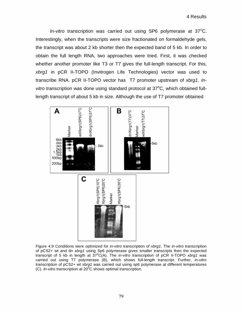
4 Results
79
In-vitro transcription was carried out using SP6 polymerase at 37oC.
Interestingly, when the transcripts were size fractionated on formaldehyde gels,
the transcript was about 2 kb shorter then the expected band of 5 kb. In order to
obtain the full length RNA, two approaches were tried. First, it was checked
whether another promoter like T3 or T7 gives the full-length transcript. For this,
xbrg1 in pCR II-TOPO (Invitrogen Life Technologies) vector was used to
transcribe RNA. pCR II-TOPO vector has T7 promoter upstream of xbrg1. In-
vitro transcription was done using standard protocol at 37oC, which obtained full-
length transcript of about 5 kb in size. Although the use of T7 promoter obtained
Figure 4.9 Conditions were optimized for in-vitro transcription of xbrg1. The in-vitro transcription
of pCS2+ wt and dn xbrg1 using Sp6 polymerase gives smaller transcripts then the expected
transcript of 5 kb in length at 37oC(A). The in-vitro transcription of pCR II-TOPO xbrg1 was
carried out using T7 polymerase (B), which shows full-length transcript. Further, in-vitro
transcription of pCS2+ wt xbrg1 was carried out using sp6 polymerase at different temperatures
(C). In-vitro transcription at 20oC shows optimal transcription.
4 Results
80
full-length mRNA but SP6 promoter used in pCS2+ vector, has been shown to
have higher transcriptional efficiency as well as transcript obtained from SP6
promoter has highest translational efficiency. Thus, another approach was used
to obtain the full-length transcript. The in-vitro transcription was carried out at
various temperatures that is at 10oC, 20
oC, and at 30
oC. As shown in figure 4.11,
at 10oC the transcript was of full-length but the amount was extremely low, while
at 30oC, the amount was sufficient but transcript were spreaded on the
formaldehyde gel indicating several short transcripts while at 20oC the transcript
amount was sufficient as well as transcript was also full length (figure 4.9.). Thus,
in future experiments, in-vitro transcripts for xbrg1 were generated at 20oC using
SP6 RNA polymerase.
4.7 Xenopus BRG1 is required for anterior-posterior axis formation
To carry out in depth analysis of developmental functions of xBRG1, a
dominant negative construct similar to dn hBRG1 was made. To obtain dominant
negative version of xBRG1, a lysine at the conserved ATP binding site (GKT), at
position 770 was point mutated to arginine (K770-R) as has been described for
hBRG1(Khavari et al., 1993) by a single point mutation using site directed
mutagenesis.
The in-vitro synthesized, wild type Xbrg1 and dominant negative Xbrg1
mRNAs were injected at a dose of 500 pg/embryo in to dorsal marginal zone at
four-cell stage along with Gfp as lineage tracer. The control embryos were
injected with in-vitro synthesized Gfp mRNA. The phenotypes were assessed at
NF stage 35. As shown in figure 4.10, dominant negative Xenopus BRG1
produced 61% embryos with head defects as seen in case of dominant negative
hBRG1 overexpression. Overexpression of wild type hBRG1 did not produce
phenotypes but on contrary, overexpression of wild type xBRG1 resulted in 53%
embryos with reduced head structures (Table 4.2). Superficially, the head defects
shown by wild type and dominant negative xBRG1 looked similar, but on closer
inspection it was found that in case of wild type xBRG1 the cement gland was

4 Results
81
preferentially reduced, while eyes remained unaffected. In contrast, over-
expression of dominant negative Xenopus BRG1 affected primarily eye
formation, while cement glands differentiated normally.
4.8 Ventral overexpression of wild type xBRG1 produces partial
secondary axis
Wild type and dominant negative xBRG1 was overexpressed on ventral
side of the embryos by injecting 500pg mRNA along with Gfp as a lineage tracer.
As shown in figure 4.10, overexpression of dominant negative xBRG1 did not
produce any abnormalities, consistent with dominant negative hBRG1
overexpression on ventral side of the embryo. In contrast, overexpression of wild
type Xenopus BRG1 on the ventral side produced partial secondary axis at a
frequency of about 65% (Table 4.2.). The overexpression of GFP did not produce
any defect.
The results obtained from overexpression of xBRG1 on the dorsal side of
the embryo revealed a possible role of BRG1 in anterior-posterior patterning of
Xenopus embryo, which are principally consistent with the results obtained by
hBRG1 overexpression.
Phenotypes produced by dorsal or ventral overexpression of wild type and
dominant negative xBRG1 could not be produced by overexpression of wild type
and dominant negative overexpression of xISWI, another SNF2 domain
containing protein, either on the dorsal side or on the ventral side of the Xenopus
embryos. For these experiments, wild type and dominant negative Xiswi in-vitro
synthesized mRNA were injected into the dorsal marginal zone or into the ventral
marginal zone.
Dominant negative xISWI variant was obtained by a single point mutation
into ATPase domain. The point mutation converted the conserved lysine residue
at position 204 into arginine. This mutation renders its ATPase domain inactive,
as reported previously for Drosophila ISWI (Deuring et al., 2000). The mRNA at a
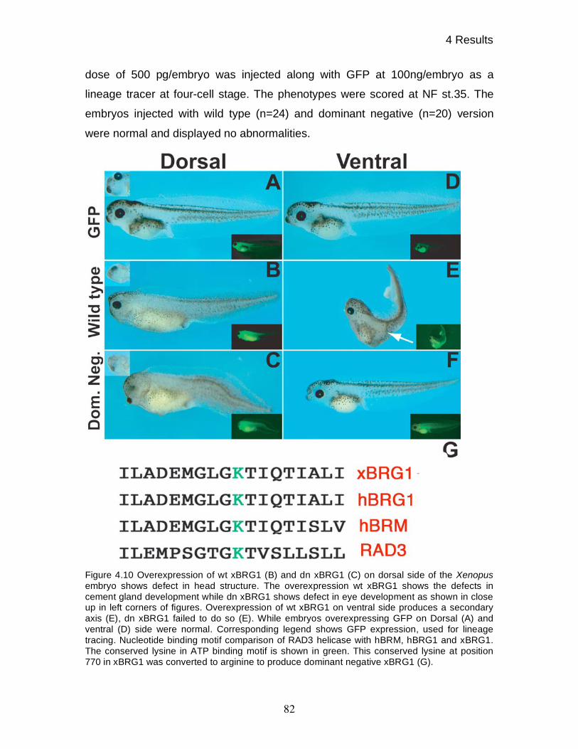
4 Results
82
dose of 500 pg/embryo was injected along with GFP at 100ng/embryo as a
lineage tracer at four-cell stage. The phenotypes were scored at NF st.35. The
embryos injected with wild type (n=24) and dominant negative (n=20) version
were normal and displayed no abnormalities.
Figure 4.10 Overexpression of wt xBRG1 (B) and dn xBRG1 (C) on dorsal side of the Xenopus
embryo shows defect in head structure. The overexpression wt xBRG1 shows the defects in
cement gland development while dn xBRG1 shows defect in eye development as shown in close
up in left corners of figures. Overexpression of wt xBRG1 on ventral side produces a secondary
axis (E), dn xBRG1 failed to do so (E). While embryos overexpressing GFP on Dorsal (A) and
ventral (D) side were normal. Corresponding legend shows GFP expression, used for lineage
tracing. Nucleotide binding motif comparison of RAD3 helicase with hBRM, hBRG1 and xBRG1.
The conserved lysine in ATP binding motif is shown in green. This conserved lysine at position
770 in xBRG1 was converted to arginine to produce dominant negative xBRG1 (G).
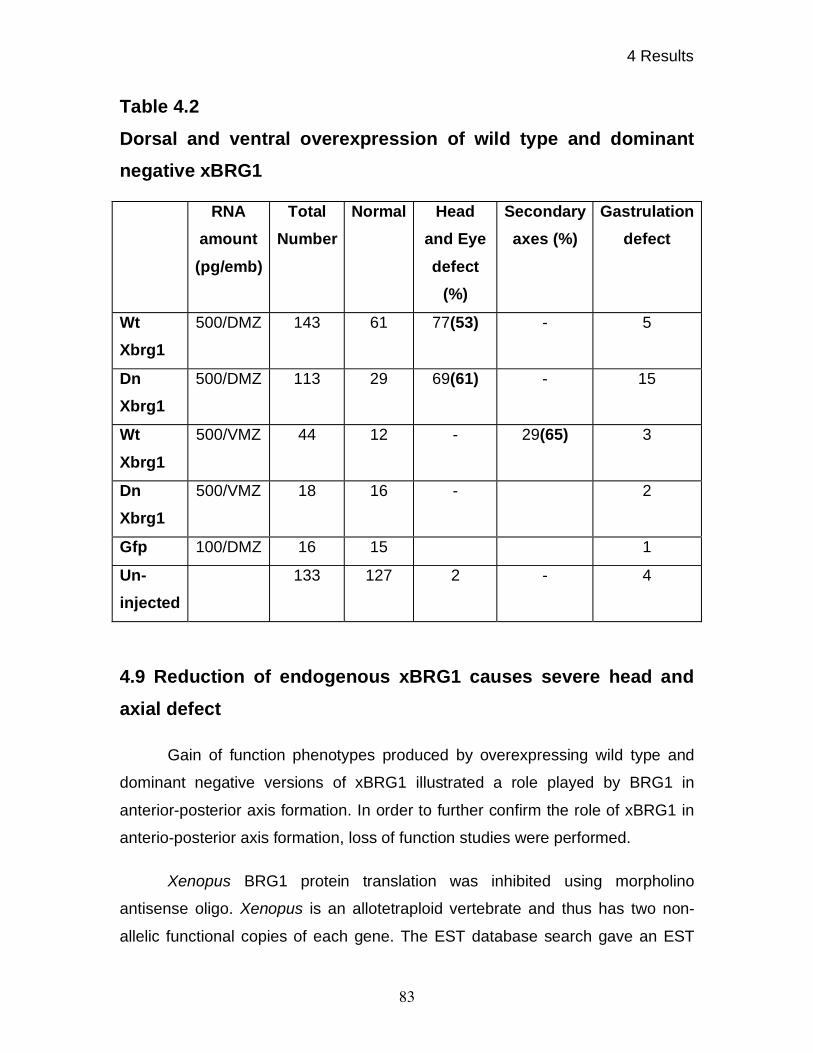
4 Results
83
Table 4.2
Dorsal and ventral overexpression of wild type and dominant
negative xBRG1
4.9 Reduction of endogenous xBRG1 causes severe head and
axial defect
Gain of function phenotypes produced by overexpressing wild type and
dominant negative versions of xBRG1 illustrated a role played by BRG1 in
anterior-posterior axis formation. In order to further confirm the role of xBRG1 in
anterio-posterior axis formation, loss of function studies were performed.
Xenopus BRG1 protein translation was inhibited using morpholino
antisense oligo. Xenopus is an allotetraploid vertebrate and thus has two non-
allelic functional copies of each gene. The EST database search gave an EST
RNA
amount
(pg/emb)
Total
Number
Normal Head
and Eye
defect
(%)
Secondary
axes (%)
Gastrulation
defect
Wt
Xbrg1
500/DMZ 143 61 77(53) - 5
Dn
Xbrg1
500/DMZ 113 29 69(61) - 15
Wt
Xbrg1
500/VMZ 44 12 - 29(65) 3
Dn
Xbrg1
500/VMZ 18 16 - 2
Gfp 100/DMZ 16 15 1
Un-
injected
133 127 2 - 4
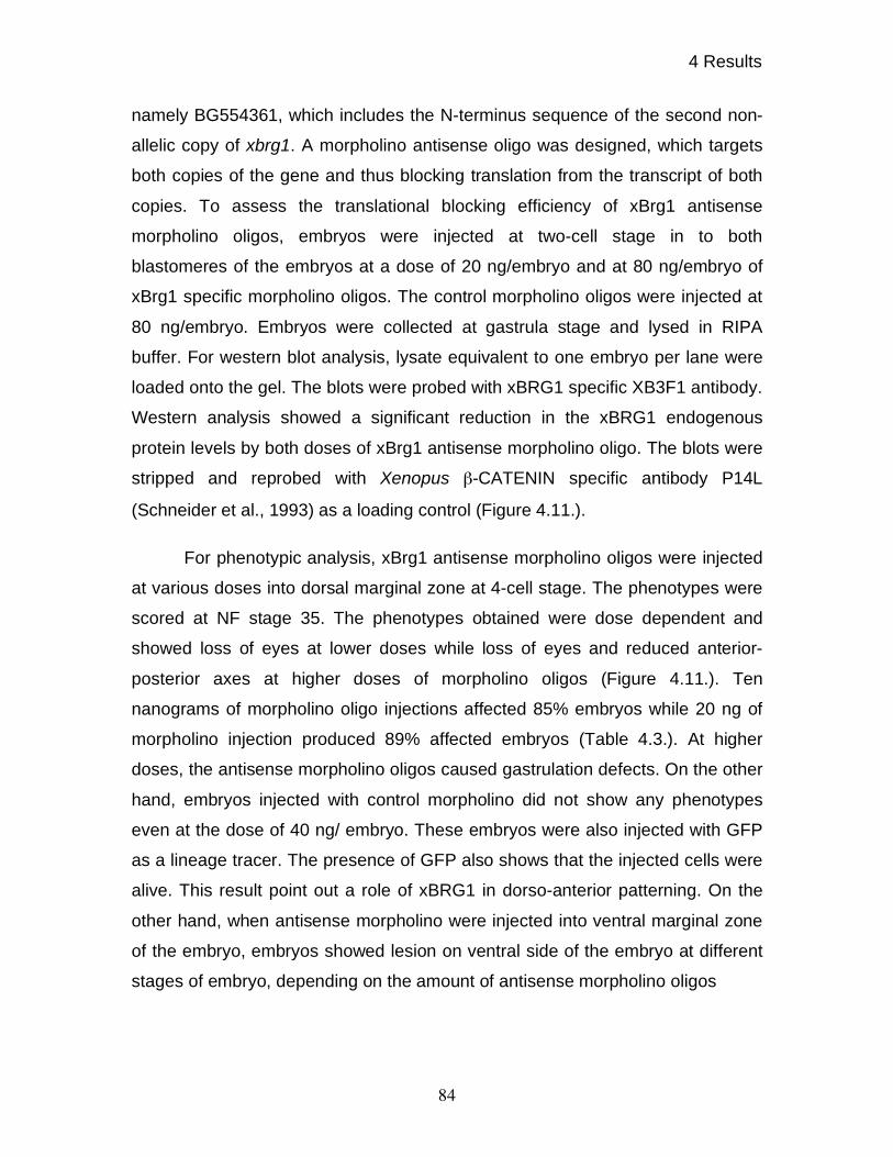
4 Results
84
namely BG554361, which includes the N-terminus sequence of the second non-
allelic copy of xbrg1. A morpholino antisense oligo was designed, which targets
both copies of the gene and thus blocking translation from the transcript of both
copies. To assess the translational blocking efficiency of xBrg1 antisense
morpholino oligos, embryos were injected at two-cell stage in to both
blastomeres of the embryos at a dose of 20 ng/embryo and at 80 ng/embryo of
xBrg1 specific morpholino oligos. The control morpholino oligos were injected at
80 ng/embryo. Embryos were collected at gastrula stage and lysed in RIPA
buffer. For western blot analysis, lysate equivalent to one embryo per lane were
loaded onto the gel. The blots were probed with xBRG1 specific XB3F1 antibody.
Western analysis showed a significant reduction in the xBRG1 endogenous
protein levels by both doses of xBrg1 antisense morpholino oligo. The blots were
stripped and reprobed with Xenopus -CATENIN specific antibody P14L
(Schneider et al., 1993) as a loading control (Figure 4.11.).
For phenotypic analysis, xBrg1 antisense morpholino oligos were injected
at various doses into dorsal marginal zone at 4-cell stage. The phenotypes were
scored at NF stage 35. The phenotypes obtained were dose dependent and
showed loss of eyes at lower doses while loss of eyes and reduced anterior-
posterior axes at higher doses of morpholino oligos (Figure 4.11.). Ten
nanograms of morpholino oligo injections affected 85% embryos while 20 ng of
morpholino injection produced 89% affected embryos (Table 4.3.). At higher
doses, the antisense morpholino oligos caused gastrulation defects. On the other
hand, embryos injected with control morpholino did not show any phenotypes
even at the dose of 40 ng/ embryo. These embryos were also injected with GFP
as a lineage tracer. The presence of GFP also shows that the injected cells were
alive. This result point out a role of xBRG1 in dorso-anterior patterning. On the
other hand, when antisense morpholino were injected into ventral marginal zone
of the embryo, embryos showed lesion on ventral side of the embryo at different
stages of embryo, depending on the amount of antisense morpholino oligos
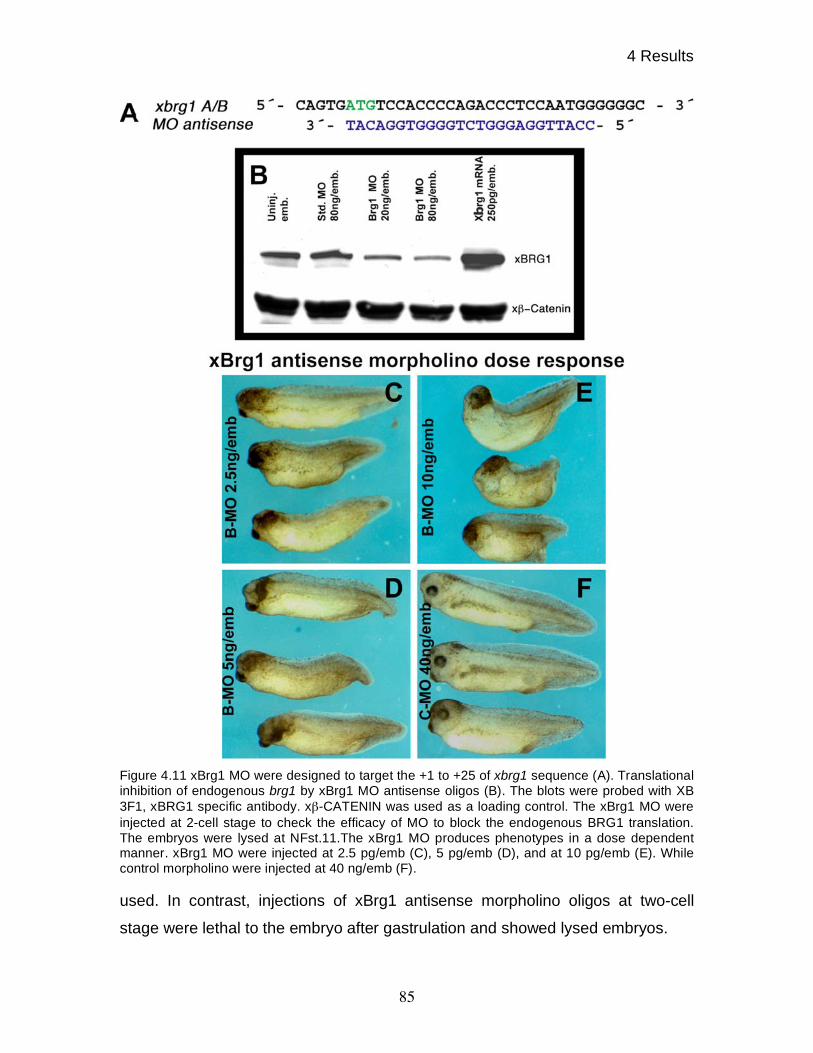
4 Results
85
Figure 4.11 xBrg1 MO were designed to target the +1 to +25 of xbrg1 sequence (A). Translational
inhibition of endogenous brg1 by xBrg1 MO antisense oligos (B). The blots were probed with XB
3F1, xBRG1 specific antibody. x -CATENIN was used as a loading control. The xBrg1 MO were
injected at 2-cell stage to check the efficacy of MO to block the endogenous BRG1 translation.
The embryos were lysed at NFst.11.The xBrg1 MO produces phenotypes in a dose dependent
manner. xBrg1 MO were injected at 2.5 pg/emb (C), 5 pg/emb (D), and at 10 pg/emb (E). While
control morpholino were injected at 40 ng/emb (F).
used. In contrast, injections of xBrg1 antisense morpholino oligos at two-cell
stage were lethal to the embryo after gastrulation and showed lysed embryos.
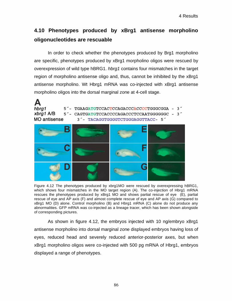
4 Results
86
4.10 Phenotypes produced by xBrg1 antisense morpholino
oligonucleotides are rescuable
In order to check whether the phenotypes produced by Brg1 morpholino
are specific, phenotypes produced by xBrg1 morpholino oligos were rescued by
overexpression of wild type hBRG1. hbrg1 contains four mismatches in the target
region of morpholino antisense oligo and, thus, cannot be inhibited by the xBrg1
antisense morpholino. Wt Hbrg1 mRNA was co-injected with xBrg1 antisense
morpholino oligos into the dorsal marginal zone at 4-cell stage.
Figure 4.12 The phenotypes produced by xbrg1MO were rescued by overexpressing hBRG1,
which shows four mismatches in the MO target region (A). The co-injection of Hbrg1 mRNA
rescues the phenotypes produced by xBrg1 MO and shows partial rescue of eye (E), partial
rescue of eye and AP axis (F) and almost complete rescue of eye and AP axis (G) compared to
xBrg1 MO (D) alone. Control morpholino (B) and Hbrg1 mRNA (C) alone do not produce any
abnormalities. GFP mRNA was co-injected as a lineage tracer, which has been shown alongside
of corresponding pictures.
As shown in figure 4.12, the embryos injected with 10 ng/embryo xBrg1
antisense morpholino into dorsal marginal zone displayed embryos having loss of
eyes, reduced head and severely reduced anterior-posterior axes, but when
xBrg1 morpholino oligos were co-injected with 500 pg mRNA of Hbrg1, embryos
displayed a range of phenotypes.
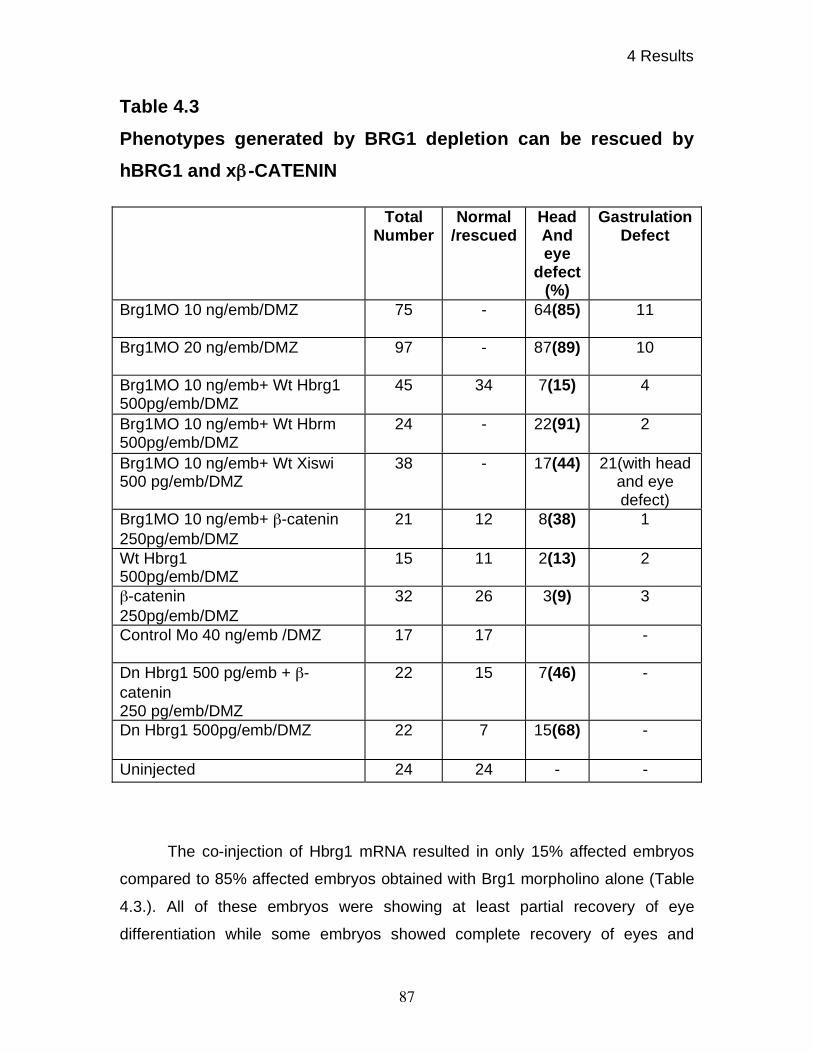
4 Results
87
Table 4.3
Phenotypes generated by BRG1 depletion can be rescued by
hBRG1 and x -CATENIN
The co-injection of Hbrg1 mRNA resulted in only 15% affected embryos
compared to 85% affected embryos obtained with Brg1 morpholino alone (Table
4.3.). All of these embryos were showing at least partial recovery of eye
differentiation while some embryos showed complete recovery of eyes and
Total Number
Normal /rescued
Head And eye
defect (%)
Gastrulation Defect
Brg1MO 10 ng/emb/DMZ 75 - 64(85) 11
Brg1MO 20 ng/emb/DMZ 97 - 87(89) 10
Brg1MO 10 ng/emb+ Wt Hbrg1
500pg/emb/DMZ
45 34 7(15) 4
Brg1MO 10 ng/emb+ Wt Hbrm
500pg/emb/DMZ
24 - 22(91) 2
Brg1MO 10 ng/emb+ Wt Xiswi
500 pg/emb/DMZ
38 - 17(44) 21(with head
and eye
defect)
Brg1MO 10 ng/emb+ -catenin
250pg/emb/DMZ
21 12 8(38) 1
Wt Hbrg1
500pg/emb/DMZ
15 11 2(13) 2
-catenin
250pg/emb/DMZ
32 26 3(9) 3
Control Mo 40 ng/emb /DMZ 17 17 -
Dn Hbrg1 500 pg/emb + -
catenin
250 pg/emb/DMZ
22 15 7(46) -
Dn Hbrg1 500pg/emb/DMZ 22 7 15(68) -
Uninjected 24 24 - -
4 Results
88
anterior-posterior axes. Interestingly, xISWI and hBRM overexpression failed to
rescue the phenotypes generated by Brg1 morpholino oligos.
These results showed that the phenotypes produced by the xBrg1
morpholino oligos were specifically produced by interference with xBRG1 protein
function.
4.11 BRG1 knock-down affects expression of various
differentiation markers
The BRG1 knock-down experiments displayed severely reduced head
structures and anterio-posterior axis formation. To find out the effect of BRG1
knock-down on organogenesis, various tissues were analyzed by in-situ
hybridization. Xenopus embryos were injected at 4-cell stage into dorsal marginal
zone with 5 ng per embryo of xBrg1 antisense morpholino oligonucleotides. The
embryos were fixed at stage 35.
Embryos were probed with neural specific -tubulin in-situ probe
(Oschwald et al., 1991). In-situ analysis showed that N- tubulin stains neural
tissue in head region as well as in neural tube. The expression levels were high
in head region and lower in neural tube. BRG1 knocked-down embryos displayed
strongly reduced N- tubulin staining in head region, while staining was absent in
neural tube region. These results showed that BRG1 knock-down reduced the
amount of neural tissue formation (figure 4.13.).
Next I examined the expression of Nkx2.5, a tinman homologue and a
known regulator of heart differentiation and morphogenesis (Tonissen et al.,
1994). As shown in figure 4.13, the expression was reduced in xBrg1 MO
injected embryos, while the expression of Nkx2.5 remained unaltered in control
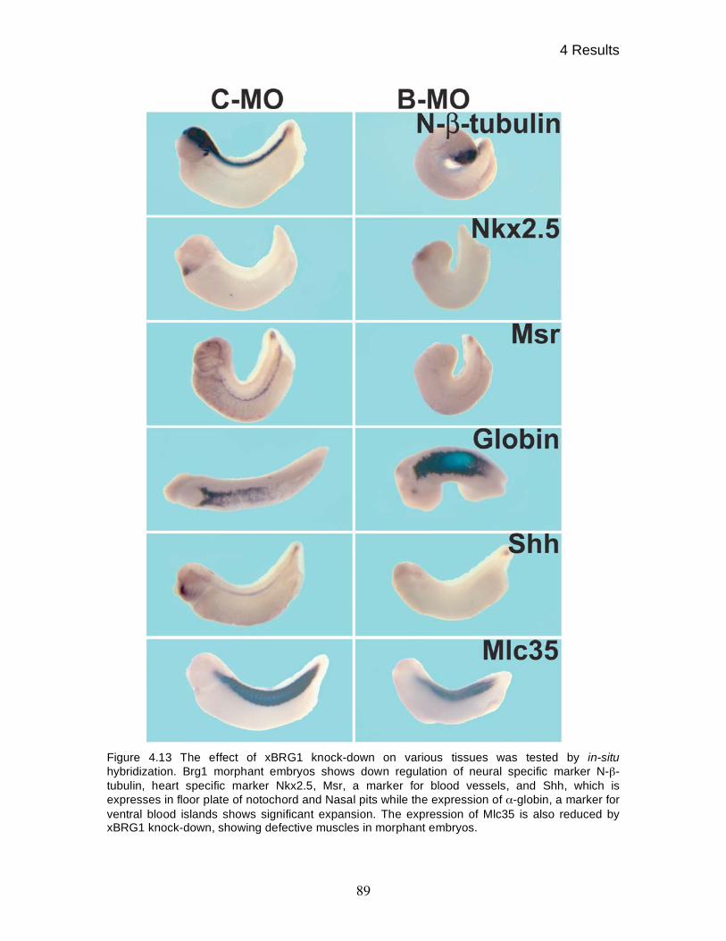
4 Results
89
Figure 4.13 The effect of xBRG1 knock-down on various tissues was tested by in-situ
hybridization. Brg1 morphant embryos shows down regulation of neural specific marker N- -
tubulin, heart specific marker Nkx2.5, Msr, a marker for blood vessels, and Shh, which is
expresses in floor plate of notochord and Nasal pits while the expression of -globin, a marker for
ventral blood islands shows significant expansion. The expression of Mlc35 is also reduced by
xBRG1 knock-down, showing defective muscles in morphant embryos.
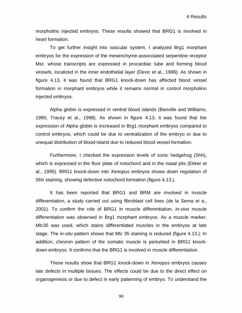
4 Results
90
morpholino injected embryos. These results showed that BRG1 is involved in
heart formation.
To get further insight into vascular system, I analyzed Brg1 morphant
embryos for the expression of the mesenchyme-assocoiated serpentine receptor
Msr, whose transcripts are expressed in procardiac tube and forming blood
vessels, localized in the inner endothelial layer (Devic et al., 1996). As shown in
figure 4.13, it was found that BRG1 knock-down has affected blood vessel
formation in morphant embryos while it remains normal in control morpholino
injected embryos.
Alpha globin is expressed in ventral blood islands (Banville and Williams,
1985; Tracey et al., 1998). As shown in figure 4.13, it was found that the
expression of Alpha globin is increased in Brg1 morphant embryos compared to
control embryos, which could be due to ventralization of the embryo or due to
unequal distribution of blood-island due to reduced blood vessel formation.
Furthermore, I checked the expression levels of sonic hedgehog (Shh),
which is expressed in the floor plate of notochord and in the nasal pits (Ekker et
al., 1995). BRG1 knock-down into Xenopus embryos shows down regulation of
Shh staining, showing defective notochord formation (figure 4.13.).
It has been reported that BRG1 and BRM are involved in muscle
differentiation, a study carried out using fibroblast cell lines (de la Serna et a.,
2001). To confirm the role of BRG1 in muscle differentiation, in-vivo muscle
differentiation was observed in Brg1 morphant embryos. As a muscle marker,
Mlc35 was used, which stains differentiated muscles in the embryos at late
stage. The in-situ pattern shows that Mlc 35 staining is reduced (figure 4.13.). In
addition, chevron pattern of the somatic muscle is perturbed in BRG1 knock-
down embryos. It confirms that the BRG1 is involved in muscle differentiation.
These results show that BRG1 knock-down in Xenopus embryos causes
late defects in multiple tissues. The effects could be due to the direct effect on
organogenesis or due to defect in early patterning of embryo. To understand the
4 Results
91
cause of these phenotypes, the genes involved in early patterning of the embryos
were investigated.
4.12 BRG1 knock-down causes down regulation of genes
required for early patterning of the dorsal mesoderm
To understand the role of xBRG1 in early patterning, the embryos were
injected with 20 ng of antisense morpholino oligos at 2-cell stage. The target
genes were checked by in-situ hybridization at relevant embryonic stages.
Xenopus nodal related 3 (xnr3) is a direct target gene of WNT/ -CATENIN
pathway (McKendry et al., 1997). The expression levels of Xnr3 were checked in
Brg1 morphant embryos. Xnr3 is expressed maximally at NF st.9 and thus
control morpholino as well as Brg1 morpholino injected embryos were fixed at
this stage. As shown in fig 4.14, the expression level of Xnr3 in Brg1 morphant
embryos is significantly reduced while the size of its expression domain remains
same compared to control morpholino injected embryos.
Chordin, a BMP antagonist, is expressed transiently in the BCNE center
(Blastula Chordin Noggin Expression center) and later in the organizer. The
expression of Chordin in the BCNE center is under the -CATENIN mediated
WNT signal (Kuroda et al., 2004). To test the expression of Chordin in BCNE
center, embryos were fixed at blastula stage. As shown in figure 4.14, the
expression of Chordin was notably reduced by knocking-down BRG1 in the
embryos compared to control embryos.
This result not only gives a second read out for Wnt target genes as well
as explains neural defects seen in whole embryos since the BCNE center later
gives rise to prospective brain and floor plate. Chordin is expressed at the dorsal
blastopore lip at the gastrula stage. As shown in legend of figure 4.14, the
expression of Chordin was reduced in the ectodermal cells while remained
unaffected in the lip. Next, the expression of Goosecoid, a homeobox gene,
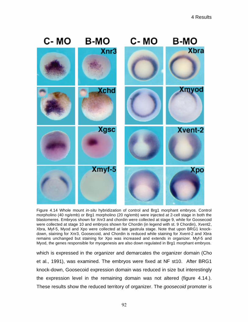
4 Results
92
Figure 4.14 Whole mount in-situ hybridization of control and Brg1 morphant embryos. Control
morpholino (40 ng/emb) or Brg1 morpholino (20 ng/emb) were injected at 2-cell stage in both the
blastomeres. Embryos shown for Xnr3 and chordin were collected at stage 9, while for Goosecoid
were collected at stage 10 and embryos shown for Chordin (in legend with st. 9 Chordin), Xvent2,
Xbra, Myf-5, Myod and Xpo were collected at late gastrula stage. Note that upon BRG1 knock-
down, staining for Xnr3, Goosecoid, and Chordin is reduced while staining for Xvent-2 and Xbra
remains unchanged but staining for Xpo was increased and extends in organizer. Myf-5 and
Myod, the genes responsible for myogenesis are also down regulated in Brg1 morphant embryos.
which is expressed in the organizer and demarcates the organizer domain (Cho
et al., 1991), was examined. The embryos were fixed at NF st10. After BRG1
knock-down, Goosecoid expression domain was reduced in size but interestingly
the expression level in the remaining domain was not altered (figure 4.14.).
These results show the reduced territory of organizer. The goosecoid promoter is
4 Results
93
known to have two binding sites, one each for WNT signal and NODAL signal.
Siamois/Twin binding on the promoter relays WNT signal. Therefore, it was
reasoned that since WNT signal is downregulated as shown by the
downregulation of Xnr3 and Chordin, I see a reduction in expression domain of
Goosecoid but probably NODAL signaling remains unaffected and thus maintains
the intensity of expression.
Xbra, a Xenopus homologue of the T-box gene brachyury, is expressed in
the pan-mesoderm (Smith et al., 1991). To find out the effect of BRG1 knock-
down on the mesoderm formation, the expression of Xbra was checked at NF
st.11. As shown in figure 4.13, the expression level of Xbra remains unchanged
in the Brg1 morphant embryos compared to control embryos showing that the
mesoderm formation remains unaffected by BRG1 knock-down.
Xvent-2, a homeobox gene, has been shown to be required for
ventralization of mesoderm (Onichtchouk et al., 1996). Xvent-2 is a direct target
of BMP signaling (Schuler-Metz et al., 2000). To find out the effect of BRG1
knock-down on ventral mesoderm formation, the expression of Xvent-2 was
checked. The expression levels of Xvent-2 remain comparable to control
embryos in Brg1 morphant embryos (figure 4.14). These results show that BRG1
knock-down does not affect BMP signaling and thus ventral mesoderm formation
remains unaffected.
Xenopus-posterior (Xpo), encodes protein with a ‘CCHC’ finger domain,
(Sato and Sargent, 1991) and has been proposed to be involved in anterio-
posterior axis organization. Xpo is one of several ventralizing genes, which can
be induced by xCAD-2 (Pillemer et al., 1998). As shown in figure 4.14, the
expression of Xpo is limited to the posterior ectoderm and mesoderm in the
control embryos but is expanded in Brg1 morphant embryos in the organizer
territory.
These results shows that knock-down of xBRG1 selectively affect Wnt
target genes and shows defective organizer formation during gastrulation of
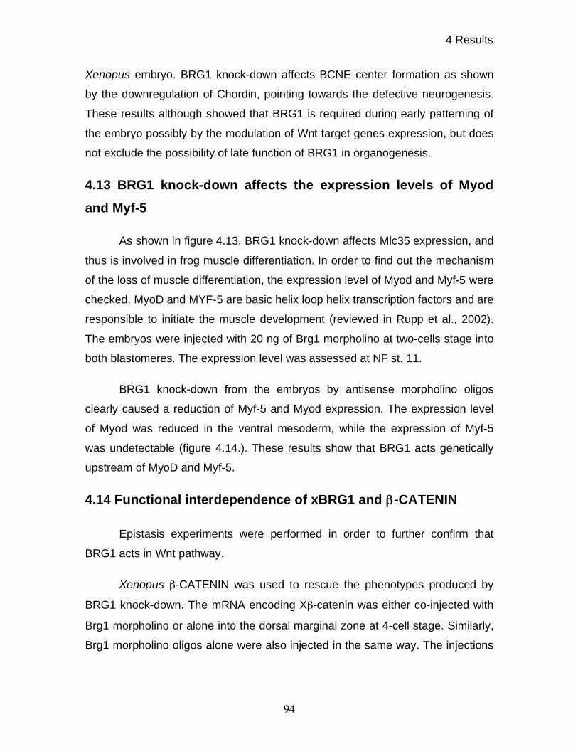
4 Results
94
Xenopus embryo. BRG1 knock-down affects BCNE center formation as shown
by the downregulation of Chordin, pointing towards the defective neurogenesis.
These results although showed that BRG1 is required during early patterning of
the embryo possibly by the modulation of Wnt target genes expression, but does
not exclude the possibility of late function of BRG1 in organogenesis.
4.13 BRG1 knock-down affects the expression levels of Myod
and Myf-5
As shown in figure 4.13, BRG1 knock-down affects Mlc35 expression, and
thus is involved in frog muscle differentiation. In order to find out the mechanism
of the loss of muscle differentiation, the expression level of Myod and Myf-5 were
checked. MyoD and MYF-5 are basic helix loop helix transcription factors and are
responsible to initiate the muscle development (reviewed in Rupp et al., 2002).
The embryos were injected with 20 ng of Brg1 morpholino at two-cells stage into
both blastomeres. The expression level was assessed at NF st. 11.
BRG1 knock-down from the embryos by antisense morpholino oligos
clearly caused a reduction of Myf-5 and Myod expression. The expression level
of Myod was reduced in the ventral mesoderm, while the expression of Myf-5
was undetectable (figure 4.14.). These results show that BRG1 acts genetically
upstream of MyoD and Myf-5.
4.14 Functional interdependence of xBRG1 and -CATENIN
Epistasis experiments were performed in order to further confirm that
BRG1 acts in Wnt pathway.
Xenopus -CATENIN was used to rescue the phenotypes produced by
BRG1 knock-down. The mRNA encoding X -catenin was either co-injected with
Brg1 morpholino or alone into the dorsal marginal zone at 4-cell stage. Similarly,
Brg1 morpholino oligos alone were also injected in the same way. The injections
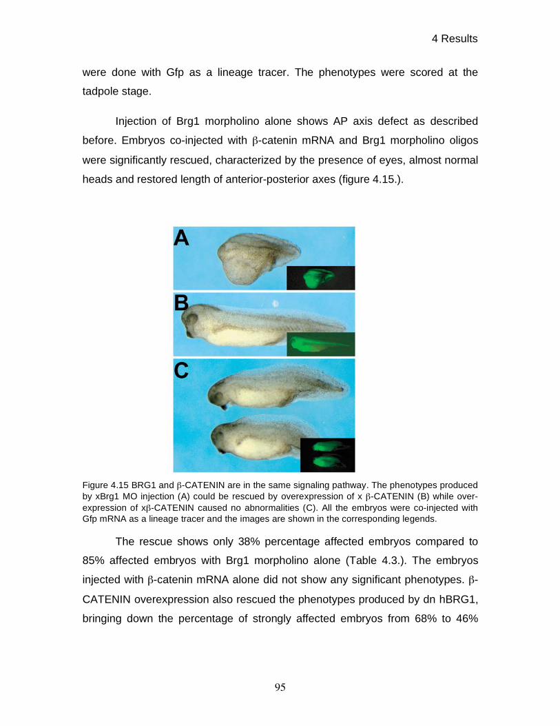
4 Results
95
were done with Gfp as a lineage tracer. The phenotypes were scored at the
tadpole stage.
Injection of Brg1 morpholino alone shows AP axis defect as described
before. Embryos co-injected with -catenin mRNA and Brg1 morpholino oligos
were significantly rescued, characterized by the presence of eyes, almost normal
heads and restored length of anterior-posterior axes (figure 4.15.).
Figure 4.15 BRG1 and -CATENIN are in the same signaling pathway. The phenotypes produced
by xBrg1 MO injection (A) could be rescued by overexpression of x -CATENIN (B) while over-
expression of x -CATENIN caused no abnormalities (C). All the embryos were co-injected with
Gfp mRNA as a lineage tracer and the images are shown in the corresponding legends.
The rescue shows only 38% percentage affected embryos compared to
85% affected embryos with Brg1 morpholino alone (Table 4.3.). The embryos
injected with -catenin mRNA alone did not show any significant phenotypes. -
CATENIN overexpression also rescued the phenotypes produced by dn hBRG1,
bringing down the percentage of strongly affected embryos from 68% to 46%
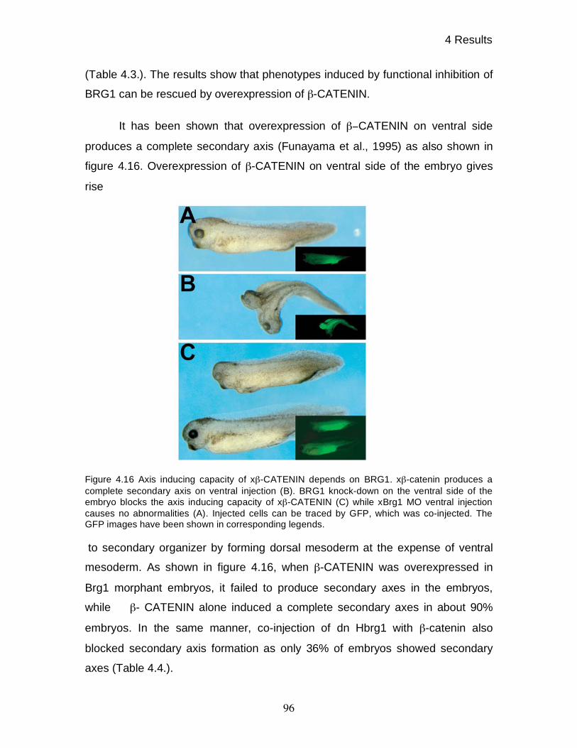
4 Results
96
(Table 4.3.). The results show that phenotypes induced by functional inhibition of
BRG1 can be rescued by overexpression of -CATENIN.
It has been shown that overexpression of CATENIN on ventral side
produces a complete secondary axis (Funayama et al., 1995) as also shown in
figure 4.16. Overexpression of -CATENIN on ventral side of the embryo gives
rise
Figure 4.16 Axis inducing capacity of x -CATENIN depends on BRG1. x -catenin produces a
complete secondary axis on ventral injection (B). BRG1 knock-down on the ventral side of the
embryo blocks the axis inducing capacity of x -CATENIN (C) while xBrg1 MO ventral injection
causes no abnormalities (A). Injected cells can be traced by GFP, which was co-injected. The
GFP images have been shown in corresponding legends.
to secondary organizer by forming dorsal mesoderm at the expense of ventral
mesoderm. As shown in figure 4.16, when -CATENIN was overexpressed in
Brg1 morphant embryos, it failed to produce secondary axes in the embryos,
while - CATENIN alone induced a complete secondary axes in about 90%
embryos. In the same manner, co-injection of dn Hbrg1 with -catenin also
blocked secondary axis formation as only 36% of embryos showed secondary
axes (Table 4.4.).
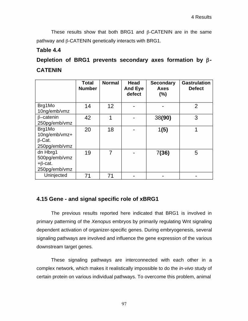
4 Results
97
These results show that both BRG1 and -CATENIN are in the same
pathway and -CATENIN genetically interacts with BRG1.
Table 4.4
Depletion of BRG1 prevents secondary axes formation by -
CATENIN
4.15 Gene - and signal specific role of xBRG1
The previous results reported here indicated that BRG1 is involved in
primary patterning of the Xenopus embryos by primarily regulating Wnt signaling
dependent activation of organizer-specific genes. During embryogenesis, several
signaling pathways are involved and influence the gene expression of the various
downstream target genes.
These signaling pathways are interconnected with each other in a
complex network, which makes it realistically impossible to do the in-vivo study of
certain protein on various individual pathways. To overcome this problem, animal
Total Number
Normal Head And Eye defect
Secondary Axes (%)
Gastrulation Defect
Brg1Mo
10ng/emb/vmz
14 12 - - 2
catenin
250pg/emb/vmz
42 1 - 38(90) 3
Brg1Mo
10ng/emb/vmz+
-Cat.
250pg/emb/vmz
20 18 - 1(5) 1
dn Hbrg1
500pg/emb/vmz
+ -cat.
250pg/emb/vmz
19 7 - 7(36) 5
Uninjected 71 71 - - -
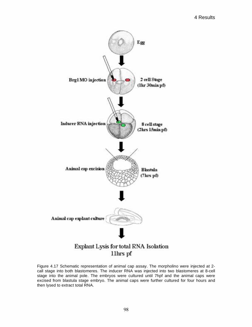
4 Results
98
Figure 4.17 Schematic representation of animal cap assay. The morpholino were injected at 2-
call stage into both blastomeres. The inducer RNA was injected into two blastomeres at 8-cell
stage into the animal pole. The embryos were cultured until 7hpf and the animal caps were
excised from blastula stage embryo. The animal caps were further cultured for four hours and
then lysed to extract total RNA.
4 Results
99
cap in-vitro induction assay was used. As stated before, animal caps are
ectodermal cells and their use reduces the complexity of the problem by allowing
to test the effect of individual signals on the expression of direct target genes.
The signaling pathways in the animal cap cells are induced by
overexpression of specific ligand for the specific pathway. Embryos were injected
with 20 ng of Brg1 morpholino at two-cell stage into both blastomeres. At the 8-
cell stage, uninjected as well as Brg1 morpholino (B-MO) injected embryos were
injected with inducer mRNA into animal pole. The caps were excised at seven
hpf and further cultured for 4 hours, before they were lysed to extract total RNA,
which was used to assess the induction of representative direct target genes of
different signaling pathways.
The direct target genes were selected based on previously reported
cycloheximide insensitive genes or the presence of response element in the
promoter. Cycloheximide treatment of the cells blocks protein synthesis and thus
blocking the effect of intermediate proteins in the signaling pathway on the
outcome of induction of the gene.
To directly check the role of BRG1 on WNT signaling, the animal caps
were induced with 50 pg of Wnt8 mRNA. Wnt8 activates WNT signaling pathway
via interaction with its receptor, which in turns blocks the phosphorylation of -
CATENIN and thus preventing its degradation. This stabilized -CATENIN is
transported to the nucleus where it releases the Tcf3 mediated repression and
thus activating Wnt target genes. In addition to xnr3 (McKendry et al., 1997),
siamois (Brannon et al., 1997; Brannon and Kimelman, 1996; Lemaire et al.,
1995) is also among other known WNT target genes. The promoter of Siamois
has been shown to have a Lef/Tcf3 binding sites
The expression of Siamois was checked by semi-quantitative RT-PCR in
the un-injected and morpholino treated animal caps along with untreated and
morpholino treated caps after induction by Wnt8 mRNA. As shown in figure 4.18,
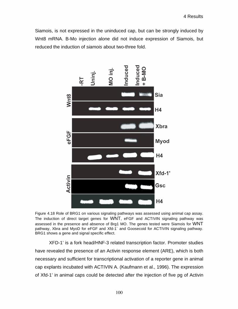
4 Results
100
Siamois, is not expressed in the uninduced cap, but can be strongly induced by
Wnt8 mRNA. B-Mo injection alone did not induce expression of Siamois, but
reduced the induction of siamois about two-three fold.
Figure 4.18 Role of BRG1 on various signaling pathways was assessed using animal cap assay.
The induction of direct target genes for WNT, eFGF and ACTIVIN signaling pathway was
assessed in the presence and absence of Brg1 MO. The genes tested were Siamois for WNT
pathway, Xbra and MyoD for eFGF and Xfd-1´ and Goosecoid for ACTIVIN signaling pathway.
BRG1 shows a gene and signal specific effect.
XFD-1’ is a fork head/HNF-3 related transcription factor. Promoter studies
have revealed the presence of an Activin response element (ARE), which is both
necessary and sufficient for transcriptional activation of a reporter gene in animal
cap explants incubated with ACTIVIN A. (Kaufmann et al., 1996). The expression
of Xfd-1’ in animal caps could be detected after the injection of five pg of Activin
4 Results
101
mRNA. When BRG1 depleted animal caps were induced by Activin mRNA
injection, the transcription of xfd-1’ remained unaffected (figure 4.18).
The promoter of goosecoid has been shown to have two signaling inputs
one from the WNT signaling through Siamois/Twin and the other from the
NODAL signaling. (Germain et al., 2000; Laurent et al., 1997). In order to test the
effect of BRG1 knock-down on goosecoid transcription under induction by nodal
signal only, animal caps were induced by 5 pg of Activin mRNA. As shown in
figure 4.18, Activin could induce goosecoid as reported previously (Green and
Smith, 1990). In contrast to the effect of BRG1 knock-down seen in whole
embryos, the induction of goosecoid remained unaffected in the absence of
BRG1 pointing out that BRG1 is not required for ACTIVIN signaling.
To find out the role of BRG1 in FGF/MAPK signaling, caps were induced
by injecting 10 pg of eFGF mRNA at 8-cell stage into animal pole in uninjected
and Brg1 morpholino injected embryos. The expression of Xbra was assessed,
which has been shown to be a direct target of eFGF (Smith et al., 1991). As
shown in figure 4.18, eFGF induces xbra in the animal caps but remains
unaffected in the absence of BRG1. Similarly, eFGF has also been shown to
induce myod transcription in the presence of cycloheximide (Fisher et al., 2002),
as I have also shown in figure 4.18, myod could be induced by eFGF. Unlike
Xbra, Myod transcripts were undetectable in the absence of BRG1. These results
pointed out that BRG1 is required for the transcription of myod.
The in-vitro induction experiments confirmed that BRG1 is predominantly
involved in WNT signaling pathway. As seen, BRG1 knock-down did not affect
activin target genes while selective affects eFGF target genes, showing that the
BRG1 affect is gene and signal specific.
4.16 Modulation of WNT-dependent gene-activation by xBRG1
Xnr3 and Chordin expression was strongly affected in the embryos upon
BRG1 knock-down (figure 4.14), while Siamois expression was mildly down-

4 Results
102
regulated in animal cap assay (figure 4.18). Furthermore, -CATENIN rescue
experiments showed that the increased WNT response could compensate for
reduced BRG1 activity (figure 4.15). Thus, it is possible that effect on siamois
down regulation was masked by over stimulation of WNT pathway. This was
investigated by testing the BRG1 knock-down effect on the transcriptional
readout of weaker WNT signal.
Figure 4.19. BRG1 sets up a signaling threshold for the optimal gene expression. The expression
of Xnr3, Siamois, and Xnr6 was tested for the normal gene expression using two doses of Wnt8
in the presence and absence of Brg1 MO. The requirement of BRG1 for the normal gene
expression was further confirmed by co-injection of Xbrg1 mRNA along with sub-optimal dose of
Wnt8.
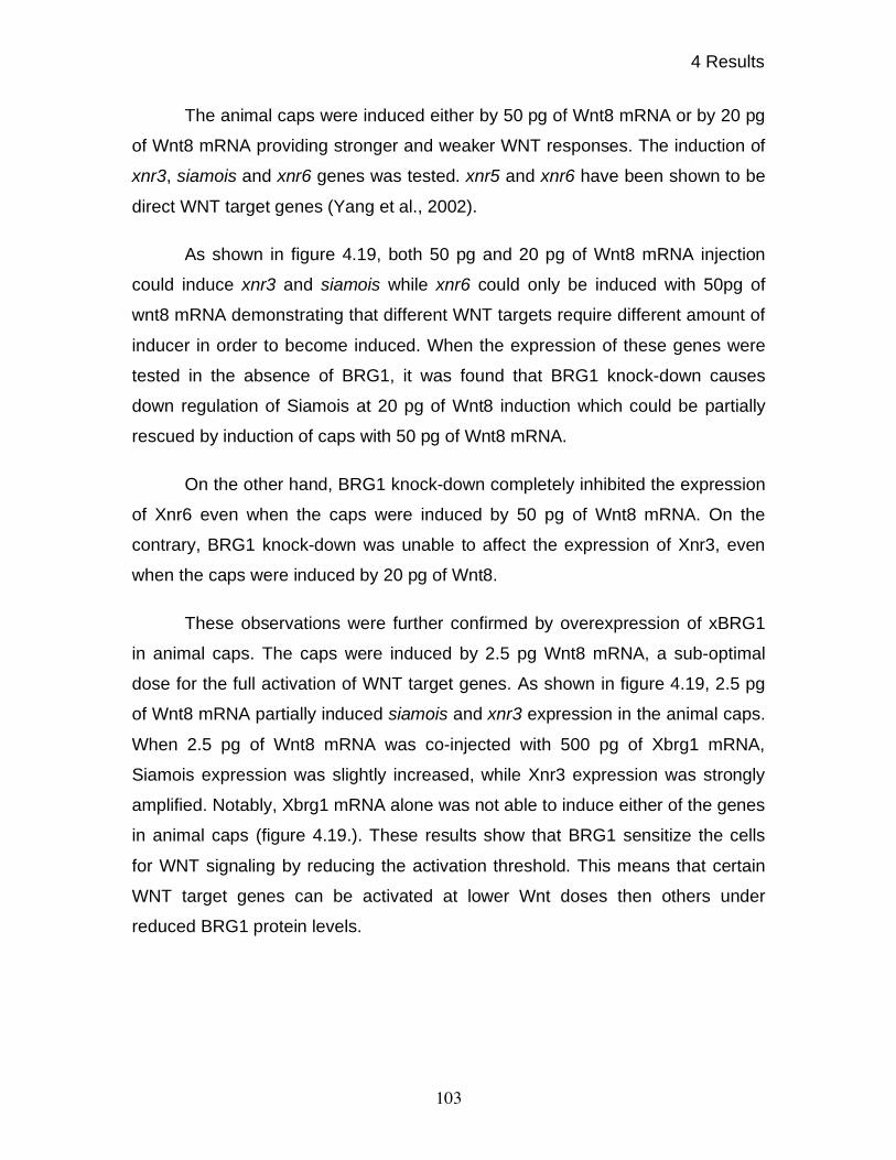
4 Results
103
The animal caps were induced either by 50 pg of Wnt8 mRNA or by 20 pg
of Wnt8 mRNA providing stronger and weaker WNT responses. The induction of
xnr3, siamois and xnr6 genes was tested. xnr5 and xnr6 have been shown to be
direct WNT target genes (Yang et al., 2002).
As shown in figure 4.19, both 50 pg and 20 pg of Wnt8 mRNA injection
could induce xnr3 and siamois while xnr6 could only be induced with 50pg of
wnt8 mRNA demonstrating that different WNT targets require different amount of
inducer in order to become induced. When the expression of these genes were
tested in the absence of BRG1, it was found that BRG1 knock-down causes
down regulation of Siamois at 20 pg of Wnt8 induction which could be partially
rescued by induction of caps with 50 pg of Wnt8 mRNA.
On the other hand, BRG1 knock-down completely inhibited the expression
of Xnr6 even when the caps were induced by 50 pg of Wnt8 mRNA. On the
contrary, BRG1 knock-down was unable to affect the expression of Xnr3, even
when the caps were induced by 20 pg of Wnt8.
These observations were further confirmed by overexpression of xBRG1
in animal caps. The caps were induced by 2.5 pg Wnt8 mRNA, a sub-optimal
dose for the full activation of WNT target genes. As shown in figure 4.19, 2.5 pg
of Wnt8 mRNA partially induced siamois and xnr3 expression in the animal caps.
When 2.5 pg of Wnt8 mRNA was co-injected with 500 pg of Xbrg1 mRNA,
Siamois expression was slightly increased, while Xnr3 expression was strongly
amplified. Notably, Xbrg1 mRNA alone was not able to induce either of the genes
in animal caps (figure 4.19.). These results show that BRG1 sensitize the cells
for WNT signaling by reducing the activation threshold. This means that certain
WNT target genes can be activated at lower Wnt doses then others under
reduced BRG1 protein levels.
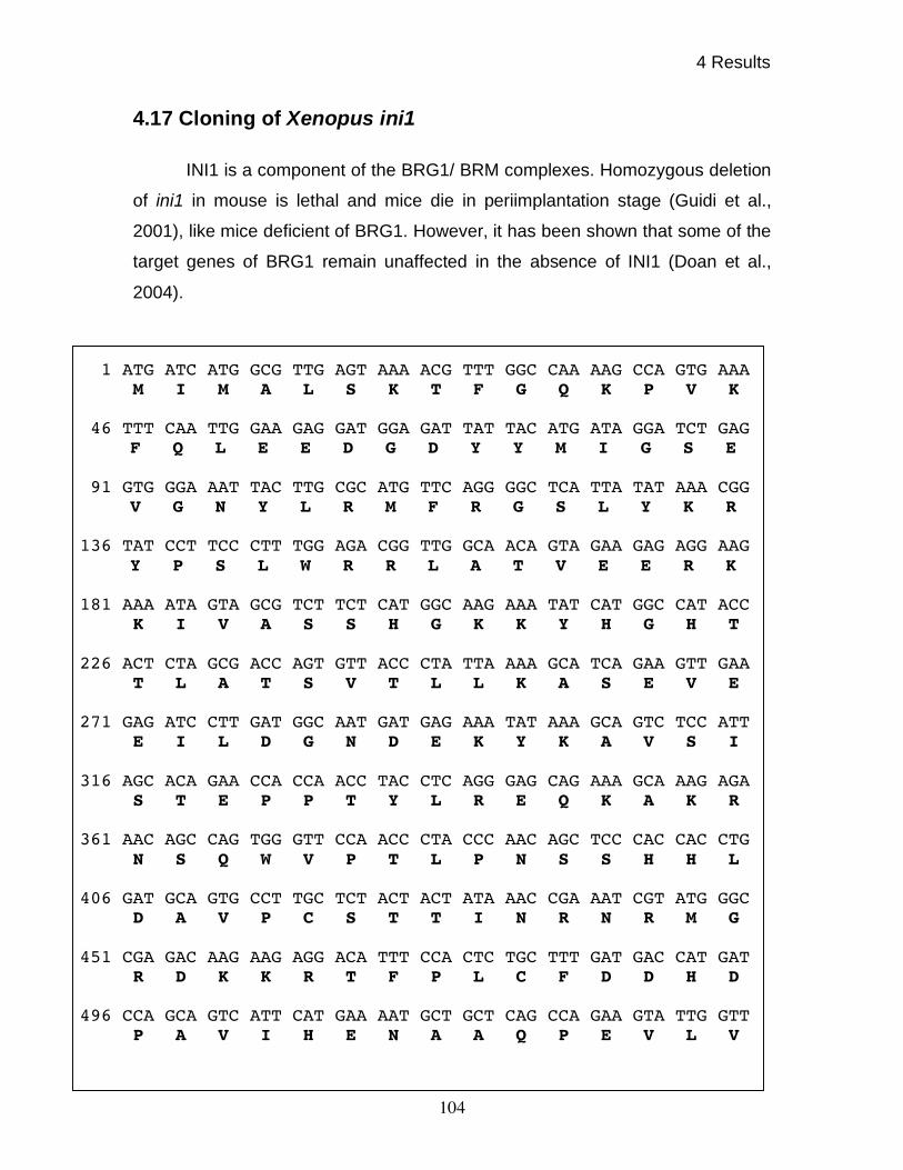
4 Results
104
4.17 Cloning of Xenopus ini1
INI1 is a component of the BRG1/ BRM complexes. Homozygous deletion
of ini1 in mouse is lethal and mice die in periimplantation stage (Guidi et al.,
2001), like mice deficient of BRG1. However, it has been shown that some of the
target genes of BRG1 remain unaffected in the absence of INI1 (Doan et al.,
2004).
1 ATG ATC ATG GCG TTG AGT AAA ACG TTT GGC CAA AAG CCA GTG AAA M I M A L S K T F G Q K P V K
46 TTT CAA TTG GAA GAG GAT GGA GAT TAT TAC ATG ATA GGA TCT GAG
F Q L E E D G D Y Y M I G S E 91 GTG GGA AAT TAC TTG CGC ATG TTC AGG GGC TCA TTA TAT AAA CGG
V G N Y L R M F R G S L Y K R 136 TAT CCT TCC CTT TGG AGA CGG TTG GCA ACA GTA GAA GAG AGG AAG
Y P S L W R R L A T V E E R K 181 AAA ATA GTA GCG TCT TCT CAT GGC AAG AAA TAT CAT GGC CAT ACC
K I V A S S H G K K Y H G H T 226 ACT CTA GCG ACC AGT GTT ACC CTA TTA AAA GCA TCA GAA GTT GAA
T L A T S V T L L K A S E V E 271 GAG ATC CTT GAT GGC AAT GAT GAG AAA TAT AAA GCA GTC TCC ATT
E I L D G N D E K Y K A V S I 316 AGC ACA GAA CCA CCA ACC TAC CTC AGG GAG CAG AAA GCA AAG AGA
S T E P P T Y L R E Q K A K R 361 AAC AGC CAG TGG GTT CCA ACC CTA CCC AAC AGC TCC CAC CAC CTG
N S Q W V P T L P N S S H H L 406 GAT GCA GTG CCT TGC TCT ACT ACT ATA AAC CGA AAT CGT ATG GGC
D A V P C S T T I N R N R M G 451 CGA GAC AAG AAG AGG ACA TTT CCA CTC TGC TTT GAT GAC CAT GAT
R D K K R T F P L C F D D H D 496 CCA GCA GTC ATT CAT GAA AAT GCT GCT CAG CCA GAA GTA TTG GTT
P A V I H E N A A Q P E V L V
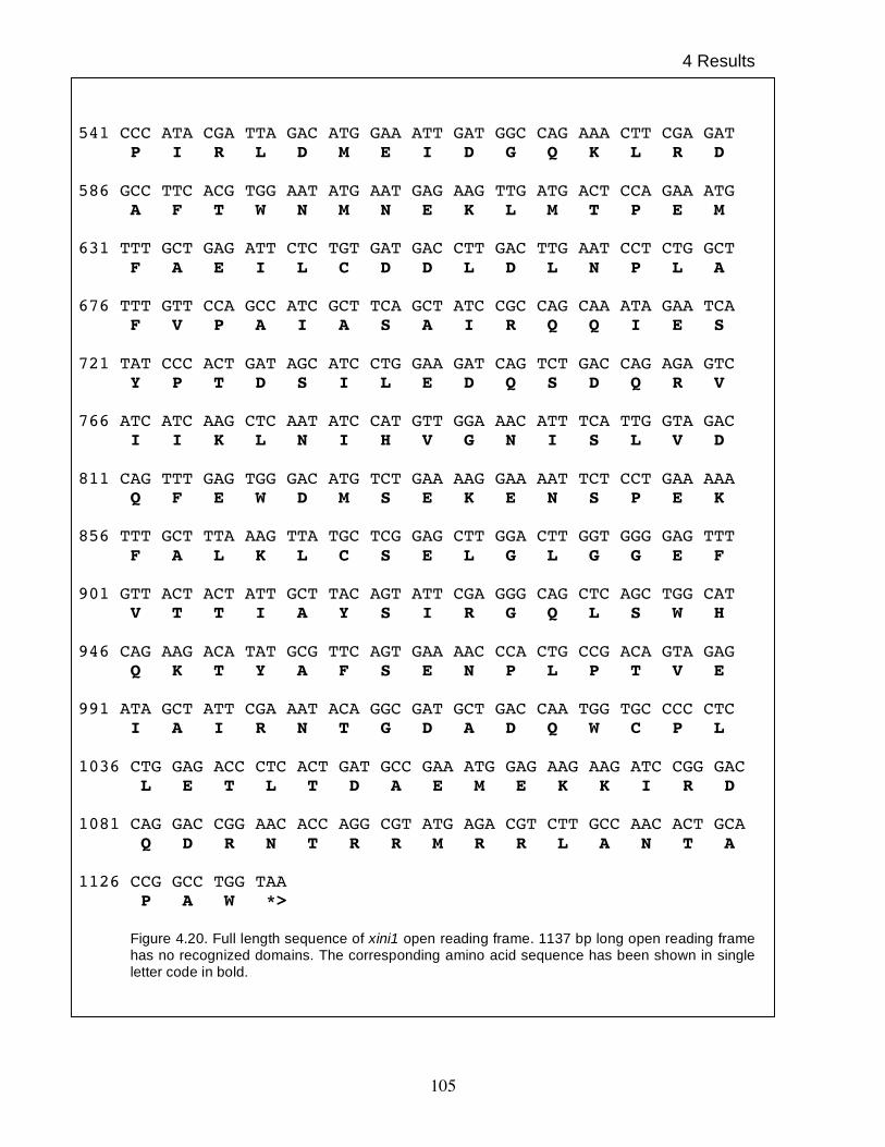
4 Results
105
541 CCC ATA CGA TTA GAC ATG GAA ATT GAT GGC CAG AAA CTT CGA GAT
P I R L D M E I D G Q K L R D 586 GCC TTC ACG TGG AAT ATG AAT GAG AAG TTG ATG ACT CCA GAA ATG
A F T W N M N E K L M T P E M 631 TTT GCT GAG ATT CTC TGT GAT GAC CTT GAC TTG AAT CCT CTG GCT
F A E I L C D D L D L N P L A 676 TTT GTT CCA GCC ATC GCT TCA GCT ATC CGC CAG CAA ATA GAA TCA
F V P A I A S A I R Q Q I E S 721 TAT CCC ACT GAT AGC ATC CTG GAA GAT CAG TCT GAC CAG AGA GTC
Y P T D S I L E D Q S D Q R V 766 ATC ATC AAG CTC AAT ATC CAT GTT GGA AAC ATT TCA TTG GTA GAC
I I K L N I H V G N I S L V D 811 CAG TTT GAG TGG GAC ATG TCT GAA AAG GAA AAT TCT CCT GAA AAA
Q F E W D M S E K E N S P E K 856 TTT GCT TTA AAG TTA TGC TCG GAG CTT GGA CTT GGT GGG GAG TTT
F A L K L C S E L G L G G E F 901 GTT ACT ACT ATT GCT TAC AGT ATT CGA GGG CAG CTC AGC TGG CAT
V T T I A Y S I R G Q L S W H 946 CAG AAG ACA TAT GCG TTC AGT GAA AAC CCA CTG CCG ACA GTA GAG
Q K T Y A F S E N P L P T V E 991 ATA GCT ATT CGA AAT ACA GGC GAT GCT GAC CAA TGG TGC CCC CTC
I A I R N T G D A D Q W C P L 1036 CTG GAG ACC CTC ACT GAT GCC GAA ATG GAG AAG AAG ATC CGG GAC
L E T L T D A E M E K K I R D
1081 CAG GAC CGG AAC ACC AGG CGT ATG AGA CGT CTT GCC AAC ACT GCA Q D R N T R R M R R L A N T A
1126 CCG GCC TGG TAA
P A W *> Figure 4.20. Full length sequence of xini1 open reading frame. 1137 bp long open reading frame
has no recognized domains. The corresponding amino acid sequence has been shown in single
letter code in bold.
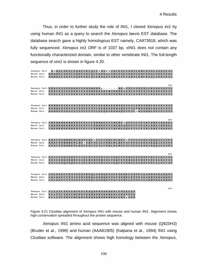
4 Results
106
Thus, in order to further study the role of INI1, I cloned Xenopus ini1 by
using human INI1 as a query to search the Xenopus laevis EST database. The
database search gave a highly homologous EST namely, CA973918, which was
fully sequenced. Xenopus ini1 ORF is of 1037 bp. xINI1 does not contain any
functionally characterized domain, similar to other vertebrate INI1. The full-length
sequence of xini1 is shown in figure 4.20.
Figure 4.21 Clustlaw alignment of Xenopus INI1 with mouse and human INI1. Alignment shows
high conservation spreaded throughout the protein sequence.
Xenopus INI1 amino acid sequence was aligned with mouse (Q9Z0H3)
(Bruder et al., 1999) and human (AAA81905) (Kalpana et al., 1994) INI1 using
Clustlaw software. The alignment shows high homology between the Xenopus,
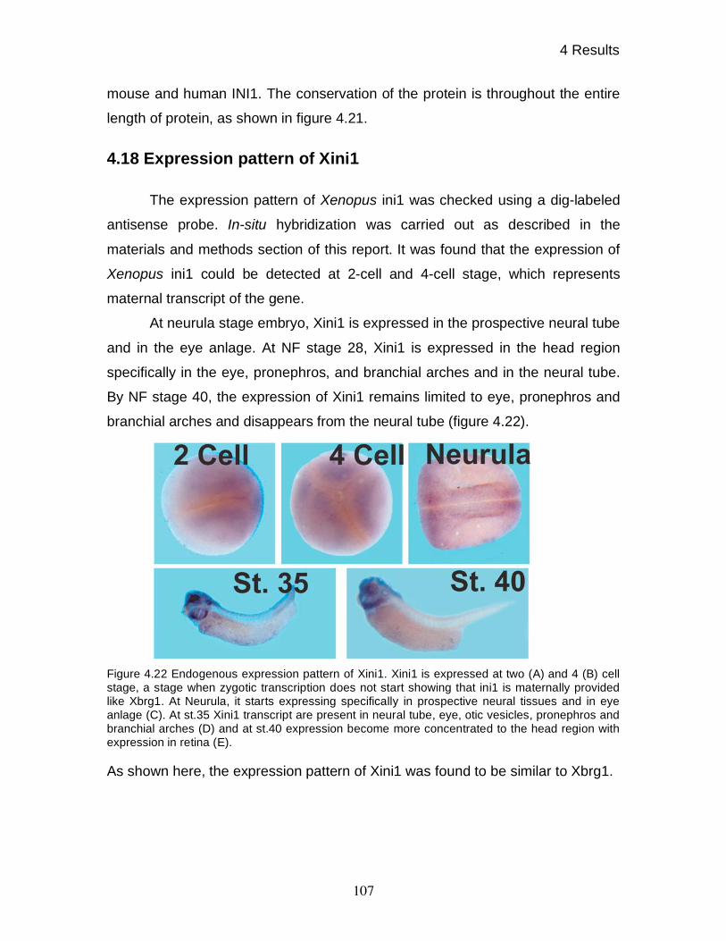
4 Results
107
mouse and human INI1. The conservation of the protein is throughout the entire
length of protein, as shown in figure 4.21.
4.18 Expression pattern of Xini1
The expression pattern of Xenopus ini1 was checked using a dig-labeled
antisense probe. In-situ hybridization was carried out as described in the
materials and methods section of this report. It was found that the expression of
Xenopus ini1 could be detected at 2-cell and 4-cell stage, which represents
maternal transcript of the gene.
At neurula stage embryo, Xini1 is expressed in the prospective neural tube
and in the eye anlage. At NF stage 28, Xini1 is expressed in the head region
specifically in the eye, pronephros, and branchial arches and in the neural tube.
By NF stage 40, the expression of Xini1 remains limited to eye, pronephros and
branchial arches and disappears from the neural tube (figure 4.22).
Figure 4.22 Endogenous expression pattern of Xini1. Xini1 is expressed at two (A) and 4 (B) cell
stage, a stage when zygotic transcription does not start showing that ini1 is maternally provided
like Xbrg1. At Neurula, it starts expressing specifically in prospective neural tissues and in eye
anlage (C). At st.35 Xini1 transcript are present in neural tube, eye, otic vesicles, pronephros and
branchial arches (D) and at st.40 expression become more concentrated to the head region with
expression in retina (E).
As shown here, the expression pattern of Xini1 was found to be similar to Xbrg1.
5 Discussion
108
5 DISCUSSION
ATP dependent BRG1 chromatin remodeling complexes are large
complexes of about 2 MDa in size. These complexes play a role in opening up
local chromatin structures by either looping or by disrupting the nucleosomes
around the promoters of various genes, making these promoters accessible for
the transcriptional machinery. It has been shown in many previous reports that
BRG1 containing chromatin remodeling complexes play a role in both repression
and activation of genes. Most of the studies came from either Yeast or
Drosophila (reviewed by Becker and Horz, 2002). The role of BRG1 has not been
studied in vertebrates since knock-out studies in mouse could not reveal the role
of these complexes in normal development (Bultman et al., 2000). In this study, I
have shown by gain and loss of function experiments that BRG1 is required for
the anterio-posterior body axis formation by specifically modulating the cellular
Wnt signaling response during early patterning of the Xenopus embryo. In
addition, I have shown that BRG1 modulated transcription is gene and signal
specific and sets up a signaling threshold for normal Wnt-dependent gene
induction.
5.1 Cloning and expression pattern
Xenopus brg1 and Xenopus ini1 were cloned by using the human
homologues as a query to search the database. The members of ATPase family
show high degree of sequence conservation among the vertebrates as also seen
in case of Xenopus homologues. In case of Xenopus BRG1, the conservation of
this protein is not just in the ATPase domain; rather it is extended throughout the
protein as also shown by Xenopus INI1, which is also conserved throughout the
whole sequence. This high conservation of sequence identity represents an
opportunity to use the existing database and provides a method for in-silico
cloning for other members of the ATPase complexes.
5 Discussion
109
Xenopus BRG1 shows about 85% identity with its human counterpart.
There were several missing regions in either of the sequences, which could be
due to different splice variants of protein. The presence of a bromodomain, which
represents a signature domain of SWI/SNF family members established this
clone as a member of SWI/SNF family member (Horn and Peterson, 2001). The
absence of a polyglutamine stretch, which is present in the BRM coding region,
was one of the distinguishing features of it. Moreover, the sequence motif EED at
the C-terminus of BRG1 is conserved in all the vertebrate homologues cloned
until date, while BRM has sequence motif DDE at the C-terminus. Whether these
differences are of functional importance is not known. These sequence features
identify this cDNA as the Xenopus homologue of BRG1. Cloning of INI1 was
undertaken to further understand the function of various subunits of BRG1
complex using database searches and was found to have significant homology
with other vertebrate counterparts.
Xbrg1 transcript are present in the maternal mRNA pool of the embryo,
which is consistent with the reported maternal expression of BRG1 protein
checked by immunobloting of protein extract from various stages of oocyte
(Gelius et al., 1999). In the same way, the Zebrafish brg1 has also been
reported to be expressed maternally (Gregg et al., 2003). The expression of
Xbrg1 at gastrula stage was ubiquitous, which becomes locally restricted from
neurula stages onward, which is also one of the conserved features of the Brg1
expression in Zebrafish. Similarly, mouse Brg1 has also been shown to have
widespread expression, which later becomes restricted to a number of tissues. In
mice, highest Brg1 expression is found in the spinal cord, the brain, parts of the
peripheral nervous system and in retina (Randazzo et al., 1994). This pattern is
comparable to Zebrafish and Xenopus (Gregg et al., 2003). The Zebrafish retina
shows expression in differentiating neuroepithelium and at later stage restricted
to ganglion cells and inner nuclear layers (Gregg et al., 2003). The similar
expression is also shown by Xbrg1 as seen in cross-sections of st. 35 embryos.
Interestingly, at stage 45, the expression of Xbrg1 was found only in the heart. In
a recent report, the expression of Smarcd3, a mouse homologue of BAF60c, a
5 Discussion
110
subunit of BRG1/BRM complex, was also found to be restricted to the heart
region until embryonic stage E9.25 (Lickert et al., 2004). Thus, the expression
pattern of Xbrg1 is very similar to the previously reported Brg1 expression
patterns in mice and Zebrafish, which concludes that expression of Brg1 is
conserved among vertebrates.
The expression pattern of Xini1 was also found to be similar to that of
Xbrg1. The expression pattern of Xbaf57 (Domingos et al., 2002) has also been
reported to be similar to that of Brg1 and Ini1. Thus, xBRG1, xINI1 and xBAF57,
which are conserved in core SWI/SNF complexes form an synexpression group
(Niehrs and Pollet, 1999) while specific subunits like BAF60c, which are part of
tissue specific SWI/SNF remodeling complexes, show a locally restricted
expression.
5.2 Role of BRG1 in Dorso-ventral patterning
5.2.1 Methodological consideration
In order to study the role of chromatin remodeling molecules in the early
development of Xenopus laevis, I used both loss and gain of function methods to
find out the role of BRG1 in normal development. The gain of function studies
were carried out by microinjection of mRNA at 4-cell stage for either wild type or
dominant negative versions of BRG1. Microinjection of mRNA at 4-cell stage
leads to overexpression of the protein in the embryo already prior to the onset of
zygotic transcription. Overexpression of a core subunit of a protein complex
might have several consequences. The exogenous protein may compete with its
endogenous counterpart for incorporation into the complex during assembly.
Alternatively, if this protein is normally limiting, more complexes may be
assembled. Increased number of dominant negative BRG1 complexes will be
competing with the endogenous protein complexes and thus will be inhibiting the
endogenous function of BRG1. On the other hand increased number of wild type
BRG1 containing complexes will co-operate with the endogenous function of
BRG1 and thus potentiate their function. Proteins involved in complexes with

5 Discussion
111
BRG1 to obtain full functional capability, are also involved in making complex
with BRM. Thus during the gain of function studies, it is possible that over-
expressed protein sequesters a subunit of the complex and thus makes it
unavailable for other complexes which can lead to non-specific effects. This
possibility of non-specific consequences was ruled out by rescue experiments, in
which the phenotype of dominant negative BRG1 protein was reverted by
overexpression of wild type BRG1 protein but not by overexpression of BRM
protein.
Gain of function studies provide useful information about the potential
function of a protein, however only loss of function studies can reveal essential
functions of a protein. The methods generally used for loss of function studies in
Xenopus are maternal knockouts or modified antisense oligonucleotides. siRNA
procedure has also been used in Xenopus embryos for loss of function studies
(Anantharam et al., 2003; Nakano et al., 2000; Zhou et al., 2002) but our
attempts to use siRNA mediated knock-down failed, similar to some other
Xenopus labs. Maternal knock-out studies (Heasman et al., 1991) are technically
difficult with limited success and do not distinguishes between zygotic or
maternal functions of the protein. The most commonly used method of knocking-
down proteins is by microinjection of modified oligonucleotides in the desired
region of embryo. Among the modified oligonucleotides, most common ones are
phosphoorthothioate antisense oligonucleotides and morpholino antisense
oligonucleotides. Phosphoorthothioate antisense oligonucleotides are
comparatively cheap, but they are known to produce non-specific effects.
Moreover, it is often also difficult to choose the correct target site for these
oligonucleotides. In recent times, morpholino antisense oligonucleotides have
been used (Heasman et al., 2000), which offer the advantage of high specificity,
easier selection of target sites, and efficient knock-down of the desired protein.
Based on these mentioned advantages and disadvantages of various methods
available, I chose to carry out loss of function studies using morpholino antisense
oligonucleotides.
5 Discussion
112
In this study, I reported that the full-length transcript of Xbrg1 could only
be obtained by in-vitro transcription at lower temperature, than the usual 37oC. It
has been reported previously that the in-vitro transcription using T3, T7 or SP6 at
37oC may result in truncated transcripts and that this problem could be overcome
by lowering the temperature (Krieg, 1990). Here, the full-length transcript of
Xbrg1 could be obtained by T7 promoter, but not by SP6 polymerase. Thus, for
the transcription of xbrg1, the use of SP6 RNA polymerase is problematic. Since
the pCS2+ vector, which is used to overexpress proteins in Xenopus, contains an
SP6 promoter upstream of translation start site, it was essential to use SP6
polymerase. In order to obtain full-length transcripts using SP6 promoter, lower
temperatures were used for transcription reaction. As per some available reports
(Ambion inc), some of the bacteriophage derived RNA polymerase show the
tendency of falling off and terminating RNA synthesis, leading to prematurely
terminated transcripts. The underlying reason for this premature termination has
not been resolved until date.
5.2.2 Specific role of BRG1 in anterio-posterior axis formation
I have shown in the report that BRG1 participates in the formation of AP
axis. These results are shown first, by overexpression of human BRG1, which
could be rescued and thus representing the specific phenotypes. Later these
results were also confirmed by overexpression of dominant negative Xenopus
BRG1. These results show that human BRG1 and Xenopus BRG1 act in a
similar manner, suggesting functional conservation. On the other hand, wild type
human BRG1 did show no phenotype on dorsal as well as on ventral
overexpression, while Xenopus BRG1 produced head defects upon dorsal
overexpression and induced an incomplete secondary axis upon ventral
overexpression. These results show that Xenopus BRG1 is more potent than
human BRG1. This might be due to either higher stability of Xenopus BRG1 or
due to some sequence divergence, xBRG1 may be capable to form more stable
complexes due to its compatibility with other endogenous components of BRG1
5 Discussion
113
complex. Overall, these results using human and Xenopus BRG1 variants
confirmed the role of BRG1 in AP axis formation.
Apart from these gain of function studies, loss of function studies provided
further evidence of the importance of BRG1 in head formation and dorsal ventral
patterning. Notably, the xBrg1 MO induced phenotypes could not be rescued by
hBRM and xISWI, demonstrating that the function of BRG1 is not redundant and
could not be taken over by other members of the SNF2 family. Thus, the results
obtained with gain and loss of function studies unambiguously established the
role of BRG1 in AP axis formation.
Overexpression of wild type and dominant negative human BRM, as well
as variants of xISWI did not cause any developmental phenotypes. These results
confirmed that the observed phenotypes with BRG1 are not due to injection
artifacts or due to toxic effects of RNA. These results obtained from
overexpression of dominant negative and wild type human BRM are in consent
with the recent reports showing that BRM knock-out mice do not show any
phenotypes, probably due to compensation by BRG1 (Reyes et al., 1998).
Moreover, it is now known that BRG1 is found in BAF and PBAF complexes
while BRM is found in BAF complexes only. Thus, it is possible that loss of BRM,
which will affect BAF complexes only, is compensated by BRG1, while the loss of
BRG1, which affect both BAF and PBAF complexes, can not be fully
compensated by BRM. Moreover other studies have also pointed out that BRG1
and BRM have transcriptional specificity (Kadam and Emerson, 2003).
Furthermore, in a recent report it was shown that the transcription of sox2, a
gene expressed in early neural cells, is dependent on BRM and not on BRG1
(Kondo and Raff, 2004) showing the differential requirement of BRM and BRG1.
The unsuccessful attempts to achieve developmental phenotypes by ISWI
overexpression may be explained differently. In particular, it is known that xISWI
is a highly abundant molecule in Xenopus oocytes (Guschin et al., 2000) and,
thus, I might not be able significantly inhibit its function by overexpression of
dominant negative protein. These results were also in consent with a recent

5 Discussion
114
report showing that mice heterozygous for ISWI were normal, but homozygous
null mutant mice dies in early embryonic stages (Stopka and Skoultchi, 2003).
5.3 BRG1 is required for normal expression of WNT target genes
When the phenotypes obtained from dorsal overexpression of wild type
and dominant negative xBRG1 were observed closely, the overexpression of
dominant negative BRG1 was found to cause preferentially loss of eyes, while
wild type xBRG1, preferentially caused loss of cement glands. In a recent report,
it was shown that the local mis-expression of Wnt8 in the head territory under
control of the cerberus promoter causes preferentially loss of cement glands
(Silva et al., 2003). A detailed model for head formation has been discussed
elsewhere (Niehrs, 1999; Niehrs, 2004). Moreover, overexpression of xBRG1 on
the ventral side produces a partial secondary axis, which indicates the formation
of weak organizer on the ventral side. The role of WNT signaling has been
convincingly shown in organizer formation as the overexpression of -CATENIN
on the ventral side causes secondary axis (Funayama et al., 1995) formation as
well as inhibition of WNT signaling on dorsal side by UV treatment causes
ventralization of the embryo (Kao and Elinson, 1988). The role of WNT pathway
in dorso-ventral patterning has been confirmed by interfering with -CATENIN by
maternal knock-downs as well as by morpholino antisense mediated knock-down
(Heasman et al., 2000; Wylie et al., 1996). The phenotypes observed in these
cases were similar to BRG1 knock-down. Thus, our results strongly support
dose-sensitive interactions between BRG1-containing remodeling complexes and
the canonical WNT signaling pathway.
In-situ hybridization analysis of genes involved in dorsal mesoderm
formation showed that indeed BRG1 affects WNT target gene expression, as
evidenced by the downregulation of Xnr3 and Chordin. Reduced expression of
Goosecoid also supports this conclusion. The goosecoid promoter is known to
have two signal inputs i.e. WNT signal and NODAL signal. WNT signaling is
relayed to the gsc promoter by the distinct WNT-targets siamois/twin, while

5 Discussion
115
NODAL signaling is mediated by SMAD2/SMAD4 heterodimers (Germain et al.,
2000). Reduced Siamois expression as checked in animal cap assay shows the
reduced WNT signal input for activation of goosecoid. Importantly, when
goosecoid was induced by ACTIVIN signaling alone in animal caps, its induction
was unaffected by BRG1 knock-down, indicating that reduced expression of
Goosecoid is due to reduced WNT signaling input. Interestingly, a nodal related
gene xnr6, which has been shown to be transcribed in pre MBT stages under the
control of WNT signaling pathway (Yang et al., 2002) was found to be
downregulated in animal cap assay under the reduced protein levels of BRG1.
Figure 5.1 BRG1 is involved in the WNT signaling pathway.
These results suggest that while NODAL signaling remains unaffected, the
reduced expression of Xnr6 reduces the nodal input on the goosecoid promoter,
resulting in reduced expression of Goosecoid. Taken together, four distinct WNT
target genes were tested and all of them were misregulated by BRG1 over-
/underexpression. I can conclude, therefore that the BRG1-CRC is essential for
proper transcriptional regulation of WNT / -CATENIN target genes.
5 Discussion
116
5.4 BRG1 containing complexes have gene and signal specific
functions
It has been claimed previously that BRG1 acts as a global transcriptional
activator (Armstrong et al., 2002). The loss of function studies presented here
point to rather selective functions of BRG1. Reduced levels of BRG1 protein
results in lower expression of Xnr3, Chordin, Myod and Myf-5, while expression
of Xbra and Xvent-2 remains unchanged and Xpo expression is increased.
Although it has been shown that WNT signaling affects Xbra expression (Schohl
and Fagotto, 2003), I did not find any change in the expression of Xbra. This
might be explained by a compensation of loss of WNT signaling input by other
signaling events like FGF signaling, required for Xbra expression (Smith et al.,
1991). Consistent with this assumption, when animal caps were induced by
eFGF for the induction of Xbra, BRG1 knock-down does not affects the
transcriptional levels of Xbra. The effect of BRG1 knock-down is not only gene
specific but also signal specific as seen in animal cap assay. On one hand,
BRG1 knock-down affects WNT signaling, shown by reduced expression of
Siamois and Xnr6, ACTIVIN/NODAL related signaling remains unaffected,
exhibited by Goosecoid and Xfd-1´.
The effect on FGF/MAPK signal appears also to be highly gene specific,
given that the Xbra mRNA levels remain normal while expression of Myod was
strongly reduced. The explanation for reduced transcription of myod came from a
recent report showing that BRG1 and MyoD interact with each other in the
presence of activated MAPK (Simone et al., 2004). It has been also shown that
MyoD promotes its own transcription by autocatalysis (Steinbach et al., 1998;
Thayer et al., 1989). Thus, it might be possible that BRG1 interacts with MyoD to
promote the transcription of myod and thus when BRG1 is knocked out, the
autocatalysis of myod will be blocked, leading to reduced transcription as seen in
animal cap induction experiments.
5 Discussion
117
BRG1 knock-down does not affect BMP signaling as checked by
expression of Xvent-2, a gene under direct control of BMP signaling. These
results were unexpected since the weakening of organizer would mean that weak
organizer would no longer be able to inhibit invasive BMP signaling mediated
ventral mesoderm formation but it is possible that the remaining activity of
organizer is still sufficient to prevent the expression of Xvent-2 in the organizer
territory. Although the expression of Xpo in organizer territory confirms that the
organizer has been weakened, its increased expression could also be due to the
repressive function of BRG1 as has been reported previously (Martens and
Winston, 2003). Taken together, these results pointed out that BRG1 acts in a
gene and signal specific manner.
5.5 Effects of BRG1 knock-down on organogenesis
The pleiotropic effects of BRG1 knock-down was shown in this report
(figure 4.13) by reduced expression of several differentiation markers including
neural n- -tubulin, heart specific Nkx2.5, muscle specific Mlc35 and blood vessel
specific Msr. These effects could be explained partly by perturbation of the
primary embryonic patterning, but it does not exclude a requirement of BRG1 for
tissue specific transcription factors. The effect on neural tissue development
observed by neural specific beta-tubulin may be explained partly by the reduced
expression of Chordin in blastula stage. chordin is a gene that is expressed in the
BCNE center. The BCNE center gives rise to future neural tissue (De Robertis
and Kuroda, 2004). A reduction of Chordin expression will impair the BCNE
center, which will ultimately affect neural tissue development. A recent report has
also demonstrated a role of Xenopus BRG1 in neurogenesis (Seo et al., 2005)
although it claims that BRG1 knock-down does not affect neurogenesis during
early patterning based on the fact that Sox2 expression was unaffected by the
BRG1 ablation. SOX2 is a member of HMG box transcription factor family,
involved in early stages of neurogenesis (Bylund et al., 2003). However, a recent
report has shown that Sox2 expression is controlled by BRM and not by BRG1
(Kondo and Raff, 2004) in a study carried out in Rats.

5 Discussion
118
As I have shown that BRG1 knock-down affects heart formation, a recent
report supported my observation and provided a mechanism. It was shown that
BAF60c, which is a component of BRG1 complex is expressed specifically in the
heart and when knocked-down using siRNA, heart formation is severely affected.
Furthermore, it was shown that BAF60c is required for transcription of Nkx2.5,
which is dependent on BRG1. Furthermore, it has been reported that knock-out
of BAF180, a signature subunit of PBAF complexes, in mice also causes heart
defects (Wang et al., 2004b). The blood vessel formation is severely inhibited in
BRG1 MO treated embryos. The process of angiogenesis requires among other
factors, MMP-2. It has been shown that the transcription of mmp-2 is dependent
on BRG1 (Ma et al., 2004). This observation points out a possibility that when
BRG1 is knocked-down, the transcription of mmp-2 might get inhibited and this
may affect angiogenesis. These observations point out a role for BRG1
containing chromatin-remodeling complex in tissue specific transcription. In later
stages of development, the expression of Mlc35 was also reduced, confirming
the reported role of BRG1 in myogenesis in which authors reported that dominant
negative BRG1 interferes with the MyoD mediated muscle program shown by the
down regulation of Myogenin and Mrf-4 (de la Serna et al., 2001). Here in this
study, I have shown that both Myod and Myf-5 are down regulated upon BRG1
knock-down and thus showing another level at which myogenesis is affected by
BRG1. A TCF binding site has been found in the myf-5 promoter, as well as it
has been shown that myf-5 can be activated immediately after activation of
animal caps by Wnt stimulation in the presence of cycloheximide (Shi et al.,
2002), suggesting it to be a direct target of Wnt signaling pathway. As shown,
upon BRG1 knock-down, Myf-5 expression is completely inhibited showing that
BRG1 is required to activate the transcription of myf-5, possibly by interfering
with Wnt signaling. Thus, it can be concluded that BRG1 is involved in several
differentiation programs at key regulatory levels.
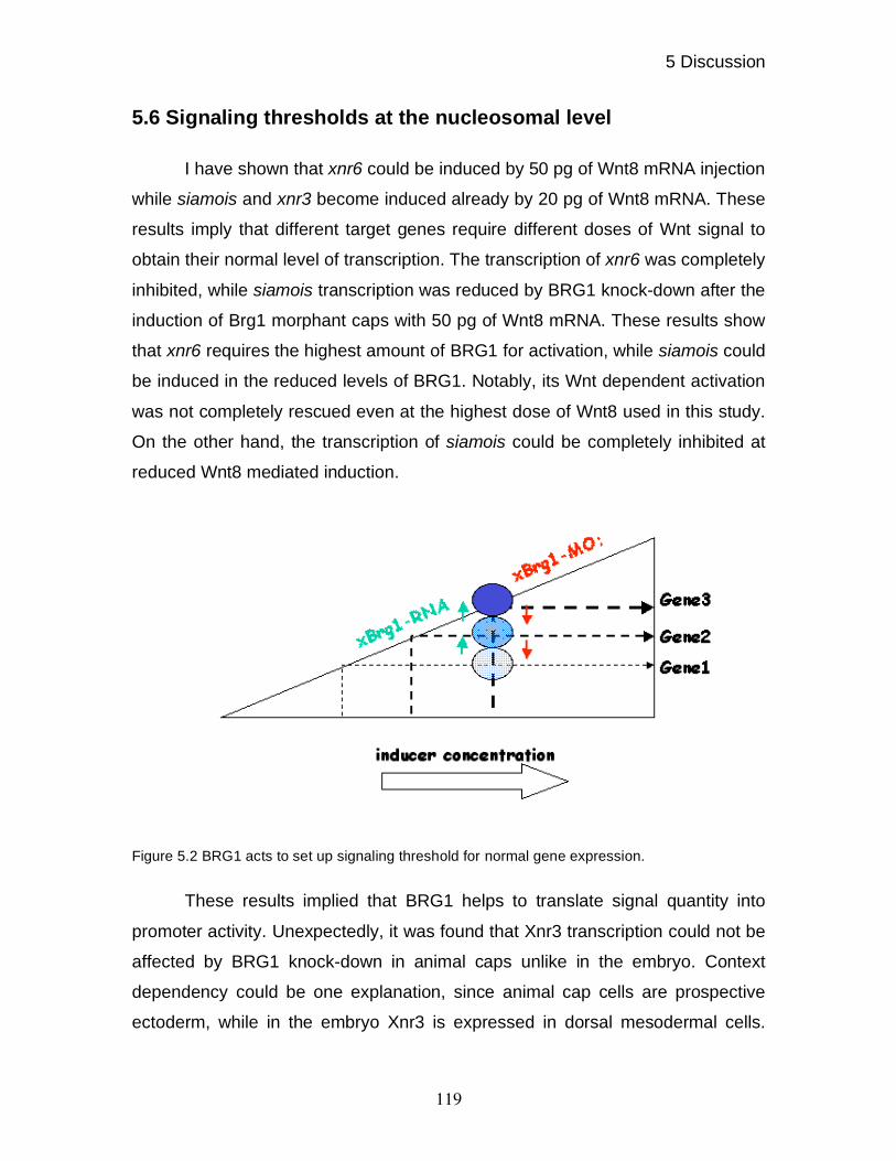
5 Discussion
119
5.6 Signaling thresholds at the nucleosomal level
I have shown that xnr6 could be induced by 50 pg of Wnt8 mRNA injection
while siamois and xnr3 become induced already by 20 pg of Wnt8 mRNA. These
results imply that different target genes require different doses of Wnt signal to
obtain their normal level of transcription. The transcription of xnr6 was completely
inhibited, while siamois transcription was reduced by BRG1 knock-down after the
induction of Brg1 morphant caps with 50 pg of Wnt8 mRNA. These results show
that xnr6 requires the highest amount of BRG1 for activation, while siamois could
be induced in the reduced levels of BRG1. Notably, its Wnt dependent activation
was not completely rescued even at the highest dose of Wnt8 used in this study.
On the other hand, the transcription of siamois could be completely inhibited at
reduced Wnt8 mediated induction.
Figure 5.2 BRG1 acts to set up signaling threshold for normal gene expression.
These results implied that BRG1 helps to translate signal quantity into
promoter activity. Unexpectedly, it was found that Xnr3 transcription could not be
affected by BRG1 knock-down in animal caps unlike in the embryo. Context
dependency could be one explanation, since animal cap cells are prospective
ectoderm, while in the embryo Xnr3 is expressed in dorsal mesodermal cells.

5 Discussion
120
Another possibility is that the amount of Wnt8 used to induce the xnr3
transcription is still high enough to counteract the reduced amount of BRG1 in
the animal cap assay. Thus, xnr3 requires the least amount of BRG1 for
remodeling of its promoter, and may be fully activated already at very low levels
of WNT signal.
This possibility was strengthened by the observation that xBRG1 could
hyperstimulate the expression of Xnr3 in sub-optimal WNT induction conditions
much stronger then Siamois induction. Thus, it can be concluded that BRG1
protein levels set up a threshold for the activation of WNT target genes at a given
signal strength as presented in the model (figure 5.2).
5.7 Specific recruitment of BRG1 to target genes
From my results, it has become clear that BRG1 modulates the
transcriptional read out of the canonical Wnt-signaling pathway required for early
patterning of the Xenopus embryo. In order to do this, BRG1 is expected to bind
to WNT-target gene promoters. A report showing interaction of BRG1 and -
CATENIN, provides a mechanism how BRG1 may become recruited to these
promoters (Barker et al., 2001). However, this result was disputed since
interaction of BRG1 and -CATENIN could not be reproduced by other labs,
which tested the interaction of in-vitro translated BRG1 and -CATENIN (Kadam
and Emerson, 2003). However, the epistasis experiments carried out during this
project showed that -CATENIN could rescue the Brg1 morpholino mediated
phenotypes. Additionally, I could also show that BRG1 is required for the
induction of secondary axis formation by -CATENIN. These results in
combination confirmed that BRG1 and -CATENIN interact genetically. Recently,
a report could show convincingly that BAF60c enhances the interaction of BRG1
and -CATENIN (Lickert et al., 2004). This observation suggested that BRG1 is
specifically recruited to the WNT target genes promoter by the interaction of
BAF60c and -CATENIN. In another case, the recruitment of BRG1 on the myod
promoter could be explained by the interaction of BRG1 and MyoD in the

5 Discussion
121
presence of activated MAPK (Simone et al., 2004). From these observations, it
was clear that various subunits of BRG1 complex are required for recruitment of
this complex to a variety of promoters other then having their role in obtaining the
maximum activity of nucleosomal remodeling by BRG1 complex.
5.8 Conclusions and outlook
In this study, the role of BRG1 was studied in early development of
Xenopus. It was shown that BRG1 is required for AP axis formation by affecting
the early patterning of the embryo. It was also shown that BRG1 affects early
patterning of Xenopus embryo by modulating WNT signal target genes.
Furthermore, it was shown that BRG1 affects myogenesis. In this study, I found
that BRG1 affects specific signaling pathways. BRG1 affected the WNT and
eFGF-signaling pathway, while the ACTIVIN signaling pathway remained
unaffected. I could also show that BRG1 sets up a signaling threshold for the
stimulation of WNT target genes.
These observations provide an entry point for the analysis of BRG1
containing remodeling complexes in vivo. It will be interesting to show the
recruitment of BRG1 on its target promoters using chromatin immunoprecipitation
(ChIP). Moreover, it will be interesting to investigate, when BRG1 is required –
i.e. before the assembly of pre initiation complex (PIC) as has been shown for
some genes (Soutoglou and Talianidis, 2002), or after the assembly of PIC.
These experiments can further be extended to find out, whether BRG1 stays
bound to promoter, or leaves it after initiating the transcription.
Furthermore, the functions of various other subunits of BRG1 complex
including INI1 can now be explored using the same strategy, used in this study.
INI1 is a common component of BRG1 and BRM complex. Therefore, it will be
interesting to find out if INI1 knock-down affects all BRG1 target genes or other
genes as well. Furthermore, it can also be tested if some of the BRG1 target
genes remain unaffected by INI1 knock-down as has been reported. These

5 Discussion
122
studies could explore the role of other subunits of BRG1 complex in early
development. Given the importance of INI1 in rhabdoid tumor development, it
may also provide further mechanistic explanation of the role of INI1 in this
disease. Other then INI1, other interesting subunits to study will be BAF180 and
BAF250 due to their specific presence in PBAF and BAF complexes respectively.
As it has been shown that BRG1 knock-down affects several tissues, the
role of BRG1 in organogenesis can also be studied. It has been shown that
BAF60c and BAF180 are required for heart formation. In Xenopus, animal cap
system can provide an easy method to study the organogenesis outside the
organism. Animal caps contain multipotent embryonic stem cells. The animal cap
tissue can be differentiated into various organs and cell types by specific
induction protocols, using variable Activin and Retinoic acid concentrations
(Ariizumi and Asashima, 2001). By this strategy, it is now possible to study the
specific role of chromatin remodeling complexes in organ development.

6 Literature
123
6 LITERATURE
Aagaard, L., Schmid, M., Warburton, P., and Jenuwein, T. (2000). Mitotic
phosphorylation of SUV39H1, a novel component of active centromeres,
coincides with transient accumulation at mammalian centromeres. J Cell Sci 113 ( Pt 5), 817-829.
Aasland, R., Stewart, A. F., and Gibson, T. (1996). The SANT domain: a putative
DNA-binding domain in the SWI-SNF and ADA complexes, the transcriptional co-
repressor N-CoR and TFIIIB. Trends Biochem Sci 21, 87-88.
Ae, K., Kobayashi, N., Sakuma, R., Ogata, T., Kuroda, H., Kawaguchi, N.,
Shinomiya, K., and Kitamura, Y. (2002). Chromatin remodeling factor encoded by
ini1 induces G1 arrest and apoptosis in ini1-deficient cells. Oncogene 21, 3112-
3120.
Agius, E., Oelgeschlager, M., Wessely, O., Kemp, C., and De Robertis, E. M.
(2000). Endodermal NODAL-related signals and mesoderm induction in
Xenopus. Development 127, 1173-1183.
Anantharam, A., Lewis, A., Panaghie, G., Gordon, E., McCrossan, Z. A., Lerner,
D. J., and Abbott, G. W. (2003). RNA interference reveals that endogenous
Xenopus MinK-related peptides govern mammalian K+ channel function in
oocyte expression studies. J Biol Chem 278, 11739-11745.
Angus-Hill, M. L., Schlichter, A., Roberts, D., Erdjument-Bromage, H., Tempst,
P., and Cairns, B. R. (2001). A Rsc3/Rsc30 zinc cluster dimer reveals novel roles
for the chromatin remodeler RSC in gene expression and cell cycle control. Mol
Cell 7, 741-751.
Ariizumi, T., and Asashima, M. (2001). In vitro induction systems for analyses of
amphibian organogenesis and body patterning. Int J Dev Biol 45, 273-279.
Armstrong, J. A., Papoulas, O., Daubresse, G., Sperling, A. S., Lis, J. T., Scott,
M. P., and Tamkun, J. W. (2002). The Drosophila BRM complex facilitates global
transcription by RNA polymerase II. Embo J 21, 5245-5254.
Armstrong, N. J., Steinbeisser, H., Prothmann, C., DeLotto, R., and Rupp, R. A.
(1998). Conserved Spatzle/Toll signaling in dorsoventral patterning of Xenopus
embryos. Mech Dev 71, 99-105.
Arents, G., Burlingame, R. W., Wang, B. C., Love, W. E., and Moudrianakis, E.
N. (1991). The nucleosomal core histone octamer at 3.1 A resolution: a tripartite
protein assembly and a left-handed superhelix. Proc Natl Acad Sci U S A 88,
10148-10152.
6 Literature
124
Artinger, M., Blitz, I., Inoue, K., Tran, U., and Cho, K. W. (1997). Interaction of
goosecoid and brachyury in Xenopus mesoderm patterning. Mech Dev 65, 187-
196.
Baetz, K. K., Krogan, N. J., Emili, A., Greenblatt, J., and Hieter, P. (2004). The
ctf13-30/CTF13 genomic haploinsufficiency modifier screen identifies the yeast
chromatin remodeling complex RSC, which is required for the establishment of
sister chromatid cohesion. Mol Cell Biol 24, 1232-1244.
Banville, D., and Williams, J. G. (1985). The pattern of expression of the
Xenopus laevis tadpole alpha-globin genes and the amino acid sequence of the
three major tadpole alpha-globin polypeptides. Nucleic Acids Res 13, 5407-5421.
Barak, O., Lazzaro, M. A., Lane, W. S., Speicher, D. W., Picketts, D. J., and
Shiekhattar, R. (2003). Isolation of human NURF: a regulator of Engrailed gene
expression. Embo J 22, 6089-6100.
Barker, N., Hurlstone, A., Musisi, H., Miles, A., Bienz, M., and Clevers, H. (2001).
The chromatin remodeling factor Brg-1 interacts with beta-catenin to promote
target gene activation. Embo J 20, 4935-4943.
Becker, P. B., and Horz, W. (2002). ATP-dependent nucleosome remodeling.
Annu Rev Biochem 71, 247-273.
Biegel, J. A., Kalpana, G., Knudsen, E. S., Packer, R. J., Roberts, C. W., Thiele,
C. J., Weissman, B., and Smith, M. (2002). The role of INI1 and the SWI/SNF
complex in the development of rhabdoid tumors: meeting summary from the
workshop on childhood atypical teratoid/rhabdoid tumors. Cancer Res 62, 323-
328.
Bochar, D. A., Wang, L., Beniya, H., Kinev, A., Xue, Y., Lane, W. S., Wang, W.,
Kashanchi, F., and Shiekhattar, R. (2000). BRCA1 is associated with a human
SWI/SNF-related complex: linking chromatin remodeling to breast cancer. Cell 102, 257-265.
Boeger, H., Griesenbeck, J., Strattan, J. S., and Kornberg, R. D. (2004).
Removal of promoter nucleosomes by disassembly rather then sliding in vivo.
Mol Cell 14, 667-673.
Boyer, L. A., and Peterson, C. L. (2000). Actin-related proteins (Arps):
conformational switches for chromatin-remodeling machines? Bioessays 22, 666-
672.
Brannon, M., Gomperts, M., Sumoy, L., Moon, R. T., and Kimelman, D. (1997). A
beta-catenin/XTcf-3 complex binds to the siamois promoter to regulate dorsal
axes specification in Xenopus. Genes Dev 11, 2359-2370.
6 Literature
125
Brannon, M., and Kimelman, D. (1996). Activation of Siamois by the Wnt
pathway. Dev Biol 180, 344-347.
Brehm, A., Langst, G., Kehle, J., Clapier, C. R., Imhof, A., Eberharter, A., Muller,
J., and Becker, P. B. (2000). dMi-2 and ISWI chromatin remodeling factors have
distinct nucleosome binding and mobilization properties. Embo J 19, 4332-4341.
Brizuela, B. J., and Kennison, J. A. (1997). The Drosophila homeotic gene moira
regulates expression of engrailed and HOM genes in imaginal tissues. Mech Dev 65, 209-220.
Brownell, J. E., and Allis, C. D. (1995). An activity gel assay detects a single,
catalytically active histone acetyltransferase subunit in Tetrahymena
macronuclei. Proc Natl Acad Sci U S A 92, 6364-6368.
Brownell, J. E., and Allis, C. D. (1996). Special HATs for special occasions:
linking histone acetylation to chromatin assembly and gene activation. Curr Opin
Genet Dev 6, 176-184.
Brownell, J. E., Zhou, J., Ranalli, T., Kobayashi, R., Edmondson, D. G., Roth, S.
Y., and Allis, C. D. (1996). Tetrahymena histone acetyltransferase A: a homolog
to yeast Gcn5p linking histone acetylation to gene activation. Cell 84, 843-851.
Bruder, C. E., Dumanski, J. P., and Kedra, D. (1999). The mouse ortholog of the
human SMARCB1 gene encodes two splice forms. Biochem Biophys Res
Commun 257, 886-890.
Bruno, M., Flaus, A., Stockdale, C., Rencurel, C., Ferreira, H., and Owen-
Hughes, T. (2003). Histone H2A/H2B dimer exchange by ATP-dependent
chromatin remodeling activities. Mol Cell 12, 1599-1606.
Bultman, S., Gebuhr, T., Yee, D., La Mantia, C., Nicholson, J., Gilliam, A.,
Randazzo, F., Metzger, D., Chambon, P., Crabtree, G., and Magnuson, T.
(2000). A Brg1 null mutation in the mouse reveals functional differences among
mammalian SWI/SNF complexes. Mol Cell 6, 1287-1295.
Bylund, M., Andersson, E., Novitch, B. G., and Muhr, J. (2003). Vertebrate
neurogenesis is counteracted by Sox1-3 activity. Nat Neurosci 6, 1162-1168.
Cairns, B. R., Erdjument-Bromage, H., Tempst, P., Winston, F., and Kornberg, R.
D. (1998). Two actin-related proteins are shared functional components of the
chromatin-remodeling complexes RSC and SWI/SNF. Mol Cell 2, 639-651.
Cairns, B. R., Kim, Y. J., Sayre, M. H., Laurent, B. C., and Kornberg, R. D.
(1994). A multisubunit complex containing the SWI1/ADR6, SWI2/SNF2, SWI3,
6 Literature
126
SNF5, and SNF6 gene products isolated from yeast. Proc Natl Acad Sci U S A 91, 1950-1954.
Cairns, B. R., Schlichter, A., Erdjument-Bromage, H., Tempst, P., Kornberg, R.
D., and Winston, F. (1999). Two functionally distinct forms of the RSC
nucleosome-remodeling complex, containing essential AT hook, BAH, and
bromodomains. Mol Cell 4, 715-7223.
Camporeale, G., Shubert, E. E., Sarath, G., Cerny, R., and Zempleni, J. (2004).
K8 and K12 are biotinylated in human histone H4. Eur J Biochem 271, 2257-
2263.
Carrozza, M. J., Utley, R. T., Workman, J. L., and Cote, J. (2003). The diverse
functions of histone acetyltransferase complexes. Trends Genet 19, 321-329.
Cheng, A. M., Thisse, B., Thisse, C., and Wright, C. V. (2000). The lefty-related
factor Xatv acts as a feedback inhibitor of nodal signaling in mesoderm induction
and L-R axes development in Xenopus. Development 127, 1049-1061.
Cho, K. W., Blumberg, B., Steinbeisser, H., and De Robertis, E. M. (1991).
Molecular nature of Spemann's organizer: the role of the Xenopus homeobox
gene goosecoid. Cell 67, 1111-1120.
Clark, R. J., and Felsenfeld, G. (1971). Structure of chromatin. Nat New Biol 229,
101-106.
Clement, J. H., Fettes, P., Knochel, S., Lef, J., and Knochel, W. (1995). Bone
morphogenetic protein 2 in the early development of Xenopus laevis. Mech Dev 52, 357-370.
Collins, R. T., Furukawa, T., Tanese, N., and Treisman, J. E. (1999). Osa
associates with the BRM chromatin remodeling complex and promotes the
activation of some target genes. Embo J 18, 7029-7040.
Collins, R. T., and Treisman, J. E. (2000). Osa-containing BRM chromatin
remodeling complexes are required for the repression of wingless target genes.
Genes Dev 14, 3140-3152.
Crosby, M. A., Miller, C., Alon, T., Watson, K. L., Verrijzer, C. P., Goldman-Levi,
R., and Zak, N. B. (1999). The trithorax group gene moira encodes a brahma-
associated putative chromatin-remodeling factor in Drosophila melanogaster. Mol
Cell Biol 19, 1159-1170.
Cui, Y., Tian, Q., and Christian, J. L. (1996). Synergistic effects of Vg1 and Wnt
signals in the specification of dorsal mesoderm and endoderm. Dev Biol 180, 22-
34.
6 Literature
127
Dale, L., Matthews, G., and Colman, A. (1993). Secretion and mesoderm-
inducing activity of the TGF-beta-related domain of Xenopus Vg1. Embo J 12,
4471-4480.
Dale, L., and Slack, J. M. (1987). Fate map for the 32-cell stage of Xenopus
laevis. Development 99, 527-551.
Dallas, P. B., Pacchione, S., Wilsker, D., Bowrin, V., Kobayashi, R., and Moran,
E. (2000). The human SWI-SNF complex protein p270 is an ARID family member
with non-sequence-specific DNA binding activity. Mol Cell Biol 20, 3137-3146.
Damelin, M., Simon, I., Moy, T. I., Wilson, B., Komili, S., Tempst, P., Roth, F. P.,
Young, R. A., Cairns, B. R., and Silver, P. A. (2002). The genome-wide
localization of Rsc9, a component of the RSC chromatin-remodeling complex,
changes in response to stress. Mol Cell 9, 563-573.
de la Serna, I. L., Carlson, K. A., and Imbalzano, A. N. (2000). Mammalian SWI-
SNF complexes contribute to activation of the hsp70 gene. Molecular and
Cellular Biology 20, 2839-2851.
de la Serna, I. L., Carlson, K. A., and Imbalzano, A. N. (2001). Mammalian
SWI/SNF complexes promote MyoD-mediated muscle differentiation. Nat Genet 27, 187-190.
De Robertis, E. M., and Kuroda, H. (2004). Dorsal-ventral patterning and neural
induction in Xenopus embryos. Annu Rev Cell Dev Biol 20, 285-308.
De Robertis, E. M., Larrain, J., Oelgeschlager, M., and Wessely, O. (2000). The
establishment of Spemann's organizer and patterning of the vertebrate embryo.
Nat Rev Genet 1, 171-181.
Deuring, R., Fanti, L., Armstrong, J. A., Sarte, M., Papoulas, O., Prestel, M.,
Daubresse, G., Verardo, M., Moseley, S. L., Berloco, M., et al. (2000). The ISWI
chromatin-remodeling protein is required for gene expression and the
maintenance of higher order chromatin structure in vivo. Mol Cell 5, 355-365.
Devic, E., Paquereau, L., Vernier, P., Knibiehler, B., and Audigier, Y. (1996).
Expression of a new G protein-coupled receptor X-msr is associated with an
endothelial lineage in Xenopus laevis. Mech Dev 59, 129-140.
Dhalluin, C., Carlson, J. E., Zeng, L., He, C., Aggarwal, A. K., and Zhou, M. M.
(1999). Structure and ligand of a histone acetyltransferase bromodomain. Nature 399, 491-496.
Doan, D. N., Veal, T. M., Yan, Z., Wang, W., Jones, S. N., and Imbalzano, A. N.
(2004). Loss of the INI1 tumor suppressor does not impair the expression of
6 Literature
128
multiple BRG1-dependent genes or the assembly of SWI/SNF enzymes.
Oncogene 23, 3462-3473.
Domingos, P. M., Obukhanych, T. V., Altmann, C. R., and Hemmati-Brivanlou, A.
(2002). Cloning and developmental expression of Baf57 in Xenopus laevis. Mech
Dev 116, 177-181.
Eberharter, A., Ferrari, S., Langst, G., Straub, T., Imhof, A., Varga-Weisz, P.,
Wilm, M., and Becker, P. B. (2001). Acf1, the largest subunit of CHRAC,
regulates ISWI-induced nucleosome remodeling. Embo J 20, 3781-3788.
Eberharter, A., and Becker, P. B. (2004). ATP-dependent nucleosome
remodeling: factors and functions. J Cell Sci 117, 3707-3711.
Eisen, J. A., Sweder, K. S., and Hanawalt, P. C. (1995). Evolution of the SNF2
family of proteins: subfamilies with distinct sequences and functions. Nucleic
Acids Res 23, 2715-2723.
Ekker, S. C., McGrew, L. L., Lai, C. J., Lee, J. J., von Kessler, D. P., Moon, R. T.,
and Beachy, P. A. (1995). Distinct expression and shared activities of members
of the hedgehog gene family of Xenopus laevis. Development 121, 2337-2347.
Elfring, L. K., Daniel, C., Papoulas, O., Deuring, R., Sarte, M., Moseley, S., Beek,
S. J., Waldrip, W. R., Daubresse, G., DePace, A., et al. (1998). Genetic analysis
of brahma: the Drosophila homolog of the yeast chromatin remodeling factor
SWI2/SNF2. Genetics 148, 251-265.
Elfring, L. K., Deuring, R., McCallum, C. M., Peterson, C. L., and Tamkun, J. W.
(1994). Identification and characterization of Drosophila relatives of the yeast
transcriptional activator SNF2/SWI2. Mol Cell Biol 14, 2225-2234.
Fainsod, A., Deissler, K., Yelin, R., Marom, K., Epstein, M., Pillemer, G.,
Steinbeisser, H., and Blum, M. (1997). The dorsalizing and neural inducing gene
follistatin is an antagonist of BMP-4. Mech Dev 63, 39-50.
Fan, H. Y., He, X., Kingston, R. E., and Narlikar, G. J. (2003). Distinct strategies
to make nucleosomal DNA accessible. Mol Cell 11, 1311-1322.
Fisher, M. E., Isaacs, H. V., and Pownall, M. E. (2002). eFGF is required for
activation of XmyoD expression in the myogenic cell lineage of Xenopus laevis.
Development 129, 1307-1315.
Flemming W.,1882 Zell substanz,Kern und Zelltheilung.Leipzig:Vogel
Friedle, H., Rastegar, S., Paul, H., Kaufmann, E., and Knochel, W. (1998). Xvent-
1 mediates BMP-4-induced suppression of the dorsal-lip-specific early response
gene XFD-1' in Xenopus embryos. Embo J 17, 2298-2307.
6 Literature
129
Fukui, Y., Furue, M., Myoishi, Y., Sato, J. D., Okamoto, T., and Asashima, M.
(2003). Long-term culture of Xenopus presumptive ectoderm in a nutrient-
supplemented culture medium. Dev Growth Differ 45, 499-506.
Funayama, N., Fagotto, F., McCrea, P., and Gumbiner, B. M. (1995). Embryonic
axes induction by the armadillo repeat domain of beta-catenin: evidence for
intracellular signaling. J Cell Biol 128, 959-968.
Furukawa, Y., Kawakami, T., Sudo, K., Inazawa, J., Matsumine, A., Akiyama, T.,
and Nakamura, Y. (1996). Isolation and mapping of a human gene (RPD3L1)
that is homologous to RPD3, a transcription factor in Saccharomyces cerevisiae.
Cytogenet Cell Genet 73, 130-133.
Gdula, D. A., Sandaltzopoulos, R., Tsukiyama, T., Ossipow, V., and Wu, C.
(1998). Inorganic pyrophosphatase is a component of the Drosophila
nucleosome remodeling factor complex. Genes Dev 12, 3206-3216.
Gelius, B., Wade, P., Wolffe, A., Wrange, O., and Ostlund Farrants, A. K. (1999).
Characterization of a chromatin remodeling activity in Xenopus oocytes. Eur J
Biochem 262, 426-434.
Gerhart, J., Danilchik, M., Doniach, T., Roberts, S., Rowning, B., and Stewart, R.
(1989). Cortical rotation of the Xenopus egg: consequences for the
anteroposterior pattern of embryonic dorsal development. Development 107 Suppl, 37-51.
Gerhart, J., and Keller, R. (1986). Region-specific cell activities in amphibian
gastrulation. Annu Rev Cell Biol 2, 201-229.
Germain, S., Howell, M., Esslemont, G. M., and Hill, C. S. (2000). Homeodomain
and winged-helix transcription factors recruit activated Smads to distinct
promoter elements via a common Smad interaction motif. Genes Dev 14, 435-
451.
Glinka, A., Wu, W., Delius, H., Monaghan, A. P., Blumenstock, C., and Niehrs, C.
(1998). Dickkopf-1 is a member of a new family of secreted proteins and
functions in head induction. Nature 391, 357-362.
Goodwin, G. H. (1997). Isolation of cDNAs encoding chicken homologues of the
yeast SNF2 and Drosophila BRM proteins. Gene 184, 27-32.
Grainger, R. M., and Gurdon, J. B. (1989). Loss of competence in amphibian
induction can take place in single nondividing cells. Proc Natl Acad Sci U S A 86,
1900-1904.
6 Literature
130
Green, G. R. (2001). Phosphorylation of histone variant regions in chromatin:
unlocking the linker? Biochem Cell Biol 79, 275-287.
Green, J. (2002). Morphogen gradients, positional information, and Xenopus:
interplay of theory and experiment. Dev Dyn 225, 392-408.
Green, J. B., Howes, G., Symes, K., Cooke, J., and Smith, J. C. (1990). The
biological effects of XTC-MIF: quantitative comparison with Xenopus bFGF.
Development 108, 173-183.
Green, J. B., New, H. V., and Smith, J. C. (1992). Responses of embryonic
Xenopus cells to activin and FGF are separated by multiple dose thresholds and
correspond to distinct axes of the mesoderm. Cell 71, 731-739.
Green, J. B., and Smith, J. C. (1990). Graded changes in dose of a Xenopus
activin A homologue elicit stepwise transitions in embryonic cell fate. Nature 347,
391-394.
Green, J. B., Smith, J. C., and Gerhart, J. C. (1994). Slow emergence of a
multithreshold response to activin requires cell-contact-dependent sharpening
but not prepattern. Development 120, 2271-2278.
Gregg, R. G., Willer, G. B., Fadool, J. M., Dowling, J. E., and Link, B. (2003).
Positional cloning of the young mutation identifies an essential role for the BRM
chromatin remodeling complex in mediating retinal cell diffrentiation. PNAS 100,
6535-6540.
Gregory, P. D., Wagner, K., and Horz, W. (2001). Histone acetylation and
chromatin remodeling. Exp Cell Res 265, 195-202.
Gregory, S. L., Kortschak, R. D., Kalionis, B., and Saint, R. (1996).
Characterization of the dead ringer gene identifies a novel, highly conserved
family of sequence-specific DNA-binding proteins. Mol Cell Biol 16, 792-799.
Guidi, C. J., Sands, A. T., Zambrowicz, B. P., Turner, T. K., Demers, D. A.,
Webster, W., Smith, T. W., Imbalzano, A. N., and Jones, S. N. (2001). Disruption
of Ini1 leads to peri-implantation lethality and tumorigenesis in mice. Mol Cell Biol 21, 3598-3603.
Gurdon, J. B., Standley, H., Dyson, S., Butler, K., Langon, T., Ryan, K.,
Stennard, F., Shimizu, K., and Zorn, A. (1999). Single cells can sense their
position in a morphogen gradient. Development 126, 5309-5317.
Gurdon, J. B., Untiler, E., Roberts, J., and Kato, K. (1993). A community effect in
muscle development. Curr Biol 3, 1-11.
6 Literature
131
Guschin, D., Geiman, T. M., Kikyo, N., Tremethick, D. J., Wolffe, A. P., and
Wade, P. A. (2000). Multiple ISWI ATPase complexes from Xenopus laevis.
Functional conservation of an ACF/CHRAC homolog. J Biol Chem 275, 35248-
35255.
Hansen, J.C. (2002) Conformational dynamics of the chromatin fiber in solution:
determinants,mechanisms,and functions. Annu Rev Biophys Biomol Struct, 31,
361-392.
Hansis, C., Barreto, G., Maltry, N., and Niehrs, C. (2004). Nuclear
reprogramming of human somatic cells by Xenopus egg extract requires BRG1.
Curr Biol 14, 1475-1480.
Harland, R. M., ed. (1991). In situ hybridization: an improved whole-mount
method for Xenopus embryos (San Diego, Academic Press, Inc.).
Hassan, A. H., Neely, K. E., and Workman, J. L. (2001). Histone
acetyltransferase complexes stabilize swi/snf binding to promoter nucleosomes.
Cell 104, 817-827.
Hassan, A. H., Prochasson, P., Neely, K. E., Galasinski, S. C., Chandy, M.,
Carrozza, M. J., and Workman, J. L. (2002). Function and selectivity of
bromodomains in anchoring chromatin-modifying complexes to promoter
nucleosomes. Cell 111, 369-379.
Haushalter, K. A., and Kadonaga, J. T. (2003). Chromatin assembly by DNA-
translocating motors. Nat Rev Mol Cell Biol 4, 613-620.
Heasman, J. (1997). Patterning the Xenopus blastula. Development 124, 4179-
4191.
Heasman, J., Holwill, S., and Wylie, C. C. (1991). Fertilization of cultured
Xenopus oocytes and use in studies of maternally inherited molecules. Methods
Cell Biol 36, 213-230.
Heasman, J., Kofron, M., and Wylie, C. (2000). Beta-catenin signaling activity
dissected in the early Xenopus embryo: a novel antisense approach. Dev Biol 222, 124-134.
Herrscher, R. F., Kaplan, M. H., Lelsz, D. L., Das, C., Scheuermann, R., and
Tucker, P. W. (1995). The immunoglobulin heavy-chain matrix-associating
regions are bound by Bright: a B cell-specific trans-activator that describes a new
DNA-binding protein family. Genes Dev 9, 3067-3082.

6 Literature
132
Holstege, F. C., Jennings, E. G., Wyrick, J. J., Lee, T. I., Hengartner, C. J.,
Green, M. R., Golub, T. R., Lander, E. S., and Young, R. A. (1998). Dissecting
the regulatory circuitry of a eukaryotic genome. Cell 95, 717-728.
Hoppler, S., and Moon, R. T. (1998). BMP-2/-4 and Wnt-8 cooperatively pattern
the Xenopus mesoderm. Mech Dev 71, 119-129.
Horn, P. J., and Peterson, C. L. (2001). The bromodomain: a regulator of ATP-
dependent chromatin remodeling? Front Biosci 6, D1019-1023.
Hsu, J. M., Huang, J., Meluh, P. B., and Laurent, B. C. (2003). The yeast RSC
chromatin-remodeling complex is required for kinetochore function in
chromosome segregation. Mol Cell Biol 23, 3202-3215.
Huang, J., Hsu, J. M., and Laurent, B. C. (2004). The RSC nucleosome-
remodeling complex is required for Cohesin's association with chromosome
arms. Mol Cell 13, 739-750.
Hudson, B. P., Martinez-Yamout, M. A., Dyson, H. J., and Wright, P. E. (2000).
Solution structure and acetyl-lysine binding activity of the GCN5 bromodomain. J
Mol Biol 304, 355-370.
Imai, S., Armstrong, C. M., Kaeberlein, M., and Guarente, L. (2000).
Transcriptional silencing and longevity protein Sir2 is an NAD-dependent histone
deacetylase. Nature 403, 795-800.
Inoue, H., Furukawa, T., Giannakopoulos, S., Zhou, S., King, D. S., and Tanese,
N. (2002). Largest subunits of the human SWI/SNF chromatin-remodeling
complex promote transcriptional activation by steroid hormone receptors. J Biol
Chem 277, 41674-41685.
Ito, T., Levenstein, M. E., Fyodorov, D. V., Kutach, A. K., Kobayashi, R., and
Kadonaga, J. T. (1999). ACF consists of two subunits, Acf1 and ISWI, that
function cooperatively in the ATP-dependent catalysis of chromatin assembly.
Genes Dev 13, 1529-1539.
Jacobson, R. H., Ladurner, A. G., King, D. S., and Tjian, R. (2000). Structure and
function of a human TAFII250 double bromodomain module. Science 288, 1422-
1425.
Jenuwein, T., and Allis, C. D. (2001). Translating the histone code. Science 293,
1074-1080.
Jonas, L., and Spannhof, L. (1971). [Electron microscopic demonstration of
mucoproteins in mesonephron bottle cells of the Xenopus laevis Daudin using
phosphostungstic acid staining]. Acta Histochem 41, 185-192.
6 Literature
133
Jones, C. M., Kuehn, M. R., Hogan, B. L., Smith, J. C., and Wright, C. V. (1995).
NODAL-related signals induce axial mesoderm and dorsalize mesoderm during
gastrulation. Development 121, 3651-3662.
Joseph, E. M., and Melton, D. A. (1998). Mutant Vg1 ligands disrupt endoderm
and mesoderm formation in Xenopus embryos. Development 125, 2677-2685.
Kadam, S., and Emerson, B. M. (2002). Mechanisms of chromatin assembly and
transcription. Curr Opin Cell Biol 14, 262-268.
Kadam, S., and Emerson, B. M. (2003). Transcriptional specificity of human
SWI/SNF BRG1 and BRM chromatin remodeling complexes. Mol Cell 11, 377-
389.
Kalpana, G. V., Marmon, S., Wang, W., Crabtree, G. R., and Goff, S. P. (1994).
Binding and stimulation of HIV-1 integrase by a human homolog of yeast
transcription factor SNF5. Science 266, 2002-2006.
Kao, K. R., and Elinson, R. P. (1988). The entire mesodermal mantle behaves as
Spemann's organizer in dorsoanterior enhanced Xenopus laevis embryos. Dev
Biol 127, 64-77.
Katsani, K. R., Mahmoudi, T., and Verrijzer, C. P. (2003). Selective gene
regulation by SWI/SNF-related chromatin remodeling factors. Curr Top Microbiol
Immunol 274, 113-141.
Kaufmann, E., Paul, H., Friedle, H., Metz, A., Scheucher, M., Clement, J. H., and
Knochel, W. (1996). Antagonistic actions of activin A and BMP-2/4 control dorsal
lip-specific activation of the early response gene XFD-1' in Xenopus laevis
embryos. Embo J 15, 6739-6749.
Kavka, A. I., and Green, J. B. (2000). Evidence for dual mechanisms of
mesoderm establishment in Xenopus embryos. Dev Dyn 219, 77-83.
Keller, R. (1991). Early embryonic development of Xenopus laevis. Methods Cell
Biol 36, 61-113.
Keller, R., and Danilchik, M. (1988). Regional expression, pattern and timing of
convergence and extension during gastrulation of Xenopus laevis. Development 103, 193-209.
Keller, R., Shih, J., and Sater, A. (1992a). The cellular basis of the convergence
and extension of the Xenopus neural plate. Dev Dyn 193, 199-217.
6 Literature
134
Keller, R., Shih, J., Sater, A. K., and Moreno, C. (1992b). Planar induction of
convergence and extension of the neural plate by the organizer of Xenopus. Dev
Dyn 193, 218-234.
Keller, R. E. (1980). The cellular basis of epiboly: an SEM study of deep-cell
rearrangement during gastrulation in Xenopus laevis. J Embryol Exp Morphol 60,
201-234.
Keller, R. E. (1981). An experimental analysis of the role of bottle cells and the
deep marginal zone in gastrulation of Xenopus laevis. J Exp Zool 216, 81-101.
Keller, R. E., Danilchik, M., Gimlich, R., and Shih, J. (1985). The function and
mechanism of convergent extension during gastrulation of Xenopus laevis. J
Embryol Exp Morphol 89 Suppl, 185-209.
Kennison, J. A., and Tamkun, J. W. (1988). Dosage-dependent modifiers of
polycomb and antennapedia mutations in Drosophila. Proc Natl Acad Sci U S A 85, 8136-8140.
Khavari, P. A., Peterson, C. L., Tamkun, J. W., Mendel, D. B., and Crabtree, G.
R. (1993). BRG1 contains a conserved domain of the SWI2/SNF2 family
necessary for normal mitotic growth and transcription. Nature 366, 170-174.
Khorasanizadeh, S. (2004). The nucleosome: from genomic organization to
genomic regulation. Cell 116, 259-272.
Klochendler-Yeivin, A., Muchardt, C., and Yaniv, M. (2002). SWI/SNF chromatin
remodeling and cancer. Curr Opin Genet Dev 12, 73-79.
Knoepfler, P. S., and Eisenman, R. N. (1999). Sin meets NuRD and other tails of
repression. Cell 99, 447-450.
Kondo, T., and Raff, M. (2004). Chromatin remodeling and histone modification
in the conversion of oligodendrocyte precursors to neural stem cells. Genes Dev 18, 2963-2972.
Kornberg, R. D. (1974). Chromatin structure: a repeating unit of histones and
DNA. Science 184, 868-871.
Krieg, P. A. (1990). Improved synthesis of full-length RNA probe at reduced
incubation temperatures. Nucleic Acids Res 18, 6463.
Kristeleit, R., Stimson, L., Workman, P., and Aherne, W. (2004). Histone
modification enzymes: novel targets for cancer drugs. Expert Opin Emerg Drugs 9, 135-154.
6 Literature
135
Kuroda, H., Wessely, O., and De Robertis, E. M. (2004). Neural induction in
Xenopus: requirement for ectodermal and endomesodermal signals via Chordin,
Noggin, beta-Catenin, and Cerberus. PLoS Biol 2, E92.
Lachner, M., and Jenuwein, T. (2002). The many faces of histone lysine
methylation. Curr Opin Cell Biol 14, 286-298.
Lachner, M., O'Sullivan, R. J., and Jenuwein, T. (2003). An epigenetic road map
for histone lysine methylation. J Cell Sci 116, 2117-2124.
Langst, G., and Becker, P. B. (2004). Nucleosome remodeling: one mechanism,
many phenomena? Biochim Biophys Acta 1677, 58-63.
Laurent, M. N., Blitz, I. L., Hashimoto, C., Rothbacher, U., and Cho, K. W. (1997).
The Xenopus homeobox gene twin mediates Wnt induction of goosecoid in
establishment of Spemann's organizer. Development 124, 4905-4916.
Laybourn, P. J., and Kadonaga, J. T. (1992). Threshold phenomena and long-
distance activation of transcription by RNA polymerase II. Science 257, 1682-
1685.
Lazzaro, M. A., and Picketts, D. J. (2001). Cloning and characterization of the
murine Imitation Switch (ISWI) genes: differential expression patterns suggest
distinct developmental roles for Snf2h and Snf2l. J Neurochem 77, 1145-1156.
LeGouy, E., Thompson, E. M., Muchardt, C., and Renard, J. P. (1998).
Differential preimplantation regulation of two mouse homologues of the yeast
SWI2 protein. Dev Dyn 212, 38-48.
Murray,K. (1964) The occurrence of Epsilon-N-Methyl Lysine in Hisone.
Biochemistry, 127, 10-15.
Lemaire, P., Garrett, N., and Gurdon, J. B. (1995). Expression cloning of
Siamois, a Xenopus homeobox gene expressed in dorsal-vegetal cells of
blastulae and able to induce a complete secondary axis. Cell 81, 85-94.
Lemon, B., Inouye, C., King, D. S., and Tjian, R. (2001). Selectivity of chromatin-
remodeling cofactors for ligand-activated transcription. Nature 414, 924-928.
Lewis, J., Slack, J. M., and Wolpert, L. (1977). Thresholds in development. J
Theor Biol 65, 579-590.
Leyns, L., Bouwmeester, T., Kim, S.-H., Piccolo, S., and De Robertis, E. M.
(1997). Frzb-1 is a secreted antagonist of Wnt signaling expressed in the
Spemann organizer. Cell 88, 747-756.
6 Literature
136
Lickert, H., Takeuchi, J. K., Von Both, I., Walls, J. R., McAuliffe, F., Adamson, S.
L., Henkelman, R. M., Wrana, J. L., Rossant, J., and Bruneau, B. G. (2004).
Baf60c is essential for function of BAF chromatin remodeling complexes in heart
development. Nature 432, 107-112.
Linder, B., Cabot, R. A., Schwickert, T., and Rupp, R. A. (2004). The SNF2
domain protein family in higher vertebrates displays dynamic expression patterns
in Xenopus laevis embryos. Gene 326, 59-66.
Luger, K., Mader, A. W., Richmond, R. K., Sargent, D. F., and Richmond, T. J.
(1997). Crystal structure of the nucleosome core particle at 2.8 A resolution.
Nature 389, 251-260.
Ma, Z., Chang, M. J., Shah, R., Adamski, J., Zhao, X., and Benveniste, E. N.
(2004). Brg-1 is required for maximal transcription of the human matrix
metalloproteinase-2 gene. J Biol Chem 279, 46326-46334.
Mahadevan, L. C., Clayton, A. L., Hazzalin, C. A., and Thomson, S. (2004).
Phosphorylation and acetylation of histone H3 at inducible genes: two
controversies revisited. Novartis Found Symp 259, 102-111; discussion 111-104,
163-109.
Marmorstein, R., and Berger, S. L. (2001). Structure and function of
bromodomains in chromatin-regulating complexes. Gene 272, 1-9.
Martens, J. A., and Winston, F. (2003). Recent advances in understanding
chromatin remodeling by Swi/Snf complexes. Curr Opin Genet Dev 13, 136-142.
Martinez-Balbas, M. A., Tsukiyama, T., Gdula, D., and Wu, C. (1998). Drosophila
NURF-55, a WD repeat protein involved in histone metabolism. Proc Natl Acad
Sci U S A 95, 132-137.
McKendry, R., Hsu, S., Harland, R. M., and Grosschedl, R. (1997). LEF-1/TCF
proteins mediate Wnt-inducible Transcription from the Xenopus nodal-related 3
promoter. Developmental Biology 192, 420-431.
Melton, D. A. (1987). Translocation of a localized maternal mRNA to the vegetal
pole of Xenopus oocytes. Nature 328, 80-82.
Meno, C., Gritsman, K., Ohishi, S., Ohfuji, Y., Heckscher, E., Mochida, K.,
Shimono, A., Kondoh, H., Talbot, W. S., Robertson, E. J., et al. (1999). Mouse
Lefty2 and zebrafish antivin are feedback inhibitors of nodal signaling during
vertebrate gastrulation. Mol Cell 4, 287-298.
6 Literature
137
Meno, C., Saijoh, Y., Fujii, H., Ikeda, M., Yokoyama, T., Yokoyama, M., Toyoda,
Y., and Hamada, H. (1996). Left-right asymmetric expression of the TGF beta-
family member lefty in mouse embryos. Nature 381, 151-155.
Mohrmann, L., Langenberg, K., Krijgsveld, J., Kal, A. J., Heck, A. J., and
Verrijzer, C. P. (2004). Differential targeting of two distinct SWI/SNF-related
Drosophila chromatin-remodeling complexes. Mol Cell Biol 24, 3077-3088.
Moon, R. T., and Kimelman, D. (1998). From cortical rotation to organizer gene
expression: toward a molecular explanation of axes specification in Xenopus.
Bioessays 20, 536-545.
Murray, K. (1964). The Occurrence of Epsilon-N-Methyl Lysine in Histones.
Biochemistry 127, 10-15.
Nakano, H., Amemiya, S., Shiokawa, K., and Taira, M. (2000). RNA interference
for the organizer-specific gene Xlim-1 in Xenopus embryos. Biochem Biophys
Res Commun 274, 434-439.
Narlikar, G. J., Fan, H. Y., and Kingston, R. E. (2002). Cooperation between
complexes that regulate chromatin structure and transcription. Cell 108, 475-487.
Nasmyth, K., Peters, J. M., and Uhlmann, F. (2000). Splitting the chromosome:
cutting the ties that bind sister chromatids. Science 288, 1379-1385.
Neely, K. E., and Workman, J. L. (2002). Histone acetylation and chromatin
remodeling: which comes first? Mol Genet Metab 76, 1-5.
Newport, J., and Kirschner, M. (1982). A major devlopmental transition in early
Xenopus embryos: II. control of the onset of transcription. Cell 30, 687-696.
Ng, H. H., Robert, F., Young, R. A., and Struhl, K. (2002). Genome-wide location
and regulated recruitment of the RSC nucleosome-remodeling complex. Genes
Dev 16, 806-819.
Niehrs, C. (1999). Head in the WNT: the molecular nature of Spemann's head
organizer. Trends Genet 15, 314-319.
Niehrs, C. (2004). Regionally specific induction by the Spemann-Mangold
organizer. Nat Rev Genet 5, 425-434.
Niehrs, C., and Pollet, N. (1999). Synexpression groups in eukaryotes. Nature 402, 483-487.
6 Literature
138
Niehrs, C., Steinbeisser, H., and De Robertis, E. M. (1994). Mesodermal
patterning by a gradient of the vertebrate homeobox gene goosecoid. Science 263, 817-820.
Nieuwkoop, P. D., and Faber, J. (1967). Normal table of Xenopus laevis
(Daudin), 2nd edn (Amsterdam, North-Holland Publishing Company).
Nieuwkoop, P. D. (1963). Pattern formation in artificially activated ectoderm
(Rana pipiens and Ambystoma punctatum). Dev Biol 7, 255-279.
Nieuwkoop,P.D.(1973) The organization center of the amphibian embryo: its
origin, spatial organization, and morphogenetic action. Adv Morphog, 10, 1-39.
Nishimatsu, S., and Thomsen, G. H. (1998). Ventral mesoderm induction and
patterning by bone morphogenetic protein heterodimers in Xenopus embryos.
Mech Dev 74, 75-88.
Nishizuka, Y., Ueda, K., Honjo, T., and Hayaishi, O. (1968). Enzymic adenosine
diphosphate ribosylation of histone and poly adenosine diphosphate ribose
synthesis in rat liver nuclei. J Biol Chem 243, 3765-3767.
Nowak, S. J., and Corces, V. G. (2004). Phosphorylation of histone H3: a
balancing act between chromosome condensation and transcriptional activation.
Trends Genet 20, 214-220.
Olave, I., Wang, W., Xue, Y., Kuo, A., and Crabtree, G. R. (2002a). Identification
of a polymorphic, neuron-specific chromatin remodeling complex. Genes Dev 16,
2509-2517.
Olave, I. A., Reck-Peterson, S. L., and Crabtree, G. R. (2002b). Nuclear actin
and actin-related proteins in chromatin remodeling. Annu Rev Biochem 71, 755-
781.
Olins, A. L., and Olins, D. E. (1974). Spheroid chromatin units (v bodies).
Science 183, 330-332.
Onichtchouk, D., Gawantka, V., Dosch, R., Delius, H., Hirschfeld, K.,
Blumenstock, C., and Niehrs, C. (1996). The Xvent-2 homeobox gene is part of
the BMP-4 signaling pathway controlling [correction of controling] dorsoventral
patterning of Xenopus mesoderm. Development 122, 3045-3053.
Oruetxebarria, I., Venturini, F., Kekarainen, T., Houweling, A., Zuijderduijn, L. M.,
Mohd-Sarip, A., Vries, R. G., Hoeben, R. C., and Verrijzer, C. P. (2004).
P16INK4a is required for hSNF5 chromatin remodeler-induced cellular
senescence in malignant rhabdoid tumor cells. J Biol Chem 279, 3807-3816.
6 Literature
139
Oschwald, R., Richter, K., and Grunz, H. (1991). Localization of a nervous
system-specific class II beta-tubulin gene in Xenopus laevis embryos by whole-
mount in situ hybridization. Int J Dev Biol 35, 399-405.
Oudet, P., Gross-Bellard, M., and Chambon, P. (1975). Electron microscopic and
biochemical evidence that chromatin structure is a repeating unit. Cell 4, 281-
300.
Owen, D. J., Ornaghi, P., Yang, J. C., Lowe, N., Evans, P. R., Ballario, P.,
Neuhaus, D., Filetici, P., and Travers, A. A. (2000). The structural basis for the
recognition of acetylated histone H4 by the bromodomain of histone
acetyltransferase gcn5p. Embo J 19, 6141-6149.
Papoulas, O., Beek, S. J., Moseley, S. L., McCallum, C. M., Sarte, M., Shearn,
A., and Tamkun, J. W. (1998). The Drosophila trithorax group proteins BRM,
ASH1 and ASH2 are subunits of distinct protein complexes. Development 125,
3955-3966.
Papoulas, O., Daubresse, G., Armstrong, J. A., Jin, J., Scott, M. P., and Tamkun,
J. W. (2001). The HMG-domain protein BAP111 is important for the function of
the BRM chromatin-remodeling complex in vivo. Proc Natl Acad Sci U S A 98,
5728-5733.
Paranjape, S. M., Kamakaka, R. T., and Kadonaga, J. T. (1994). Role of
chromatin structure in the regulation of transcription by RNA polymerase II. Annu
Rev Biochem 63, 265-297.
Parseghian, M. H., and Hamkalo, B. A. (2001). A compendium of the histone H1
family of somatic subtypes: an elusive cast of characters and their
characteristics. Biochem Cell Biol 79, 289-304.
Pera, E. M., and De Robertis, E. M. (2000). A direct screen for secreted proteins
in Xenopus embryos identifies distinct activities for the Wnt antagonists Crescent
and Frzb-1. Mech Dev 96, 183-195.
Peterson, C. L., Zhao, Y., and Chait, B. T. (1998). Subunits of the yeast
SWI/SNF complex are members of the actin-related protein (ARP) family. J Biol
Chem 273, 23641-23644.
Phillips, D. M., and Johns, E. W. (1965). A Fractionation of the Histones of Group
F2a from Calf Thymus. Biochem J 94, 127-130.
Piccolo, S., Agius, E., Leyns, L., Bhattacharyya, S., Grunz, H., Bouwmeester, T.,
and De Robertis, E. M. (1999). The head-inducer Cerberus is a multifunctional
anatgonist of NODAL, BMP and Wnt signals. Nature 397, 707-710.
6 Literature
140
Pillemer, G., Yelin, R., Epstein, M., Gont, L., Frumkin, Y., Yisraeli, J. K.,
Steinbeisser, H., and Fainsod, A. (1998). The Xcad-2 gene can provide a ventral
signal independent of BMP-4. Mech Dev 74, 133-143.
Plessow, S., Koster, M., and Knochel, W. (1991). cDNA sequence of Xenopus
laevis bone morphogenetic protein 2 (BMP-2). Biochim Biophys Acta 1089, 280-
282.
Poot, R. A., Dellaire, G., Hulsmann, B. B., Grimaldi, M. A., Corona, D. F., Becker,
P. B., Bickmore, W. A., and Varga-Weisz, P. D. (2000). HuCHRAC, a human
ISWI chromatin remodeling complex contains hACF1 and two novel histone-fold
proteins. Embo J 19, 3377-3387.
Prothmann, C., Armstrong, N. J., and Rupp, R. A. (2000). The Toll/IL-1 receptor
binding protein MyD88 is required for Xenopus axes formation. Mech Dev 97, 85-
92.
Ramachandran, A., Omar, M., Cheslock, P., and Schnitzler, G. R. (2003). Linker
histone H1 modulates nucleosome remodeling by human SWI/SNF. J Biol Chem 278, 48590-48601.
Ramakrishnan, V. (1997). Histone H1 and chromatin higher-order structure. Crit
Rev Eukaryot Gene Expr 7, 215-230.
Randazzo, F. M., Khavari, P., Crabtree, G., Tamkun, J., and Rossant, J. (1994).
brg1: a putative murine homologue of the Drosophila brahma gene, a homeotic
gene regulator. Dev Biol 161, 229-242.
Rando, O. J., Zhao, K., Janmey, P., and Crabtree, G. R. (2002).
Phosphatidylinositol-dependent actin filament binding by the SWI/SNF-like BAF
chromatin remodeling complex. Proc Natl Acad Sci U S A 99, 2824-2829.
Reifsnyder, C., Lowell, J., Clarke, A., and Pillus, L. (1996). Yeast SAS silencing
genes and human genes associated with AML and HIV-1 Tat interactions are
homologous with acetyltransferases. Nat Genet 14, 42-49.
Reisman, D. N., Sciarrotta, J., Wang, W., Funkhouser, W. K., and Weissman, B.
E. (2003). Loss of BRG1/BRM in human lung cancer cell lines and primary lung
cancers: correlation with poor prognosis. Cancer Res 63, 560-566.
Reyes, J. C., Barra, J., Muchardt, C., Camus, A., Babinet, C., and Yaniv, M.
(1998). Altered control of cellular proliferation in the absence of mammalian
brahma (SNF2alpha). Embo J 17, 6979-6991.
6 Literature
141
Roberts, C. W., Galusha, S. A., McMenamin, M. E., Fletcher, C. D., and Orkin, S.
H. (2000). Haploinsufficiency of Snf5 (integrase interactor 1) predisposes to
malignant rhabdoid tumors in mice. Proc Natl Acad Sci U S A 97, 13796-13800.
Roberts, C. W., Leroux, M. M., Fleming, M. D., and Orkin, S. H. (2002). Highly
penetrant, rapid tumorigenesis through conditional inversion of the tumor
suppressor gene Snf5. Cancer Cell 2, 415-425.
Roberts, C. W., and Orkin, S. H. (2004). The SWI/SNF complex--chromatin and
cancer. Nat Rev Cancer 4, 133-142.
Robinson, R. C., Turbedsky, K., Kaiser, D. A., Marchand, J. B., Higgs, H. N.,
Choe, S., and Pollard, T. D. (2001). Crystal structure of Arp2/3 complex. Science 294, 1679-1684.
Rouleau, M., Aubin, R. A., and Poirier, G. G. (2004). Poly(ADP-ribosyl)ated
chromatin domains: access granted. J Cell Sci 117, 815-825.
Rowning, B. A., Wells, J., Wu, M., Gerhart, J. C., Moon, R. T., and Larabell, C. A.
(1997). Microtubule-mediated transport of organelles and localization of beta-
catenin to the future dorsal side of Xenopus eggs. Proc Natl Acad Sci U S A 94,
1224-1229.
Rupp, R. A., Singhal, N., and Veenstra, G. J. (2002). When the embryonic
genome flexes its muscles. Eur J Biochem 269, 2294-2299.
Rupp, R. A., Snider, L., and Weintraub, H. (1994). Xenopus embryos regulate the
nuclear localization of XMyoD. Genes Dev 8, 1311-1323.
Sambrook,J., Fritsch,E.F. and Maniatis,T. (1989). Molecular cloning.a laboratory
manual. New York.
Sasai, Y., Lu, B., Steinbeisser, H., Geissert, D., Gont, L. K., and De Robertis, E.
M. (1994). Xenopus chordin: a novel dorsalizing factor activated by organizer-
specific homeobox genes. Cell 79, 779-790.
Sato, S. M., and Sargent, T. D. (1991). Localized and inducible expression of
Xenopus-posterior (Xpo), a novel gene active in early frog embryos, encoding a
protein with a 'CCHC' finger domain. Development 112, 747-753.
Schafer, D. A., and Schroer, T. A. (1999). Actin-related proteins. Annu Rev Cell
Dev Biol 15, 341-363.
Schneider, S., Herrenknecht, K., Butz, S., Kemler, R., and Hausen, P. (1993).
Catenins in Xenopus embryogenesis and their relation to the cadherin-mediated
cell-cell adhesion system. Development 118, 629-640.
6 Literature
142
Schneider, S., Steinbeisser, H., Warga, R. M., and Hausen, P. (1996). Beta-
catenin translocation into nuclei demarcates the dorsalizing centers in frog and
fish embryos. Mech Dev 57, 191-198.
Schohl, A., and Fagotto, F. (2003). A role for maternal beta-catenin in early
mesoderm induction in Xenopus. EMBO J 22, 3303-3313.
Schuler-Metz, A., Knöchel, S., Kaufmann, E., and Knöchel, W. (2000). The
homeodomain transcription factor Xvent-2 mediates autocatalytic regulation of
BMP-4 expression in Xenopus embryos. Journal of Biological Chemistry 275,
34365-34374.
Seo, S., Richardson, G. A., and Kroll, K. L. (2005). The SWI/SNF chromatin
remodeling protein Brg1 is required for vertebrate neurogenesis and mediates
transactivation of Ngn and NeuroD. Development 132, 105-115.
Shen, X., Mizuguchi, G., Hamiche, A., and Wu, C. (2000). A chromatin
remodeling complex involved in transcription and DNA processing. Nature 406,
541-544.
Shen, X., Ranallo, R., Choi, E., and Wu, C. (2003). Involvement of actin-related
proteins in ATP-dependent chromatin remodeling. Mol Cell 12, 147-155.
Shi, D. L., Bourdelas, A., Umbhauer, M., and Boucaut, J. C. (2002). Zygotic
Wnt/beta-catenin signaling preferentially regulates the expression of Myf5 gene
in the mesoderm of Xenopus. Dev Biol 245, 124-135.
Shiio, Y., and Eisenman, R. N. (2003). Histone sumoylation is associated with
transcriptional repression. Proc Natl Acad Sci U S A 100, 13225-13230.
Sif, S., Saurin, A. J., Imbalzano, A. N., and Kingston, R. E. (2001). Purification
and characterization of mSin3A-containing Brg1 and hBrm chromatin remodeling
complexes. Genes Dev 15, 603-618.
Silva, A. C., Filipe, M., Kuerner, K. M., Steinbeisser, H., and Belo, J. A. (2003).
Endogenous Cerberus activity is required for anterior head specification in
Xenopus. Development 130, 4943-4953.
Simone, C., Forcales, S. V., Hill, D. A., Imbalzano, A. N., Latella, L., and Puri, P.
L. (2004). p38 pathway targets SWI-SNF chromatin-remodeling complex to
muscle-specific loci. Nat Genet 36, 738-743.
Slack, J. M. (1987). Embryology: we have a morphogen! Nature 327, 553-554.
Slack, J. M. (1993). Embryonic induction. Mech Dev 41, 91-107.

6 Literature
143
Slack, J. M., Darlington, B. G., Heath, J. K., and Godsave, S. F. (1987).
Mesoderm induction in early Xenopus embryos by heparin-binding growth
factors. Nature 326, 197-200.
Smith, J. C., Price, B. M., Green, J. B., Weigel, D., and Herrmann, B. G. (1991).
Expression of a Xenopus homolog of Brachyury (T) is an immediate-early
response to mesoderm induction. Cell 67, 79-87.
Smith, R. A., Halpern, R. M., Bruegger, B. B., Dunlap, A. K., and Fricke, O.
(1978). Chromosomal protein phosphorylation on basic amino acids. Methods
Cell Biol 19, 153-159.
Smith, W. C., and Harland, R. M. (1992). Expression cloning of noggin, a new
dorsalizing factor localized to the Spemann organizer in Xenopus embryos. Cell 70, 829-840.
Soutoglou, E., and Talianidis, L. (2002). Coordination of PIC assembly and
Chromatin remodeling during diffrentiation-induced gene activation. Science 295,
1901-1904.
Spannhof, L., and Dittrich, S. (1967). [Histophysiological studies on the bottle-
shaped cells of the mesonephron of Xenopus laevis Daudin under experimental
conditions]. Z Zellforsch Mikrosk Anat 81, 407-415.
Spemann,H and Mangold,H. (1924) Über Induktion von Embryonalanlagen durch
Implantation artfremder Organisatoren. Arch. Mikrosk. Anat.
Entwicklungsmechan. 100, 599-638.
Standley, H. J., Zorn, A. M., and Gurdon, J. B. (2001). eFGF and its mode of
action in the community effect during Xenopus myogenesis. Development 128,
1347-1357.
Steinbach, O. C., Wolffe, A. P., and Rupp, R. A. (2000). Histone deacetylase
activity is required for the induction of the MyoD muscle cell lineage in Xenopus.
Biol Chem 381, 1013-1016.
Steinbach, O. C., and Rupp, R. A. (1999). Quantitative analysis of mRNA levels
in Xenopus embryos by reverse transcriptase-polymerase chain reaction (RT-
PCR). Methods Mol Biol 127, 41-56.
Steinbach, O. C., Ulshofer, A., Authaler, A., and Rupp, R. A. (1998). Temporal
restriction of MyoD induction and autocatalysis during Xenopus mesoderm
formation. Dev Biol 202, 280-292.
Steinbach, O. C., Wolffe, A. P., and Rupp, R. A. (1997). Somatic linker histones
cause loss of mesodermal competence in Xenopus. Nature 389, 395-399.
6 Literature
144
Stern, M., Jensen, R., and Herskowitz, I. (1984). Five SWI genes are required for
expression of the HO gene in yeast. J Mol Biol 178, 853-868.
Stopka, T., and Skoultchi, A. I. (2003). The ISWI ATPase Snf2h is required for
early mouse development. Proc Natl Acad Sci U S A 100, 14097-14102.
Strahl, B. D., and Allis, C. D. (2000). The language of covalent histone
modifications. Nature 403, 41-45.
Sudarsanam, P., Iyer, V. R., Brown, P. O., and Winston, F. (2000). Whole-
genome expression analysis of snf/swi mutants of Saccharomyces cerevisiae.
Proc Natl Acad Sci U S A 97, 3364-3369.
Sudarsanam, P., and Winston, F. (2000). The Swi/Snf family nucleosome-
remodeling complexes and transcriptional control. Trends Genet 16, 345-351.
Sun, B. I., Bush, S. M., Collins-Racie, L. A., LaVallie, E. R., DiBlasio-Smith, E. A.,
Wolfman, N. M., McCoy, J. M., and Sive, H. L. (1999). derriere: a TGF-beta
family member required for posterior development in Xenopus. Development 126, 1467-1482.
Szerlong, H., Saha, A., and Cairns, B. R. (2003). The nuclear actin-related
proteins Arp7 and Arp9: a dimeric module that cooperates with architectural
proteins for chromatin remodeling. Embo J 22, 3175-3187.
Tamkun, J. W., Deuring, R., Scott, M. P., Kissinger, M., Pattatucci, A. M.,
Kaufman, T. C., and Kennison, J. A. (1992). brahma: a regulator of Drosophila
homeotic genes structurally related to the yeast transcriptional activator
SNF2/SWI2. Cell 68, 561-572.
Thayer, M. J., Tapscott, S. J., Davis, R. L., Wright, W. E., Lassar, A. B., and
Weintraub, H. (1989). Positive autoregulation of the myogenic determination
gene MyoD1. Cell 58, 241-248.
Thisse, C., and Thisse, B. (1999). Antivin, a novel and divergent member of the
TGFbeta superfamily, negatively regulates mesoderm induction. Development 126, 229-240.
Thomas, J. O., and Travers, A. A. (2001). HMG1 and 2, and related 'architectural'
DNA-binding proteins. Trends Biochem Sci 26, 167-174.
Thomsen, G. H., and Melton, D. A. (1993). Processed Vg1 protein is an axial
mesoderm inducer in Xenopus. Cell 74, 433-441.

6 Literature
145
Tonissen, K. F., Drysdale, T. A., Lints, T. J., Harvey, R. P., and Krieg, P. A.
(1994). XNkx-2.5, a Xenopus gene related to Nkx-2.5 and tinman: evidence for a
conserved role in cardiac development. Dev Biol 162, 325-328.
Tracey, W. D., Jr., Pepling, M. E., Horb, M. E., Thomsen, G. H., and Gergen, J.
P. (1998). A Xenopus homologue of aml-1 reveals unexpected patterning
mechanisms leading to the formation of embryonic blood. Development 125,
1371-1380.
Trotter, K. W., and Archer, T. K. (2004). Reconstitution of glucocorticoid receptor-
dependent transcription in vivo. Mol Cell Biol 24, 3347-3358.
Tsuchiya, E., Uno, M., Kiguchi, A., Masuoka, K., Kanemori, Y., Okabe, S., and
Mikayawa, T. (1992). The Saccharomyces cerevisiae NPS1 gene, a novel CDC
gene which encodes a 160 kDa nuclear protein involved in G2 phase control.
Embo J 11, 4017-4026.
Tsukiyama, T. (2002). The in vivo functions of ATP-dependent chromatin-
remodeling factors. Nat Rev Mol Cell Biol 3, 422-429.
Vaquero, A., Loyola, A., and Reinberg, D. (2003). The constantly changing face
of chromatin. Sci Aging Knowledge Environ 2003, RE4.
Varga-Weisz, P. D., Wilm, M., Bonte, E., Dumas, K., Mann, M., and Becker, P. B.
(1997). Chromatin-remodeling factor CHRAC contains the ATPases ISWI and
topoisomerase II. Nature 388, 598-602.
Vazquez, M., Moore, L., and Kennison, J. A. (1999). The trithorax group gene
osa encodes an ARID-domain protein that genetically interacts with the brahma
chromatin-remodeling factor to regulate transcription. Development 126, 733-
742.
Versteege, I., Medjkane, S., Rouillard, D., and Delattre, O. (2002). A key role of
the hSNF5/INI1 tumour suppressor in the control of the G1-S transition of the cell
cycle. Oncogene 21, 6403-6412.
Versteege, I., Sevenet, N., Lange, J., Rousseau-Merck, M. F., Ambros, P.,
Handgretinger, R., Aurias, A., and Delattre, O. (1998). Truncating mutations of
hSNF5/INI1 in aggressive paediatric cancer. Nature 394, 203-206.
Vignali, M., Hassan, A. H., Neely, K. E., and Workman, J. L. (2000). ATP-
dependent chromatin-remodeling complexes. Mol Cell Biol 20, 1899-1910.
Wade, P. A., Gegonne, A., Jones, P. L., Ballestar, E., Aubry, F., and Wolffe, A. P.
(1999). Mi-2 complex couples DNA methylation to chromatin remodeling and
histone deacetylation. Nat Genet 23, 62-66.
6 Literature
146
Wang, S., Krinks, M., Lin, K., Luyten, F. P., and Moos, M. J. (1997). Frzb, a
secreted protein expressed in the Spemann organizer, binds and inhibits Wnt-8.
Cell 88, 757-766.
Wang, S., Yan-Neale, Y., Zeremski, M., and Cohen, D. (2004a). Transcription
regulation by histone deacetylases. Novartis Found Symp 259, 238-245;
discussion 245-238, 285-238.
Wang, W. (2003). The SWI/SNF family of ATP-dependent chromatin remodelers:
similar mechanisms for diverse functions. Curr Top Microbiol Immunol 274, 143-
169.
Wang, W., Cote, J., Xue, Y., Zhou, S., Khavari, P. A., Biggar, S. R., Muchardt,
C., Kalpana, G. V., Goff, S. P., Yaniv, M., et al. (1996a). Purification and
biochemical heterogeneity of the mammalian SWI-SNF complex. Embo J 15,
5370-5382.
Wang, W., Xue, Y., Zhou, S., Kuo, A., Cairns, B. R., and Crabtree, G. R. (1996b).
Diversity and specialization of mammalian SWI/SNF complexes. Genes Dev 10,
2117-2130.
Wang, Z., Zhai, W., Richardson, J. A., Olson, E. N., Meneses, J. J., Firpo, M. T.,
Kang, C., Skarnes, W. C., and Tjian, R. (2004b). Polybromo protein BAF180
functions in mammalian cardiac chamber maturation. Genes Dev 18, 3106-3116.
Weaks, D. L., and Melton, D. A. (1987). A maternal mRNA localized to the
vegetal hemisphere in Xenopus eggs codes for a growth factor related to TGF-
beta. Cell 51, 861-867.
Wilsker, D., Patsialou, A., Dallas, P. B., and Moran, E. (2002). ARID proteins: a
diverse family of DNA binding proteins implicated in the control of cell growth,
differentiation, and development. Cell Growth Differ 13, 95-106.
Wong, A. K., Shanahan, F., Chen, Y., Lian, L., Ha, P., Hendricks, K., Ghaffari, S.,
Iliev, D., Penn, B., Woodland, A. M., et al. (2000). BRG1, a component of the
SWI-SNF complex, is mutated in multiple human tumor cell lines. Cancer Res 60,
6171-6177.
Wong, M. C., Scott-Drew, S. R., Hayes, M. J., Howard, P. J., and Murray, J. A.
(2002). RSC2, encoding a component of the RSC nucleosome remodeling
complex, is essential for 2 microm plasmid maintenance in Saccharomyces
cerevisiae. Mol Cell Biol 22, 4218-4229.
Woodland, H. R., and Jones, E. A. (1987). The development of an assay to
detect mRNAs that affect early development. Development 101, 925-930.
6 Literature
147
Workman, J. L., and Kingston, R. E. (1998). Alteration of nucleosome structure
as a mechanism of transcriptional regulation. Annu Rev Biochem 67, 545-579.
Wylie, C., Kofron, M., Payne, C., Anderson, R., Hosobuchi, M., Joseph, E., and
Heasman, J. (1996). Maternal beta-catenin establishes a 'dorsal signal' in early
Xenopus embryos. Development 122, 2987-2996.
Xiao, H., Sandaltzopoulos, R., Wang, H. M., Hamiche, A., Ranallo, R., Lee, K.
M., Fu, D., and Wu, C. (2001). Dual functions of largest NURF subunit NURF301
in nucleosome sliding and transcription factor interactions. Mol Cell 8, 531-543.
Xue, Y., Canman, J. C., Lee, C. S., Nie, Z., Yang, D., Moreno, G. T., Young, M.
K., Salmon, E. D., and Wang, W. (2000). The human SWI/SNF-B chromatin-
remodeling complex is related to yeast rsc and localizes at kinetochores of
mitotic chromosomes. Proc Natl Acad Sci U S A 97, 13015-13020.
Yang, J., Tan, C., Darken, R. S., Wilson, P. A., and Klein, P. S. (2002). Beta-
catenin/Tcf-regulated transcription prior to the midblastula transition.
Development 129, 5743-5752.
Yokomori, K. (2003). SMC protein complexes and the maintenance of
chromosome integrity. Curr Top Microbiol Immunol 274, 79-112.
Zeng, L., and Zhou, M. M. (2002). Bromodomain: an acetyl-lysine binding
domain. FEBS Lett 513, 124-128.
Zhang, J., Houston, D. W., King, M. L., Payne, C., Wylie, C., and Heasman, J.
(1998). The role of maternal VegT in establishing the primary germ layers in
Xenopus embryos. Cell 94, 515-524.
Zhang, J., and King, M. L. (1996). Xenopus VegT RNA is localized to the vegetal
cortex during oogenesis and encodes a novel T-box transcription factor involved
in mesodermal patterning. Development 122, 4119-4129.
Zhang, Y. (2003). Transcriptional regulation by histone ubiquitination and
deubiquitination. Genes Dev 17, 2733-2740.
Zhang, Y., and Reinberg, D. (2001). Transcription regulation by histone
methylation: interplay between different covalent modifications of the core
histone tails. Genes Dev 15, 2343-2360.
Zhang, Z. K., Davies, K. P., Allen, J., Zhu, L., Pestell, R. G., Zagzag, D., and
Kalpana, G. V. (2002). Cell cycle arrest and repression of cyclin D1 transcription
by INI1/hSNF5. Mol Cell Biol 22, 5975-5988.

6 Literature
148
Zhang, Y. (2003) Transcriptional regulation by histone ubiquitination and
deubiquitination. Genes Dev, 17 2733-2740.
Zhao, K., Wang, W., Rando, O. J., Xue, Y., Swiderek, K., Kuo, A., and Crabtree,
G. R. (1998). Rapid and phosphoinositol-dependent binding of the SWI/SNF-like
BAF complex to chromatin after T lymphocyte receptor signaling. Cell 95, 625-
636.
Zhou, Y., Ching, Y. P., Kok, K. H., Kung, H. F., and Jin, D. Y. (2002). Post-
transcriptional suppression of gene expression in Xenopus embryos by small
interfering RNA. Nucleic Acids Res 30, 1664-1669.

Abbreviation
149
ABBREVIATIONS
A Adenine
Ab Antibody
AC Animal caps
AP Alkaline phosphatase
AP axis Anterior-posterior axis
APS Ammonium persulfate
BCIP 5-Bromo-4-chloro-3-indolyl-phosphate
BCNE Blastula Chordin Noggin Expression center
BMP Bone morphogenic protein
bp base pair
BSA Bovine serum albumin
C Cytosine
CNS Central nervous system
CS Chicken serum
dd H2O Double distilled water
DE Dorsal ectoderm
Dig Digoxygenin
DMZ Dorsal marginal zone
Dn Dominant negative
DTT Dithiothreitol
EDTA Ethylene diamine tetra acetic acid
eFGF Embryonic fibroblast growth factor
FGF Fibroblast growth factor
G Guanine
h Hours
HCG Human chorionic gonadotropin
HEPES 4-(2-Hydroxyethyl)-piperazine-1-ethanesulfonic acid
HMG High mobility group
hpf Hours post fertilization

Abbreviation
150
ICC Immunocytochemistry
kb kilo base pair
LMZ lateral marginal zone
MBS modified Barth`s saline
Min minutes
MMR Marc ’s modified Ringer ’s Solution
T Thymine
VMZ Ventral marginal zone
Wt wild type

Curriculum Vitae
151
CURRICULUM VITAE
First Name: Nishant
Family Name: Singhal
Sex: Male
Date of Birth: 2 July 1974
Place of Birth: Kalagarh, India
Nationality: India
Education:
Master of Science
M.Sc. in Biotechnology from Department of Biotechnology, Himachal Pradesh
University, Shimla, India, June 1997.
PhD
Dissertation at the Department of Molecular Biology, Adolf Butenandt Institute,
Ludwig Maximilians University, Munich, Germany. June 2001 to March 2005.
Publication
Rupp, R. A., Singhal, N., and Veenstra, G. J. (2002). When the embryonic
genome flexes its muscles. Eur J Biochem 269, 2294-2299.