the role of the stem cell in rauscher murine leukaemia - repub
TRANSCRIPT

THE ROLE OF THE STEM CELL IN RAUSCHER MURINE LEUKAEMIA
PROEFSCHRIFT
TER VERKRIJGING VAN DE GRAAD VAN
DOCTOR IN DE GENEESKUNDE AAN DE
MEDISCHE FACULTEIT TE ROTTERDAM
OP GEZAG VAN DE DEKAAN DR. J. MOLL,
HOOGLERAAR IN DE FACULTEIT DER GENEESKUNDE,
TEGEN DE BEDENKINGEN VAN HET COLLEGE VAN DEKANEN
UIT DE FACULTEIT DER GENEESKUNDE,
TE VERDEDIGEN OP WOENSDAG 4 OKTOBER I 972
TE I 6.00 UUR PRECIES
DOOR
EMILE JOAN PHILIPPE BROMMER
GEBOREN TE ALKMAAR IN 1933
I972
BRONDER-OFFSET N.Y.- ROTTERDAM

PROMOTOR: PROF. DR. D.W. VAN BEKKUM
COREFERENTEN: PROF. DR. M.J. DE VRIES
PROF.DR.M.FRENKEL

Aan mijn opvoeders en opleiders

CONTENTS
INTRODUCTION 9 Malignant transformation 9 Leukaemogenesis 16 Stem cells in acute leukaemia 20 Purpose and design of present study 27 References 29
RAUSCHER MURINE LEUKAEMIA 36 Choice of experimental model 36 Rauscher Murine Leukaemia 38 The neoplastic nature of Rauscher erythroblastosis 42 The target of Rauscher leukaemia virus 44 References 50
MATERIALS & METHODS 54 Experimental animals 54 Virus 54 Histological techniques 55 Cytology 55 Haematology 55 Radiation 56 Spleen colony assay 56 Chromosome analysis 57 Preparation of antiserum 57 Incubation of spleen cells with antisera 58 Absorption of antiserum with normal mouse spleen cells 58 References 59

NATURAL HISTORY OF RAUSCHER DISEASE 60 Studies in BALB/ c mice 60
Survival 60 Pathology 61 Haematology 65 Conclusions 69
Influence of the dose of RLV 69 Rauscher leukaemia in C57BL mice 71 Rauscher disease in splenectomized mice 73 The role of the bone marrow 75 Transplantation studies 77 The influence of erythropoietin on the evolution of Rauscher erythroblastosis 81 References 84
THE STEM CELL IN RAUSCHER ERYTHROBLASTOSIS 85 Stem cell assay 85 Histology of spleen colonies 89 Repopulating capacity of leukaemic spleen cell suspensions 89 Effect of anti-RLV antiserum on CFU's 91 References 94
THE RESTORED MOUSE 95 Survival 96 Spleen weight 97 Histology 99 Haematology I 0 I Immunoglobulins I 0 I Liver function I 0 I Cause of death I 04 Conclusion I 05 References I 07
DISCUSSION I 08 References 121
SUMMARY 124
SAMENVATTING 127
ACKNOWLEDGEMENTS 133


CHAPTER I
INTRODUCTION
The essential problem of malignancy is associated with the nature of the disturbance which leads to the purposeless multiplication and spread of cells in the body of the afflicted individual.
Before overt manifestations of malignancy - cancer or leukaemia - are discernable, a whole series of events takes place some of which can be investigated either clinically or experimentally. The most interesting objects of current research are the identification of a causative agent and the kinetics of the proliferation of the transformed cells at the expense of the normal cells of the body. The knowledge gathered from these investigations will hopefully provide a basis for the institution of a therapy which results in lasting remissions while avoiding the damage of normal cells such as occurs with the present forms of treatment.
I. Malignant transformation "Transformation" of cells has been originally defined as a change
in morphology and growth pattern of cultured cells in vitro caused by the transforming agent, e.g. a virus. In the living organism cells which have been transformed to malignant cells do not always display these features as distinctively as in tissue culture and the definition could be modified to: the change resulting in the capacity of cells to proliferate under environmental conditions in which normal cells do not.
Transformed cells in vitro often have altered nutritional requirements. Deviation from normal metabolism is demonstrated in several
9

types of cancer cells and has led to numerous theories on the nature of malignancy. The great interest displayed from the dawn of cancer research in pin-pointing the essential metabolic deviation and using this as a base for a potentially selective attack, is understandable. The efforts to reveal differences in metabolism between normal and malignant cells and to apply these to therapy have been largely unrewarding, but this approach recently proved fruitful as demonstrated by the usefulness of L-asparaginase in certain clinical cases of lymphoma and acute lymphoblastic leukaemia (Crowther 1971 ). However, it has not thrown much light on the cause of malignant transformation.
Smithers (1962) warned against spending too much energy in the cytological approach to cancer and compared this type of approach to trying to solve our traffic problems by studying the internalcombustion engine. He preferred to call cancer a disease of organisation. The mechanism underlying the autonomy of malignant cells should therefore not only be sought within the cells themselves but also in local factors and in regulatory systems of the body. On the other hand, it is a matter of dispute whether autonomy is an essential feature of malignancy. Endocrine tumours often remain partially dependent on hormonal stimulation, although their neoplastic nature seems beyond doubt.
A related aspect of malignancy is transplantability, which enables the cells to multiply at places distant from their origin, because they proliferate independently from the contribution of cells from a precursor compartment. This can be either within the body in which the neoplasia originated or, after transplantation, in another individual. However, the propagation of malignant growth by the transfer of one single or a small number of cells is not always successful. As malignantly transformed cells may exhibit new "transplantation"-antigens which can elicit an immune reaction, sometimes even in the primary host, the milieu can be prohibitive for the growth of these cells. The failure to demonstrate transplantability therefore does not disprove malignancy.
Most virus-induced neoplasms, the malignancy of which is uncontested, are not transplantable. Apart from immunological defense mechanisms other factors are involved as well.
The regulatory mechanisms of the living organism being absent in vitro, the irregular growth pattern of transformed cells observed in
10

tissue cultures must be attributed to defective cell-to-cell interrelations which normally are mediated either by humoral substances or by mechanical contact.
The importance of the structure of the membrane for the phenomenon of contact inhibition was recently revealed by the discovery of the adherence of concanavalin A, an agglutinin isolated from jack bean meal, to the surface of transformed fibroblasts, and the consequent reversal of the growth pattern to normal (Burger e. a. 1970). Apparently, the contact inhibition of division depends on the molecular configuration of certain parts of the cell membrane. This view has been substantiated by the exposure of agglutinin receptor sites on the surface of normal cells by treatment with proteolytic enzymes which resulted in a transient escape from growth control (Sefton and Rubin 1970, Burger 1970b ). Trypsin treated normal cells, however, did not give rise to tumours in vivo (In bar e.a. 1972). The results of the application of concanavalin A and related substances in vivo are regarded with interest. Although it is unlikely that the membrane alterations are the only factor responsible for the proliferative derangement of cancer cells, the manipulation of cell contact can be expected to give insight in the relative importance of this and of other factors.
The proliferation of malignant cells in vivo gives the impression of being unregulated and unrestrained. The mitotic activity of the cells seems to be set at a constant level, irrespective of the need. Sometimes it is greater than normal but often it is lower (Baserga 1965), and due to the longevity of the progeny there is an increase in the number of cells which do not confine themselves to natural boundaries but invade the structures beyond, hamper other cells and damage tissues by pressure or interference with blood or nerve supply. However, ignoring the hormone dependant neoplasias, in many instances the proliferative activity of tumour cells appears to be correlated with population density: the larger the tumour, the lower the frequency of cell devision (Laird, 1965). In blastic leukaemia substantial differences in mitotic activity have been found according to the phase of the disease (Clarckson e.a. 1970). The often observed rise in mitotic index and in labeling index after chemotherapy has been associated with the decrease in population density in the bone marrow (ibid.). It is unknown as to how far this phenomenon is influenced by contact inhibition or by the availability of nutrients or by other
II

factors. In untreated disease, the paradoxical situation occurs in which the least mitotic activity is observed in the most advanced and widespread cancer. Both in leukaemia and in solid tumours the accretion of the cell mass in advanced cases is due to the proliferation of a minority of cells (Hauschka 1953). The long life span of their progeny results in an accumulation of cells.
The altered ratio of cell birth and cell death causes a clinically detectable tumour to increase in size. This has important consequences especially for the evaluation of the efficacy of therapy (Bagshawe 1968).
Apart from the "anti-social" behaviour of cancer cells a shift to the prevalence of immature cells within the malignant cell population can be observed. This so-called "dedifferentiation" of solid tumours - comparable to the accumulation of blast cells in leukaemia - suggests a maturation defect. Although the development of some types of cancer and leukaemia could be explained by a maturation block as the primary expression of malignant transformation this block appears not to be an essential attribute of malignancy. Furthermore, in many cases it is not irreversible (Pierce e.a. 1971 ).
For a unifying concept of cancer all the above-mentioned features of malignancy have to be reduced to the same denominator. The current concept of molecular biology offers the possibility to conceive malignancy either as a consequence of structural derangements of the DNA-chain in the cell nucleus (mutation theory) or as a defective or aberrant programming of gene function (epigenetic theory). The DNA-chain constitutes the template for the various RNA molecules participating in the production of proteins and enzymes which characterise the tumour metabolically. Presumably, the DNA-chain is also responsible for the transmission of malignant properties of tumour cells to their daughter cells.
At present, the effect of the various carcinogenic agents are sought in changes of the genome of the cell. Of the three well-known groups of carcinogens - chemicals, radiation and virus - the latter presently enjoys the strongest claim as being directly involved in malignant transformation. Arguments for this theory are: I) the isolation of infective virus particles from spontaneous animal
tumours; 2) the fact that for tumour induction often only one dose of virus is
12

sufficient; 3) the short incubation period after virus inoculation in comparison
with the usually very long latent period after application of radiation or chemicals;
4) the facility to induce malignant transformation in vitro with oncogenic virus;
5) the isolation of infective virus particles from radiation-induced or chemically induced tumours, suggesting the unmasking of latent virus. Furthermore, in an increasing number of animal neoplasms virus
particles or virus antigens are being encountered. There is also an ever increasing list of publications describing the electron microscopic discovery of virus particles even in human cancer. However, in many of these cases the observed structures have later been recognized as cellular organelles and not as virus; in other well decomented cases, evidence for a causative relationship remains lacking.
Hitherto it has been impossible to prove the infectious role of the virus particles isolated from malignant human tumours. This does not rule out the virus as oncogenic factor in human pathology because it is known that a virus can be hidden in the tumour it had induced as was demonstrated by the classical example of the Shope-papilloma virus: cell free extracts of the papillomas of the feral cotton-tail rabbit give rise to fast growing tumours in rabbits. These tumours do not contain free virus and extracts of it are not infective. Only by serological techniques can the presence of the virus be demonstrated.
Recent progress in the elaboration of sophisticated methods to rescue such so-called defective virus from tissues will undoubtedly enlarge the list of viral malignancies, possibly also in man.
The only human malignancy for which there was until recently strong evidence for a viral etiology is Burkitt's lymphoma, but now indications of a similar aetiology are accumulating for other tumours also.
Recently Moore e.a. ( 1971) isolated virus particles from human milk of different groups of lactating women. They found a correlation betwe.en the incidence of virus and the history of breast cancer in the populations studied. The resemblance to the aetiological factor of murine mammary carcinoma, the so-called "milk-factor" (Bittner 1936), later identified as a virus and morphologically similar to the human virus, is a striking one and "makes one believe that the
13
isolated virus may well prove to be the causative agent of human breast cancer" (Edit. Nature '71 ).
Although in later studies the authors have been compelled to change the morphological classification of the virus particles and to withdraw their finding of a difference in incidence of virus in patients with and without a family history of breast cancer, the evidence of a viral genesis of human breast tumours has not been refuted (Sarkar and Moore 1972).
The fact that cancer and leukaemia apparently do not have an epidemic distribution is no argument against the viral aetiology in view of what is known of other viral diseases in man, e.g. herpes. Obviously other factors play a decisive role in the outbreak of the disease. In experimental animals genetic susceptibility, age, sex, cocarcinogens, etc. play a role in the induction of tumours. Evidence for the multi-factorial etiology of human leukaemia has been presented by Gunz (1970).
According to the type of nucleic acid they contain, viruses can be divided in two categories: DNA- and RNA-viruses. Both categories have their representatives in the group of oncogenic viruses. As to be expected each type of oncogenic virus has its own way of converting normal to neoplastic cells. After penetration into the cell nucleus a DNA-virus can presumably insert itself in the DNA-chain of the host. After this, one of two events may occur, either production of virus, leading to the death of the cell, or transformation of the cell into a potentially malignant state. It has been shown for the SV-40 virus that at least one and probably several cell devisions are required for transformation (Todaro and Green 1966, Sachs 1965). The resulting changes will in principle be irreversible and transmitted from cell to daughter cells and indistinguishable from the alterations in the genome that one is accustomed to call mutations. In rare instances reversion of concomittant cell surface changes and cellular behaviour has been observed (Rabinowitz and Sachs, 1970).
Oncogenic RNA-viruses differ in their action on the host cell from DNA-virus in that they can replicate without killing the cell. Moreover, they can achieve transformation and production of virus particles at the same time in one cell. In 1964 Temin proposed the hypothesis that RNA-virus produced a DNA-copy to secure these functions in the cell and in its progeny. In 1970 this theory was substantiated by the demonstration of the enzyme RNA-dependent
14
DNA-polymerase, within an RNA-virus (Temin and Mizutani 1970, Baltimore 1970). Since then this enzyme, now referred to as "reverse transcriptase" has been found in virtually all known oncogenic RNAviruses (Spiegelman e.a. 1970; Green e.a. 1970, Hatanaka e.a. 1970, Gallo 1972). The enzyme can induce the formation of a DNAsequence starting from a RNA-chain as template, the reverse of the once postulated "central dogma" of molecular biology.
The detection of this reverse transcriptase in malignant tissue has even been interpreted in favour of the presence of oncogenic RNAvirus, e.g. in milk obtained from patients with breast cancer (Schlom e.a. 1971).
To explain tumour induction and virus release by radiation or chemical carcinogens, a pre-existing virus-DNA-segment in the genome of the cells has been suggested. Remarkably, this principle was first proposed for an RNA-virus. Bentvelzen (1968), working with mammary tumour virus, provided experimental evidence for the genetic transmission of a "provirus", which could instruct for virusRNA under certain conditions. He postulated that a DNA-sequence, complementary to the RNA of the free virus, was present in the genome of several mouse strains. In normal circumstances the transcription of the provirus would be repressed in accordance with the Jacob and Monod model for bacteria. Radiation or chemical carcinogens (urethan) were postulated as derepressing the transcription of the DNA-copy and resulting in the production of virus-RNA and the transformation of the cell.
This provirus concept has been supported by Weiss (1972) who was able to demonstrate the presence of a DNA-copy of an avian leukosis virus (an RNA-virus) in the nuclear DNA of normal chicken cells by using the nucleic acid hybridization technique. A specific affinity between isolated viral RNA and chicken DNA suggested the presence of complementary structures in both nucleic acid chains.
Huebner and Todaro (1969) extended this theory to a unifying concept of cancer, assuming that the cells of most if not all vertebrate species have DNA-copies of C-type RNA virus in their genomes - "oncogenes" - which are vertically transmitted in a covert, "switched off' form. Physical or chemical inducers or senescence could "switch on" the oncogene and - depending on other factors -this would result in any type of cancer.
As a corollary of this hypothesis, in cancer cells the oncogene
IS

should be activated and transcribed. If available techniques are sufficiently sensitive, it might be anticipated that cytoplasmic RNA transcribed from these oncogenes could be demonstrated within malignant cells. Indeed, the nucleic acid hybridization technique was enabled Spiegelman and co-workers to provide evidence for the presence of RNA in human breast cancer cells which is complementary to the DNA copy obtained in vitro from a murine mammary tumour virus (Axel e.a. 1972). This complementary RNA was not found in non-malignant human breast tissue. Furthermore, there was no hybridization with DNA copied from leukaemogenic virus. Remarkably, these results were obtained with mouse mammary tumour virus, as sufficient quantities of virus isolated from human milk were not available. These observations therefore suggest that human malignancy might be expressed by the activity of genes which have much in common with those in the corresponding tumours in mice. Although the RNA specific for murine mammary tumour also occurs as an infective virus particle, the conclusion that human cancer is virusmediated is not warranted, even if an identical RNA sequence would be demonstrated within the virus particles isolated from human milk. The data stress, however, the similarity of the genes which play a role in the malignant transformation in mice and man.
Although virologists implicate physical and chemical agents in their theories on viral carcinogenesis it must be realized that many chemical compounds and radiation can in principle act by producing immunosuppression and thereby interfere with the removal of cells which are transformed by other means. In accordance with the stimulation theory of cancer, regeneration in response to toxic or mechanical injury will also predispose to carcinogenesis.
Finally, a difference in toxicity of carcinogenic agents for normal cells on the one hand and transformed cells on the other, could lead to selection of the latter and thus promote the development of cancer.
H. Leukaemogenesis Leukaemias in general constitute diseases characterized by im
moderate proliferation of haemopoietic cells which, just like other cancer cells, cross their natural barrier and infiltrate other organs and the blood. The fact that mature blood cells may already under normal circumstances pass into the blood stream does not preclude the
16

importance of cell-to-cell contact within the bone marrow and other haemopoietic organs, a factor which has only scarcely been explored.
In acute leukaemias the normal diversity of cells in the bone marrow is replaced by a monotonous picture of blast cells. The predominating cell type is called myeloblast or lymphoblast according to the degree of similarity which exists between the leukaemic cells and the normal precursors of myelopoiesis and lymphopoiesis, respectively.
The presence of a few granules in the cytoplasm of the blast cells and of transitional stages towards the promyelocyte among the blast cells are arguments for the diagnosis of myeloblastic leukaemia. The impression is gained of a maturation arrest at the myeloblast stage. In lymphoblastic leukaemia stages of maturation are less easily discernable. Here too, the morphological picture suggests a maturation defect analogous to the myeloblastic leukaemia and probably comparable to the "de-differentiation" of solid tumours.
Apart from morphological differences observed by light microscopy, there are, however, few criteria for the separation of these two kinds of acute blastic leukaemia. Careful electron microscopic studies by Bessis and Lajtha (1971) paradoxically revealed incipient myelocyte granulation in blast cells of so-called lymphoblastic leukaemia! Only monoblastic leukaemia appeared to be characterized by a separate type of blast cell. The more favourable results of chemotherapy in acute lymphoblastic leukaemia as compared with those in myeloblastic leukaemia might be associated with the stage of maturation of the majority of the blast cells and probably also with the age distribution of the patients, the acute lymphoblastic leukaemia being the predominant type in children. At present, data on the kinetics of the different morphological types of acute leukaemia are insufficient to deal with them separately.
With regard to the aetiology of human acute blastic leukaemia many causative agents have been incriminated, such as exposure to benzene, ionizing radiation, cytotoxic agents and other drugs capable of inducing bone marrow aplasia, and a virus. Only the aetiological role of relatively large doses of radiation can be regarded as proven, mainly by the association of leukaemia and therapeutic irradiation of the spine in ankylosing spondylitis, by the high incidence of leukaemia among radiologists and by the burst of leukaemia among the
17

survivors of the atomic bomb explosions in Hiroshima and Nagasaki (Hempelman 1960). However, lower doses do not seem to be harmless in this respect; low doses of radiation applied during diagnostic radiation of pregnant women raises the probability of leukaemia in their children by 40-50% (Stewart e.a. 1958, Ager e.a. 1965, McMahon 1962, Graham e. a. 1963, Gibson e. a. 1969). How radiation induces leukaemia is not yet clear but several possibilities have been considered: I) release or activation of a (pro)virus; 2) immunosuppression, permitting the survival of transformed cells
which would otherwise have been eliminated; 3) regeneration, making the tissue more susceptible for neoplastic
conversion; 4) chromosomal injury.
In mice the release of infective virus by radiation - even from low leukaemia strains- is well established (see Upton 1968).
A recent investigation of the prevalence of leukaemia and lymphoma among the atomic bomb victims of Hiroshima and Nagasaki suggested that leukaemia was associated with bone marrow hypoplasia, caused by relatively low radiation doses, whereas lymphomas were induced by higher doses which interfered with the immunological homoiostasis (Anderson e.a. 1972).
Congenital human disorders, accompanied by chromosome defects seem to predispose for the contraction of leukaemia (see Schroeder 1971 ). Whether the chromosome breaks are related to the action of a virus or to the activation of a provirus is still conjecturaL
For benzene as a causative agent presumptive evidence also exists (Vigliani 1964). The reports of cases of blastic leukaemia after transient bone marrow aplasia should possibly be regarded in the light of the promoting effect of toxicity and regeneration or of other indirect factors involved in carcinogenesis.
Despite intensive research direct proof of the viral aetiology of human leukaemia is still lacking. Yet, since the discovery by Epstein and Barr (1964) of a herpes virus in a cultured cell line of Burkitt lymphoma and of the serological evidence (Gunvim e.a. 1970, Zur Hausen e.a. 1970) for the causal relationship of this virus with both Burkitt lymphoma and infectious mononucleosis, (a "self-limiting leukaemia", Dameshek 1968), a widespread inclination to accept the viral aetiology of human leukaemia has been noted (Swaen 1969,
18
Dameshek 1969, Gunz 1970, Epstein 1971). The difficulties to prove this are manifold. In spite of many ex
perimental attempts, the proof of the viral aetiology of murine leukaemias had to wait half a century after the first successful cell-free transmission of avian leukosis by Ellerman and Bang in 1908 (reviewed by Tio 1927, Furth 1968, Gross 1970).
The availability of inbred strains of mice with a genetically determined susceptibility has promoted the research considerably and especially so after the discovery by Gross (1951) that neonatal mice were more prone to develop leukaemia after inoculation of infectious material than were adult animals.
The failure to detect infective particles in the past is all the more understandable now that it is known that a virus may disappear almost completely within the cell it has transformed into a tumour cell.
Often the virus can be traced only by serological means, e.g. Shope's papilloma virus in the domestic rabbit, or by other indirect approaches. Some viruses are defective and need a helper virus (Huebner e.a. 1966) to rescue it from infected cells. If indeed a virus is produced from human leukaemic tissue (Dmochowski 1966, Priori e.a. 1971) the proof of the causal relationship can for obvious reasons not be provided experimentally. Injection of human leukaemic tissue in primates has been consistently unsuccessful (Rauscher 1968). Infection of human cells cultured in vitro seems to be the only promising substitution (Wright e.a. 1969, Todaro e.a. 1970).
The EB-virus, which has been related to Burkitt's lymphoma and infectious mononucleosis, is a DNA-virus. In contradistinction, all known animal leukaemia viruses are of the RNA, C-type variety, and some investigators favour the idea that human leukaemia may be associated with C-type RNA viruses as well (Dmochowski 1970, Todaro e.a. 1970, Gross 1970, Gallo e.a. 1970). Indeed, C-type particles have been observed in human leukaemia (Braunsteiner 1960, Ames e.a. 1966, Dmochowski 1966, Dalton e.a. 1968). Perhaps the sucrose density sedimentation method as proposed by Todaro e.a. ( 1970) will prove to be rewarding in this respect in the future.
An exciting development in this field is the demonstration by Spiegelman and co-workers of RNA in human leukaemic cells which hybridizes with DNA copied from Rauscher leukaemia virus, but not with DNA-copies of unrelated viruses (Hehlman e.a. 1972). This
19

observation is analogous to that in human breast cancer, cited above. It might indicate that malignant haematopoietic cell proliferation both in the mouse and in man is accompanied - or even induced -by the formation of a distinctive mRNA. This RNA can assume the shape of an infective virus particle, at least in the mouse.
The epidemiological evidence produced so far for a virus in human leukaemia is scant (Fraumeni ! 969). However, the sporadic occurrence and the apparent noncontagious character are consistent with ordinary or even ubiquitous virus, rather than with a highly infectious one (Fink 1968, McBeath e.a. 1968). It is questionable whether one distinct type of virus will be incriminated as the causative agent, or whether many types can produce the same disorder. The fortuitous success of Spiegelman c.s. who chose Rauscher leukaemia virus for the detection of oncogene activity in human leukaemia cells seems to curtail the possibilities.
The observations of Fialkow (1971) and Thomas e.a. (1972) of a donor-type blastic leukaemia in two irradiated patients who received their brother's bone marrow, has been adduced in favour of an external agent, i.e. a virus, by the advocates of the virus theory. Believers in an immunological breakdown as a permissive factor in leukaemogenesis, on the other hand, prefer a different explanation (Fischer 1971 ). Pertinent observations are awaited with interest.
Stem cells in acute leukaemia The careful histological examination of haematopoietic organs at
the turn of the century has focused attention on the origin of the various differentiated blood elements from a relatively small number of progenitor cells. A long debate has developed among pathologists and haematologists on the question whether all types of cells were derived during lifetime from one common ancestor or from two or more classes of precursor cells with a limited differentiating capacity (reviewed by Bloom !938). Only in recent years this problem seems to be settled in favour of the "unitarians", at least in the mouse (Becker e.a. 1963, Wu e.a. 1967, Fowler e.a. !967) and in the rat (Nowell e.a. 1970): and there are no reasons why one should expect a different situation in man. In this concept haematopoietic stem cells are regarded as undifferentiated cells which are capable of unlimited self-replication and each of which has the potency to respond to an adequate stimulus to differentiate into any type of primitive
20

haematopoietic cell, ultimately providing a number of mature, specialized blood cells. The morphological identification proved to be difficult to establish because the concentration of these cells in the bone marrow is very low. The most likely candidate seemed to be a lymphocyte-like cell or "transitional lymphocyte" (Yoffey 1960, Cudkowicz e.a. 1964, Metcalf 1971 ), as was already postulated more than half a century ago (Dominici 1902, Maxim ow 1909, etc., see Bloom I.e.). Only recently, provocative evidence has been provided that the cell which fulfils the criteria for the pluripotent stem cell is distinct from the lymphocyte. This cell could be identified electron microscopically after its concentration in mouse bone marrow suspensions either by freezing and thawing (Rubinstein and Trobaugh 1970) or by gradient centrifugation (van Noord e.a. 1970, van Bekkum e. a. 1971 ).
Despite the difficulties in recognizing the pluripotent stem cell in a population of haematopoietic cells from bone marrow or mouse spleen, a quantitative estimation of the stem cell content is possible. A practical technique has been provided by Till and McCulloch (1961) based on the potentiality of the pluripotent stem cell to grow out to visible colonies at the surface of the spleen of irradiated mice. During the last few years important steps to an in vitro method for the assay of stem cells have been taken by Pluznik and Sachs (1965) and by Bradley and Metcalf (I 966) and one is hopeful to achieve an in vitro system which is more or less equivalent to the classical spleen colony assay (Dicke 1970). The in vitro method has some obvious advantages over the in vivo techniques as regards the manipulation of all sorts of humoral stimulatory and inhibitory factors. The achievement of megakaryocyte differentiation in such colonies by the addition of thrombopoietin-rich plasma to the agar culture (Nakeff e.a. 1970) is a striking example, and demonstrates at the same time the pluripotency of the colony forming cells.
On the analogy of the pluripotent stem cell in the mouse and rat, the functioning of a similar stem cell in man has become the more likely. The clue to the existence of a pluripotent haemopoietic stem cell in human bone marrow comes principially from the circumstantial evidence for the occurrence of an abnormal chromosome, the Philadelphia chromosome, both in myeloblasts and in erythroblasts as well as in megakaryocytes in chronic myeloid leukaemia (Whang e.a. 1963, Tough e.a. 1963). Likewise, indirect evidence has been
21
provided that the chromosomal aberrations sometimes encountered in acute myeloblastic leukaemia also occur in erythropoietic cells (Krogh Jensen 1967). In cases of acute myeloblastic leukaemia and in preleukaemic states the same abnormal karyograms have been found in bone marrow preparations and in cultures of peripheral blood, stimulated by phytohaemagglutinin. As lymphocyte metaphases are preferentially obtained with this latter method, this observation might indicate the existence of a common precursor to myelopoiesis and lymphocytopoiesis and even the involvement of this common stem cell in leukaemogenesis (Leeksma 1969).
In recent years also for human material quantitative methods are available for the estimation of the stem cells in the bone marrow (see van Bekkum and Dicke 1972). However, it has not yet been proved that any of the currently used assay systems actually measures the pluripotent stem cell.
Notwithstanding the excellent achievements of chemotherapy in the past decade, which has seen the increase of the remission rate of acute granulocyte leukaemia to more than 50% (Rosenthal e.a. 1972) and of childhood leukaemia to almost 100% (Hamilton Fairley 1971, Holland 1972), cure of leukaemia is seldom attained. The relapse rate regretfully remains high. According to Skipper ( 1968), virtually all leukaemic cells have to be eradicated to obtain a complete remission. However, may instances of unpredictably good response have been noted (Bernard 1965, Bierman 1967, Burchenal 1968). The factors which determine the response of a particular case of leukaemia to treatment are largely unknown. Even which cells are specifically attacked by the cytotoxic agents has not yet been satisfactorily resolved. It is generally assumed that rapidly dividing cells are selectively affected, although this is difficult to reconcile with the observations that the greater part of the leukaemic blast cells are not proliferating but inactive and out of the mitotic cycle (Killmann 1968c). If a complete remission is achieved promyelocytes and myelocytes appear after a phase of bone marrow aplasia and often norrnal haematopoiesis is restored for a certain period of time. Regretfully, nearly always leukaemia recurs after a variable interval. Knowledge of the source of the normal bone marrow cells which repopulate the bone marrow in remission and of the leukaemic blast cells which reappear in relapse is scanty. Concerning this problem several hypotheses have
22

' Stem cells Differentiating and proliferating: ' compartments ' :Mature blood cells
Clonal theory of leukaemogenesis: leukaemic clone suppresses the progeny
of normal stem cells
II External influence theory: newly recruited cells from stem cell compartment
are transformed to leukaemic cells
Ill Leukaemia major - leukaemia minor theory of leukaemogenesis: all stem cells
are leukaemic, but some of their progeny may appear norl'"flal
23

been brought forward of which the following three will be discussed (see schemata page 23). I. Killmann (1968b) has tried to elucidate both aspects of the problem by postulating a mutation of a pluripotential haematopoietic stem cell once in ontogeny or in adult life. Assuming that the original set of stem cells with which every individual is endowed from birth, gives off its members one by one, each being capable of populating the haematopoietic system for a limited period of time, the release of a leukaemic precursor depends on chance. When it is the tum of a leukaemic stem cell to feed the haematopoietic apparatus, a leukaemic progeny will appear and if the aberrant cells are viable and not eliminated by immunological or other mechanisms, leukaemia will ensue. After the induction of complete remission by chemotherapy the bone marrow is apparently repopulated by a surviving normal stem cell, the stem cells being less susceptible to agents whose efficacy depends on the mitotic activity of the target cells. However, as soon as the original leukaemic stem cell grows out to an appreciable number of cells, clinical leukaemia relapses.
Chromosome analysis has provided evidence for the involvement of pluripotent stem cells in acute leukaemia. However, so far there are no strong arguments in favour of a constant influx of cells from this pluripotent stem cell pool into the population of leukaemic blasts. Attemps to demonstrate the alternative, i.e. the self maintenance of the leukaemic blasts, have yielded contradictory results. The application of radioactive labeling techniques, especially the incorporation of tritium labeled thymidine (3HTdR) analysed by autoradiography, has revealed that in many tumours including leukaemias, the neoplasia consists of a small number of actively proliferating cells as against a majority of mitotically inactive, although immature cells (Mendelsohn 1962, Gavosto e.a. 1964, Killman 1965, Baserga 1963, Mauer e.a. 1966, Frindel e.a. !968). The growth fraction of leukaemic blasts at the time of diagnosis has been estimated to be in the order of 10-20% (Gavosto e.a. 1967, Mauer e.a. 1966, Killmann !968a) in contrast to an overall labeling index of about 30% in normal granulopoiesis and a much higher growth fraction of normal myeloblasts. Consequently, the accretion of the cell mass in leukaemia must be due to the multiplication of a relatively small number of progenitor cells. The question whether these cells belong to the leukaemic blast cell population per se or to a separate
24

(pluripotent) compartment or both is one of the most intriguing. In studying the proliferating blast cells Gavosto e.a. ( 1967) found
that more than 50% of these cells had decreased in size and remained small after the first devision i.e. between I 0 and 17 hours after pulse labeling (injection of 3HTdR in vivo) with the label being found in a non-proliferating pool on subsequent days. From this observation he concluded that the proliferating compartment was not self-maintaining (Gavosto I.e.). However, the re-entry of the (small) nonproliferating blast cells into the proliferative pool after therapeutic intervention or stimulation by U.V. light has been demonstrated by several investigators (Gabutti e.a. 1969, Saunders 1969, Chan e.a. 1969, Clarkson e.a. 1970, Strijkmans e.a. 1970, Lampkin e.a. 1972), suggesting that the small secondarily labeled non-proliferating blast cells are not end cells but resting cells. It has not been decided whether the re-entry of temporarily non-proliferating blasts into the cycle can account for the maintainance of the proliferating cell pool in a steady state or whether an influx from an unrecognized stem cell compartment is necessary.
According to the clonal theory there is a normal stem cell pool besides the supposedly leukaemic cells. When all less primitive cells are destroyed by chemotherapy, the difference in proliferative speed will favour the return of normal haematopoiesis. In fact, contrary to the former belief that neoplastic cells devide faster than normal cells, recent research has shown that in many tumours and in most cases of blastic leukaemia the malignant cells divide slower than normal cells (Baserga 1965, Killmann 1965). The average generation time, i.e. the interval of time between successive mitoses, is longer than normal in leukaemia (Killmann 1963, Saunders e.a. 1967) and the blast cell production rate, which has been calculated by Killman (1968c) from available data, is less than the production rate of normal multiplicating granulocytic cells.
Thus it is conceivable that after chemotherapy the normal stem cells repopulate the bone marrow and release normally functioning blood cells before the leukaemic cells return in detectable numbers.
The results of the in vitro colony growth technique applied to human leukaemic bone marrow revealed the paucity of colony forming cells in relapse (Senn e.a. 1967, Harris and Freiriech 1970) and an almost normal number in remission (Greenberg e.a. 1971) of blastic leukaemia. Concomitantly, the colony stimulating activity
25

emanating from normal granulocytes was lacking in relapse of blastic leukaemia and returned in remission (Robinson e.a. 1970, Greenberg e.a. 1971 ). The latter author regarded these findings in favour of the clonal origin of leukaemic blast cells.
II. An alternative hypothesis is that in leukaemia the stern cells remain normal, the leukaemic transformation occurring only on their way to differentiation into committed haernatopoietic precursor cells. This hypothesis assumes the presence of a transforming agent throughout the lPukaernic disease, possibly a virus. An argument for an external factor in man has recently been provided by the observation of Fialkow e.a. (1971) and Thomas e.a. (1972): in their patients acute leukaemia relapsed after total body irradiation and bone marrow transplantation; the leukaemic blasts in relapse proved to be of donor origin, suggesting, the presence of a transforming agent in the host.
This external influence theory seems difficult to reconcile with the conception of an unicellular origin of the leukaemic tissue as is held in the sleeper-to-feeder stern cell hypothesis of Killrnann (1968b) ~ in which the functional state of a stern cell depends on the distance from the fertilized ovum ~ unless one supposes that the transformation occurs after the sleeper or even after the feeder stage. This latter idea is supported by chromosome studies in human leukaemia. In many cases of blastic leukaemia chromosomal abnormalities are found. However, the same chromosomal changes are never seen in all rnetaphases and if one admits that all cells with a particular chromosome pattern belong to one clone, different clones can be found in the leukaemic population of blood or bone marrow of the same patient. The observation of Leeksrna e.a. ( 1970) of chromosomal aberrations emerging and disappearing during a preleukaernic state without altering the clinical course also substantiates the view that the leukaemic cell mass as a whole might consist of multiple and succeeding clones which are not necessarily derived from one leukaemic stern cell.
The increase in the incidence of new karyotypes with increasing survival in chronic myeloid leukaemia (Whang-Peng, see Gallo 1972) also argues for this conception.
III. The third hypothesis which tries to explain the occurrence of mature cells in complete remission is the "leukaemia minor" -theory
26

of Killman ( 1968b ). Leukaemic transformation need not be an all or non phenomenon. If conditions are favourable transformed precursor cells might differentiate into mature, morphologically slightly abnormal end cells, so-called "leukaemia minor" cells, in contrast to leukaemia major cells. Indeed, in acute leukaemia abnormalities in mature granulocytes are often seen, e.g. poor granularity, Dohle bodies, pseudo-Pelger-cells, etc., the origin of which is poorly understood. Even if morphologically indistinguishable from normal the function of neutrophils in leukaemic patients can be substantially impaired (Holland e.a. 1971 ).
According to Killmann's hypothesis, haematopoiesis in remission could temporarily be run by leukaemic precursor cells. One of the sources of "leukaemia minor" cells could be the leukaemic blasts. Indeed, leukaemic myeloblasts not only can resume DNA-synthesis as discussed before, but also evidence has been provided that they can give rise to mature granulocytes in vitro (Robinson e.a. 1970). This hypothesis raises the rather pessimistic conception that all efforts to eradicate the leukaemic cell population are in vain, because remission depends on the progeny of leukaemic cells! However, virologists have propagated the consoling knowledge that oncogenic factors may be stowed away in the genome of a cell without causing transformation. In the future, the repression of the oncogene might become the prime goal for the maintenance therapy of complete remission of acute leukaemia.
Purpose and design of the present study At present more arguments are needed to prove the validity of
any of the hypotheses on leukaemogenesis. The main object of this study was to investigate the role of the pluripotent stem cell in leukaemia. For this purpose an experimental model was chosen, namely Rauscher Murine Leukaemia. The arguments for this choice are discussed in Chapter II. In the same chapter a survey of the litterature is given on Rauscher leukaemia, especially regarding the neoplastic nature of the disease and concerning the target of the virus.
Since this disease displayed a different course when it was induced by different experimenters, as discussed in Chapter II, the first aim of this study was to check the natural history of Rauscher Leukaemia in mice, under the circumstances prevailing in our laboratory and induced by the virus preparation available (Chapter IV).
27

To characterize the disease further and to examine the autonomous growth potential of the leukaemic cells, these experiments were extended with studies on the transplantability of the leukaemic cells (Chapter IV).
Taking advantage of the availability of a stem cell assay in mice, the presence of stem cells in the enlarged leukaemic spleens of the infected mice was assessed quantitatively by the spleen colony assay (Chapter V).
Evidence was sought for the differentiative capacity of the stem cells of the leukaemic spleens by the analysis of the types of cells constituting the colonies which emerge from the leukaemic stem cells when they are injected in lethally irradiated recipients, and by the attempt to prolong the survival of the lethally irradiated mice by the injection of leukaemic spleen cells (Chapter V).
The question whether the stem cells are involved in this murine leukaemia was approached by the search for viral antigens upon the cell membrane of the colony forming stem cells (Chapter V).
Leukaemia developed in mice restored with leukaemic spleen cells after lethal irradiation. Nevertheless, these animals survived longer than unirradiated mice, infected with the virus. Some characteristics of these restored mice are recorded in Chapter VI.
The results of the experiments are discussed in general and a hypothesis on leukaemogenesis is proposed (Chapter VII).
28

REFERENCES
Ager, E.A., L.M. Schuman, H.M. Wallace, M.M. Rosenfield, W.H. Gullen (1965): "Epidemiological study of childhood leukemia". J. Chronic Dis. 18: 113. ·
Ames, R.P., J.T. Sobota, R.L. Reagan, M. Karon (1966): "Virus-like particles and cytopathic activity in urine of patients with leukemia". Blood 28: 465.
Anderson, R.E., H. Nishiyama, Y. II, K. Ishida, N. Okabe (1972): "Pathogenesis of radiation-related leukaemia and lymphoma". Lancet 1: 1060.
Axel, R., J. Schlom, S. Spiegelman (1972): "Presence in human breast cancer of R.N.A. homologous to mouse mammary tumour virus R.N.A." Nature 235: 32.
Baltimore D. (1970): "RNA-dependent DNA polymerase in virions of RNA tumour viruses".Nature 226: 1209.
Bagshawe, K.D. (1968): ''Tumour growth and anti-mitotic action". Brit. J. Cancer 22: 698. Baserga, R. (1963): "Mitotic cycle of ascites tumor cells".Arch. Path. 75: 156. Baserga, R. (1965): The relationship of the cell cycle to tumour growth and control of cell
division: a review. Cancer Research 25: 581. Becker, A.J., E.A. McCulloch, J.E. Till (1963): "Cytological demonstration of the clonal
nature of spleen colonies derived from transplanted mouse marrow cells". Nature 197o 452.
Van Bekkum, D.W. and K.A. Dicke (1972): "In vitro culture of hemopoietic cells". Proceedings of a workshop symposium held at the Radiobiological institute TNO, Rijswijk, 1971.
Van Bekkum, D.W., M.J. van Noord, B. Maat and K.A. Dicke (1972): "Attempts at identification of hemopoietic stem cell in mouse". Blood 38: 547.
Bentvelzen, P. (1968): "Genetical control of the vertical transmission of the MU:hlbock mammary tumour virus in the GR mouse strain". Thesis, Amsterdam, Hollandia Publ. House 1968.
Bernard, J. (1965): "Long duration of complete remissions in acute leukaemia". Cancer Research 25: 1673.
Bessis, M. and L.G. Lajtha (1971): "Pathology of cell organelles and differentiation in leukemic cells". Proceedings Vth International Symposium on Comparative Leukaemia Research. Padova 1971.
Bierman, H. (1967): "The leukemias- Proliferative or Accumulative?". Blood 30: 238. Bittner, J.J. (1936): "Some possible effects of nursing on the mammary gland tumor in
cidence in mice". Science 84: 162. Bloom, W. (1938): "Theories of blood cell formation". in: "Handbook of Hematology".
Ed. H. Downey- Harnisch Hamilton Med. Books. London 1938 p.416. Boggs, D.R., J.C. Marsh, P.A. Chervenick, C.R. Bishop, G.E. Cartwright, M.M. Wintrobe
(1967): "Factors influencing hematopoietic spleen colony formation in irradiated mice. II. The effect of foreign materials". J. Exp. Med. 126: 851.
Bradley, T.R. and D. Metcalf (1966): "The growth of mouse bone marrow cells in vitro". Austr. J. exp. Biol. med. Sci 44: 287.
Braunsteiner, H., K. Fellinger, F. Pakesch (1960): "On the occurrence of virus-like bodies in human leukemia". Blood 15: 476.
Burchenal, J.H. (1968): "Long term survivors in acute leukemia". In: "Proceedings of the International Conference on Leukaemia-Lymphoma". Ed.: C.J. Zarafonetis. Lea and Febiger, Philadelphia 1968, p.469.
Burger, M.M. (1970): "Proteolytic enzymes initiating cell division and escape from contact inhibition of growth". Nature 227: 170.
29
Burger, M.M., K.D. Nooman (1970b): "Restoration of normal growth by covering of agglutinin sites on tumour cell surface". Nature 228: 512.
Chan, B.W.B., E.G.J. Hayhoe, J.A. Bullimore (1969): "Effect of extracorporeal irradiation of the blood on bone marrow activity in acute leukaemia". Nature 221: 972.
Chervenick, P.A. and D.R. Boggs (1970): "Bone marrow colonies: stimulation in vitro by supernatant from incubated human blood cells". Science 169: 691.
Clarkson, B., J. Fried, A. Strife, Y. Sakai, K. Okhita (1970): "Studies of cellular proliferation in human leukaemia. III. Behaviour of leukaemic cells in three adults with acute leukaemia given continuous infusions of 3H-thymidine for 8 or 10 days". Cancer 25: 1237.
Crowther, D., C.J.T. Bateman, C.P. Vartan, J.M.A. Whitehouse, J.S. Malpas, G. Hamilton Fairley, R. Bodley Scott (1970): "Combination chemotherapy using L-asparaginase, daunorubicin, and cytosine arabinoside in adults with acute myelogenous leukaemia". Brit. Med. J. IV: 513.
Crowther, D. (1971): "L-asparaginase and human malignant disease". Nature 229: 168. Cudkowicz, G., A.C. Upton, C.H. Smith, D.G. Gosslee, W.L. Hughes (1964): "An approach
to the characterization of stem cells in mouse bone marrow". Ann. New York A c. Sci. 114: 571.
Dalton, A.J., W.P. Rowe, E.Z. Mitchell, W.E. Pugh (1968): "Detection of virus particles in leukemia-lymphoma by electron microscopy". In: "Proceedings of the International Conference on Leukaemia-Lymphoma". Ed.: C.J. Zarafonetis. Lea and Febiger, Philadelphia 1968. p.87.
Dameshek, W. (1969): "Leukemia - Defmition and characterization from a comparative viewpoint". Proc. III. Int. Symposium on Comparative Leukemia Research 1969. Cherry Hill- N.J. USA 19.69. S. Karger, Basel, 1970.
Dicke, K.A. (1970): "Bone marrow transplantation after separation by discontinuous albumin density gradient centrifugation". Leiden, Academisch Proefschrift 1970.
Dicke, K.A., M.G.C. Platenburg, D.W. van Bekkum (1971): ''Colony formation in agar: in vitro assay for haemopoietic stem cells". Cell Tiss.. Kinet 4: 463.
Dmochowski, L. (1966): Electron microscope studies of leukemia in animals and man". In: Subviral Carcinogenesis Ed. Yohei ITO 1966.
Dmochowski, L. (1970): "Current status of the relationship of viruses to leukemia, lymphoma and solid tumors". In: "Leukemia-Lymphoma", Proceedings XIV. Clin. Conf. in Cancer, Houston 1969, Year book Med. Publ. Chicago.
Dominici, H. (1902) "Polynycl€aires et macro phages" .Arch. Med. exp. et d' A nat. Path. 14: !.
Duttera, M.J., J. Whang-Peng, J.M.C. Bull, P.P. Carbone (1972): "Cytogenetically abnormal cells in vitro in acute leukaemia". Lancet I: 715.
Editorial (News and Views) (1971): "Human breast cancer virus?" Nature 229: 593. Ellermann, V. and 0. Bang (1908): "Experimentelle Leukamie bei HU.hnern". Zentra/blatt
Bakterio/. I. Band 46: 4. Epstein, M.A., B.G. Achong, Y.M. Barr (1964): "Virus particles in cultured lymphoblasts
from Burkitts lymphoma". Lancet I: 702. Epstein, M.A. (1971): "The possible role of viruses in human cancer". Lancet I: 1344. Ernst, P., V. Andersen, S.A. Killmann (1971): "Cell cycle effect of extracorporeal irradia
tion of the blood in acute myeloid leukaemia". Scand. J. Haemat. 8: 21. Fialkow, P.J., E.D. Thomas, 1.1. Bryant (1971): "Leukaemia transformation of engrafted
marrow cells in vivo". Lancet 1: 251. Fink, M.A. (1968): ''Studies of anti "C-type particle" fluorescent antibody in human leu
kemia: status report". In: "Leukemia an Animals and Man", Basel1968 by H.J. Bendixen.
30

Fisher, S. (1971): "Development of leukaemia in donor cells". Lancet I: 644. Fowler, J.H., A.M. Wu, J.E. Till, E.A. McCulloch and L. Simonovitch (1967): "The cellular
composition of haemopoietic spleen colonies". J. Cell. Physiol. 69: 65. Fraumeni, J.F. jr. (1969): "Clinical epidemiology of leukemia". Seminars in Hematology 6:
250. Frindel, E., E. Malaise, M. Tubiana (1968): ''"Cell proliferation kinetics in five human solid
tumours". Cancer 22: 611. Furth, J. (1968): "An historical sketch of experimental leukemia". In: M.A. Rich: Experi~
mental Leukemia, Amsterdam·New York 1968, p.1. Gilden, R.V., W.P. Parks, R.J. Huebner, G.J. Todaro (1971): "Murine leukaemia virus
group·specific antigen in the C-type virus-containing human cell line, ESP-1 ". Nature 233: 102.
Graham, S. (1965): "Preconception, intrauterine and postnatal irradiation as related to leukemia". Nat. Cancer Inst. Monogr. 19: 347.
Green, M., M. Rokutanda, K. Fujinaga, R.K. Ray, H. Rokutanda, C. Gurgo (1970): "Mechanism of carcinogenesis by RNA tumour viruses, I. An RNA·dependent DNA poly· merase in murine sarcoma viruses". Proc. US Nat. Ac. Sci. 67: 385.
Greenberg, P.L., W.C. Nichols, S.L. Schrier (1971): "Granulopoiesis in acute myeloid leukemia and preleukemia". New Eng J. Med. 22: 1225.
Gross, L. (1951): "Spontaneous" leukemia developing in c3H mice following inoculation in infancy, with AK-leukemia extracts, or AK-embryos". Proc. Soc. Exp. Bioi. Med. 76: 27.
Gross, L. (1966): "General considerations". In: Oncogenic Viruses, 1966, p.l. Gunven, P., G. Klein, G. Henle, W. Henle, P. Clifford (1970): "Epstein-Barr virus in
Burkitt's lymphoma and nasopharyngeal Carcinoma". Nature 228: 1053. Gunz, F.W. (1970): "Problems in leukemia etiology". XIII. Int. Congr. Hemat., Miinchen,
Plenary Sessions, Lehmans Verlag, MUnchen, p.48. Gabutti, V., A. Pileri, R.D. Tarocco, F. Gavosto, E.H. Cooper (1969): "Proliferative poten
tial of out of cycle leukaemia cells".Nature 224: 375. Galbraith, P.R. and E.G. Advincula (1972): "Observations on the myelocyte to tissue transit
time (MTT) in acute leukaemia and other proliferative disorders". Brit. J. Haemat. 22: 453.
Gallo, R.C., S.S. Yang and R.C. Ting (1970): "RNA-dependent DNA Polymerase of human acute leukaemia cells". Nature 228: 927.
Gallo, R.C. (1972): "RNA dependent DNA polymerase in viruses and cells: views on the current state". Blood 39: 117.
Gavosto, F., A. Pileri, C. Bachi, L. Pegorado (1964): "Proliferation and maturation defect in acute leukaemic cells". Nature 203: 92.
Gavosto, F., A. Pileri, V. Gabutti, P. Masera (1967): "Non selfmaintaining kinetics of proliferating blasts in human acute leukaemia". Nature 216: 188.
Gavosto, F. (1970): "The proliferative kinetics of the acute leukaemias in relation to their treatment". Revue Europ. d' Etud. Clin. Bioi. 15: 1042.
Gibson, R., I.D.J. Bross, S. Graham, A.M. Lilienfeld, L.M. Schuman, M.L. Levin, J.E. Dowd (1968): "Leukemia in children exposed to multiple risk factors". New Eng. J. Med. 279: 906.
Hamilton Fairley, G. (1971): "The treatment of acute myeloblastic leukaemia". Brit. J. Haemat. 20: 56 7.
Harris, J. and E.J. Freireich (1970): "In vitro growth of myeloid colonies from bone marrow of patients with acute leukemia in remission". Blood 35: 61.
Hatanaka, M., R.J. Huebner, R.V. Gilden (1970): "DNA polymerase activity associated with RNA tumour viruses". Proc. US Nat. Ac. Sci. 67: 143.
31
Hauschka, T.S. (1953): "Cell population studies on mouse ascites tumors". Ann. N.Y. A cad. Sci. 16: 64.
Hehlmann, R., D. Kuff, ~-Spiegelman (1972): "'RNA in human leukemic cells related to the RNA of a mouse leukemia virus". Proc. Nat. A cad. Sci. 69: 435.
Hempelmann, L.H. (1960): "Epidemiologic studies of leukaemia in persons exposed to ionizing radiation". Cancer Research 20: 18.
Henderson, E.S. (1969): "Treatment of acute leukemia". Seminars in Hematology 6: 271. Hitotsumachi, Z. Rabinowitz and L. Sachs (1971): "Chromosomal control or reversion in
transformed cells". Nature 231: 511. Holland, J. (1971): "E pluribus omnium: presidential address''. Cancer Research 31: 1319. Holland, J.F. (1970); "Therapy of acute leukemia". Plenary Sessions XIII. Int. Congress of
Hematology, MU:nchen 1970, p.58. Holland, 'l.F., H. Senn, T. Banerjee (1971): "Quantitative studies of localized leukocyte
mobilization in acute leukemia". Blood 31: 499. Huebner, R.J., Hartley, J.W., W.P. Rowe, W.T. Lane, W.I. Capps (1966): "Rescue of the
defective genome of maloney sarcoma virus from a non-infectious hamster tumor and the production of pseudo type sarcoma viruses with various murine leukemia viruses". Proc. Nat. Ac. Sci. 56: 1164.
Huebner, R.J. and G.J. Todaro (1969): "Oncogenes of RNA tumor viruses as determinants of cancer".Proc. Nat. Ac. Sci. 64: 1087.
Inbar, M., Hannah Ben-Bassat and Leo Sachs (1972): "Membrane changes associated with malignancy". Nature New Biology 236: 3.
Iscove, N.N., J.S. Senn, J.E. Till and E.A. McCulloch (1971): "Colony formation by normal and leukemic human marrow cells in culture: Effect of conditioned medium from human leucocytes".Blood 37: 1.
Killmann, S.A., E.P. Cronkite, J.S. Robertson, T.M. Fliedner and V.P. Bond (1963): "Estimation of phases in the life cycle of leukemic cells from labeling in human beings in vivo with tritiated thymidine". Lab .. Invest. 12: 671.
Killmann, S.A. (1965): "Proliferative activity of blast cells in leukemia and myelofibrosis. Acta Med. Scand. 178: 263.
Killmann, S.A. (1968a): "Kinetics of normal granulocytopoiesis and leukemic blast cells in man". XII. Congress Int. Soc. Hemat. Plenary Sessions Papers p.187.
Killmann, S.A. (1968b): "Acute leukemia: Development, remission/relapse pattern, relationship between normal and leukemic hemopoiesis, and the "Sleeper-to-Feeder" stem cell hypothesis". Series Haemat. 1: 103.
Killmann, S.A. (1968c): "Acute leukemia. The kinetics of leukemic blast cells in man. An analytical review". Series Haemat. 1: 38.
Krogh Jensen, M. (1967): "Chromosome studies in acute leukemia. Ill. Chromosome constitution of bone marrow cells in 30 cases".ActaMed. Scand. 182:629.
Laird, A.K. (1965): "Dynamics of tumour growth: comparison of growth rates and extrapolation of growth curve to one cell". Bn·t. J. Cancer 19: 278.
Lampkin, B.C., N.B. McWilliams, A.M. Mauer (1972): "Cell kinetics and chemotherapy in acute leukemia". Sem Hemat. 9: 211.
Leeksma, C.H.W. (1969): "Chromosomale afwijkingen bij hematologische aandoeningen" (Summary in English). In: Nineteenth Yearbook for Cancer Research and Fight against Cancer in the Netherlands. DeBussy- Amsterdam. p.l15.
Leeksma, C.H.W., H. Suwarno-Soetedjo, A. Sideri..Cascaniano (1970): "Long term cytogenetic studies of myeloid and lymphoid cells in preleukemia". XIII. Int. Congress Hemat. Miinchen. Abstract Volume. p.19.
MacMahon, B. (1962): "Prenatal X-ray exposure and childhood cancer". J. Nat. Cancer Inst. 28: 1173.
32

Mauer, A.M. and V. Fisher (1966): Characteristics of cell proliferation in four patients with untreated acute leukemia". Blood 28: 428.
Maximow, A.A. (1909): "Der Lymphozyt als gemeinsame Stammzelle der verschiedenen Blutelemente in der embryonalen Entwicklung und im postfOtalen Leben der Siiugetiere". Folifl Haemat. (Frankfurt) 8: 125.
McBeath, S.S. and D.G. Harnden (1968): "Complement-fixing antivirus antibodies in patients with leukemia". Blood 32: 231.
Mendelsohn, M.L. (1962): "Autoradiographic analysis of cell proliferation in spontaneous breast cancer of c3H mouse. III. The growth fraction". J. Nat. Cane. Inst. 28: 1015.
Metcalf, D., M.A.S. Moore, N. Williams (1971): "Identification of bone marrow transitional lymphocytes as progenitors of granulocytes and monocytes". 1st Meeting Europ. Div. Int. Soc. Haemat. Milano. (Abstract) p.l75.
Moore, D.H., J. Charney, B. K.ramarsky, E.Y. Lasfargues, N.H. Sarkar, M.J. Brennan, S.H. Burrows, S.M. Sirsat, J.C. Paymaster, A.B. Vaidya (1971): "Search for a human breast cancer virus". Nature 229: 611.
Nakeff, A. and F.G. van den Berg (1970): "Haemopoietic stem cell differentiation into megakaryocytopoiesis in vivo and in vitro". Annual Report 1970. Organ. for Health Res. TNO., p.l19.
Noord, M.J. van, C.F. Hollander (1970): "Tentative morphological description of stem cells with electron microscopic methods". XIII. Internat. Hemat. Munich. Abstract Vol. p.405.
Nowell, P.C. (1960): "Differentiation of human leukemic leukocytes in tissue culture". Exp. Cell Res. 19: 267.
Nowell, P.C. and D. Hungerford (1966): "The etiology of leukemia: some comments on current studies". Seminars in Hematology 3: 114.
Nowell, P.C., B.E. Hirsch, D.H. Fox, D.C. Wilson (1970): "Evidence for the existence of multi potential lympho-hematopoietic stem cells in the adult rat". J. Cell. Physiol. 75: 151.
Paran, M., L. Sachs, Y. Barak, P. Resnitzky (1970): "In vitro induction of granulocyte differentiation in hematopoietic cells from leukemic and nonleukemic patients". Proc. Nat. Ac. Sci. USA 67: 1542.
Pierce, G.B. and C. Wallace (1971): "Differentiation of malignant to benign cells". Cancer Research 31: 127.
Pluznik, D.H. and L. Sachs (1965): "The cloning of normal mast cells in tissue culture" J. Cell. Camp. Phys. 66: 319.
Priori, E.S., L. Dmochowski, B. Myers, J.R. Wilbur (1971): "Constant production of type C virus particles in a continuous tissue culture derived from pleural effusion cells of a lymphoma patient". Nature 232: 61.
Rabinowitz, Z. and L. Sachs (1970): "Control of reversion of properties in transformed cells". Nature 225: 136.
Rauscher, F.J. jr. (1968): "The search for etiologic agents in human leukemia and lymphoma". In: Plenary Sessions XII. Congress Internat. Soc. Hemat. 1968 New York.
Robinson, W.A. and B.L. Pike (1970):- .. Colony growth of human bone marrow cells in vitro". In: F. Stahlman: "Hemopoietic cellular proliferation, Grune and Stratton, New York 1970, p.249.
Robinson, W.A., J.E. Kurnick, B.L. Pike (1970): "Colony growth of human leukemic cells in vitro". XIII. Int. Congress Hemat., Mi.inchen, Abstract Volume, p.350.
Rubinstein, A.S., F.E. Trobaughjr. (1970): "Ultrastructural identification of a hematopoietic cell precursor in frozen murine bone marrow". XIII. Intern. Congress Hemat. Munich. Abstract. Vol. p.l63.
33

Sachs, L. (1965): "A theory on the mechanism of carcinogenesis by small desoxyribonucleic acid tumour viruses". Nature 207: 1272.
Sarkar, N.H. and D.H. Moore (1972): "On the possibility of a human breast cancer virus". Nature 236: 103.
Saunders, E.F., B.C. Lampkin, A.M. Mauer (1967): "Variation of proliferative activity in leukemic cell populations of patients with acute leukemic". J. Clin. Invest. 46: 1356.
Saunders, E.F. and A.M. Mauer (1969): "Reentry of nondividing cells into a proliferative phase in acute childhood leukemia". J. Clin.lnvest. 48: 1299.
Schroeder, T.M. and R. Kurth (1971): "Analytical review: spontaneous chromosomal breakage and high incidence ofleukemia in inherited disease". Blood 37: 96.
Sefton, B.M. and H. Rubin (1970): "Release from density dependent growth inhibition by proteolytic enzymes". Nature 227: 843.
Senn, J.S., E. A. McCulloch, J.E. Till (1967): "Comparison of colony-forming ability of normal and leukaemic human marrow in cell culture". Lancet 196711, p.597.
Shadduck, R.K., A. Winkelstein, N.G. Nunna (1972): "Cyclic leukemic cell production in CML". Cancer 29: 399.
Shope, R.E. (1932): ''A filtrable virus causing a tumor-like condition in rabbits and its relationship to virus myxomatosum". J. Exp. Med. 56: 803.
Skipper, H.E. (1968): "Cellular kinetics associated with "curability" of experimental leukemias". In: Perspectives in Leukemia. Ed. Dameshek and Dutcher. Grune and Stratton, New York 1968, p.l87.
Smithers, D.W. (1962): "Cancer, an attack on cytologism". Lancet 1: 493. Spiegelman, S., A. Burny, M.R. Das, J. Keydar, J. Schlom, M. Travnicek, K. Watson (1970):
"DNA-directed DNA polymerase activity in oncogenic RNA viruses". Nature 227: 1029.
Stewart, A., J. Webb, D. Hewitt (1958): "Survey of childhood malignancies". Brit. Med. J. 1495.
Strijkmans, P., G. Delalieux, J. Manaster, M. Socquet (1970): "The potentiality of out-ofcycle acute leukemia cells to synthesize DNA". Blood 36: 697.
Swaen, G.J.V. (1969): "Het witte bloed". Openbare les. Scheltema en Holkema N.V./Amsterdam.
Temin, H.M. (1964): "Nature of the p~ovirus of Rous sarcoma". Nat. Cancer Jnst. Monogr. 17:557.
Temin, H.M., S. Mizutani (1970): "RNA-dependent DNA polymerase in virions of Rous virus". Nature 226: 1211.
Thomas, E.D., J. Bryant, C.D. Buckner, R.A. Clift, A. Fefer, F.L. Johnson, P. Neiman, R.E. Ramberg, R. Storb (1972): "Leukaemic transformation of engrafted human marrow cells in vivo". Lancet 1: 1310.
Till, J.E. and E.A. McCulloch (1961): "A direct measurement of the radiation sensitivity of normal mouse bone marrow cells". Rad. Res. 14: 213.
Tio Tjwan Gie (1927): "Over leukaemie bij dieren en over een overentbare cavia-leukose". Thesis. (Uitg. N.V. 't Raedthuis, Amsterdam). Amsterdam, Academisch Proefschrift.
Todaro, G.J. and H. Green (1966): "Cell growth and the initiation of transformation by SV 40". Proc. Nat. A c. Sci. U.S. 55: 302.
Todaro, G., v. Zeve, S.A. Aaronson (1970): "Virus in cell culture derived from human tumour patients". Nature 226: 104 7.
Tough, LM., P.A. Jacobs, W.M. Court Brown, A.G. Bailde, E.R.D. Williamson (1963):
34
"Cytogenetic studies on bone marrow in chronic myeloid leukaemia". Lancet I: 844.

Upton, A.C. and G.E. Cosgrovejr. (1968): "Radiation·induced leukemia". In: M.A. Rich: Experimental Leukemia. North Holland Publ. Comp. Amsterdam. Appleton· Century-crofts· New York, p.l31.
Vigliani, E. and G. Saita (1964): "Benzene and leukemia". New Eng. J. Med. 271: 872. Weiss, R.A. (1972): "Helper cells and helper viruses". In: "RNA viruses and host genome in
oncogenesis". Ed.: P. Emmelot and P. Bentvelzen, North Holland, Amsterdam 1972.
Whang, J., E. Frei, III, J.H. Tjio, P.P. Carbone and G. Brecher (1963): "The distribution of the Philadelphia cluomosome in patients with cluonic myelogenous leukemia". Blood 22: 664.
Wright, B.S. and Korol, W. (1969): "Infections of human embryonic cell culture with the Rauscher Murine leukemia virus"'. Cancer Research 29: 1886.
Wu, A.M., J.E. Till, L. Siminovitch and A. E. McCulloch (1967): "A cytological study of the capacity for differentiation of normal hemopoietic colony forming cells". J. Cell. Physiol. 69: 177.
Yoffey, J.M. (1960): "The lymphomyeloid Complex". In: Ciba Foundation Symposium: "Haemopoiesis, Cell production and its regulation". Ed. Wolstenholme and O'Connor. Churchill, London 1960, p.l.
Zur Hausen, H., H. Schulte·Holthausen, G. Klein, W. Henle, G. Henle (1970): "EBV·DNA in biopsies of Burkitt Tumours and anaplastic carcinomas of the nasopharynx". Nature 228: 1056.
35

CHAPTER II
RAUSCHER MURINE LEUKAEMIA
A major obstacle in the understanding of the mechanism of leukaemogenesis is the ignorance of the level at which the presumptive leukaemogenic agent has its impact upon the haematopoietic system. This gap in our knowledge should be closed, not only to arrive at a more rational chemotherapy, but also to allow the development of other therapeutic approaches, e.g. immunotherapy. Should the therapy be aimed at the pluripotent stem cell, either by selection of the therapeutic agent or by manipulation of the stem cell into a susceptible state? Or should the pluripotential stem cell be spared? Especially when efforts are undertaken to treat leukaemia by the correction of the deranged differentiation, knowledge of the involvement of the various stages of differentiation in haematopoiesis is essential.
Choice of the experimental model For the study of the role of the stem cell in leukaemogenesis an
animal model was selected. A murine leukaemia appeared to be the most convenient because of the ready availability of the test animal and the ease with which leukaemias can be induced in mice. Furthermore, in the mouse the stem cell population can be estimated quantitatively by the spleen colony assay, as indicated before. Until equivalent techniques for the essay of the pluripotent stem cell are feasable in other animal species and man, the mouse has a great advantage for the study of haematopoietic cell differentiation.
Several types of spontaneous leukaemia have been encountered in
36

mice and various types can be induced, either by radiation or by chemical carcinogens, by virus infection or by transplantation of leukaemic cells. Most leukaemias in the mouse, among them spontaneously occurring leukaemias as well as induced ones, are of thymic origin and can be compared with lymphatic leukaemias in other animals and man. Since differentiation of lymphocytes is difficult to recognize morphologically, these leukaemias were considered not to be suitable for the proposed study. Myelogenous leukaemias have the disadvantage to occur only rarely even if specifically induced; moreover the incubation period is generally long (Siegler and Rich 1967). At the time this study was started a myelogenous leukaemia was not available.
Transplantable leukaemias are mostly caused by the proliferation of a single or few cells which grow exponentially after transfer to a new recipient without a normal differentiation pattern. The kinetics of the proliferation of transplanted leukaemia cells differ strongly from those of spontaneous Jeukaemias, especially with respect to the growth fraction, - which is almost I in the L 121 0 leukaemia, - the absence of cells in G
0 and the time parameters of the cell cycle
(Gavosto 1970). Furthermore, a pluripotent precursor of the transplantable neoplastic cells cannot be detected. Spleen colonies can be obtained by injecting leukaemic cells in irradiated and often even in non-irradiated recipients (Bruce e.a. 1963, Tanaka and Lajtha 1969), but there is no evidence yet that the tumour-colony forming cells are pluripotent stem cells. In the transplantable leukaemias the spleen colony formation only permits the estimation of the fraction of "stem cells" in the leukaemic population, in the sense of those cells which are endowed with the ability of self replication and are responsible for the proliferation of the leukaemic population. ln the most widely used type of transplantable leukaemia. L 1210, this fraction is ± I 00%. In some transplantable myeloid leukaemias, e.g. that studied by Tanaka e.a. ( 1970), an indication can be obtained that some colonies might have arisen from multipotential precursors. Metcalf e.a. ( 1969, 1970) observed colony formation in vitro by cells from a myelomonocytic leukaemia, originally transplantated in mice by Warner e.a. (1969). They observed various cells of the myeloid series as well as "macrophages" in these colonies. However, until the culture systems are further improved so as to permit induction of any direction of differentiation, no conclusions can be attained
37
about the pluripotency of the cells forming the colonies in vitro. In conclusion, transplantable leukaemias are not suitable as models for human leukaemias when the study of differentiation is the goal.
The advantages of a virus-induced leukaemia as an experimental model as compared with chemically and radiation-induced leukaemias are among others the high rate of response of the appropriate host to virus infection and - at least with some viruses - a short incubation period. As available models for lymphatic leukaemias were considered unsuitable for reasons mentioned before, an erythroblastic leukaemia was chosen, namely that induced by Rauscher Leukaemia virus (RL V). This disease is very similar to Friend disease (Dmochowski e.a. 1966, Siegler and Rich 1967), which was discovered earlier (Friend 1957) and was regarded by others (Metcalf e.a. 1959) "an excellent object for the study of tumour-virus relationships".
Rauscher Murine Leukaemia In 1962 Rauscher described a hitherto unknown murine leukae
mia in BALB/c mice. This leukaemia was fortuitously elicited by the inoculation of a filtered extract of a serially transplanted intraperitoneally growing lymphoblastoma, obtained in an attempt to isolate an oncogenic virus from the Schoolman-Schwartz lymphoblastoma. The filterable leukaemogenic agent which could not be reisolated from the original tumour, proved to be a C-type virus, present in large amounts in plasma pellets and within the megakaryocytes of leukaemic animals.
Serial passage of virus recovered from the spleens of these mice resulted in increased potency which was manifested by a higher incidence of leukaemia (95-100%) and by a decrease in the interval of time between inoculation and splenomegaly or death (Rauscher 1962).
If a large dose of virus was injected in young BALB/ c mice, the spleens became palpable within 10-15 days and most animals died between 28 and 35 days after inoculation. The spleens of these mice eventually became grossly enlarged and reached weights of 3-6 gr. The texture of the spleens was spongy due to the presence of multiple haemorrhages. Usually, hepatomegaly was also observed. Lymph nodes and thymus were normal or only moderately enlarged. Microscopic examination revealed a crowding of nucleated red cells
38
and erythroblasts in the red pulp of the spleen and in the liver sinusoids. In some lymph nodes an accumulation of granulocytes was found. The thymus appeared to be atrophic. Peripheral blood smears showed a large number of nucleated red cells.
In mice which survived the early splenomegaly a lymphocytic leukaemia was noted, beginning 30-45 days after inoculation of the virus. Thymus, lymph nodes and spleen in these animals were infiltrated by large mononuclear basophilic cells. In the liver these cells accumulated in the portal triads. Many lymphocytic cells appeared in the peripheral blood.
To explain the dual character of the disease the possibility of the presence of two viruses in the same preparation was considered. However, no evidence for this was found at that time, neither by electron microscopy of the tissues of the erythroblastic and lymphocytic disease, nor by serial passage or dilution of the preparations.
Rats or newborn c57BL mice inoculated with large doses of virus, developed lymphocytic leukaemia without preceding erythroblastosis (Rauscher 1962).
Since the original description of this new murine leukaemia the disease has been studied in many laboratories. Regarding the initial splenomegaly and erythroblastic proliferation, these observations have been widely confirmed. This aspect of the disease appeared to be indistinguishable from the erythroblastic disease induced by the Friend virus (Siegler and Rich, 1964) and from the murine erythroblastosis in C3HF /G3 mice described by Kirsten e.a. (1967). A similar disease has been induced in BALB/c mice by the combination of antilymphocyte serum and Moloney leukaemia virus (Varet e.a., 1971 ), which is presumably contaminated by erythroblastosisinducing viruses.
In BALB/c mice infected with a high dose of Rauscher virus a severe anaemia was found (Boiron e.a. 1965, Brodsky e.a. 1969, Seidel 1972, Ebert e.a. 1972). The decrease of the haematocrit was accompanied by a considerable reticulocytosis which correlated with the dose of virus (Brodsky e.a. 1969), but, obviously, failed to compensate for the lack of erythrocytes. The anaemia has been ascribed to a combination of haemolysis and bone marrow failure and was potentiated by splenectomy (Brodsky I.e.), which suggests that splenic erythroblasts contribute some mature cells to the peripheral blood. However, Seidel (1972) observed a rise of the reticulo-
39

cyte count of only twice normal despite a decrease of the haematocrit to less than half its normal value, indicating an insufficient erythrocyte production in response to the severe anaemia. As the spleen is crowded with erythroblasts, this suggests that the splenic erythropoiesis in Rauscher leukaemia is mainly ineffective.
Although in all laboratories experimenting with this murine leukaemia the virus stems from the original one isolated by Rauscher, the observations with respect to the late phase of the disease differ considerably.
Boiron e.a. (1965) made a systematic histological and cytological study of Rauscher disease in BALB/c mice, C57BL mice and in Wistar rats. In BALB/c mice they found an initial erythroblastic proliferation. A second phase of generalized myeloblastic leukaemia was seen in most animals who received such a low dose of virus that they survived the first phase. This myeloblastic leukaemia originated in the bone marrow and seemed to proceed from immature granulocytic cells discernable already in earlier stages by careful histological and cytological examination. In the opinion of these investigators Rauscher disease in BALB/ c mice is a true erythroleukaemia.
The neonatally infected c57BL mice reacted with a myelocytic leukaemia, sometimes presenting itself as a chloroleukaemia. A lymphatic leukaemia was never observed in mice inoculated with Rauscher virus. In the infected rats, however, lymphatic leukaemia was found exclusively. Others observed also in the second stage of Rauscher disease in mice only lymphatic leukaemias (Siegler and Rich 1964, Dunn and Green 1966, Brodsky e.a. 1967, 1968).
Fieldsteel e.a. (1969a) reevaluated the reported dual ability of Rauscher's original first passage isolate. Both splenic disease and lymphatic leukaemia were observed but not in the same animal. With later passages of the virus the lymphatic disease could not be induced in mouse strains which were susceptible to the erythroblastosis. These authors concluded that the Rauscher virus preparations probably contained a mixture of at least two viruses, one of which induced the early splenic response and the other a lymphatic leukaemia after a long incubation period. To explain the failure of later passages of the virus to induce lymphatic leukaemia in BALB/ c mice they advanced the idea of a preponderance of the erythroblastosis-inducing agent, which kills most animals long before the incubation period of the lymphatic leukaemia is expired, as already suggested by Dunn
40

and Green (1966). There is evidence that the dual character of Friend disease, an
early erythroblastic response and late lymphatic leukaemia, is due to the presence of at least two viruses in the preparations. Dawson e.a. ( 1966) showed that continued passage of Friend virus in rats yielded a virus which had permanently lost the capacity to induce the erythroblastic phase, but retained the capacity to induce lymphatic leukaemia. Steeves e.a. (1970, 1971) demonstrated the defectiveness of the erythroblastosis-inducing virus and showed that it needs a helper virus to display its pathogenicity. This helper function was exerted by the lymphatic leukaemia virus, present in a much higher concentration than the erythroblastosis virus.
By similar approaches evidence has been provided for the presence of a dual population of viruses in Rauscher virus preparations. Swaen (1966) found that the virus produced a thymic lymphoma in rats. The virus recovered from these rats produced only lymphatic leukaemia in BALB/c mice, suggesting that one component out of a mixture of viruses has been separated in this biological system. In a similar experiment Gross (1966) found that antiserum against Gross virus inhibited the induction of lymphomas by the rat-adapted Rauscher virus. He concluded that the Rauscher virus consisted of a mixture of Friend and Gross virus.
lshimoto and Maeda (1970) have carried out serial transplantations of lymphomas originally induced by Rauscher virus in C57BL/6 mice. The initial virus preparations and the cell free extracts of the first passages rapidly induced hepatosplenomegaly with erythroblastosis in susceptible mouse strains (SMA and BALB/c) as well as lymphomas in c57BL/6 mice. Extracts of long-transplanted lymphomas did not initiate early erythroblastosis. However, the ability to induce lymphomas in c57BL/6 mice did not disappear. This divergence of the activities of the virus to induce either erythroblastosis or lymphoma with successive transplant passages might also be interpreted as evidence for the presence of two different viruses in the original preparation.
Further evidence for a complex of viruses in the Rauscher virus preparations has been obtained by titration studies, using the spleen focus formation as a measure for the response to the virus: after inoculation of a low dose of virus into susceptible mice clusters of cells
41

appear at the surface of the spleens; these clusters are composed of primitive erythroid cells which are thought to originate from one transformed cell in each cluster. These clusters or foci can be counted with the naked eye. Whereas Pluznik e.a. (1964) observed a linear relationship between the dose of virus and the number of foci, Bentvelzen (1972) found an exponential rise of the number of foci with increasing dose of virus, indicating a multiple hit event. This not only suggests that there is a mixture of viruses in the injected preparations, but also that at least two viruses help each other in producing the effect (Brommer e.a. 1971 ).
Probably the many passages of the virus in the various laboratories have modified the composition of the different virus preparations used. For example, one of the recognized contaminants in some laboratories (Hanna e.a., 1970, Bennet e.a., 1970) but absent in others (Bentvelzen 1971) is the lactic dehydrogenase-elevating virus. It is conceivable that various amounts of this or other viruses might play a role as helper virus or even as primary infectants and define the outcome of the disease.
In conclusion, the erythroblastosis is characteristic for the murine disease induced by Rauscher virus; the experience of the students of Rauscher leukaemia differs with respect to the second phase, some describing a lymphoma or lymphatic leukaemia, others a myelocytic leukaemia.
From the quoted literature it seems likely that the virus preparations contain a mixture of virus. The erythroblastosis virus probably is only pathogenic if promoted by a helper virus, be it a lymphatic leukaemia virus or a virus, inducing myelocytic leukaemia.
The neoplastic nature of Rauscher.erythroblastosis About the leukaemic nature of the second phase of Rauscher's
disease there seems to be no doubt in the literature. The similarities to other leukaemias obviously are great enough to warrant the epithet malignant for this disease. However, with respect to the first phase of this disease, opinions about the nature of the erythroblastic proliferation diverge.
The arguments in favour of a malignant process include the uniform proliferation of largely undifferentiated elements and the invasion of these cells into the spleen and the liver, resulting in the
42

destruction of the architecture of these organs (Boiron e.a. 1965). The neoplastic character of the erythroblastosis is pleaded also by Siegler ( 1968) who prefers to can it a leukaemia by virtue of the dissemination of the cells to the periferal blood and its fatality for the host. In his opinion, the lack of autonomy of the erythroblastic cens does not contradict the neoplastic nature of the disease. In connection with this he refers to other malignant tumours which do not form metastases and are not readily transplantable. The same argumentation has been applied to Friend disease (Metcalf e.a., 1959).
On the other hand Dunn and Green (1966) argue for the nonneoplastic nature of Rauscher's disease. Their arguments include the rapidity of induction, which would be unusual for animal Jeukaemias, and the presence of cens in various stages in differentiation in the enlarged organs, instead of a uniform cen population. However, a certain amount of criticism can be raised against the latter point. In many human tumours, e.g. in wen differentiated squamous cen carcinoma, a considerable fraction of cens is more or less differentiated without giving doubt as to their neoplastic nature. The arguments that the disease is confined to the haematopoietic organs and that subcutaneous transfer to other mice does not result in local growth, is not valid because this depends on the Jack of autonomy of the erythroid cens, which is not a conditio sine qua non for malignancy. The accumulation of proliferating erythroblasts in liver and spleen and the failure of these cells to mature at a normal rate was not regarded sufficient for defining it as neoplastic. It can, however, be argued that these aberrations are in fact characteristic features of malignancy, discriminating it from simple hyperplasia and from aspecific reactions upon stimuli and infective agents.
Other advocates for the non-neoplastic nature of the altered erythropoiesis in the initial phase of Rauscher Leukaemia are Brodsky e.a. ( 1967). They derive their arguments from the reversion of some erythropoietic parameters in the third week after infection. It seems questionable, however, whether the ferrokinetics they studied, reflect the proliferation of the immature cen mass in spleen · and liver.
Spontaneous regression of erythroblastosis induced by Friend or Rauscher virus has been described either in a particular strain of mice (Dawson & Fieldsteel 1969, 1971) or after X-irradiation (GallienLartigue e.a. 1969). In both cases a correlation with diminishing virus
43

titers has been found. These observations cannot be adduced in favour of the non-neoplastic nature of Friend and Rauscher disease in BALB/ c mice because the susceptibility of the host to the virus resp. the X-ray treatment plays a major role. Moreover, spontaneous regression has also been observed in human leukaemia (see Dameshek and Gunz 1958).
In conclusion, the workers in this field disagree in their opinion about the question whether the erythroblastosis induced by Friend or Rauscher virus be neoplastic or otherwise. On the one hand, this phase of the disease might be classified as malignant by its clinical behaviour, but other arguments for the neoplastic nature are difficult to obtain. The common criteria of malignancy, among which autonomy and infiltrative growth, are hardly to prove in leukaemia. On the other hand, it is difficult to maintain the compensatory character of the splenomegaly in erythroblastosis if the spleen weight surpasses 2 grams! Admittedly, the anaemia may in part be due to haemolysis and compensatory mechanisms may contribute to the erythroblastic proliferation, but this does not refute the neoplastic nature of the underlying disease. Likewise, in the Di Guglielmo syndrome in man the erythroblastic proliferation can partially be regarded as an attempt to compensate for the low erythrocyte count which results from a myeloproliferative disorder (Adamson and Finch 1970). Thus, even if the erythroblastosis of Rauscher disease were mainly a temporary phenomenon of an irrefutably malignant leukaemia, as most authors agree, the study of the stem cell in this phase of Rauscher disease might be relevant to leukaemogenesis in general.
The target of Rauscher Leukaemia Virus The erythroblastic proliferation dominates the morphological pic
ture of Rauscher disease from the very beginning of the pathological changes. A few days after injection of the virus, groups of young cells can be found in the spleen, often designated reticulum cells, which soon differentiate to erythroid cells, coalesce to large areas and bring about the enormous splenic enlargement. In the sinusoids of the liver these cells appear shomewhat later than in the spleen. Their number gradually increases until the liver parenchyma is largely replaced by the immature erythroblasts. Eventually erythroblasts also crowd the bone marrow (Rauscher 1962, Dunn and Green 1966, Siegler 1968, Seidel 1979). In the terminal stages normoblasts enter
44

the blood stream where they may constitute 30-60% of the nucleated cells (Hopkins e.a. 1965, Seidel 1969) or more (Boiron e.a. 1965).
Understandably, this disease has been regarded as a disorder of the red cell system. This view was substantiated by the elegant experiments of Pluznik e.a. (1966) which permitted to define a target for the virus. These experiments were based upon the spleen focus forming ability of Rauscher virus. If injected into intact susceptible mice in relatively low doses the virus induces macroscopically visible clusters of cells upon the surface of the spleen which can be shown to consist of erythroid precursor cells. Short-term exposure of the recipient animals to hypoxia in order to stimulate erythropoiesis before the injection of the virus, resulted in an increase of the number of spleen foci. Suppression of erythropoiesis by polycythaemia caused a decrease of the number of foci. Concominantly the titer of virus in the blood rose or fell. The authors concluded that cells in an early stage of erythroid differentiation are the target of RLV. When they presented these data at the Conference on Murine Leukaemia in Philadelphia in 1965 the view of the authors was supported by Axelrad who had performed analogous experiments with Friend leukaemia.
The experiments performed by Tambourin e.a. ( 1969) with Friend leukaemia to define the target cells for this virus provided similar data. These investigators measured the rate of increase of the spleen weight after virus injection as a parameter for the response to the virus. They observed an acceleration of the course of Friend disease whenever the erythropoiesis was stimulated, and a slowing down of the process when the erythropoiesis was inhibited either by polycythaemia or by actinomycin D (Tambourin e.a. 1971 ). They concluded that the infected cells were probably identical to the erythropoietin responsive cell (ERC), defined by Gurney e.a. (1962). The question whether the affected cell be uni- or multi potential was left open.
The observations by Pluznik e.a. imply that the course of the disease is determined by the number of differentiated erythroid cells, present at the moment of infection, whereas the French authors assume that it depends on the number of undifferentiated cells. This incongruity is further stressed by the observation of the French group that virus-infected polycythaemic mice responded to i.p. injections of erythropoietin with an accelerated spleen growth. This
45

signifies in the first place that the virus itself exerts no erythropoietin-like effect and in the second place that erythropoietin is needed for the full effect of the virus on erythropoiesis, not only in the initial phase, but also for the maintenance of the proliferation. Indeed, Dunn e.a. ( 1966) could prolong the survival of mice infected with Rauscher virus by suppressing erythropoietin production by repeated blood transfusion.
The discrepancy between the different observations could be explained by postulating that the virus attacks at a still more primitive level of haematopoiesis and that the ensuing proliferation of haematopoietic cells remains dependent on regulatory mechanisms. Erythropoietin-induced differentiation into the erythroid direction would "trigger" the unrestrained proliferation underlying the erythroblastosis.
A distinct strain of virus, isolated from the Friend virus complex by Mirand (1967) does display an erythropoietin-like effect, as has been demonstrated in hypertransfused polycythaemic mice (Mirand 1968). The virus itself produces polycythaemia in susceptible mice in conjunction with the erythroblastosis and splenomegaly, inherent to the Friend virus complex. The effects of this Mirand-strain of virus cannot be compared to Rauscher's disease without further comment.
From several data reported in the literature it can be deduced that a more primitive cell than a proerythroblast or an erythropoietin-responsive cell is involved in leukaemogenesis upon Friend virus injection.
In spite of the predilection of the spleen for the proliferation of leukaemic cells in this disease (Metcalf 1959) the bone marrow appears to contain as many or even more target cells for virus replication and transformation on a per cell basis than the spleen, as shown by Thomson (1969). Since the bone marrow contains more colony forming cells than the spleen she suggested that these are the target cells for the virus.
A more direct approach to the question which cell type harboured the virus has been performed by providing evidence for the presence of viral antigen in the colony forming cells both of the spleen (Steeves 1968, Thomson and Axelrad 1968) and of the bone marrow (Thomson !969). The technique employed was a modification of the spleen colony assay: the spleen or bone marrow cells to be injected were incubated with mouse- anti-Friend virus antiserum
46

and Guinea pig complement. As compared to the control suspension incubated with Guinea pig serum only, a great reduction of the number of colony forming units was noted, amounting to 95% (Steeves I.e.) or even more (Thomson I.e.). In their original articles the authors conclude that spleen and bone marrow of the infected mice contained a mixture of normal colony forming units and "tumourcolony forming units". However, the few cells escaping the cytotoxic action of the antiserum are not necessarily different from the sensitive cells. On the other hand, the designation "tumour-colony forming unit" seems to be premature. Although the spleen colonies produced by the injection of cells from Friend virus-infected spleens resemble the foci provoked by the injection of the virus in susceptible mice (Thomson and Axelrad 1968), there are to our knowledge no reports in the literature which state the incapability of the virus bearing colony forming cells to differentiate to normal end cells and to repopulate the damaged haematopoietic organs of irradiated recipients and prolong their survival. In Rauscher disease (Brommer and Bentvelzen 1970) the colony forming cells derived from the infected spleens proved to be capable to restore the haematopoietic system of supralethally irradiated mice. The number of cells required for 50% survival after at least 30 days was not significantly different from that of normal spleen cells. There was no indication of a minor population of normal colony forming cells together with a majority of "tumour-colony" forming cells. The failure to prolong survival of lethally irradiated mice by the injection of Friend virus-induced leukaemic cells, harvested from an established tissue culture line (Rossi and Friend 1967), permits no conclusions about the repopulating ability of spleen cells from virus-infected animals.
For the electron microscopic localization of the virus the spleen colony technique has been employed by Rossi e.a. (1971). For its replication Friend virus requires living haematopoietic cells. The inability of the virus to replicate and to produce viraemia in lethally irradiated mice could be prevented by the injection of bone marrow or spleen cells prior to the injection of the virus as was demonstrated previously (Axelrad and Thomson, 1969). The virus will lodge and multiply in the spleen colonies. Electron microscopic examination of II days old colonies in restored and virus-infected mice revealed virus particles, budding from the surface of primitive haematopoietic
47

cells, but not of the recognizable erythroid cells. Of course, the presence of virus within a cell and even the replica
tion of virus mediated by the genome of the host cell, is not equivalent to the transformation of this cells. Only if other changes are noted concomitantly, for example behavioral alterations, transformation can be assumed to have occured. The autonomous growth potential displayed by haematopoietic cells one day after Friend virus infection - as manifested by the capacity to proliferate in unirradiated hybrids - has been regarded as the earliest sign of Friend virus-induced transformation (Rossi e.a. 1970). Possibly this reflects such a behavioral change of the colony forming cell.
The idea that Rauscher virus attacks the stem cell was already launched by Siegel and Morton in 1966b. They observed depressed antibody formation in mice infected with Rauscher virus and supposed "a competition between virus and antigen for a stem cell that has immunoproliferative potentials as well as being subject to neoplastic direction by the virus, these two courses of development being mutually exclusive". The administration of Freund adjuvant prior to the virus inoculation appeared to render the mice more susceptible to the virus. Supposing that Freund adjuvant caused an increase in the number of stem cells, this observation would support the hypothesis of the competitive interference at the stem cell level. Indeed Boggs e.a. (1967) and McNeill ( 1970) showed that Freund adjuvant produced an increase in the number of colony forming units in the bone marrow and the spleen of mice. However, the competition of these two systems at the level of the stem cell is independent of the site of impact of the respective stimuli. In the same experimental system Siegel and Morton tried to retard the leukaemic response to Rauscher virus by extensive antigenic stimulation of the mice before virus inoculation. The antigen stimulated animals showed an increased survival as compared to the unstimulated control animals. This effect appeared not to be due to the production of a virus neutralizing antibody but instead was attributed by the authors to a decrease in the number of target cells available to the virus as a consequence of their assumed prior commitment by immunological hyperstimulation (Siegel and Morton, 1967).
Similar observations have been done by other investigators. Ceglowski and Friedman (1968, 1969) noted the relationship be-
48

tween susceptibility to the virus and immunodepression. They suggested that the target cells for both leukaemogenesis and immunogenesis might be similar or related, leaving open the possibility that the common stem cell was involved.
Seidel (!972) assumed that the changes in granulopoiesis and thrombocytopoiesis found during the erythroblastic phase of Rauscher leukaemia in BALB/ c mice are the expression of the leukaemic involvement of a common stem cell. This would suggest that the pluripotent stem cell be the target of the virus.
In conclusion, many data can be collected from the literature, which are indicative of, or at least compatible with the involvement of a primitive, possibly pluripotent haematopoietic cell in Friend and Rauscher disease. In addition, the virus might attack directly some differentiated cell types, like the megakaryocyte, as observed in electron microscopy (Rauscher 1962) or deduced from the early, dosedependent reduction of the platelet life span (Brodsky e.a., 1968) and presumably the normoblasts, as judged from the data presented by Reilly and Schloss (!971 ), who found Rauscher virus within circulating erythrocytes as early as four days after virus inoculation. The sequelae of the settling of the virus in these differentiated cells are inconspicious in comparison with the effects of the transformation of more primitive precursor cells. The presence of the virus within blood platelets and red blood cells might account in part for their shortened life span. On the other hand, the intrusion of the virus into a primitive haematopoietic cell could entail the proliferation and accumulation of immature cells, impeding the outpouring of functioning blood cells, which would result in the enlargement of spleen and liver, in the interference with vital functions and in a lethal outcome.
The same principles ~ the proliferation and accumulation of immature haematopoietic cells, the resulting enlargement of spleen and liver and an inadequate output of mature blood cells ~ characterise human (blastic) leukaemia. According to these criteria the murine leukaemia selected seems to be a good experimental model.
49

REFERENCES
Axelrad, A. (1966): "Genetic control of susceptibility to Friend leukemia virus in mice: studies with the spleen focus assay method". Nat. Cancer Inst. Monogr. 22: 619.
Axelrad, A. and S. Thomson (1969): "Radiosensitivity and growth of Friend leukemia virus studied with the spleen focus assay method". Int. J. Cancer 4: 179.
Bennet, M. and G. Cudkowicz (1968): "Hemopoietic progenitor cells of the mouse incapable of selfreplication". Proc. Soc. exp. Bioi. (N.Y.) 129: 99.
Bennet, M. and R.A. Steeves (1970): "Immunocompetent cell functions in mice infected with Friend leukemia virus". J. Nat. Cane. Inst. 44: 1107.
Bentvelzen, P. (1971): Personal communication. Bentvelzen, P., A.M. Aarssen, J. Brinkhof (1972): "Defectivity of Rauscher murine erythro
blastosis virus. (in press). Boggs, D.R., J.C. Marsh, P.A. Chervenick, C.R. Bishop, G.E. Cartwright, M.M. Wintrobe
(1967): "Factors influencing hematopoietic spleen colony formation in irradiated mice. II. The effect of foreign materials". J. Exp. Med. 126: 851.
Boiron, M., J.P. Levy, J. Lasneret, S. Oppenheim (1965): "Pathogenesis of Rauscher leukaemia".J. Nat. Cane. lnst. 35: 865.
Brodzky, I., L.H. Dennis, S.B. Kahn, I.W. Brady (1966): "Normal mouse erythropoiesis I. The role of the spleen in mouse erythropoiesis. Cancer Research 26: 198.
Brodsky, I., S.B. Kahn, E.M. Ross, G. Petkov, S.D. Braverman (1967): "Prelymphoid leukemia phase of Rauscher virus infection~'. J. Nat. Cane. lnst. 38: 779.
Brodsky, I., E.M. Ross, S.B. Kahn, S.D. Braverman (1968): "Effect of cortisol on Rauscher virus infection". Cancer Research 28: 297.
Brodsky, I., E.M. Ross, S.B. Kahn, G. Petkov (1968): "The effect of a leukemia virus on thrombopoiesis". Cancer Research 28: 2406.
Brodsky, I. (1969): "Thrombocytopenia and Prelymphoid Leukemic Signs in AKR mice" Nature 223: 198.
Brodsky, I. and N.V. Dimikov (1969): "P~telet metabolism in Rauscher virus leukemia". J. Nat. Cane. Inst. 43: 385.
Brodsky, I. and S.B. Kahn (1969): "Effect of a leukemia virus (RV) on erythropoiesis". J. Nat. Cane. Inst. 42: 39.
Brommer, E.J.P: and P. Bentvelzen (1970): "Leukaemia-virus and haematopoieti; cell differentiation in mice". Folia Med. Neerl. 13: 136.
Brommer, E.J.P., A.M. Aarssen, P. Bentvelzen (1971): "Leukaemogenesis in BALB/c mice by the Rauscher virus". In: Annual Report 1971, Publication of the Radiobiological Institute TNO, Rijswijk 1971, p.67.
Bruce, W.R. and H. van der Gaag (1963): "A quantitative assay for the number of murine lymphoma cells capable of proliferation in vivo". Nature 199: 79.
Ceglowski, W.S., H. Friedman (1968): "Immunosuppressive effects of Friend and Rauscher leukaemia disease viruses on cellular and humoral antibody formation". J. Nat. Cancer lnst. 40: 983.
Ceglowski, W.S. and H. Friedman (1969): "Murine Virus Leukemogenesis: relation between susceptibility and immunodepression". Nature 224: 1318.
Chakrabarty, A.K., H. Friedman, W.S. Ceglowski (1969): ''Rapid changes of nuclease activity in spleens of leukaemia virus infected mice". Nature 224: 1319.
Chan, G., M.W. Rancourt, W.S. Ceglowski (1968): "Leukemia virus suppression of antibodyforming cells: ultrastructure of infected spleens". Science 159: 437.
Chirigos, M.A., F.J. Rauscher, I.A. Kamel, G.R. Fanning, A. Goldin (1963): "Studies with the murine leukemogenic Rauscher virus. I. Chemotherapy studies with in vivo and in vitro assay systems". Cancer Research 23: 762.
so

Chirigos, M.A. and R.W. Marsh (1966): "Reversal by syngenic spleen cells of inhibitory effect of drugs and irradiation on Friend virus". Antimicrob. Agents Chemoth. 6: 489.
Dameshek, W. and F. Gunz (1958): "Leukemia". Grune and Stratton, New York and London 1958, p.305.
Dawson, P.J., W.M. Rose, A.H. Fieldsteel (1966): "Lymphatic leukaemia in rats and mice inoculated with Friend virus".Brit. J. Cane. 20: 114.
DaWson, P.J., W.M. Rose, A.M. Fieldsteel (1968): "Relationship between Friend virus and an associated lymphatic leukaemia virus". Brit. J. Cancer 22: 569.
Dawson, P.J. and A.H. Fieldsteel (1969): "Pathogenesis of chronic Friend disease in hybrid BDF 1 mice". Cancer Res. 29: 1206.
Dmochowski, L. (1965): "Electron microscopic studies of leukemia in Animals and Men". In: E.G.J. Hayhoe, Current Research in Leukaemia, p.23, Cambridge University Press.
Dmochowski, L., C. Recher, T. Tanaka, T. Yumoto, J.A. Sykes, L. Young (1966): "Studies on the biologic relationship of some murine leukemia viruses". Cancer Res. 26: 382.
Dunn, Th.B. and A.W. Green (1966): ''Morphology of BALB/c mice inoculated with Rauscher Leukemia Virus".!. Nat. Cane. Inst. 36:987.
Dunn, Th.B., R.A. Malmgren, P.C. Carney, A.W. Green (1966): "PTU and transfusion modification of the effects of Rauscher virus in BALB/ c mice". J. Nat. Cane. Inst. 36: 1003.
Fieldsteel, A.H., P.J. Dawson and C. Murahapa (1969): "Induction of lymphatic leukaemia in BALB/c mice from the original isolate of Rauscher virus". Brit. J. Cancer 23: 806.
Fieldsteel, A.H., C. Kurshira, P.J. Dawson (1969): "Moloney leukemia virus as a helper in retrieving Friend virus from a noninfective reticulum cell sarcoma". Nature 223: 1274.
Friend, C. (1957): "Cell free transmission in adult Swiss mice of a disease having the character of a leukemia". J. Exp. Med. 105: 307.
Gallien-Lartigue, 0., P. Tambourin, F. Wendling and F. Zajdela (1969): "Spontaneous hematopoietic recovery of Friend virus-infected mice after heavy X-irradiation". J. Nat. Cancer Inst. 42: 1061.
Gavosto, F. (1970): "The proliferative kinetics of the acute leukaemias in relation to their treatment". Revue Europ. d' Etud Clin. Biol. 15: 1042.
Gross, L. (1964): "Attemps at classification of mouse leukaemia viruses. Mouse leukaemia virus type A and the Friend virus".Acta. Haemtlt. (Basel) 32: 81.
Gross, L. (1966): "The Rauscher Virus: A mixture of the Friend virus and of the mouse leukaemia virus (Gross)? "Acta. Haemat. (Basel) 35: 200.
Gurney, C.W., L.G. Lajtha and R. Oliver (1962): "A method of investigation of stem cell kinetics". Brit. J. Cancer 8: 461.
Hanna jr ., M.G., H.E. Walburg jr ., R.L. Tyndall, M.J. Snodgrass (1970): "Histoproliferative effect of Rauscher leukaemia virus on lymphatic tissue. II. Antigen-stimulated germfree and conventional BALB/c mice". Proc. Soc. Bioi. Med. 134: 1132.
Harven, E. de and C. Friend (1960): "Further electron microscopic studies of a mouse leukemia induced by cell-free flltrates". Biophys. Biochem Cytol. 7: 747.
Harven, E. de, G. Ross, J. Haddad, C. Friend (1970): "Studies on viremia (FLV) in lethally irradiated mice with or without hematopoietic reconstitution". Proc. Am Ass. Cancer Research 1970: 20 (no.76).
Hopkins, J.C. and B.Y. Siegel (1965): "Occurrence of abnormal cell in murine virus~induced leukemia".Acta Haemat. 33: 159.
51

Ishimoto, A. and M. Maeda (1970): "Studies on the susceptibility of C57Bl/6 mice to Rauscher virus. I. Properties of Rauscher virus~induced C57Bl/6 lymphomas". J. Nat. Cancer Inst. 44: 361.
Kirsten, W.H., L.A. Mayer, R.L. Wollmann and M.I. Pierce (1967): "Studies on a murine erythroblastosis virus". J. Nat. Cancer Inst. 38: 1003.
McNeill, T.A. (1970): "Antigenic stimulation of bone marrow colony forming cells. III. Effect in vivo".Immu.nology 18: 61.
Metcalf, D., J. Furth, R.F. Buffet (1959): "Pathogenesis of mouse leukemia caused by Friend virus". Cancer Research 19: 52.
Metcalf, D., M.A.S. Moore, N.L. Warner (1969): "Colony formation in vitro by myelomono~ cytic leukemic cells". J. Nat. Cancer Inst. 43: 983.
Metcalf, D., M.A.S. Moore (1970): "Factors modifying stem cell proliferation of myelomonocytic leukemic cells in vitro and in vivo". J. Nat. Cancer Inst. 44: 801.
Mirand, E. A. and J.T. Grace (1962): "Induction of leukemia in Rats with Friend Virus". Virology 17: 364.
Mirand, E.A. (1967): "Virus induced erythropoiesis in hypertransfused polycythaemic mice". Science 156: 832.
Mirand, E.A. (1968): "Virus induced polycythaemia in mice: erythropoiesis without erythropoietin". Proc. Soc. Exp. Bioi. Med. 128: 844.
Odaka, T. and M. Matsukura (1969): "Inheritance of susceptibility to Friend mouse leukemia virus". IV. Reciprocal alteration of innate resistance or susceptibility by bone marrow transplantation between cogenic strains". J. Virology 4: 837-843.
Pluznik, D.H. and L. Sachs (1966): "Quantitation of a murine leukemia virus with a spleen colony assay". J. Nat. Cancer Inst. 33: 535.
Pluznik, D.H., L. Sachs, P. Resnitzky (1966): "The mechanism of Leukemogenesis by the Rauscher Leukemia Virus". Nat. Cane. Inst. Mono 22: 3.
Rauscher, F.J. (1962): "Virus-induced disease of mice characterized by erythrocytopoiesis and lymphoid leukemia". J. Nat. Cancer Inst. 29·. 515.
Reilly, C.A. and G.T. SChloss (1971): "The erythrocyte as virus carrier in Friend and Rauscher virus leukemias". Cancer Research 31: 841.
Rossi, G.B. and C. Friend (1967): "Erythrocytic maturation of virus induced leukemic cells in spleen clones". Proc. Nat. Acad. Sci. USA 58: 1373.
Rossi, G.B., G. Cudkowicz, C. Friend (1970): "Evidence for transformation of spleen cells one day after infection of mice with Friend leukemia virus". J. Exp. Med. 131: 765.
Rossi, G.B., C. Friend (1970): "Further studies on the biological oroporties of Friend Virusinduced Leukemic cells differentiating along the erythrocytic pathway". J. Cell. Physiol. 76: 159.
Rossi, G.B., E. de Harven, J.R. Haddad and C. Friend (1971): "Studies on Friend virus~ induced viremia in lethally irradiated mice with or without hematopoietic repopulation"./nt. J. Cancer 7: 303.
Sachs, L. (1968): "Virus~cell interactions in leukemia and the mechanism of leukemogenesis". In: "Perspectives in Leukemia", p.81. Grune and Stratton, New York 1968. Ed.: W. Dameshek and R.M. Dutcher.
Seidel, H.J. (1969): "Histologische Untersuchungen der Erythropoiese bei 2 Miiusestammen nach Infektion mit dem Rauscher-Virus". Verhandlung 13, Tagung der Deutsche Gesellschaft fUr Haematologie. Lehmans Verlag, Mti.nchen, p.l19.
Seidel, H.J. (1972): "Die Blutzellbildung bei der Rauscher~Leukiimie (Miiusestamm BALB/c) und ihre Beeinflussung durch Hypertransfusion". Zeitschrift fUr Krebsforschungund Klinische Onkologie, 77: 115.
Siegel, B.V. (1964): "Mouse erythroleukaemia of viral aetiology". Nature 201: 1042.
52

Siegel, B.V. and J.L Morton (1966): "Depressed antibody response in the mouse infected with Rauscher Leukemia Vii:us".Immunology 10:559.
Siegel, B.V. and J.I. Morton (1966): "Serum agglutinin levels to sheep red blood cells in mice infected with Rauscher virus". Proc. Soc. Exp. Bioi. Med. 123: 467.
Siegel, B.V. and J.l. Morton (1967): "Influence of immunologic hyperstimulation on murine vii:alleukemogenesis". Blood 29: 585.
Siegel, B.V. and J.I. Morton (1969): "Retardation of murine viral leukemogenesis by prolonged antigenic stimulation". Experientia 25/2: 186.
Siegler, R. and M.A. Rich (1964): "Comparative pathogenesis of murine viral lymphoma". Cancer Research 24: 1406.
Siegler, R. and M.A. Rich (1967): "Pathogenesis of viral induced myeloid leukemia in mice". J. Nat. Cancer lnst. 38: 31.
Siegler, R. (1968): "Pathology of murine leukemia". In: "Experimental leukemia". M.A. Rich. Ed.: North Holland Publishing Company Amsterdam. Appleton-CenturyCrofts, New York 1968, p.86.
Steeves, R.A. (1968): "Cellular antigen of Friend vii:us induced leukemias". Cancer Research 28: 338.
Steeves, R.A. and R.J. Eckner (1970): "Host induced changes in infectivity of Friend Spleen focus-forming virus". J. Nat. Inst. 44: 587-594.
Steeves, R.A., R.J. Eckner, M. Bennett, E.A. Mirand, P.J. Trudel (1971): "Isolation and characterization of a lymphatic leukemia virus in the Friend virus complex". J. Nat. Cancer Inst. 46: 1209.
Swaen, G.J.V. (1966): "Development of thymic neoplasms in rats inoculated with a murine leukemia virus (Rauscher)". J. Nat. Cancer Inst. 36: 1027.
Tambourin, P., F. Wendling, N. Barat, F. Zajdela (1969): "Influence de diff6rents facteurs d'hom6ostase erythropoi6tiques sur l'6volution de la leuc6mie de Friend''. Nouv. Revue Fr. Hem 9: 461.
Tambourin, P. and F. Wendling (1971): '"Malignant transformation and erythroid differentiation by polycythaemia inducing Friend virus". Nature New Biology 234: 230.
Tanaka, T. and L.G. Lajtha (1969): "Characteristics of Murine Myeloid Leukaemia colonies in the spleen". Brit. J. Cancer 23: 197.
Tanaka, T., A.W. Craig, L.G. Lajtha (1970): "A kinetic study on murine myeloid leukaemia". Brit. J. Cancer 24: 138.
Thomson, S. and A.A. Acelrad (1968): "A quantitative spleen colony assay method for tumor cells induced by Friend Leukemia Virus infection in mice". Cancer Research 28: 2105.
Thomson, S. (1969): "A system for quantitative studies on interactions between Friend leukemia virus and haemopoietic cells''. Proc. Soc. Exp. Bioi. Med. 130: 227.
Varet, B., J.P. Levy, J.C. Leclerc, F.M. Kourilsky (1971): "Effect of antithymocytic serum on viral leukemia, erythroblastosis, and sarcoma in mice". Int. J. Cancer 7: 313.
Warner, N.L., M.A.S. Moore, D. Metcalf (1969): "A transplantable myelornonocytic leukemia in BALB/c mice: cytology, karyotype, and muramidase content". J. Nat. Cancer Inst. 43: 963.
Zajdela, F., P. Tambourin, F. Wendling, 0. Pierre (1968): Formation de particules virales sur la membrane d'6rythrocytes de Souris inject6es avec le virus de Friend". C. R. Ac. Sc. Pan·s 267: 2394.
53

CHAPTER III
MATERIALS AND METHODS
Experimental animals All animals used in the experiments to be described were from
inbred strains of mice, kept in this laboratory. I) Most experiments were carried out in BALB/c mice from an
inbred strain obtained from the Charles River Breeding Laboratories, North Wilmington, Massachussets, U.S.A., as germfree animals in 1967 and conventionalized in Rijswijk in 1968. The mice were originally bred by Andervont in the National Cancer Institute. Bethesda, Maryland, U.S.A.
2) C57BL/Rij mice; this strain was obtained in 1953 from Milhlbock, Amsterdam (origin: Little, Jackson Laboratories).
3) Chromosome studies were done with CBA/T6T6 mice obtained from Boranic (Zagreb, Jugoslavia) in 1966, as well as with F1 hybrids of BALB/c x CBA/T6T6. As co-isogenic controls FJ hybrids of BALB/c x CBA/Rij were used in these experiments; the CBA/Rij mice were obtained from Milhlbock in Amsterdam in 1953 (origin: Bonser, Leeds).
Mice were routinely kept in groups of 4 to 5 together in transparent polycarbonate cages. Food pellets and tap water were supplied ad libitum. For the irradiated animals the drinking water was acidified to pH 3 by adding HCI in order to inhibit bacterial growth.
Virus The Rauscher Leukaemia Virus (RL V) used in these experiments
was obtained in 1969 from the Netherlands Cancer Institute,
54

Amsterdam, to which it was provided in 1967 by Dr. F. Rauscher jr. from the National Cancer Institute, Bethesda, Maryland (U.S.A.).
For the present studies virus preparations were made from a pool of frozen stored spleens of BALB/ c mice, infected with the virus. The spleens, weighing at least 800-1000 mg., were homogenized in an Omnimixer (Sorvall, type OM 1220) with 5 x their volume of Phosphate Buffered Saline (PBS); the homogenate was centrifuged for 15 minutes at 6000 rpm in a conventional table centrifuge and the supernatant was collected. The pellet was homogenized in 5 x its volume of PBS. After centrifugation the supernatant was added to the first yield. This stock of RLV was divided in lots of 2-3 ml, frozen at ~2ooc and after one day stored in liquid nitrogen in glass ampoules or in sealed plastic tubing, until use.
Histological techniques For histological examination the organs were fixed in Bouin's
solution or in 4% buffered formalin, processed in an Autotechnicon 2A (Technicon Company, Chauncey, New York, U.S.A.) and embedded in paraffin. Sections of 5 J1 were cut and stained with haematoxilin-phloxin-saffron.
Cytology Spleen imprints and bone marrow smears on glass slides were
stained with May-Grunwald-Giemsa stain.
Haematology Blood was withdrawn under light aether anaesthesia by orbital
puncture with a heparinized capillary haematocrit tube. After filling the haematocrit tube 4 to 5 drops of blood were collected in a plastic vial, containing a small amount of EDT A-powder for anticoagulation. One drop of blood was smeared on a glass slide for differential counts of the nucleated cells. For leukaemic blood better results were obtained with sedimentation preparations, prepared in the apparatus devised by Sayk (1960) and modified by Bats (1963), using 20% fetal calf serum as a suspension medium; the air-dried preparations were stained with May-Grunwald-Giesma stain. Haematocrits were read after centrifugation in a microhaematocrit centrifuge (International model B, International Equipment Company, Boston Mass. U.S.A.). Nucleated cells were counted in a Coulter Counter,
55

Model A, (Coulter Electronics, St. Albans, Herts, U.S.A.) equipped with a I 00 11 aperture, after addition of 3 drops of Zaponin (Coulter Electronics, Dunstable, Beds, England) to I 0 ml of a dilution of the blood in Isoton (Coulter Electronics). Platelets were counted either in Biirker counting chambers under phase contrast microscopy according to Brecher and Cronkite (1950) or in the Coulter Counter, equipped with a 30 11 aperture, as devised by Nakeff (to be published).
Radiation The animals were irradiated with a Philips-Miiller M.G. 300
(C.H.F. Muller GMBH, Hamburg, Germany) operated under the following conditions: 300 kV, I 0 rnA, beam H.V.L 3.0 mm Cu. The mice were exposed in a flat perspex box, maximally 20 at a time, at a distance of 50 em from the focus of the X-ray tube. Dose rate: 60 rads per minute, measured with a dummy.
Spleen colony assay Splenic stem cells were measured by the colony forming tech
nique according to Till and McCulloch (1961 ). Spleen cell suspensions were prepared by mincing the spleen with scissors in a watch glass with Hanks' solution and by pressing the fragments gently through double layer nylon gauze (van Bekkum and de Vries, 1967). Cell counts were performed in a haemocytometer after dilution with trypan blue solution to permit enumeration of the viable cells. The cell suspensions were kept at 0-40C until injection. Of each spleen cell suspension various concentrations were made in Hank's solution, ranging from JOS to I o6 cells per 0.5 mi. This amount of each suspension was injected intravenously into the recipients under light aether anaesthesia. The recipients, I 0 weeks old female BALB/ c mice, pretreated with 700 rads whole body irradiation 1-4 hours previously, were sacrified I 0 days later. The spleens were fixed in Tellyesniczky's solution. Superficial colonies were counted with the naked eye.
The number of colonies produced by I o6 spleen cells was calculated for each dilution and the mean of these figures was referred to as the number of colony forming units (CFU).
Background counts in simultaneously irradiated, non-injected control mice were negligible: less than one colony in 20 spleens.
56

For the histological evaluation of the cell types in the colonies, the spleens were processed as indicated above and sectioned semiserially.
Chromosome analysis Chromosome analysis was performed by a modification of the
method of Bunker (1965). The spleens were removed aseptically and cell suspensions were
prepared. The cells were washed in sterile Hanks' solution, diluted to 106-2.106 cells per ml in Hanks-Eagle medium containing 17% fetal calf serum and 4 Jlg colchicine per mi. These cell suspensions were incubated for 3 hours at 37oc in flat I 0 ml culture bottles. After the incubation, the cells were collected - both those in suspension and those sticking to the glass walls - and spinned down in a centrifuge tube (5 minutes at 1000 rpm). The supernatant was discarded and 3 ml of a 0.91% tri-sodium citrate-2H20 solution, previously warmed to 37°C, was added. The cells were resuspended by gentle sucking and squirting through a wide-mouthed Pasteur pipette, incubated for 15 minutes at 37°C and centrifuged again for 5 minutes at 800 rpm. The supernatant was discarded and 3 ml of a freshly prepared fixative was added (acetic acid I part, methanol 2 parts). The tube was left at room temperature for I 0-15 minutes, centrifuged again for 5 minutes at 800 rpm and approximately I ml of fresh fixative was added.
Squash preparations were made by dropping the cells suspended in the last change of fixative on cleansed glass slides just dipped in 25% aethanol. The slides were flamed briefly, air-dried, fixed in methanol and stained for I 0 minutes with Giesma stain.
Preparation of antiserum Virus particles were isolated from the plasma of infected mice on
a continuous sucrose gradient of 5 to 20%, according to the device of Chenaille e.a. ( 1968). From each of 3 tubes of the gradient the band containing the virus particles was harvested and suspended in 3 ml of Tris-buffer. Complete Freund adjuvant was added until the mixture emulsified and then the emulsion was injected intramuscularly into a 2 kg rabbit. Two weeks later the procedure was repeated with incomplete Freund adjuvant. Four and six weeks later purified virus was injected without adjuvant. Two weeks after the last injection the
57

serum of the rabbit was tested and if the titer was sufficient the animal was bled.
The serum was tested by precipitation of virions in the double diffusion plate technique of Ouchterlony, performed by Dr. Bentvelzen. Radiobiological Institute TNO, Rijswijk.
Incubation of spleen cells with antisera One to 15 ml of serum and an equal volume of a cell suspension
containing 10 7 cells per ml were incubated for 45 minutes at 37°C; every 10 minutes the cells were resuspended by gentle shaking. Thereafter, the suspensions were diluted with Hanks Balanced Salt Solution to the desired concentration.
Absorption of antiserum with normal mouse spleen cells Equal volumes of rabbit serum and cell suspensions containing
1 o7 cells per ml were incubated for 45 minutes at 37°C; the cells were repeatedly resuspended by gentle shaking.
After incubation, the suspensions were centrifuged for 15 minutes at 2000 rpm in a table centrifuge; the supernantant was harvested and used as absorbed antiserum.
58

REFERENCES
Bekkum, D. van and M.J. de Vries (1967): "Radiation Chimaeras''. Logos Press. Bats, G.T.A.M., L.N. Went, A. Schaberg (1963): "Een toestel voor cytologisch onderzoek
van de liquor cerebrospinalis". Ned. Tijdschrift v. Geneesk. 107: 445. Brecher, G. and E.P. Cronkite (1950): "Morphology and enumeration of blood platelets". J.
Appl. Physiol. 3o 365. Bunker, M.C. (1965): "Chromosome preparations from solid tumors of the mouse: a direct
method". Canad. J. Genet. Cytol. 7: 78. Chenaille, Ph., A. Tavitian, M. Boiron (1968): "Preservation of the leukemogenic activity
and elimination of contaminants during purification of Rauscher virus". Europ. J. Cancer 3: 511.
Nakeff, A. "A method for quantitative separation of mouse peripheral blood platelets from whole blood by the use of silicone fluid". To be-published.
Sayk, J. (1960): "Cytologie der Cerebrospinalfliissigkeit". Fischer Verlag, Jena. Till, J.E. and E.A. McCulloch (1961): "A direct measurement of the radiation sensitivity of
normal mouse bone marrow cells". Rad. Res. 14: 213.
59
CHAPTER IV
NATURAL IDSTORY OF RAUSCHER DISEASE
As a point of departure for all further experiments and to allow a comparison with results presented by others, an orientating study of the natural history of Rauscher disease was carried out in two strains of mice kept in this laboratory and infected with the virus available.
Virtually all BALB/ c mice receiving a high dose of the Rauscher Leukaemia Virus (RL V) preparation at the age of 4 weeks responded with a rapidly developing splenomegaly, a deterioration of the general condition, as manifested by a hunched back, shaggy fur, pallor and stunted growth. Mortality was higher after the third week of infection.
Neonatally infected C57BL mice became leukaemic only after several months. Details are described below.
A. Studies in BALB/c mice
1. Survival To study the survival of mice infected with RLV, 30 female
BALB/c mice, aged 4 weeks, were inoculated i.p. with 0.2 ml of the undiluted RLV-extract and kept in groups of 5 mice per cage. The dead animals were removed and the survivors recorded daily. The results are depicted in Fig. L
As can be seen from the curve, 50% of the infected mice died during the fourth week; 90% of the animals died before the 37th day.
60

%survival
100
80
80
40
20
0 0 7 14 21 28 35 42
days after RLV inoculation
Fig.l. Survival of BALB/c mice infected with Rauscher Leukaemia Virus (RLV).
2. Pathology In the animals autopsied after spontaneous death some yellowish
or reddish fluid was often found in the peritoneal cavity; large amounts of blood were rarely seen unless the fragile spleen had been damaged during the autopsy. Rupture of the spleen appeared not to be a frequent cause of death, in contrast to the statement of Siegler (1968).
A systematic study was undertaken to follow the evolution of the pathology of Rauscher disease from infection until the terminal stages. An experimental group and a control group were studied simultaneously. a) 24 BALB/ c mice, 4 weeks of age, were inoculated intra peritoneally with 0.3 ml of the undiluted RLV-extract and divided into 6 groups, kept separately in numbered cages, four animals per cage. Twice a week a group of 4 animals was sacrificed in the order of the cage number. b) 24 uninfected BALB/c mice of the same age as the preceding group were kept together in a large-size cage and twice a week 4 animals were taken at random and sacrificed.
The mice were killed by cervical dislocation. Before sacrifice blood was withdrawn under aether anaesthesia by orbital puncture for haematological investigation. The organs were inspected in situ and the spleen was removed and weighed. A piece of the liver, the
61

thymus, a kidney, the sternum and one or more axillary, inguinal or mesenteric lymph nodes were collected for histological examination. Imprints were made of the cut surface of the spleen and the femoral bone marrow was smeared on microscope slides for cytological examination.
The spleen The most impressive morbid changes in the course of Rauscher
disease were seen in the spleen and in the liver. From day 7 onwards the spleen weight increased almost exponentially until a weight of approximately 3 grams was reached after 3 to 4 weeks (Fig.2). The enormous spleen weights reported by Rauscher (1962), amounting to 6 grams, were never attained in these mice. Initially, the spleen tissue
spleen weighllmgl
•
2000
100l
0~~~--~~~----~---0 7 14 21 28
days after RLV inoculation
Fig.2. Spleen weights of BALB/c mice after infection with Rauscher Leukaemia Virus (RLV).
had a firm, homogeneous consistence and a greyish colour, but during the third week multiple haemorrhages appeared. Only rarely a spleen of more than 2 grams was encountered without macroscopically visible haemorrhages; most heavy spleens were swollen by large blood-filled spaces, bulging the tightly stretched splenic capsule. A slight injury to this thin capsule caused its rupture and the extrusion of a considerable quantity of bloody fluid, amounting to a few milli-
62

liters, comparable to the total circulating blood volume of a normal mouse. It is understandable that minor traumata may rupture such a spleen and cause the death of the animal (Siegler 1968).
Microscopically, an increase in haematopoiesis was seen in the spleen as early as 4 days after RLV injection. Both granulopoiesis and erythropoiesis appeared more active than in the normal control spleens. Many megakaryocytes could be found in the spleens of the RLV-infected mice. After the first week of infection the lymphatic tissue of the spleen was gradually displaced by erythropoiesis, represented by erythroblasts in various stages of maturation. The majority of the cells were very young and almost undifferentiated (reticulum) cells. Only few mature orthochromatic normoblasts were present. The granulopoiesis, too, remained active, as manifested by the presence of groups of myeloblasts, myelocytes and mature neutrophils. However, after the 18th da,y from RL V inoculation the spleen appeared to be occupied largely by erythroblasts in all stages of development. Little was left of the preexisting lymphatic structures of the spleen; only few small lymphocytes could be detected in the remnants of the white pulpa. In occasional fields foci of polymorph young lymphoid cells were encountered up to the fourth week. In later stages of the disease haemorrhages became conspicious in the majority of the spleens. The borders of these haemorrhages sometimes showed signs of fibrosis.
The liver The liver increased gradually in size and acquired a greyish tinge.
Finally its consistence became firm and rigid, apparently due to its infiltration by foreign cells.
Microscopically, one week after RLV-injection small clusters of myelocytes and erythro blasts were seen in the portal triads. Also a few megakaryocytes were found, but these were present in the control livers as well. The haematopoietic infiltration proceeded steadily and in the fourth week after infection not only the portal triads, but also the liver sinusoids and the central veins were stuffed with erythroid cells, displacing the liver parenchyma. Occasional foci of granulopoiesis could be discerned even in terminal stages.
The bone marrow The changes in the bone marrow were on the whole less dramatic
63

than in the spleen and in the liver. Initially, the granulopoiesis appeared to be stimulated and increased in proportion to the erythropoiesis. Even up to three weeks after RLV-infection a varying degree of myeloid hyperplasia was evident. In the fourth week the erythropoiesis usually became preponderant and from day 28 onwards the erythroblasts crowded the bone marrow, replacing other cell types. However, both myelocytes, neutrophils and megakaryocytes remained present in the bone marrow until death.
Other organs Thymic enlargement was never found in the RLV-infected
BALB/c mice, neither in this series nor in subsequent experiments. This finding is in contrast to the experience of Siegler and Rich (1964) in Swiss ICR mice, infected either with Friend virus or with Rauscher virus. Microscopically, we found only slight changes in the thymus: in the cortex of the lobules lymphoblasts were present with numerous mitotic figures.
64
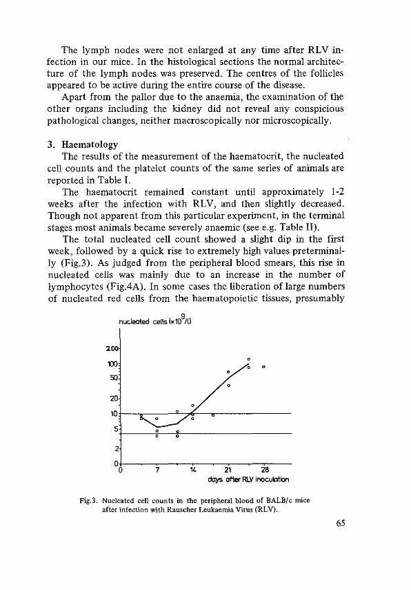
The lymph nodes were not enlarged at any time after RLV infection in our mice. In the histological sections the normal architecture of the lymph nodes was preserved. The centres of the follicles appeared to be active during the entire course of the disease.
Apart from the pallor due to the anaemia, the examination of the other organs including the kidney did not reveal any conspicious pathological changes, neither macroscopically nor microscopically.
3. Haematology The results of the measurement of the haematocrit, the nucleated
cell counts and the platelet counts of the same series of animals are reported in Table I.
The haematocrit remained constant until approximately l-2 weeks after the infection with RLV, and then slightly decreased. Though not apparent from this particular experiment, in the terminal stages most animals became severely anaemic (see e.g. Table H).
The total nucleated cell count showed a slight dip in the first week, followed by a quick rise to extremely high values preterminally (Fig.3). As judged from the peripheral blood smears, this rise in nucleated cells was mainly due to an increase in the number of lymphocytes (Fig.4A). In some cases the liberation of large numbers of nucleated red cells from the haematopoietic tissues, presumably
200
m 50
9 nucleated cells (x10 ill
0
0 0 0
0
20
10t---.~:--__.__,..::---.,-------
5.t--~r-~----------------
7 14 21 28 clays after RLV inocublion
Fig.3. Nucleated cell counts in the peripheral blood of BALB/c mice after infection with Rauscher Leukaemia Vuus (RLV).
65

A
B
',(,.' /.>
'c~. , •.
Fig.4. Peripheral blood of BALB/c mouse infected with Rauscher Leukaemia Virus 18 days previously. A. lymphocytes and large mononuclear cells (MGG x 800) B. nucleated red cells (MGG x 450).
66
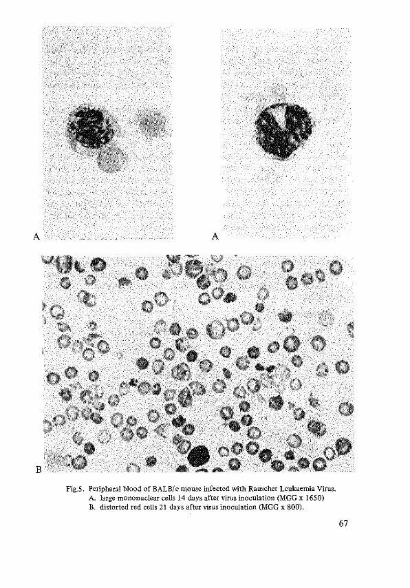
A A
B
Fig.5. Peripheral blood of BALB/ c mouse infected with Rauscher Leukaemia Virus. A. large mononuclear cells 14 days after virus inoculation (MGG x 1650) B. distorted red cells 21 days after virus inoculation (MGG x 800).
67

the spleen, accounted for a substantial contribution to the total nucleated cell count (Fig.4B). Especially in these cases many distorted erythrocytes were seen, resembling the schistocytes and burrcells of human microangiopathic haemolytic anaemia or disseminated intravascular coagulation (Fig.5B).
In the second week after RL V many large mononuclear cells were discernable in the peripheral blood, characterized by a big round or oval nucleus with a loose chromatin structure and abundant clear blue cytoplasm, sometimes containing a few purple granules (Fig.5A). In subsequent smears these cells were not detected in appreciable numbers. Perhaps these cells are the same as those described by Hopkins and Siegel (1965) who regarded them pathognomonic for virus-induced leukaemias. Since these cells disappeared in the course of the disease they must probably be regarded as stimulated lymphocytes, inherent to the viral infection and should not be regarded as typical Rauscher leukaemia cells.
The platelet count dropped in this series to approximately I 0% of the initial value, the decrease starting in the second week (Fig.6). The occurrence of splenic haemorrhages might in part be due to the severe thrombocytopenia. However, haemorrhages usually were not found at other sites in the leukaemic mice, even when sacrificed moribund.
tt-ro"rtlocytes (x 1:? II)
•
• •
• 100 •
• 50 •
20 o+-----.-----r----.-----.---------------
0 7 14 21 28 days after RLV inoculation
Fig.6. Platelet counts in the peripheral blood of BALB/c mice after infection with Rauscher Leukaemia Virus (RLV)
68 Normal range indicated by horizontal lines.

Conclusions From the observation of the natural history of Rauscher disease
in BALB/c mice the following conclusions may be drawn: I) In BALB/ c mice Rauscher virus induces erythroblastosis with accumulation of immature cells of the erythroid series in the spleen, the liver and eventually in the bone marrow, accompanied by anaemia, thrombocytopenia and general deterioration, culminating in death. 2) Splenic rupture was not the common cause of death. 3) Granulopoiesis in BALB/ c mice remained active throughout the course of the disease; there were, however, no indications of myeloid leukaemia as reported by Boiron e.a. ( 1965) and by Seidel ( 1971 ). 4) Lymphatic leukaemia was never seen in BALB/ c mice, although sometimes very high lymphocyte counts were found in the peripheral blood; this is in agreement with the experience of Boiron e.a. (I.e.). 5) Thrombocytopenia could not be attributed to a lack of megakaryocytes, neither in the early nor in the later phase of the disease, as has been concluded by Brodsky e.a. (1968) from their observations.
B. Influence of the dose of RL V The incubation period of a viral disease is the time required for
the multiplication and spread of the virus in the infected individual until so many cells are involved that signs of the disease become manifest. High doses of virus, therefore, need a shorter period of latency than lower doses, provided that a greater number of target cells are attained by the larger inoculum. For Rauscher disease this was checked in the following experiment:
30 BALB/c mice, 4 weeks of age, were infected by i.p. injection of 0.2 ml of the undiluted virus extract; in a parallel series of 30 mice of the same strain and age 0.2 ml of a J0-1 dilution of the extract was injected. At day 3, 7, 10, 14, 17 and 21, groups of 4 mice of each series were sacrificed after withdrawal of blood for cell counts, and their spleens were weighed.
Dilution of the virus preparation to I o-2 and I o-3 resulted in very long incubation periods, making systematic studies unfeasible. Still smaller amounts of virus seemed to cause no detectable leukaemia at all.
69

Results: By a I O-f old dilution of the virus the onset of splenomegaly was delayed for ± 7 days (Fig. 7). The doubling time of the spleen weight was slightly shorter for the higher than for the lower dose: ± 3.5 days respectively. The rise of the peripheral nucleated cell count was delayed for a few days by the dilution of the viruspreparation (Fig.8). The same phenomenon was observed in the changes of the platelet counts (Fig.9).
70
spleen weight (mg)
31Xll 2000
11Xll
500
200
100
o+-----.-----.----,----------0 7 14 21
days after RLV inoculation
Fig.7. Spleen weights of BALB/c mice after infection with the undiluted and a 10-fold diluted preparation of RLV. Each point represents the mean of 4 spleens; standard deviation indicated by vertical bar.
o--o 0.2 ml RLV undiluted ---• 0.2 ml RLV, 10-1
nucleated cells
100
50
20
10
5
1109/1 I
2
1+-----.-----.-----.--0 7 14 21
days after RLV-inoculation
Fig.8. Nucleated cell counts in the peripheral blood of BALB/c mice infected with an undiluted and a 10-fold diluted preparation of Rauscher Leukaemia Virus (RL V). Each point represents the mean of 4 mice.

o-----o 0.2 ml RLV undiluted •---_. 0.2ml RLV.10-1
thrombocytes 110'11)
1000
500
200
100
50
2
1+----.~---.----.--0 7 14 21
days after RLV-inoculation
Fig.9. Platelet counts in BALB/c mice after infection with an un~ diluted and a lO~fold diluted preparation of RLV. Each points represents the mean ±S.D. of 3-4 mice.
Conclusions: The delayed onset of splenic growth suggests that less target cells are affected by the smaller inoculum of virus and that multiplication of virus and subsequent infection of more target cells before the overt manifestations of the disease occur, rather than the proliferation of a fixed number of transformed cells from the outset. It is not clear why the splenic enlargement is delayed for a longer period than the changes of the platelet and nucleated cell counts. Perhaps, the number of test animals was too small to attach much importance to the difference.
C. Rauscher leukaemia in C57BL mice Boiron e.a. (1965) have shown that the same Rauscher virus pre
paration can induce erythroblastosis in BALB/c mice and myeloblastic leukaemia in C57BL mice. In contradistinction, Ishimoto and Maeda ( 1970) describe only lymphomas in this strain of mice after infection with Rauscher virus.
To compare our virus with that used by the cited authors, a litter of C57BL mice was infected neonatally with RLV by intraperitoneal injection. The animals were kept together and sacrificed when organs became palpably enlarged. Eventually 6 mice could be evaluated, respectively 183, 197,270 en 291 days after infection.
71

Results: Surprisingly, three different types of leukaemia were encountered: 1) Two mice were sacrificed 183 days after infection because ab
dominal masses could be palpated. The leukocyte count was 80 x 1 o9 /1.; in the peripheral blood smear mature granulocytes predominated: 84%; young myeloid cells B>%, lymphocytes 6%, monocytes 2% and smudge cells 6\>%. Haematocrit 37.5%. Besides the splenomegaly there also was an enlarged liver and a large mesenteric lymph node. Other lymph nodes were only moderately enlarged; the thymus was normal in size. The cut surface of the abdominal "lymphoma" was yellowish-green; the imprints showed predominantly mature granulocytes. Granulocytic infiltrations were seen in other lymph nodes and in the liver. The bone marrow was hypercellular and here, too, mature granulocytes prevailed. Remarkably, many megakaryocytes were found among the granulocytes in all organs. The disease should be classified as a chloroma-like granulocytic leukaemia.
2) Two mice sacrificed 197 days after the infection presented with splenomegaly, hepatomegaly, generalized lymph node enlargement and a huge thymus. The peripheral leukocyte counts were 30 and 106 x 1 o9 /1 consisting virtually entirely of lymphocytes. Lymphocytes, too, were the sole cell type in lymph nodes and thymus; the liver was heavily infiltrated with lymphocytes and in the spleen these cells predominated as well. Obviously, these animals had a lymphatic leukaemia.
3) The animals sacrificed at day 270 and 291 had splenomegaly, hepatomegaly and mesenteric lymph node enlargement. The thymus seemed to be normal. The blood picture was characterized by a leukocytosis with many immature granulocytes. The enlarged organs were stuffed with the same type of young myeloid cells, mainly myelo blasts and promyelocytes. Besides, in the spleen many foci of erythroblasts were seen. These animals had a myeloid leukaemia, but differing from that observed in the first group of mice with respect to the immaturity of the cell population.
Conclusion: Although an exhaustive analysis could not be carried out an important conclusion might be drawn from these observations, viz. the ability of RL V to induce granulocytic leukaemia in
72

C57BL mice. It is not warranted to ascribe the lymphatic leukaemia observed in two mice in this series to the injected virus, because lymphatic leukaemia is a naturally occurring disease in this strain of mice. In BALB/c mice, however, lesions comparable to those in the C57BL mice suffering from granulocytic leukaemia never were found. This is an argument against the leukaemic nature of the neutrophilia and the myeloid hyperplasia found in the erythroblastic phase of Rauscher disease in BALB/ c mice.
D. Rauscher disease in splenectomized mice Dunn and Green (1966) noted an intensification of the hepatic
involvement in erythroblastosis if BALB/c mice were splenectomized prior to RLV infection. Erythroblast foci were also found in lymph nodes.
Yokoro and Thorell (1966) studied Rauscher disease in splenectomized mice of a different strain of mice (NMRI). They demonstrated that the development of a leukaemic blood picture and hepatomegaly could not be prevented by splenectomy. The progression of the erythroblastosis appeared to be Jess fulminant than in intact animals and early mortality due to splenic rupture was of course eliminated.
To study the role of the spleen in the natural history of Rauscher disease in BALB/c mice a group of 6 mice was infected with the virus immediately after splenectomy had been performed at the age of 5 weeks. In all animals the leukocyte count and the haematocrit were followed until death occurred spontaneously. A group of 6 nonsplenectomized mice served as controls. Blood was withdrawn bi-weekly by cutting the tail under aether anaesthesia; histology of the dead animals was performed routinely.
Results: The most striking differences between the two groups (Table II) were seen in the rate of development of a leukaemic blood picture and in the survival time. The extensive liver involvement was also a feature of the splenectomized mice; presumably this depended on the protracted course of the disease, allowing the leukaemic infiltration to proceed in this organ.
In both groups the haematocrit might have been influenced by the repeated withdrawal of blood from each animal, the more so as in some animals the tail wound bled profusely. Nevertheless, after a
73

Table II. Mean values of nucleated cell counts and haematocrit measurements of splenectomized BALB/c mice infected with RLV compared with non-splenectomized control mice, infected at the same time.
Intact Splenectomized and infected controls infected mice
Day after Nucleated cells Haemotocrit Nucleated cells Haemotocrit infection (x106/l)
(%) (x 106/1)
(%)
4 9,700 39.5 15,400 ----7 8,000 40.3 11,400 37.4
11 6,600 35.0 6,780 35.8
18 70,300 26.1 8,980 31.8
21 42,800 29.0 12,800 27.4
25 17,000 26.5 10,300 23.3
28 10,900 23.1
32 21,000 26.4
35 59,120 25.9
39 121,000 22.5
46 124,100 26.0
53 209,000 27.5
60 171,000 29.0
67 286,000 33.5
74 146,000 ----
severe drop in the haema tocrit in the splenectomized mice in the first three to four weeks the animals seemed to recuperate from the anaemia to a certain extent. Independently, the total nucleated cell count rose steadily, probably reflecting the proliferation of leukaemic cells in the haematopoietic organs. At postmortem examination of the splenectomized mice especially the enormous liver was striking, invaded by greyish-white leukaemia tissue. The other organs were not invaded by tumour cells. The thymus was not enlarged.
Microscopical examination of the liver confirmed the extensive infiltration of the foreign cells in the liver parenchyma. Sinusoids,
74

central veins and portal tracts were crowded with primitive cells, resembling those seen in the non-splenectomized mice, in addition to more differentiated erythroblasts.
The bone marrow appeared to be crowded with cells; both erythropoieses and myelopoiesis were abundant and megakaryocytes were present as welL Apart from some fields containing many young myeloid elements, the maturation of the different cell lines seemed to be normaL Especially in the cytological preparations the number of lymphocytes was striking.
Conclusions: From these experiments the following conclusions may be drawn: I) the presence of the spleen is not essential for the induction of erythroblastosis in mice, although in splenectomized mice it takes more time for the virus to kill the host; 2) in the absence of the spleen the tumour cells accumulate preferentially in the liver; 3) the presence of large masses of tumour cells in the liver sinusoids is independent of the presence of the spleen; these cells are not necessarily transported from there to the liver, as has been argued by Siegler (1968); 4) the haematocrit does not reflect the growth of the leukaemic cell mass, which is an argument against the hypothesis of Brodsky e.a. (1969) who associate the changes in the ferrokinetics with the progression or regression of Rauscher erythroblastosis.
D. The role of the bone marrow The development of Rauscher erythroblastosis in mice without
spleens indicates that target cells for the virus are present in other organs, as welL Thomson (1969) and Yokoro e.a. (1966) have suggested that bone marrow cells are affected primarily by the erythroblastosis virus.
Lethal irradiation prevents the replication of erythroblastosis virus by the eradication of haematopoiesis (Axelrad and Steeves 1964). Extracellular virus is rapidly inacticated at body temperature, so that propagation of infection must result from virus replicated within target cells of the host. Shielding of part of the haematopoietic organs during otherwise lethal irradiation may preserve enough target cells for the virus to secure its survivaL
75

In the following experiment one hind leg of BALB/ c mice was shielded during irradiation with an otherwise lethal dose in order to investigate the presence of target cells in the bone marrow.
40 BALB/ c mice, I 0 weeks of age, were irradiated after anaesthesia with nembutal. One hind leg was shielded for part of the irradiation period so as to reduce the dose of radiation to the extremity to 100-500 rads whereas the remainder of the body received 700 rads. The same day 0.2 ml of undiluted RLV extract was injected intraperitoneally. After 21 days the mice were killed and their spleens weighed.
Results: The mice developed splenomegaly which varied inversely to the dose of radiation delivered to the hind leg (Fig. I 0).
spleen weight
400 (mgl (10}
300 (8} I I
200 (5}
I 100
(3}
I 0
500 400 300 200 dose of radiation to one leg (reDs)
Fig.lO. Spleen weights, measured 21 days after whole body irradiation with shielding of a hind limb and subsequent infection with RLV. Number of mice in each dose-group indicated between brackets. Vertical bars= standard deviation.
Conclusion: The fact that splenomegaly developed in these animals in spite of the irradiation of the liver and the spleen confirmed that the virus may meet its target elsewhere, apparently in the bone marrow. Presumably, these infected cells migrate to the spleen and the liver.
The negative correlation between the dose of radiation received by the bone marrow and the size of the spleen 21 days later suggests that the target cells for the virus are sensitive to irradiation, which prevents the replication of the virus, or that the splenomegaly depends on the number of cells capable of migrating to the spleen, or both. 76

F. Transplantation studies In contrast to the lymphocytic leukaemia induced by Rauscher
(1962), the initial erythroblastic disease induced by the same virus cannot be transplanted in the classical sense, i.e. by continuous proliferation of the transferred cells. If viable cells of the enlarged erythroblastic spleens are injected subcutaneously into uninfected mice of the same strain, typical Rauscher disease ensues, which, however, seems to be virus-born and not caused by proliferation of transplanted cells. The incubation time is similar to that observed after virus injection, no local growth can be seen at the site of deposition of the cells and the disease starts in the spleen and is restricted to the haematopoietic organs, mainly the spleen and the liver, just like the primary, virus-induced disease (Dunn e.a. 1966).
Although these transplantation studies have been carried out in syngeneic mice, the possibility of an immunologic rejection of the transplanted cells has to be considered notwithstanding the impaired immune system in Rauscher virus-infected mice (Siegel and Morton 1966, Hiiyry e.a. 1970). Virus-induced antigenic alterations of the cellular surface might behave as transplantation antigens, which might elicit an immunological response in the host, severe enough to kill the cells bearing these antigens.
An alternative possibility for the disappearance of the local deposits of subcutaneously injected leukaemic spleen cells might be the maturation and transportation of the cells. In this case the splenic onset could be explained by the migration of stem cells to the spleen. To rule out these possibilities the following experiments were carried out.
I. To exclude the possibility of a potent new histocompatibility antigen present upon the membrane of the spleen cells of Rauscher virus-infected mice, transplantation of these cells was performed to sublethally irradiated mice. One year old recipients were selected because they were thought to be less susceptible to the virus than younger mice.
Five female BALB/ c mice, one year of age, received 200 rads whole body irradiation. The same day approximately 108 leukaemic spleen cells were injected subcutaneously into the neck region. The mice were observed until leukaemic.
77

Results: The first mouce died spontaneously 32 days after the spleen cell transplantation with a grossly enlarged spleen; no local tumour could be found. The other test animals were sacrificed 7 weeks after the transplantation, because in all an enlarged spleen could be palpated. In the neck region local tumours could not be palpated throughout the observation period. Dissection of the site of injection revealed no foreign cell deposits. The spleens were massively enlarged and the microscopic aspect of the spleen, the liver and the other organs was similar to that of virus-infected younger animals.
Conclusion: Assuming that the sublethal irradiation was a sufficient immunosuppression for the virus-infected mice, the absence of local growth of transplanted leukaemic spleen cells cannot be ascribed to virus-induced histocompatibility antigens. In this experiment migration of cells to the spleen and liver and proliferation of transported cells within these organs could not be excluded. Therefore a comparable experiment was carried out with the use of a chromosome marker to permit the determination of the origin of the proliferating spleen cells in the leukaemic recipients.
II. In order to rule out the possibility that the transferred leukaemia was due to the migration of transplanted stem cells into the haematopoietic organs of the recipients, in the present experiment the spleen cells were injected intravenously in maximally immunodepressed animals (350 rads whole body irradiation). A donor-recipient combination was selected which differed only in the presence of a marker chromosome. The FJ hybrids of BALB/c x CBA/T6T6 were shown to be susceptible to the Rauscher virus; splenomegaly developed in these mice like in BALB/ c mice.
Cell suspensions were prepared of three enlarged spleens, weighing 800-1000 mg, obtained from BALB/c mice infected with RLV 14 days previously; 106 viable cells were injected intravenously into female F 1 (BALB/ c x CBA/T6T6) mice, who had received 350 rads whole body irradiation. The mice were observed until the spleens were palpably enlarged.
After an interval of 46 days spleens were palpable and three mice with spleen weights of 500-900 mg were sacrificed. The chromosome constitution of the spleen cells was determined (Fig. II).
78

recipient: BALB/c x CBA/T6T6 donor: BALB/c
.e. I 350 rads. 5x1o6 viable spleen
//1\\ ./? I ~--_j
cells Lv.
I (4-6 wks)
j
~
Fig.ll. Transplantation of leukaemic cells into sublethally irradiated coisogenic recipients with a different chromosome pattern. Experimental design for the determination of the origin of the proliferating spleen cells by chromosome analysis.
Results: In the squash preparations a total of 58 meta phases were suitable for analysis. In each of these 58 metaphases the T6-marker could be recognized (Fig.l2).
Fig.l2. Metaphase showing T6 marker chromosome (arrow) in spleen cell of female F 1 (BALB/ c x CBA/T6T6) in which leukaemia developed after injection of leukaemic spleen cells of BALB/c mouse.
79

Conclusion: The cells in mitosis must have been derived from the recipient and not from the donor, whose cells had a normal karyogram without chromosomal marker.
III. The presence of a minority of donor cells within the recipient spleen could not be excluded. For this reason the experiment was repeated, slightly modified: donors were FJ hybrids of BALB/c x CBA/T6T6, infected with a low dose of virus (0.3 ml w-1 dilution i.p.) at the age of 13 weeks and sacrificed II weeks later when the spleens were very large. Spleen cell suspensions were made and washed in Hanks BSS to remove excess virus. After centrifugation the supernatant was discarded and the pellet resuspended in Hanks' BSS to a concentration of 2.5 x 10⁸ cells/mi. Of this cell suspension 0.5 ml was injected in 6 F1 hybrids of BALB/c x CBA/Rij, which had been irradiated with 350 rads whole body irradiation one day before. Each recipient received 1.25 x 10⁸ cells i.p. Two month later chromosome analysis was performed of the spleen cells of 4 recipient mice with splenomegaly.
Results: Many metaphases were present in the squash preparations; 100 metaphases containing 40 chromosomes were screened for the presence of a T6 marker chromosome. This was detected in 6 out of I 00 metaphases.
Conclusion: The fact that only 6% of the metaphases was of donor karyotype indicates that more than 90% of the proliferating spleen cells consisted of secondarily infected host cells. Only a small number of donor cells contributed to the splenomegaly. The bulk of the injected cells had disappeared. It may be concluded, that the majority of the cells in the leukaemic spleens are not capable of sustaining an autonomous proliferation upon transplantation, even in immunosuppressed syngeic or co-isogenic recipients. By inference, the leukaemic cell population in the primary infected animal is probably not self-maintaining. Presumably, a constant influx of newly recruited precursor cells is needed to preserve or - a fortiori - to increase the number of leukaemic cells and to produce splenomegaly. The primitive erythroid cells populating the spleen and the liver of the leukaemic animals either mature without multiplication or divide slowly after transplantation into sublethally irradiated recipients so
80

that mitoses belonging to the transplanted leukaemic cell population could not be found.
If sufficient stem cells are present in the inoculum the appearance of the progeny of these stem cells in the haematopoietic organs -and in the chromosome preparations - can be anticipated. The donor karyotypes detected among the spleen cells of mice, which received 108 leukaemic spleen cells presumably are derived from injected stem cells. The leukaemia thus is not transplanted in the common sense, but rapidly propagated by spread of the virus in the haematopoietic tissue of the recipient.
The influence of erythropoietin on the evolution of Rauscher erythroblastosis
Endogenous erythropoietin production is depressed by hypertransfusion. To evaluate the influence of erythropoietin on the splenogemaly of Rauscher erythroblastosis packed red blood cells were injected intraperitoneally in BALB/ c mice before and after inoculation with RLV. Assuming that the observed effects were due to the depression of erythropoietin levels, it was attempted to abrogate these effects by the administration of exogenous erythropoietin.
A control group of I 0 BALB/ c mice and three experimental groups of 5 animals each received RLV at day 0. Packed cells were given either 6 and 3 days before or 2, 4 and 7 days after the infection.
Erythropoietin (Anaemic sheep erythropoietin, step III, Connaught, Toronto) was administered in experiment III by intraperitoneal injection of 5 U per mouse at day 8, I 0 and 12.
The experimental set up is depected in Figure 13.
Results: As is shown in Table III, the results suggest that the development of splenomegaly could be impeded by blood transfusions, whether given before or after the infection with RLV and that erythropoietin promoted the splenic growth in mice in which the splenomegaly was previously retarded by blood transfusions.
Conclusions: Theoretically, if measures applied to the recipient animals before the injection of RLV influence the course of the subsequent disease, their action could be attributed to the change in
81

82
'" Numb.< 05 ml 10-l of mico
1 " Con~ls
'" v 'Y v
EKp. I .. _,
v hp. II
'Y'l'l • '
v Exp. Ill 'Y 'Y ' .. _,
T
"· "· "· ++.J. ' '" "
'\l • 0 5 ml packed reel eollo i.p,
EP, • 5 U oryth<Oj>Oi<tin i.p.
"
"
"
t
" Fig.13. Schematic representation of the experiments performed in order to
investigate the influence of blood transfusions and erythropoietin on the spleen weight 21 days after infection with RLV.
Table III. The influence of hypertransfusion before (experiment 1), and after (experiment II) infection with Rauscher leukaemia virus upon the spleen weight; cf. fig.13.
Spleen Weight (mg)
day 8 day 21
Controls 202 + 75' 2156 + 402' - -
Exp. I 699 + 575' -
Exp. II 1169 + 796' -
Exp. Ill 1662 + 501' -
* mean + S.D.

the number of target cells available for the virus. Thus, hypertransfusion of mice before the inoculation with RLV might inhibit the splenic enlargement by the decrease of the number of early erythroblasts which are present at the moment of infection. Indeed, this was suggested by Pluznik e.a. ( 1966), who found a lower number of virus-induced spleen foci if blood transfusions were given to the recipients before the virus.
An alternative explanation is provided by the assumption that the effect of the hypertransfusion lasts longer than the few days between the injection of blood and the inoculation of virus. Presumably, the hypertransfusion will depress the production of endogenous erythropoietin for some more days. This might interfere with the recruitment of differentiated erythroid cells during the first few days after the virus inoculation, preventing the formation of virus-induced foci and the subsequent enlargement of the spleen.
The validity of this hypothesis is strengthened by the observed effect of the blood transfusions administered after the inoculation of RL V. Thus, it is possible to influence the course of Rauscher disease even after the virus had reached its target. Dunn e.a. (1966) could prolong the survival of RLV-infected BALB/c mice for several months. The last experiment (Ill) in our series indicates that this inhibitory effect of blood transfusions can be neutralized by the administration of exogenous erythropoietin. The erythroblastosis thus appears to be hormone-dependent.
83

REFERENCES
Axelrad, A.A. and R.A. Steeves (1964): "Assay for Friend leukemia virus: rapid quantitative method based on enumeration of macroscopic spleen foci in mice". Virology 24: 513.
Boiron, M., J.-P. Levy, J. Lasneret, S. Oppenheim (1965): "Pathogenesis of Rauscher leukemia". J. Nat. Cancer Inst. 35: 865.
Brodsky, 1., E.M. Ross, S.B. Kahn, G. Petkov (1968): "The effect of a leukemia virus on thrombopoiesis". Cancer Research 28: 2406.
Brodsky. I., and S.B. Kahn (1969): "Effect of a leukemia virus (RV) on erythropoiesis". J. Nat. Cane. Inst. 42: 39.
Chi, C.H. and B. LagerlOf (1968): "Effects of radiation damage to bone marrow on susceptibility of chicks to erythroleukemia virus". Acta. Rad. 7: 353.
Dunn, Th.B. and A.W. Green (1966): "Morphology of BALB/c mice inoculated with Rauscher Leukemia Virus". J. Nat. Cane. Inst. 36: 987.
Dunn, Th.B., R.A. Malmgren, P.C. Carney, A.W. Green (1966): "PTU and transfusion modi~ fication of the effects of Rauscher virus in BALB/c mice". J. Nat. Cane. Inst. 36: 1003.
Hopkins, J.C. and B. V. Siegel (1965): "Occurrence of abnormal cell in murine virus~induced leukemia". Acta Haemat. 33: 159.
lshimoto, A. and M. Maeda (1970): "Studies on the susceptibility of c57BL/6 mice to Rauscher virus. I. Properties of Rauscher virus~induced c57BL/6lymphomas". J. Nat. Cancer Inst. 44: 361.
Pluznik, D.H., L. Sachs, P. Resnitsky (1966): "The mechanism of Leukemogenesis by the Rauscher Leukemia Virus". Nat. Cane. Inst. Mono 22: 3.
Rauscher, F.J. (1962): "Virus~induced disease of mice characterised by erythropoiesis and lymphoid leukemia". J. Nat. Cancer Inst. 29: 515.
Seidel, H.J. (1971): "Target cell characterization for Rauscher leukemia virus in vivo". In: Proceedings V. International Symposium on Comparative Leukemia Research. Padova 1971. In the press.
Siegler, R. and M.A. Rich (1964): "Comparative pathogenesis of murine viral lymphoma". Cancer Research 24: 1406.
Siegler, R. (1968): "Pathology of murine leukemia". In: "Experimental leukemia". M.A. Rich, Ed.: North Holland Publishing Company Amsterdam. Appleton~
Century-Crofts~ New York 1968, p.86. Thomson, S. and A.A. Axelrad (1968): "A quantitative spleen colony assay method for
tumor cells induced by Friend Leukemia Virus infection in mice". Cancer Research 28: 2105.
Thomson, S. (1969): "A system for quantitative studies on interactions between Friend Leukemia Virus and hemopoietic cells". Proc. Soc. Exp. Biol. Med. 130: 227.
Yokoro and Thorell (1966): "Cytology and pathogenesis of Rauscher virus disease in splenectomized mice". Cancer Res. 26: 536~543.
84
CHAPTER V
THE STEM CELL IN RAUSCHER ERYTHROBLASTOSIS
From the available literature no defmite conclusions could be drawn about the involvement of the pluripotent stem cell in Rauscher Leukaemia. In this study, therefore, efforts were made to perform a quantitative estimation of the number of splenic stem cells by the spleen colony assay of Till and McCulloch ( 1961 ), to check the functional integrity of the stem cells, and to look for the presence of virus within the pluripotent stem cell.
Stem cell assay In lethally irradiated mice erythroblastosis virus survives only
shortly and no spleen foci are formed (Axelrad and Steeves, 1964). The virus injected together with the leukaemic spleen cell suspensions will not exert influence upon the colony assay by adding virusinduced foci which might resemble spleen colonies. In the following experiment this was confirmed.
In 4 groups of I 0 female BALB/ c mice, I 0 weeks of age, irradiated with 700 rads, dilutions of a normal spleen cell suspension were injected without and with RLV. For this purpose two suspensions of normal spleen cells were prepared containing 2 x 10⁵ and 5 x 10⁵ cells per 0.5 mi. Just before the injection of these suspensions into the irradiated recipients, to one half of each suspension RLV was added to a final concentration of 10-l. Each group of mice received either 2 x 10⁵ or 5 x 10⁵ spleen cells with or without RLV. Spleen colonies were counted after I 0 days. Because many mice died spontaneously before the tenth day, the experiment was repeated. The results of both series are given in Table IV.
85

Table IV. Spleen colony assay of normal spleen cell suspensions without and with the addition of RLV in vitro. The results of two separate experiments ("a" and "b") are reported; n =number of spleens in which colonies have been counted.
Number of cells injected Number of colonies counted without and with RLV n per spleen (mean)
a) 2 X 10⁵ 3 2.0
b) 2 X 10⁵
10 1.1
a) 5 X 10⁵
6 6. I
b) 5 X 10⁵ 9 2.2
a) 2x 10⁵ +RLV 1.0
b) 2 X 10⁵ + RLV 5 2.2
a) 5 X 10⁵ + RLV 3 3,6
b) 5x 10⁵ +RLV 4 4,5
CFU per 106
injected spleen cells
10.0} 7.-f' 5.5
12.2} 8.3
4.4
5.0} 8.0
11.0
7.2} 8.1
9.0
Results: In both experiments the number of spleen colonies counted in the mice which received virus differed only slightly from the number of colonies counted in the control group. In the first experiment (a) the difference was in favour of the controls (without RL V), in the second (b) in favour of the group which received virus. It was concluded that the method could be applied in Rauscher leukaemia to measure the colony forming units (CFU).
In order to follow the changes in the number of colony forming units in the course of the induction and the establishment of Rauscher erythroblastosis, the number of CFU's present in the spleen was estimated at various intervals after the infection of BALB/ c mice with Rauscher Leukaemia Virus (RL V).
Two procedures were followed. In the first, all donor animals were infected with RL V simultaneously and at various intervals thereafter spleen colony assays were performed of spleen cell suspensions of small groups of infected mice. In the second procedure, the
86

donors were infected with RLV at various intervals before their sacrifice and the colony assays were performed simultaneously for all groups. The results of both experiments were similar.
I. Female BALB/c mice, 4 weeks of age were infected with 0.2 ml of the undiluted RLV preparation; a few hours later the first group of donors were sacrificed for splenic CFU estimation. Twice a week 3-6 mice were killed for the performance of a spleen colony assay as described in Chapter Ill. The experiment was repeated several times. Cell suspensions were made either of pooled spleens (A) or of single spleens (B).
The colony counts obtained in these experiments are shown in Table V, experiment rA and JB.
Table V. Number of colony forming units per 106 spleen cells of BALB/c mice infected with RLV, determined at various intervals after the infection. Between brackets the number of spleens in which the colonies were counted.
Days after Rl V-Experiment lA Experiment 1
8 inoculation Experiment II
of donors (pooled spleens) (single spleens) (pooled spleens)
0 75 100 - 31 29 -(13) (/8) ( 8) ( 8)
1 - - - - - 93 (15)
3-4 60 29 47 39 27 99 ( 9) (15) (12) (17) ( 8) (18)
7-8 66 - - - - -(14)
10-11 - 25 27 55 34 103 ( 8) (11) (17) (13) (15)
14-15 86 - - - - 61 (13) (15)
17-18 - - 31 25 - -(13) (29)
21-22 15 - 39 22 - 109 (12) ( 8) ( 4) (20)
28-29 39 - - - - -(20)
87
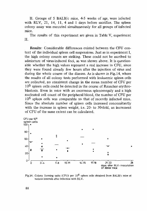
II. Groups of 5 BALBI c mice, 4-5 weeks of age, were infected with RLV, 21, 14, II, 4 and I days before sacrifice. The spleen colony assay was executed simultaneously for all groups of infected mice.
The results of this experiment are given in Table V, experiment II.
Results: Considerable differences existed between the CFU content of the individual spleen cell suspensions. Just as in experiment I, the high colony counts are striking. These could not be ascribed to admixture of virus-induced foci, as was shown above. It is questionable whether the high values represent a real increase in CFU, since they were found already few hours after the injection of virus and during the whole course of the disease. As is shown in Fig.l4, where the results of all colony tests performed with leukaemic spleen cells are collected, no c6nsistent change in the mean number of CFU per I o6 spleen cells could be detected in the course of Rauscher erythroblastosis. Even in mice with an enormous splenomegaly and a high nucleated cell count of the peripheral blood, the number of CFU per I o6 spleen cells was comparable to that of recently infected mice. Since the absolute number of spleen cells increased concomitantly with the increase in spleen weight, i.e. 20- to 30-fold, an increment of CFU of the same extent can be calculated.
CFU per 106 spleen celts 100 •
• BO • • • 60 •
40 • ~ • ~ • • • • •
20 • • • 0
0 3-4 7-B 10-11 14-15 17-1B 21-22 28 days after RLV -inoculation of donor mice
Fig.l4. Colony forming units (CFU) per 106 spleen cells obtained from BALB/c mice at various intervals after infection with RLV.
88

Histology of spleen colonies A histological analysis was performed of spleen colonies produced
in lethally irradiated mice by the injection of either normal or leukaemic spleen cells. Leukaemic spleen cells were obtained from mice, which were infected at least three weeks previously with a high dose of RLV, i.e. 0.2-0.5 ml of the undiluted cellfree spleen extract.
One hundred colonies of both series were examined and the cell types constituting the colonies were determined. The percentage of colonies which consisted either of erythro blasts, of myelocytes and myeloblasts, of megakaryocytes or of a combination of these cell types was calculated.
Results: As can be seen from Table VI the colony forming cells of the leukaemic spleens could give rise to a variety of cell types. The
Table VI. Spleen colonies in lethally irradiated mice, 10 days after the injec~· tion of normal (N) and leukaemic (L) spleen cells, arranged according to the type of cells constituting the colony. Between brackets the number of colonies in each category apparently consisting of only one cell type.
Erythroid Myeloid Megakaryoti c Undetermined Total
N. 66 (20) 68 (20) 22 (3) 9 (4) 100
L. 71 (35) 51 ( ll) 24 (5) 9 (5) 100
similarity in distribution of the types of colonies produced by leukaemic and by normal spleen cells is striking. Among the colonies produced by leukaemic cells only a slightly greater percentage of erythroid colonies was found. It may be concluded that the stem cells in the leukaemic spleen retain their pluripotency, at least to a certain extent.
Repopulating capacity of leukaemic spleen cell suspensions In Rauscher erythroblastosis splenic stem cells not only can form
colonies of different histological types in the spleens of irradiated mice, but also are capable of repopulating the depleted haematopoietic organs in these mice and can build up a new blood forming system. This can be deduced from the observation that a sizeable
89

fraction of lethally irradiated mice survives for 30 days or more after the injection of spleen cells, obtained from the enlarged spleens of mice suffering from Rauscher erythroblastosis. Preliminary data even suggested that equal numbers of either leukaemic or normal spleen cells were required for the survival of 50% of the irradiated mice for at least 30 days (Brommer e.a. 1970). These experiments have been extended and the results could be reproduced. All data pertinent to the survival of lethally irradiated and restored mice were conformed to a pro bit curve, shown in Fig.l5.
From these lines it can be inferred that the bone marrow syndrome can be overcome by the restitution of leukaemic cells almost as effectively as with normal spleen cells. Thus, the conclusion seems warranted that the stem cells in the leukaemic spleens not only maintain their relative numbers but also retain their pluripotency: they can give rise to a variety of colonies and the distribution of the
%survival 99
o normal spleen eel ls • leukaemic spleen cells
90
70
50
30
10
106 1Q1
number of spleen cells injected
Fig.l5. Probit curves of the survival of BALB/c mice 30 days after lethal irradiation and restoration with leukaemic spleen cells versus normal spleen cells; 95% confidence limits indicated by horizontal bars.
90
histological types is almost normal; they are capable of restoring lethally irradiated mice almost as effectively as normal stem cells, suggesting that they may produce normal functioning end cells.
In order to investigate as to how far the stem cells escape the viral attack a method was selected to demonstrate the presence or absence of the virus from these cells.
Effect of anti-RLV antiserum on CFU's Attempts were made to demonstrate the presence of viral anti
gens upon the surface of the colony forming cells in the spleens of mice infected with Rauscher virus. For this purpose spleen cell suspensions were incubated with rabbit anti-RLV antiserum before the injection into irradiated recipients; if less colonies would arise from these incubated cells than from buffer-incubated controls, this would indicate the eradication of a corresponding number of colony forming cells, presumably by the interaction of the antiserum with virus-specific antigens upon the stem cell membrane. Analogous experiments have been performed in Friend disease by Steeves (1968), Thomson and Axelrad (1968) and Thomson (1969).
I. Donors were female BALB/c mice, inoculated with 0.5 ml of undiluted RL V at the age of 8 weeks and sacrificed 17 days later. Cell suspensions were prepared from 4 enlarged spleens; 4 normal spleens were processed in the same way. The cell suspensions were diluted in Hanks BSS to contain 107 viable cells per ml; 15 ml of each suspension were incubated for 45 minutes at 37°C with an equal volume of rabbit antiserum or with control medium. After incubation the suspensions were centrifuged for I 0 minutes at 1000 rpm. The supernatant was discarded and the pellets were resuspended in Hanks BSS so as to obtain cell concentrations of 5 x I o5 and I o5 cells per 0.5 ml, calculated from the initial cell counts. Of each suspension 0.5 ml was injected intravenously into 20 irradiated mice for a spleen colony assay. Ten days later the recipients were sacrificed and the spleen colonies counted.
Results: In a pilot study, unabsorbed rabbit sera were used. The rabbit anti-RLV antiserum appeared to be cytotoxic both for leukaemic and for normal mouse spleen cells (Table VII). In the control experiment with normal rabbit serum a toxic effect upon normal
91

spleen cells was noted. Remarkably, the majority of the leukaemic CFU's survived after the incubation with the same rabbit serum, suggesting that certain receptors were absent from the surface of the leukaemic cells. The experiment was repeated with rabbit sera absorbed with normal mouse spleen cells (Table VIII).
In these experiments, again, the high values of the colony counts obtained with leukaemic spleen cell suspensions are noteworthy.
Table VII. Cytotoxic effect of rabbit anti~RLV antiserum on the colony forming units of normal and leukaemic spleens, compared to the effect of the suspension medium and normal rabbit serum (non-absorbed sera! ).
colony forming units/ 106
spleen cells Incubation medium
normal spleen cells leukaemic spleen cells
Hank's balanced salt solution 18 87
normal rabbit serum 60
rabbit anti-RLV 0 0
Table VIII. Cytotoxic effect of rabbit anti-RLV antiserum on the colony forming cells of normal and leukaemic spleens, compared to the effect of suspension medium, non-absorbed and absorbed normal rabbit serum. Each figure represents the mean colony count in 20 spleens 10 days after grafting of the incubated cells in lethally irradiated recipients. (abs NMSC =absorbed with normal mouse spleen cells).
Incubation medium
Hank's balanced salt solution
normal rabbit serum
normal rabbit serum - abs NMSC
anti-RLV I
anti-RLV I - abs NMSC
anti-RLV II
anti-RLV 11 - abs NMSC
92
6 CFU/ 10 spleen cells
normal spleen cells leukaemic spleen cells
28 130
38
10
30
9
36
40
155
2
4
18
41

Rabbit anti-RLV antiserum proved to be cytotoxic for leukaemic spleen cells and to eradicate the CFU's from the cell suspensions effectively. Normal spleen cells were also killed by the rabbit antiserum, but this effect appeared to be due to a factor present in normal rabbit serum. Since normal rabbit serum absorbed with normal mouse spleen cells did not kill leukaemic spleen cells, the eradication of leukaemic CFU's by the anti-RLV antiserum could be ascribed to the specific action of thls antiserum. In this way indirect evidence was obtained for the presence of viral antigens upon the surface of the leukaemic stem cells. Moreover, other antigens had apparently disappeared from the cell membrane of the leukaemic spleen cells.
Conclusion: As for the susceptibility of the CFU to anti-RLV, Rauscher leukaemia is similar to Friend leukaemia, in whlch Steeves (1968) and Thomson (1969) demonstrated the abolition of the splenic "tumour colony forming cells" by anti-Friend virus antiserum. As stated before, these "tumour colony forming cells" possibly represent haematopoietic precursor cells. It is tempting to assume that the antigenic changes are the result of the viral activity within the genome of the colony forming cells. Thls would imply that the virus had been present withln the colony forming cells or in their precursors during one or a few cell divisions before the assay.
A remarkable finding was the cytotoxic effect of normal rabbit serum to normal mouse spleen cells. On the analogy of the cytotoxic action of rabbit serum to lymphoid cells of mice and rats (Palm 1961, Boyse e.a. 1970, Colley e.a. 1970) thls might be an antibody effect. Presumably, the altered sensitivity of the leukaemic spleen cells to the naturally occurring cytotoxic factor in rabbit serum is the consequence of the appearance of viral antigens upon the cellular membrane. The possibility that it constitutes an independent change can, however, not be dismissed. Thls would be another argument for the transformation of the haematopoietic stem cell under the influence of Rauscher virus.
93

REFERENCES
Axelrad, A.A. and R.A. Steeves (1964): "Assay for Friend leukemia virus: rapid quantitative method based on enumeration of macroscopic spleen foci in mice". Virology 24: 513.
Brommer, E.J.P. and P. Bentvelzen (1970): "Leukaemia-virus and haematopoietic cell differentiation in mice". Folia Med. NeerL 13: 136.
Boyse, E.A., L. Hubbaxd, E. Stockert (1970): "Improved complementation in the cytotoxic test". Transp/atation 10: 446.
Colley, D.G. and B.H. Waksman (1970): "Cytotoxic effect of normal rabbit serum on rat lymphoid cells". Transplantation 9: 395.
Steeves, R.A. (1968): "Cellular antigen of Friend virus induced leukemias". Cancer Research 28: 338.
Thomson, S. and A.A. Axelrad {1968): "A quantitative spleen colony assay method for tumor cells induced by Friend Leukemia Virus infection in mice". Cancer Research 28: 2105.
Thomson, S. (1969): "A system for quantitative studies on interactions between Friend leukemia virus and haematopoietic cells". Proc. Soc. Exp. Bioi. Med. 130: 227.
Till, J.E. and E.A. McCulloch (1961): "A direct measurement of the radiation sensitivity of normal mouse bone marrow cells". Rad. Res. 14: 213.
94
CHAPTER VI
THE RESTORED MOUSE
The bone marrow syndrome which develops after lethal irradiation can readily be overcome by the injection of suspensions of normal isogeneic bone marrow or spleen cells. Mice restored in this way are apparently able to cope with the weal and woe of life and have a normal life span. The formation of a new haematopoietic system out of the injected pluripotent stem cells reconstitutes the ability to combat bacterial infections, to prevent bleeding and to maintain a normal haemoglobin level.
If instead of cells from normal spleen or bone marrow, spleen cells are injected, derived from a donor mouse with a leukaemia-like disease - i.e. Rauscher erythroblastosis - not only leukaemic, nonfunctioning tissue proliferates, as might be expected, but also normal blood elements are formed. These blood cells ensure the survival of the recipients for at least a month, whereas the non-injected controls die within two weeks. (Chapter V).
In mice restored with leukaemic cells, however, viral infection is inevitable: washing the cells does not remove the virus because it is also localized within the cells and incubation with anti-RLV antiserum kills the stem cells and counteracts the repopulation of the haematopoietic organs. Thus, mice restored with leukaemic cells, develop erythroblastosis in due course. Nevertheless, these animals can live much longer than a month, as appears from the next experiment.
95

Survival 40 BALB/ c mice, I 0 weeks of age, irradiated with 700 rads whole
body irradiation, received I o6, respectively 5x I o5 cells intravenously, obtained from the enlarged spleens of 7 weeks old donor mice, which had been infected with RLV 21 days previously. The recipients were kept for approximately three months and the surviving animals were recorded daily. The results are depicted in Fig.l6. After 3 months the leukaemic survivors were in a very bad condition and for that reason all animals were killed.
700rads
0 %survival
40
20
············· 5.106 normal spleen cells -- 1~ leukaemic spteen cells ----- 5.10 leukaemic spteen cells
+ ........................................................ ] : ................................................ .
.. _1
' ' L----------------------------·
60 80 100 days
Fig.l6. Survival of BALB/c mice after lethal irradiation and restoration with nor~ mal and leukaemic spleen cells. The experiment was terminated when the condition of the animals was critical. * mice killed and used for another experiment.
As the curve of the percentage surviving mice shows, their chance to survive as long as 80 days is approximately 55%. Most animals died during the critical period between day 8 and 14, when also many mice treated with normal spleen cells die. Thereafter, they have a fair chance to survive 2 - 3 months. The cause of death will be studied below.
The long survival - as compared to primary infected intact animals - could theoretically be due either to a slow progression of the erythroblastosis or to a longer time-lag before a perceptible proliferation of leukaemic cells commences. This was excluded by the following experiments.
96

Spleen weight 75 BALB/c mice, 10 weeks of age, irradiated with 700 rads
whole body irradiation, were restored with either 106 or 5.106 spleen cells from a leukaemic donor mouse, infected with RL V 21 days before, or with I o6 normal spleen cells. The restored mice were kept in numbered cages in groups of 5. Twice a week one group was sacrificed in the order indicated by the numbers of the cages, and the spleens were weighed (Fig. I?).
5.'06 ieukcemic spleen cells spleen weigh!lmgl
20CIJ
1000
500
300 200
100 I
30 •
I
o+--.--.--.--.--.--.--r~ 0 4 7 10 14 17 21 24 2Bdays
1Qb 105 106
n.sc. l.bm.c. l.sc.
. + .ll'. -
• . •
30days after 700 rod W8l
Fig.17. Spleen weights of BALB/c mice restored after 700 rads whole body irradiation (WBI) with 5 x 10⁶ leukaemic spleen cells (curve), 105 normal spleen cells (n.s.c.), 105 leukaemic bone marrow cells (Lb.m.c.) or 106 leukaemic spleen cells (l.s.c.). Leukaemic cells were obtained from syngeneic mice infected with RLV 17 to 21 days prior to this experiment.
The spleen weight dropped to approximately one third of the initial value after the irradiation; whereas the spleen weight of mice restored with normal spleen cells returned to normal, the spleens of mice receiving leukaemic cells grew exponentially until a mean weight of ± 700 mg was attained at day 18. Thereafter the spleen weight remained constant for at least ten days, when the experiment was terminated.
Although the starting point was somewhat lower than in intact animals, there was no time-lag in the growth of the spleen. Neither
97

was a slower progression of the spleen weight noted, the slope of the curve being even steeper than that of the spleen weight of infected, unirradiated mice. It is not clear why there is a maximum at a weight of less than one gram. The following possibilities were envisaged: I) the number of stem cells in the irradiated mouse is limiting; 2) there is a greater demand for differentiated cells, distracting a proportion of the stem cells.
The first possibility was investigated by injecting different numbers of spleen and bone marrow cells.
Bone marrow and spleen cell suspensions were prepared from the organs of BALB/ c mice, inoculated with 0.3 ml of the undiluted RLV extract, resp. 14 and 21 days previously. Graded numbers of viable cells were injected in lethally irradiated recipients: 10 weeks old BALB/c mice; the radiation dose was 725 rads. After 30 days the surviving animals were killed and their spleens weighed (Table IX) and Fig. 1 7).
In these experiments no relation was found between the number of bone marrow or spleen cells injected and the spleen weight 30
Table IX. Spleen weight of BALB/c mice 21 days after 700 rads whole body irradiation and grafting of various numbers of bone marrow and spleen cells obtained 14, respectively 21 days after the infection of syngeneic donor mice with RLV.
Day after
Number ond origin RLV-
Number of mice Spleen weight in inoculation mg of injected cells of donors in jected/survi vi ng {mean)
5 1 0 bone morrow 14 12 I 4 880
21 lO I 4 775
3 • 105 .. 14 10 I 5 706
21 10 I 2 725 106 .. 14 10 I 6 860
6 10 spleen cells 14 10 I 400
21 lO I 5 1110
3 • 106 .. 14 10 I 6 550
21 10 I 0
Jl .. 21 10 I 4 850
98

days later; the maximal spleen weight was already attained with the lowest number of cells administered in this experiment. Consequently, the number of stem cells injected seems not to be limiting for the splenic growth, at least within the range of cell numbers studied.
The interval of time between the infection of the donor mice with virus and the use of their spleens for the preparation of the cell suspension had no influence upon the growth of the spleens in mice restored with the leukaemic spleen cell suspensions.
Conclusion: Three weeks after the reconstitution, the haematopoietic apparatus seems to be completely restored. Consequently, the withdrawal of cells for differentiation into other directions than the erythroid cell line is not a likely cause of the cessation of the growth of the spleen.
Presumably, the expanding leukaemic cell mass needs a constant influx of cells from a normal stem cell pool. As this stem cell pool is destroyed in irradiated mice and regains its original size only after a certain period of time, this might be a limiting factor in the increase of leukaemic cells in spleen and liver.
Remarkably, the mice restored with leukaemic spleen cells survived much longer than non-irradiated mice infected with RLV. Presumably, this long survival is associated with the cessation of spleen growth. However, notwithstanding this seeming arrest in the leukaemic proliferation, the animals became progressively ill and died after two or three months in a very bad general condition. The factors responsible for this deterioration will be considered below.
The presence of infective virus in the blood of the restored mice was investigated by the injection of plasma into weanling BALB/c mice. The rapid induction of erythroblastosis indicated that there was a high titer of virus in the plasma of the animals restored with leukaemic spleen cells. Another finding was that the injection of fresh virus preparations in to mice restored with leukaemic spleen cells did not accelerate the evolution of splenomegaly. Obviously, the virus titer in these animals does not constitute a limiting factor. Presumably all target cells in these animals have been in contact with the virus and other factors determine their fate.
Histology The injected leukaemic stem cells home in the haematopoietic
99

organs and form colonies just like normal stem cells. In the spleen, the colonies which have a varying cellular constitu
tion, coalesce after II to 14 days and populate the whole spleen, which soon exceeds its original size. Under the microscope an abundance of haematopoietic cells can be discerned, differentiated in all directions. Lymphocytes seem to return in the perivascular shealths, erythroblasts are seen dispersed in the red pulpa and there are areas where myelocytes and mature granulocytes predominate. Megakaryocytes also contribute to the repopulation of the spleen. The splenic capsule is often thickened by fibrosis.
In the liver, foci of foreign cells can be found which are mostly constituted of poorly differentiated cells with a bloated, round nucleus, probably primitive erythroid elements, and mature erythroblasts. In later stages, the liver parenchyma is heavily infiltrated with these cells as well as with myelocytes and some megakaryocytes.
All haematopoietic cell lines are abundantly represented in the bone marrow. Though in some preparations mainly erythroid elements are detectable, there is no evidence for a crowding out of the granulopoiesis. On the contrary, myelocytes and mature granulocytes often dominate the picture, especially in later stages, when the cellularity of the bone marrow is extremely high.
Table X. Haematocrit, nucleated cell counts and platelet counts (means ±S.D.) in the peri~ pheral blood of BALB/c mice at various intervals after 700 rads whole body ir~ radiation and restoration with leukaemic spleen cells. n =number of mice.
Day after Hoematocrit Nucleated cells Platelets grafting (%)
(x 106/1) (x 106 /1) n n n
7 5 46.5 ::': 2.8 5 1090 + 360 5 75,000 ::': 64,000 -14 4 25 + 9.0 4 670 + 360 3 15,000 ::': 4,300 -17 4 22 + 6.7 3 10,700 ::': 5100 2 27,000 ::': 6,400
21 3 33 + 8.7 3 30,000 ::': 14,700 3 46,000 ::':· 30,200
31 7 35.4 ::': 2.7 7 32,700 ::': 15,500 6 205,000 ::': 181,000
38 4 40.4 ::': 5.1 4 13,400 ::': 3700 4 90,000 ::': 30,900
49 3 35.6 ::': 9.1 2 41,400 ::': 30,600 4 53,000 ::': 12,500
61-77 5 40.6 ::': 6.5 2 91,500 ::': 41,000 -
100

Haematology From different experiments the data concerning the haematocrit,
total nucleated cell counts and platelet counts were collected to get an impression of the functional status of the haematopoietic apparatus at various intervals after the restoration (Table X). As judged from these figures, after a drop in the haematocrit in the first two or three weeks, a slight increase seems to occur in the fourth week, indicating some differentiation of the stem cells into the erythroid direction, resulting in functional end cells.
The leukocyte count recovers in the third week. Very high peripheral leukocyte counts are encountered at day 21. The peripheral blood in these restored animals did not differ from that in primary infected, non-irradiated mice.
The platelet count, too, showed a tendency to recover after leukaemic spleen cell transplantation. In a single experiment values were found, which did not differ much from those seen in mice restored with normal spleen cells. In most experiments, rather low values were found throughout the observation period.
Immunoglobulins In 4 mice restored 65 days previously with leukaemic spleen cells,
immunoglobulin levels in the plasma were estimated semi-quantatively by immunoelectrophoresis, agar gel-electrophoresis and the Wiemetechnique (Wieme 1959), kindly performed by Dr Radl, Institute for Experimental Gerontology, TNO, Rijswijk.
All immunoglobulin levels appeared to be depressed substantially as compared to normal controls (Fig.l8) There were no M-components present, as have been found in a human baby suffering from a severe immune deficiency syndrome during the recovery of lymphopoiesis after thymus grafting and bone marrow transplantation (Radl e.a. 1972).
Liver function The massive infiltration of the liver by the haematopoietic cells
understandibly leads to some impairment of liver function. It was attempted to substantiate this by chemical analysis of the blood. A reliable technique for the measurement of ammonia levels in the mouse was not available. An estimation of the ammonia level in the plasma of two mice restored 65 days previously, kindly performed
101

Experimental animal nr.
2
3
4
Normal control
Fig.18. Immunoelectrophoresis of plasma of BALB/c mice 65 days after lethal irradiation and restoration with leukaemic spleen cells, showing an appreciable reduction of the immunoglobulin levels. Antiserum: rabbit anti-mouse immunoglobulins (nr 156).
by Mr Blijenberg, Academic Hospital Dijkzigt, Rotterdam, revealed high values, roughly twice normal (controls: normal uninfected BALB/c mice, same age).
102

Table XL SGOT-estimations in 5 restored mice, 61, 68 and 75 days after 700 rads whole body irradiation and grafting of 5 x 10⁶ leukaemic spleen cells, compared with normal BALB/c mice and RLV-infected controls, 21 days after infection.
Mouse nr. 61 days 68 days 75 days
112 - 5 520 t -112 - 8 234 214 240
112 - lO - 251 260
112 - 18 244 191 240
112 - 21 298 t -
Normal 30,5 + 6.6*
controls
RLV-infected ~ 68
controls
Table XII. SGPT-estimations in 5 restored BALB/c mice, 61, 68 and 7 5 days after 700 rads whole body irradiation and grafting of 5 x 10⁶ leukaemic spleen cells, compared with normal BALB/c mice and leukaemic controls, 21 days after virus injection.
Mouse nr, 61 days 68 days
112 - 5 34 t 112 - 8 24.8 77
112 - 10 - 103
112 - 18 39,6 61
112 - 21 132 t Normal 17,4 :!: 7,6* controls
RLV-infected 14,1 + controls -
t died spontaneously mean of 8 mice
1.1 **
** mean + S.D. of 5 mice
75 days
-103
26
26
-
103

Transaminase levels were estimated by Mr van Bezooyen, Institute for Experimental Gerontology, TNO, Rijswijk. SGOT-levels were extremely elevated as compared with simultaneously estimated controls (Tabel XI). This could in part be due to haemolysis. The SGPT-levels (Tabel XII) were also substantially elevated, suggesting necrosis of liver cells. However, these values were nor so high as to explain the death of the animals.
Cause of death The death of the mice restored after lethal irradiation with leu
kaemic spleen cells could not be attributed to progressive leukaemic infiltration in the haematopoietic tissues or the liver. In order to determine the cause of death 4 restored mice were observed attentively and either sacrificed moribund and autopsied or examined soon after spontaneous death.
Remarkably, lymph nodes could not be found in these animals. After the irradiation and transplantation of leukaemic spleen cells the thymus apparently did not recuperate either. The only lymphatic tissue which could be detected consisted of a few questionable lymphocyte foci in the spleen.
In the small intestine lymphatic follicles were absent. The intestinal epithelium was intact. In several animals infestation by worms was noticed, which is a common finding in healthy animals, too, but reactive changes were conspiciously absent, in accordance with an immune deficiency.
The kidneys were normal. The most dramatic changes were found in the lungs. In each of
the 4 mice one lung appeared solid and oedematous on macroscopic inspection; sometimes also part of the other lung was affected. Microscopically, the alveoli appeared to contain fibrin, the alveolar walls were thickened by hyaline material, characteristic for hyaline membrane pneumonia. The alveolar septa were oedematous and displayed an increased cellularity; many giant cells were seen, some of which contained large, bright red nuclear inclusion bodies, suggesting a viral genesis of the pneumonia. A few erythroblasts were encountered in the affected alveolar septs. There was, however, no evidence for substantial leukaemic infiltration in the lungs (Fig.l9). These changes provide a satisfactory explanation for the death of the mice.
Presumably, an immunological deficiency is the major factor
104
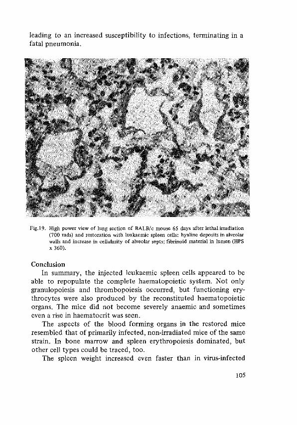
leading to an increased susceptibility to infections, terminating in a fatal pneumonia.
Fig.19. High power view of lung section of BALB/c mouse 65 days after lethal irradiation (700 rads) and restoration with leukaemic spleen cells: hyaline deposits in alveolar walls and increase in cellularity of alveolar septs; fibrinoid material in lumen (HPS X 360).
Conclusion In summary, the injected leukaemic spleen cells appeared to be
able to repopulate the complete haematopoietic system. Not only granulopoiesis and thrombopoiesis occurred, but functioning erythrocytes were also produced by the reconstituted haematopoietic organs. The mice did not become severely anaemic and sometimes even a rise in haematocrit was seen.
The aspects of the blood forming organs in the restored mice resembled that of primarily infected, non-irradiated mice of the same strain. In bone marrow and spleen erythropoiesis dominated, but other cell types could be traced, too.
The spleen weight increased even faster than in virus-infected
105

mice, but, curiously, after three weeks it attained a plateau. The number of stem cells injected seemed not to be a limiting factor for the ultimate spleen weight. The fibrosis of the splenic capsule and the absence of massive haemorrhages in the organ obviously played a role. However, the long survival time indicates that the leukaemic proliferation is slower in the restored than in a normal mouse. The explanation of this phenomenon might be sought in a decreased influx of newly recruited cells from intact bone marrow into the leukaemic population.
Notwithstanding the arrest in spleen growth and the absence of leukaemic proliferation, the animals died after 1-3 months, in contrast to the control mice restored with normal spleen cells. Death could be attributed to pneumonia, presumably of viral origin. Since the Rauscher virus itself has no effect upon the lungs, a superinfection due to the immunological deficiency of the animals restored with leukaemic tissue must be assumed.
The lethally irradiated mouse, restored with leukaemic spleen cells can be compared with a patient in complete remission after chemotherapy of blastic leukaemia: in both situations the haematopoietic organs are repopulated by cells, derived from stem cells present among a leukaemic cell population. In this respect, it might be a suitable model of complete remission in human leukaemia.
106
REFERENCES
Galien- Lartigue, 0., P. Tambourin, F. Wendling and F. Zajdela (1969): "Spontaneous hematopoietic recovery of Friend virus-infected mice after heavy X-irradiation". J. Nat. Cancer Inst. 42: 1061.
Ridl, J., L.J. Dooren, V.P. Eijsvoogel, J.J. van Went, W. Hijmans (1972): "An immunological study during posHransplantation follow-up of a case of severe combined immunodeficiency". Clin. Exp. Immunology. 10: 367.
Wieme, R.J. (1959): "Studies on agar gel electrophoresis. Techniques-Applications". Arscia, Bruxelles 1959.
107

CHAPTER VII
DISCUSSION
The main objectives for the study of an animal model for human leukaemia are in general: I) to gain insight in the pathogenesis of the disease, and 2) to test cytotoxic agents and therapeutic regimens. The transplantable leukaemias, especially L 1210, have proved to be excellent models for the evaluation of the efficacy of anti-tumour agents and have contributed to the introduction of effective schemes for the treatment of clinical leukaemia (Skipper 1968). These leukaemias, however, display some essential differences from human leukaemia with respect to growth fraction (Gavosto 1970) and the autonomy of the leukaemic cells. If virus-induced murine leukaemias were similar in this respect to human leukaemia these might constitute a more suitable model for the study of leukaemogenesis.
Since the initiation of this study the likelihood of a possible viral genesis of human leukaemia has increased, as judged from the many reports in the litterature dealing with this subject. Recent studies by Spiegelman c.s. (Hehlmann e.a. 1972) even suggest that a nucleic acid sequence similar to Rauscher virus might have causal significance both in murine and in human leukaemia. If this proves to be true, the study of Rauscher disease in particular might have more implications than supposed until recently.
The natural history of Rauscher disease in BALB/ c mice, as observed in this laboratory, corresponds with the splenic and hepatic erythroblastosis or erythroblastic leukaemia, described by other authors, whether induced by Friend or by Rauscher virus. The disease was restricted to the haematopoietic organs, especially spleen
108

and liver, the changes in the bone marrow being difficult to interpret. Infiltrations of leukaemic cells in other organs, as described by Yokoro e.a. (1966), were not encountered.
In BALB/c mice there was no evidence for leukaemic proliferation of cell types other than erythroid elements. The granulopoiesis remained active throughout the course of the disease, without indications of a maturation arrest, accumulation of young cells or metastases of myeloid cells. In some neonatally infected C57BL mice, however, granulocytic leukaemia developed with chloroma-like cell masses in mesenteric lymph nodes similar to the laesions described by Boiron e.a. ( 1965). This demonstrates the leukaemogenic potential of the Rauscher virus preparations used, with respect to the granulocytic cell line. The absence of such lesions in BALB/ c mice infected with the same virus, suggests that the stimulated granulopoiesis which was seen in the early phase of erythroblastosis, is not the expression of myelocytic leukaemia but rather an aspecific reaction to the viral infection.
Although the virus has been demonstrated by others within megakaryocytes and blood platelets, this is not tantamount to malignant transformation of these cells. In the present study the number of megakaryocytes was not abnormally high neither in the bone marrow nor in the spleen of the majority of the infected BALB/ c mice, as judged from the histological sections and the imprint preparations. Exceptionally, however, rather large numbers of megakaryocytes were encountered in the leukaemic spleens. The possibility of neoplastic proliferation of megakaryocytes, supposed by Brodsky e.a. (1968), cannot be excluded but does not seem likely.
In his original article Rauscher (1962) described the development of lymphomas in rats infected with the newly discovered virus. Several other investigators have gained the same experience. The virus preparations used in this study, too, could induce lymphoma in rats. In BALB/c mice, however, lymphomas and thymic enlargement were never observed, which in all likelihood excludes a proliferative process of lymphatic origin. The often observed lymphocytosis in the peripheral blood of these mice must be attributed to aspecific stimulation or mobilization, rather than to neoplastic proliferation of lymphocytes.
The splenic disease in BALB/ c mice thus appears to be restricted to the pathological increase of erythroblasts and their precursors.
109

The supposition of Brodsky e.a. ( 1969) that the erythroblastosis is a compensatory mechanism brought into action to cope with the shortened erythrocyte survival time, does not account for the magnitude of the splenomegaly and the liver involvement, even in face of a severe anaemia, let alone in case of a slight or moderate anaemia. A direct action of the virus on the kinetics of the erythroblasts must be assumed. The leukaemic cell mass largely consists of immature cells. Consequently, it might be inferred that the virus one way or other hampers the normal maturation of the infected haematopoietic cells. Only a minority of the red cells proceeds to a functional end stage, although they are often appreciably altered morphologically. The longevity of the immature cells contributes to the accumulation of leukaemic cells in spleen and liver. In these respects erythroblastosis resembles human leukaemia and combined with the fact that the process pursues its course relentlessly until the death of the affected animal, the erythroblastosis may be regarded as a malignancy.
If it may be assumed that the spleen colony assay in tbis viral leukaemia can be interpreted in the same way as in the normal system, the present data indicate that in Rauscher leukaemia normal stem cells are present in the spleen. In the course of the disease their number increases I 0 to 30 fold.
Theoretically, the colonies arising from the transplanted leukaemic spleen cells might be foci of tumour cells, as Thomson (1969) concluded, when she induced colony formation in irradiated mice by the injection of spleen cells obtained from mice infected with Friend virus. However, analysis of the colonies in the present experiments revealed an almost normal differentiative capacity of the splenic CFU's of leukaemic mice. It is therefore justified to call the cells wbich give rise to these colonies: 'colony forming units'; they may serve as a parameter for the pluripotent stem cell pool, instead of being merely 'tumour colony forming units' (Thomson 1969, Steeves 1968).
The normal functional capacity of the colony forming cells from leukaemic spleens is substantiated by the long survival of mice which receive 6 x 10⁵ or more spleen cells after lethal irradiation. The existence of a dual population of stem cells, normal next to leukaemic, is bighly unlikely in view of the small difference in repopulating capacity of normal and leukaemic spleen cells.
The normal functional capacity of the stem cells of mice infected
110

with an erythroblastosis virus has recently been confirmed: Wendling e.a. (1972) infected mice with the polycythaemia-inducing strain of Friend virus and observed a six-fold increase of the number of splenic colony forming units which followed the rise in the number of spleen cells. The distribution of the various types of colonies, 25-30 days after infection, did not differ much from normal.
These experiments give no clue as to the origin of the colony forming cells. They might be generated in the spleen by the multiplication of the stem cells present at the time of infection. Stem cells are known to increase in number under the influence of various stimuli, among others immunological challenge (Boggs e.a. 1967, McNeill 1970), hypoxia (Okunewick e. a. 1969) and bleeding (Marsh e.a. 1969). Multiplication of stem cells induced by the virus seems less probable but cannot be excluded. Though in individual spleens the number of CFU's per 106 cells sometimes greatly surpassed the values found in normal controls, the mean values did not differ from normal and did not change during the development of the leukaemia. The fact that the number of CFU's keeps pace with the total number of spleen cells suggests a physiological mechanism.
Rencricca e.a. ( 1970) provided evidence for the migration of stem cells from the bone marrow to the spleen in mice with an increased demand for erythroid precursor cells, provoked by phenylhydrazin induced haemolytic anaemia. The increase in the numbersofCFU'sin the spleen coincided with a decrease of those in the tibia, as had been found previously by Marsh e.a. ( 1968). Furthermore, the CFU's in the bone marrow entered in active cell cycle whereas those in the spleen were resting. Hodgson e.a. ( 1968) reported an increase in CFU content of the peripheral blood in rats with phenylhydrazin-induced haemolytic anaemia. Both data might indicate an increased release of stem cells from the bone marrow in response to anaemia. A similar compensatory mechanism might play a role in the splenomegaly or erythroblastosis. Stephenson e.a. ( 1972) found as much as 165 "tumour colony forming units" per I o6 cells in the peripheral blood of those mice in which an elevated nucleated cell count of more than 100 x 109/1 developed after infection with Friend virus. Although the factors responsible for the release of stem cells into the peripheral blood are not known, neither in leukaemic, nor in normal mice, the data are in keeping with the possibility that this mechanism contributes to the increase in CFU's in the enlarged spleen in erythroblastosis.
Ill

In addition to the action of the causative agent, the virus, an erythropoietic stimulus appears to influence the development of erythroblastosis, since hypoxia or exogenous erythropoietin promotes and polycythaemia induced by repeated blood transfusions delays splenomegaly (Tambourin e.a. 1969). At first sight this seems irreconciliable to the classical conception of the autonomous behaviour of malignant cells. However, even continuous hypertransfusion could not arrest the gradual progression of splenomegaly or prevent the premature death of mice infected with Rauscher or Friend virus (Dunn e.a. 1966, Tambourin I.e.). Besides, the cellular proliferation of several other tumours, both animal and human, can be influenced by hormone action, e.g. mammary carcinoma and prostatic carcinoma, the malignant nature of these tumours being beyond doubt. The erythroid hyperplasia in Di Guglielmo's syndrome has been shown to decrease concomitantly with the erythropoietin excretion after hypertransfusion and thus appears to be influenced by physiological factors (Adamson and Finch 1970). Furthermore, Metcalf ( 1971) provided evidence for the responsiveness of human leukaemic blast cells to regulation, mediated by humoral substances. Recent observations on the oscillating course of some cases of chronic myeloid leukaemia (Shadduck e.a. 1972) are in favour of the responsiveness of this type of leukaemia to feed back regulation. Thus, the umesponsiveness of cancer and leukaemia cells to physiological regulator mechanisms is not an absolute criterium. Apart from this, the most effective point of impact upon a cell renewal system, like haematopoiesis, is not the end cell but the stem celL In the case of murine erythroblastosis it is conceivable that the hormone erythropoietin induces the differentiation of a primitive precursor cell into the erythroid direction and thus feeds the neoplastic population. The stem cells remain resting until a differentiating stimulus makes them enter into the expanding leukaemic cell population.
Indeed, a constant influx of cells from a precursor pool into the erythroblastic compartment is essential for the spread of Rauscher leukaemia in the infected mouse. This could be deduced from the lack of autonomy of the majority of the proliferating spleen cells. After transplantation of leukaemic spleen cells into sublethally irradiated recipients, the greater part of the dividing cells in the enlarged spleens of the recipients were host-derived, indicating that new cells had been recruited from the host. Partial shielding of the bone mar-
1!2
row greatly influenced the ultimate spleen weight in irradiated mice infected with RLV; obviously, the splenic disease is dependent on the immigration of cells from the bone marrow in these circumstances. In unirradiated mice these cells might arrive at the spleen from the same source.
Many attempts have been made to determine the target cells for Rauscher and Friend virus. Although the presence of viral antigens on the colony forming cells has been demonstrated in Friend disease (Steeves 1968, Thomson e.a. 1968, Thomson 1969), this has not led to the conclusion that the colony forming cells might be the origin of erythroblastosis. The effect of hypoxia and polycythaemia on the evolution of erythroblastosis has been adduced in favour of the hypothesis that the erythropoietin responsive cell (ERC) be the target for Rauscher and Friend virus. However, these findings are compatible also with the induction of erythroblastosis via the infection of a more primitive precursor. Likewise, many attempts to promote or to counteract the evolution of Rauscher disease by influencing the stem cell pool (Brommer and Bentvelzen 1971 b) fail to differentiate between the ERC and the colony forming cells as the target for the virus.
The virus seems not to be over-particular in the choice of its host cells. On the contrary, it is capable of infecting many types of cells. Virus particles have been found in megakaryocytes (Rauscher 1962), in reticulocytes (Zajdela e.a. 1968) and in other tissues without malignant transformation. The production of virus particles and transformation of cells by RNA-virus are separate processes. Murine leukaemia virus can replicate in various tissues but presumably only transforms haematopoietic cells; e.g. mouse mammary tumour virus multiplicates in haematopoietic cells without giving rise to·neoplasms of blood forming organs, stressing the importance of the epigenetic status of the host cell (Bentvelzen e.a. 1970). In the oncogene theory of Huebner and Todaro a distinction is made between the "virogene", instructing for the production of viral proteins, and the "oncogene", the transformation-inducing gene.
In the present study evidence was provided for the infection of the pluripotent, colony forming cells in Rauscher disease. These cells produce viral proteins, detectable as antigens on the surface of the colony forming cells. Nevertheless, the fate of these cells is not sub-
113

stantially changed. The results obtained by restoration of lethally irradiated mice with spleen cells derived from virus-infected syngeneic animals indicate that the virus-bearing stem cells can give rise to normal haematopoietic cells. As judged from the thymic aplasia and the low immunoglobulin levels in the restored mouse, only the lymphoid cells seem not to recover adequately and the immunological deficiency constitutes a life-threatening defect in these animals which, as a consequence, eventually die with pulmonary infections.
If the leukaemia virus is incorporated in the DNA-chain of the pluripotent stem cells, it may be assumed that the genome of all progeny cells is endowed with the viral genes. However, in BALB/ c mice, the virus only induces appreciable changes in erythroid cells, resulting in malignant erythroblastosis, whereas granulopoiesis and thrombopoiesis proceed almost normally (Fig.20). Thus the development of malignancy seems to depend on the direction of differentia-
114
o<o.z-0 0 ------0
~<~ 0 o<~r::::l Erythropoiesis
0 (}~o<-o 0 0 ~~o<-o 0
Stem cell Committed
pool stem cells
Fig.20. Differentiation-dependent transformation hypothesis of leukaemogenesis. The progeny of precursor cells committed and stimulated to differentiate in one direction reveals leukaemic features; other cell lines develop normally although all cells carry the genetic material capable of instructing for leukaemic transformation. (Differences in committed cells indicated by external symbols).

tion of the target cells. In different mouse strains or in rats other cell lines are more vulnerable.
To account for the almost normal development of virus-infected haematopoietic stem cells into granulocytes and megakaryocytes and the malignant derailment of erythroblasts, the hypothesis is presented that the transforming principle of Rauscher leukaemia virus, when penetrated into the cells of BALB/ c mice, is associated with a site of the host DNA-chain which instructs for erythroid differentiation.
This association might be effectuated in several ways. First of all, the oncogenic component of the virus might be incorporated in a part of the host DNA-chain involved in erythroid differentiation. On the analogy of the specific "puff patterns" in the giant chromosomes of Chironomus (Beermann 1952) and Drosophila (Berendes 1965), it is probable that for each differentiated cell type in higher organisms one or more specific sites of the DNA-chain are unfolded and activated by a specific stimulus to arrive at the specialized function of the cell. This mechanism obviously is not restricted to ontogenesis; it must be functioning also in the adult animal since in many tissues continuous replacement of cells takes place until death, e.g. in the skin, in intestinal and other epithelia and in the bone marrow. Undifferentiated stem cells are preserved in these organs during the whole life span. They are capable of responding to adequate stimuli with differentiation, proliferation and maturation. If it is assumed that a potentially carcinogenic alteration, e.g. a (pro )virus, is inserted into the DNA-chain during a preceding nuclear division, it is conceivable that malignancy, induced by this fault in the genome will be manifested only when the DNA-sequence concerned is prepared for transcription. In a primitive, undifferentiated cell this latent malignancy will not be expressed. Only the evolution of a particular specialized cell function will reveal the presence of the carcinogenic factor. The same hypothesis can be applied to the vertically transmitted, "germinal" provirus or oncogene.
Secondly, the interference of gene products of the infecting virus with the production or function of cellular regulator substances might result in a hormone dependent transformation. Only in those cells in which the maturation depends on the function of these specific regulator substances the machinery of the cell will be disturbed and in appropriate circumstances malignancy may become manifest.
115

Presumably, erythropoietin acts by derepression of that segment of the genome which codes for the initial step in erythroid differentiation (Hodgson 1970). In the case of Rauscher erythroblastosis, erythropoietin would also initiate the transcription of the oncogene in differentiating precursor cells or unmask it otherwise, triggering the malignant derailment of erythropoiesis.
As this hypothesis has also to account for the development of other types of haematopoietic neoplasms in other animals, induced by the same virus, it must be assumed that the genetic constitution of the host determines the site or sites of the DNA chain were the (pro)virus is inserted or with which it interferes indirectly. In C57BL mice this would be a segment coding for granulopoietic differentiation. The knowledge of the localization of genes upon chromosomes is only in its initial phase and more refined methods than hitherto available are required to enable the detection of the site of incorporation of a viral gene in the DNA-chain of infected cells and segments with specialized functions.
According to this hypothesis it should be impossible or at least very difficult to induce transformation in the absence of the appropriate differentiating stimulus. Perhaps, the discrepancy between the ample evidence for radiation carcinogenesis in vivo in clinical and experimental medicine, and the difficulty to induce transformation by radiation under certain conditions in vitro (Klein 1966), should be regarded in this light.
In lethally irradiated recipients the infected transplanted stem cells differentiate normally and are capable of reconstituting the complete haematopoietic system, except for the lymphatic organs. In the restored mouse erythropoiesis proceeds at a sufficient rate to maintain the haematocrit at a slightly depressed level. From this it may be inferred that under certain conditions the leukaemic erythroblasts can reach maturity, notwithstanding the hypothetical presence of an oncogene in the segment of the genome, instructing for this direction of differentiation. This may be compared with the observa· tion of Friend e.a. (1971), who induced haemoglobin production in cells obtained from a cultured cell line of murine leukaemia cells (Friend leukaemia) by the addition of dimethyl sulfoxide (DMSO).
Reversion of malignancy has been reported in experimental pathology (Seilern-Aspang e.a. 1962, MacPherson 1965, Marin e.a.
I 16
1969, etc.) and in clinical pathology (see Pierce e.a. 1971), as well as in plant tumours (Braun 1965). Since single cells isolated from a malignant tumour - a mouse teratoma - were capable of giving rise to several types of differentiated cells, it is not necessary to have recourse to clonal selection to explain the regression of malignancy. Maturation can sometimes be induced in undifferentiated tumours by autotransplantation, suggesting the need of essential nutrients or other factors, or by growing them in vitro (Flaxman 1972). Induction of maturation of leukaemic blasts has been achieved in cultures of leukaemic cells obtained from the bone marrow or the peripheral blood of patients with leukaemia (Nowell 1960, Farnes e.a. 1961, Robinson 1970, Paran e.a. 1970, Ichikawa e.a. 1970, Metcalf e.a. 1971, etc.). In some instances differentiation of malignant cells has been attributed to starvation (Rusch e.a. 1970) or to removal of nutrients from the culture (Seeds e. a. 1970). Of course, all examples of reversion of malignancy are not necessarily based on the same mechanism. The most likely factors in the restored mouse seem to be the combination of a strong stimulus for proliferation and differentiation emanating from the environment and the absence of inhibitory factors. This might eventually conduce to an epigenetic event which might be the final common pathway of multiple mechanisms leading to the same effect: differentiation and maturation of previously blocked immature cells.
If neoplastic transformation is due to the action of an oncogene, transmitted within the genome of the cells, it must be assumed that this oncogene has been inactivated again in the cells reversed to normal. From the observations of the giant chromosomes it is known that the "puff patterns" change in the course of differentiation (Berendes 1967). Previously active puffs regress in subsequent differentiation stages. Likewise, the genes necessary for the initiation of a specialized cell function - e.g. erythropoiesis - will be inactivated in more mature stages. If the proposed hypothesis is correct, the oncogene linked to these host genes either structurally or functionally, will be suppressed conjointly, just like an acquired oncogenic provirus which does not cause malignant transformation in every cell. Interestingly, reversion of transformation of hamster cells, transformed by a virus, has been shown to occur without loss of the viral genome (Rabinowitz e.a. 1969, 1970) and could be attributed to a suppressor, regulated by loci upon an identifyable group of chromo-
117

somes (Hitotsumachi e.a. 1971). However, it is difficult to accept that mammalian cells possess regulatory genes commissioned to suppress acquired viral genes. Probably, the traced loci contain regulator genes which repress DNA-segments with which the oncogenes are associated.
In conclusion, erythroblastosis induced by Rauscher or Friend virus may be conceived as the ultimate result of the disturbance of cellular processes elicited by the transcription of a provirus incorporated in the DNA-chain of the host. The viral genes are in one way or the other associated with erythroid differentiation. Consequently, only the maturation of erythroblasts is defective. Erythropoietin triggers responsive cells (ERC) to differentiate and to participate in the leukaemic process. In turn, the pluripotent stem cell compartment receives a stimulus to compensate for the loss of ERC's (Lamerton 1970). Since the majority of the red cells do not reach maturity, the
feed back stimulation
(erythropoietin)
Erythropoiesis
Other cell lines
Fig.21. Hypothesis on the leukaemogenesis of Rauscher disease. Exogenous and endogenous erythropoietin enhance leukaemic proliferation. The lack of functioning red cells, maintained by the impaired maturation, provides a positive feed back to the stem cell compartment, stimulating the differentiation into erythroblasts. By the competing demand on the stem cell pool the differentiation of other cell lines may be hampered.
stimulus to the precursor pools does not cease. The continuous delivery of erythroblasts results in the accumulation of immature cells in spleen and liver (Fig.21 ).
118
Although the most conspicious abnormality in this disease is the increased cell mass in spleen and liver, largely consisting of immature red cells, the viral infection is not restricted to the red cell series. The most primitive haematopoietic cells, detectable by their function of pluripotent precursors are affected by the virus. These virus-bearing stem cells only wait for a physiological, differentiating stimulus to enter into the leukaemic process. Remarkably, if they are induced to differentiate to granulocytes or megakaryocytes their development will be virtually normal. Only the erythroid cell line derails and manifests malignant changes. Presumably, the expression of the malignancy ultimately depends on the physiological stimulation of that segment of the genome in which the provirus is incorporated.
If the conclusions from this experimental model might be transferred to human leukaemia this would afford some interesting perspectives. The model demonstrates that a disease which is induced by viral infection has much in common with some types of human leukaemia.
In the experimental model the leukaemic cells do not display autonomous proliferation. They are not self-maintaining and for the progression of the disease a constant influx is needed of cells, recruited from the pluripotent stem cell pool. This would favour the view of Gavosto (1970) on human acute leukaemia and corresponds to the external influence theory. However, in erythroblastosis the recruited cells are infected already in an early stage, prior to the challenge to enter into the differentiating compartment. Presumably, the oncogene is incorporated in the genome of the stem cells and will be transmitted to daug)Jter cells. This suggests that the persistence of a leukaemogenic agent is not obligatory for the progression of the leukaemia. If, indeed, the pluripotent haematopoietic stem cells are infected in human leukaemia as has been demonstrated in Rauscher leukaemia, this might constitute a serious obstacle for therapeutic success. It would be impossible to eradicate all infected cells without killing the patient. The achievement of complete remissions in acute leukaemia does not refute this because relapse ensues in the majority of the cases. The lethally irradiated mouse restored with leukaemic cells seems to be a realistic model for complete remission of human leukaemia. Here, too, haematopoiesis is run by differentiated leukaemic cells.
119

It is to be hoped that human stem cells are not so readily attacked by leukaemogenic agents as murine haematopoietic stem cells by the Rauscher virus. For the treatment of human blastic leukaemia this might have the implication that bone marrow transplantation after the complete eradication of the pre-existing stem cells and after anti-viral therapy would be the only way to get rid of the leukaemogenic germs. All regimens permitting the proliferation of autologous stem cells will allow the re-introduction of cells endowed with the (acquired) provirus and will fail to cure the disease, unless devices will be developed tore-initiate the maturation of the leukaemic blast cells in the patient. If human leukaemia will prove to be hormonedependent like murine erythroblastosis, this might open new perspectives for the treatment of leukaemic patients.
120
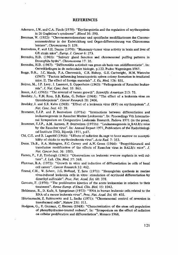
REFERENCES
Adamson, J.W. and C.A. Finch (1970): "Erythropoietin and the regulation of erytlrropoiesis in DiGuglielmo's syndrome". Blood 36: 590.
Beerman, W. (1952): "Chromomerenkonstanz und spezifische modifl.kationen der Chromo~ somenstruktur in der Entwicklung und Organ-d.ifferenzierung von Chironomus tentans". Chromosoma 5: 139.
Bentvelzen, P. and J.H. Daams (1970): "Mammary-tumor virus activity in brain and liver of GR strain mice". Europ. J. Cancer 6: 273.
Berendes, H.D. (1965): "Salivary gland function and chromosomal puffing patterns in Drosophila hydei''. Chromosoma 17: 35.
Berendes, H.D. (1967): "Differentiele activiteit van genes als basis van celdifferentiatie".In: Ontwikkelingen in de moleculaire biologie. p.122. Pudoc Wageningen 1967.
Boggs, D.R., J.C. Marsh, P.A. Chervenick, C.R. Bishop, G.E. Cartwright, M.M. Wintrobe (1967): "Factors influencing hematopoietic spleen colony formation in irradiated mice. II. The effect of foreign materials". J. Ex. Med. 126: 851.
Boiron, M., J.P. Levy, J. Lasneret, S. Oppenheim (1965): "Pathogenesis of Rauscher leukaemia". J. Nat. Cane. Inst. 35: 865.
Braun, A.C. (1965): "The reversal of tumor growth". Scientific American 213: 75. Brodsky, I., E.M. Ross, S.B. Kahn, G. Petkov (1968): "The effect of a leukemia virus on
thrombopoiesis". Cancer Research 28: 2406. Brodsky, I. and S.B. Kahn (1969): "Effect of a leukemia virus (RV) on erythropoiesis".J.
Nat. Cane. Inst. 42: 39. Brommer, E.J.P. and P. Bentvelzen (1971a): "Interactions between differentiation and
leukaemogenesis in Rauscher Murine Leukaemia". In: Proceedings Vth International Symposium on Comparative Leukemia Research. Padova 1971 (in the press).
Brommer, E.J.P., A.M. Aarssen, P. Bentvelzen (1971b): "Leukaemogenesis in BALB/c mice by the Rauscher virus". In: Annual Report 1971, Publication of the" Radiobiological Institute TNO, Rijswijk 1971, p.67.
Chi, C.H. and B. LagerlOf (1968): "Effects of radiation da.nage to bone marrow on susceptibility of chicks to erythroleukemia virus".Acta Rad. 7: 353.
Dunn, Th.B., R.A. Malmgren, P.C. Carney and A.W. Green (1966): "Propylthiouracil and transfusion modification of the effects of Rauscher virus in BALB/ c mice". J. Nat. Cancer Inst. 36: 1003.
Farnes, P., F.E. Trobaugh (1961): "Observations on leukemic IT''U!OW explants in well culture". J. Lab. Clin. Med. 51: 568.
Flaxman, B.A. (1972): "Growth in vitro and induction of differentiation in cells of basal cell cancer". Cancer Research 32: 462.
Friend, C.H., W. Scherr, J.G. Holland, T. Sato (1971): "Hemoglobin synthesis in murine virus-induced leukemia cells in vitro: stimulation of erythroid differentiation by dimethyl sulfoxide".Proc. Nat. Acad. Sci. 68: 378.
Gavosto, F. (1970): "The proliferative kinetics of the acute leukaemias in relation to their treatment". Revue Europ. d'Etud. Clin. BioL 15: 1042.
Hehlmann, R., D. Kufe, S. Spiegelman (1972): "RNA in human leukemic cells related to the RNA of a mouse leukemia virus". Proc. Nat. A cad. Sci 69: 435.
Hitotsumachi, Z. Rabinowitz and L. Sachs (1971): "Chromosomal control of reversion in transformed cells". Nature 231: 511.
Hodgson, G., E. Guzman, C. Herrera (1968): "Characterization of the stem cell population of phenylhydrazine-treated rodents". In: .. Symposium on the effect of radiation on cellular proliferation and differentiation". Monaco 1968.
121

Hodgson, G. (1970): "Mechanism of action of erythropoietin". In: Regulation of Hematopoiesis. Ed. A.S. Gordon. Appleton-Century-Crofts, New York 1970.
Ichikawa, Y. (1970): "Further studies on the differentiation of a cell line of myeloid leukemia".!. Cell. Physiol. 76: 175.
Klein, J .C. (1966): "Malignant transformation in vitro". J. Nat. Cancer Inst. 37: 655. Lamerton, D. (1970): "Adaptation of hemopoietic systems to environmental conditions".
In: XIII. International Congress of Hematology. Plenary Sessions, p.132. Munich 1970.
McPherson, I. (1965): "Reversion in hamster cells transformed by Rous sarcoma virus". Science 148: 1731.
Marin, G. and I. MacPherson (1969): "Reversion of polyoma-transformed cells: retransformation, induced antigens and tumorigenicity". J. Virol. 3: 146.
Marsh, J.C., D.R. Boggs, P.A. Chervenick, G.E. Cartwright, M.M. Wintrobe (1968): "Factors influencing hematopoietic spleen colony formation in irradiated mice. IV. The effect of erythropoietic stimuli''. J. Cell. Physiol. 71: 65.
McNeill, T.A. (1970): "Antigenic stimulation of bone marrow colony forming cells. I. Effect of antigens on normal bone marrow cells in vitro".Immunology 18: 39.
McNeill, T.A. (1970): "Antigenic stimulation of bone marrow colony forming cells. III. Effect in vivo". Immunology 18: 61.
Metcalf, D.: "Abnormal regulation of granulopoiesis in human acute granulocytic leukemia". Proceedings of Vth International Symposium on Comparative Leukemia Research, Padova 1971.
Metcalf, D. (1971): "Clinical applications of the agar culture technique for hemopoietic cells". Europ. J. Cancer 16: 855.
Nowell, P.C. (1960): "Differentiation of human leukemia leukocytes tissue culture''. Exper. Cell Res. 19: 267.
Okunewick, J.P., K.M. Hartley, J. Darden (1969): "Comparison of radiation sensitivity, endogenous colony formation, and erythropoietin response following prolonged hypoxia exposure". Rad. Research 38: 530.
Paran, M., L. Sachs, Y. Barak, P. Resnitzky (1970): "In vitro induction of granulocytic differentiation in hematopoietic cells from leukemic and non-leukemic patients". Proc. Nat. Acad. Sci. 67: 1542.
Pierce, G.B. and C. Wallace (1971): "Differentiation of malignant to benign cells". Cancer Research 31:127.
Proceedings of a workshop/symposium on in vitro culture of hemopoietic cells, held at the Radiobiological Institute, TNO, Rijswijk, September/october 1971. Publicating of Radiobiol. Inst. TNO, Rijswijk 1972.
Rabinowitz, Z. and L. Sachs (1969): "The formation of variants with a reversion of properties of transformed cells". Virology 38: 336.
Rabinowitz, Z., L. Sachs (1970): "Control of the reversion of properties in transformed cells''. Nature 225: 136.
Rauscher, F.J. (1962): "Virus-induced disease of mice characterised by erythrocytopoiesis and lymphoid leukemia". J. Nat. Cancer Inst. 29: 515.
Rencricca, N.J., V. Rizzolo, D. Howard, P. Duffy, F. Stahlman (1970): "Stem cell migration and proliferation during severe anemia". Blood 36: 764.
Seeds, N.W., A.G. Gilman, T. Amana, M.W. Nirenberg (1970): "Regulation of axon formation by clonal lines of a neural tumor". Proc. Nat. A c. Sci USA 66: 160.
Seilern-Aspang, F. and K. Kratochwill (1962): "Induction and differentiation of an epithelial tumour in the newt (Triturus cristatus)". J. Embr. and Exp. Morpho!. 10: 337.
122

Shadduck, R.K., A. Winkelstein, N.G. Nunna (1972): "Cyclic leukemic cell production in CML". Cancer 29: 399.
Steeves, R.A. (1968): "Cellular antigen of Friend virus induced leukemias". Cancer Research 28: 338.
Stephenson, J.R., A.A. Axelrad, D.L. McLeod (1972): "Erythroid nature of the response to Friend leukemia virus infection in mice". J. Nat. Cancer Inst. 48: 531.
Tambourin, P., F. Wendling, N. Barat, F. Zajdela (1969): "Influence de diff6rents facteurs d'hom6ostase 6rythropoi6tiques sur revolution de Ia leucemie de Friend". Nouv. Revue Fr. Hhn. 9: 461.
Thomson, S. (1969): "A system for quantitative studies on interactions between Friend Leukemia virus and haemopoietic cells". Proc. Soc. Exp. Bioi. Med. 130: 227 _
Thomson, S. and A.A. Axelrad (1968): "A quantitative spleen colony assay method for tumor cells induced by Friend Leukemia Virus infection in mice". Cancer Research 28: 2105.
Wendling, F., P.E. Tambourin and P. Julien (1972): "Hematopoietic CFU in mice infected by the polycythemia~inducing Friend virus. I. Number of CFU, and differentia~ tion pattern in the spleen colonies"./nt. J. Cancer 9: 554.
Yokoro and Thorell (1966): "Cytology and pathogenesis of Rauscher virus disease in splenectomized mice". Cancer Res. 26: 536~543.
Zajdela, F., P. Tambourin, F. Wendling, 0. Pierre (1968): "Formation de particules virales sur la membrane d'erythrocytes de Souris injectees avec le virus de Friend". C.R.Ac.Sc. Paris 267: 2394.
123

SUMMARY
Rauscher leukaemia virus (RL V) induces erythroblastic proliferation in BALB/ c mice if a sufficiently large dose of the virus is injected. The incubation period of the disease is dose-dependent, indicating that the number of target cells attained by the virus determines the course of the erythroblastosis, just as in non-oncogenic viral infections.
Lethally irradiated BALB/c mice with partially shielded bone marrow acquire the same disease upon infection with RLV. The virus obviously may fmd its target in bone marrow cells. Presumably these cells migrate to the spleen and the liver where they divide and accumulate. In splenectomized mice, infected with RLV, the most impressive changes are found in the liver; metastatic deposits were not encountered. These animals survived longer than the non-splenectomized controls. In C57BL/Rij mice, infected neonatally, the same virus induces either lymphatic or myeloid leukaemia.
Transplantation of leukaemic cells from Fl(BALB/c x CBA/ T6T6) into F1(BALB/c x CBA/Rij) and vice versa revealed the lack of autonomy of transferred leukaemic spleen cells. In the recipients the disease was propagated by the spread of the virus; the proliferating spleen cells could be identified by chromosomal analysis and the large majority appeared to be host cells.
The relative number of stem cells in the suspensions of the spleens of BALB/c mice with erythroblastosis, measured by the spleen colony assay, remained approximately constant during the
124

development of splenomegaly. The absolute number of splenic stem cells thus increased proportionally to the total number of spleen cells, i.e. 20- to 30-fold. Arguments for the migration of newly recruited stem cells from the bone marrow to the spleen are discussed.
The stem cells from the leukaemic spleens had a normal differentiative capacity, as judged from the distribution of histological types of the colonies which arose in lethally irradiated recipients upon injection of leukaemic spleen cells. Furthermore, the leukaemic spleen cell suspensions displayed an almost normal repopulating capacity in lethally irradiated recipients. Leukaemic spieen cell suspensions could prolong the survival of these animals for several months.
In contrast to the normal behaviour of the stem cells in the leukaemic mice, there is overwhelming evidence that they are affected by the virus. The majority of the leukaemic colony forming cells were killed by the incubation with specific antisera against Rauscher virus. Apparently, the stem cells bear viral antigens, suggesting that the stem cells are among the target cells of the virus. Besides, the leukaemic colony forming cells were shown to be less sensitive to normal rabbit serum than normal mouse colony forming cells. This might be an argument for a virus-induced cellular change which could be a step in the leukaemic transformation of the stem cell in Rauscher erythroblastosis.
To account for the discrepancy of the normal differentiation of granulocytes and megakaryocytes and the leukaemic derailment of the erythroblasts which originate from the same stem cells, the following hypothesis was proposed: the viral oncogenes only cause leukaemic transformation when activated together with those host genes which instruct for erythropoiesis. Consequently, physiological regulator mechanisms influence leukaemic transformation, resulting in a hormone-dependency of the disease. A model for leukaemogenesis is proposed for Rauscher erythroblastosis in which a feed back regulation sustains the erythroblastic expansion.
In lethally irradiated mice, restored by the injection of leukaemic spleen cells, splenomegaly and erythroblastic infiltration in the liver develop. Since leukaemic and normal haematopoiesis in these animals originate from stem cells which were present during the leukaemia,
125

the situation in the restored mouse is comparable to a chemotherapeutically induced remission of human leukaemia. Therefore, the mouse restored in this way might be an appropriate model for (complete) remission in acute human leukaemia.
The spleen weight of lethally irradiated mice restored with leukaemic spleen cells did not exceed I 000 mg, whereas the spleen weight in virus-infected normal mice may amount to 2 - 3000 mg! Moreover, the animals survived for I or 2 months after this weight had been attained. A deficient recruitment of bone marrow cells in irradiated mice might be among the factors causing the cessation of splenic growth. These restored mice are immunological cripples and die probably from opportunistic infections.
Finally, some perspectives for the conception and the treatment of human leukaemia are discussed in the light of the presented data.
126

SAMENVATTING
Het beschreven onderzoek had tot doe! het inzicht in de pathogenese van leukaemieen te verdiepen.
Verscheidene theorieen over het wezen van maligne ontaarding zijn te herleiden tot veranderingen in het genoom van de eel of in de expressie van bepaalde genen. De argumenten dat in vele gevallen een virus verantwoordelijk is stapelen zich op. Volgens sommigen is een "oncogeen" zelfs in aile lichaamscellen in het genoom ingebouwd en kan de expressie hlervan geluxeerd worden door ouderdom of door allerlei invloeden van buitenaf, zoals ioniserende straling, physische en chemische agentia.
Zowel bij carcinomen als bij leukaemieen krijgt men de indruk, dat slechts een klein percentage van de cellen zich actief deelt en verantwoordelijk is voor de groei van de gehele tumor, resp. de leukaemische celpopulatie. Bij lymphoblasten-leukaemieen is aangetoond dat inactieve blasten weer in mitose kunnen komen en aldus weer kunnen bijdragen tot de celvermeerdering. Daarnaast vermoedt men dat de leukaemische celpopulatie wordt gevoed door het stamcelcompartiment.
Het feit dat na effectieve chemotherapie een complete remissie kan optreden met normale bloedvorming, doet vermoeden dat er stamcellen tijdens de acute leukaemie onaangetast blijven en in gunstiger omstandigheden in staat zijn normale progenituur te krijgen. Tegenover de 'clonale' theorie, waarin verondersteld wordt dat de gehele leukaemische populatie uit een of hoogstens enkele leukae-
127

mische stamcellen ontstaat, staat de exogene theorie, waarin aangenomen wordt dat de cellen die het stamcelcompartiment verlaten getransformeerd worden door een uitwendige oorzaak, b.v. een virus. In beide gevallen zou de meerderheid van de stamcellen normaal zijn. Een derde theorie pleit voor de leukaemische verandering van aile stamcellen. Een complete remissie zou slechts schijnbaar zijn: a! rijpen de bloedcellen uit tot - oppervlakkig beschouwd - normale eindstadia, in wezen zi.in het leukaemische cellen. Geavanceerde technieken zijn in staat subtiele verschillen ten opzichte van normale ce!len aan te tonen.
Voor elk van de drie theorieen zijn vele steekhoudende argumenten aan te voeren. Het directe bewijs was echter niet te leveren omdat de haematopoietische stamcel bij de mens tot voor kort slecht toeganke!ijk was voor onderzoek en omdat men om trent de oorzaak van menselijke leukaemieen nog steeds in het duister tast. Daarom werd een experimenteel model gekozen voor het onderzoek van de pluripotente haematopoietische stamcel. Een door virus gei:nduceerde leukaemie bij muizen leek het meest geschikt.
Door Rauscher werd in 1962 een virus gevonden dat in daarvoor gevoelige muizenstammen een woekering van rode bloedcellen veroorzaakte, culminerend in een sterke vergroting van lever en milt, het verschijnen van onrijpe cellen in het perifere bloed en een vroegtijdige dood van de gei:nfecteerde dieren. Deze z.g. erythroblastose is vrijwel identiek aan de door Friend beschreven erythro-leukaemie en is wei vergeleken met de ziekte van Di Guglielmo bij de mens.
Het natuurlijke verloop van deze erythroblastose werd vergeleken met de beschrijvingen die door andere onderzoekers werden gepubliceerd. Bij BALB/ c muizen, besmet met Rauscher-virus werd een typische erythroblastose gezien zonder aanwijzingen voor woekering van lymphatische of myeloide elementen. Bij een andere muizenstam daarentegen - de C57BL muis - werden door hetzelfde virus zowel lymphatische als myeloide leukaemieen ge1nduceerd, hetgeen er op zou kunnen wijzen dat niet zozeer het virus alswel de gastheer het type van de leukaemie bepaalt. Vermindering van de dosis toegediend virus resulteerde in een uitstel van de miltvergroting, overigens trad een identiek ziektebeeld op. Bij te sterke verdunning (< I0-3, uitgaande van het onverdunde preparaat) konden geen tekenen van leukaemie worden waargenomen. Vermoedelijk is een zekere hoeveel-
128

heid van het virus in het Iichaam van het gefnfecteerde dier nodig om tot ziekteverschijnselen te kunnen leiden.
Bij de erythro blastose werden de belangrijkste veranderingen gevonden in de milt. Werd de milt verwijderd v66r de infectie met het virus dan traden de grootste veranderingen op in de lever. Het virus bereikt zijn doe! echter ook wanneer lever en milt bestraald worden: door tijdens letale bestraling tijdelijk een achterpoot van de muizen af te schermen kon aannemelijk worden gemaakt dat het effect van virus-besmetting afhankelijk was van het aantal intacte beenmergcellen.
Door middel van transplantatie van leukaemische miltcellen die in de gastheer herkenbaar waren aan een chromosomale merker, kon worden vastgesteld dat de leukaemische miltcellen niet autonoom groeiden. Dit maakte het waarschijnlijk, dat voor de handhaving en de uitbreiding van de leukaemische celpopulatie een constante recrutering van cellen uit een stamcel-compartiment noodzakelijk was. Gezien het voorafgaande zou het beenmerg hierin een belangrijke rol kunnen spelen.
Bij muizen is een quantitatieve bepaling van het aantal stamcellen mogelijk met behulp van de miltkolonie-proef van Till & McCulloch. Haematopoietische stamcellen, ingespoten in letaal bestraalde muizen, geven aanleiding tot de vorming van kolonies op het oppervlak van de milt. Iedere kolonie wordt gevormd door een eel; het aantal kolonies dat ontstaat uit een bepaalde hoeveelheid ingespoten cellen is een maat voor het aantal stamcellen in de ingespoten celsuspensie. De eenheid is de "kolonie-vormende eenheid", uitgedrukt in C F U I I o6 cellen. Deze techniek werd toegepast voor het bepalen van het aantal stamcellen in de milt van leukaemische muizen. Hierbij bleek, dat het aantal CFU's per I o6 miltcellen in het verloop van de incubatie-periode en ontwikkeling van de leukaemie gelijk bleef ondanks de enorme groei van de milt in de zelfde periode. Het absolute aantal CFU's stijgt dus evenredig met het aantal miltcellen, d.w.z. tot 20 a 30 maal de uitgangswaarde.
Het vermogen tot differentiatie van deze stamcellen leek ongestoord, te oordelen naar de verdeling van de kolonies naar celtype. Bovendien bleek het mogelijk door intraveneuze injectie van leukaemische miltcellen de levensduur van letaal bestraalde muizen te ver-
129

lengen. De stamcellen uit de leukaemische milten zijn dus in staat functionerende eindcellen te vormen. Rieruit zou men kunnen concluderen dat tijdens deze virus-leukaemie de stamcellen normaal blijven.
Door specifieke, tegen het Rauscher-virus gerichte cytotoxische antilichamen bleken vrijwel aile kolonievormende cellen ge!nactiveerd te worden. Dit wijst op de aanwezigheid van virus-antigenen op het oppervlak van de stamcellen. De conclusie, dat het virus aangrijpt op zeer primitieve voorstadia van de haematopoiese lijkt derhalve gerechtvaardigd.
De leukaemische CPU's waren in dezelfde proeven minder gevoelig voor natuurlijke cytotoxische antilichamen, welke voorkomen in de gebruikte konijne-sera, dan de CPU's van normale muizen; blijkbaar missen de besmette starncel!en bepaalde antigene determinanten. Dit kan geduid worden als een uiting van de transformatie van de pluripotente stamcel.
Remming van de erythropoiese door bloedtransfusies remt ook de ontwikkeling van de erythroblastose, terwijl stimulering van de erythropoiese d.m.v. exogeen erythropoietine de erythroblastose juist bevordert. Dit is door verscheidene auteurs aangevoerd als argument voor de hypothese dat een erythroblastose zou ontstaan door de transformatie van de z.g. erythropoietine-gevoelige voorloper van de erythroide reeks, die aangemerkt wordt als de "target cell" van Rauscher- en Friend-virus. Ret feit, dat verschillende rijpingsstadia van de rode celreeks besmet worden door het virus wijst erop, dat het virus zich niet beperkt tot een "target cell". Niet aile besmette cellen ontaarden. Voor de expressie van de maligne ziekte is het blijkbaar noodzakelijk dat de eel zich in een bepaalde richting differentieert. In het geval van de erythroblastose is het de erythroide differentia tie die het oncogeen de gelegenheid biedt de eel te transformeren. Prikkels tot differentia tie in de richting van myelopoiese of thrombopoiese Iaten in de BALB/c muis het oncogeen ongemoeid en leiden tot de vorming van normale leukocyten en thrombocyten.
Ter verklaring van deze discrepantie werd de volgende hypothese opgesteld. Ret virus, of een DNA-copie ervan, veroorzaakt slechts maligne ontaarding wanneer het geactiveerd wordt tesamen met genen, die een rol spelen bij de differentia tie van de rode celreeks. Op
130

analoge wijze zouden tumoren van endocriene organen beinvloed kunnen worden door hormonale prikkels. Wellicht bepaalt de plaats waar een provirus wordt geincorporeerd in de DNA-keten van de gastheercel het specifieke effect.
De muizen, die herstellen van letale bestraling door de injectie van leukaemische stamcellen, krijgen eveneens leukaemie. Merkwaardigerwijs stijgt hun miltgewicht niet boven 800 il. 1000 mg. Deze dieren sterven pas na 2 il. 3 maanden aan intercurrente infecties, vermoedelijk doordat het immunologische apparaat zich niet herstelt van de schade door de bestraling. lnmiddels blijkt de haematocriet een stijging te vertonen van ± 30 tot ± 40%. Ondanks de aanwezigheid van virus rijpt een groat aantal erythroblasten uit tot functionerende eindcellen. Om dit te verklaren wordt een vergelijking getrokken met de spontane regressie, die is waargenomen bij sommige muizenleukaemieen en met het phenomeen van de reversibele transformatie. Getracht wordt dit te verenigen met de hypothese dat sommige oncogenen uitsluitend werkzaam zijn wanneer ze tesamen met bepaalde gastheer-genen geactiveerd worden, hetzij onder invloed van dezelfde humorale substantie (erythropoietine?) hetzij door synergistische werking van beider producten.
Wanneer het geoorloofd is de conclusie van dit dier-model te projecteren op bepaalde vormen van menselijke leukaemieen, kan in de eerste plaats gesteld worden dat er vele pun ten van overeenkomst zijn tussen de bestudeerde leukaemie bij de muis en verschlllende vormen van leukaemie bij de mens. Er is weinig in tegenspraak met de mogelijkheid dat leukaemie bij de mens door een overeenkomstige fout in het genoom zou kunnen worden veroorzaakt, eventueel door een virus.
De aanwijzingen die werden gevonden voor een constante recrutering van cellen uit een stamcelcompartiment komt overeen met de opvatting van Gavosto over de blastenleukaemie. Aangezien de mogelijkheid blijkt te bestaan dat de haematopoietische cellen a! in een zeer vroeg stadium besmet worden, pleit de afwezigheid van infectieus virus tijdens de ziekte niet tegen een virale aetiologie. Een complete remissie zou slechts schljnbaar zijn, zoals voorgesteld door de "Leukaemia-minor"-theorie van Killmann. Deze zou mogelijk gemaakt worden door een onderdrukking van het oncogeen, dat echter
131

in de cellen aanwezig blijft. De letaal bestraalde muis, welke in Ieven wordt gehouden door de
transplantatie van leukaemische cellen !ijkt een goed model voor de bestudering van een complete remissie, zoals bij een menselijke leukaemie wei eens geinduceerd kan worden met krachtige chemotherapie. Waarschijn!ijk zijn subtielere behandelingsmethoden nodig om een derge!ijke complete remissie ad libitum te verlengen. Misschien opent het inzicht dat bepaalde vormen van !eukaemie hormonaal beinvloedbaar zijn nieuwe perspectieven.
132

ACKNOWLEDGEMENTS
With regard to the realization of this thesis, the author is much indebted to Professor Dr. D.W. van Bekkum for the facilities offered at the Radiobiological institute TNO and for the inspiring discussions which proved to be material to the completion of this work; to Professor Dr. M.J. de Vries for introducing him in murine pathology as well as for his valuable advices and criticism, and to Professor Dr. M. Frenkel who generously provided the opportunity to participate in scientific research outside his clinic.
The author thanks Dr. Peter Bentvelzen for his stimulating enthusiasm and his actual help in devising and evaluating the experiments.
The author wishes to express his gratitude to the members of the technical and administrative staff of the Radiobiological Institute for their collaboration, with special reference to Mrs. M. van Doorninck, Miss Els Schotman, Mrs. L. v.d. Burg-Verwey, Mr. A.M. Aarssen and Mr. J. Brinkhof for expert technical assistance.
Much support came from the Institute of Experimental Gerontology (Director Dr. C.F. Hollander), where the histological preparations were processed; transaminase levels were determined by Dr. C.P.A. van Bezooijen and immunoglobulins were estimated by Dr. J. Radl, which is gratefully acknowledged.
The author is grateful to Miss H. Spijker of the Audiovisual Department of the Medical Faculty of Rotterdam for the preparation of the figures and tables and to Mrs. W. van Harstkamp-Roodbeen for the development and printing of the microphotographs.
Valuable help came from Mrs. J. Veringmeier-van Zanten, Mrs. M.A. Lelieveld-Muller and especially from Miss Elsa Goeree, who typewrited the manuscript.
Finally, the author thanks all others whose collaboration and support contributed to the accomplishment of this thesis.
133


CURRICULUM VITAE AUCTORIS
Schrijver dezes werd op 23 januari 1933 geboren te Alkmaar. Zijn lagere en middelbare schoolopleiding kreeg hij gedeeltelijk in Winschoten, gedeeltelijk in Haarlem. Na het afleggen van het eindexamen Gymnasium {3 te Haarlem studeerde hij medicijnen aan de Universiteit van Amsterdam. In 1961 werd hij aldaar bevorderd tot arts. Na de vervulling van de militaire dienstplicht kreeg hij zijn opleiding tot internist bij Dr. G. Blomhert aan de Zuidwal te 's Gravenhage. Een haematologische stage op de afdeling van Dr. C.H.W. Leeksma in hetzelfde ziekenhuis legde de grondslag voor zijn interesse voor dit onderdeel van de inwendige geneeskunde, waarin hij van 1967 tot 1968 nadere ervaring opdeed op de Polikliniek Haematologie van het Academisch Ziekenhuis te Leiden (Hoofd Dr. F.J. Cleton).
Sedert 1968 is de schrijver werkzaam als chef de policlinique van de afdeling Interne Geneeskunde II (Hoofd Prof. Dr. M. Frenkel) van het Academisch Ziekenhuis Dijkzigt te Rotterdam. Vanaf 1969 vervult hij tevens de functie van haematoloog in het laboratorium voor haematologische cytologie van de afdeling Pathologische Anatomie II (Hoofd Prof. Dr. M.J. de Vries) van de Medische Faculteit te Rotterdam. Daarnaast werd hem de gelegenheid geboden wetenschappelijk onderzoek te verrichten in het Radiobiologisch Instituut T.N.O. te Rijswijk (Directeur Prof. Dr. D. W. van Bekkum). De experimenten waarop dit proefschrift is gebaseerd werden verricht op het virologisch laboratorium van dit instituut bij Dr. P. Bentvelzen.
135
