the role of the osteocyte in orthodontic tooth …
TRANSCRIPT

1
THE ROLE OF THE OSTEOCYTE IN
ORTHODONTIC TOOTH MOVEMENT IN
THE RAT DENTO-ALVEOLAR COMPLEX
Doctor of Clinical Dentistry (Orthodontics) Thesis
Dinesh Sanmuganathan
B.D.S. (USyd)
Orthodontic Unit
School of Dentistry
Faculty of Health Sciences
The University of Adelaide
South Australia
AUSTRALIA
2011

2
Table of Contents Table of Contents............................................................................................................................. 2
Figures and Tables .......................................................................................................................... 5
Glossary of Abbreviated Terms...................................................................................................... 7
Declaration ..................................................................................................................................... 10
Acknowledgements ....................................................................................................................... 11
SECTION 1 ...................................................................................................................................... 12
Introduction .................................................................................................................................... 13
LITERATURE REVIEW ................................................................................................................... 14
Overview of Alveolar Bone Structure ..................................................................................... 15
Osteoclast.................................................................................................................................. 16
Osteoblast ................................................................................................................................. 17
i) Osteoblast differentiation......................................................................................................19
Osteocyte................................................................................................................................... 20
i) Size of the cells ....................................................................................................................22
ii) Distribution...........................................................................................................................22
iii) Formation of the cells—lineage transitions .........................................................................23
iv) Osteocyte Receptors ..........................................................................................................24
v) Functional Role of Osteocytes in Bone ...............................................................................24
Osteoblastic and Osteocytic Responses to Mechanical Stimuli.......................................... 25
Orthodontic Tooth Movement.................................................................................................. 28
i) Mineralised tissue response to applied mechanical load .....................................................32
ii) Non-mineralised tissue response to applied mechanical load.............................................33
Sclerostin................................................................................................................................... 35
Apoptosis .................................................................................................................................. 41
i) Pathways of apoptosis..........................................................................................................41
ii) Osteoblast and osteocyte apoptosis....................................................................................42
iii) Regulation of osteoblast and osteocyte apoptosis..............................................................43
iv) Osteocyte Apoptosis and its Role in Targeted Bone Removal ...........................................43
References...................................................................................................................................... 45
SECTION 2 ...................................................................................................................................... 54
Statement of Purpose .................................................................................................................... 55
ARTICLE ONE................................................................................................................................. 56
Abstract ..................................................................................................................................... 57
Introduction ............................................................................................................................... 58
Materials and Methods ............................................................................................................. 60

3
i) Animals………… ..................................................................................................................60
ii) Appliances and Animal Manipulations .................................................................................60
iii) Harvesting of tissue ............................................................................................................62
iv) Section preparation.............................................................................................................62
v) Immunohistochemistry.........................................................................................................63
vi) Histoquantification ..............................................................................................................63
vii) Statistics and Error Study ..................................................................................................65
Results ....................................................................................................................................... 66
i) Error Study ...........................................................................................................................66
ii) Comparison of region 1 and region 2 within treated group..................................................66
iii) Comparison of day 7 and day 14 within RHS of treated group...........................................67
iv) Comparison of RHS MP and DB differences between treated and control groups ............69
v) Comparison of MP and DB differences between left and right sides in treated group ........70
Discussion................................................................................................................................. 71
Conclusion ................................................................................................................................ 74
Acknowledgments .................................................................................................................... 75
References................................................................................................................................. 76
ARTICLE TWO ................................................................................................................................ 78
Abstract ..................................................................................................................................... 79
Introduction ............................................................................................................................... 80
Materials and methods ............................................................................................................. 81
i) Animals, Appliances and Animal Manipulations ...................................................................81
ii)Tooth movement measurements ..........................................................................................82
iii) Harvesting of tissue ............................................................................................................82
iv) Section preparation.............................................................................................................83
v) Immunohistochemistry.........................................................................................................84
vi) Histoquantification ..............................................................................................................84
Results ....................................................................................................................................... 87
Discussion................................................................................................................................. 89
i) Application of orthodontic force to rat molars .......................................................................89
ii) Sclerostin-immunoreactivity.................................................................................................91
Conclusion ................................................................................................................................ 92
Acknowledgments .................................................................................................................... 93
References................................................................................................................................. 94
SUMMARY ...................................................................................................................................... 97
4
APENDICES .................................................................................................................................. 101
Appendix 1............................................................................................................................... 102
The Application of Orthodontic Force on the Rat Molars.......................................................102
Appendix 2............................................................................................................................... 104
Measurement of tooth movement..........................................................................................104
Appendix 3............................................................................................................................... 106
Tooth measurement results...................................................................................................106
Appendix 4............................................................................................................................... 109
Harvesting of Tissue..............................................................................................................109
Appendix 5............................................................................................................................... 113
Avidin-Biotin Peroxidase Complex immunolabelling protocol................................................113
Appendix 6............................................................................................................................... 114
AEC and counter-staining protocol....................................................................................... .114

5
Figures and Tables
Literature Review
Figure 1: A light micrograph of a osteoclasts resorbing an old osteon…………………………....17
Figure 2: An example of a functional syncytium………………………………………………….….21
Figure 3: Alveolar bone metabolism showing “tension” and “compression”………………….…..29
Figure 4: Cell sequence in mechanosensing, transduction and response………….…………….31
Figure 5: The sequence of bone remodelling stages during orthodontic treatment………..……33
Figure 6: Effects of sclerostin on osteoblasts………………………………………………..………36
Figure 7: Proposed regulation of osteon remodelling by osteocytic sclerostin expression……..39
Figure 8: Classical apoptosis signaling pathways……………………………………….…………..42
Article 1
Article 2
Figure 1: Rat model with closed coil spring…………………………………………………………. 61
Table 1: Sample sizes………………………………………………………………………………….61
Figure 2: Schematic representation of section through maxillary molars……………………….. 62
Figure 3: Arrangement of specimen sections on slides…………………………………………….63
Figure 4: An example of the AEC staining and histoquantification method used………………..64
Table 2: Error study for total osteocyte count ……………………………………………………….66
Table 3: Adjusted means of proportion of positively stained osteocytes…….……………………67
Table 4: Type 3 Tests of Fixed Effects……………………………………………………………… 67
Table 5: Adjusted means of the treated group at day 7 and day 14……………………………... 68
Table 6: Type 3 test, treated group at day 7 and day 14…………………………..……………... 68
Table 7: Sclerostin staining differential between the treated and control groups………………. 69
Table 8: Type 3 test, sclerostin staining differential, treated versus control groups………… 69
Table 9: Sclerostin staining differential between left and right sides in the treated group……...70
Table 10: Type 3 test, sclerostin staining differential, left and right sides in the treated
group……………………………………………………………………………………………………. 70
Figure 1: Schematic representation of a section cut through the maxillary molars.....................83
Figure 2: Arrangement of specimen sections on slides ..............................................................83
Figure 3: Sclerostin positive osteocytes and sclerostin-negative osteocytes. ............................85
Figure 4: An example of the AEC staining and histoquantification method used.. .....................86
Table 1: Tooth movement and differential sclerostin measurements over day 0 – day 7...........87
Figure 5: Scatter plot, differential +ve Sclr percentage and distance from day 0 to day 7..........87
Table 2: Tooth movement and differential sclerostin measurements over day 0 – day 14.........88
Figure 6: Scatter plot, differential +ve Sclr percentage and distance from day 0 to day 7..........88

6
Appendix 1
Figure 1: Animal was placed onto a purpose-built holding rack................................................103
Figure 2: Photograph of experimental spring setup with wire ligature ......................................103
Appendix 2
Figure 1: Impression taken with special tray.............................................................................104
Figure 2: Impression landmarks................................................................................................105
Figure 3: Illustration of centroid determination..........................................................................105
Appendix 3 Table 1: Tooth movement measurements of Day 0..................................................................106
Table 2: Tooth movement measurements of Day 7..................................................................107
Table 3: Tooth movement measurements of Day 14................................................................108
Appendix 4
Figure 1: Perfusion apparatus...................................................................................................109
Figure 2: Animal at completion of perfusion..............................................................................110

7
Glossary of Abbreviated Terms
Ab Antibody
ABC Avidin-biotin complex
AEC 3-Amino-9-ethylcarbazole
Ag Antigen
ALP Alkaline phosphatase
ATP Adenosine-5’-triphosphate
BMP Bone morphogenetic protein
BMU Basic metabolic unit
Ca2+ Calcium ions
cAMP Cyclic adenosine monophosphate
CGRP Calcitonin gene related peptide
CSF Colony stimulating factor
DNA Deoxyribonucleic acid
ECM Extracellular matrix
EDTA Ethylenediaminetetra-acetic acid
ER Oestrogen receptor
hMSC Human mesenchymal stem cells
Ig Immunoglobulin
IGF Insulin-like growth factor
IL Interleukin
IMVS Institute of Medical and Veterinary Science
IP3 Inositol triphosphate
IR Immunoreactive
IU International units
K Potassium
LRP 5/6 Lipoprotein receptor related protein 5/6
M Molar (molarity)

8
M1 Maxillary first molar
M2 Maxillary second molar
M-Csf Macrophage colony-stimulating factor
MEPE Matrix extracellular
MMP Matrix metalloproteinases
mRNA Messenger ribonucleic acid
MSC Mesenchymal stem cells
NGS Normal goat serum
NHS Normal horse serum
NO Nitric oxide
NT Neurotrophin
O.C.T. Optimal cutting temperature
OTM Orthodontic tooth movement
PBS Phosphate buffered solution
PDL Periodontal ligament
PTH Parathyroid hormone
PTHrP Parathyroid hormone-related protein
RANKL Receptor activator of nuclear factor kappa-β ligand
RER Rough endoplasmic reticulum
RNA Ribonucleic acid
TBS Tris Buffered Solution
TGF Transforming growth factor
TNF Tumour necrosis factor
TRAP Tartrate-resistant acid phosphatase
Trk Tyrosine receptor kinase

9
Abbreviations of length
mm Millimetre
μm Micron
nm Nanometre
Abbreviations of time
d Day
h Hour
min Minute
s Second
wk Week
y Year
Abbreviations of volume
l Litre
ml Millilitre
µl Microlitre
Abbreviations of weight
g Gram
kg Kilogram
mg Milligram
ng Nanogram

10
Declaration
This work contains no material which has been accepted for the award of any other
degree or diploma in any university or other tertiary institution and, to the best of my
knowledge and belief, contains no material previously published or written by another
person, except where due reference has been made in the text.
I give consent for this copy of my thesis, when deposited in the University Library,
being made available for loan and photocopying, subject to the provisions of the
Copyright Act 1968.
The author acknowledges that copyright of published works contained within this thesis
(as listed below) resides with the copyright holder(s) of those works.
I also give permission for the digital version of my thesis to be made available on the
web, via the University’s digital research repository, the Library catalogue, the
Australasian Digital Theses Program (ADTP) and also through web search engines,
unless permission has been granted by the University to restrict access for a period of
time.
Dr Dinesh Sanmuganathan
Dated:

11
Acknowledgements
I express my appreciation and gratitude to the following people for their invaluable
assistance in the completion of this thesis.
Professor Wayne J. Sampson, P.R. Begg Chair in Orthodontics, University of Adelaide.
Associate Professor Craig W. Dreyer, Orthodontics Unit, School of Dentistry, University
of Adelaide.
Dr. Angela Pierce, School of Dentistry, University of Adelaide.
Dr Kencana Dharmapati, Anatomical Sciences, School of Medicine, University of
Adelaide.
Dr. Henry S.H. Ho, Specialist Orthodontist, Sydney, New South Wales.
Dr. James Moses, Specialist Orthodontist, Adelaide, South Australia.
Mrs Nadia Gagliardi and Ms. Gail Hermanis, Technical Officers, Anatomical Sciences,
School of Medicine, University of Adelaide.
Mr Thomas Sullivan, Statistician, Data Management and Analysis Centre, University of
Adelaide
Ms. Sandie Hughes, Laboratory Officer, Oral Pathology, University of Adelaide.
Professor P. Mark Bartold, Director, Colgate Dental Research Centre, Adelaide Dental
Hospital, Adelaide.
Australian Society of Orthodontists Foundation for Research and Education for their
funding.
My parents, Lilani and Rudran, brother Rumesh and sister Shanya for their support.

12
SECTION 1

13
Introduction
Since the middle of the 19th century the importance of mechanical stimuli in the
maintenance and structure of skeletal tissues has been recognised.[1] It is now
accepted that skeletal tissue is dynamic, incorporating cycles of bone resorption and
bone formation; a process which helps to restore the skeleton while preserving its
structural integrity.[2] This remodelling is organised by cells of the osteoblast and
osteoclast lineage and involves a complicated network of cell-cell and cell-matrix
interactions, systemic hormones, locally produced cytokines and growth factors.[3]
Orthodontic and orthopaedic practice have much in common, in that both fields involve
a fundamental understanding of bone biology, particularly the relationship that
mechanical stress has on the various cell types found in bone. Mechanical strain on a
tooth, as opposed to bone, is a more complicated phenomenon requiring changes in
both the periodontal ligament (PDL) as well as the supporting alveolar bone, which are
tissue types that house vastly different cell populations and remodelling characteristics.
The signal transduction events that convert the mechanical strain to a molecular
biological response, and subsequent remodelling of the supporting tissues, are yet to
be fully elucidated.[3] Bone biologists have recently ascribed to the osteocyte a key
role in this mechanotransduction, a role that was traditionally given to the osteoblast. A
more fundamental understanding of the cellular and molecular elements that regulate
and drive these events is not only pivotal in understanding how these processes occur
but also needed for development of a means to control them to make orthodontic tooth
movement more efficient. [4]

14
LITERATURE REVIEW
15
Overview of Alveolar Bone Structure
The skeleton makes up approximately 20% of human body weight. Its major functions
incorporate mechanical support and motility, protection of internal organs, storage and
metabolism of Ca2+ and phosphate, and regulating haematopoiesis. Bone is dynamic
in nature, being controlled by changes in hormones, growth factors, mechanical
loading, nutrition and other as yet unidentified factors.[5]
Within mammals the teeth are attached to the bones of the jaws via an attachment
gomphosis apparatus consisting of cementum, periodontal ligament and alveolar bone.
This apparatus allows enough flexibility to withstand the forces of mastication.[6]
Alveolar bone specifically refers to that bone containing the sockets for the teeth. It
consists of an outer cortical plate, a central spongiosa and bundle bone (the bone lining
the alveolus). The cortical plates are continuations of the compact bone of the principal
mass of the maxilla and mandible. Bundle bone provides the attachment for the PDL
fibre bundles.[7]
Like all other bone, alveolar bone is in a state of constant remodelling through an
orchestrated interplay between the removal of old bone and its replacement with new
bone.[6] There is considerable variation, at the histological level, in the morphology of
alveolar bone produced by resorption or deposition of bone as it responds to the
functional demands placed on it.

16
Cells of Alveolar Bone
Bone effector cells of the osteoclast and osteoblast lineage are responsible for the size,
shape and strength of bones. The joint activity of these two cell types drive the
turnover, repair and developmental sculpting of the skeleton including both modelling
and modelling drift.[8]
Osteoclast
The multinucleated osteoclast represents the main bone resorption cell.[7] Osteoclasts
are derived from bone marrow haematopoietic stem cells sharing the same precursor
as macrophages.[5] It is by far the largest of the bone cells (50 to 100 µm) and their
precursors. They are usually found in association with bony surfaces, occupying
shallow depressions known as Howship’s lacunae.[6] Mature osteoclasts have
abundant mitochondria, numerous lysosomes, and free ribosomes.[9] However, the
most remarkable morphological feature of the osteoclast is their ruffled border which is
a complex system of finger-shaped projections of the membrane, the function of which
is to mediate the resorption of the calcified bone matrix.[10] This border is completely
surrounded by another specialized area, called the clear zone. The cytoplasm within
this zone has a uniform appearance, containing bundles of actin-like filaments.
Immediately underneath the osteoclast, the clear zone seals off a resorbing
compartment on the bone surface. It is the ability of the clear zone to seal off this area
of bone surface that allows the formation of a microenvironment suitable for the
operation of the resorptive apparatus.[9] Within the ruffled border membrane is an
ATP-driven proton pump (the so-called vacuolar H1-ATPase). This pump creates an
acidic environment within the resorption site which is responsible for the dissolution of
the mineral component (hydroxyapatite) of the bone matrix.[9] Matrix
metalloproteinases (MMP’s) and cathepsins K, B, and L are secreted by the osteoclast
into the area of bone resorption and are responsible for the degradation of the protein

17
components of the matrix, chiefly collagen.[11] Degraded bone matrix components are
endocytosed along the osteoclast’s ruffled border and subsequently transcytosed to the
contralateral membrane area, where they are released into the extracellular
environment.[12] Osteoclasts feature the presence of high amounts of the
phosphohydrolase enzyme, tartrate-resistant acid phosphatase (TRAPase), and this
feature is commonly used for the detection of osteoclasts in bone specimens.[9]
Figure 1: A light micrograph indicating large multinucleated osteoclasts resorbing an old
osteon.[6]
Osteoblast
Osteoblasts arise from multipotent mesenchymal stem cells that have the capacity to
differentiate into osteoblasts, adipocytes, chondrocytes, myoblasts, or fibroblasts.[13]
The proteins that constitute the bone matrix are produced and secreted by the fully
differentiated osteoblasts.[14] This matrix is subsequently mineralized under the control
of the same cells.[9]

18
Osteoblasts, when most active, form a layer of cuboidal cells with strong basophilic
cytoplasm, and a prominent nucleus that lies towards the basal end of the cell. There is
a pale juxtanuclear area indicating the site of the Golgi complex. They also contain a
prominent rough endoplasmic reticulum (RER) with numerous vesicles and
mitochondria. Microtubules and microfilaments are present and particularly prominent
beneath the secreting membrane of the cell. As bone deposition proceeds, osteoblasts
become incorporated into the matrix as osteocytes.[15]
Osteoblasts do not function alone but are found in groups along the bone surface,
where they line the layer of bone matrix that they are producing. Toward the end of the
matrix-secreting period, 15% of mature osteoblasts are entrapped in the new bone
matrix and differentiate into osteocytes. On the contrary, some cells remain on the
bone surface, becoming flat lining cells.
Bone formation occurs in three successive phases: the production and the maturation
of osteoid matrix, followed by mineralization of the matrix. In normal adult bone, these
processes occur at the same rate so that the balance between matrix production and
mineralization is equal. Initially, osteoblasts produce osteoid by rapidly depositing
collagen. This is followed by an increase in the mineralization rate to equal that of
collagen synthesis. In the final stage the rate of collagen synthesis decreases and
mineralization continues until the osteoid becomes fully mineralized.[9]
As stated above, a major product of the bone-forming osteoblast is type I collagen.
Initially, this protein is secreted in the form of a precursor, which contains peptide
extensions at both the amino-terminal and carboxyl ends of the molecule. Proteolytic
removal of these propeptides and additional extracellular processing results in mature
three-chained type I collagen molecules that assemble themselves into a collagen fibril.
Numerous other proteins are synthesized by the osteoblast. These proteins, such as

19
osteocalcin and osteonectin are incorporated into the bone matrix and constitute 40%
to 50% of the noncollagenous proteins of bone.[9] Glycosaminoglycans are other
osteoblast-derived proteins and are attached to one of two small core proteins: PGI (or
biglycan) and decorin. Numerous other proteins such as osteopontin, bone
sialoprotein, fibronectin, vitronectin, and thrombospondin serve as attachment factors
that interact with integrins.
While responsible for producing the osteoid matrix, mature osteoblasts play an
essential role in creating the microenvironment that allows the mineralization of this
osteoid by deposition of hydroxyapatite.[16] Local concentrations of calcium and
phosphate are regulated by osteoblasts in such a way as to promote the formation of
hydroxyapatite crystals. Osteoblasts express relatively high amounts of alkaline
phosphatase, an enzyme thought to play a role in bone mineralisation.[9] This is
highlighted by the fact that a genetic deficiency of alkaline phosphatase leads to
hypophosphatasia, a condition characterised by defective bone mineralization.[17] The
precise mechanism of mineralisation and the role of alkaline phosphatase in this
process still remains unclear. Bone mineralisation lags behind matrix production and, in
remodelling sites in the adult bone, occurs at a distance of 8–10 μm from the
osteoblast. Matrix synthesis determines the volume of bone but not its density.
Mineralisation of the osteoid increases the density of bone by removal of water, but
does not alter its volume.[9]
i) Osteoblast differentiation
Marrow mesenchymal stem cells (MSC’s) are pluripotent progenitors that can
differentiate into bone, cartilage, muscle and fat cells.[5] Many factors have been
implicated in regulating osteoblast differentiation (and subsequent bone formation) from
these mesenchymal stem cells. Bone morphogenetic protein 2 (BMP-2), BMP-6 and
BMP-9 are among the most potent inducers of osteogenic differentiation. The bone

20
morphogenetic proteins (BMP’s) are a large family of dimeric proteins within the
Transforming Growth Factor β superfamily of cytokines.[18] They were originally
identified as the active components within the osteo-inductive extracts derived from
bone and they are now known to be involved in a wide range of signalling functions that
mediate tissue interactions during development. The discovery of Smad-mediated
signals revealed the precise functions of BMPs in osteoblast differentiation.[19]
Transcription factors, Runx2 and Osterix, are essential molecules for inducing
osteoblast differentiation, as indicated by the fact that both Runx2-null mice and
Osterix-null mice have neither osteoblasts or bone tissue.[19]
There is increasing evidence suggesting that the canonical Wnt signalling pathway may
play as important a role in regulating osteoblastic differentiation.[20] The Wnt family
consists of a large number of secreted glycoproteins that are involved in embryonic
development, tissue induction and axis polarity.[20] Wnt ligands bind to seven
transmembrane-spanning receptors of the frizzled family and coreceptors of the
LRP5/6 gene, leading to an accumulation of the intracellular signalling molecule β-
catenin which is translocated to the nucleus where it initiates transcription of target
genes.[21] Loss-of-function mutations in the LRP5 gene cause the low-bone-mass
phenotype of the autosomal recessive disorder osteoporosis-pseudoglioma syndrome
and conversely, gain in function mutations of LRP5 result in patients with a high-bone-
mass phenotype. These findings suggest that balanced function of LRP5 is critical to
osteoblastic proliferation and differentiation.[20]
Osteocyte
The osteocyte represents a terminally differentiated non-proliferative cell of the
osteoblast lineage. As osteoblasts secrete bone matrix (osteoid), some of them
become embedded in lacunae and are from that point referred to as osteocytes. As
such, the osteocyte is considered non-migratory because it is entrenched within the

21
bone matrix. Each osteocyte extends numerous (perhaps as many as 60) small
cytoplasmic processes within bone canaliculi.[8,22] They form a network permeating
the entire bone matrix. These long cell processes are rich in microfilaments that are
organized during the formation of the matrix and before its calcification. It has been
shown that these cytoplasmic processes are approximately half the diameter of the
canaliculi in which they reside, leaving a sufficient gap for two processes to lie side by
side.[23] Each osteocyte communicates with its neighbours and with the surface lining
cells of bone by means of gap junctions.[24] A gap junction allows ions and compounds
of low molecular weight to pass between the two neighbouring cells without having to
pass into extracellular space.[25] Theoretically, this allows the cells to communicate
with each other, forming an extensive syncitial network through bone.[22] Osteocytes
are, therefore, in close communication with bone-lining cells at quiescent bone
surfaces, osteoblasts at sites of new bone formation and the pericytes of capillaries
and sinusoids supplying nutrients to osteocytes and other bone cells. They are also in
indirect gap-junction communication with the osteoblast precursor cells of the marrow
stroma.[24]
Figure 2: A schematic diagram of a functional syncytium comprising osteocytes, osteoblasts,
bone marrow stromal cells and endothelial cells. [9]
NOTE: This figure is included on page 21 of the print copy of the thesis held in the University of Adelaide Library.

22
i) Size of the osteocytes
Osteocyte size varies among species. The murine osteocyte lacuna dimension is of the
order of 5 μm by 20 μm [23] with a gap present between the cell and the lacuna wall.
The canaliculi range between 50 and 100 nm in diameter, where the cytoplasmic
processes, as discussed, are approximately half the diameter of the canaliculi.[23]
ii) Distribution
The distribution of osteocytes and their dendritic processes in lamellar bone, and to a
lesser extent woven bone, is not entirely random. In theory, this distribution is set at the
time of new bone formation.[8] Polarity is displayed by osteocytes in terms of the
distribution of their cell processes. The cell membrane facing the bone surface has the
highest number of these processes termed vascular dendrites.[26] Pallumbo et al.
hypothesised that the maximum functional length of these cell processes might be the
determining factor in incorporating the associated osteoblast into bone matrix.[26]
Osteocytes represent the most common cell type in bone.[24] While their
lacuna/canalicular system only represents 1% of bone fluid volume, it has substantial
surface area for molecular exchange, estimated to be some 400 times that of the entire
Haversian and Volkmann system combined and more than 100 fold that of the
trabecular bone surface.[27] This vast internal surface area potentially provides a
mechanism to quickly alter bone mineral homeostasis, growth factor content and
degree of mineralisation of bone. Osteocytes must have the capability to inhibit
mineralization on this large bone surface as, in their absence, micropetrosis
(mineralisation of the canaliculi and to a lesser extent the osteocyte lacunae)
occurs.[28] As the osteoblast becomes encased in bone matrix, the metabolic activity
of the osteoblast/osteocyte decreases, despite the fact that the osteocyte has been
historically thought to be a metabolically inert cell due to its number of cellular
organelles.[8] In fact, osteocytes are capable of significant molecular synthesis and

23
modification. The nervous system is a similar type of communication system,
comprising large numbers of low activity cells in a syncitial network, which has been
claimed to represent the most efficient design for the transmission of metabolically
expensive signals over long distances.[29]
iii) Formation of the cells—lineage transitions
The early stages of any lineage are poorly defined and this is particularly so for cells
derived from the mesenchymal stem cells within the bone marrow environment. These
stem cells, of which the osteocyte is one derivative, are particularly problematic due to
the increasing levels of cell plasticity being reported.[30]
Osteocyte formation is the result of an infrequent lineage transition from bone surface
resident osteoblasts to this non-proliferative terminally differentiated cell (it is estimated
only 10-20% of osteoblasts differentiate into non-proliferative osteocytes).[24] The
second end-form of the osteoblastic lineage is the bone lining cell.[31] During the
process of bone formation, an osteoblast, will be entombed in matrix as an ‘‘osteoid
osteocyte” as outwardly advancing newly formed osteoid is laid down. As the cell is
carried away, the future osteocyte maintains contact with the advancing osteoblasts at
the surface via extending cellular processes.[24] The surrounding osteoid matrix then
undergoes mineralisation under the control of enzymes, including osteocyte-derived
cacein kinase II.[32] However, the exact cellular/molecular mechanisms that regulate
this process are not fully understood. It has been hypothesised that osteocytes produce
signals that decrease the bone apposition rate of osteoblasts during refilling of BMUs
(basic multicellular units) hence enabling the recruitment of osteoblasts to become
osteocytes.[24] The evolution of an osteoblast to osteocyte involves a number of
significant changes including reduced susceptibility to apoptotic death, permanent
removal from the cell cycle and production of dendritic processes. As such it would be
expected that changes in gene expression at this time would be prominent.[24]
24
iv) Osteocyte Receptors
Mature osteocytes express most of the receptors known to play important roles in bone
metabolism. These include ER α and β (oestrogen receptor α and β), PTH, vitamin D3,
corticosteroids, and TGF-β. Certain osteocytic receptors, ligands and molecular
transporters are commonly associated with components of nerve tissue, suggesting a
role for some components of the nervous system in bone function. Osteocytic
production of nerve growth factor after fracture and the presence of glutamate
transporters are intriguing given the proposed function of the osteocytes as a syncitial
sensing and information transfer system.[8]
v) Functional Role of Osteocytes in Bone
The definition of an osteocyte is descriptive of its location as opposed to its function.
Bone biologists are still unclear about the precise role of the osteocyte in bone but
several theories have been postulated concerning their function and their contribution
to the process of bone remodelling.[8] These theories include; osteolysis, maintaining
systemic mineral homeostasis, sensing the strains produced in response to mechanical
loading of bones, production of signals that affect osteoblast/osteoclast function as well
as the expression of molecules that directly affect the turnover process.[24,33] If the
majority of these theories are correct then the osteocyte is a multi-functional “smart
cell” that acts as the conductor of the orchestra that is local bone turnover.[8]

25
Osteoblastic and Osteocytic Responses to Mechanical Stimuli
The architecture of bone is sensitive and shaped by mechanical forces.[34] Unloaded
bone, such as in the case of long-term bed rest or space travel, is resorbed whereas
increased exercise or gravity is associated with increased bone mineral density.[35]
Bone cells can respond to various mechanical stimuli including stretch, fluid flow and
hydrostatic pressure. However, in the literature these responses are widely varied.[34]
Osteoblast proliferation on mechanical stimulation has been found in some studies,
whereas others have found decreased cell numbers. Burger and Veldhuijzen
hypothesised that differential responses to mechanical stimulation are based on the
level of strain applied. They reason that osteoblasts respond to high stress levels with
increased proliferation but physiologic levels of stress decrease cell numbers.[36] The
level of differentiation of human osteoblasts has been shown to affect the response of
the osteoblast to mechanical strain.
Weyts et al. showed that physiologic stretch levels induce apoptosis in young
osteoblast cultures (7-day).[34] In more mature cultures, apoptosis was not triggered
by the same level of stretching and in day 14 cultures stretching increased cellular
proliferation. Protection of older cultures against stretch-induced apoptosis was not
related to accumulation of a mineralised matrix, or the absence of a matrix to adhere to
in young cultures, because collagen I matrix was provided to all cells from day 1 of
culture. The authors conceptualised that the differentiation-dependent changes in
stretch responses are due to intrinsic changes in mechanosensitivity of human
osteoblasts during differentiation and may reflect differential expression or maturation
of a mechanosensor (i.e., integrins, G protein coupled receptors, ion-channels, or the
cytoskeleton). As such, the absence of a response on the level of apoptosis or
proliferation in late stages of osteoblast differentiation may not necessarily reflect
insensitivity of cells in this phase to mechanotransduction; but the tight regulation of

26
this response by differentiation does imply its significance and is congruent with the
role of the osteoblastic lineage as a mechanotransductor in bone. [34]
Given that osteocytes are terminally differentiated osteoblasts, it is a logical
extrapolation that they, too, possess a mechanosensory apparatus. However, because
osteocytes, unlike osteoblasts, are housed in a mineralised matrix they are ideally
positioned to detect changes in environmental perturbations. It has, therefore, been
postulated that the osteocyte plays a central role in orchestrating local bone
remodelling where physical loading triggers them to modulate local bone
homeostasis.[8] Osteocytes most likely detect these changes and fatigue-induced bone
microdamage through their dendritic processes extending throughout bones within the
canalicular system [37] and their apoptosis might provide site-specific repair
signals.[38] In support of this notion, induction of microcracks in rat ulnae by fatigue
loading induced apoptosis of osteocytes adjacent to microcracks, but not in distant
osteocytes.[39] More importantly, resorption of the affected sites followed. Very high
strains also increased osteocyte apoptosis in rat ulnae, possibly by signalling to surface
lining osteoblasts.[39]
It has also been theorised that control of osteoclast and osteoblast apoptosis is
important in the overall control of bone remodelling.[40] The control mechanisms are
not fully understood but the fluid flow hypothesis proposes that locally evoked strain
derived from the displacement of fluid in the canaliculi plays an important role.[41]
When loading occurs, interstitial fluid is squeezed through the thin layer of non-
mineralized matrix surrounding the cell bodies and cell processes, resulting in local
strain at the cell membrane and activation of the affected osteocytes.[41]
Osteocyte lacunal density has been found to be variable, not only between individuals
[42] and with age [43], but also under altered mechanical stimuli.[43] The exact location

27
of the mechanosensor in the osteocyte is still to be determined but the presence of a
primary (non-motile) cilium on osteocytes has been reported and is known to sense
mechanical stimuli in other cell types.[44] It has also been found that a mesh of
extracellular material, primarily proteoglycans, is present in at least some canaliculi and
might assist in the amplification of fluid flow derived from mechanical signals.[45]
These cells respond both in vivo and in vitro to increased load-induced strains by
modifying a number of important molecules.[8] Anabolic signals, such as nitric oxide,
prostaglandins, and ATP, are released within seconds of osteocyte loading.[46] A few
minutes after the onset of mechanical loading, glucose 6-phosphate dehydrogenase, a
marker of cell metabolism, is elevated in osteocytes [47], and an increase in c-FOS
mRNA is observed within 2 hrs.[48] Within 4 hours, transforming growth factor β and
insulin-like growth factor mRNA expression are increased.[49]
Increases in osteocyte-specific markers have also been published, including
E11/gp3.8, dentin matrix protein 1 (found in a tooth movement model), matrix
extracellular phosphoglycoprotein (MEPE), and sclerostin.[50] The targets for all these
signals, through the network of cell-cell communications, are bone-surface cells and
osteoblasts. It was reported recently that SOST (the gene for sclerostin) and sclerostin
protein levels were dramatically reduced by mechanical loading of bone.[51]
Mechanical strain may also trigger osteocytes to send signals for activation of the bone
resorption cascade through expression of activator for NF-κB ligand (RANKL),
secretion of macrophage colony-stimulating factors (M-CSF), and through their own
apoptosis at the sites of micro-damage or micro-cracks.[50] Gene expression analysis
suggests that osteocytes indirectly control osteoclast differentiation through modulation
of RANKL expression in osteoblasts. In addition, humoral factors produced and
released through canaliculi into the bone marrow may regulate the differentiation and
activity of osteoclasts.[52] With these types of control mechanisms, it is plausible to

28
conclude that the osteocyte acts as the chief mechanosensor in bone, which has also
been recently confirmed by targeted ablation of osteocytes in a mouse model.[52]
�
Orthodontic Tooth Movement
The skeleton is subject to intermittent loads during locomotion and other forms of
physical activity. Similarly, alveolar bone is loaded intermittently during mastication, but
is subject to a more continuous deformation or strain during orthodontic tooth
movement.[53] In non-alveolar bone, the application of mechanically induced strain
appears to trigger primarily an osteogenic response [53], without any apparent
resorptive effect. Furthermore, this osteogenic response of bone to external loading
appears to be due to a reactivation of quiescent bone-lining cells [54], and is not
dependent on a preliminary phase of bone resorption [55]. Alveolar bone, however,
fundamentally differs from the rest of the skeleton in this response as when it is subject
to a continuous physical strain, it undergoes both resorption (osteoclastogenesis) and
deposition (osteogenesis), the extent of which is dependent upon the magnitude,
direction, and duration of the applied force.[3] The balance favours bone deposition on
the side of the alveolar wall undergoing tension and bone resorption on the side of the
wall undergoing compression, allowing the tooth to “move” along the force vector being
applied to it.(Figure 3) Osteoclastogenesis and osteogenesis are not only pivotal for
orthodontic tooth movement but also for tooth eruption.[4]

29
Figure 3: Alveolar bone undergoes constant metabolism, but in response to continuous force it
favours bony deposition on the side of the alveolar wall undergoing “tension” and bony
resorption on the side of the wall undergoing “compression”.
Mechanosensing is the phenomenon by which cells are capable of sensing structural
changes in the extra-cellular matrix (ECM), caused by external loading. Mechanical
forces exerted on teeth by orthodontic appliances strain both the extracellular matrix
(ECM) and cells of the periodontium (alveolar bone, PDL, gingiva, and associated
blood vessels and neural elements). The effect is initially physical in nature and is
followed closely by a biological response.[50] This interaction generates significant
changes in both the structural and functional components of the ECM, cell membrane,
cytoskeletal elements, nucleus, and several other cytoplasmic organelles that
subsequently synthesise and mobilise a variety of molecules inside and outside the
cells.[3,56]
Cell adhesion molecules, like the integrins, allow direct transmission of the tensile,
compressive, and shear forces from the ECM into the cell and vice versa, and are
considered essential for cellular survival, growth, and mobility.[50] This transmission
helps to maintain the cells in their active form, termed tensegrity, capable of responding

30
rapidly to various mechanical, physical, and other challenges. In addition to the energy
provided by the mechanical load, this sensing triggers a biochemical reaction that
provides the additional energy required for the biological system to respond to the
elevated environmental demands.[57] Each cellular group within paradental tissue is
equipped with mechanosensors and as such a mechanical stimulus may activate
multiple sets of these sensors, leading to a series of separate downstream cellular
events in various tissue types.[50] Subsequent changes in cytoskeletal protein
structure and function propagate the signalling process into the nucleus and, ultimately
the genome, via signalling proteins (such as hedgehog, transforming growth factors)
and calcium ions. The eventual outcome of these chains of events is enhanced or
suppressed gene expression, with transference of the signal back to the cytoplasm
through mRNAs, reaching the ribosomes, generating protein synthesis and secretion
and an associated cellular response that could include mitosis, cell motility or
programmed cell death.[58] (Figure 4) This process is termed mechanotransduction
which simply is the conversion of a mechanical input into a biochemical one.[50]
The unique feature of mechanotransduction in orthodontics is the interaction that
occurs between various tissues, both mineralized and non-mineralized, and their
associated neurovascular elements. For tooth movement to be perpetuated these
tissues need to be remodelled, a task performed by cells of the alveolar bone
(osteoblasts, osteoclasts, and osteocytes), PDL (gingival fibroblasts), blood vessels
(endothelial cells), and neural tissues (dendritic and neural cells). Other cell types
essential for this process are derivatives of the immune system (inflammatory cells and
osteoclast progenitors). There is an individual response pattern for each of these
tissues as far as the mechanosensing, transduction, and response mechanisms are
concerned.[3,58]

31
Figure 4: The sequence followed by cells in mechanosensing, transduction and response.[50]

32
i) Mineralised tissue response to applied mechanical load
When an orthodontic force is applied to a tooth it bends alveolar bone and both
compresses and stretches the PDL. This leads to a sequence of events within the
dento-alveolar complex where the alveolar bone adjacent to the compressed PDL
becomes the site of intense bone resorption, while the stretched PDL interfaces with
sites of active osteogenesis. As the alveolus bends there is flow of bone interstitial
fluid, evoking shear stress in the mineralized extracellular matrix (ECM) and
deformation of the alveolar bone osteocytes in the lacunae and of the dendrites in the
canaliculi. Osteoblasts, which maintain direct contact with osteocytes will, in turn,
respond to the osteocytic signal one of two ways depending on the direction of the
applied force. The first response, on the “tension” side is a direct one where the
osteoblasts initiate and are responsible for appositional bony deposition. The second
response, on the “compression” side results in the activated osteoblasts conveying
signals to approaching osteoclasts, enticing these cells to start resorbing the alveolar
bone, and informing them on the proper time to cease their resorptive activities. These
osteoclasts for bone resorption arise from conversion within the local population of
monocytes/macrophages or migration of progenitor cells from alveolar bone marrow
cavities to the strained PDL.[59] Within the compression side osteocyte signalling
mechanisms, such as sclerostin, also inhibit osteoblast differentiation, function and
mineralisation.

33
Figure 5: The sequence of bone remodelling stages during orthodontic treatment. The roles
played by osteocytes osteoblasts and osteoclasts are illustrated.[50]
ii) Non-mineralised tissue response to applied mechanical load
Gingival and periodontal fibroblasts differ in their roles in paradental tissue remodeling.
While PDL fibroblasts are predominantly involved in the synthesis and degradation of
their own ECM, gingival fibroblasts are involved in bone remodelling events.[50] A
reduction in collagen production (type I and IV) has been reported in the compressed
PDL, as well as increased type IV collagen production at PDL tension sites after 72 h of
force application.[60]
34
The role of blood vessels in orthodontic tooth movement has been the subject of recent
scrutiny, where angiogenesis and remodeling of existing blood vessels help functional
adaptation to the new environment created. Reports show an initial reduction and a
later increase in the number of PDL blood vessels following orthodontic force
application.[60,61] These blood vessels also play a major role in the mechanical force-
induced sterile necrosis that occurs in the compressed periodontium by acting as a
source of cytokines and chemokines, where demonstrable increases in expression of
IL-1β, IL-1 receptor, IL-6, IL-6 receptor, IL-8 receptor, IL-11, and TNF-α have been
shown.[50]
Neural elements also play a role where released neurotransmitters, such as Substance
P and calcitonin gene related peptide (CGRP), interact with endothelial cells of the
paradental capillary network, enticing them to bind circulating leukocytes, promoting
their migration into the paradental ECM.[62] These migratory leukocytes in turn
produce ample amounts of chemokines and cytokines upon entering these tissues. It is
this combination of cytokines, along with those produced by the native paradental cells,
that evoke and maintain the remodeling events of the PDL and alveolar bone.[63]
In summary, orthodontic tooth movement involves the use of a mechanical strain that is
subsequently converted to a desirable, and somewhat predictable, biological response
within the dento-alveolar complex. The effectors of this force-induced remodeling are
the cells of the paradental tissue that use mechanosensing, transduction, and response
phenomena to respond to applied mechanical forces.[50]

35
Sclerostin
Sclerostin is the protein product encoded by the SOST gene, located on chromosome
17q12-q21.[64] Sclerostin is a known negative regulator of osteoblast differentiation
and function and acts as one inhibitor to bone formation.[8,65] In its role as a key
inhibitor, sclerostin helps determine the normal extent of bone formation and,
consequently, protects against the deleterious effects of uncontrolled bone growth.[66]
Its effect is highlighted by two rare high bone mass diseases, Sclerosteosis and Van
Buchem disease, both of which have been linked to inactivating mutations in the SOST
gene.[65,67] The administration of an anti-sclerostin antibody has been shown to
increase bone formation and restore the bone loss upon ovariectomy in rodents.[68] It
has been suggested that the inhibition of sclerostin (through sclerostin neutralizing
antibodies) may, therefore, be an attractive strategy for conditions in which bone loss is
a significant component, such as postmenopausal and senile osteoporosis.[66]
Conversely, transgenic mice overexpressing Sost exhibit low bone mass.[69]
On the basis of its amino acid sequence, sclerostin belongs to the DAN family of
glycoproteins.[21] This group of proteins has been shown to antagonize both bone
morphogenetic proteins (BMP’s) and the Wnt signalling pathway, growth factors of
pivotal importance in bone formation.[8,70] BMPs and Wnts are critical for
osteoblastogenesis because they provide the initial and essential stimulus for
commitment of multipotential mesenchymal progenitors to the osteoblast lineage.[71]
Similarly, sclerostin has been demonstrated to potently antagonise and negatively
regulate several members of the bone morphogenetic protein (BMP) family.[72]
Sclerostin is also believed to work by binding to lipoprotein receptor-related protein 5/6
(LRP5/6), inhibiting canonical Wnt-signalling and thus impairing osteoblast
differentiation and function.[65] Although this binding to LRP5/6 antagonises Wnt
signalling, sclerostin and Wnt do not appear to compete for binding of this co-
36
receptor.[21] The precise mechanism through which sclerostin expression inhibits Wnt-
mediated bone formation is still unclear, where it may be transported to the bone
surface by the osteocytic canaliculi or it may induce another signalling molecule in
osteocytes which is then transported to the osteoblasts to inhibit bone formation.[21]
While most of the literature points to sclerostin operating through inhibition of BMP and
Wnt pathways, it cannot be ruled out that it may have its own, as yet unknown,
receptors to which they bind.[70]
It is believed that sclerostin inhibits bone formation via a number of ways. These
include:
1. negatively regulating osteoblast differentiation;
2. negatively regulating osteoblast proliferation;
3. suppressing mineralisation by osteoblastic cells and
4. stimulating osteoblast apoptosis.[21,70]
Figure 6: A schematic drawing demonstrating the possible inhibitory effects of sclerostin on the
proliferation and differentiation of osteoblasts and its agonistic effects on osteoblast
apoptosis.[70]
NOTE: This figure is included on page 36 of the print copy of the thesis held in the University of Adelaide Library.

37
Sclerostin is a factor that was thought to be exclusively expressed by the osteocyte,
where high levels have been detected in their lacunae and canalicular system.[70,72] It
has been demonstrated that osteoclasts, osteoblasts, bone lining cells (retired
osteoblasts that have ceased forming bone and cover most of the bone surface) and
periosteal osteoblasts exhibited no sclerostin staining.[66] While postnatally it is clear
that osteocytes and no other cells in bone express sclerostin, recent evidence has
shown distinct immunolocalisation of SOST in the cementocytes and mineralising
periodontal ligament cultures.[21,72] This is hardly surprising given that cementum and
bone are rather similar hard tissue substances, where osteocytes and cementocytes,
together with their canalicular network, share many morphological and cell biological
characteristics. Unlike osteocytes in bone there is no clear evidence that cementocytes
have a functional role in the tissue homeostasis of cementum.[72] While SOST protein
expression seems to be limited to terminally differentiated cells within an embedded
mineralised matrix, SOST mRNA is expressed by many non-mineralised tissues,
especially during embryogenesis, including the heart, aorta, kidney and liver.[21]
The distribution of sclerostin-positive osteocytes is not homogenous throughout bone
where section mapping has shown a differential spatial relationship, with sclerostin-
negative osteocytes located significantly closer to bone surfaces.[66] In vivo and in
vitro murine and human studies have shown that recently embedded osteocytes,
including those within unmineralised osteoid, were almost all negative for
sclerostin.[21] As such, sclerostin protein expression by new osteocytes is delayed until
the cells have matured and are surrounded by a mineralised matrix.[66] It has been
postulated by Poole et al. that sclerostin secretion by newly embedded osteocytes is
delayed such that the cells must mature or receive a later signal that triggers sclerostin
expression. They conceptualise that newly embedded osteocytes secrete sclerostin
only after the local onset of mineralization to inhibit cortical bone formation and osteon
infilling by cells of the osteoblast lineage.(Figure 7) This allows a sufficient, but not

38
excessive, amount of cortical bone to be formed to fill in osteonal and other canals
without compressing their contents. As such, trabecular thickness is maintained at an
appropriate thickness.[66] In contrast, active osteoblasts within forming osteons are
protected from inhibition by sclerostin by a layer of sclerostin-negative osteocytes,
permitting bone formation to continue at specific sites (Figure 7) including the
periosteum. As such, the osteocyte has a regulatory role in maintaining bone structure
and strength through differential expression of sclerostin. The observations by Poole et
al. shift the emphasis away from sclerostin as a possible regulator of preosteoblast
proliferation (through interaction with bone morphogenetic proteins) toward a role in
determining bone’s microarchitectural development through a very precise
geographical expression and a selective local modulator of mature osteoblast
function.[66] In a similar way, sclerostin expression in cementum may be a method for
development of cemental microarchitecture.
As discussed earlier, osteocytes are thought to sense loading stimuli and regulate
remodelling and bone turnover processes. The findings by Poole suggest that
osteocytes, which have access to surface osteoblasts and bone lining cells through
their dendritic connections, may provide the key inhibitory signal (sclerostin) that
inactivates bone surfaces. Mechanical loading on non-alveolar bone was found to
reduce sclerostin expression, thereby providing a mechanism by which bone formation
upon mechanical strain is increased.[70]

39
Figure 7: Schematic diagram of the proposed regulation of a remodelling cortical osteon by
osteocytic sclerostin expression.[66]
Sclerostin has been reported to selectively initiate the apoptosis of osteoblastic cells.
Human mesenchymal cells treated with sclerostin displayed a marked increase in
caspase activity and elevated levels of fragmented histone-associated DNA in these
cells were detected.[73] This phenomenon could serve as one of the mechanisms by
which sclerostin modulates the survival of osteoblasts and ultimately osteoblastic
function. Sclerostin was also found to significantly decrease alkaline phosphatase
(ALP) activity and the proliferation of differentiating human mesenchymal cells (hMSC).
NOTE: This figure is included on page 39 of the print copy of the thesis held in the University of Adelaide Library.

40
Sclerostin-induced changes in ALP activity and the survival of hMSC cells were
partially restored by BMP-6, suggesting the involvement of additional growth
factors.[73]
Intermittent parathyroid hormone application has strong anabolic effects on bone.
SOST and sclerostin expression was found to be reduced in vitro and in vivo in
response to parathyroid hormone treatment, and SOST is a direct target gene of
parathyroid hormone.[74,75] This suggests that downregulation of SOST expression
may play a role in mediating parathyroid hormone action in bone.
Thus, one way for osteocytes to regulate bone remodelling would be by altering the
secretion of sclerostin. Accordingly, it was shown that mechanical stimulation in vivo
reduced the expression of sclerostin by osteocytes.[76] Also, it is possible that
osteocyte death is a signal for bone formation because the level of sclerostin would
decrease. Sevetson et al. demonstrated that the osteoblast differentiation factor
Cbfa1/Runx2 increases SOST expression.[77] Up to now, there is some disagreement
on the effect of parathyroid hormone (PTH) on SOST expression. One study showed
that intermittent PTH treatment reduced SOST expression.[74] In contrast, Bellido et al.
found that PTH given intermittently to mice did not alter the levels of SOST, but PTH
given continuously did decrease SOST expression.[75]
Somewhat interestingly, Poole et al. found no reduction in the recruitment of osteocytes
(as inferred from osteonal lacunar density) in the osteons of bone specimens removed
at operation from 3 cases of sclerosteosis and 3 controls. This finding suggests that
sclerostin primarily influences the later stage of bone formation rather than earlier
events such as osteocyte recruitment.[66]

41
Apoptosis
Programmed cell death, termed apoptosis, is an essential cellular mechanism used by
organisms to reduce cell number in order to attain precise control of organ
development and function. In its simplicity it is a form of individual cell suicide. When
cells become apoptotic they contract, lose attachment to their neighbours, undergo
chromatin condensation and margination and cleavage of the DNA into inter-
nucleosomal size fragments which retain their organelles. This is followed by
packaging of the cell contents into membrane-bound vesicles (apoptotic bodies) that
have specific membrane surface molecule signatures that aid their rapid removal from
the system by phagocytic cells.[40,78] This rapid deletion allows apoptosis to occur
with minimal or no inflammatory response in direct contrast to necrosis.[80] Apoptosis
is not initiated simply to delete cells. The death process gives rise to a large number of
signalling molecules that can dramatically modulate the behaviour of cells in the locality
of the deletion and far beyond.[78]
i) Pathways of apoptosis
Two major signalling pathways control apoptosis.[79] One is initiated by death
receptors, members of the TNF receptor family with an intracellular death domain, and
the other is the mitochondrial pathway that involves mitochondrial release of
cytochrome c.[40] Regardless of the source of activation, the final mechanistic pathway
in apoptosis is the activation of a family of proteolytic enzymes called caspases.[80]
These enzymes induce the morphological features of apoptosis by cleaving specific
substrates.

42
Figure 8: Model of death ligand/death receptor and mitochondria-mediated classical apoptosis
signalling, including Bcl-2 members. [40]
ii) Osteoblast and osteocyte apoptosis
Osteoblast apoptosis plays a critical role during embryonic limb development, skeletal
maturation, bone turnover in the adult skeleton, and in bone repair and regeneration
processes.[80] This balance of proliferation, cell differentiation, apoptosis and cell
progression to the osteocyte phenotype affects the pool of osteoblasts available to
maintain bone homeostasis. It is clearly evident from the literature that there are many
factors which affect osteoblast survival but the mechanistic pathways by which cell
survival is affected or compromised are not well understood.[73]
Osteoblast apoptosis explains the fact that 50–70% of the osteoblasts initially present
at a remodelling site of human bone cannot be accounted for after counting the lining
cells and osteocytes at that site.[9] Osteocyte apoptosis could be of importance to the
origination and/or progression of the bone multicellular unit (BMU), the unit responsible
for a localised area of bone remodelling. Osteocytes are the only cells in bone with the
infrastructure to sense the need for remodelling at a specific time and place and, as
such, it makes sense that they could drive this turnover. Furthermore, osteocytes are in
direct physical contact with lining cells on the bone surface and targeting of osteoclast

43
precursors to a specific location on bone depends on a “homing” signal given by lining
cells.[37] What seems evident now is that the life span of the cells that form the BMU
(that is the osteclasts, osteoblasts, and osteocytes) is an important regulator of bone
mass, strength and turnover.[40]
iii) Regulation of osteoblast and osteocyte apoptosis
It is now appreciated that all major regulators of bone metabolism including bone
morphogenetic proteins (BMPs), Wnts, other growth factors and cytokines, integrins,
oestrogens, androgens, glucocorticoids, PTH and PTH-related protein (PTHrP),
immobilization, and the oxidative stress associated with aging contribute to the
regulation of osteoblast and osteocyte life span by modulating apoptosis. Moreover,
osteocyte apoptosis has emerged as an important regulator of remodelling on the bone
surface and a critical determinant of bone strength, independent of bone mass.[83]
Osteoblast apoptosis has been shown in vitro to be stimulated by the activation of
death receptors with TNF or CD95 ligand.[82,83] While osteoblasts and osteocytes
express CD95 and its ligand, their in vivo role in the apoptosis of these cells is yet to be
elucidated.[40]
iv) Osteocyte Apoptosis and its Role in Targeted Bone Removal
One of the more characteristic osteocyte behaviours is their apoptotic death.[8] The
impact of a loss of cells from a functional syncitium has an impact not only on
neighbouring cell types but also those in communication via the canalicular system.
Frost found that with increasing age there was an increasing incidence of empty
osteocyte lacunae in human bone.[28] Reduced osteocyte lacunal density has also
been noted in osteoporotic bone [84], and individuals with fewer osteocyte lacunae in
their bone have a reduced ability to repair accumulating microdamage.[85] It was
initially thought that necrosis was responsible for osteocytic death under these
conditions; however, it is now known that osteocyte apoptosis is a relatively common

44
event in both pathological and healthy human bone.[39] Noble and coworkers have
hypothesised that apoptosis of osteocytes helps target the resorption process to
specific areas following microdamage.[86] To support this hypothesis, large numbers of
apoptotic osteocytes have been observed to surround resorption pits induced in
response to fatigue microdamage.[38] Osteocytic apoptosis associated with regions of
damage was shown to precede osteoclastic activity raising the possibility that it may be
the apoptotic process that generates the targeting signal for osteoclastic bone
destruction.[86] When conditional ablation of osteocytes has occurred, the bones
demonstrated a dramatic increase in bone resorption, a decrease in bone formation,
trabecular bone loss and loss of response to unloading.[52] The results of these data
can be interpreted two ways. It may indicate osteocytes play a role in the inhibition of
osteoclastic resorption or alternatively the increased resorption could be due to the
death-induced signals from osteocytes.[8] Parfitt postulated that there are two types of
bone remodelling in adults: targeted and background or untargeted remodelling. He
has suggested that no more than 10–20% of remodelling in adult humans normally falls
into the first category. [87] This has not been tested in alveolar bone, and as such it
would be interesting to test the model with orthodontic tooth movement, which one
would assume would be a form of targeted bone remodelling.

45
References
�
1. von Meyer H. Die Architektur der Spongiosa. Archiv für Anatomie und Physiologie
1867;47:615-628.
2. Frost H. Bone remodelling dynamics. Springfield: Charles C Thomas, 1963.
3. Miekle M. The tissue, cellular, and molecular regulation of orthodontic tooth
movement: 100 years after Carl Sandstedt. European Journal of Orthodontics
2006;28:221-240.
4. Wise GE, King GJ. Mechanisms of tooth eruption and orthodontic tooth movement.
Journal of Dental Research 2008;87(5):414-434.
5. Chau W, Leong W, Li B. Signalling pathways governing osteoblast proliferation,
differentiation and functioning. . Histology and Histopathology 2009;24:1593-1606.
6. Ten Cate A. Oral Histology, 5 Edition: Mosby, 1998.
7. Davidovitch Z. Cell biology associated with orthodontic tooth movement. In:
Berkovitz B, Moxham B, Newman H, editors. The periodontal ligament in health and
disease. Philadelphia: Mosby-Wolfe, 1995. p. 259-278.
8. Noble B. The osteocyte lineage. Archives of Biochemistry and Biophysics
2008;473:106-111.
9. Manolagas S. Birth and Death of Bone Cells: Basic Regulatory Mechanisms and
Implications for the Pathogenesis and Treatment of Osteoporosis. Endocrine Reviews
2000;21(2):115-137.
10. Roodman G. Advances in bone biology: the osteoclast. Endocrine Reviews
1996;(17):308-332.
11. Bossard M, Tomaszek T, Thompson S, Amegadzie B, Hanning C, Jones C, et al.
Proteolytic activity of human osteoclast cathepsin K—expression, purification,
activation, and substrate identification. J Biol Chem 1996;271:12517-12524.

46
12. Salo J, Lehenkari P, Mulari M, Metsikko K, Va¨a¨na¨nen H. Removal of osteoclast
bone resorption products by transcytosis. Science 1997;276:270-273.
13. Bianco P, Riminucci M, Gronthos S. Bone marrow stromal stem cells: nature,
biology, and potential applications. Stem Cells 2001;19:180-192.
14. Robey P, Boskey A. The biochemistry of bone. In: Marcus R, Feldman D, Bilezikian
J, Kelsey J, editors. Osteoperosis. New York: Academic Press, 1995 p. 95-103.
15. Berkovitz B, Shore RC. Cells of the periodontal ligament. In: Berkovitz B, Moxham
B, Newman H, editors. The periodontal ligament in health and disease. Philadelphia:
Mosby-Wolfe, 1995. p. 9-33.
16. Boskey A. Matrix proteins and mineralization: an overview. Connect Tissue Res
1996; 35:357–363.
17. Whyte M. Hypophosphatasia and the role of alkaline phosphatase in skeletal
mineralization. Endocrine Reviews 1994;15:439–461.
18. Kingsley D. The TGF- B superfamily: new members, new receptors, and new
genetic tests of function in different organisms. Genes Development 1994;8:133-146.
19. Katagiri T, Takahashi N. Regulatory mechanisms of osteoblast and osteoclast
differentiation, 2002.
20. Si W, Kang Q, Luu H, Park J, Luo Q, Song W, et al. CCN1/Cyr61 is regulated by
the canonical Wnt signal and plays an important role in Wnt3A-induced osteoblast
differentiation of mesenchymal stem cells. Mollecular and cellular biology
2006;26(8):2955-2964.
21. Moester M, Lowik C, Papapoulos S, Van Bazooijen R. Sclerostin: Current
knowledge and future perspectives. Calcified Tissue International 2010;87:99-107.
22. Hadjidakis D, Androulakis I. Bone Remodelling. Annals of the New York Academy
of Sciences 2006;1092:385-396.
23. Su M, Jiang H, Zhang P, Liu Y, Wang E, Hsu A, et al. Knee-Loading Modality
Drives Molecular Transport in Mouse Femur. Annals of Biomedical Engineering
2006;34:1600-1606.

47
24. Noble B. The role of the osteocyte in bone turnover. Bone 2008;43(Supplement
1):s22.
25. Cowin S. Mechanosensation and fluid transport in living bone. Journal of
Musculoskeletal Neuronal Interactions 2002;2(3):256-260.
26. Palumbo C, Palazzini S, Marotti G. Morphological study of intercellular junctions
during ostocyte differentiation. Bone 1990;11(6):401-406.
27. Martin R, Burr D, Sharkey N. Skeletal Tissue Mechanics. New York: Springer-
Verlag, 1998.
28. Frost H. Micropetrosis. Journal of Bone and Joint Surgery American 1960;(42-
A):144-150.
29. Laughlin S, de Ruyter can Steveninck R, Anderson J. The metabolic cost of neural
information. Nat Neurosci 1998;1(1):36-41.
30. Jiang Y, Vaessen B, Lenvik T, Blackstad M, Reyes M, Verfaillie C. Multipotent
progenitor cells can be isolated from postnatal murine bone marrow, muscle, and brain.
Experimental Hematology 2002;30(8):896-904.
31.Noble B, Reeve J. Osteocyte function, osteocyte death and bone fracture
resistance. Molecular and Cellular Endocrinology 2000;159:7-13.
32. Suzuki Y, Kubota T, Koizumi T, Satoyoshi M, Teranaka T, KAwase T, et al.
Extracellular processing of bone and dentin proteins in matrix mineralization.
Connective Tissue Research 1996;35(1-4):223-229.
33. Bonewald L. Osteocytes: A proposed multifunctional bone cell. Journal of
Musculoskeletal Neuronal Interactions 2002;2(3):239-241.
34. Weyts F, Bosmans B, Niesling R, van Leeuwen P, Weinans H. Mechanical Control
of Human Osteoblast Apoptosis and Proliferation in Relation to Differentiation. Calcified
Tissue International 2003;72:505-512.
35. Martin R. Toward a unifying theory of bone remodeling. Bone 2000;26:1-6.

48
36. Burger E, Veldhuijzen J. Influence of mechanical factors on bone formation,
resorption, and growth in vitro. In: Hall B, editor. Bone. Boca Raton, FL: CRC Press,
1993.
37. Parfitt A, Mundy G, Roodman G, Hughes D, Boyce B. A new model for the
regulation of bone resorption, with particular reference to the effects of
bisphosphonates. Journal of Bone and Mineral Research 1996;11:150-159.
38. Verbogt O, Gibson G, Schaffler M. Loss of osteocyte integrity in association with
microdamage and bone remodelling after fatigue in vivo. Journal of Bone and Mineral
Research 2000;15(1):60-67.
39. Noble B, Stevens H, Loveridge N, Reeve J. Identification of apoptotic changes in
osteocytes in normal and pathological human bone. Bone 1997;20:273-282.
40. Xing L, Boyce B. Regulation of apoptosis in osteoclasts and osteoblastic cells.
Biochemical and Biophysical Research Communications 2005;328:709-720.
41. Goulet G, Cooper D, Coombe D, Zernicke R. Influence of cortical canal architecture
on lacunocanalicular pore pressure and fluid flow. Computerised Methods
Biomechanical and Biomedical Engineering 2008;11:379-387.
42. Tomkinson A. Effects of oestrogen on the maintenance of bone mass and bone cell
viability: Cambridge University; 1998.
43. Mullender M, van der Meer D, Huiskes R, Lips P. Osteocyte density changes in
aging and osteoporosis. Bone 1996;18:109-113.
44. Malone A, Anderson C, Stearns T, Jacobs C. Primary cilia in bone. Journal of
Musculoskeletal Neuronal Interactions 2007;7(4):301.
45. Han Y, Cowin S, Schaffler M, Weinbaum S. Mechanotransduction and strain
amplification in osteocyte cell processes. Proceeding of the National Academy of
Sciences USA 2004;101(47):16689–16694.
46. Bakker A, Soejima K, Klein-Nulend J, Burger E. The production of nitric oxide and
prostaglandin E(2) by primary bone cells is shear stress dependent. Journal of
Biomechanics 2001;34:671-677.

49
47. Dodds R, Ali N, Pead M, Lanyon L. Early loading-related changes in the activity of
glucose 6-phosphate dehydrogenase and alkaline phosphatase in osteocytes and
periosteal osteoblasts in rat fibulae in vivo. Journal of Bone and Mineral Research
1993;8:261-267.
48. Kawata A, Mikuni-Takagaki Y. Mechanotransduction in stretched osteocytes-
temporal expression of immediate early and other genes. Biochemical and Biophysical
Research Communications 1998;246:404-408.
49. Wildemann B, Schmidmaier G, Brenner N, Hüning M, Stange R, Haas N.
Quantification, localization, and expression of IGF-I and TGF-beta1 during growth
factor-stimulated fracture healing. . Calcified Tissue International 2004;74:388-397.
50. Krishnan V, Davidovitch Z. On a path to unfolding the biological mechanisms of
orthodontic tooth movement. Journal of Dental Research 2009;88:597-608.
51. Robling A, Niziolek P, Baldridge L, Condon K, Allen M, Alam I. Mechanical
stimulation of bone in vivo reduces osteocyte expression of Sost/sclerostin. Journal of
Biological Chemistry 2008;283:5866-5875.
52. Tatsumi S, Ishii K, Amizuka N, Li M, Kobayashi T, Kohno K, et al. Targeted
Ablation of Osteocytes Induces Osteoporosis with Defective Mechanotransduction. Cell
Metabolism 2007;5(6):464–475.
53. Hěrt J, Lisková M, Landa J. Reaction of bone to mechanical stimuli. Part 1:
Continuous and intermittent loading of tibia in rabbit. . Folia Morphologica (Praha)
1971;19:290–300.
54. Chambers T, Evans M, Gardner T, Turner-Smith A, Chow J. Induction of bone
formation in rat tail vertebrae by mechanical loading. Bone and Mineral 1993;20:167–
178.
55. Chow J, Wilson A, Chambers T, SW F. Mechanical loading stimulates bone
formation by reactivation of bone lining cells in 13-week-old rats. Journal of Bone and
Mineral Research 1998;13:1760 – 1767.

50
56. Kerrigan J, Mansell J, Sandy J. Matrix turnover. Journal of Orthodontics
2000;27(227-233).
57. Poirier C, Iglesias P. An integrative approach to understanding mechanosensation.
Brief Bioinformation 2007;8:258-265.
58. Masella R, Meister M. Current concepts in the biology of orthodontic tooth
movement. American Journal of Orthodontics and Dentofacial Orthopaedics
2006;129:458-468.
59. Xie R, Kuijpers-Jagtman AM, Maltha JC. Osteoclast differentiation during
experimental tooth movement by a short-term force application: an
immunohistochemical study in rats. . Acta Odontologica Scandinavia 2008;66:314-320.
60. Anastasi G, G C, Matarese G, Rizzo G, Nucera R, Mazza M, et al. An
immunohistochemical, histological, and electron-microscopic study of the human
periodontal ligament during orthodontic treatment. International Journal of Molecular
Medicine 2008;21:545-554.
61. Ren Y, Maltha JC, Stokroos I, Liem R, Kuijpers-Jagtman AM. Effect of duration of
force application on blood vessels in young and adult rats. . American Journal of
Orthodontics and Dentofacial Orthopaedics 2008;133:752-757.
62. Andrade I, Silva TA, Silva GAB, Teixeira AL, Teixeira MM. The role of tumor
necrosis factor type 1 in orthodontic tooth movement. Journal of Dental Research
2007;86:1089-1094.
63. Garlet TP, Coelho U, Repeke CE, Silva JS, Cunha Fde Q, Garlet GP. Differential
expression of osteoblast and osteoclast chemoattractants in compression and tension
sides during orthodontic movement. Cytokine 2008;42:330-335.
64. Yavropoulou M, Yovos J. The role of Wnt signalling pathway in osteoblast
commitment and differentiation. Hormones 2007;6(4):279-294.
65. Lacey D. Inhibition of sclerostin as an anabolic therapeutic approach for bone loss.
Bone 2008;43(Supplement 1):S22.

51
66. Poole K, van Bazooijen R, Loveridge N, Hamersma H, Papapoulos S, Lowik C, et
al. Sclerostin is a delayed secreted product of osteocytes that inhibits bone formation.
The FASEB Journal 2005:1842-1844.
67. Winkler D, Sutherland M, Yu C, Skonier J, Hayes T, Geoghegan J. Osteocyte
control of bone formation via sclerostin, a novel BMP antagonist. EMBO 2003;22:6267-
6276.
68. Warmington K, Morony S, Sarosi I. Sclerostin antagonism in adult rodents, via
monoclonal antibody mediated blockade, increases bone mineral density and
implicates sclerostin as a key regulator of bone mass during adulthood. Journal of
Bone and Mineral Research 2004;19:S56.
69. Loots G, Kneissel M, Keller H, Baptist M, Chang J, Collette N, et al. Genomic
deletion of a long-range bone enhancer misregulates sclerostin in Van Buchem
disease. Genome Research 2005;15:928-935.
70. ten Dijke P, Krause C, de Gorter D, Lowik S, Rutger L, Bezooijen V. Osteocyte-
derived sclerostin inhibits bone formation: its role in bone morphogenetic protein and
Wnt signalling. Journal of Bone ane Joint Surgery 2008;90:31-35.
71. Canalis E, Economides A, Gazzerro E. Bone morphogenetic proteins, their
antagonists, and the skeleton. Endocrine Reviews 2003;24:218-235.
72. Jäger A, Götz W, Lossdörfer S, Rath-Deschner B. Localization of SOST/sclerostin
in cementocytes in vivo and in mineralizing periodontal ligament cells in vitro. Journal
of Periodontal Research 2009;45(2):246-254.
73. Sutherland M, Geoghegan J, Yu C, Turcott E, Skonier J, SWinkler D, et al.
Sclerostin promotes the apoptosis of human osteoblastic cells: a novel regulation of
bone formation. Bone 2004;35(4):828-835.
74. Keller H, Kneissel M. SOST is a target gene for PTH in bone. Bone 2005;37:148-
158.
75. Bellido T, Ali A, Gubrij I, Plotkin L, Fu Q, O’Brien C, et al. Chronic elevation of
parathyroid hormone in mice reduces expression of sclerostin

52
by osteocytes: a novel mechanism for hormonal control of osteoblastogenesis.
Endocrinology 2005;146:4577-4583.
76. Robling A, Bellido T, Turner C. Mechanical stimulation in vivo reduces osteocyte
expression of sclerostin. Journal of Musculoskeletal Neuronal Interactions 2006;6:354.
77. Sevetson B, Taylor S, Pan Y. Cbfa1/RUNX2 directs specific expression of the
sclerosteosis gene (SOST). Biological Chemistry 2004;279:13849–13858.
78. Noble B. Microdamage and apoptosis. European Journal of Morphology
2005;42(1):91-98.
79. Gupta S. Molecular signaling in death receptor and mitochondrial pathways of
apoptosis (Review). International Journal of Oncology 2003;22:15-20.
80. Hock J, Krishnan V, Onyia J, Bidwell J, Milas J, Stanislaus D. Osteoblast Apoptosis
and Bone Turnover. Journal of Bone and Mineral Research 2001;16(6):975-984.
81. Jilka R, Weinstein R, Parfitt A, Manolagas S. Quantifying Osteoblast and Osteocyte
Apoptosis:Challenges and Rewards. Journal of Bone and Mineral Research
2007;22(10):1492-1501.
82. Tsuboi M, Kawakami A, Nakashima T, Matsuoka N, Urayama S, Kawabe Y, et al.
Tumor necrosis factor-alpha and interleukin-1beta increase the Fas-mediated
apoptosis of human osteoblasts. J Lab Clin Med 1999;134:222-231.
83. Kitajima I, Nakajima T, Imamura T, Takasaki I, Kawahara K, Okano T, et al.
Induction of apoptosis in murine clonal osteoblasts expressed by human T-cell
leukemia virus type I tax by NF-kappa B and TNF-alpha. Journal of Bone and Mineral
Research 1996;11:200-210.
84. Dunstan C, Somers N, Evans R. Osteocyte death and hip fracture. Calcified Tissue
International 1993;53 (Suppl. 1):S113–S117.
85. Burr D. Remodeling and the repair of fatigue damage. Calcified Tissue International
1993;53 (suppl. 1):s75-s80.

53
88. Noble B, Peet N, Stevens H, Brabbs A, Mosley J, Reilly G, et al. Mechanical
loading: biphasic osteocyte survival and targeting of osteoclasts for bone destruction in
rat cortical bone. American Journal of Physiology 2003;284(4):C934-C943.
89. Parfitt A. Skeletal heterogeneity and the purposes of bone remodelling. Implications
for the understanding of osteoporosis. In: Marcus R, Feldman, D., Kelsey, J., editor.
Osteoperosis. New York: Academic Press, 1996. p. 315-329.
�
�
�
�
�
�
�
�
�
�
�
�

54
SECTION 2

55
Statement of Purpose
Within bone biology there has been a paradigm shift away from the osteoblast, and
toward the osteocyte being the chief cell responsible for governing local bone turnover.
Sclerostin may play a key role in this regulation as it has been shown to be exclusively
and differentially expressed by osteocytes in response to mechanical strain in non-
alveolar bone. Interestingly its effect within alveolar bone, as far as we know, has yet
to be investigated. Previous studies have looked at sclerostin expression in bone
subject to either mechanical loading (bony apposition) or unloading (bony resorption)
but due to the unique remodelling characteristics of alveolar bone, our experimental
design will allow for both phenomena to be looked at within the one model.
1. Aims
Paper 1 – To examine changes that occur in the distribution of osteocytes positively
stained for sclerostin in the alveolar bone of orthodontically stimulated rat teeth.
Paper 2 – To examine whether there is a linear association between the rate of
orthodontic movement experienced by a tooth and the differential sclerostin expression
across it.
2. Hypotheses
Paper 1 - The null hypothesis to be investigated is there is no statistically significant
difference in the expression of sclerostin by osteocytes in the alveolar bone of a rat
molar when subject to orthodontic tooth movement.
Paper 2 - The null hypothesis to be investigated is there is no statistically significant
association between differential sclerostin expression around a tooth and the amount of
orthodontic movement it achieves.�
56
�������������
ARTICLE ONE
DIFFERENTIAL SCLEROSTIN EXPRESSION IN RAT ALVEOLAR BONE
EXPOSED TO MECHANICALLY INDUCED TOOTH MOVEMENT
FOR SUBMISSION TO ARCHIVES OF ORAL BIOLOGY
�
�
�
�
�
�

57
Abstract
Design: Orthodontic 100cN NiTi pullcoil springs were lightly ligated to the right maxillary
first molar and maxillary incisors of 12 Sprague-Dawley rats. The rats were divided into
two groups with sacrifice at either 7 or 14 days. The left first maxillary molars from each
animal served as internal controls. Six animals from a third, matched group did not
have an orthodontic spring attached and were sacrificed to provide an additional
untreated external control group. Serial horizontal frozen sections were obtained to
allow direct comparison between untreated and experimental sides of the mesial first
maxillary molar root. Immunohistochemical tissue labelling was performed, involving
the avidin-biotin peroxidase technique and goat polyclonal anti-mouse sclerostin
(SOST) as the primary antibody. Histomorphometric analysis of digital
photomicrographs allowed comparison and quantification of immunolabelled
osteocytes within alveolar bone.
Results: A comparison of the immunohistochemical SOST staining showed a
statistically significant (p<0.05) increase in the difference in positively stained
osteocytes between the mesio-palatal (compression zone) and disto-buccal (tension
zone) alveolar bone surfaces in both the day 7 and day 14 treated groups when
compared to external and internal controls.
Conclusion: Data suggest that sclerostin expression is altered in both the compression
and tension zone of alveolar bone in rat molars subject to orthodontic tooth movement.
Objective: Sclerostin, a negative regulator of osteoblast differentiation and function, has
been shown to be exclusively and differentially expressed by osteocytes in response to
mechanical strain. The aim of the study was to examine changes in this distribution
within the alveolar bone of orthodontically stimulated rat teeth.

58
Introduction
Bone is subject to intermittent loads during locomotion and other forms of physical
activity. Similarly, alveolar bone is also loaded intermittently during mastication, but is
subject to a more continuous deformation or strain during orthodontic tooth
movement.[1] In non-alveolar bone, the application of mechanically induced strain
appears to trigger primarily an osteogenic response [2], without any apparent
resorptive effect. Alveolar bone, however, fundamentally differs from the rest of the
skeleton when subject to a continuous physical strain. It undergoes both resorption
(osteoclastogenesis) and deposition (osteogenesis), the extent of which is dependent
upon the magnitude, direction, and duration of the applied force. This balance favours
bone deposition on the side of the alveolar wall undergoing tension and bone
resorption on the side of the wall undergoing compression, allowing the tooth to “move”
along the force vector being applied to it. [3]
This mechanotransduction, the conversion of a physical strain into a biological
response, involves a complex series of interactions between disparate tissue types,
both mineralized and non-mineralized, and their associated neurovascular elements.
For tooth movement to be perpetuated these tissues need to be remodelled, a task
performed by cells of the alveolar bone (osteoblasts, osteoclasts, and osteocytes),
periodontal ligament (gingival fibroblasts), blood vessels (endothelial cells), and neural
tissues (dendritic and neural cells). Each of these tissues has an individual response
pattern as far as the mechanosensing, transduction, and response mechanisms are
concerned.[1,4]
The osteocyte represents a terminally differentiated non-proliferative cell of the
osteoblast lineage. As osteoblasts secrete bone matrix (osteoid), some become
embedded in lacunae and are, from that point, referred to as osteocytes. As such, the
osteocyte is considered non-migratory because it is entrenched within this bone matrix.

59
Osteocytes extend numerous small cytoplasmic processes within canaliculi, forming a
network that permeates the entire bone matrix.[5,6] Each osteocyte communicates with
its neighbours and with the surface lining cells of bone by means of gap junctions,
theoretically allowing for an extensive syncitial network through bone.[6] It is the
density, rigid housing, extensive distribution and interconnectivity of this cell population
that has led bone biologists to believe that these cells are ideally positioned to serve as
the main mechanosensors within bone and to be the chief orchestrators of the
response to this mechanical strain through the effector cells responsible for skeletal
resorption and formation.[7] The discovery of sclerostin, a mechanically modulated
osteocyte-specific factor which has shown a predilection for reaching effector cell
populations (osteoblasts), has provided a long sought-after molecular basis for
osteocytic reception, initiation, and spatial control of mechanotransduction.
Among bone cells, sclerostin is exclusively expressed by the osteocyte, in which high
levels have been detected in both their lacunae and canalicular system.[8,9] Sclerostin,
the protein product of the Sost gene [10], is a potent inhibitor of bone formation.[5,11] It
is believed that sclerostin inhibits bone formation through numerous pathways,
including negative regulation of osteoblast differentiation and proliferation, as well as
suppression of mineralisation by osteoblastic cells and the stimulation of osteoblast
apoptosis.[8] In its role as a key inhibitor, sclerostin helps determine the normal extent
of bone formation and, consequently, protects against the deleterious effects of
uncontrolled bone growth.[12] Its effect is highlighted by two rare high bone mass
diseases, Sclerosteosis and Van Buchem disease, both of which have been linked to
inactivating mutations in the SOST gene.[11,13] Previous studies have investigated the
regulation of Sost/Sclerostin in non-alveolar bone by the mechanical environment using
a model for either enhanced loading or reduced loading, but not simultaneously.[10] By
looking at alveolar bone, a tissue that undergoes separate and distinct areas of bony
apposition and resorption in response to mechanical loading, a model is provided in

60
which the relationship these phenomena have with Sost/Sclerostin expression can be
studied concurrently. The null hypothesis to be investigated is that there is no
differential expression of sclerostin in the alveolar bone of the rat molar when subject to
orthodontic tooth movement. We hypothesize that the side of the alveolar bone
undergoing resorption (compression side) will have an increase in sclerostin
expression and conversely the side undergoing deposition (tension side) will have a
reduction in Sost expression in response to a continuous strain.
Materials and Methods
Approval for the study was obtained through the University of Adelaide Animal Ethics
Committee number M/023/2006 and the experimental procedures and care of animals
was in accordance with the European Convention for the Protection of Vertebrate
Animals used for Experimental and Other Scientific Purposes.
i) Animals
Eighteen, eight week old Sprague-Dawley rats were used for this study and were
contained at the Animal House facility of the Medical School of the University of
Adelaide. The rats weighed between 250 - 350g and were fed commercially
manufactured standard pellets (Parastoc Feed, Ridley AgriProducts, Murray Bridge,
Australia) and water, ad libitum.
ii) Appliances and Animal Manipulations
All rats were anaesthetised via a subcutaneuous injection containing a combination of
Ketamine (100mg/ml) (Ketamil Injection, Troy Laboratories, Smithfield, Australia) and
Xylazine (Xylazil, 20mg/ml) at a dosage of 2ml/kg. The two drugs were mixed at the
ratio 2:1 (Ketamine:Xylazine) and diluted 1:1 with sterile water for injection. In twelve
animals, a 100cN superelastic nickel titanium closing spring (Sentalloy, GAC, New

61
York, USA) was bonded to the right maxillary first molar and to the maxillary incisors
using 0.010” stainless steel ligatures and light cured bonding material (Z100 composite
resin, 3M ESPE, St. Paul, USA) (Figure 1). This was to impart a mesially directed
heavy force to the right maxillary first molar. Another six animals, which were not
subjected to orthodontic movement, were used to establish age-matched, untreated
external controls.
Figure 1: The closed coil spring was placed between the maxillary rat incisor and first molar
imparting a mesially directed force on the molar, and an equal and opposite distally directed
force on the incisor (diagram modified from Ren et al. 2004).[14]
Animals were provided with identification numbers and sacrificed at day 0, 7 or 14 as
shown in Table 1. A power study was undertaken where, for a sample of 18, there was
an 80% power to detect a moderate effect (effect size ≥ 0.75) when comparing the two
sides, assuming a 2-tailed p-value of 0.05 to indicate statistical significance.
DAY 0 7 14
TOTAL 6 6 6
Table 1: Sample sizes. Apart from Day 0, an orthodontic spring was attached to the maxillary
right first molar of all animals.

62
iii) Harvesting of tissue
The animals were sacrificed via cardiac left ventricular perfusion with 4%
paraformaldehyde, after an intraperitoneal administration of chloral hydrate 5g/100ml
H2O (5mL). Of the twelve animals that had spring placement performed at day 0, six
animals were perfused at 7 days and six animals were perfused at 14 days. The six
untreated animals were also sacrificed, using the same perfusion protocol involving
200ml 0.1% sodium nitrite, and a 400ml solution of 50:50 para-formaldehyde and p-
benzoquinone under positive pressure. Each animal had the maxilla removed and
stored in the 50:50 fixative solution for two hours, followed by 4% EDTA decalcifying
solution pH 7.4 for 8 weeks. The decalcified tissue was impregnated with an
embedding medium (OCT, Sakura Finetek, Torance, CA, USA) for cryosectioning.
iv) Section preparation
Horizontal sections 25μm thick were obtained using a Leitz 1720 cryostat at -20°C. The
maxillae were orientated so that the crowns of teeth were uppermost. Sections were
collected sequentially from the furcation in an apical direction to the root apex (Figure
2). Such sections allowed for direct comparison between untreated and experimental
sides of the alveolar bone circumference.
Figure 2: Schematic representation of the level at which the sections were cut through maxillary
first molar (M1) and maxillary second molar (M
2) roots below the furcation.

63
Serial sections were placed consecutively over 12 slides, so that each slide contained
sections representing different levels of the alveolar bone for comparison (Figure 3).
Positive controls were provided via separate slides of rat tibia included in the staining
runs.
Figure 3: Arrangement of specimen sections on slides.
v) Immunohistochemistry
The even-numbered slides from each animal were used for immunohistochemical
labelling of sclerostin. Slides from the different groups were labeled in the same
solutions simultaneously with positive control tissue (rat tibia) known to have SOST-
immunoreactive tissue. The avidin-biotin peroxidase complex technique was used
involving goat anti-mouse-SOST polyclonal antibody (R & D Systems, MN, USA) as the
primary antibody. The anti-mouse-SOST was diluted from lyophilised form (200μg/ml)
to 1:75 in PBS. The optimal primary antibody concentration used was found through a
pilot study using similar tissue to the area studied. Secondary antibody labelling and
detection were accomplished using the Vectastain Universal Quick-Kit (Vector Lab.
Inc.) with 3-amino-9-ethylcarbazole (AEC) as the chromogen. For a negative control,
primary antibody was omitted (R & D Systems, MN, USA).
vi) Histoquantification
The immunolabelled sections were photographed at 20x magnification (Leica Q550 IW
and Leica QWin image analysis software) and stored as JPEG files of 300 – 350

64
kilobytes. For image quantification, a protocol developed by Robling et al. was used.(9)
The number of sclerostin positive osteocytes, defined as those osteocyte cell bodies
exhibiting red (AEC) staining (Figure 4), and the number of sclerostin-negative (Sclr-)
osteocytes, defined as those osteocyte cell bodies exhibiting blue (haematoxylin)
staining, were counted on each section. To measure a depth effect, two areas of
interest within each side of interest were measured. The percentage of sclerostin-
positive cells (Sclr+) was calculated as the number of Sclr+ cells divided by the total
number of cells (Sclr+ plus Sclr–) in four areas of interest.
Figure 4: The middle panel shows a transverse section of the mesial root immunolabelled for
sclerostin at a magnification of 5x. At this power individual osteocytes cannot be identified. The
orange arrow represents the direction of orthodontic tooth movement, where the mesio-palatal
surface in this case represents the area of “compression” and the disto-palatal surface the area
of “tension”. At this magnification a representative section of alveolar bone from the areas of
tension and compression are chosen. The upper and lower panels are photomicroraphs taken
at a magnification of 20x. The upper panel represents the mesio-palatal region of interest (area
of “compression”). Here sclerostin positive osteocytes can be visualized by the red staining of
their cell bodies and/or cell processes within the canaliculae. The lower panel represents the

65
disto-buccal surface (area of “tension”). The upper and lower panels have been divided into two
region of interest, R1 and R2. R1 was compared to R2 to measure a potential depth effect, with
R1 being the region of alveolar bone closest to the molar. A percentage difference between
positively stained osteocytes on mesio-palatal (MP) and disto-buccal (DB) surfaces was
calculated and compared between the control groups (internal and external) and treated groups
(7 days and 14 days).
vii) Statistics and Error Study
All statistical analysis was performed using SAS Version 9.2 (SAS Institute Inc., Cary,
NC, USA). An error study involved re-counting the total number of osteocytes in 40
sections that were picked at random (10 from each region), and re-measured.
Systematic error was assessed with the paired t test, and random error was assessed
with the coefficient of reliability. Definitive data analysis was carried out with a linear
mixed effects model and a Type 3 test of fixed effects.

66
Results
i) Error Study
Recounting of the total number of osteocytes in each of the four regions showed no
statistically significant difference between the two sets of replicates. From the data in
Table 2, it can be seen that the confidence interval includes zero in all cases and the p
values are all acceptable. On examination of the values for the coefficient of reliability
in Table 2, there was no statistically significant random error in the study. This was also
confirmed by the fact that the means and standard deviations of the differences were
small.
Variable Mean Difference 1-2
SD 95% CI P π
MP count (R1) 1.3 3.1 0.13 0.39 1.0
MP count (R2) 1.1 2.3 0.04 0.10 1.0
DB count (R1) 2.2 4.2 0.32 0.84 1.0
DB count (R2) 1.8 3.7 0.11 0.24 1.0
Table 2: Error study for total osteocyte count (n = 40).
ii) Comparison of region 1 and region 2 within treated group
To test for a difference in the proportion of positive osteocytes between region 1 and
region 2, a linear mixed effects model was fitted to the data. In the model, region (1 vs.
2) was entered as a fixed effect (predictor variable), while rat ID was entered as a
random effect to account for the dependence between repeated measurements from
the same rat. Table 3 shows the mean proportion of positive osteocytes for each
region, as estimated by the linear mixed effects model.

67
Adjusted Means
Effect Region Mean Standard Error
Region Region 1 0.43 0.015
Region Region 2 0.43 0.015
Table 3: Adjusted means of proportion of positively stained osteocytes.
Table 4 shows the significance of predictor variables in the linear mixed effects model.
In this case there was no evidence for a difference between the two regions (p = 0.94).
Type 3 Tests of Fixed Effects
Effect Num DF Den DF F Value P Value
region 1 83 0.01 0.94
Table 4: Type 3 Tests of Fixed Effects.
Overall conclusion
Within the groups there was no evidence for a difference in the mean proportion of
positive osteocytes between region 1 and region 2 (mean 0.43 vs. 0.43, p = 0.94).
Subsequently, for all further analyses, region was ignored.
iii) Comparison of day 7 and day 14 right side of treated group
For each RHS tooth within the treated group, the difference in the proportion of positive
osteocytes between the MP (pressure zone) and DB (tension zone) locations was
calculated. To test whether this outcome (the difference) was shared between day 7
and day 14 treated rats, a linear mixed effects model was fitted to the data. Day (7 vs
14) was entered as a fixed effect in the model, while rat ID was entered as a random
effect (Table 5).

68
Adjusted Means
Effect day Mean Standard Error
day 7 0.23 0.027
day 14 0.16 0.027
Table 5: Adjusted means of the treated group (right side) at day 7 and day 14.
On day 7 the mean difference in the proportion of positive osteocytes between the
pressure and tension locations was 0.2253, whereas on day 14 the mean difference
was 0.1601.
There was no significant difference in the outcome between the two groups (p = 0.11)
(Table 6).
Type 3 Tests of Fixed Effects
Effect Num DF Den DF F Value P Value
day 1 12 2.92 0.1133
Table 6: Type 3 tests of fixed effects of the treated group (right side) at day 7 and day 14.
Overall conclusion
In the right side of the treated group, there was no evidence for a difference in the
outcome (MP-DB proportion of positive osteocytes ) between the day 7 and day 14
treated rats (mean 0.2253 vs. 0.1601, p = 0.11). Subsequently for all further analyses
the day 7 and day 14 treated rats were pooled together into a single 'treated' group.
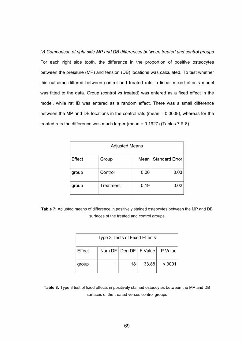
69
iv) Comparison of right side MP and DB differences between treated and control groups
For each right side tooth, the difference in the proportion of positive osteocytes
between the pressure (MP) and tension (DB) locations was calculated. To test whether
this outcome differed between control and treated rats, a linear mixed effects model
was fitted to the data. Group (control vs treated) was entered as a fixed effect in the
model, while rat ID was entered as a random effect. There was a small difference
between the MP and DB locations in the control rats (mean = 0.0008), whereas for the
treated rats the difference was much larger (mean = 0.1927) (Tables 7 & 8).
Adjusted Means
Effect Group Mean Standard Error
group Control 0.00 0.03
group Treatment 0.19 0.02
Table 7: Adjusted means of difference in positively stained osteocytes between the MP and DB
surfaces of the treated and control groups
Type 3 Tests of Fixed Effects
Effect Num DF Den DF F Value P Value
group 1 18 33.88 <.0001
Table 8: Type 3 test of fixed effects in positively stained osteocytes between the MP and DB
surfaces of the treated versus control groups

70
Overall conclusion
There was a significant difference between control and treated rats in the outcome
(MP-DB proportion of positively stained osteocytes) (mean 0.0008 vs. 0.1927, p <
0.0001).
v) Comparison of MP and DB differences between left and right sides in treated group
For each tooth, the difference in the proportion of positive osteocytes between the
pressure (MP) and tension (DB) locations was calculated. To test whether this outcome
differed between left and right sides within the treated group, a linear mixed effects
model was fitted to the data. Side (left vs. right) was entered as a fixed effect in the
model, while rat ID was entered as a random effect (Tables 9 & 10).
Adjusted Means
Effect Side Mean Standard Error
Side Left -0.03 0.02
Side Right 0.19 0.02
Table 9: Adjusted means of difference in positively stained osteocytes between in MP and DB
surfaces between left and right sides in the treated group.
Type 3 Tests of Fixed Effects
Effect Num DF Den DF F Value P Value
side 1 35 98.28 <.0001
Table 10: Type 3 test of fixed effect in positively stained osteocytes between in MP and DB
surfaces between left and right sides in the treated group.
71
Overall conclusion
There was a significant difference between left and right sides in treated rats in the
outcome (MP-DB proportion positive osteoctyes) (mean -0.0316 vs. 0.1927, p<0.0001).
Discussion
The main objective in this study was to determine if mechanical stimulation altered
sclerostin regulation within the alveolar bone of rats. Our results indicate that sclerostin
levels are tightly regulated by mechanical strain evidenced by a differential SOST
expression across the “pressure” (mesio-palatal) and “tension” (disto-buccal) sides of
alveolar bone of an orthodontically treated sample when compared to both a non-
orthodontically strained and an internal control group.
No statistically significant regional differences were found within the disto-buccal or
mesio-palatal surfaces of either the control or tested groups. This was somewhat
unexpected and could have resulted from the force levels in the closed coil spring
being too high. Heavy forces were used to guarantee tooth movement, but the
amplitude could have exceeded the discriminatory mechanosensory threshold of the
osteocytes measured. Also, if the area we measured was too small, the force may
have been sensed over a larger distribution of osteocytes. This area of measurement
was somewhat curtailed by local anatomy, where often the mesial and mesio-buccal
root of the first molar lie in close proximity to each other. No statistical differences were
found between corresponding sites in the day 7 and day 14 test group. This was
expected given the fact that we were using a closed coil spring that, due to its
composition (Nickel-Titanium), exerted a continuous force even though the length
between which it was activated was reducing as the teeth came towards each other. A
difference may have been detected had the spring been made from a material that had

72
a steeper force-deflection curve (eg stainless steel) or had the spring been removed
after a 7 day period.
While the mechanistic pathway of sclerostin has yet to be fully elucidated, several
pathways have been suggested in the literature. Wnt signalling has been shown to be
crucial in the mechanotransduction cascade, as indicated by the observation that the
Lrp5-deficient mouse skeleton is unable to respond anabolically to mechanical
stimulation.[15] Several experiments have demonstrated enhanced Wnt signalling in
bone cells as a result of mechanical stimulation. For example, stretched osteoblasts
harvested from transgenic mice exhibited activation of canonical Wnt signalling after
stretching.[16] Robling and co-workers have shown the inhibitory effects of sclerostin
on Lrp function and bone formation, implicating it as a mechanically suppressed signal,
providing an attractive regulatory mechanism that permits enhanced Wnt signalling
upon mechanical stimulation.[10]
The mineralisation of bone, that is the formation of hydroxyapatite, is a highly regulated
process. The details of this regulation are also not fully understood but sclerostin has
recently been proposed as a potential source of control of mineralisation within mature
bone.[17] The TNF-family member, TWEAK, inhibits mineralisation by human
osteoblasts. This action was accompanied by TWEAK inducing the formation of both
SOST/sclerostin mRNA and protein.[18] When human osteoblasts had recombinant
exogenous sclerostin applied to them, it was found that mineral deposition was highly
sensitive to sclerostin, with concentrations as low as 10 ng/ml inhibiting cell layer-
associated Ca2+. The inhibition of mineralisation by sclerostin was associated with
reduced expression by the osteoblasts of mRNA encoding the bone formation markers
osteocalcin and collagen I, and increased expression of the pre-osteocyte marker E11.
This implies that sclerostin acts in part by inhibiting the progression of cells through to
the mature osteocyte stage.[17] Consistently, the same study also showed that DMP1

73
expression was also reduced in response to sclerostin treatment.[17] These results
indicate that sclerostin inhibits the differentiation of human osteoblasts and/or their
ability to regulate mineral deposition.
Our study showed that osteocytes on either side of alveolar bone under mechanical
strain had a different response to alteration in sclerostin expression, facilitating tooth
movement in the direction of the force vector applied to it. It would be interesting to
know how this discriminatory mechanotransduction takes place, and whether it lies
within the osteocyte, or is downstream to signals from the periodontal ligament.
Another issue that remains unclear is the identity of the upstream factors that control
sclerostin expression and whether sclerostin can be modulated by known classical
early mediators of mechanotransduction (e.g. ATP, Ca, PGE2, and NO) or if sclerostin
is controlled by other novel mechanisms.
The osteocyte-specific expression profile of sclerostin provides a long sought-after
molecular mechanism by which osteocytes per se can relay mechanical information
biochemically to the effector cell populations (chiefly osteoblasts and osteoclasts).
From the results of this study, we hypothesise that when significant local strains are
sensed by the osteocyte cell population on the side of tension, sclerostin levels are
decreased, and the local osteoblast cell populations are released from their
suppressed Wnt signaling state, ultimately resulting in new local bone formation in the
region. Conversely, when significant local strains are sensed by the osteocyte cell
population on the side of compression, sclerostin levels are increased with continued
Wnt signalling suppression, leading to a prevention of local bone formation.

74
Conclusion
Sclerostin levels were significantly altered in the alveolar bone around rat molars
subject to orthodontic tooth movement. These data highlight the role of the osteocyte in
mechanically induced bone formation and suppression and provide the first known
evidence for osteocyte-specific control of mechanotransduction in alveolar bone.
Furthermore, sclerostin presents a novel mechanism for modulating alveolar bone
turnover and mineralisation in response to mechanical strain. Pharmacologic
modulation of sclerostin signalling emerges as an obvious potential therapy for
improved treatment efficiency in orthodontic treatment.
75
Acknowledgments
Funding: The Australian Society of Orthodontists Foundation for
Research and Education provided partial funding for the project.
Conflict of interest: There is no foreseeable conflict of interest.
Ethical approval: The ethics approval was obtained from the University of
Adelaide Animal Ethics Committee (M/023/2006).
Statistician: Mr Thomas Sullivan, Statistician, Discipline of Public Health,
University of Adelaide.
Bone biologist: Dr Brendon Noble, Scottish Mechanotransduction Consortium,
University of Edinburgh Medical School, Edinburgh, UK. ��
�
�
�
�
�
�
�
�

76
References �
1. Meikle M. The tissue, cellular, and molecular regulation of orthodontic tooth
movement: 100 years after Carl Sandstedt. European Journal of Orthodontics
2006;28:221-240.
2. Hěrt J, Lisková M, Landa J. Reaction of bone to mechanical stimuli. Part 1:
Continuous and intermittent loading of tibia in rabbit. . Folia Morphologica (Praha)
1971;19:290–300.
3. Melsen B. Tissue reaction to orthodontic tooth movement- a new paradigm.
European Journal of Orthodontics 2001;23:671-681.
4. Masella R, Meister M. Current concepts in the biology of orthodontic tooth
movement. Am J Orthod Dentofacial Orthop American Journal of Orthodontics and
Dentofacial Orthopaedics 2006;129:458-468.
5. Noble B. The osteocyte lineage. Archives of Biochemistry and Biophysics
2008;473:106-111.
6. Hadjidakis D, Androulakis I. Bone Remodelling. Annals of the New York Academy of
Sciences 2006;1092:385-396.
7. Bonewald L, Johnston M. Osteocytes, Mechanosensing and Wnt Signaling. Bone
2008;42(4):606-615.
8. ten Dijke P, Krause C, de Gorter D, Lowik S, Rutger L, Bezooijen v. Osteocyte-
derived sclerostin inhibits bone formation: its role in bone morphogenetic proteing and
Wnt signalling. Journal of Bone ane Joint Surgery 2008;90:31-35.
9. Jäger A, Götz W, Lossdörfer S, Rath-Deschner B. Localization of SOST/sclerostin in
cementocytes in vivo and in mineralizing periodontal ligament cells in vitro. Journal of
Periodontal Research 2009;45(2):246-254.
10. Robling A, Niziolek P, Baldridge L, Condon K, Allen M, Alam I. Mechanical
stimulation of bone in vivo reduces osteocyte expression of Sost/sclerostin. Journal of
Biological Chemistry 2008;283:5866-5875.

77
11. Lacey D. Inhibition of sclerostin as an anabolic therapeutic approach for bone loss.
Bone 2008;43(Supplement 1):S22.
12. Poole K, van Bazooijen R, Loveridge N, Hamersma H, Papapoulos S, Lowik C, et
al. Sclerostin is a delayed secreted product of osteocytes that inhibits bone formation.
The FASEB Journal 2005:1842-1844.
13. Winkler D, Sutherland M, Yu C, Skonier J, Hayes T, Geoghegan J. Osteocyte
control of bone formation via sclerostin, a novel BMP antagonist. EMBO 2003;22:6267-
6276.
14. Ren Y, Maltha JC, Kuijpers-Jagtman AM. The rat as a model for orthodontic tooth
movement- a critical review and a proposed solution. European Journal of Orthodontics
2004;26:483-490.
15. Sawakami K, Robling A, Ai M, Pitner N, Liu D, Warden S, et al. The Wnt co-
receptor LRP5 is essential for skeletal mechanotransduction but not for the anabolic
bone response to parathyroid hormone treatment. Journal of Biological Chemistry 2006
281:23698–23711.
16. Hens J, Wilson K, Dann P, Chen X, Horowitz M, Wysolmerski J. TOPGAL mice
show that the canonical Wnt signaling pathway is active during bone development and
growth and is activated by mechanical loading in vitro. Journal of Bone and Mineral
Research 2005;20:1103–1113.
17. Lim H, Wijenayaka A, Welldon K, Atkins G, Findlay D. Sclerostin inhibits
mineralisation by human osteoblasts. Bone 2010;47:S121.
18. Vincent C, Findlay D, Welldon K, Wijenayaka A, Zheng T, Haynes D, et al.
Proinflammatory cytokines TNF-related weak inducer of apoptosis (TWEAK) and
TNFalpha induce the mitogen-activated protein kinase (MAPK)-dependent expression
of sclerostin in human osteoblasts. . Journal of Bone and Mineral Research
2009;24:1434-1449.

78
�� ARTICLE TWO
RATE OF ORTHODONTIC TOOTH MOVEMENT AND
DIFFERENTIAL SCLEROSTIN EXPRESSION IN THE RAT
FOR SUBMISSION TO THE ARCHIVES OF ORAL BIOLOGY

79
Abstract
Design: Orthodontic NiTi pullcoil springs(100cN) were ligated to the right maxillary first
molar and maxillary incisors of 12 eight week old Sprague-Dawley rats. The rats were
divided into two groups; one sacrificed at 7 and the other at 14 days. Serial frozen
sections were obtained and immunohistochemical tissue labelling was performed,
involving the avidin-biotin peroxidase technique and goat polyclonal anti-mouse
sclerostin as the primary antibody. Histomorphometric analysis of digital
photomicrographs allowed quantification and comparison of immunolabelled
osteocytes resident in the mesio-palatal and disto-buccal alveolar bone of the maxillary
first molar mesial root. This differential was expressed as a percentage. The amount of
tooth movement between the incisors and first molars was measured using an indirect
method and compared with the differential sclerostin staining percentage of each
animal. A linear regression analysis was performed using the paired sets of data with
GraphPad Prism 5 software.
Results: The analysis showed there was no significant association between differential
sclerostin staining and the amount of tooth movement measured at day 7 (p = 0.16)
and a significant association at day 14 (p = 0.03).
Conclusion: Tooth movement and sclerostin expression are affected by multiple
variables; however, a linear association between osteocyte-driven sclerostin
expression and the rate of tooth movement may be present once hyalinised tissue has
been eliminated.
Objective: Osteocytic changes involving the release of sclerostin have been shown to
occur in the alveolar bone of teeth subjected to orthodontic tooth movement. The aim
of this study was to examine whether differential sclerostin expression around a tooth
has a linear association with measured orthodontic tooth movement in the rat.

80
Introduction
Alveolar bone is loaded intermittently during mastication, but is subject to a more
continuous deformation or strain during orthodontic tooth movement.[1] Alveolar bone
significantly differs from the rest of the skeleton in its remodelling response when
placed under a mechanical load. The balance favours bone deposition on the side of
the alveolar wall undergoing tension and bone resorption on the side of the wall
undergoing compression, allowing the tooth to “move” along the force vector being
applied to it. This complex cascade of events involves a mechanical signal being
converted into a biological response. Recent literature has focused on the osteocyte as
the chief mechaonosensor within bone.
Sclerostin, a protein product encoded by the SOST gene, is a known negative regulator
of osteoblast differentiation and function, and acts as a inhibitor to bone formation.[2-4]
It has also been hypothesised that sclerostin released by the mature osteocyte may
have a role in regulating bone mineralisation surrounding the “osteoid osteocyte”. In its
role as a key inhibitor, sclerostin helps determine the normal extent of bone formation
and, consequently, protects against the deleterious effects of uncontrolled bone
growth.[5] Its effect is highlighted by two rare high bone mass diseases, Sclerosteosis
and Van Buchem disease, both of which have been linked to inactivating mutations in
the SOST gene.[3,6] The administration of an anti-sclerostin antibody has been shown
to increase bone formation and restore the bone loss upon ovariectomy in rodents.[7] It
has been suggested that the inhibition of sclerostin (through sclerostin neutralizing
antibodies) may, therefore, be an attractive strategy for conditions in which bone loss is
a significant component, such as postmenopausal and senile osteoporosis.[5]
Conversely, transgenic mice overexpressing sclerostin have been shown to exhibit low
bone mass.[8]

81
The results of a previous study (Article 1) indicated that teeth subjected to orthodontic
tooth movement showed an increase in the difference between positively stained
osteocytes between mesio-palatal and disto-buccal surfaces of alveolar bone around
the rat first molar. The present study aims to evaluate a potential association between
the amount of this differential and the amount of orthodontic tooth movement achieved
over a given period of time. The null hypothesis to be investigated is there is no
association between differential sclerostin expression and the amount of orthodontic
tooth movement.
�
Materials and methods
The ethics approval for this study was provided by the University of Adelaide Animal
Ethics Committee number M/023/2006 and the experimental procedures and care of
animals was in accordance with the European Convention for the Protection of
Vertebrate Animals used for Experimental and Other Scientific Purposes.
i) Animals, Appliances and Animal Manipulations
This study used twelve, eight week old Sprague-Dawley rats which were contained at
the Animal House facility of the Medical School of the University of Adelaide.
Anaesthesia for the rats was administered via a subcutaneuous injection containing a
combination of Ketamine (100mg/ml) (Ketamil Injection, Troy Laboratories, Smithfield,
Australia) and Xylazine (Xylazil, 20mg/ml) at a dosage of 2ml/kg. Twelve animals had a
100cN superelastic nickel titanium closing spring (Sentalloy, GAC, New York, USA)
ligated to the right maxillary first molar and to the maxillary incisors using 0.010”
stainless steel ligatures and a light cured bonding material (Z100 composite resin, 3M
ESPE, St. Paul, USA). This imparted a mesially directed force to the right maxillary first
molar, using a protocol developed by Noxon et al. (9).

82
ii)Tooth movement measurements
A protocol developed by Ho [10] for molar-to-incisor distance measurement was used
on the anaesthetised animal via direct and indirect means (see Appendix 2). For the
direct method, a digital calliper was used to take three sets of readings from the labial
surface of the incisor to the mesial surface of the first molar and a mean value was
calculated from the three readings. Readings were performed by the same operator,
and recorded at day 0, 7 and 14. In the present study, the measurements at day 0 to
day 7 and day 0 to 14 were used to form paired data, along with the differential
sclerostin staining percentages for each animal (animals were sacrificed at either day 7
or day 14 for immunological staining). The indirect method involved a polyvinyl siloxane
impression (Imprint 3, 3M ESPE) of the maxillary arch using a special tray for the
anaesthetised animal. Images of the impressions were taken with a MZ16FA stereo
microscope and were calibrated using Image analySIS software. Computer software
was used to measure the distance between three constructed centroid points
representing the incisors and the first molar on the digitised images (Appendix 2).
iii) Harvesting of tissue
The animals were sacrificed via cardiac left ventricular perfusion with fixative solutions,
after an intraperitoneal administration of chloral hydrate 5g/100ml H2O (5ml). The
perfusion protocol involving 200ml 0.1% sodium nitrite and a 400ml solution of 50:50
4% para-formaldehyde and p-benzoquinone injected under positive pressure. Of the
twelve animals that had spring placement and injection performed at day 0, six animals
were perfused at 7 days and six animals were perfused at 14 days. Each animal had
the maxilla removed and stored in the 50:50 fixative solution for two hours, followed by
4% EDTA decalcifying solution pH 7.4 for 8 weeks. The decalcified tissue was placed
within an embedding medium (OCT, Sakura Finetek, Torance, CA, USA) for
cryosectioning.

83
iv) Section preparation
Horizontal sections 25μm thick were obtained using a Leitz 1720 cryostat at -20°C. The
maxillae were orientated so that the crowns of teeth were uppermost. Sections were
collected sequentially from the furcation in an apical direction to the root apex (Figure
1).
Figure 1: Schematic representation of the level at which the section was cut through maxillary
first molar (M1) and maxillary second molar (M2) roots below the furcation.
Serial sections were placed consecutively over 12 slides, so that each slide contained
sections representing different levels of the alveolar bone for comparison (Figure 2).
Positive controls were provided via separate slides of rat tibia included in the staining
runs.
Figure 2: Arrangement of specimen sections on slides

84
v) Immunohistochemistry
For immunohistochemical labelling of sclerostin, the avidin-biotin peroxidase complex
technique was used involving goat anti-mouse-SOST polyclonal antibody (R & D
Systems, MN, USA) as the primary antibody. The anti-mouse-SOST was diluted from
lyophilised form (200μg/ml) to 1:75 in PBS. The optimal primary antibody concentration
was calculated through a pilot study using similar tissue to the area studied. Secondary
antibody labeling and detection was accomplished using the Vectastain Universal
Quick-Kit (Vector Lab. Inc.) with 3-amino-9-ethylcarbazole (AEC) as the chromogen.
Slides from the different groups were labeled in the same solutions along with both
positive and negative control tissue (rat tibia) known to have SOST-immunoreactive
tissue. For a negative control, primary antibody was omitted (R & D Systems, MN,
USA).
vi) Histoquantification
A photomicroscope was used to capture immunolabeled sections. For image
quantification, the number of sclerostin positive osteocytes, defined as those osteocyte
cell bodies exhibiting red (AEC) staining (Figure 3), and the number of sclerostin-
negative (Sclr-) osteocytes, defined as those osteocyte cell bodies exhibiting blue
(haematoxylin) staining, were counted on the mesio-palatal (MP) and the disto-buccal
(DB) section of alveolar bone surrounding the mesial root of the first maxillary molar
(Figure 4). These were areas of interest because the force vector was directed mesio-
palatally and, as such, the MP surface was expected to be a compression site
(undergo bone resorption) and the DB surface a tension site (undergo bony
apposition). The percentage of sclerostin-positive cells (Sclr+) was calculated as the
number of Sclr+ cells divided by the total number of cells (Sclr+ plus Sclr–).
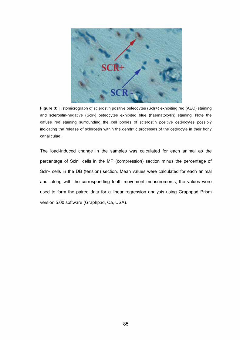
85
Figure 3: Histomicrograph of sclerostin positive osteocytes (Sclr+) exhibiting red (AEC) staining
and sclerostin-negative (Sclr-) osteocytes exhibited blue (haematoxylin) staining. Note the
diffuse red staining surrounding the cell bodies of sclerostin positive osteocytes possibly
indicating the release of sclerostin within the dendritic processes of the osteocyte in their bony
canaliculae.
The load-induced change in the samples was calculated for each animal as the
percentage of Sclr+ cells in the MP (compression) section minus the percentage of
Sclr+ cells in the DB (tension) section. Mean values were calculated for each animal
and, along with the corresponding tooth movement measurements, the values were
used to form the paired data for a linear regression analysis using Graphpad Prism
version 5.00 software (Graphpad, Ca, USA).

86
�Figure 4: The middle panel shows a transverse section of the mesial root immunolabelled for
sclerostin at a magnification of 5x. At this power, individual osteocytes can not be identified. The
orange arrow represents the direction of orthodontic tooth movement, where the mesio-palatal
surface in this case represents the area of “compression” and the disto-palatal surface the area
of “tension”. At this power, representative sections of alveolar bone from the areas of tension
and compression were chosen. The upper panel represents the mesio-palatal region of interest.
The lower panel represents the disto-buccal surface. A percentage difference between
positively stained osteocytes in mesio-palatal (MP) and disto-buccal (DB) surfaces was
calculated and compared between treated groups (7 days and 14 days).
�
�
��

87
Results
The tooth movement results are shown in Appendix 3. Measurement calculations were
performed using SAS Version 9.1 software (SAS Institute Inc., Cary, NC, USA), and a
linear regression analysis of scatter plot diagrams using the paired data was performed
(Tables 1 and 2, and Figures 5-8).
�
Animal
% of Scl +ve
osteocytes (MP)
% of Scl +ve
osteocytes (DB)
Differential % of Scl +ve osteocytes
(MP-DB)
Distance day 0 to
day 7 Indirect (mm)
102 58.16 28.48 29.68 0.2
104 67.75 36.94 30.81 0.58
106 57.12 42.41 14.71 0.76
301 47.84 32.93 14.91 1.85
302 50.50 24.79 25.71 1.04
303 57.48 38.14 19.34 0.81
�
Table 1: Tooth movement measured between the right maxillary first molars and incisors over
day 0 – day 7. Percentage of osteocytes positively stained for sclerostin was recorded for the
mesial root of the treated maxillary first molars.
Figure 5: Scatter plot graph representing the paired data between the differential +ve Sclr
percentage and the indirectly measured distance (incisor to 1st molar) from day 0 to day 7. The
solid line shows the line of best fit. Dotted lines show 95% confidence levels. R square value =
0.4284. Sy.x = 0. 4688. The slope is not significantly non-zero (P = 0.1584).
�

88
Animal
% of Scl +ve
osteocytes (MP)
% of Scl +ve
osteocytes (DB)
Differential % of Scl +ve osteocytes
(MP-DB)
Distance day 0 to day 14
Indirect (mm)
201 50.52 34.48 16.05 1.83
401 57.48 38.14 19.34 2.28
402 51.14 33.06 18.07 1.64
405 51.95 39.30 12.64 1.94
407 56.51 32.92 23.59 2.45
408 52.60 46.23 06.37 1.32
Table 2: Tooth movement measured between the right maxillary first molars and incisors over
day 0 – day 14. Percentage of osteocytes positively stained for sclerostin was recorded for the
mesial root of the treated maxillary first molars.
�
Figure 6: Scatter plot graph representing the paired data between the differential +ve Sclr
percentage and the indirectly measured distance (incisor to 1st molar) from day 0 to day 14. The
solid line shows the line of best fit. Dotted lines show 95% confidence levels. R square value =
0.7064. Sy.x = 0. 2508. The slope is considered significant (P = 0.0361).
�
�
�
�
�
�
�
�
�
�

89
Discussion
Adult Sprague-Dawley rats were used in this study because of their availability, ease of
handling, cost and their biological and structural similarities to human periodontium and
alveolar bone. Male rats were used to negate any hormonal-related variables.[11] The
rat model also allows for better control of genetic variability, easier monitoring of animal
number and conditions (diet and environment), increased sample size and more
manageable size of the region to be studied (the maxillary molar). Sprague Dawley
rats are an outbred strain of rat and ideally an inbred strain of animal would have
allowed for isogenicity, but not necessarily phenotypic uniformity due to individual
variations in the environment of each animal. This heterogeneticity may account for an
increased individual variability in the response to the treatment effect.[12]
The present study incorporated tissue obtained from a previous study [10], after which
the maxillary tissue of twelve treated animals were stored in EDTA medium for up to 18
months in a dark store room at room temperature. Although every effort was made to
preserve this tissue in its optimal state, the lengthy storage period may have enabled
some protein denaturation.
i) Application of orthodontic force to rat molars
Various methods have been described for application of an orthodontic force to a rat
molar, each with varying degrees of success. Waldo and Rothblatt used an orthodontic
elastic between rat molars to stimulate orthodontic movement.[13] Orthodontic modules
(alastics) have also been used, and are often made from polyurethane, which does not
show ideal elastic characteristics. Time, temperature, salivary pH and water absorption
have all been found to result in force loss, permanent deformation and strength loss of
elastomerics modules.[14] Using this tooth movement model, O’Hara et al. noted that
only 66% (26 out of 42) of the animals were found to have the elastic modules retained
at the time of sacrifice, where the longer the module was left between the teeth, the

90
greater the risk of its loss.[15] The study also showed there was no consistency in the
direction of tooth movement and level of response. O’Hara’s tooth movement model
also involved a force being delivered to the tooth via decompression (as opposed to
contraction) of an elastomeric, causing tooth movement to be translatory, tipping or
rotational depending upon the depth of elastomeric module insertion, tightness of
interproximal contact, and duration of application. Unfortunately, there has been
inadequate evidence in the literature regarding the force decay rate, the amount of
force decay and the dimensional changes of elastomeric modules during
compression.[14]
The method used in this experimental sample was based on a modified version of the
Brudvig and Rygh protocol, which advocated the use of a closed coil spring ligated
between an eyelet on an incisor band and the upper first molar.[16-21] The present
study used a continuous force level of 100 cN generated by a NiTi coil spring, although
forces as low as 10cN (which if equated by surface area would be equivalent to a
170cN force exerted on a human molar) have been advocated by Ren et al.[14] While it
could be considered that the force levels delivered in our study were too high, we
wanted to maximise the likelihood of orthodontic tooth movement. A steel ligature was
used to tie in the rat molar, which could have caused localised trauma and a
subsequent inflammatory response. Tooth movement is also based on a modified
inflammatory response and thus this overlap could have confounded our results.
Bonding the spring to the rat molar could alleviate this issue, but had the added risk of
spring loss during mastication. A mesially directed force on the rat maxillary molar was
used in order to oppose the normal physiological conditions these teeth naturally
encounter as they have been shown to distally drift at a rate of 7.7μm/day in adult male
Sprague Dawley rats.[12]

91
ii) Sclerostin-immunoreactivity
The linear regression analysis showed a statistically significant association between
the difference in osteocytes positively stained for sclerostin on either side of the MB
root of the maxillary first molar and the amount of tooth movement measured (p < 0.05)
from day 0 to day 14. However, there was no significant association between
differential sclerostin staining and the amount of tooth movement measured (p > 0.05)
from day 0 to day 7. This may be a true representation of the underlying biological
phenomena or may result from small sample sizes, variability in measurements or
possible method errors.
Studies over the past 100 years have shown that a phenomena termed hyalinization is
often observed during the initial phases of tooth movement in both animal and human
models.[1] On the pressure side of tooth movement, the first biological event that takes
place following force application is a disturbance of blood flow in the periodontal
ligament (PDL). Cessation of blood flow results in a sterile cell death in the compressed
area of the PDL, a process termed hyalinization. An influx of phagocytotic cells such as
macrophages, foreign body giant cells and osteoclasts invade from the adjacent
undamaged areas of the PDL and eliminate this necrotic tissue, a process termed
undermining resorption. It has been purported that until this hyalinised tissue has been
removed, tooth movement will not take place.[22] Hyalinisation is considered to be an
undesirable, and somewhat unavoidable sequela of orthodontic tooth movement as the
need to remove the necrotised tissue prolongs the initiation of tooth movement.
Previous studies have shown that, in a rat model, this hyalinised layer appears as
quickly as 24 hours following orthodontic force application; varies vastly and is thought
to be related to a multitude of factors including the magnitude and direction of the
orthodontic force, and type of tooth movement initiated.[22] The commonly accepted
theory within the field of orthodontics was that complete hyalinisation tissue removal
was needed before tooth movement could start but it has been recently shown that

92
even in the later stages of tooth movement small hyalinised patches can still be
found.[23] The results of our study tie in to the undermining resorption phenomena as
our day 7 readings could be distorted by the fact that not enough hyalinised tissue was
removed, and thus the rate of tooth movement was inhibited. An additional confounder
could be the fact that bone formation in the deeper region of the pressure side has
been previously demonstrated, which would serve to result in fewer sclerostin positive
osteocytes.[24] At day 14, it is feasible that enough undermining resorption would have
occurred in order for tooth movement to progress, thus allowing for a linearity between
sclerostin expression and orthodontic tooth movement.
Conclusion
The results of the present study indicate there is a statistically significant association
between the differential sclerostin staining intensity and the amount of tooth movement
measured (p>0.05) after a 14 day period. This linear association between sclerostin
staining intensity and amount of tooth movement achieved suggests that while rate of
tooth movement and sclerostin expression are affected by multiple variables, a linear
association between the two may be present once hyalinised tissue has been
eliminated.

93
Acknowledgments�
Funding: The Australian Society of Orthodontists Foundation for
Research and Education provided partial funding for the project.
Conflict of interest: There is no foreseeable conflict of interest.
Ethical approval: The ethics approval was obtained from the University of
Adelaide Animal Ethics Committee (M/023/2006).
Statistician: Mr Thomas Sullivan, Statistician, Discipline of Public Health,
University of Adelaide.
Bone biologist: Dr Brendon Noble, Scottish Mechanotransduction Consortium,
University of Edinburgh Medical School, Edinburgh, UK.

94
References
1. Meikle M. The tissue, cellular, and molecular regulation of orthodontic tooth
movement: 100 years after Carl Sandstedt. European Journal of Orthodontics
2006;28:221-240.
2. Noble B. The osteocyte lineage. Archives of Biochemistry and Biophysics
2008;473:106-111.
3. Lacey D. Inhibition of sclerostin as an anabolic therapeutic approach for bone loss.
Bone 2008;43(Supplement 1):S22.
4. Yavropoulou M, Yovos J. The role of Wnt signalling pathway in osteoblast
commitment and differentiation. Hormones 2007;6(4):279-294.
5. Poole K, van Bazooijen R, Loveridge N, Hamersma H, Papapoulos S, Lowik C, et al.
Sclerostin is a delayed secreted product of osteocytes that inhibits bone formation. The
FASEB Journal 2005:1842-1844.
6. Winkler D, Sutherland M, Yu C, Skonier J, Hayes T, Geoghegan J. Osteocyte control
of bone formation via sclerostin, a novel BMP antagonist. EMBO 2003;22:6267-6276.
7. Warmington K, Morony S, Sarosi I. Sclerostin antagonism in adult rodents, via
monoclonal antibody mediated blockade, increases bone mineral density and
implicates sclerostin as a key regulator of bone mass during adulthood. Journal of
Bone and Mineral Research 2004;19:S56.
8. Loots G, Kneissel M, Keller H, Baptist M, Chang J, Collette N, et al. Genomic
deletion of a long-range bone enhancer misregulates sclerostin in Van Buchem
disease. Genome Research 2005;15:928-935.
9. Noxon SJ, King GJ, Gu G, Huang G. Osteoclast clearance from periodontal tissues
during orthodontic tooth movement. Am J Orthod Dentofacial Orthop 2001;120(5):466-
476.
10. Ho SH. An immunohistochemical study of neurotrophic factors and associated cells
in the rat dento-alveolar complex subjected to orthodontic forces. Thesis. University of
Adelaide, Adelaide; 2007.

95
11. King GJ, Latta L, Rutenberg J, Ossi A, Keeling SD. Alveolar bone turnover and
tooth movement in male rats after removal of orthodontic appliances. Am J Orthod
Dentofacial Orthop 1997;111(3):266-275.
12. King GJ, Keeling SD, McCoy EA, Ward TH. Measuring dental drift and orthodontic
tooth movement in response to various initial forces in adult rats. . American Journal of
Orthodontics and Dentofacial Orthopaedics 1991;99(5):456-465.
13. Waldo CM, Rothblatt JM. Histological response to tooth movement in the laboratory
rat. J Dent Res 1954;33(4):481-486.
14. Ren Y, Maltha JC, Kuijpers-Jagtman AM. The rat as a model for orthodontic tooth
movement- a critical review and a proposed solution. European Journal of Orthodontics
2004;26:483-490.
15. O'Hara A. An immunohistochemcial investigation into the expression of nerve
growth factor and its receptors in the rat dento-alveolar complex subjected to
orthodontic forces. Thesis University of adelaide, Adelaide; 2005.
16. Brudvik P, Rygh P. Non-clast cells start orthodontic root resorption in the periphery
of hyalinized zones. Eur J Orthod 1993;15(6):467-480.
17. Brudvik P, Rygh P. The initial phase of orthodontic root resorption incident to local
compression of the periodontal ligament. Eur J Orthod 1993;15(4):249-263.
18. Brudvik P, Rygh P. Multi-nucleated cells remove the main hyalinized tissue and
start resorption of adjacent root surfaces. Eur J Orthod 1994;16(4):265-273.
19. Brudvik P, Rygh P. Root resorption beneath the main hyalinized zone. Eur J Orthod
1994;16(4):249-263.
20. Brudvik P, Rygh P. Transition and determinants of orthodontic root resorption-
repair sequence. Eur J Orthod 1995;17(3):177-188.
21. Brudvik P, Rygh P. The repair of orthodontic root resorption: an ultrastructural
study. Eur J Orthod 1995;17(3):189-198.

96
22. von Bohl M, Kuijpers-Jagtman AM. Hyalinization during orthodontic tooth
movement: a systematic review on tissue reactions. European Journal of Orthodontics
2009;31(1):30-36.
23. von Bohl M, Maltha, JC, Von den Hoff JW Kuijpers-Jagtman AM. Focal
hyalinization during experimental tooth movement in beagle dogs. American Journal of
Orthodontics and Dentofacial Orthopaedics 2004;125:615-623.
24. Melsen B. Tissue reaction to orthodontic tooth movement- a new paradigm.
European Journal of Orthodontics 2001;23:671-681.

97
SUMMARY

98
The literature review described the structure and function of alveolar bone, showing it
to be a dynamic, complex and highly structured tissue. A particular emphasis was
placed on both mechanotransduction, the conversion of a mechanical force into a
biologic response and sclerostin, an osteogenic inhibitor uniquely expressed by
osteocytes. Alveolar bone was shown to have unique remodelling characteristics when
a mechanical strain was placed through it. This response differentiates it from bone
found elsewhere in the body and much of the molecular biology that controls this local
bone turnover has yet to be elucidated.
In this study, the mineralised response to orthodontic tooth movement was investigated
in the dento-alveolar region of Sprague-Dawley rat molars subject to an orthodontic
force. The main aim of this study was to examine changes that occur in the distribution
of osteocytes positively stained for sclerostin in the alveolar bone of orthodontically
stimulated teeth. Furthermore, the study aimed to find out if there was a linear
association between the rate of orthodontic movement experienced by a tooth and the
differential sclerostin expression across it.
The findings in paper one showed a statistically significant change in the sclerostin
expression profile of alveolar bone, when subject to an orthodontic force. This
difference was attributable to an increase in sclerostin positive osteocytes on the
compression side and a decrease in sclerostin positive osteocytes on the tension side
of the rat molar. This is a remarkable finding in that it ascribes a key role to the
osteocyte in the processes of orthodontic tooth movement and local alveolar bone
turnover.
The second study involved measuring tooth movement, indirectly, within the treated
sample and correlating this data with the differential sclerostin staining intensity
obtained from the previous study. No significant association was found in the

99
comparison with the 7 day group; however, a significant association between the
amount of differential sclerostin expression and tooth movement was found for the day
14 group. This difference may be attributable and correlate to the length of time taken
to remove the hyalinised layer.
There were some limitations to the present study. These were as follow:
o Limited sample size.
o Heavy orthodontic force used to establish movement in the rat molar.
o Length of storage time of decalcified maxillary tissue prior to cryosectioning.
o Thickness of the sections.
o Measurements were only conducted over a 14 day period.
o Only one operator was used to measure tooth movement and count positively
stained osteocytes.
Some of the strength of the current study were as follows:
o A protocol was established that routinely delivered tooth movement.
o Power study used to determine sample size.
o Internal and external controls were used.
o Quantitative approach to measure cellular response.
o Error study to determine measurement accuracy.
o Blinding of the operator when measuring data.
Despite the limitations of the study, all the proposed aims have been achieved. The
results refuted all the initial null hypotheses and the overall conclusions drawn from the
data provided suggest that sclerostin plays a significant role in the conversion of a
mechanical signal into a biological response within alveolar bone. Given there are no
other studies that have investigated the role of sclerostin within alveolar bone it makes
extrapolation and corroboration of our results difficult. It emphasises the need for

100
supplementary studies to be conducted in this area, particularly given the results
shown. Future research should focus on gaining a further insight into the series of
events that precede and follow sclerostin expression, and how far downstream in the
mechanotransductory sequence the osteocytic response lies. Can the osteocyte be
considered to be the chief orchestrator of mechanically driven local alveolar bone
turnover or is it merely part of a response mechanism secondary to other local events?
Further exploration into the cellular events that are coupled with sclerostin expression,
particularly osteoblast apoptosis, control of osteoid mineralisation, and osteoclastic
activity could help determine this.

101
APENDICIES

102
Appendix 1.
The Application of Orthodontic Force on the Rat Molars
Twelve animals were prepared in a previous study. The animals were weighed and
anaesthetized to allow for placement of the closed-coil spring, and also prior to
perfusion fixation at the end of the study.
1. Placement of Closed-Coil Spring: The rats were anaesthetized with Ketamine
(100mg/ml) (Ketamil Injection, Troy Laboratories, Smithfield Australia) and Xylazine
(Xylazil, 20mg/ml) via intraperitoneal injection at a dosage of 2ml/kg of body weight.
The two drugs were mixed at the ratio of 2:1 (Ketamine: Xylazine) and then diluted 1:1
with sterile water for injection. This allowed a short period of anaesthesia allow for the
placement of closed coil springs. Anaesthesia was tested via observation of a plantar
reflex and chest movements.
2. Application of Orthodontic Force: The anaesthetised animal was placed onto a
holding rack (figure 1). The mouth was gently propped open by two rings attached with
elastic bands to the rack. A split-mouth design was used with the experimental side on
the right, and the contralateral as the control. A 100 cN super-elastic NiTi closing coil
spring (Sentalloy, GAC, New York, USA Catalogue number 10-000-06) was used to
impart a mesially-directed force to the right maxillary first molar tooth. The springs
were ligated with 0.010” stainless steel ligature wire anteriorly to the maxillary incisors
and posteriorly to the cervical region of the right maxillary first molar. Springs were
previously calibrated such that the force levels studied were within its superelastic
range. Light curing bonding material (3M ESPE Z100 Restorative, St. Paul, USA) was
used for bonding the 0.010” stainless steel ligature end of the closed-coil spring to the
specific tooth. Care was taken not to injure the surrounding soft tissue (Figure 2).

103
Figure 1: Animal was placed onto a purpose-built holding rack
Figure 2: Photograph of experimental spring setup with wire ligature

104
Appendix 2.
Measurement of tooth movement
1. Tooth movement was measured indirectly. Polyvinyl siloxane impressions
(Imprint 3, 3M ESPE) were taken intra-orally with a special customised acrylic tray at
Days 0, 7 and 14 (Figure 1). Photos of the impressions were taken under light
microscopy (MZ16FA stereo microscope) and were calibrated by Image Analysis
software (analySIS® a SOFT IMAGING SYSTEM product).
Figure 1: Impression taken with special tray
2. The image capturing procedure involved recording the animal number and date
of impression. A millimetre rule was photographed adjacent to the set impression, and
the image captured and digitised using Image analysis software.
3. Landmarks of the first molar of both sides and the central incisors were located
and digitised in a specific order and their X and Y coordinates were collected (Figure
2). Four anatomical landmarks along the circumference of the clinical crown were
recorded for the incisors, the first and second molars. These were the distobuccal,
mesiobuccal, mesiopalatal and distopalatal points, and a centroid was constructed by
using these four points (Figure 3).

105
5. Measurements were made between the centroids of the first and second molars,
and between the centroids of the first molar and incisors, for both the left and right
sides. These were recorded and tabulated at specified time points (Appendix 3).
Figure 2: Impression landmarks
Figure 3: Illustration of centroid determination

106
Appendix 3.
Tooth measurement results
Tooth Movement (Day 0)
rat no. C T Molar 1-2
101s * * *
102s 16.80 16.42 1.91
103s 16.17 16.27 2.09
104n 16.26 16.72 2.06
105n * * *
106n 16.65 16.68 2.09
201s 14.79 15.22 2.30
202s 14.38 14.95 2.01
203s 15.22 15.23 2.32
204n * * *
205n 14.84 15.08 2.16
206n * * *
301s 15.93 16.19 2.34
302s 16.30 16.49 2.33
303s 15.86 15.96 2.25
304s 15.92 16.15 2.19
305n 16.00 16.11 2.05
306n 16.00 16.06 2.18
307n 16.19 16.26 2.36
308n 15.94 16.02 2.51
401s 15.84 15.82 2.19
402s 15.94 16.38 2.20
403s 15.71 15.58 2.32
404s 16.06 16.16 2.31
405n 15.41 15.73 2.05
406n 16.05 15.78 2.38
407n 15.25 15.05 2.20
408n 15.69 15.56 2.37
Table 1: Tooth movement measurements of Day 0 (* = animals which died during the
experimental procedure, C = control left side, T = treated right side)

107
Tooth Movement (Day 7)
rat no. C T Molar 1-2
101s * * *
102s 16.81 16.22 2.73
103s * * *
104n 16.33 16.15 2.44
105n * * *
106n 16.62 15.92 2.67
201s 14.41 14.24 2.59
202s 10.91 10.52 2.55
203s 14.69 14.16 2.71
204n * * *
205n 15.02 14.51 2.56
206n * * *
301s 15.35 14.34 2.98
302s 16.05 15.45 2.64
303s 15.90 15.15 2.78
304s 15.54 14.91 2.44
305n 15.41 14.81 2.54
306n 15.75 15.33 2.60
307n 15.98 15.57 2.76
308n 15.60 14.96 2.69
401s 15.49 14.82 2.50
402s 15.95 15.65 2.46
403s 15.46 14.96 2.70
404s * * *
405n 15.03 14.72 2.25
406n * * *
407n 14.77 13.62 2.53
408n 15.42 14.83 2.63
Table 2: Tooth movement measurements of Day 7 (* = animals which died during the
experimental procedure, C = control left side, T = treated right side).

108
Tooth Movement (Day 14)
rat no. C T Molar 1-2
201s 14.31 13.39 3.01
202s * * *
203s * * *
204n * * *
205n * * *
206n * * *
401s 14.68 13.54 2.92
402s 15.72 14.74 2.50
403s * * *
404s * * *
405n 14.40 13.79 2.54
406n * * *
407n 14.32 12.60 3.36
408n 15.38 14.24 2.76
Table 3: Tooth movement measurements of Day 14 (* = animals which died during the
experimental procedure, C = control left side, T = treated right side)

109
Appendix 4.
Harvesting of Tissue
1. Animal Sacrifice: A second anaesthetic procedure, involving administered intra-
peritoneal administration of chloral hydrate 5g/100ml H2O (5 ml per animal), was
required immediately prior to sacrifice to provide anaesthesia for intra-cardiac perfusion
of fixative.
2. Perfusion of Animal: The animals were perfused at either 7 or 14 days after coil
spring placement at the Animal House, Medical School. The chest cavity was opened
and rib cage removed to allow open access to the heart and aorta. A wide bore needle
of the perfusion apparatus was placed into the left ventricle and the right atrium was cut
to allow venous blood returning to the heart to drain. Each animal was initially perfused
with 200-300ml of previously prepared 0.1% sodium nitrite solution for 5 minutes to
cause sufficient vasodilation to facilitate tissue perfusion with the fixative (Figure 1).
Figure 1: Perfusion apparatus
3. Perfusion with Fixative: 200ml of p-benzoquinone solution and 200ml of 4% para-
formaldehyde were combined and perfused into the left ventricle under positive
pressure for approximately 15 minutes (Figure 2). A satisfactory level of fixation was

110
indicated when the extremities became brown in colour. The removal of the cranial
vault allowed visual inspection of the brain and the degree of tissue perfusion.
Figure 2: Animal at completion of perfusion, note the brown colour of all tissues and extremities
4. Dissection of the maxilla: The maxilla and cranial vault were dissected out and
superfluous soft tissues removed. The dissected maxilla and cranium were then placed
into 0.2% p-benzoquinone and 2% para-formaldehyde for approximately 2 hours.
5. Decalcification: After 2 hours of immersion in the fixative solution, the tissues
were then placed in 4% EDTA solution at ph 7.0. This solution was changed every
second day for 14 days and then twice a week for a further 6 weeks, giving the
minimum total decalcification time of 8 weeks. These tissues were then examined
radiographically to confirm decalcification prior to usage. The maxillae from the
seventeen animals were stored in the EDTA solution until use in the current study.
6. Cryo-protection: In preparation for cryo-protection, the maxilla and the brain stem
were dissected further into two portions, separating the maxilla from the brainstem with
the trigeminal ganglion attached. The tissues were impregnated with embedding
medium (O.C.T. Sakura Finetek USA Inc Torance, CA 90504 USA), via a gradual

111
increase in concentration and gentle agitation. Over an 8 day period, the concentration
of the embedding medium was steadily increased in a 30 % sucrose solution until
100% embedding medium was obtained by day 8.
7. Sectioning: Specimens were sectioned horizontally at 25 μm on a Leitz 1720
cryostat at -20°C with the blade set at 6°. Sections were collected directly onto pre-
coated slides. Each maxilla was orientated so that the crowns of the teeth were
uppermost. The initial transverse sections through to the pulpal regions were removed
and discarded. The slides were placed into a slide box, and left over night at room
temperature to allow the sections to fully dry and adhere to the slide. The following day
the slide box was sealed into a plastic bag, labelled and placed into a -20°C refrigerator
until required.
8. Immunohistochemical Staining Protocol: After cryosectioning, the sample was
incubated with blocking agents. 1% H2O2 (in 70% methanol in 0.1M PBS) was used to
block endogenous peroxidase and then rinsed in PBS. The sections were then
incubated in 3% NGS (normal goat serum) in Milli Q water for one hour, to prevent non-
specific binding. After blocking and rinsing, the sample was incubated with goat
polyclonal anti-mouse SOST antibodies and rinsed. The sample was then incubated
with a biotin labelled secondary antibody (Biotin conjugated affinity purified anti-rabbit
IgG (Goat), which acts to bind to the primary antibody, to amplify the resultant amount
of visual staining.
An enzyme-based immuno-staining method was used, involving the secondary
antibody that is tagged with biotin. This Secondary antibody labeling and detection
were accomplished using the Vectastain Universal Quick kit (Vector Labs, Inc. Cat N.o
PK-7800). Diaminobenzidine (DAB) was used as the chromogen, resulting in a brown
precipitate forming where the antibody has bound. To enhance visualisation prior to

112
viewing under the light microscope the immunolabeled sections were counterstained
with haematoxylin and then coverslipped.
Positive immunohistochemistry controls were provided by sections of rat tibia which
had been perfused and placed in fixative according to the same protocol as the
maxillary tissue.
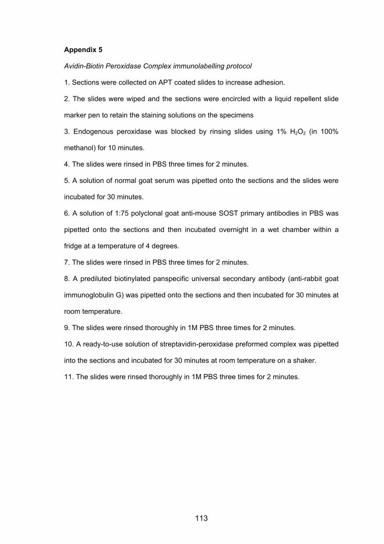
113
Appendix 5
Avidin-Biotin Peroxidase Complex immunolabelling protocol
1. Sections were collected on APT coated slides to increase adhesion.
2. The slides were wiped and the sections were encircled with a liquid repellent slide
marker pen to retain the staining solutions on the specimens
3. Endogenous peroxidase was blocked by rinsing slides using 1% H2O2 (in 100%
methanol) for 10 minutes.
4. The slides were rinsed in PBS three times for 2 minutes.
5. A solution of normal goat serum was pipetted onto the sections and the slides were
incubated for 30 minutes.
6. A solution of 1:75 polyclonal goat anti-mouse SOST primary antibodies in PBS was
pipetted onto the sections and then incubated overnight in a wet chamber within a
fridge at a temperature of 4 degrees.
7. The slides were rinsed in PBS three times for 2 minutes.
8. A prediluted biotinylated panspecific universal secondary antibody (anti-rabbit goat
immunoglobulin G) was pipetted onto the sections and then incubated for 30 minutes at
room temperature.
9. The slides were rinsed thoroughly in 1M PBS three times for 2 minutes.
10. A ready-to-use solution of streptavidin-peroxidase preformed complex was pipetted
into the sections and incubated for 30 minutes at room temperature on a shaker.
11. The slides were rinsed thoroughly in 1M PBS three times for 2 minutes.

114
Appendix 6
AEC and counter-staining protocol
1. A ready-made AEC solution was placed on the slides for 18 minutes. The solution
was kept in the dark to ensure the reaction did not proceed further.
2. Section were washed for 5 minutes with Milli-Q water.
3. The sections were stained with haematoxylin for 10 seconds and washed in tap
water to remove the excess stain. They were dipped in lithium carbonate for 30
seconds, followed by a wash in tap water.
4. The sections were mounted using aquamount and covered with large coverslips.