the role of stat3 activation in glomerulonephritis
TRANSCRIPT

9
The Role of STAT3 Activation in Glomerulonephritis
Fumio Tsuji, Osamu Katsuta and Hiroyuki Aono Research and Development Center, Santen Pharmaceutical Co., Ltd.
Japan
1. Introduction
Glomerulonephritis is a renal disease characterized by inflammation of the glomeruli, i.e., small blood vessels, in the kidneys. It may present with isolated hematuria and/or proteinuria or as nephritic syndrome, acute renal failure, or chronic renal failure. Glomerulonephritis has several different pathological patterns that can be broadly grouped into non-proliferative and proliferative types. Non-proliferative types include minimal change glomerulonephritis, focal segmental glomerulosclerosis, and membranous glomerulonephritis. Proliferative types include IgA nephropathy, post-infectious glomerulonephritis, membranoproliferative/mesangiocapillary glomerulonephritis, and rapidly progressive glomerulonephritis (Miller et al., 2010). Of the many factors involved in the inflammatory response, cytokines are the most important factors that bind to their receptors and activate signal transduction pathways. Progress in our understanding of inflammatory signaling pathways has led to the identification of the involvement of nuclear factor-κB (NF-κB), mitogen-activated protein kinases (MAPKs) such as p38, and Janus tyrosine kinase-signal transducer and activator of transcription (JAK-STAT) pathways (O’Neill, 2006). The NF-κB and p38 pathways are activated by the stimulation of interleukin-1 (IL-1) and tumor necrosis factor (TNF). The principal signaling pathways activated by IL-1 and TNF are the NF-κB and stress-activated MAPK pathways, whereas the JAK-STAT pathway is activated by many other cytokines.
2. STAT family
JAK-STAT is an important tyrosine kinase pathway activated by almost all cytokines (Ihle, 2001) that are well known for their role in the progression of renal diseases (Johnson, 1997). The STAT family consists of seven members (Table 1). The STAT proteins are unique transcription factors containing Src homology (SH) 2 and phosphotyrosine-binding domains. The SH2 domain interacts with sites of tyrosine phosphorylation to recruit STATs to receptor complexes. Following tyrosine phosphorylation of STATs, dimerization occurs between the SH2 domains. Tyrosine phosphorylation around amino acid position 700 (STAT1, tyrosine 701; STAT3 tyrosine 705) is essential for dimerization of STATs and the concomitant nuclear translocation of the dimer (Kaptein et al., 1996; Shuai et al., 1993). The basic model of cytokine pathways depends on tyrosine phosphorylation by JAK proteins non-covalently bound to specific receptors. The JAK family consists of four members: JAK1,
www.intechopen.com

An Update on Glomerulopathies – Etiology and Pathogenesis
172
JAK2, JAK3, and Tyk2. In addition, the activity of the C-terminal transactivation domain of STATs is at least partially regulated by serine phosphorylation (serine 727 in STAT1 and STAT3) that does not involve the JAK protein (Wen et al., 1995). The kinase responsible for this serine phosphorylation depends on the signaling pathway and cellular context.
Family member
Chromosomal locationActivating cytokines
Phenotype of knockout mice
Murine Human
STAT1 1 2q12–33 IFNs, IL-6 Viable, normal development, IFN
functions eliminated, tumors increased, growth control impaired.
STAT2 10 12q13–14.1 IFNs Viable, normal development, IFN
functions affected.
STAT3 11 17q11.2–22 IL-6 family Embryonic lethal, extraembryonic
endoderm defects, cell survival impaired.
STAT4 1 2q12–33 IL-12 Viable, normal development, IL-12
functions eliminated, Th1 differentiation impaired.
STAT5A 11 17q11.2–22 Numerous Viable, mammary gland deficiency, prolactin responsiveness eliminated.
STAT5B 11 17q11.2–22 Numerous Viable, partial loss of growth hormone
functions.
STAT6 10 12q13–14.1 IL-4, IL-13 Viable, normal development, IL-4
functions eliminated. Th2 differentiation impaired.
Table 1. Properties of STATs.
3. STAT3
STAT3 is a member of the JAK-STAT signaling pathway (Zhong et al., 1995). Cytoplasmic STAT3 in unstimulated cells is activated by recruitment through the SH2 domain to phosphorylated motifs within complexes of cytokine receptors, growth factor receptors, and non-receptor tyrosine kinases. Determination of STAT3 functions based on knockout mice studies have been difficult because mouse embryos die early in embryogenesis, prior to gastrulation (Takeda et al., 1997). STAT3 is essential for the early development of mouse embryos. Recently, some studies have succeeded in isolating STAT3 from individual tissues by the Cre-loxP method (Akira, 2000), which circumvents the problem of embryonic lethality, and revealed the roles of STAT3 in a wide variety of tissues (Table 2). A surprising result from such studies has been the identification of a multitude of and sometimes contradictory roles of STAT3 in biological processes including cell growth, cell growth suppression and apoptosis, and cell motility. The phenotypes resulting from the loss of STAT3 in adult tissues include failure of cell survival, impaired apoptosis, loss of negative feedback regulation, and impaired cell migration and wound healing (Levy & Lee, 2002). Of great current interest is the persistently active STAT3, which occurs in a wide variety of human tumors (Bowman & Jove, 1999). Overexpression and/or elevated protein tyrosine kinase activity of the epidermal growth factor receptor (EGFR), Src, and other protein
www.intechopen.com

The Role of STAT3 Activation in Glomerulonephritis
173
kinases is associated with the progression of numerous human cancers. As a consequence, growing evidence indicates that abnormal STAT3 signaling in response to hyperactive protein tyrosine kinase activity is frequent in human tumors and is associated with the progression of oncogenesis (Garcia & Jove, 1998). Furthermore, STAT3 can be converted into an oncogene by experimental mutation (Bromberg et al., 1999). A persistently active protein is required because introduction of a dominant-negative form of STAT3 into head and neck cancer cells or multiple myeloma cells causes apoptosis of recipient cancer cells (Bowman et al., 2000). Persistent STAT3 activation in head and neck cancer is associated with mutations in EGFR or mutations that result in the production of excess ligands or normal receptors (Song & Grandis, 2000). In some multiple myelomas, excess production of IL-6 might be the underlying defect (Catlett-Falcone et al., 1999). IL-6-dependent accumulation of long-lived plasma cells occurs due to elevated levels of a key regulatory protein, Bcl-xL, a member of the Bcl-2 family of proteins that prevent apoptosis. Constitutive activation of STAT3 signaling, an important component of the IL-6 pathway, directly contributes to the induction of Bcl-xL gene expression. Thus, constitutive activation of STAT3 signaling in response to IL-6 promotes tumor cell survival and malignant progression of multiple myelomas by directly inducing expression of a key apoptosis regulatory protein. All these results suggest that STAT3 may be essential for many cell functions.
Target tissue Phenotype
Skin Impaired second hair cycle, wound repair and keratinocyte
migration
Thymic epithelium Age-dependent thymic hypoplasia, hypersensitivity to stress
T lymphocytes Impaired IL-6-dependent survival and IL-2R┙ expression
Monocytes/neutrophils Enhanced inflammatory responses and Th1 differentiation,
chronic colitis
Granulocytes Enhanced proliferation owing to impaired negative feedback
Mammary epithelium Defective apoptosis, delayed mammary involution
Liver Impaired acute phase response
Neurons Impaired cell survival
Table 2. Tissue-specific roles of STAT3 (Levy & Darnell, 2002).
4. STAT3 in gp130 cytokine signaling
Several cytokines including IL-6, IL-11, leukemia inhibitory factor, oncostatin M, ciliary neurotrophic factor, and cardiotrophin share a gp130 subunit that is required to activate JAKs and STATs (Stahl et al., 1994). These cytokines are competent to activate STAT3 and induce various pleiotrophic responses that include hematopoiesis regulation, immune response, and neuronal differentiation (Snick, 1990). gp130-associated kinases JAK1, JAK2, and Tyk2 become activated on stimulation, and the cytoplasmic tail of gp130 is phosphorylated (Heinrich et al., 1998). STAT3 is recruited to phosphorylated tyrosine residues in the YXXQ motif including the tyrosine residue Y705 of gp130, where it is activated and dimerized. It subsequently enters the nucleus and regulates gene expression (Hirano et al., 2000). STAT3 is essential for gp130-mediated cell survival and G1 to S cell cycle transition signals. Both c-myc and pim have been identified as target genes of STAT3 and together can compensate for STAT3 in cell survival and cell cycle transition. On the
www.intechopen.com

An Update on Glomerulopathies – Etiology and Pathogenesis
174
other hand, the SH2 domain-bearing protein tyrosine phosphatase (SHP)-2 is recruited to the phosphorylated tyrosine residue Y759 in gp130, where it becomes activated and forms a complex with adaptor/docking proteins, Gab1 and Gab2, leading to activation of the Ras-MAPK pathway (Hibi & Hirano, 2000). Several in vitro experiments have shown that SHP-2- and STAT3-mediated signal transduction pathways initiated through gp130 are involved in growth, differentiation, and gene expression in various cell lines (Fukuda et al., 1996; Hirano et al., 2000; Nakajima et al., 1996). SHP-2-mediated ERK/MAPK activation has been suggested to play a negative role in STAT3-mediated biological responses (Jain et al., 1998; Sengupta et al., 1998). The tyrosine residue Y759 also provides the binding site for the suppressor of cytokine signaling 3 (SOCS3) protein that negatively regulates gp130 signals (Nicholson et al., 2000; Schmitz et al., 2000). To clarify the roles of SHP-2- and STAT3-mediated signal transduction pathways in vivo, a series of knock-in mouse lines was generated in which gp130-mediated STAT3 or SHP-2 signals were selectively disrupted. This disruption was achieved by mutating the tyrosine residues in all YXXQ motifs or the tyrosine residue Y759 to phenylalanine (gp130FXXQ/FXXQ and gp130F759/F759 mice, respectively) (Ohtani et al., 2000). Analyses of these mice indicated that SHP-2-mediated or Y759-dependent signals negatively regulate the biological responses elicited by STAT3-mediated signals in vivo and that the balance of positive and negative signals generated through gp130 is skewed or shifted toward positive STAT3 signaling in gp130F759/F759 mice (Fig. 1).
JAKsIL-6
R
IL-6
STAT3
Y759gp
130
YXXQYXXQYXXQYXXQ
SHP-2
Acute phase protein production
Th1 cytokine production
B cell differentiation
IgG2a, IgG2b production
InhibitionImportant for
development
ERK
activation
Resistance to
Listeria infection
JAKsIL-6
R
IL-6
STAT3
F759gp
130
YXXQYXXQYXXQYXXQ
SHP-2PI3K
Gab
STAT3STAT3
SOCS3
STAT3STAT3
Lack of inhibitory signal
Augmentation of STAT3 response
Splenomegaly and lymphadenopathy
Hyperimmunoglobulinemia
Autoantibody production
Abnormality of dendritic and T cells
Arthritis
(A) (B)
JAKsIL-6
R
IL-6
STAT3
Y759Y759gp
130
YXXQYXXQYXXQYXXQYXXQYXXQYXXQYXXQ
SHP-2
Acute phase protein production
Th1 cytokine production
B cell differentiation
IgG2a, IgG2b production
InhibitionImportant for
development
ERK
activation
Resistance to
Listeria infection
JAKsIL-6
R
IL-6
STAT3
F759F759gp
130
YXXQYXXQYXXQYXXQYXXQYXXQYXXQYXXQ
SHP-2PI3K
Gab
STAT3STAT3STAT3STAT3
SOCS3
STAT3STAT3STAT3STAT3
Lack of inhibitory signal
Augmentation of STAT3 response
Splenomegaly and lymphadenopathy
Hyperimmunoglobulinemia
Autoantibody production
Abnormality of dendritic and T cells
Arthritis
(A) (B)
Fig. 1. The role of gp130 signals in vivo. A: STAT3/YXXQ and SHP-2/Y759 signals induce responses independent of each other. SHP-2/Y759 signals negatively regulate the biological responses elicited by STAT3-mediated signals. B: The balance of positive and negative signals generated through gp130 is skewed or shifted toward positive STAT3 signaling in gp130F759/F759.
www.intechopen.com

The Role of STAT3 Activation in Glomerulonephritis
175
The SHP-2 signal-deficient mice (gp130F759/F759) were born normally but displayed splenomegaly and lymphadenopathy and an enhanced acute phase reaction. In contrast, the STAT3 signal-deficient mice (gp130FXXQ/FXXQ) died perinatally, similar to the gp130-deficient mice. The gp130F759/F759 mice showed prolonged gp130-induced STAT3 activation. Importantly, these mice in a mixed background with 129 and C57BL/6 spontaneously develop a rheumatoid arthritis-like autoimmune disease in old age (Atsumi et al., 2002). The mice show severe immunological abnormalities, including autoantibody production, increased memory/activated T cells, impaired thymic negative selection, and peripheral clonal deletion. Development of a rheumatoid arthritis-like disease is entirely dependent on mature lymphocytes, but abnormally enhanced homeostatic proliferation of CD4 T cells is caused by augmented production of IL-7 by non-hematopoietic stromal cells through a STAT3-dependent process (Sawa et al., 2006). In mice showing hyperactivation of STAT1/STAT3, T cell recruitment and CCL5/RANTES expression is enhanced (McLoughlin et al., 2005). A recent study described an IL-17A-triggered positive-feedback loop of IL-6 signaling that involved activation of the transcription factors NF-κB and STAT3 in fibroblasts (Ogura et al., 2008). Although the underlying mechanisms by which STAT3 regulates tissue fibrosis are not fully understood, its activation is required for upregulation of transforming growth factor (TGF)-┚ signaling, activation/proliferation of myofibroblasts, and deposition of extracellular matrix proteins (Ma & Zhuang, 2011). All these results suggest that STAT3 plays an important role in the function of multiple cells.
5. STAT3 activation in renal cells
Various factors show STAT3 activation in glomerular mesangial and proximal tubule cells. High glucose activates the growth-promoting enzyme JAK2 and its latent STAT transcription factors (STAT1, STAT3, and STAT5) and increases TGF-┚ and fibronectin synthesis in mesangial cells (Wang et al., 2002). High glucose-induced tyrosine phosphorylation of JAK2, STAT1, and STAT3 as well as TGF-┚ and fibronectin synthesis are abolished by a specific JAK2 inhibitor, AG-490. However, antisense oligonucleotide studies have shown that STAT1 activation is more important than STAT3 activation for TGF-┚ and fibronectin synthesis. Angiotensin II induces phosphorylation of JAK2, STAT1, STAT3, STAT5A/STAT5B and SHP-2, and angiotensin II-induced phosphorylation is enhanced by high glucose levels (Amiri et al., 2002). Angiotensin II-induced growth and collagen IV synthesis are also increased under high glucose conditions. Transfection of glomerular mesangial cells with JAK2 antisense oligonucleotides blocks angiotensin II-induced growth and collagen IV synthesis under both normal and high glucose conditions. In renal proximal tubule cells, IL-6 increases phosphorylation of STAT3, phosphoinositide-3 kinase (PI3K)/Akt, MAPKs, and NF-κB (Lee et al., 2007). IL-6 also stimulates ┙-methyl-D-[14C]glucopyranoside uptake, which is indicative of active transport in renal proximal tubule cells. The increased uptake can be blocked by pretreatment with a STAT3 inhibitor, a PI3K inhibitor, an Akt inhibitor, MAPK inhibitors, and NF-κB inhibitors. High glucose also induces activation of Raf-1, p42/p44 MAPK, JAK2, STAT1, and STAT3 (but not STAT5) in renal tubular epithelial cells (Huang et al., 2007). Moreover, severe oxidative stress leads to tyrosine phosphorylation of STAT3 at Y705 (Arany et al., 2006). This event depends on the activation of EGFR and JAK2 and is directly linked to cell death because inhibition of STAT3 function enables cells to survive severe oxidative stress.
www.intechopen.com

An Update on Glomerulopathies – Etiology and Pathogenesis
176
6. STAT3 activation in an animal model of glomerulonephritis
In an animal model of mesangial proliferative glomerulonephritis induced by the injection of anti-Thy1.1 antibody, STAT3 is phosphorylated in mesangial cells (Yanagita et al., 2001a). STAT3 phosphorylation peaks 8 days after the anti-Thy1.1 injection. Inhibition of growth arrest-specific gene 6 by the extracellular domain of its receptor Axl or warfarin abolishes STAT3 phosphorylation (Yanagita et al., 2001a) and inhibits mesangial cell proliferation (Yanagita et al., 2001b) in vivo. STAT3 can be expressed and activated in the kidneys of rats with immune complex glomerulonephritis using BSA as an antigen (Zhang et al., 2005). These rats also have increased macrophage infiltration (detected by a surface marker for monocytes/macrophages, ED-1), with some cells showing simultaneous expression of p-STAT3 and ED-1; this may contribute to inflammatory proliferation in the glomeruli and accumulation of extracellular matrix proteins. Inhibition of angiotensin-converting enzyme (ACE) with fosinopril downregulates STAT3 activation and ED-1 influx, and these effects may attenuate renal damage in this model. Hyperglycemia induces activation of JAK2 and STATs, including STAT3, in vivo (Banes et al., 2004). Phosphorylation of JAK2, STAT1, STAT3, and STAT5 in the glomeruli by streptozotocin (STZ) injection is reduced in rats treated with the ACE inhibitor captopril, angiotensin II type 1 receptor antagonist candesartan, or AG-490. Furthermore, both candesartan and AG-490 inhibit STZ-induced increases in urinary protein excretion. Knockdown of STAT3 activity in vivo prevents diabetic glomerulopathy (Lu et al., 2009). While the number of glomeruli does not differ between diabetic STAT3 knockdown and reference mice, the diabetic STAT3 knockdown mice exhibit significantly less proteinuria, mesangial expansion, glomerular cell proliferation, and macrophage infiltration than the diabetic reference mice. Reduction in STAT3 activity abrogates the stimulation of inflammatory markers, including IL-6, intercellular adhesion molecule-1, and monocyte chemotactic protein (MCP)-1, and blocks nuclear translocation of NF-κB. JAK2-STAT3 proteins may be involved in the early kidney damage associated with diabetes. AG490 also ameliorates adriamycin-induced nephritic syndrome in mice (Li et al., 2007). JAK-STAT signaling is activated in adriamycin nephropathy. Phosphorylation of JAK2, STAT1, and STAT3 is significantly inhibited by AG490. Proteinuria, glomerulosclerosis, tubulointerstitial lesions, and renal ┙-smooth muscle actin expression are significantly suppressed by AG490 treatment. In addition, AG490 inhibits MCP-1 mRNA expression accompanied by reduced interstitial infiltration of macrophages and T cells. Activation of JAK-STAT signaling is involved in the progression of glomerular diseases with proteinuria. While AG490 inhibits phosphorylation of STAT1 and STAT3, a novel selective inhibitor of STAT3, S3I-201, has been synthesized. S3I-201 preferentially inhibits STAT3 DNA-binding activity and diminishes tyrosine phosphorylation of STAT3 (Siddiquee et al., 2007). In a mouse model of renal interstitial fibrosis induced by unilateral ureteral obstruction, STAT3 was activated and administration of S3I-201 attenuated both STAT3 activation and extracellular matrix protein deposition following injury (Pang et al., 2010). S3I-201 reduced infiltration of the injured kidney by inflammatory cells and suppressed injury-induced expression of fibronectin, ┙-smooth muscle actin, and collagen type 1 proteins, as well as expression of multiple cytokines. Furthermore, S3I-201 inhibited proliferation and preferentially induced apoptosis in renal interstitial fibroblasts of the obstructed kidney. Inhibition of STAT3 signaling may hold therapeutic potential for fibrotic kidney diseases. IL-6 expression is increased in S-(1,2 dichlorovinyl)-L-cysteine-induced acute renal tubule necrosis (Vaidya et al., 2003) and renal ischemia-reperfusion injury (Lemay et al., 2000) in mice. IL-6-deficient mice are resistant to
www.intechopen.com
The Role of STAT3 Activation in Glomerulonephritis
177
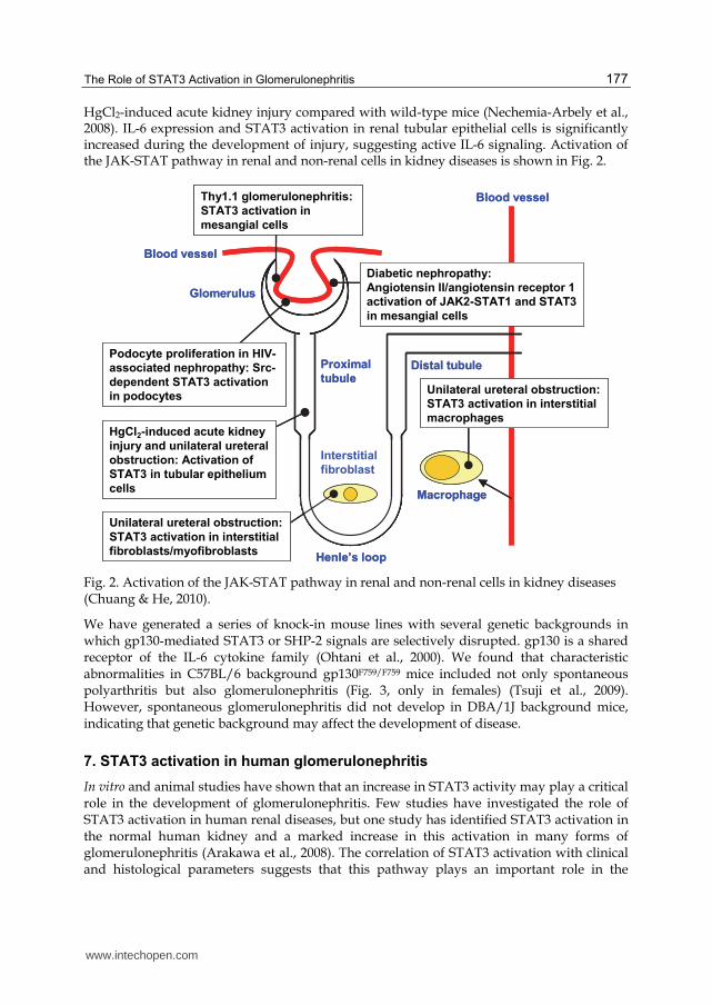
HgCl2-induced acute kidney injury compared with wild-type mice (Nechemia-Arbely et al., 2008). IL-6 expression and STAT3 activation in renal tubular epithelial cells is significantly increased during the development of injury, suggesting active IL-6 signaling. Activation of the JAK-STAT pathway in renal and non-renal cells in kidney diseases is shown in Fig. 2.
Thy1.1 glomerulonephritis:
STAT3 activation in
mesangial cells
Unilateral ureteral obstruction:
STAT3 activation in interstitial
fibroblasts/myofibroblasts
Glomerulus
Proximal
tubuleDistal tubule
Henle’s loop
Interstitial
fibroblast
Macrophage
Blood vessel
Blood vessel
Podocyte proliferation in HIV-
associated nephropathy: Src-
dependent STAT3 activation
in podocytes
Diabetic nephropathy:
Angiotensin II/angiotensin receptor 1
activation of JAK2-STAT1 and STAT3
in mesangial cells
HgCl2-induced acute kidney
injury and unilateral ureteral
obstruction: Activation of
STAT3 in tubular epithelium
cells
Unilateral ureteral obstruction:
STAT3 activation in interstitial
macrophages
Fig. 2. Activation of the JAK-STAT pathway in renal and non-renal cells in kidney diseases (Chuang & He, 2010).
We have generated a series of knock-in mouse lines with several genetic backgrounds in which gp130-mediated STAT3 or SHP-2 signals are selectively disrupted. gp130 is a shared receptor of the IL-6 cytokine family (Ohtani et al., 2000). We found that characteristic abnormalities in C57BL/6 background gp130F759/F759 mice included not only spontaneous polyarthritis but also glomerulonephritis (Fig. 3, only in females) (Tsuji et al., 2009). However, spontaneous glomerulonephritis did not develop in DBA/1J background mice, indicating that genetic background may affect the development of disease.
7. STAT3 activation in human glomerulonephritis
In vitro and animal studies have shown that an increase in STAT3 activity may play a critical role in the development of glomerulonephritis. Few studies have investigated the role of STAT3 activation in human renal diseases, but one study has identified STAT3 activation in the normal human kidney and a marked increase in this activation in many forms of glomerulonephritis (Arakawa et al., 2008). The correlation of STAT3 activation with clinical and histological parameters suggests that this pathway plays an important role in the
www.intechopen.com

An Update on Glomerulopathies – Etiology and Pathogenesis
178
pathogenesis of kidney diseases. In addition, mesangial expansion and podocyte loss in the glomeruli are important early features of diabetic nephropathy. JAK1, JAK2, and JAK3 as well as STAT1 and STAT3 are expressed at higher levels in patients with diabetic nephropathy than in control subjects (Berthier et al., 2009). Immunohistochemistry showed strong JAK2 staining in glomerular and tubulointerstitial compartments of patients with diabetic nephropathy compared with control subjects. These data suggest a direct relationship between tubulointerstitial JAK-STAT expression and the progression of kidney failure in patients with type 2 diabetic nephropathy and can be used to distinguish progressive human diabetic nephropathy. Further studies are needed to clarify the role of STAT3 activation in human glomerulonephritis.
Fig. 3. Glomerulonephritis in a gp130F759/F759 mouse (female, 12 months). Deposition of hyaline droplets in the subendothelial zone is shown.
8. Conclusions
The JAK-STAT pathway is a pleiotropic cascade essential to cytokine and growth hormone receptor signaling. Signaling through the JAK-STAT pathway is important for the kidney’s response to injury in disease. STAT3 activation is observed in an animal model of glomerulonephritis and human glomerulonephritis, and the STAT3 pathway inhibition ameliorates the renal conditions in some animal models. Further studies are needed to clarify the role of STAT3 activation in human glomerulonephritis, but the STAT3 pathway inhibition may be one of the potential therapeutic approaches for renal diseases.
9. References
Akira, S. (2000). Roles of STAT3 defined by tissue-specific gene targeting. Oncogene, Vol.19, No.21, pp. 2607-2611, ISSN 0950-9232
www.intechopen.com

The Role of STAT3 Activation in Glomerulonephritis
179
Amiri, F.; Shaw, S.; Wang, X.; Tang, J.; Waller, J.L.; Eaton, D.C. & Marrero, M.B. (2002). Angiotensin II activation of the JAK/STAT pathway in mesangial cells is altered by high glucose. Kidney International, Vol.61, No.5, pp. 1605-1616, ISSN 0085-2538
Arakawa, T.; Masaki, T.; Hirai, T.; Doi, S.; Kuratsune, M.; Arihiro, K.; Kohno, N. & Yorioka, N. (2008). Activation of signal transducer and activator of transcription 3 correlates with cell proliferation and renal injury in human glomerulonephritis. Nephrology Dialysis Transplantation, Vol.23, No.11, pp. 3418-3426, ISSN 0931-0509
Arany, I.; Megyesi, J.K.; Nelkin, B.D. & Safirstein, R.L. (2006). STAT3 attenuates EGFR-mediated ERK activation and cell survival during oxidant stress in mouse proximal tubular cells. Kidney International, Vol.70, No.4, pp. 669-674, ISSN 0085-2538
Atsumi, T.; Ishihara, K.; Kamimura, D.; Ikushima, H.; Ohtani, T.; Hirota, S.; Kobayashi, H.; Park, S.; Saeki, Y.; Kitamura, Y. & Hirano, T. (2002). A point mutation of Tyr-759 in interleukin 6 family cytokine receptor subunit gp130 causes autoimmune arthritis. The Journal of Experimental Medicine, Vol.196, No.7, pp. 979-990, ISSN 0022-1007
Banes, A.K.; Shaw, S.; Jenkins, J.; Redd, H.; Amiri, F.; Pollock, D.M. & Marrero, M.B. (2004). Angiotensin II blockade prevents hyperglycemia-induced activation of JAK and STAT proteins in diabetic rat kidney glomeruli. American Journal of Physiology Renal Physiology, Vol.286, No.4, pp. F653-F659, ISSN 0363-6127
Berthier, C.C.; Zhang, H.; Schin, M.; Henger, A.; Nelson, R.G.; Yee, B.; Boucherot, A.; Neusser, M.A.; Cohen, C.D.; Carter-Su, C.; Argetsinger, L.S.; Rastaldi, M.P.; Brosius, F.C. & Kretzler, M. (2009). Enhanced expression of janus kinase-signal transducer and activator of transcription pathway members in human diabetic nephropathy. Diabetes, Vol.58, No.2, 469-477, ISSN 0012-1797
Bowman, T. & Jove, R. (1999). STAT proteins and cancer. Cancer Control, Vol.6, No.6, pp. 615-619, ISSN 1073-2748
Bowman, T.; Garcia, R.; Turkson, J. & Jove, R. (2000). STATs in oncogenesis. Oncogene, Vol.19, No.21, pp. 2474-2488, ISSN 0950-9232
Bromberg, J.F.; Wrzeszczynska, M.H.; Devgan, G.; Zhao, Y.; Pestell, R.G.; Albanese, C. & Darnell, J.E., Jr. (1999). Stat3 as an oncogene. Cell, Vol.98, No., pp. 295-303, ISSN 0092-8674
Catlett-Falcone, R.; Landowski, T.H.; Oshiro, M.M.; Turkson, J.; Levitzki, a.; Savino, R.; Ciliberto, G.; Moscinski, L.; Fernández-Luna, J.L.; Nuñez, G.; Dalton, W.S. & Jove, R. (1999). Constitutive activation of Stat3 signaling confers resistance to apoptosis in human U266 myeloma cells. Immunity, Vol.10, No.1, pp. 105-115, ISSN 1074-7613
Chuang, P.Y. & He, J.C. (2010). JAK/STAT signaling in renal diseases. Kidney International, Vol.78, No.3, pp. 231-234, ISSN 0085-2538
Fukada, T.; Hibi, M.; Yamanaka, Y.; Takahashi-Tezuka, M.; Fujitani, Y.; Yamaguchi, T.; Nakajima, K. & Hirano, T. (1996). Two signals are necessary for cell proliferation induced by a cytokine receptor gp130: involvement of STAT3 in anti-apoptosis. Immunity, Vol.5, No.5, pp. 449-460, ISSN 1074-7613
Garcia, R. & Jove, R. (1998). Activation of STAT transcription factors in oncogenic tyrosine kinase signaling. Journal of Biomedical Science, Vol.5, No.2, pp. 79-85, ISSN 1423-0127
Heinrich, P.C.; Behrmann, I.; Müller-Newen, G.; Schaper, F. & Graeve, L. (1998). Interleukin-6-type cytokine signalling through the gp130/Jak/STAT pathway. Biochemical Journal, Vol.334, No.2, pp. 297-314, ISSN 0264-6021
www.intechopen.com

An Update on Glomerulopathies – Etiology and Pathogenesis
180
Hibi, M. & Hirano, T. (2000). Gab-family adapter molecules in signal transduction of cytokine and growth factor receptors, and T and B cell antigen receptors. Leukemia & Lymphoma, Vol.37, No.3-4, pp. 299-307, ISSN 1042-8194
Hirano, T.; Ishihara, K. & Hibi, M. (2000). Roles of STAT3 in mediating the cell growth, differentiation and survival signals relayed through the IL-6 family of cytokine receptors. Oncogene, Vol.19, No.21, pp. 2548-2556, ISSN 0950-9232
Huang, J.S.; Chuang, L.Y.; Guh, J.Y.; Huang, Y.J. & Hsu, M.S. (2007). Antioxidants attenuate high glucose-induced hypertrophic growth in renal tubular epithelial cells. American Journal of Physiology Renal Physiology, Vol.293, No.4, pp. F1072-F1082, ISSN 0363-6127
Ihle, J.N. (2001). The Stat family in cytokine signaling. Current Opinion in Cell Biology, Vol.13, No.2, pp. 211-217, ISSN 0955-0674
Jain, N. ; Zhang, T.; Fong, S.L.; Lim, C.P. & Cao, X. (1998). Repression of Stat3 activity by activation of mitogen-activated protein kinase (MAPK). Oncogene, Vol.17, No.24, pp. 3157-3167, ISSN 0950-9232
Johnson, R.J. (1997). Cytokines, growth factors and renal injury: where do we go now? Kidney International, Vol.52, Suppl.63, pp. S2-S6, ISSN 0085-2538
Kaptein, A.; Paillard, V. & Saunders, M. (1996). Dominant negative Stat3 mutant inhibits interleukin-6-induced Jak-STAT signal transduction. The Journal of Biological Chemistry, Vol.271, No.11, pp. 5961-5964, ISSN 0021-9258
Lee, Y.J.; Heo, J.S.; Suh, H.N.; Lee, M.Y. & Han, H.J. (2007). Interleukin-6 stimulates ┙-MG uptake in renal proximal tubule cells: involvement of STAT3, Pi3K/Akt, MAPKs, and NF-kB. American Journal of Physiology Renal Physiology, Vol.293, No.4, pp. F1036-F1046, ISSN 0363-6127
Lemay, S.; Rabb, H.; Postler, G. & Singh, A.K. (2000). Prominent and sustained up-regulation of gp130-signaling cytokines and of the chemokine MIP-2 in murine renal ischemia-reperfusion injury. Transplantation, Vol.69, No.5, pp. 959-963, ISSN 0041-1337
Levy, D.E. & Darnell, J.E. (2002). STATs: Transcriptional control and biological impact. Nature Reviews Molecular Cell Biology, Vol.3, No.9, pp. 651-662, ISSN 1471-0072
Levy, D.E. & Lee, C. (2002). What does STAT3 do? The Journal of Clinical Investigation, Vol.109, No.9, pp. 1143-1148, ISSN 0021-9738
Li, R.; Yang, N.; Zhang, L.; Huang, Y.; Zhang, R.; Wang, F.; Luo, M.; Liang, Y. & Yu, X. (2007). Inhibition of JAK/STAT signaling ameliorates mice experimental nephritic syndrome. American Journal of Nephrology, Vol.27, No.6, pp. 580-589, ISSN 0250-8095
Lu, T.C.; Wang, Z.H.; Feng, X.; Chuang, P.Y.; Fang, W.; Shen, Y.; Levy, D.E.; Xiong, H.; Chen, N. & He, J.C. (2009). Knockdown of Stat3 activity in vivo prevents diabetic glomerulopathy. Kidney International, Vol.76, No.1, pp. 63-71, ISSN 0085-2538
Ma, L. & Zhuang, S. (2011). The role of STAT3 in tissue fibrosis. Current Chemical Biology, Vol.5, No.1, pp. 44-51, ISSN 1872-3136
McLoughlin, R.M.; Jenkins, B.J.; Grail, D.; Williams, A.S.; Fielding, C.A.; Parker, C.R.; Ernst, M.; Topley, N. & Jones, S.A. (2005). IL-6 trans-signaling via STAT3 directs T cell infiltration in acute inflammation. Proceedings of the National Academy of Sciences of the United States of America, Vol.102, No.27, pp. 9589-9594, ISSN 0027-8424
Miller, F.P.; Vandome, A.F. & McBrewster J. (2010). Glomerulonephritis, Alphascript Publishing, ISBN 978-613-1-76981-8, USA
www.intechopen.com

The Role of STAT3 Activation in Glomerulonephritis
181
Nakajima, K.; Yamanaka, Y.; Nakae, K.; Kojima, H.; Ichiba, M.; Kiuchi, N.; Kitaoka, T.; Fukada, T.; Hibi, M. & Hirano, T. (1996). A central role for Stat3 in IL-6-induced regulation of growth and differentiation in M1 leukemia cells. The EMBO Journal, Vol.15, No.14, pp. 3651-3658, ISSN 0261-4189
Nechemia-Arbely, Y.; Barkan, D.; Pizov, G.; Shriki, A.; Rose-John, S.; Galun, E. & Axelrod, J.H. (2008). IL-6/IL-6R axis plays a critical role in acute kidney injury. Journal of the American Society of Nephrology, Vol.19, No.6, pp. 1106-1115, ISSN 1046-6673
Nicholson, S.E.; De Souza, D.; Fabri, L.J.; Corbin, J.; Willson, T.A.; Zhang, J.; Silva, A.; Asimakis, M.; Farley, A.; Nash, A.D.; Metcalf, D.; Hilton, D.J.; Nicola, N.A. & Baca, M. (2000). Suppressor of cytokine signaling-3 preferentially binds to the SHP-2-binding site on the shared cytokine receptor subunit gp130. Proceedings of the National Academy of Sciences of the United States of America, Vol.97, No.12, pp. 6493-6498, ISSN 0027-8424
Ogura, H.; Murakami, M.; Okuyama, Y.; Tsuruoka, M.; Kitabayashi, C.; Kanamoto, M.; Nishimura, M.; Iwakura, Y. & Hirano, T. (2008). Interleukin-17 promotes autoimmunity by triggering a positive-feedback loop via interleukin-6 induction. Immunity, Vol.29, No.4, pp. 628-636, ISSN 1074-7613
Ohtani, T.; Ishihara, K.; Atsumi, T.; Nishida, K.; Kaneko, Y.; Miyata, T.; Itoh, S.; Narimatsu, M.; Maeda, H.; Fukada, H.; Itoh, M.; Okano, H.; Hibi, M. & Hirano, T. (2000). Dissection of signaling cascades through gp130 in vivo: Reciprocal roles for STAT3- and SHP2-mediated signals in immune responses. Immunity, Vol.12, No.1, pp. 95-105, ISSN 1074-7613
O’Neill, L.A.J. (2006). Targeting signal transduction as a strategy to treat inflammatory diseases. Nature Reviews Drug Discovery, Vol.5, No.7, pp. 549-563, ISSN 1474-1776
Pang, M.; Ma, L.; Gong, R.; Tolbert, E.; Mao, H.; Ponnusamy, M.; Chin, Y.E.; Yan, H.; Dworkin, L.D. & Zhuang, S. (2010). A novel STAT3 inhibitor, S3I-201, attenuates renal interstitial fibroblast activation and interstitial fibrosis in obstructive nephropathy. Kidney International, Vol.78, No.3, pp. 257-268, ISSN 0085-2538
Sawa, S.; Kamimura, D.; Jin, G.H.; Morikawa, H.; Kamon, H.; Nishihata, M.; Ishihara, K.; Murakami, M. & Hirano, T. (2006). Autoimmune arthritis associated with mutated interleukin (IL)-6 receptor gp130 is driven by STAT3/IL-7-dependent homeostatic proliferation of CD4+ T cells. The Journal of Experimental Medicine, Vol.203, No.6, pp. 1459-1470, ISSN 0022-1007
Schmitz, J.; Weissenbach, S.; Haan, S.; Heinrich, P.C. & Schaper, F. (2000). SOCS3 exerts its inhibitory function on interleukin-6 signal transduction through the SHP2 recruitment site of gp130. The Journal of Biological Chemistry, Vol.275, No.17, pp. 12848-12856, ISSN 0021-9258
Sengupta, T.K.; Talbot, E.S.; Scherle, P.A. & Ivashkiv, L.B. (1998). Rapid inhibition of interleukin-6 signaling and Stat3 activation mediated by mitogen-activated protein kinases. Proceedings of the National Academy of Sciences of the United States of America, Vol.95, No.19, pp. 11107-11112, ISSN 0027-8424
Shuai, K.; Stark, G.R.; Kerr, I.M. & Darnell, J.E., Jr. (1993). A single phosphotyrosine residue of Stat91 required for gene activation by interferon-gamma. Science, Vol.261, No.5129, pp. 1744-1746, ISSN 0036-8075
Siddiquee, K.; Zhang, S.; Guida, W.C.; Blaskovich, M.A.; Greedy, B.; Lawrence, H.R.; Yip, M.L.R.; Jove, R.; McLaughlin, M.M.; Lawrence, N.J.; Sebti, S.M. & Turkson, J. (2007).
www.intechopen.com

An Update on Glomerulopathies – Etiology and Pathogenesis
182
Selective chemical probe inhibitor of Stat3, identified through structure-based virtual screening, induces antitumor activity. Proceedings of the National Academy of Sciences of the United States of America, Vol.104, No.18, pp. 7391-7396, ISSN 0027-8424
Snick, J.V. (1990). Interleukin-6: An overview. Annual Review of Immunology, Vo.8, pp. 253-278, ISSN 0732-0582
Song, J.I. & Grandis, J.R. (2000). STAT signaling in head and neck cancer. Oncogene, Vol.19, No.21, pp. 2489-2495, ISSN 0950-9232
Stahl, N.; Boulton, T.G.; Farruggella, T.; Ip, N.Y.; Davis, S.; Witthuhn, B.A.; Quelle, F.W.; Silvennoien, O.; Barbieri, G.; Pellegrini, S.; Ihle, J.N. & Yancopoulos, G.D. (1994). Association and activation of Jak-Tyk kinases by CNTF-LIF-OSM-IL-6 beta receptor components. Science, Vol.263, No.5143, pp. 92-95, ISSN 0036-8075
Takeda, K.; Noguchi, K.; Shi, W.; Tanaka, T.; Matsumoto, M.; Yoshida, N.; Kishimoto, T. & Akira, S. (1997). Targeted disruption of the mouse Stat3 gene leads to early embryonic lethality. Proceedings of the National Academy of Sciences of the United States of America, Vol.94, No.8, pp. 3801-3804, ISSN 0027-8424
Tsuji, F.; Yoshimi, M.; Katsuta, O.; Takai, M.; Ishihara, K. & Aono, H. (2009). Point mutation of tyrosine 759 of the IL-6 family cytokine receptor, gp130, augments collagen-induced arthritis in DBA/1J mice. BMC Musculoskeletal Disorders, Vol.10, No.23, ISSN 1471-2474
Vaidya, V.S.; Shankar, K.; Lock, E.A.; Dixon, D. & Mehendale, H. (2003). Molecular mechanisms of renal tissue repair in survival from acute renal tubule necrosis: role of ERK1/2 pathway. Toxicologic Pathology, Vol.31, No.6, pp. 604-618, ISSN 0192-6233
Wang, X.; Shaw, S.; Amiri, F.; Eaton, D.C. & Marrero, M.B. (2002). Inhibition of the JAK/STAT signaling pathway prevents the high glucose-induced increase in TGF-┚ and fibronectin synthesis in mesangial cells. Diabetes, Vol.51, No.12, pp. 3505-3509, ISSN 0012-1797
Wen, Z.; Zhong, Z. & Darnell, J.E., Jr. (1995). Maximal activation of transcription by Stat1 and Stat3 requires both tyrosine and serine phosphorylation. Cell, Vol.82, No.2, pp. 241-250, ISSN 0092-8674
Yanagita, M.; Arai, H.; Nakano, T.; Ohashi, K.; Mizuno, K.; Fukatsu, A.; Doi, T. & Kita, T. (2001a). Gas6 induces mesangial cell proliferation via latent transcription factor STAT3. The Journal of Biological Chemistry, Vol.276, No.45, pp. 42364-42369, ISSN 0021-9258
Yanagita, M.; Arai, H.; Ishii, K.; Nakano, T.; Ohashi, K.; Mizuno, K.; Vernum, B.; Fukatsu, A.; Doi, T. & Kita, T. (2001b). Gas6 regulates mesangial cell proliferation through Axl in experimental glomerulonephritis. The American Journal of Pathology, Vol.158, No.4, pp. 1423-1432, ISSN 0002-9440
Zhang, W.; Chen, X.; Shi, S.; Wei, R.; Wang, J.; Yamanaka, N. & Hong, Q. (2005). Expression and activation of STAT3 in chronic proliferative immune complex glomerulonephritis and the effect of fosinopril. Nephrology Dialysis Transplantation, Vol.20, No.5, pp. 892-901, ISSN 0931-0509
Zhong, Z.; Wen, Z. & Darnell, J.E., Jr. (1994). Stat3: a STAT family member activated by tyrosine phosphorylation in response to epidermal growth factor and interleukin-6. Science, Vol.264, No.5155, pp. 95-98, ISSN 0036-8075
www.intechopen.com

An Update on Glomerulopathies - Etiology and PathogenesisEdited by Prof. Sharma Prabhakar
ISBN 978-953-307-388-0Hard cover, 276 pagesPublisher InTechPublished online 06, September, 2011Published in print edition September, 2011
InTech EuropeUniversity Campus STeP Ri Slavka Krautzeka 83/A 51000 Rijeka, Croatia Phone: +385 (51) 770 447 Fax: +385 (51) 686 166www.intechopen.com
InTech ChinaUnit 405, Office Block, Hotel Equatorial Shanghai No.65, Yan An Road (West), Shanghai, 200040, China
Phone: +86-21-62489820 Fax: +86-21-62489821
The book has fourteen chapters which are grouped under different sections: Immune System andGlomerulonephritis, Animal Models of Glomerulonephritis, Cytokines and Signalling Pathways, Role of Cellsand Organelles in Glomerulonephritis and Miscellaneous. While the purpose of this volume is to serve as anupdate on recent advances in the etio-pathogenesis of glomerulopathies, the book offers the current andbroad based knowledge in the field to readers of all levels in the nephrology community.
How to referenceIn order to correctly reference this scholarly work, feel free to copy and paste the following:
Fumio Tsuji, Osamu Katsuta and Hiroyuki Aono (2011). The Role of STAT3 Activation in Glomerulonephritis,An Update on Glomerulopathies - Etiology and Pathogenesis, Prof. Sharma Prabhakar (Ed.), ISBN: 978-953-307-388-0, InTech, Available from: http://www.intechopen.com/books/an-update-on-glomerulopathies-etiology-and-pathogenesis/the-role-of-stat3-activation-in-glomerulonephritis

© 2011 The Author(s). Licensee IntechOpen. This chapter is distributedunder the terms of the Creative Commons Attribution-NonCommercial-ShareAlike-3.0 License, which permits use, distribution and reproduction fornon-commercial purposes, provided the original is properly cited andderivative works building on this content are distributed under the samelicense.