the role of soluble and insoluble fibers during
TRANSCRIPT

The role of soluble and insoluble fibers
during fermentation of Chicory root pulp
Uttara S. Ramasamy

Thesis committee
Promoters
Prof. Dr. H. Gruppen Professor of Food Chemistry Wageningen University
Prof. Dr. H. A.Schols Personal chair at the Laboratory of Food Chemistry Wageningen University
Other members
Dr. Ad de Laat, Cosun Food Technology Centre, Roosendaal, the Netherlands Prof. Dr. Anne Meyer, Technical University of Denmark Prof. Dr. Edith Feskens, Wageningen University Prof. Dr. Hauke Smidt, Wageningen University
This research was conducted under the auspices of the Graduate School VLAG (Advanced studies in Food Technology, Agro-biotechnology, Nutrition and Food Sciences).

The role of soluble and insoluble fibers
during fermentation of Chicory root pulp
Uttara S. Ramasamy
Thesis
Submitted in fulfilment of the requirements for the degree of doctor
at Wageningen University
by the authority of Rector Magnificus
Prof. Dr M.J. Kropff,
in the presence of the
Thesis Committee appointed by the Academic Board
to be defended in public
on Friday 6 June 2014
at 4 p.m. in the Aula

Uttara S. Ramasamy
The role of soluble and insoluble fibers during fermentation of Chicory root pulp
152 pages
PhD thesis, Wageningen University, Wageningen, NL (2014) With references, with summaries in English and Dutch
ISBN: 978-94-6173-965-0

Abstract This thesis was aimed at understanding the in vitro fermentability of soluble and insoluble fibers in chicory root pulp (CRP). First, CRP and ensiled chicory root pulp (ECRP) were characterized for cell wall polysaccharides (CWPs). Both CRP and ECRP were rich in CWPs (56-58 w/w (%)) and had rather similar sugar compositions. The CWPs consist of 62 % pectin, 11% hemicellulose and 27% cellulose. Pectin and xyloglucan were acetylated and the rhamnogalacturonan-I segments of pectin were branched mostly with arabinan. Compared to CRP, ECRP has four times more soluble pectin. In vitro fermentability in a batch model for 24 h using human faecal inoculum, showed that fibers in both CRP (51% carbohydrate utilisation) and ECRP (59% carbohydrate utilisation) were fermentable, especially pectin (80-87%). The increased levels of soluble pectin (arabinan, homogalacturonan and galactan) and the hypothesized open cell wall structure in ECRP contributed to a quicker fermentation and a higher level of carbohydrate utilization compared to CRP. In contrast to batch fermentation, fermentation in the dynamic TNO In vitro model of the colon (TIM-2) was rapid (57% carbohydrate utilisation in 2 h). ECRP carbohydrates (85%) were less fermented in 24 h compared to CRP carbohydrates (92%) due to lower utilisation of ECRP insoluble fibers than CRP insoluble fibers. It was hypothesized that soluble fibers that are readily fermentable and dominantly present in ECRP, programmed the microbiota in TIM-2 to fully adapt to these soluble fibers. After their utilization, the microbiota was not able to adapt towards the fermentation of insoluble fibers. Analysis of enzyme activities during batch fermentation of CRP showed increased levels of arabinofuranosidase, -galactosidase, endo-arabinanase, endo-galactanase, exo-polygalacturonase, pectin de-esterifying enzymes and endo-polygalacturonase. They synergistically contributed to degrading pectin in CRP from 12 to 24 h of fermentation.

Table of contents
Abstract
Chapter 1 General introduction 1
Chapter 2 Structural and water holding characteristics of untreated and ensiled chicory root pulp
27
Chapter 3 The effect of soluble and insoluble fibers on the fermentation of chicory root pulp
47
Chapter 4 The fate of chicory root pulp polysaccharides during fermentation in the TNO In vitro model of the colon (TIM-2)
73
Chapter 5 Characteristics of bacterial enzymes as present after fermentation of chicory root pulp by human faecal microbiota
97
Chapter 6 General discussion 119
Summary, Samenvatting 135
Acknowledgements 143
About the author 147


1
Chapter 1
General introduction

2
1.1. Background
The use of fibers from agricultural by-products for food purposes is gaining importance.
Most commonly used for food purposes are fibers arising from the cereal processing
industry (1). The cereal based by-products are abundant in xylan and -glucan fibers (1, 2).
In contrast, by-products from the fruit and vegetable processing industry, which are
dominant in pectin fibers, are very limited in use (3).
Chicory root pulp (CRP) is an agricultural by-product obtained after industrial extraction of
inulin from the chicory root. It is rich in pectin fibers, followed by cellulose and
hemicellulose (3). The pulp also contains some residual inulin, which has not been
completely removed during the industrial extraction process. For economic reasons, the
agricultural by-product is used for animal feed. In order to substantiate the use of the pulp
as a fiber supplement for human consumption, a multidisciplinary project ‘Novel food
fibers’ was started. Main research areas in this project dealt with determining the chemical
and techno-functional characteristics of fibers in CRP, studying in vitro and in vivo effects
of CRP and immune-modulatory effects of defined fibers as present in CRP. This thesis, as
a part of the above mentioned project, aimed at understanding the effect of CRP cell wall
polysaccharides and their arrangement in the cell wall network on the in vitro
fermentability of polysaccharides by human colonic bacteria.
1.2. Dietary fibers
Dietary fibers have been claimed to contribute to health benefits, such as adding bulk and
softness to stool (4, 5), controlling blood glucose levels (6), lowering blood cholesterol
levels (7), providing immuno-modulatory effects (8), providing satiety and facilitating
weight reduction (9, 10) and reducing the risks of gastrointestinal disorders and colon
cancer (11). The term dietary fiber (DF) encompasses plant cell wall polysaccharides
(CWPs), such as pectin, hemicellulose and cellulose, which are resistant to digestion in the
human small intestine and become available for (partial) fermentation in the colon by the
human microbiota (12).

General introduction
3
1.3. Plant cell wall polysaccharides
Plant cell walls support cell membranes surrounding plant cells and provide rigidity,
strength and shape to plant cells (13). They are built of polysaccharides, structural proteins
and lignin (14, 15). The plant cell wall in dicotyledonous plants mainly consists of the
middle lamella and the primary cell wall. The middle lamella consists mostly of pectic
polysaccharides which glue plant cells together while the primary cell wall is characterized
by a higher degree of organization of polysaccharide (16) giving the cell wall rigidity and
structure (figure 1.1). Normally, the primary cell wall of dicotyledonous plants contains
around 35% pectin, 30% cellulose, 30% hemicellulose and 5% protein (17, 18). In contrast,
the primary cell wall of monocots are abundant in arabinoxylan and cellulose, while they
contain <10% pectin (19).
Figure 1-1. Schematical plant cell wall model of dicotyledons as proposed by McCann and Roberts (20).

4
Figure 1-2. Schematic representation of plant polysaccharide structures. DHA: 3-deoxy-D-manno-2-octulosonic acid; KDO: 3-deoxy-D-lyxo-2-heptulosaric acid.
The chemical structure and variations of polysaccharides are described below:
Pectin
Pectin is a heterogenous group of polysaccharides consisting of homogalacturonan (HG),
rhamnogalacturonan (RG) I and II, arabinan, galactan, arabinogalactan and
xylogalacaturonan (XGA).
HG. Pectin mostly comprises HG which contains -D-galacturonic acid (GalA) residues
linked by 1,4-linkages (21). The GalA residues may be esterified with methanol at C-6
and/or acetylated at O-2 and /or O-3 (22) (figure 1.2). The distribution and the extent to
which methyl esters and acetyl groups are present vary from source to source.

General introduction
5
HG may have different substitutions such as present in: i) RG II: This structural element is
well conserved and present in low levels. Sugars of 2-O-methyl xylose, 2-O-methyl fucose,
apiose, aceric acid, 2-keto-3-deoxy-D-lyxo heptulosaric acid (DHA) and 2-keto-3-deoxy-D-
manno octulosonic acid (kdo) form four well defined side chains of a backbone of nine
GalA residues to form the RG II (23) (figure 1.2). ii) Xylogalacturonan (XGA): HG is
substituted with single xylose units (24) or iii) Apiogalacturonan (AGA): HG is substituted
with apiose residues to form AGA aspresent in duck weed (25, 26).
RG I. The RG I consists of alternating -(1,2)-linked rhamnosyl and -(1,4)-linked
galacturonic acid residues (27). The rhamnosyl residues can be substituted at O-4 by side
chains composed of arabinose and/or galactose residues forming arabinans, galactans and
arabinogalactans (AG) I and II (28) (figure 1.2). Arabinan is composed of a backbone of -
L-(1,5)-linked arabinofuranosyl residues, which may be substituted with -
arabinofuranosyl residues at the O-2 and/or O-3 position (15, 29, 30) (figure 1.2). The
backbone of AG I is composed of -1,4 linked galactose units which are substituted with -
1,5- linked arabinose units at the O-3 position (15). The backbone of AG II is composed of
-1,3 linked galactose units which are substituted with galactose units at O-6 which may be
further substituted with single arabinose residues (31) (figure 1.2).
Figure 1-3. Schematic representation of two different models A. the smooth and hairy regions model and B. the rhamnogalacturonan model describing the hypothetical pectin structure, adapted from Vincken et al (32).
Arabinan, galactan and arabinogalactan I and II present as neutral sugar side chains of
rhamnogalacturonan (RG) constitute the so called hairy regions of pectin (33). Two popular
models are used to describe the pectin structure: The smooth and hairy region model (21)

6
and the RG I backbone model (32)(figure 1.3). In the first model, the hairy regions are
interspersed with smooth regions HG containing 70-100 GalA residues (21, 34). In
addition, RG II is believed to be an integral part of HG (35). In the second model, HG is a
side chain of RG I as are the neutral sugar side chains (32).
Cellulose
Cellulose is insoluble and provides a framework to plant cells. Cellulose is built of -1,4
linked glucose units (figure 1.2) which make a fibril. An association of many crystalline
densely packed fibrils together with less ordered, amorphous regions of less densely packed
cellulose together forms a microfibril. The microfibrils are hydrogen bonded along their
length (14, 36, 37).
Hemicellulose
Hemicellulose is a class of many different polysaccharides, such as, xyloglucans, xylans
and mannans. Predominant polysaccharides in dicotyledons are xyloglucans. These
polymers have a -1,4 linked backbone of glucose residues as in cellulose but which is 75
% of the residues are substituted at O-6 by -xylose (figure 1.2). Further extensions of these
xylose side chains may include arabinosyl, galactosyl or fucosyl units at O-2 position (38-
40). In addition, O-acetylation of galactose and/or glucose residues is observed (39).
Xyloglucans bind to cellulose microfibrils in the native cell wall by hydrogen bond
interactions (38).
Xylans have a -1,4 linked backbone of xylose residues, which may be unsubstituted as
homoxylans or may be substituted with arabinose or glucuronic acid or both to form
arabinoxylans, glucuronoxylans or glucuronoarabinoxylans respectively (figure 1.2). They
are characteristic of monocots such as, cereals (26, 41, 42). In dicots, such as potato and
sugar beet, they are also present, but in low quantities (42).
Mannans have a mannose -1,4 linked backbone (figure 1.2). The backbone may be
substituted with -1,6-linked galactose residues forming galactomanans. Mannans may also
contain glucose units in their backbone forming (galacto)glucomannans (43).

General introduction
7
Figure 1-4. Schematic presentation of possible pectin- pectin crosslinks in a cell wall: A. Egg box model of calcium-pectin crosslink (44), B. Ferulic acid crosslink, for example 5-5-diferulic acid esterfied with neutral sugar side chains of pectin (45), C. Rhamnogalacturonan II diester, (35), D. Uronyl ester of pectin with a hydroxyl group of another polysaccharide chain (46).
Cell wall network / architecture
Cellulose microfibrils and xyloglucan are interlinked via hydrogen bonds forming a rigid
network (15, 47) (figure 1.1). Depending on the proportions of xyloglucan and pectin in the
cell wall, pectin may as well interact with cellulose (48). For cell walls containing
reasonable levels of xyloglucan as in pea (49) and blackberries (50), pectins bind loosely to
cellulose and fill the interstices within the cellulose/ xyloglucan network (48). In case of
relatively low amounts of xyloglucan, e.g. in sugar beet (51), pectins replace xyloglucan as
they bind to cellulose microfibrils (48, 52).
Pectins have also been shown to interact with hemicellulose via its neutral sugar side chains
(47). Pectins also interact with proteins via arabino-3,6-galactans (53). Proteins in the cell
wall are mostly hydroxyproline rich glycoproteins which include extensins, arabinogalactan

8
proteins, and lectins (31). Pectins may also be linked with each other via the following
crosslinks (see also figure 1.4):
A. Calcium crosslinking between unesterified GalA residues. Usually, blocks of more
than 10 unesterified GalA residues give rise to pectin molecules sensitive to Ca2+
crosslinking (44).
B. Ferulic acid crosslinking. The propenoic double bonds of ferulic acid may
crosslink pectins (45). The ferulic acids are found in RG I and are attached to the
O-2 position of arabinose and to O-6 position of galactose (45, 54-56). Usually
found in low concentrations, sugar beet pulp contains 0.7% ferulic acid of cell wall
material. Around 22 % of the total ferulates exist as ferulic acid dehydromers in
sugar beet pectin (56).
C. Rhamnogalacturonan (RG) II boron diester crosslinking. RGII components are
crosslinked via a borate diester (35). RG II is present predominantly as a dimer
(figure 1.4) in plant cell walls (57).
D. Uronyl ester crosslinking. A covalent ester bond is formed between galacturonic
acid in pectins and hydroxyl group of neighbouring polysaccharides crosslinking
two (pectin) chains (46, 50). However, strong evidence for the presence of such
esters is still lacking.
1.4. Enzymes degrading plant cell wall polysaccharides In general, CWPs can be degraded by two classes of enzymes: i. Glycosidases that
hydrolyse single sugars or small oligomers from the ends of oligomers and exo-enzymes
that hydrolyse from the ends of polymers and ii. Endo enzymes that cleave linkages within
a polymer backbone give rise to a drastic decrease of the molar mass of the polymer. Three
types of cell wall degrading enzymes exist: 1. Glycosyl Hydrolases (E.C. 3.2.1) cleave the
glycosidic linkage between two sugar units and introduce a water molecule, 2.
Polysaccharide Lyases (E.C. 4.2.2) which cleave the glycosidic linkage by introducing a
double bond and 3. Carbohydrate Esterases (E.C. 3.1.1) that remove non carbohydrate
substituents such as methyl esters, ferulic acid groups and acetyl groups from

General introduction
9
carbohydrates. Degradation of each polysaccharide requires a specific set of enzymes as
discussed below:
Cellulose degrading enzymes
Cellulose can be degraded by three types of enzymes. i. Endo-glucanases which hydrolyse
cellulose randomly producing reducing ends; ii. Exo-glucanases which liberate D-glucose
from cellulose or cellodextrin and also liberate cellobiose (cellobiohydrolase) from
cellulose in a processive manner and iii. -glucosidases which form D-glucose from
cellobiose (58). Hydrolysis of crystalline cellulose is made efficient by the carbohydrate
binding modules (CBM) linked to cellulases. CBMs attach to the substrate and the linker
between the CBM and the catalytic site provides sufficient freedom for the catalytic domain
to move around the binding module attached to the substrate. This increases the proximity
of the catalytic domain to the substrate for hydrolysis (59).
Hemicellulose degrading enzymes
Xyloglucan degrading enzymes. Xyloglucan requires different enzymes for degradation of
the backbone and the side chains. Backbone degradation is either by non-specific endo-
glucanase or xyloglucan specific endo-glucanase and -glucosidases (40, 60). Side chains
are degraded by -xylosidases, -galactosidases and acetyl esterases (60, 61). Xylan
degrading enzymes: Xylan backbone can be degraded by endo-xylanases and -xylosidases
whereas arabinose substituents can be removed by arabinofuranosidase (60). Glucuronic
acid can be cleaved by glucuronidases (62).
Mannan degrading enzymes. The mannan backbone is degraded by endo-mannanase (60)
while the linkage between glucose residues in the mannan backbone can be hydrolysed by
endoglucanase and side chains of mannan are degraded by -galactosidase.
Pectin degrading enzymes
Different enzymes are required for degrading pectic polysaccharides. These enzymes are shown in figure 1.5. The HG can be degraded by exo- and endo-polygalacturonase. The action of endopolygalacturonase is hindered by esterified GalA residues, due to which other enzymes, such as pectin methyl esterase and pectin acetyl esterase, are also required to enhance degradation of esterified HG. Besides, esterified HG can also be cleaved by pectin lyase (figure 1.5).

10
Figure 1-5. Sites of action by pectin degrading enzymes. PG, polygalacturonase; PME, pectin methyl esterase, PAE, pectin acetyl esterase; RG, rhamnogalacturonan (63)
RG I degradation is catalysed by RG-hydrolases. The RG galacturonohydrolase liberates galacturonic acid from RG (64) while the RG rhamnohydrolase is an exo-enzyme which releases rhamnose from the non-reducing end of RG and RG rhamnogalacturonohydrolase is an endo-enzyme which cleaves between rhamnose and galacturonic acid (65). RG is cleaved by RG lyase (RGL) (66). Esters on RG I are also removed by RG-acetyl esterases. Acetyl groups and side chains attached to Rha hinder the action of RGH and RGL. Arabinan can be degraded by endoarabinanases, which cleave -1,5-linkages within a linear backbone (67). Exo-arabinanases cleave arabinose from the non-reducing end of the -1,5 linked backbone (68) or arabinotriose from branched arabinan (69). Arabinofuranosidases hydrolyse -arabinofuranosyl residues present as terminal substituents on the -1,5 linked backbone and on branched arabinan (70). Degradation of galactan and the backbone of AG I and AG II requires activities of endo-galactanase and -galactosidase. 1,4 galactanase cleaves AG I backbone and 1,3,6 galactanase cleaves the backbone of AG II. Arabinose substituents are removed by arabinofuranosidase and arabinopyranosidase.

General introduction
11
1.5. Fiber rich agricultural by-products
Agricultural by-products from industrial processing include roots and tubers (71-73),
vegetables and fruits (74, 75), and cereals (76, 77).
Table 1-1. Constituent monosaccharide content of agricultural by-products
w/w % Sources Rha Fuc Ara Xyl Man Gal Glc UA Total Reference
Chicory root pulp 1 0 7 3 2 4 23 28 69 (78)
Sugar beet pulp 1 0 22 2 1 5 23 13 67 (79)
Potato pulp 2 0 4 1 1 12 14 11 44 (80)
Apple pulp 1 1 8 6 1 6 29 24 76 (81)
Rape seed meal 2 0 19 8 6 10 40 15 36 (82)
Sunflower cake 0 0 3 6 1 1 13 7 32 (77)
Alfafa meal 0 0 3 6 1 2 15 7 33 (77)
Soybean meal 0 0 3 2 1 4 7 5 22 (77)
Linseed meal 1 0 4 7 0 3 12 4 30 (77)
Corn fiber 0 0 12 18 2* 3 14 4 53 (83) Corn cobs 0 0 2 28 1* 1 33 2 67 (83) Corn stover 0 0 3 19 1* 1 30 2 56 (83) Brewers spent grain 0 0 8 15 0 1 16 2 42 (84) Palm cake 0 0 1 3 31 2 8 2 47 (77)
Wheat bran pulp 0 0 22 32 0 1 21 2 79 (77)
Coconut cake 0 0 1 1 31 3 6 1 42 (77)
Rye bran 0 0 8 21 0 1 11 1 42 (77)
Barley dehulled 0 0 2 4 0 0 6 0 13 (77)
Oats feed meal 0 0 1 2 0 0 5 1 9 (77) Rhamnose (Rha), Fucose (Fuc), Arabinose (Ara), Xylose (Xyl), Mannose (Man), Galactose (Gal), Glucose (Glc), Uronic acid (UA), *Values represent Man+Rha.
They have been used for feed purposes for reasons such as, high dietary fiber content, high
protein content etc (76, 77). Agricultural by-products rich in polysaccharides are shown in
table 1.1. These by-products have been utilised for feed purposes.
As shown, the by-products may differ significantly in the constituent monosaccharide
composition. The by-products can be broadly distinguished as pectin rich by-products, such
as CRP, sugar beet pulp, potato pulp, apple pulp and rape seed meal and (hemi)cellulose
rich by-products. Among (hemi)cellulose rich by-products, mannan is abundant in palm
cake (31 w/w%) and coconut cake (31 w/w%) whereas xylans are abundant in corn fiber

12
(18 w/w%), corn cobs (28 w/w%), corn stover (19 w/w%), brewers spent grain (15 w/w%),
wheat bran pulp (32 w/w%), and rye bran (21 w/w%). Differences in pectic polysaccharides
can also be seen within pectin rich by-products. The pectin in CRP contains less arabinan (7
w/w %) compared to sugar beet pulp (22 w/w %).
Chicory root pulp
Chicory (Cichorium intybus L.) root is used industrially for the extraction of inulin. The
pulp obtained is used as animal feed. Pectin in chicory root pulp is highly methyl esterified
(68) and acetylated (36)(78). The gelling behaviour of chicory root pectins extracted by a
combination of protease and cellulase enzymes has been studied (85). It was found that
chicory root pectins gelled with sucrose at an acidic pH, but not as strong as citrus pectins
that have a higher GalA content than chicory root pectins. Research on chicory root cell
wall fibres has only been started since a few years (3, 78, 85, 86). Hence, not much
information is available on the different populations of polysaccharides and their
interactions in CRP.
Water holding capacity (WHC) of cell wall materials
Physical attributes of cell wall materials, such as water holding capacity (WHC), have an
influence on their functionality such as fermentability or in application of fibers to food.
The WHC is in turn determined by the molecular structure of the CWPs and the cell wall
architecture (87). Sugar beet pulp has a WHC (5.5 mL/g) (88). Treatments like autoclaving
or grinding of sugar beet pulp reduced the WHC of sugar beet pulp (89). The WHC could
also be modulated by different means such as enzymatic treatment of the plant cell wall or
processing of the plant cell wall.
Agricultural by-products containing plant cell wall polysaccharides may be fermented to
different extents in the colon. The fermentation of such plant cell wall based dietary fibers
is elaborated in the following section.
1.6. Fermentation of dietary fibers
Fermentation of dietary fibers in the colon is determined by the colonic bacteria and the
type of fibers entering the colon. The colon is densely populated with microbiota (mostly
anaerobic) accumulating to approximately 1012 bacteria per g dry weight of colonic contents

General introduction
13
(90). The type of bacteria present in the colon determines the health of the colon. Figure 1.6
(91) shows the dominant populations of bacteria in the colon.
Figure 1-6. Schematic presentation of composition and health effects of the predominant human faecal bacteria (91).
Most abundant bacteria belong to the genus Bacteroides, which are partly health promoting
and partly harmful. The health promoting bacteria include Bifidobacteria, Eubacteria and
Lactobacilli. Streptococci, E.coli, and Enterococci are partly beneficial and partly
pathogenic, whereas Veillonellae, Clostridia, Staphylococci, Proteus, Pseudomonas
aeruginosa are pathogenic when present in the colon (91).
The proportions of species in the colon could change depending on the type of fiber
consumed. Examples of bacteria that degrade different types of polysaccharides are shown
in table 1.2. Most bacteria that degrade cell wall polysaccharides fall in two genera,
Bifidobacteria and Bacteroides (92). Bacteroides ovatus, Bacteroides thetaiotaomicron and
Bacteroides uniformis were able to ferment an array of polysaccharides (92). Fermentation
of oligosaccharides using human faecal inoculum showed that rhamnogalacturono-

14
oligosaccharides were only fermentable by Bacteroides spp.(93). Highly branched xylans
from wheat flour and sorghum were also fermentable by Bacteroides spp. (93). Table 1-2. Polysaccharides that are fermentable by intestinal bacteria (92, 94, 95)
Polysaccharide Bacteria able to utilize polysaccharides
Pectin (<70% galacturonic acid) B. thetaiotaomicron, B. ovatus,
Bacteroides “3452 A”,
B. fragilis subspecies a, B.
vulgatus, Eubacterium eligens
Xylan (linear) B. ovatus, B. eggerthii,
B. fragilis subspecies a,
B. vulgatus, Bifidobacterium adolescentis,
Bifidobacterium infantis
Galactomannans
Guar gum (mannose:galactose 1.8) B. ovatus, B. uniformis,
locust bean gum (mannose:galactose 3.5) Ruminococcus albus
Arabinogalactan B. thetaiotaomicron, B. ovatus,
B. ‘3452 A ‘, B. uniformis,
B. vulgatus, B. “T4-1
Bıfldobacterium longum
Cellulose Bacteroides sp.
Enzymes produced by the colonic microbiota
B. thetaiotaomicron known to be highly abundant among Bacteroides sp. (96) has been
estimated to include 236 glycoside hydrolase genes, 15 polysaccharide lyase genes, 20
carbohydrate esterase genes and 16 carbohydrate binding modules (97-99). Expression of
enzymes by these genes during the course of fermentation is up-regulated based on the
polysaccharides present during fermentation (97).
The enzymes active in degrading polysaccharides during fermentation may be extracellular
or bound to the bacterial cell wall. Degradation of soluble viscous polysaccharides has been
found to involve extracellular enzymes whereas degradation of insoluble polysaccharides
by Bacteroides has been found to involve cell wall bound enzymes (92). Degradation of an
insoluble cell wall material first requires the adhesion of fermenting bacteria to the cell wall
polysaccharide (100). The mechanism of adhesion, however, is poorly understood. It might

General introduction
15
involve substrate binding modules from enzymes and structural proteins and carbohydrate
moieties on glycoproteins (98).
Fermentation metabolites
Fermentation of DFs in the colon produces short chain fatty acid (SCFAs), such as acetate,
butyrate and propionate, and organic acids, such as succinate and lactate, as well as gases
such as hydrogen and methane (91). The organic acids are usually intermediates that are
converted to SCFAs. These are further metabolized (consumed) by the host as an energy
source (101) and also contribute to health benefits. Propionate is gluconeogenetic (synthesis
of glucose) and inhibits the synthesis of cholesterol from acetate, thus at least controlling
the levels of serum cholesterol (102). Butyrate has beneficial effects on inflammatory
responses (103) and on the cell cycle. It promotes cellular differentiation, has anti-
neoplastic properties and has effects on apoptosis (104, 105).
Cross feeding of polysaccharides by bacteria and competition also exist among bacterial
populations (106). For example, oligofructose and inulin are known to cause a bifidogenic
shift (increase in Bifidobacteria) in the microbial composition of the colon (107). The
subsequent increase in butyrate is not caused by Bifidobacteria but by other colonic
bacteria, thereby suggesting crossfeeding between Bifidobacteria and butyrate producing
bacteria (106). Crossfeeding has also been demonstrated between resistant starch degrading
Bifidobacteria and lactate-converting, butyrate producing colon bacteria (108, 109).
Models used for studying colonic fermentation
Fermentation of DF can be studied in different types of models generally classified as in
vitro or in vivo models. Compared to in vivo fermentation models, in vitro fermentation
models can be used without ethical constraints and they enable analysis of time dependant
degradation of fiber in the colon. In addition, in vitro fermentation experiments are more
simple to perform than in vivo fermentation experiments and there is no interference of
other food components in the investigation of the effect of fibers themselves.
In vitro fermentation of fibers can be studied in numerous types of models. The simplest of
the models commonly used is a closed system model represented by sealed bottles
containing suspensions of faecal microbiota, fiber substrates and being maintained under
anaerobic conditions (110). Such batch fermentations have been performed on fibers, such

16
as guar gum, alginate, retrograded starch, glucomannan, cellulose, -glucans, inulin,
oligofructose, high methyl esterified citrus pectin, soy pectin and xanthum gum (111), beet
pulp (112, 113) and citrus pulp (113) using human faecal inoculum.
In contrast to batch models, dynamic in vitro gut fermentation models are single or multiple
chemostats inoculated with fecal microbiota and operated under physiological temperature,
pH and anaerobic conditions (110, 114-117). Single chemostats, such as the TIM-2 (TNO
In Vitro Model)(116) are used to mimic the proximal colon while multiple chemostats are
used to mimic the proximal, central and descending parts of the colon such as the three
stage culture model (115). The TIM-2 has been combined with TIM-1 (small intestinal
model) (118) resulting in a digestive system to study drug delivery and advanced nutritional
studies (119-121).
Although fermentation in a batch model is simple, inexpensive and high throughput, the
fermentation requires a strong buffer to control the pH due to the accumulation of main
fermentation metabolites SCFA. In contrast, in continuous gut fermentation models, the
fermentation metabolites are not accumulated but dialysed out. Water is absorbed and
fermentation metabolites are removed via a hollow fiber membrane in TIM-2 (116). In
addition, due to peristaltic mixing of components, such models are closer to simulating the
human colon than batch models. TIM-2 also has the capacity to handle high microbial
densities as found in vivo and high substrate concentrations compared to the batch
fermentation (116).
Effect of polysaccharide arrangement on fermentability
Although dietary fibers (e.g. by-products) contain pectin, hemicellulose and cellulose,
differences in the structural features of individual polysaccharides and the molecular
arrangement of these polysaccharides and can contribute to different levels of fermentation
(112). Differences in fermentability have also been shown for cell wall materials having the
same constituent monosaccharide composition but modified cell wall networks by
processing. In vitro fermentation of beet pulp and autoclaved sugar beet pulp with a
different polysaccharide network than beet pulp showed that removal of pectic
polysaccharides by autoclaving increased the degradability of cellulose. This suggested that
accessibility of a polysaccharide within the network towards fermentation enzymes depends

General introduction
17
on the arrangement of polysaccharides within a network (89). This also suggested that
solubilisation of fibers from a network or increase in soluble fiber levels makes the
remaining polysaccharides in the insoluble network more accessible towards enzymes
(112).
Delayed fermentability of a cell wall material has positive effects, such as, preventing
proteolytic fermentation in the colon as the cell wall material passes through the colon.
Proteolytic fermentation is undesired because it leads to the production of ammonia
amongst other metabolites, and thereby leads to a pH that is near neutral to allow growth of
pathogens and increase vulnerability of the distal colon to chronic gut disorders (122-126).
Physical properties of cell wall materials such as, WHC and particle size have been shown to affect the colonic functionality and fermentability. Cell wall materials with a high WHC cause an increase in the volume of gastric contents thereby reducing their passage through the gut and thus the time taken for gastric emptying (127). Thus, fibers with a high WHC are not completely utilised, but are beneficial because they add bulk to the stool in addition to the microbiota and prevent constipation (5).
1.7. Thesis outline
Not much is known on the different polysaccharide populations and interactions between
polysaccharides in CRP. In addition, the fermentability of CRP has not been studied. As
mentioned, the aim of this thesis is to understand the effect of CRP cell wall
polysaccharides and their arrangement in the cell wall network on the in vitro
fermentability of polysaccharides by human colonic bacteria. The first part of the research
involved characterization of cell wall polysaccharides in chicory root pulp (Chapter 2).
The effect of ensiling on the cell wall polysaccharide network was also described in this
chapter. Chapter 3 reveals the fate of cell wall polysaccharides in CRP during the in vitro
fermentation of the pulp and the effect of soluble fibers and insoluble fibers on the
fermentation of the pulp. The fate of cell wall polysaccharides is also determined during in
vitro fermentation in the continuous flow model TIM-2 (Chapter 4). Next, the type, levels
and efficiency of bacterial enzymes produced during fermentation of CRP in a batch model
is determined and the effect of CWP arrangement on enzymatic degradation is explained in
Chapter 5. Findings of all parts of research are discussed in Chapter 6.

18
1.8. References
1. Collins, H. M.; Burton, R. A.; Topping, D. L.; Liao, M.-L.; Bacic, A.; Fincher, G. B., Review: Variability in fine structures of noncellulosic cell wall polysaccharides from cereal grains: potential importance in human health and nutrition. Cereal Chemistry 2010,87, 272-282. 2. Ebringerova, A.; Heinze, T., Xylan and xylan derivatives–biopolymers with valuable properties, 1. Naturally occurring xylan structures, isolation procedures and properties. Macromolecular Rapid Communications 2000, 21, 542-556. 3. Femenia, A.; Robertson, J. A.; Waldron, K. W.; Selvendran, R. R., Cauliflower (Brassica oleracea L), globe artichoke (Cynara scolymus) and chicory witloof (Cichorium intybus) processing by-products as sources of dietary fibre. Journal of the Science of Food and Agriculture 1998, 77, 511-518. 4. Chen, H. L.; Haack, V. S.; Janecky, C. W.; Vollendorf, N. W.; Marlett, J. A., Mechanisms by which wheat bran and oat bran increase stool weight in humans. American Journal of Clinical Nutrition 1998, 68, 711-719. 5. Cherbut, C.; Salvador, V.; Barry, J. L.; Doulay, F.; Delort-Laval, J., Dietary fibre effects on intestinal transit in man: involvement of their physicochemical and fermentative properties. Food Hydrocolloids 1991, 5, 15-22. 6. Montonen, J.; Knekt, P.; Jarvinen, R.; Aromaa, A.; Reunanen, A., Whole-grain and fiber intake and the incidence of type 2 diabetes. American Journal of Clinical Nutrition 2003, 77, 622-629. 7. Brown, L.; Rosner, B.; Willett, W. W.; Sacks, F. M., Cholesterol-lowering effects of dietary fiber: a meta-analysis. American Journal of Clinical Nutrition 1999, 69, 30-42. 8. Schley, P. D.; Field, C. J., The immune-enhancing effects of dietary fibres and prebiotics. British Journal of Nutrition 2002, 87, 221-230. 9. Slavin, J. L., Position of the american dietetic association: health implications of dietary fiber. Journal of the American Dietetic Association 2008, 108, 1716-1731. 10. Birketvedt, G. S.; Shimshi, M.; Thom, E.; Florholmen, J., Experiences with three different fiber supplements in weight reduction. Medical Science Monitor 2005, 11, I5-I8. 11. Liu, S. M.; Stampfer, M. J.; Hu, F. B.; Giovannucci, E.; Rimm, E.; Manson, J. E.; Hennekens, C. H.; Willett, W. C., Whole-grain consumption and risk of coronary heart disease: results from the Nurses' Health Study. American Journal of Clinical Nutrition 1999, 70, 412-419. 12. DeVries, J. W.; Camire, M. E.; Cho, S.; Craig, S.; Gordon, D.; Jones, J. M.; Li, B.; Lineback, D.; Prosky, L.; Tungland, B. C., The definition of dietary fiber. Cereal Foods World 2001, 46, 112-129. 13. Jarvis, M. C.; McCann, M. C., Macromolecular biophysics of the plant cell wall: Concepts and methodology. Plant Physiology and Biochemistry 2000, 38, 1-13. 14. Selvendran, R. R., The plant cell wall as a source of dietary fiber: chemistry and structure. American Journal of Clinical Nutrition 1984, 39, 320-337. 15. Carpita, N. C.; Gibeaut, D. M., Structural models of primary cell walls in flowering plants: Consistency of molecular structure with the physical properties of the walls during growth. Plant Journal 1993, 3, 1-30.

General introduction
19
16. Zykwinska, A.; Rondeau-Mouro, C.; Garnier, C.; Thibault, J. F.; Ralet, M. C., Alkaline extractability of pectic arabinan and galactan and their mobility in sugar beet and potato cell walls. Carbohydrate Polymers 2006, 65, 510-520. 17. Fry, S. C.; Brian, T., Cell walls and fibers. In Encyclopedia of Applied Plant Sciences, Murphy, D. J.; Thomas, B.; Murphy, D. J.; Murray, B. G.; Murray, B. G., Eds. Elsevier: Oxford, UK, 2003; pp 75-87. 18. Baydoun, E. A. H.; Fry, S. C., [2-H-3] Mannose incorporation in cultured plant-cells - investigation of L-galactose residues of the primary-cell wall. Journal of Plant Physiology 1988, 132, 484-490. 19. Carpita, N. C., Structure and biogenesis of the cell walls of grasses. Annual Review in Plant Physiology and Plant Molecular Biolology 1996, 47, 445-476. 20. McCann, M.; Roberts, K., Plant cell wall architecture: the role of pectins. In Progress in Biotechnology. Pectins and Pectinases. Proceedings of an International Symposium., ed.; Visser, J.; Voragen, A. G. J., Eds. Wageningen, Netherlands., 1996; Vol. 14, pp 91-107. 21. Schols, H. A.; Voragen, A. G. J., Complex pectins: structure elucidation using enzymes. In Progress in Biotechnology. Pectins and pectinases. , Visser, J.; Voragen, A. G. J., Eds. Elsevier: Amsterdam, Netherlands, 1996; Vol. 14, pp 3-19. 22. Ridley, B. L.; O'Neill, M. A.; Mohnen, D., Pectins: Structure, biosynthesis, and oligogalacturonide-related signaling. Phytochemistry 2001, 57, 929-967. 23. Doco, T.; Williams, P.; Vidal, S.; Pellerin, P., Rhamnogalacturonan II, a dominant polysaccharide in juices produced by enzymic liquefaction of fruits and vegetables. Carbohydrate Research 1997, 297, 181-186. 24. Schols, H. A.; Bakx, E. J.; Schipper, D.; Voragen, A. G. J., A xylogalacturonan subunit present in the modified hairy regions of apple pectin. Carbohydrate Research 1995, 279, 265-279. 25. Pan, Y.-T.; Kindel, P. K., Characterization of particulate -apiosyl- and -xylosyltransferase from Lemna minor. Archives of Biochemistry and Biophysics 1977, 183, 131-138. 26. Caffall, K. H.; Mohnen, D., The structure, function, and biosynthesis of plant cell wall pectic polysaccharides. Carbohydrate Research 2009, 344, 1879-1900. 27. McNeil, M.; Darvill, A. G.; Fry, S. C.; Albersheim, P., Structure and function of the primary cell walls of plants. Annual Review of Biochemistry 1984, 53, 625-663. 28. Lau, J. M.; McNeil, M.; Darvill, A. G.; Albersheim, P., Treatment of rhamnogalacturonan I with lithium in ethylenediamine. Carbohydrate Research 1987, 168, 245-274. 29. Schols, H. A.; Posthumus, M. A.; Voragen, A. G. J., Hairy (ramified) regions of pectins. 1.Structural features of hairy regions of pectins isolated from apple juice produced by the liquefaction process. Carbohydrate Research 1990, 206, 117-129. 30. Huisman, M. M. H.; Brull, L. P.; Thomas-Oates, J. E.; Haverkamp, J.; Schols, H. A.; Voragen, A. G. J., The occurrence of internal (1=>5)-linked arabinofuranose and arabinopyranose residues in arabinogalactan side chains from soybean pectic substances. Carbohydrate Research 2001, 330, 103-114.

20
31. Gaspar, Y.; Johnson, K. L.; McKenna, J. A.; Bacic, A.; Schultz, C. J., The complex structures of arabinogalactan-proteins and the journey towards understanding function. Plant Molecular Biology 2001, 47, 161-176. 32. Vincken, J.-P.; Schols, H. A.; Oomen, R. J.; Beldman, G.; Visser, R. G.; Voragen, A. G., Pectin—The hairy thing. In Advances in Pectin and Pectinase research, Voragen, A. G. J.; Schols, H. A.; Visser, R., Eds. Springer: Wageningen, Netherlands, 2003; pp 47-59. 33. de Vries, J. A.; den Uijl, C. H.; Voragen, A. G. J.; Rombouts, F. M.; Pilnik, W., Structural features of the neutral sugar side chains of apple pectic substances. Carbohydrate Polymers 1983, 3, 193-205. 34. Thibault, J. F.; Renard, C. M. G. C.; Axelos, M. A. V.; Roger, P.; Crepeau, M. J., Studies of the length of homogalacturonic regions in pectins by acid hydrolysis. Carbohydrate Research 1993, 238, 271-286. 35. Ishii, T.; Matsunaga, T.; Pellerin, P.; O'Neill, M. A.; Darvill, A.; Albersheim, P., The plant cell wall polysaccharide rhamnogalacturonan II self-assembles into a covalently cross-linked dimer. Journal of Biological Chemistry 1999, 274, 13098-13104. 36. Kroon-Batenburg, L. M. J.; Kroon, J., The crystal and molecular structures of cellulose I and II. Glycoconjugate Journal 1997, 14, 677-690. 37. Gibeaut, D. M.; Carpita, N. C., Biosynthesis of plant cell wall polysaccharides. FASEB Journal 1994, 8, 904-915. 38. Vierhuis, E.; Schols, H. A.; Beldman, G.; Voragen, A. G. J., Structural characterization of xyloglucan and xylans present in olive fruit (Olea europaea cv koroneiki). Carbohydrate Polymers 2001, 44, 51-62. 39. Jia, Z.; Cash, M.; Darvill, A. G.; York, W. S., NMR characterization of endogenously O-acetylated oligosaccharides isolated from tomato (Lycopersicon esculentum) xyloglucan. Carbohydrate Research 2005, 340, 1818-1825. 40. Pauly, M.; Andersen, L. N.; Kauppinen, S.; Kofod, L. V.; York, W. S.; Albersheim, P.; Darvill, A., A xyloglucan-specific endo-beta-1,4-glucanase from Aspergillus aculeatus: expression cloning in yeast, purification and characterization of the recombinant enzyme. Glycobiology 1999, 9, 93-100. 41. Darvill, J. E.; McNeil, M.; Darvill, A. G.; Albersheim, P., Structure of plant-cell walls.11.Glucuronoarabinoxylan, a 2nd hemicellulose in the primary-cell walls of suspension-cultured sycamore cells. Plant Physiology 1980, 66, 1135-1139. 42. Saulnier, L.; Guillon, F.; Sado, P. E.; Rouau, X.; Johannis, P. K., Plant cell wall polysaccharides in storage organs: Xylans (food applications). In Comprehensive Glycoscience, Johannis, P. K., Ed. Elsevier: Oxford, UK, 2007; pp 653-689. 43. Timell, T. E., Recent progress in the chemistry of wood hemicelluloses. Wood Science and Technology 1967, 1, 45-70. 44. Morris, E. R.; Powell, D. A.; Gidley, M. J.; Rees, D. A., Conformations and interactions of pectins: I. Polymorphism between gel and solid states of calcium polygalacturonate. Journal of Molecular Biology 1982, 155, 507-516. 45. Ralet, M. C.; André-Leroux, G.; Quéméner, B.; Thibault, J. F., Sugar beet (Beta vulgaris) pectins are covalently cross-linked through diferulic bridges in the cell wall. Phytochemistry 2005, 66, 2800-2814. 46. Lamport, D. T. A., Cell wall metabolism. Annual Review of Plant Physiology 1970, 21, 235-270.

General introduction
21
47. Keegstra, K.; Talmadge, K. W.; Bauer, W. D.; Albershe.P, Structure of plant-cell walls .3. Model of walls of suspension-cultured sycamore cells based on interconnections of macromolecular components. Plant Physiology 1973, 51, 188-196. 48. Zykwinska, A.; Thibault, J. F.; Ralet, M. C., Competitive binding of pectin and xyloglucan with primary cell wall cellulose. Carbohydrate Polymers 2008, 74, 957-961. 49. Hayashi, T.; Maclachlan, G., Pea xyloglucans and cellulose.1.Macromolecular organisation. Plant Physiology 1984, 75, 596-604. 50. Hilz, H. Characterization of cell wall polysaccharides in bilberries and black currants. PhD thesis, Wageningen University, Wageningen, The Netherlands, 2007, ISBN: 9085046246. 51. Renard, C. M. G. C.; Jarvis, M. C., A cross-polarization, magic-angle-spinning, 13C-nuclear-magnetic-resonance study of polysaccharides in sugar beet cell walls. Plant Physiology 1999, 119, 1315-1322. 52. Zykwinska, A. W.; Ralet, M. C. J.; Garnier, C. D.; Thibault, J. F. J., Evidence forin vitro binding of pectin side chains to cellulose. Plant Physiology 2005, 139, 397-407. 53. Oxley, D.; Bacic, A., Structure of the glycosylphosphatidylinositol anchor of an arabinogalactan protein from Pyrus communis suspension-cultured cells. Proceedings of the National Academy of Sciences of the United States of America 1999, 96, 14246-14251. 54. Guillon, F.; Thibault, J. F., Further characterization of acid- and alkali-soluble pectins from sugar beet pulp. LWT - Food Science and Technology 1988, 21, 198-205. 55. Colquhoun, I. J.; Ralet, M. C.; Thibault, J. F.; Faulds, C. B.; Williamson, G., Structure identification of feruloylated oligosaccharides from sugar-beet pulp by NMR spectroscopy. Carbohydrate Research 1994, 263, 243-256. 56. Waldron, K. W.; Ng, A.; Parker, M. L.; Parr, A. J., Ferulic acid dehydrodimers in the cell walls of Beta vulgaris and their possible role in texture. Journal of the Science of Food and Agriculture 1997, 74, 221-228. 57. O'Neill, M. A.; Warrenfeltz, D.; Kates, K.; Pellerin, P.; Doco, T.; Darvill, A. G.; Albersheim, P., Rhamnogalacturonan-II, a pectic polysaccharide in the walls of growing plant cell, forms a dimer that is covalently cross-linked by a borate ester. In vitro conditions for the formation and hydrolysis of the dimer. Journal of Biological Chemistry 1996, 271, 22923-22930. 58. Workman, W. E.; Day, D. F., Purification and properties of -glucosidase from Aspergillus terreus. Applied and Environmental Microbiology 1982, 44, 1289-1295. 59. Schwarz, W. H., The cellulosome and cellulose degradation by anaerobic bacteria. Applied Microbiology and Biotechnology 2001, 56, 634-49. 60. de Vries, R. P.; Visser, J., Aspergillus enzymes involved in degradation of plant cell wall polysaccharides. Microbiology and Molecular Biology Reviews 2001, 65, 497-522. 61. Hayashi, T., Xyloglucans in the primary-cell wall. Annual Review of Plant Physiology and Plant Molecular Biology 1989, 40, 139-168. 62. Hinz, S. W. A.; Pouvreau, L.; Joosten, R.; Bartels, J.; Jonathan, M. C.; Wery, J.; Schols, H. A., Hemicellulase production in Chrysosporium lucknowense C1. Journal of Cereal Science 2009, 50, 318-323.

22
63. Kühnel, S. Characterization of cell wall degrading enzymes from Chrysosporium lucknowense C1 and their use to degrade sugar beet pulp. PhD thesis, Wageningen University, Wageningen, Netherlands, 2011, ISBN: 9085859786. 64. Mutter, M.; Beldman, G.; Pitson, S. M.; Schols, H. A.; Voragen, A. G. J., Rhamnogalacturonan alpha-D-galactopyranosyluronohydrolase - An enzyme that specifically removes the terminal nonreducing galacturonosyl residue in rhamnogalacturonan regions of pectin. Plant Physiology 1998, 117, 153-163. 65. Mutter, M.; Beldman, G.; Schols, H. A.; Voragen, A. G., Rhamnogalacturonan alpha-L-rhamnopyranohydrolase. A novel enzyme specific for the terminal nonreducing rhamnosyl unit in rhamnogalacturonan regions of pectin. Plant Physiology 1994, 106, 241-250. 66. Mutter, M.; Colquhoun, I. J.; Beldman, G.; Schols, H. A.; Bakx, E. J.; Voragen, A. G., Characterization of recombinant rhamnogalacturonan alpha-L-rhamnopyranosyl-(1,4)-alpha-D-galactopyranosyluronide lyase from Aspergillus aculeatus. An enzyme that fragments rhamnogalacturonan I regions of pectin. Plant Physiology 1998, 117, 141-152. 67. Rombouts, F. M.; Voragen, A. G. J.; Searle-van Leeuwen, M. F.; Geraeds, C. C. J. M.; Schols, H. A.; Pilnik, W., The arabinanases of Aspergillus niger — Purification and characterization of two -l-arabinofuranosidases and an endo-1,5- -l-arabinanase. Carbohydrate Polymers 1988, 9, 25-47. 68. Sakamoto, T.; Thibault, J.-F., Exo-arabinanase of Penicillium chrysogenum able to release arabinobiose from -1, 5-L-arabinan. Applied and Environmental Microbiology 2001, 67, 3319-3321. 69. Kaji, A.; Shimokawa, K., New exo-type arabinase from Erwinia carotovoraIAM1024. Agricultural and Biological Chemistry 1984, 48, 67-72. 70. Montes, R. A. C.; Ranocha, P.; Martinez, Y.; Minic, Z.; Jouanin, L.; Marquis, M.; Saulnier, L.; Fulton, L. M.; Cobbett, C. S.; Bitton, F., Cell wall modifications in Arabidopsis plants with altered -L-arabinofuranosidase activity. Plant Physiology 2008,147, 63-77. 71. Mayer, F.; Hillebrandt, J.-O., Potato pulp: microbiological characterization, physical modification, and application of this agricultural waste product. Applied Microbiology and Biotechnology 1997, 48, 435-440. 72. Kelly, P., Sugar beet pulp — A review. Animal Feed Science and Technology 1983, 8, 1-18. 73. Robert, C.; Devillers, T.; Wathelet, B.; Van Herck, J. C.; Paquot, M., Use of a Plackett-Burman experimental design to examine the impact of extraction parameters on yields and compositions of pectins extracted from chicory roots (Chicorium intybus L.). Journal of Agricultural and Food Chemistry 2006, 54, 7167-7174. 74. Joshi, V. K.; Sandhu, D. K., Preparation and evaluation of an animal feed byproduct produced by solid-state fermentation of apple pomace. Bioresource Technology 1996, 56, 251-255. 75. Owen, E.; Jayasuriya, M., Use of crop residues as animal feeds in developing countries. Research and Development in Agriculture 1989, 6, 129-138. 76. Pustjens, A. M.; Schols, H. A.; Kabel, M. A.; Gruppen, H., Characterization of cell wall polysaccharides from rapeseed (Brassica napus) meal. Carbohydrate Polymers 2013,98, 1650-1656.

General introduction
23
77. Knudsen, K. E. B., Carbohydrate and lignin contents of plant materials used in animal feeding. Animal Feed Science and Technology 1997, 67, 319-338. 78. Panouille, M.; Thibault, J. F.; Bonnin, E., Cellulase and protease preparations can extract pectins from various plant byproducts. Journal of Agricultural and Food Chemistry 2006, 54, 8926-8935. 79. Oosterveld, A.; Beldman, G.; Schols, H. A.; Voragen, A. G. J., Arabinose and ferulic acid rich pectic polysaccharides extracted from sugar beet pulp. Carbohydrate Research 1996, 288, 143-153. 80. Ramaswamy, U. R.; Kabel, M. A.; Schols, H. A.; Gruppen, H., Structural features and water holding capacities of pressed potato fibre polysaccharides. Carbohydrate Polymers 2013, 93, 589-596. 81. Renard, C. M. G. C.; Baron, A.; Guyot, S.; Drilleau, J. F., Interactions between apple cell walls and native apple polyphenols: quantification and some consequences. International Journal of Biological Macromolecules 2001, 29, 115-125. 82. Pustjens, A. M.; de Vries, S.; Gerrits, W. J.; Kabel, M. A.; Schols, H. A.; Gruppen, H., Residual carbohydrates from in vitro digested processed rapeseed (Brassica napus) meal. Journal of Agricutural and Food Chemistry 2012, 60, 8257-8263. 83. Van Eylen, D.; van Dongen, F.; Kabel, M.; de Bont, J., Corn fiber, cobs and stover: Enzyme-aided saccharification and co-fermentation after dilute acid pretreatment. Bioresource Technology 2011, 102, 5995-6004. 84.
-products yield different classes of xylo-oligosaccharides. Carbohydrate Polymers 2002, 50, 47-56. 85. Zykwinska, A.; Gaillard, C.; Boiffard, M. H.; Thibault, J. F.; Bonnin, E., "Green labelled" pectins with gelling and emulsifying properties can be extracted by enzymatic way from unexploited sources. Food Hydrocolloids 2009, 23, 2468-2477. 86. Zykwinska, A.; Boiffard, M. H.; Kontkanen, H.; Buchert, J.; Thibault, J. F.; Bonnin, E., Extraction of green labeled pectins and pectic oligosaccharides from plant byproducts. Journal of Agricultural and Food Chemistry 2008, 56, 8926-8935. 87. Renard, C. M. G. C.; Crépeau, M. J.; Thibault, J. F., Influence of ionic strength, pH and dielectric constant on hydration properties of native and modified fibres from sugar-beet and wheat bran. Industrial Crops and Products 1994, 3, 75-84. 88. Rosell, C. M.; Santos, E.; Collar, C., Mixing properties of fibre-enriched wheat bread doughs: A response surface methodology study. European Food Research and Technology 2006, 223, 333-340. 89. Guillon, F.; Barry, J.-L.; Thibault, J.-F., Effect of autoclaving sugar-beet fibre on its physico-chemical properties and its in vitro degradation by human faecal bacteria. Journal of the Science of Food and Agriculture 1992, 60, 69-79. 90. Berg, R. D., The indigenous gastrointestinal microflora. Trends in Microbiology 1996, 4, 430-435. 91. Gibson, G. R.; Roberfroid, M. B., Dietary modulation of the human colonic microbiota: Introducing the concept of prebiotics. Journal of Nutrition 1995, 125, 1401-1412.

24
92. Salyers, A. A., Energy sources of major intestinal fermentative anaerobes. The American Journal of Clinical Nutrition 1979, 32, 158-163. 93. Van Laere, K. M. J.; Hartemink, R.; Bosveld, M.; Schols, H. A.; Voragen, A. G. J., Fermentation of plant cell wall derived polysaccharides and their corresponding oligosaccharides by intestinal bacteria. Journal of Agricultural and Food Chemistry 2000,48, 1644-1652. 94. Betian, H.; Linehan, B.; Bryant, M.; Holdeman, L., Isolation of a cellulolytic Bacteroides sp. from human feces. Applied and Environmental Microbiology 1977, 33, 1009-1010. 95. Salyers, A. A.; Vercellotti, J. R.; West, S. E.; Wilkins, T. D., Fermentation of mucin and plant polysaccharides by strains of Bacteroides from the human colon. Applied and Environmental Microbiology 1977, 33, 319-322. 96. Bjursell, M. K.; Martens, E. C.; Gordon, J. I., Functional genomic and metabolic studies of the adaptations of a prominent adult human gut symbiont, Bacteroides thetaiotaomicron, to the suckling period. Journal of Biological Chemistry 2006, 281, 36269-36279. 97. Sonnenburg, J. L.; Xu, J.; Leip, D. D.; Chen, C.-H.; Westover, B. P.; Weatherford, J.; Buhler, J. D.; Gordon, J. I., Glycan foraging in vivo by an intestine-adapted bacterial symbiont. Science 2005, 307, 1955-1959. 98. Flint, H. J.; Bayer, E. A.; Rincon, M. T.; Lamed, R.; White, B. A., Polysaccharide utilization by gut bacteria: potential for new insights from genomic analysis. Nature Riviews Microbiology 2008, 6, 121-131. 99. Xu, J.; Bjursell, M. K.; Himrod, J.; Deng, S.; Carmichael, L. K.; Chiang, H. C.; Hooper, L. V.; Gordon, J. I., A genomic view of the human Bacteroides thetaiotaomicronsymbiosis. Science 2003, 299, 2074-2076. 100. Lynd, L. R.; Weimer, P. J.; van Zyl, W. H.; Pretorius, I. S., Microbial Cellulose Utilization: Fundamentals and Biotechnology. Microbiology and Molecular Biology Reviews 2002, 66, 506-577. 101. Manning, T. S.; Gibson, G. R., Prebiotics. Best Practice and Research: Clinical Gastroenterology 2004, 18, 287-298. 102. Wolever, T. M. S., Relationship between dietary fiber content and composition in foods and the glycemic index. American Journal of Clinical Nutrition 1990, 51, 72-75. 103. Brouns, F.; Arrigoni, E.; Langkilde, A. M.; Verkooijen, I.; Fassler, C.; Andersson, H.; Kettlitz, B.; van Nieuwenhoven, M.; Philipsson, H.; Amado, R., Physiological and metabolic properties of a digestion-resistant maltodextrin, classified as type 3 retrograded resistant starch. Journal of Agricultural and Food Chemistry 2007, 55, 1574-1581. 104. Lipkin, M.; Reddy, B.; Newmark, H.; Lamprecht, S. A., Dietary factors in human colorectal cancer. Annual Review of Nutrition 1999, 19, 545-586. 105. Nkondjock, A.; Shatenstein, B.; Maisonneuve, P.; Ghadirian, P., Specific fatty acids and human colorectal cancer: an overview. Cancer Detection and Prevention 2003,27, 55-66. 106. De Vuyst, L.; Leroy, F., Cross-feeding between bifidobacteria and butyrate-producing colon bacteria explains bifdobacterial competitiveness, butyrate production, and gas production. International Journal of Food Microbiology 2011, 149, 73-80.

General introduction
25
107. Wang, X.; Gibson, G. R., Effects of the in vitro fermentation of oligofructose and inulin by bacteria growing in the human large intestine. Journal of Applied Bacteriology 1993, 75, 373-380. 108. Belenguer, A.; Duncan, S. H.; Calder, A. G.; Holtrop, G.; Louis, P.; Lobley, G. E.; Flint, H. J., Two routes of metabolic cross-feeding between Bifidobacterium adolescentisand butyrate-producing anaerobes from the human gut. Applied and Environmental Microbiology 2006, 72, 3593-3599. 109. Duncan, S. H.; Louis, P.; Flint, H. J., Utilizing bacteria, isolated from human feces, that produce butyrate as a major fermentation product. Applied and Environmental Microbiology 2004, 70, 5810-5817. 110. Payne, A. N.; Zihler, A.; Chassard, C.; Lacroix, C., Advances and perspectives in in vitro human gut fermentation modeling. Trends Biotechnology 2012, 30, 17-25. 111. Jonathan, M. C.; Borne, J. J. G. C. v. d.; Wiechen, P. v.; Souza da Silva, C.; Schols, H. A.; Gruppen, H., In vitro fermentation of 12 dietary fibres by faecal inoculum from pigs and humans. Food Chemistry 2012, 133, 889-897. 112. Auffret, A.; Barry, J. L.; Thibault, J. F., Effect of chemical treatments of sugar-beet fiber on their physicochemical properties and on their in vitro fermentation. Journal of the Science of Food and Agriculture 1993, 61, 195-203. 113. Sunvold, G. D.; Hussein, H. S.; Fahey, G. C.; Merchen, N. R.; Reinhart, G. A., In vitro fermentation of cellulose, beet pulp, citrus pulp, and citrus pectin using fecal inoculum from cats, dogs, horses, humans, and pigs and ruminal fluid from cattle. Journal of Animal Science 1995, 73, 3639-36348. 114. Macfarlane, G. T.; Macfarlane, S.; Gibson, G. R., Validation of a three-stage compound continuous culture system for investigating the effect of retention time on the ecology and metabolism of bacteria in the human colon. Microbial Ecology 1998, 35, 180-187. 115. Cinquin, C.; Le Blay, G.; Fliss, I.; Lacroix, C., New three-stage in vitro model for infant colonic fermentation with immobilized fecal microbiota. FEMS Microbiology Ecology 2006, 57, 324-336. 116. Minekus, M.; Smeets-Peeters, M.; Bernalier, A.; Marol-Bonnin, S.; Havenaar, R.; Marteau, P.; Alric, M.; Fonty, G.; Huis in't Veld, J. H., A computer-controlled system to simulate conditions of the large intestine with peristaltic mixing, water absorption and absorption of fermentation products. Applied Microbiology and Biotechnology 1999, 53, 108-114. 117. Molly, K.; Woestyne, M.; Verstraete, W., Development of a 5-step multi-chamber reactor as a simulation of the human intestinal microbial ecosystem. Applied Microbiology and Biotechnology 1993, 39, 254-258. 118. Minekus, M.; Marteau, P.; Havenaar, R.; Huis In't Veld, J., A multicompartmental dynamic computer-controlled model simulating the stomach and small intestine. Alternatives to laboratory animals 1995, 23, 197-209. 119. Blanquet-Diot, S.; Soufi, M.; Rambeau, M.; Rock, E.; Alric, M., Digestive stability of xanthophylls exceeds that of carotenes as studied in a dynamic in vitrogastrointestinal system. Journal of Nutrition 2009, 139, 876-883. 120. Souliman, S.; Beyssac, E.; Cardot, J.-M.; Denis, S.; Alric, M., Investigation of the biopharmaceutical behavior of theophylline hydrophilic matrix tablets using USP methods

26
and an artificial digestive system. Drug Development and Industrial Pharmacy 2007, 33, 475-483. 121. Souliman, S.; Blanquet, S.; Beyssac, E.; Cardot, J.-M., A level A in vitro/in vivocorrelation in fasted and fed states using different methods: applied to solid immediate release oral dosage form. European Journal of Pharmaceutical Sciences 2006, 27, 72-79. 122. Brown, R., Effect of lactulose and other laxatives on ileal and colonic pH as measured by a radiotelemetry device. Gut 1974, 15, 999-1004. 123. Drasar, B. S.; Rowland, I., 2 - The bacterial flora of the intestine. In Role of the gut flora in toxicity and cancer, Rowland, I., Ed. Academic Press: London, UK, 1988; pp 23-38. 124. Cummings, J. H.; Macfarlane, G. T., The control and consequences of bacterial fermentation in the human colon. Journal of Applied Microbiology 1991, 70, 443-459. 125. Roediger, W. E. W., The colonic epithelium in ulcerative-colitis - an energy-deficiency disease. Lancet 1980, 2, 712-715. 126. Bufill, J. A., Colorectal-cancer - evidence for distinct genetic categories based on proximal or distal tumor location Annals of Internal Medicine 1990, 113, 779-788. 127. Takahashi, T.; Furuichi, Y.; Mizuno, T.; Kato, M.; Tabara, A.; Kawada, Y.; Hirano, Y.; Kubo, K.-y.; Onozuka, M.; Kurita, O., Water-holding capacity of insoluble fibre decreases free water and elevates digesta viscosity in the rat. Journal of the Science of Food and Agriculture 2009, 89, 245-250.

27
Chapter 2
Structural and water holding
characteristics of untreated and ensiled
chicory root pulp
Published as:
Ramasamy S. U.; Gruppen H.; Schols H.A.
Structural and water holding characteristics of untreated and ensiled chicory root pulp.
Journal of Agricultural and Food Chemistry, 2013, 61, 6077-6085
Characteristics of untreated and ensiled chicory root pulp
28
Abstract
Cell wall polysaccharides (CWPs) from chicory root pulp (CRP) and the effect of ensiling
on CWP structure to reduce the water-holding capacity (WHC) were studied. Sequential
extractions of CRP showed that hot water extraction solubilized arabinan-rich pectin and
inulin, each representing 6% of all CRP sugars. A significant amount of pectic sugars
(46%) rich in uronic acid from CRP was solubilized by chelating agents. Both dilute alkali
extraction, which solubilized branched pectin (14% from CRP), and concentrated alkali
extraction, which solubilized hemicellulose dominant in xyloglucans (2.5%) mostly of the
XXXG type and mannan (0.9%), from CRP CWPs seemed to influence the WHC of CRP.
Alkali-insoluble residue (39% of CRP sugars) mainly comprised cellulose and some
branched pectin (17% from CRP). Ensiling reduced the methyl esterification of pectin,
caused degradation of homogalacturonan and rhamnogalacturonan, and possibly modified
the xyloglucan, mannan, and glucan network, reducing the WHC from 6 mL/g to 3.4 mL/g.

CHAPTER 2
29
2.1. Introduction
Chicory roots (Cichorium intybus) are industrially used for the extraction of inulin, a
prebiotic fiber ingredient used in many food applications (1). Chicory root pulp (CRP)
obtained after inulin extraction is rich in cell wall polysaccharides (CWPs), predominantly
pectin (2, 3). The use of CRP as a fiber supplement in food applications could be promising
since plant fibers have been claimed to contribute to health benefits.
Supplementing foods with fiber from such byproducts requires the understanding of its
functional properties, such as water-holding capacity (WHC) and swelling behavior. These
physical properties are determined by the molecular structure of the cell wall
polysaccharides and the cell wall architecture (4).
The constituent monosaccharide composition of chicory root pulp has been studied before
(2, 5). Differences were found in the degree of esterification, sugar contents, and average
molar mass of pectins extracted with acid and those with enzymes, such as cellulases and
proteases (5). Despite the differences between the pectins obtained by the two treatments,
they have in common that they were highly methyl esterified (49−53) and acetylated
(12−17). Besides the characterization of extracted pectins from CRP, not much information
is present on the interactions between different subpopulations of pectin and other CWPs
present in the chicory root cell wall.
Processing of raw pulp materials may cause different waterbinding capacities, as has been
illustrated for autoclaving of sugar beet pulp (6), grinding of sugar beet pulp and citrus pulp
(7), and ensiling of potato pulp with inoculants involving degradation of starch and pectin
(8). This clearly points toward interaction between CWPs, which modulates the water-
binding capacity of the material. Different polysaccharides within the cell wall can be
studied by sequentially solubilizing polysaccharides with extractants of increasing severity
(9). The aim of this present research was to characterize the composition of CWPs of CRP
and their role toward the WHC of the pulp. Furthermore, the effect of ensiling on the CWP
structure and WHC was investigated.

Characteristics of untreated and ensiled chicory root pulp
30
2.2. Materials and Methods
Plant Material. CRP was obtained industrially after extraction of inulin from chicory root with hot water at 80 °C. The pulp was dried at 120 °C and was kindly provided by Sensus B.V. (Roosendaal, The Netherlands). The pulp was milled using a 0.5 mm sieve in a Retsch mill (ZM 200, Retsch, Haan, Germany). Ensiling of wet, unheated CRP (ECRP) was performed at Cosun Food Technology Center (Roosendaal, The Netherlands). A container of 20 L was filled completely with the pulp, and air was evacuated by applying pressure on the pulp before sealing the container. The fermentation by endogenous bacteria was performed at 20−25 °C for 21 days. The ensiled pulp was subsequently heat dried at 120 °C and milled to a particle size of 0.5 mm.
Sequential Extraction of Cell Wall Polysaccharides from Plant Materials. The first treatment involved extracting solids from CRP and ECRP with hot water at 80 °C for 1 h. The substrate:extractant ratio was 1:30 (w/v). The suspension was centrifuged (20 min, 38000g, 20 °C) to obtain the extract. The extraction was repeated until no sugars could be detected in the extract using the phenol sulfuric acid color assay. This check was also performed for other extractions. The extracts were combined and dialyzed against demineralized water using cellulose dialysis membranes (cutoff 12−14 kDa for proteins, Visking, Medicell International, London, UK), freeze-dried, and denoted as hot water soluble solids (HWSS). The final residue obtained was also freeze-dried and denoted as water unextractable solids (WUS). The WUS was treated with chelating agents (0.05 M EDTA/0.05 M ammonium oxalate) in 0.05 M sodium acetate buffer, pH 5.2 at 70 °C for 1 h. The substrate:extractant ratio was 1:50 (w/v). Next, the suspension was centrifuged (20 min, 38000g, 20 °C) to obtain the extracts. The combined extracts and the residue were dialyzed subsequently against 0.1 M ammonium acetate buffer, pH 5.2, and against demineraliszed water before freeze-drying. The dialyzed extract, denoted as chelating agent soluble solids (CHSS), and the residue, denoted as chelating agent unextractable solids (CHUS), were freeze-dried. The CHUS was further treated with dilute alkali (0.05 M NaOH containing 0.02 M NaBH4) at 0 °C for 1 h. The extracts obtained after centrifugation (20 min, 38000g, 20 °C) were combined and, independently from the residue, neutralized and dialyzed subsequently against 0.05 M sodium acetate buffer, pH 5.2, and demineralized water before freeze-drying. The dialyzed

CHAPTER 2
31
extract, denoted as dilute alkali soluble solids (DASS), and the residue, denoted as dilute alkali unextractable solids (DAUS), were freeze-dried. The DAUS was finally treated with concentrated alkali (4 M NaOH with 0.02 M NaBH4) at 0 °C for 1 h. The extracts obtained after centrifugation (20 min, 38000g, 20 °C) were combined and, independently from the residue, treated the same as for the dilute alkali extraction. This dialyzed extract was denoted as concentrated alkali soluble solids (CASS), and the residue was denoted as concentrated alkali unextractable solids (CAUS).
Enzyme Treatments of CASS. Hemicellulose digests of CASS were obtained by incubating 2.5 mg of CASS in 1 mL of 10 mM sodium acetate buffer (pH 5.0) with enzymes. Separate enzyme incubations were performed at 40 °C for 24 h using xyloglucan-specific endoglucanase (XEG, EC 3.2.1.151 from Aspergillus aculeatus, 2.26U,(10)), endoxylanase (X, EC 3.2.1.8 from Aspergillus awamori 0.00047U, (11)), and endomannanase (M, EC 3.2.1.78 from Aspergillus niger 0.046U (12)). Following incubation, the enzymes were inactivated at 100 °C for 5 min.
Characterization Sugar Composition. The total constituent monosaccharide content and composition were determined after prehydrolysis with 72% w/w sulfuric acid at 30 °C for 1 h followed by hydrolysis with 1 M sulfuric acid at 100 °C for 3 h. The monosaccharides formed upon hydrolysis were derivatized to alditol acetates and analysed by gas chromatography using inositol as an internal standard (13). The automated colorimetric m-hydroxydiphenyl assay was used to determine the total uronic acid (UA) content (14). Measurements were performed in duplicate. Overall, the coefficient of variation for the measurement of the sugar composition was below 6%. Rhamnose (Rha) levels were used to calculate the rhamnogalacturonan (RG) backbone content of a fraction assuming the RG backbone consists of Rha:UA of 1:1. The rhamnogalacturonan (RG) content was calculated as RG backbone plus arabinose plus galactose. The homogalacturonan (HG) content was calculated as total UA content minus UA content present in RG backbone (15).
Molecular Weight Distribution. High-performance size exclusion chromatography (HPSEC) was performed on an Ultimate 3000 HPLC (Dionex, Sunnyvale, CA, USA) using three TSK-Gel columns connected in series (4000−3000−2500 SuperAW; 150 × 6 mm). The columns were preceded by a TSK Super AW-L guard column (35 × 4.6 mm). All

Characteristics of untreated and ensiled chicory root pulp
32
columns were from Tosoh Bioscience (Tokyo, Japan). Sodium nitrate (0.2 M) was used as an eluent at a flow rate of 0.6 mL/min. A volume of 20 L of the sample (2.5 mg/ mL in 0.01 M sodium acetate buffer, pH 5.0) was injected and eluted at 55 °C. Solubles were detected using a refractive index detector, Shodex type RI 101 (Showa Denko, Japan). The software used for acquiring the data was Chromeleon version 7. The molecular mass distribution of polysaccharides was determined using pullulan standards (Polymer Laboratories, Varian Inc., Palo Alto, CA, USA) in the molecular mass range 0.18−790 kDa.
Fructan Content. Samples of 1 mg/mL in 0.05 M sodium acetate buffer, pH 4.7, were treated with 10 L of inulinase (Fructozyme L, Novozymes, Bagsvaerd, Denmark). The hydrolysis was performed at 50 °C for 18 h. The enzymes were inactivated by boiling for 10 min. After 20 times dilution, 25 L of the digest was injected into a Dionex ICS 3000 system (Dionex) for high performance anion exchange chromatography (HPAEC) using a Dionex ICS 3000 autosampler. The system was equipped with a Dionex CarboPac PA-1 column (2 × 250 mm) in combination with a Carbopac PA-1 guard column (2 × 50 mm). The system was equipped with pulsed amperometric detection. Fructose, glucose, and saccharose were eluted at 0.3 mL/min using a gradient with 0.1 M NaOH (A) and 1 M NaOAc in 0.1 M NaOH (B): 0−15 min from 100% A to 85% A and 15% B, followed by a washing step for 9 min with 100% B and an equilibration step for 14 min with 100% A. Total fructan concentration (Cf) was calculated using the equation Cf = k(Ff + Gf), in which Ff is total fructose released from the fructans, Gf is total glucose released from the fructans, and k is a correction factor for water uptake of monosugars after hydrolysis. k = [180DP − 18(DP − 1)]/(180DP)(16) in which DP is the degree of polymerization.
MALDI-TOF MS. Matrix-assisted laser desorption ionization time-of-flight mass spectrometry (MALDI-TOF MS) was performed for oligomer analysis using an Ultraflextreme workstation (Bruker Daltonics, Bremen, Germany). Mass spectra were obtained in positive mode using a nitrogen laser of 337 nm. After a delayed extraction in a time of 200 ns, the ions were accelerated to a kinetic energy of 12 kV and detected using the reflector mode. The laser intensity was adjusted to obtain clear mass spectra. A minimum of 100 mass spectra were used. Prior to obtaining mass spectra of unknown samples, a series of maltodextrins (mass range of 350−2350 Da) were used for calibration. Samples were desalted using AG 50W-X4 resin, and 1 L of the desalted sample was added to a dried spot of matrix of 1 L of 10 mg/ mL 2,5-dihydroxybenzoic acid (Bruker

CHAPTER 2
33
Daltonics) in 50% (v/v) acetonitrile on the MALDI plate. The sample was dried, and 1 L of matrix was spread over the dried spot and dried.
Degree of Acetylation and Methyl Esterification. The degree of methyl esterification (DM) and degree of acetylation (DA) of polysaccharides were determined by adding 0.8 mL of 0.4 N sodium hydroxide in 2-propanol/water (50/50 v/v) to 10 mg for 4 h and analysing the acetic acid and methanol released by HPLC (17). The DA and DM were calculated as moles of acetic acid or methanol per 100 mol of UA respectively.
Protein Content. The protein content (N × 6.25) was determined on a Thermo Quest NA 2100 nitrogen and protein analyser (Interscience, Breda, The Netherlands) by combustion of the sample. D-Methionine (Acros Organics, NJ, USA) was used for calibration, and cellulose (Fluka, Buchs, Switzerland) was used as a blank.
Water-Holding Capacity. The WHC of the material was determined using Baumann’s apparatus (18). The apparatus was equipped with a glass filter of porosity level G2 (Duran, Wertheim, Germany). The apparatus was set at 25 °C prior to analysis. A blank reading without the substrate was set as the starting point for measurement. Approximately 10−80 mg of sample was placed on the glass filter, and the amount of water absorbed until saturation was determined. All samples were analysed in triplicate. Evaporation of water over time was measured for a blank filter in triplicate. This loss in water was used to correct the amounts of water held by samples.
2.3. Results and discussion
Carbohydrates in Chicory Root Pulp The carbohydrate contents and molar sugar compositions of CRP and its extracts obtained by sequential extraction are shown in table 2.1. The carbohydrate content of CRP is 64% w/w. Dominant sugars are uronic acid (UA, 38 mol %), glucose (Glc, 31 mol %), and arabinose (Ara, 15 mol %). The values are in agreement with earlier findings (2). The high pectin content makes CRP a good alternative to all cereal-based fibers, especially due to the absence of off flavors compared to other fiber-rich agricultural by-products such as sugar beet pulp. CRP contains pectins that are highly methyl esterified (DM of 70). CRP is highly acetylated (DA of 43), as has been seen before for sugar beet pectin (DA of 35) (19). Similar high levels of DM and DA have been reported for chicory root Alcohol Insoluble

Characteristics of untreated and ensiled chicory root pulp
34
Residue (AIR), 68 and 36, respectively (5). The protein content (table 2.1) in CRP is 7.6% w/w and is similar to the protein content of chicory root AIR (7.4% w/w) (5). About 81% of proteins are recovered in the WUS fraction. The presence of proteins in acid-extracted high-Mw pectins (500 kDa) from chicory root AIR has been reported before (5). Yields of carbohydrates extracted sequentially from CRP are shown in table 2.1. HWSS represents only 11% of all carbohydrates present in CRP and indicates a poor extractability of CWP in hot water. CHSS contained 29% of the carbohydrates, while DASS and CASS represented 9% and 4% of the CRP carbohydrate yield, respectively. The final residue represented 39% of the carbohydrates present in CRP.
Table 2-1. Constituent monosaccharide composition of chicory root pulp (CRP) and fractions derived from the pulp
Carbohydrate yield Mol %
Total Sugars DM DA Protein
[g in fraction
per100g in pulp] Rha Fuc Ara Xyl Man Gal Glc UA % w/w % % %w/w
CRP 100 1 0 15 4 4 7 31 38 64 70 43 7.6 HWSS 11 1 0 31 1 10 7 13 37 50 90 27 3.5 WUS 87 1 0 12 4 2 7 33 40 68 70 46 7.5
CHSS 29 1 0 11 0 1 4 1 82 52 49 14 CHUS 52 1 0 10 6 3 7 53 19 59 52 91 DASS 9 2 0 31 1 1 17 2 46 52
DAUS 45 1 0 9 8 3 7 61 11 62 CASS 4 0 2 5 29 17 7 35 5 56 CAUS 39 1 0 10 4 1 7 67 10 67 DM/DA: Degree of methyl/acetyl esterification expressed as moles of methanol esters /acetyl groups per 100 moles uronic acid respectively. Rhamnose (Rha), Fucose (Fuc), Arabinose (Ara), Xylose (Xyl), Mannose (Man), Galactose (Gal), Glucose (Glc), Uronic acid (UA)
HWSS The constituent monosaccharide composition of HWSS revealed (table 2.1) water-soluble pectins rich in both galacturonic acid (37 mol %) and arabinose (31 mol %). Pectin is dominantly present in homogalacturonan (HG) segments, as concluded from the ratio UA:Rha 35:1. Among the pectic sugars present in CRP (GalA, Ara, Gal), 24% of all Ara is soluble in hot water. These water-soluble pectins are highly methyl esterified (DM 90) and

CHAPTER 2
35
highly acetylated (DA 27). The hot water treatment resulted in the extraction of mannose- and glucose-containing material (table 2.1). Since inulin might be present (2), and fructose from inulin is reduced with NaBH4 to produce a mixture of mannitol and glucitol during the analysis of alditol acetates (20), the extract was treated with inulinase and analysed by HPSEC (figure 2.1). It clearly showed degradation of a population with an average molecular mass of 3 kDa. This indicated that inulin is present in HWSS and has approximately an average DP of 18. HPAEC analysis confirmed the presence of inulin and DP range (data not shown). The inulin content in this dialyzed extract was determined to be 24.2% w/w, representing 5.2% of all carbohydrates present in CRP carbohydrates. Dialysis did not remove much inulin since inulin in the undialysed extract represented 5.9% of all sugars from CRP. This indicated that industrial extraction removed most of the inulin oligomers with DP 18 from CRP.
Figure 2-1. HPSEC elution patterns of HWSS before (solid line) and after treatment with inulinase (dotted line)
Sugar Composition and Distribution of CWP in Extracts from WUS CRP mostly comprises water-insoluble carbohydrates (table 2.1), as 87% of all carbohydrates from CRP were retained in WUS. The WUS contained 68% w/w carbohydrates and was dominant in UA (40 mol %), Glc (33 mol %), and Ara (12 mol %). Pectins in WUS are rich in methyl esters (DM 70) and acetyl groups (DA 46). The extracts obtained from WUS are described below.

Characteristics of untreated and ensiled chicory root pulp
36
CHSS As the major extract in terms of yield from the insoluble cell wall network, the pectic sugars (GalA+Rha+Ara +Gal) in CHSS constitute 46% of all pectic sugars from CRP. The extract is abundant in HG (82 mol % UA). While representing only 26% of all rhamnogalacturonan (RG) backbones from CRP, 60% of all HG from CRP is represented in CHSS. Ara + Gal side chains constitute only 18% of all Ara +Gal from CRP. Although CHSS pectins are less methyl esterified than HWSS pectins, they still contain a significant amount of methyl esters (DM 49). Besides methyl esterification, CHSS pectins are also acetylated (DA 14). Following the extraction of calcium-bound pectin by the chelating agent, pectins still containing esters may remain unextracted by the chelating agent in the residue (CHUS). Since sequential alkali extraction will remove methyl esters and acetyl groups, CHUS was analysed for DA and DM (table 2.1). It was found that pectin in CHUS (with only 27% of all UA from CRP) was highly methyl esterified (DM 52). Acetyl groups constituted 57% of all acetyl groups from CRP. If all the acetyl groups were assumed to be present on pectin, the DA could be estimated as 91. This is quite high, but possible (21). Nevertheless, it should be taken into account that some nonpectic CWPs also bear acetyl groups (22). DASS DASS is a minor fraction, rich in branched pectin compared to the other extracts. It represents 14% of the CRP pectic sugars. An increased proportion of RG backbone over HG (HG:RG backbone 9) compared to CHSS (HG:RG backbone 30) and higher branching of the RG backbone with side chains of Ara and Gal (Ara+Gal/Rha 21) compared to CHSS (Ara+Gal/Rha 11) are seen. Among the side chains, the Gal:Ara ratio is higher in DASS (0.6) than in CHSS (0.4) and HWSS (0.2). This increase in Gal:Ara ratio with extraction severity indicates an increase in pectin complexity containing galactose in the entangled network of pectins. CASS Upon extraction with 4 M alkali, CASS was found to mainly contain hemicellulose, as seen from glucose (35 mol %), xylose (29 mol %), and mannose (17 mol %). Hemicelluloses from CRP have not been studied before. The xylose:glucose ratio in CASS is 0.8. The ratio of xylose:glucose of 0.75 is indicative of xyloglucan of the XXXG type in which three out of four glucose units (G) are substituted with xylose units (X) (23).

CHAPTER 2
37
Figure 2-2. A. HPSEC elution pattern of CASS from chicory root pulp (CRP) treated with xyloglucan specific endoglucanase (XEG), xylanase (X) and mannanase (M), B. MALDI-TOF mass spectrum of xyloglucan oligomers obtained after XEG treatment of CASS
Although indicative for xyloglucans, the ratio of xylose:glucose of 0.8 cannot rule out the presence of xylan. The presence of XXXG-type xyloglucan in CRP CASS was confirmed by digestion with xyloglucan-specific endoglucanase. HPSEC analysis showed degradation of the high molecular mass material after digestion with XEG (figure 2.2A). Oligosaccharides formed upon degradation of xyloglucan were identified by the mass to

Characteristics of untreated and ensiled chicory root pulp
38
charge ratios of their sodium adducts with MALDI-TOF MS (figure 2.2B). The spectrum showed four peaks corresponding to sodium adducts of XXXG (m/z = 1085) as the major peak, XLXG or XXLG (m/z = 1247) in which L corresponds to XXXG substituted with Gal, and XXFG (m/z = 1393) and XLFG (m/z = 1555) in which F corresponds to XLXG or XXLG further substituted with fucose (Fuc). Gal and Fuc decorations on xyloglucan were also indicated from the sugar composition of the extract: Gal (7 mol %) and Fuc (2 mol %). Apart from xyloglucans, the presence of mannans in CASS was indicated from the sugar composition. Digestion of CASS with mannanase or xylanase showed degradation of the material with apparent molecular masses of 9 and 3−160 kDa, respectively (figure 2.2A). These digestions further indicate the presence of mannans and xylans in CASS. This was confirmed from HPAEC and MALDI-TOF MS, which indicated the presence of hexose oligomers for mannans and pentose oligomers substituted with 4-O-methylglucuronic acid for xylans (data not shown). CAUS The residue retained as the major fraction after 4 M alkali treatment is rich in Glc (67 mol %); 87% of all Glc from CRP is retained in the residue. Although only 4 mol % xylose (Xyl) is present, 44% of all Xyl from CRP is present in CAUS. This residual Xyl could arise from xylans or xyloglucans bound to cellulose microfibrils that remain unextracted with concentrated alkali (10, 24, 25). Next to cellulose, this fraction still contains some RG branched with Ara and Gal side chains (Ara+Gal/Rha 18), as also seen for DASS pectins. CAUS contains 30% of all Ara+Gal present in CRP, thereby constituting a significant proportion of Ara- and Gal-rich pectin. Most of the Gal is believed to originate from pectin side chains, since xyloglucans bearing Gal decorations would be present in minor levels due to the low content of Xyl (4 mol %). Side chains of Ara and Gal may play a role in anchoring pectins to cellulose microfibrils through covalent bonds (26).
WHC of Residues Obtained from Sequential Extractions In order to understand how CWPs and their network contribute to the WHC of CRP, residues obtained after sequential extractions of CWPs were analysed for their WHCs (Figure 2.3). The WHC for CRP was 6 mL/g. This value is in the same range as found for other pulp materials, such as sugar beet pulp (5.5 mL/g) (27). The WHCs for WUS, CHUS, DAUS, and CAUS were 13, 14, 22, and 13 mL/g, respectively. Despite recovering most of CRP polysaccharides in WUS (87% of CRP PS), the increase in the WHC of WUS can be

CHAPTER 2
39
due to an increase in the porosity of the cell wall caused by hot water treatment (28, 29). Removal of 35% of WUS CWPs by the chelating agent did not alter the WHC of the network, as seen from similar values of WHC for WUS and CHUS. Thus the removal of HG-rich pectin has no influence on the WHC of CRP. The high WHC for DAUS, despite representing still 87% of CWPs from CHUS, could arise from a different arrangement of CWPs in the network due to removal of branched pectins (DASS). Although such a network is abundant in cellulose, it contains hemicellulose and still some branched pectin, which may be important for hydration. Since the residue has been subjected to dilute alkali, esters lost from polysaccharides may have altered the WHC of the network. Removal of even minor amounts of CASS CWPs (representing only 9% of DAUS CWPs) resulted in a much lower WHC for CAUS compared to DAUS. CASS CWPs dominant in xyloglucan followed by mannan-rich hemicellulose (representing 2.5% and 0.9% of all CRP CWPs, respectively) possibly suggest their positive role in the WHC of DAUS. In addition, entrapped xyloglucans have been found to show motional properties intermediate between cellulose and pectin (30). Thus, they may contribute to WHC when bound to cellulose microfibrils. The observed WHC for CAUS may be an overestimation because of alkali-swollen cellulose (31, 32).
Figure 2-3. Water holding capacity (WHC) of chicory root pulp (CRP) and ensiled CRP (ECRP) and fractions derived from the pulps where WUS:water unextractable solids, CHUS: chelating agent unextractable solids, DAUS: dilute alkali unextractable solids, CAUS: concentrated alkali unextractable solids.
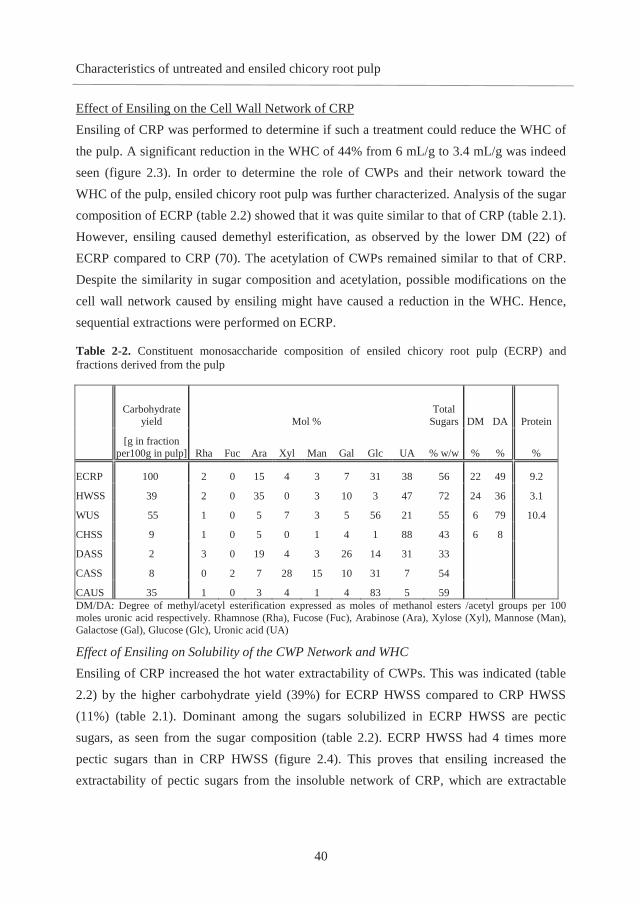
Characteristics of untreated and ensiled chicory root pulp
40
Effect of Ensiling on the Cell Wall Network of CRP Ensiling of CRP was performed to determine if such a treatment could reduce the WHC of the pulp. A significant reduction in the WHC of 44% from 6 mL/g to 3.4 mL/g was indeed seen (figure 2.3). In order to determine the role of CWPs and their network toward the WHC of the pulp, ensiled chicory root pulp was further characterized. Analysis of the sugar composition of ECRP (table 2.2) showed that it was quite similar to that of CRP (table 2.1). However, ensiling caused demethyl esterification, as observed by the lower DM (22) of ECRP compared to CRP (70). The acetylation of CWPs remained similar to that of CRP. Despite the similarity in sugar composition and acetylation, possible modifications on the cell wall network caused by ensiling might have caused a reduction in the WHC. Hence, sequential extractions were performed on ECRP.
Table 2-2. Constituent monosaccharide composition of ensiled chicory root pulp (ECRP) and fractions derived from the pulp
Carbohydrate
yield Mol % Total
Sugars DM DA Protein
[g in fraction
per100g in pulp] Rha Fuc Ara Xyl Man Gal Glc UA % w/w % % %
ECRP 100 2 0 15 4 3 7 31 38 56 22 49 9.2
HWSS 39 2 0 35 0 3 10 3 47 72 24 36 3.1
WUS 55 1 0 5 7 3 5 56 21 55 6 79 10.4
CHSS 9 1 0 5 0 1 4 1 88 43 6 8
DASS 2 3 0 19 4 3 26 14 31 33
CASS 8 0 2 7 28 15 10 31 7 54
CAUS 35 1 0 3 4 1 4 83 5 59 DM/DA: Degree of methyl/acetyl esterification expressed as moles of methanol esters /acetyl groups per 100 moles uronic acid respectively. Rhamnose (Rha), Fucose (Fuc), Arabinose (Ara), Xylose (Xyl), Mannose (Man), Galactose (Gal), Glucose (Glc), Uronic acid (UA)
Effect of Ensiling on Solubility of the CWP Network and WHC Ensiling of CRP increased the hot water extractability of CWPs. This was indicated (table 2.2) by the higher carbohydrate yield (39%) for ECRP HWSS compared to CRP HWSS (11%) (table 2.1). Dominant among the sugars solubilized in ECRP HWSS are pectic sugars, as seen from the sugar composition (table 2.2). ECRP HWSS had 4 times more pectic sugars than in CRP HWSS (figure 2.4). This proves that ensiling increased the extractability of pectic sugars from the insoluble network of CRP, which are extractable

CHAPTER 2
41
only under more severe conditions for CRP (figure 2.4A). The pectins in ECRP HWSS were shown to be degraded during ensilage, as can be seen from the elution profile by HPSEC (figure 2.4A). The dominant population of low molecular mass (Mw) less than 3 kDa indicated that dialysis was not sufficient to remove soluble material of low molecularmass. Removal of higher proportions of pectin from the cell wall network and reduced Mw and DM of pectin could explain a lower WHC for ECRP WUS (6 mL/g) compared to CRP WUS (13 mL/g). Furthermore, the characteristics of the residual CWPs in the insoluble network may also contribute to a lower WHC for ECRP WUS.
Figure 2-4. Distribution of polysaccharides in fractions derived from A. 100 g of chicory root pulp (CRP) and B. 100 g of ensiled chicory root pulp (ECRP)
Effect of Ensiling on the Extractability of CWPs and WHC Fractions obtained from the insoluble cell wall network (WUS) of ECRP had lower WHCs compared to those from CRP (figure 2.3). Ensiling increased the extractability of HG by 4 times in HWSS than CRP HWSS, having the consequence of a 4 times lower HG recovery

Characteristics of untreated and ensiled chicory root pulp
42
in ECRP CHSS compared to CRP CHSS (figure 2.4). HPSEC analysis of the extracted HG-rich pectin (88 mol % UA) in ECRP CHSS pointed to a lower Mw compared to CRP CHSS (figure 2.5B). Thus, increased removal of HG from WUS and degradation of residual HG-rich pectin in WUS during ensiling lowered the WHC of ECRP WUS (6 mL/g) compared to CRP WUS (13 mL/g). ECRP CHUS (sum of DASS, CASS, and CAUS) contains 3 times less RG and 4 times less HG than the fractions in CRP CHUS (figure 2.4). Ara-rich pectin was degraded in CHUS during ensiling, as indicated from low Ara:Rha in ECRP DASS (7:1) and CAUS (5:1) (table 2.1) compared to CRP DASS (13:1) and CAUS (11:1). The lower proportion of HG and RG and degraded Ara-rich pectin reduced the WHC of ECRP CHUS (7.8 mL/g) compared to CRP CHUS (14 mL/g) (figure 2.3). Furthermore, the WHCs for ECRP DAUS (10 mL/g) and CAUS (5 mL/g) were lower compared to CRP DAUS (22 mL/g) and CAUS (13 mL/g) (figure 2.3), respectively. ECRP CAUS derived from ECRP DAUS contains 3 times less pectic sugars than CRP CAUS (figure 2.4). Other polysaccharides, such as xyloglucan, glucan, and mannan were also modified in ECRP CAUS, increasing their extractability in ECRP CASS, as indicated from a higher carbohydrate yield (table 2.2) compared to CRP CASS and similar sugar composition to CRP CASS (table 2.1).
2.4. Conclusions
Studies on WHC showed that the WHC of chicory root pulp may change depending on the arrangement of cell wall polysaccharides within the plant cell wall network. The removal of branched pectin seemed to influence the WHC strongly. Besides, alkali-soluble xyloglucan and mannan-rich hemicellulose were indicated to contribute positively to the WHC. Ensiling degraded arabinose-rich pectin, homogalacturonan, and rhamnogalacturonan and modified the network consisting of xyloglucan, mannan, and glucan, all of which caused a significant reduction in the WHC. Following the characterization of chicory root pulp before and after ensiling, the use of CRP as a fiber supplement may have become closer to application. We now know that small changes in architecture as a result of mild processing or even by a targeted enzyme treatment will enable modulation of the WHC of CRP, resulting in improved properties of the CRP in the final fiber-rich product.

CHAPTER 2
43
2.5. Acknowledgements
This project is jointly financed by the European Union, European Regional Development Fund and The Ministry of Economic Affairs, Agriculture and Innovation, Peaks in the Delta, the Municipality of Groningen, the Province of Groningen, and the Dutch Carbohydrate Competence Center (CCC WP2a) and Royal Cosun.

Characteristics of untreated and ensiled chicory root pulp
44
References
1. Roberfroid, M. B., Inulin-type fructans: Functional food ingredients. Journal of Nutrition 2007, 137, 2493-2502. 2. Femenia, A.; Robertson, J. A.; Waldron, K. W.; Selvendran, R. R., Cauliflower (Brassica oleracea L), globe artichoke (Cynara scolymus) and chicory witloof (Cichorium intybus) processing by-products as sources of dietary fibre. Journal of the Science of Food and Agriculture 1998, 77, 511-518. 3. Robert, C.; Emaga, T. H.; Wathelet, B.; Paquot, M., Effect of variety and harvest date on pectin extracted from chicory roots (Cichorium intybus L.). Food Chemistry 2008,108, 1008-1018. 4. Renard, C. M. G. C.; Crépeau, M. J.; Thibault, J. F., Influence of ionic strength, pH and dielectric constant on hydration properties of native and modified fibres from sugar-beet and wheat bran. Industrial Crops and Products 1994, 3, 75-84. 5. Panouille, M.; Thibault, J. F.; Bonnin, E., Cellulase and protease preparations can extract pectins from various plant byproducts. Journal of Agricultural and Food Chemistry 2006, 54, 8926-8935. 6. Guillon, F.; Barry, J.-L.; Thibault, J.-F., Effect of autoclaving sugar-beet fibre on its physico-chemical properties and its in vitro degradation by human faecal bacteria. Journal of the Science of Food and Agriculture 1992, 60, 69-79. 7. Auffret, A.; Ralet, M. C.; Guillon, F.; Barry, J. L.; Thibault, J. F., Effect of grinding and experimental conditions on the measurement of hydration properties of dietary-fibers. Food Science and Technology-Lebensmittel-Wissenschaft & Technologie 1994, 27, 166-172. 8. Okine, A.; Hanada, M.; Aibibula, Y.; Okamoto, M., Ensiling of potato pulp with or without bacterial inoculants and its effect on fermentation quality, nutrient composition and nutritive value. Animal Feed Science and Technology 2005, 121, 329-343. 9. Voragen, A. G. J.; Coenen, G. J.; Verhoef, R. P.; Schols, H. A., Pectin, a versatile polysaccharide present in plant cell walls. Structural Chemistry 2009, 20, 263-275. 10. Pauly, M.; Albersheim, P.; Darvill, A.; York, W. S., Molecular domains of the cellulose/xyloglucan network in the cell walls of higher plants. Plant Journal 1999, 20, 629-639. 11. Kormelink, F. J. M.; Searle-Van Leeuwen, M. J. F.; Wood, T. M.; Voragen, A. G. J., Purification and characterization of three endo-(1,4)- -xylanases and one -xylosidase from Aspergillus awamori. Journal of Biotechnology 1993, 27, 249-265. 12. Düsterhöft, E. M.; Bonte, A. W.; Voragen, A. G. J., Solubilisation of non-starch polysaccharides from oil-seed meals by polysaccharide-degrading enzymes. Journal of the Science of Food and Agriculture 1993, 63, 211-220. 13. Englyst, H. N.; Cummings, J. H., Simplified method for the measurement of total non-starch polysaccharides by gas-liquid chromatography of constituent sugars as alditol acetates. Analyst 1984, 109, 937-942. 14. Thibault, J. F., Automated-method for the determination of pectic substances. Lebensmittel-Wissenschaft & Technologie 1979, 12, 247-251.

45
15. Ramaswamy, U. R.; Kabel, M. A.; Schols, H. A.; Gruppen, H., Structural features and water holding capacities of pressed potato fibre polysaccharides. Carbohydrate Polymers 2013, 93, 589-596. 16. Stöber, P.; Bénet, S.; Hischenhuber, C., Simplified enzymatic high-performance anion exchange chromatographic determination of total fructans in food and pet food limitations and measurement uncertainty. Journal of Agricultural and Food Chemistry 2004, 52, 2137-2146. 17. Voragen, A. G. J.; Schols, H. A.; Pilnik, W., Determination of the degree of methylation and acetylation of pectins by h.p.l.c. Food Hydrocolloids 1986, 1, 65-70. 18. Baumann, H., Apparatur nach Baumann zur Bestimmung der Flüssigkeitsaufnahme von pulvrigen Substanzen. Fette, Seifen, Anstrichm. 1966, 68, 741-743. 19. Rombouts, F. M.; Thibault, J. F., Sugar beet pectins: Chemical structure and gelation through oxidative coupling. In Chemistry and Function of Pectins, Marshall L. Fishman; Jen, J. J., Eds. American Chemical Society: Washington D.C., USA, 1986; Vol. 310, pp 49-60. 20. Davison, P. K.; Young, R., Gas chromatography of carbohydrates the quantitative determination of the free sugars of plants as their trimethylsilyl ethers. Journal of Chromatography A 1964, 41, 12-21. 21. Schols, H. A.; Posthumus, M. A.; Voragen, A. G. J., Hairy (ramified) regions of pectins. 1.Structural features of hairy regions of pectins isolated from apple juice produced by the liquefaction process. Carbohydrate Research 1990, 206, 117-129. 22. Carpita, N. C.; Gibeaut, D. M., Structural models of primary cell walls in flowering plants: Consistency of molecular structure with the physical properties of the walls during growth. Plant Journal 1993, 3, 1-30. 23. Hilz, H.; de Jong, L. E.; Kabel, M. A.; Schols, H. A.; Voragen, A. G. J., A comparison of liquid chromatography, capillary electrophoresis, and mass spectrometry methods to determine xyloglucan structures in black currants. Journal of Chromatography A 2006, 1133, 275-286. 24. Hayashi, T.; Maclachlan, G., Pea xyloglucans and cellulose.1.Macromolecular organisation. Plant Physiology 1984, 75, 596-604. 25. Bootten, T. J.; Harris, P. J.; Melton, L. D.; Newman, R. H., WAXS and 13C NMR study of Gluconoacetobacter xylinus cellulose in composites with tamarind xyloglucan. Carbohydrate Research 2008, 343, 221-229. 26. Zykwinska, A.; Thibault, J. F.; Ralet, M. C., Competitive binding of pectin and xyloglucan with primary cell wall cellulose. Carbohydrate Polymers 2008, 74, 957-961. 27. Rosell, C. M.; Santos, E.; Collar, C., Mixing properties of fibre-enriched wheat bread doughs: A response surface methodology study. European Food Research and Technology 2006, 223, 333-340. 28. Leboeuf, E.; Thoiron, S.; Lahaye, M., Physico-chemical characteristics of cell walls from Arabidopsis thaliana microcalli showing different adhesion strengths. Journal of Experimental Botany 2004, 55, 2087-2097. 29. Cosgrove, D. J., Relaxation in a high-stress environment: the molecular bases of extensible cell walls and cell enlargement. The Plant Cell Online 1997, 9, 1031-1041.

Characteristics of untreated and ensiled chicory root pulp
46
30. Dick-Structure and interactions of plant cell-wall polysaccharides by two- and three-dimensional magic-angle-spinning solid-state NMR. Biochemistry 2011, 50, 989-1000. 31. Dinand, E.; Chanzy, H.; Vignon, R. M., Suspensions of cellulose microfibrils from sugar beet pulp. Food Hydrocolloids 1999, 13, 275-283. 32. Sarko, A.; Nishimura, H.; Okano, T., Crystalline alkali-cellulose complexes as intermediates during mercerization. In The structures of cellulose, Atalla, R. H., Ed. American Chemical Society: Washington DC, USA, 1987; Vol. 340, pp 169-177.

47
Chapter 3
The effect of soluble and insoluble
fibers on the fermentation of chicory
root pulp
Submitted for publication
Ramasamy S. U.; Venema K.; Schols H.A.; Gruppen H.

CHAPTER 3
48
Abstract
The aim of this research was to study the in vitro fermentation of chicory root pulp (CRP)
and ensiled CRP (ECRP) using human faecal inoculum. Analysis of carbohydrate levels in
fermentation digests showed that 51% of all CRP carbohydrates were utilized after 24 h of
fermentation. For ECRP, having the same cell wall polysaccharide composition as CRP,
but with solubilisation of four times more of CRP pectin due to ensiling, the fermentation
was quicker than CRP as 11% more carbohydrates were utilized within the first12 hours.
The level of fiber utilization for ECRP after 24 h was increased by 8% compared to CRP.
This effect on fiber utilization from ECRP seemed to arise from i) increased levels of
soluble pectin fibers (arabinan, homogalacturonan and galactan) and ii) a more
hypothesized open structure of the remaining cell walls in ECRP which was more
accessible to degradation than the CRP cell wall network.

The effect of soluble and insoluble fibers on CRP fermentation
49
3.1. Introduction
Dietary fiber (DF) includes cell wall polysaccharides (CWPs), such as pectin, hemicellulose
and cellulose, which are resistant to absorption or digestion in the human small intestine
and become available for (partial) fermentation in the colon by the human microbiota (1).
Some types of DF have been reported to contribute to gut health. Pectin has been shown to
reduce adhesion of pathogens to intestinal epithelial cells (2), reduce ammonia generation
(3), induce intestinal mucosal proliferation and maintain intestinal integrity (4).
Fermentation of DFs in the colon promotes the growth of microbiota. Short chain fatty
acids (SCFA’s) produced by fermentation of DF entering the proximal colon, lower the pH
of the proximal colon and inhibit the growth of pathogens (5). While the availability of DF
in the proximal colon is beneficial, the absence of DF in the distal colon may lead to a
switch to protein fermentation by the microbiota, which amongst others leads to the
production of ammonia, and thereby leads to a pH that is near neutral (6, 7). Such
conditions make the distal colon vulnerable to chronic gut disorders (8, 9). Thus, it would
be beneficial to use a mixture of DFs, part of which are fermented in the proximal colon
and part of which remain available for further fermentation in the distal colon. Research on
the in vitro fermentability of pectin-rich agricultural by-products, such as sugar beet pulp,
has been performed using human faecal inoculum. More than 80% of the initial
carbohydrates in sugar beet pulp were utilized within 24 h of fermentation indicating high
fermentability of the pulp (10). Furthermore, the fermentability may be influenced by
processing resulting in a modified cell wall network as has been shown for autoclaved
sugar beet pulp (10). Chicory root pulp (CRP) obtained after inulin extraction is rich in
CWPs (58% w/w), predominantly pectin (62%), followed by cellulose (27%) and
hemicellulose (11%) as DFs (11). In addition to CWPs, CRP contains (6% w/w) residual
levels of the storage polysaccharide, inulin as a DF (11). The use of CRP as a DF
supplement in food applications could thus be promising. While abundant in insoluble
fibres of different populations of CWPs, the fermentability of CRP has not been studied.
Processed CRP, such as ensiled CRP (ECRP), has a loosened cell wall network with a
reduced water holding capacity (WHC) (3.4 mL/g) compared to CRP (6 mL/g) and contains
four times more soluble pectin fibers than CRP (11). Such a modification of the cell wall

CHAPTER 3
50
network with more soluble fibers may have an effect on the extent and rate of fermentation
of CRP. Thus, the aim of this research was to determine the effect of processed and
unprocessed cell walls of CRP on the in vitro fermentability of the pulp using human faecal
inoculum.
3.2. Materials and Methods
CRP and ECRP were obtained from Cosun Food Technology Center (Roosendaal, The
Netherlands) and have been described previously (11). Since they contain dietary fibers and
no starch and no digestible oligosaccharides that would be digested in the stomach or the
small intestine, no pre-digestion was performed on these materials.
In vitro fermentation:
The fermentation medium used in this study was modified standard ileal efflux medium
(SIEM) according to Ladirat et al. (12) Tween 80 was omitted from the fermentation
medium because it influenced analysis of polysaccharide degradation products. All medium
components were purchased from Tritium Microbiology (Veldhoven, The Netherlands).
The pH of the fermentation medium was adjusted to 5.8, simulating the pH in the proximal
colon. The inoculum was a standardised pool of adult faecal inoculum according to the
validated protocol described elsewhere (13) and prepared from eight healthy European
adults (25-45 years old) who did not receive antibiotics during 2 months before donation.
The inoculum (6 mL) was incubated in the modified SIEM (54 mL) for activation (12).
CRP and ECRP (100 mg) were added to 9 mL of the modified SIEM in 25 mL bottles for
each time point. The activated inoculum (1 mL) was added to the fermentation medium and
flushed with CO2. The bottles were immediately closed with rubber stoppers and sealed
with aluminium caps. Fermentations were performed in duplicate for each substrate at 37
°C and shaken at 300 rpm until 24 h. A blank with only microbiota was taken as a control.
Fermentation digests were inactivated at 100°C for 5 min and split into two parts. One part
was reserved for direct analysis and the other part of the fermentation digest was freeze
dried for further characterization. Part of the dried fermentation digest was suspended in
millipore water (10mg/mL) and centrifuged (10 min, 9,000 x g, 4°C) to obtain soluble and
insoluble fractions. The insoluble fraction was freeze dried and used along with the original

The effect of soluble and insoluble fibers on CRP fermentation
51
dried fermentation digest for analysing constituent monosaccharide content and
composition. Constituent monosaccharide content in the soluble fraction was calculated
from the difference in individual monosaccharide contents between the dried fermentation
digest and the dried insoluble fraction of the fermentation digest.
Analytical methods
Constituent monosaccharide composition.
The constituent monosaccharide content and composition were determined using a pre-
hydrolysis step with 72 (w/w%) sulphuric acid at 30°C for 1 h followed by hydrolysis with
1 M sulphuric acid at 100°C for 3 h. The monosaccharides formed upon hydrolysis were
derivatized to alditol acetates and analysed by gas chromatography using inositol as an
internal standard (14). The colorimetric m-hydroxydiphenyl assay (15) was used to
determine the total uronic acid (UA) content.
Molecular weight distribution
Fermentation digests were centrifuged (10min, 18,000 x g, 24°C) to obtain the soluble
fraction which was analysed for molecular weight distribution using High Performance
Size Exclusion Chromatography (HPSEC) on an Ultimate 3000 HPLC (Dionex, Sunnyvale,
CA, USA) as described before (11). Three TSK-Gel columns in series (4000-3000-2500
SuperAW; 150 x 6 mm) were used for the analysis. They were preceded by a TSK Super
AW-L guard column (35 x 4.6mm). All columns were from Tosoh Bioscience (Tokyo,
Japan). The molecular weight was estimated based on a standard curve of pullulan
standards.
Oligosaccharide profiling
High Performance Anion Exchange Chromatography (HPAEC) was performed on a
ICS5000 system (Dionex), equipped with a Dionex CarboPac PA-1 column (2 x 250 mm)
in combination with a Carbopac PA-1 guard column (2 x 50 mm). Ten μL of the soluble
fraction of the fermentation digests (diluted twice with millipore water) was injected into
the system using a Dionex ICS5000 autosampler. The system was equipped with pulsed
amperometric detection. The flow rate was 0.3mL/min and the gradient used was as

CHAPTER 3
52
described elsewhere (16). The software used was Chromeleon version 7 (Dionex).
Oligomers of galacturonic acid (DP 1 to DP 5) were used as standards to identify and
quantify galacturonic acid oligomers in the elution profile.
Analysis of short chain fatty acid (SCFAs) and organic acids
SCFAs and organic acids (lactate and succinate) were analysed as described elsewhere (17).
Microbiota composition
Samples from a twin batch fermentation of CRP were analysed for microbiota composition.
The chemical analyses of fermentation of CRP (SCFA production, monosaccharide
utilization) are correlated to the microbiota composition. The microbiota composition for
ECRP fermentation was not performed due to limitations in the sample amount. The
microbiota compositions for 2 independent duplicates of a 9 h fermentation digest of CRP
(supplementary figure 3.S1) were similar indicating reproducibility for fermentations of the
cell wall material.
Molecular techniques
DNA from the fermentation digests was isolated using the AGOWA mag Mini kit (DNA
Isolation Kit, AGOWA, Berlin, Germany), according to the manufacturer’s instructions.
Generation of the PCR amplicon library was performed by amplification of V5-V7
hypervariable region of the small subunit ribosomal DNA gene (16S rDNA). All samples
were analysed as described elsewhere (18).
3.3. Results and discussion
Characteristics of fibers in chicory root pulp and ensiled chicory root pulp
Table 1 shows the constituent monosaccharide composition of chicory root pulp (CRP) and
ensiled CRP (ECRP) (11). CRP is abundant in carbohydrates (64 w/w%) of which 58
w/w% represents cell wall polysaccharides (CWPs) and 6 w/w% represents minor levels of
the storage polysaccharide inulin with an average DP of 18 (11). The CWPs calculated
from the mol% monosaccharide composition and total sugar content (w/w %) (table 3.1)
contain 62 w/w% pectin (uronic acid + rhamnose + arabinose + galactose), 27% cellulose

The effect of soluble and insoluble fibers on CRP fermentation
53
(arising from glucose not present in xyloglucan) and 11% hemicellulose (xylose + mannose
+ fucose + glucose in xyloglucan) as characterised earlier (11). Table 3-1. Constituent monosaccharide composition of of chicory root pulp (CRP) and ensiled chicory root pulp (ECRP)
Mol % (w/w %) Carbohydrate yield
Rha Fuc Ara Xyl Man Gal Glc UA Total
[g in fraction per100g
in CRP/ECRP]
CRP 1 0 15 4 4 7 31 38 64* 100
CRP Soluble 1 0 27 1 13 6 19 33 50 12
CRP Insoluble 1 0 12 4 2 7 33 40 68 87
ECRP 2 0 15 4 3 7 31 38 56 100
ECRP Soluble 2 0 27 0 3 8 3 57 62 48
ECRP Insoluble 1 0 5 7 3 5 56 21 55 55 Rhamnose (Rha), Fucose (Fuc), Arabinose (Ara), Xylose (Xyl), Mannose (Man), Galactose (Gal), Glucose (Glc), Uronic acid (UA), * contains 6 w/w % inulin. Results from an earlier study (11).
The carbohydrates present in the soluble fraction represent only 12% of the CRP
carbohydrates. As determined earlier (11), half of the soluble fraction is arabinose rich
pectin and the other half is inulin. 87% of the carbohydrates from CRP are insoluble and
have similar polysaccharide composition as the total CRP (table 3.1). The ECRP is rather
similar in composition to CRP (table 3.1), but contains no inulin (11).
As indicated by the higher carbohydrate recovery (45%) in the soluble fraction from ECRP
compared to that from CRP (12%) (table 3.1), ECRP has more soluble fibers than CRP.
Dominant among the sugars solubilised are the pectic sugars as seen from the sugar
composition (table 3.1). As determined from an earlier study (11), the soluble fraction of
ECRP represents 4 times more pectic sugars than that from CRP. The pectic sugars from
the insoluble network of CRP are only extractable under severe conditions, like chelating
agents and alkali (11). Apparently, they can be solubilised by ensiling. As a consequence,
the insoluble fraction of ECRP represents a lower recovery of carbohydrates (55%)
compared to CRP (87%) and is enriched in (hemi)cellulose (table 3.1).
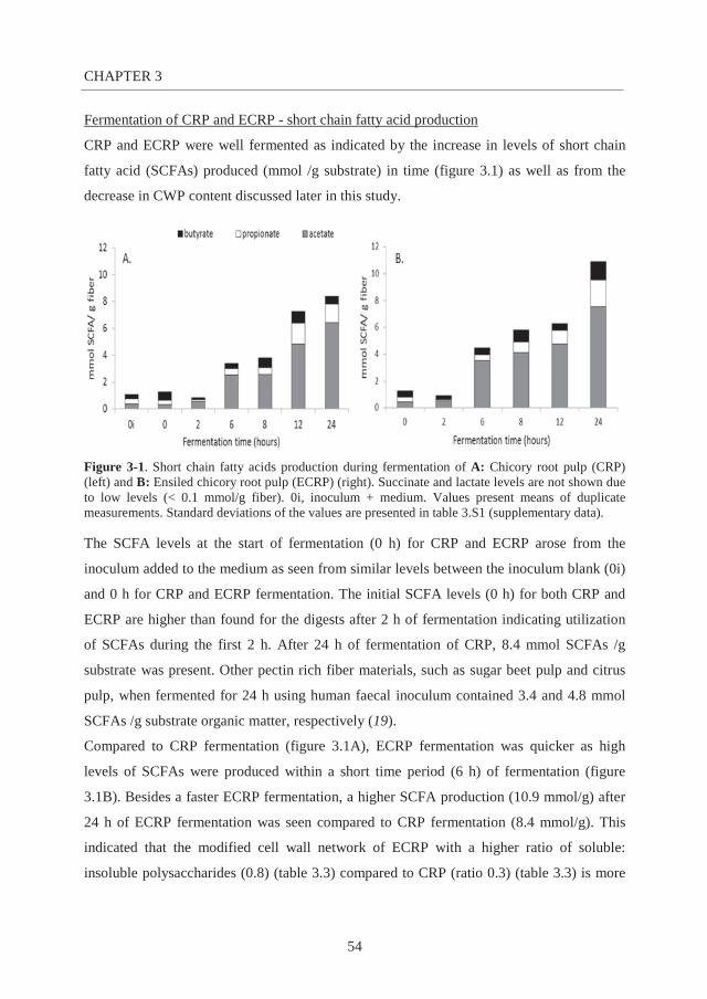
CHAPTER 3
54
Fermentation of CRP and ECRP - short chain fatty acid production
CRP and ECRP were well fermented as indicated by the increase in levels of short chain
fatty acid (SCFAs) produced (mmol /g substrate) in time (figure 3.1) as well as from the
decrease in CWP content discussed later in this study.
Figure 3-1. Short chain fatty acids production during fermentation of A: Chicory root pulp (CRP) (left) and B: Ensiled chicory root pulp (ECRP) (right). Succinate and lactate levels are not shown due to low levels (< 0.1 mmol/g fiber). 0i, inoculum + medium. Values present means of duplicate measurements. Standard deviations of the values are presented in table 3.S1 (supplementary data).
The SCFA levels at the start of fermentation (0 h) for CRP and ECRP arose from the
inoculum added to the medium as seen from similar levels between the inoculum blank (0i)
and 0 h for CRP and ECRP fermentation. The initial SCFA levels (0 h) for both CRP and
ECRP are higher than found for the digests after 2 h of fermentation indicating utilization
of SCFAs during the first 2 h. After 24 h of fermentation of CRP, 8.4 mmol SCFAs /g
substrate was present. Other pectin rich fiber materials, such as sugar beet pulp and citrus
pulp, when fermented for 24 h using human faecal inoculum contained 3.4 and 4.8 mmol
SCFAs /g substrate organic matter, respectively (19).
Compared to CRP fermentation (figure 3.1A), ECRP fermentation was quicker as high
levels of SCFAs were produced within a short time period (6 h) of fermentation (figure
3.1B). Besides a faster ECRP fermentation, a higher SCFA production (10.9 mmol/g) after
24 h of ECRP fermentation was seen compared to CRP fermentation (8.4 mmol/g). This
indicated that the modified cell wall network of ECRP with a higher ratio of soluble:
insoluble polysaccharides (0.8) (table 3.3) compared to CRP (ratio 0.3) (table 3.3) is more

The effect of soluble and insoluble fibers on CRP fermentation
55
fermentable than that of CRP. The ratio of acetate: propionate: butyrate was similar
between CRP (77:16:6) and ECRP (69:18:13) after 24 h of fermentation. Taking into
account the similar carbohydrate composition of CRP and ECRP, this suggested that
fermentation of an open cell wall network has limited influence on the utilization by the
microbiota. This similarity in the SCFA ratio has also been observed for fermentations of
sugar beet pulp and autoclaved beet pulp (10).
Minor levels of intermediate fatty acids (20), lactate and succinate (<0.1 mmol/g), were
observed throughout the fermentation period. The fermentation of higher oligomers of
inulin (discussed later) indeed resulted in very little butyrate and lactate formation.
Fermentation of higher oligomers of inulin (DP >20) by the microbiota has been reported to
hinder the production of butyrate and lactate than short oligomers of inulin (DP < 10) (21,
22).
Molecular weight distribution of soluble fibers
HPSEC elution profiles of solubles from CRP and ECRP fermentation digests are shown in
figure 3.2. For CRP fermentation, an increase in the amount of soluble material
corresponding to high molecular weight (Mw) (8 – 10 min; 47- 788 kDa) was seen after 2 h
of inoculation. From 6 h onwards, an almost complete disappearance in the high Mw
fraction suggested degradation/ utilization of the high Mw polysaccharide. This implies
either rapid utilization of fibers solubilised during fermentation or direct utilization of
insoluble fibers directly by the microbiota system.
Compared to the elution profile of CRP fermentation blank (0 h), the elution profile of the
ECRP blank showed a 52% higher amount of high Mw material, as expected from the
higher pectin solubility(11). This material is present in two populations, (8–9 min; 212-788
kDa) and (9-10 min; 47-212 kDa). After 6 h of fermentation, a decrease is seen in the
intensity of both peaks indicating degradation of both the populations. These populations
are almost completely utilized after 8 h of fermentation. As for CRP fermentation, almost
no high Mw solubles could be seen after 12 and 24 h of ECRP fermentation.

CHAPTER 3
56
Figure 3-2. HPSEC elution patterns of fermentation digests during fermentation of A. Chicory root pulp (CRP) and B. Ensiled chicory root pulp (ECRP). Pullulan was used as a calibrant.
Oligomer utilization
In order to determine oligomers formed/utilized during CRP and ECRP fermentation,
soluble fractions from fermentation digests were analysed using HPAEC (figure 3.3).
Before the start of CRP fermentation (0 h), higher oligomers of inulin (average DP of 18)
(11) were observed. Since short oligomers of inulin were removed by the industrial
extraction from the root, the pulp only contains higher oligomers of inulin, with an average
DP of 18. Within 6 h, inulin is almost completely utilized (11).
During fermentation, CWPs are degraded to lower Mw material and in a previous study
using other sources for CWPs than CRP, intermediate oligosaccharides could be clearly
recognised (16). However, in this study, no oligomers formed from CWPs were observed
during CRP fermentation. Bacteroides sp., becoming dominant during CRP fermentation,
as discussed later, are known to be motile and to possess ‘specialized polysaccharide
utilization systems’ in which cell surface proteins are involved in binding to different
polysaccharides and transporting the oligomers that are formed directly over the bacterial

The effect of soluble and insoluble fibers on CRP fermentation
57
membrane (23, 24). Such systems likely prevent diffusion of oligomers. Another possibility
for the absence of oligomers could arise from rapid cross feeding of oligomers by other
fermenting bacteria (25).
Before the start of ECRP fermentation (0 h), a number of peaks can be identified as
oligomers of galacturonic acid (GalA) based on standards of GalA oligomers used (figure
3.3B). These oligomers represent approximately 14% of total ECRP GalA and arose from
ECRP after the process of ensiling. These oligomers are shown to decrease within the first
6 h of ECRP fermentation pointing to their utilization. Similar to the CRP fermentation, no
new oligomers are formed.
Figure 3-3. HPAEC elution profile of fermentation digests during fermentation of A. Chicory root pulp (CRP) and B. Ensiled chicory root pulp (ECRP). Legend: Galacturonic acid (GalA), (GalA)2: GalA dimer, (GalA)3: GalA trimer.

CHAPTER 3
58
Changes in CRP fiber composition during fermentation
Direct utilization of CWPs during CRP fermentation can be revealed from the decrease in
the total carbohydrate content as well as from the constituent monosaccharides during
fermentation.
The constituent monosaccharide compositions of CRP fermentation digests are shown in
table 3.2. Low concentrations (w/w %) of carbohydrates in all the fermentation digests are
due to the presence of high concentrations of other fermentation media components, such as
proteins and salts (12, 13). The constituent monosaccharide composition of the CRP
fermentation blank (0 h) (table 3.2) is similar to that of CRP (table 3.1) indicating that the
carbohydrates present in the medium before fermentation do not contain carbohydrates
other than from CRP fibers. After 24 h of fermentation, 51% of CRP total carbohydrates
were utilized (figure 3.4A). Table 3-2. Residual carbohydrate levels and constituent monosaccharide composition of digests obtained from in vitro fermentation of chicory root pulp (CRP) and ensiled CRP (ECRP)
Fermentation time
Residual carbohydrate levels Mol % (w/w %)
(hours) (mg from 90 mg starting
material) Rha Fuc Ara Xyl Man Gal Glc UA Total
CRP 0 (blank) 56 2 0 14 4 5 8 35 32 7 0.00 0.05 0.69 0.25 0.67 0.10 0.36 0.19 0.00
2 46 2 0 14 4 5 8 35 32 7 0.03 0.00 0.05 0.06 0.02 0.05 0.06 0.12 0.04
12 42 2 0 14 5 3 8 35 34 7 0.01 0.00 0.00 0.02 0.02 0.01 0.11 0.29 0.44
24 28 2 0 4 8 4 6 65 10 4 0.15 0.03 0.02 0.55 0.08 0.13 0.50 0.25 0.04
ECRP 0 (blank) 55 2 0 14 4 3 8 35 34 7 0.00 0.00 0.06 0.01 0.03 0.01 0.32 0.03 0.32
2 48 2 0 15 4 3 8 36 32 7 0.20 0.02 0.10 0.16 0.38 0.35 1.55 2.43 0.29
12 35 2 0 6 6 4 7 53 22 5 0.25 0.06 0.69 0.50 0.36 1.17 0.02 1.43 0.71
24 22 2 0 2 9 5 5 68 10 4 0.43 0.04 0.10 0.33 0.04 0.41 0.27 1.08 0.22
Rhamnose (Rha), Fucose (Fuc), Arabinose (Ara), Xylose (Xyl), Mannose (Man), Galactose (Gal), Glucose (Glc), Uronic acid (UA). The values presented are means of duplicate measurements, with standard deviation in italics.

The effect of soluble and insoluble fibers on CRP fermentation
59
This is rather similar to the degradability of sugar beet fiber showing 60% carbohydrate
utilization after 24 h of fermentation with human faecal inoculum (26).
Until 12 h of fermentation, the constituent monosaccharide composition remained similar
although 25% of all carbohydrates were utilized. This indicated unspecific utilization of
CWPs in the first 12 h of fermentation. This is also seen in figure 3.4A from 0 to 12 h of
fermentation. As the fermentation continued from 12 h to 24 h, the carbohydrate content
was further reduced by another 25% and the constituent monosaccharide composition
changed considerably. This suggests a more specific utilization of CWPs from 12 h
onwards. The calculated decrease in arabinose (Ara), galactose (Gal), and GalA by 86%,
63%, and 84% respectively, after 24 h (figure 3.4A) indicated enhanced utilization of
arabinan, galactan and homogalacturonan (HG) from CRP respectively. The unexpected
decrease in glucose utilization from 12 to 24 h (figure 3.4A) might arise from an increase in
bacterial cell walls containing glucose (27).
Figure 3-4. Utilization (%) of monosaccharides during in vitro fermentation originally present in A.Chicory root pulp (CRP) and B. Ensiled chicory root pulp (ECRP). Values are means of duplicate measurements with errors shown by vertical bars on the curves.
The enzyme machinery from the fermenting bacteria seemed to be so efficient in degrading
arabinan that the 14% level of resistant arabinan after 24 h of fermentation is only half of

CHAPTER 3
60
the arabinan remaining in the cell wall matrix after severe extraction by strong alkali (11).
This indicated that even part of the heavily entangled arabinan was utilized during
fermentation.
Changes in ensiled CRP fiber composition during fermentation
The presence of more soluble fibers in a more open cell wall network in ECRP compared to
CRP was hypothesized to have an effect on the fiber utilization during fermentation of the
cell wall network. The constituent monosaccharide compositions of the fermentation
digests obtained during fermentation of ECRP are shown in table 3.2.
After 24 h of fermentation, 59% of all carbohydrates are utilized from ECRP (figure 3.4B).
The 8% higher carbohydrate utilization compared to CRP indicates a slightly higher
fermentability of ECRP over CRP as has been seen from the higher SCFA production as
discussed earlier. Unexpectedly, the increase in SCFA production (30%) is much higher
than the 8% higher carbohydrate utilization. ECRP fermentation was also quicker as was
indicated from the high level of carbohydrate utilization (36%) within a short time period of
12 h compared to only 25% carbohydrate utilization for 12 h of CRP fermentation (table
3.2).
As for CRP fermentation, specific CWPs consisting of Ara, GalA and Gal are favourably
utilized the most during ECRP fermentation, but to a larger extent compared to CRP
fermentation (figure 3.4). As seen in figure 3.4, the sugar utilization for ECRP is more
pronounced than CRP fermentation after 12 h than after 24 h of fermentation. At 12 h, the
sugar utilization was determined to be (31 and 74%) Ara, (20 and 58%) GalA, and (32 and
43%) Gal for CRP and ECRP respectively (figure 3.4). This rapid increase in carbohydrate
utilization coincides with the higher levels of soluble fibers composed of Ara, Gal and
GalA in ECRP. This indicated that processing of CRP, increasing the levels of soluble
fibers and modifying the cell wall network, increased the degradability and the rate of
fermentation of specific CWPs.

The effect of soluble and insoluble fibers on CRP fermentation
61
Utilization of soluble/insoluble fibers from cell wall materials during fermentation
The presence of soluble fibers and/or more open CWP network of the remaining insoluble
fibers in ECRP seem to enhance fermentation. In order to elaborate on this observation, the
levels of both soluble and insoluble fibers in digests are shown in figure 3.5.
Utilization of soluble/insoluble fibers from CRP:
The insoluble fibers in CRP are utilized only after 12 h when all soluble fibers have been
utilized. This is also in agreement with HPSEC in which no soluble fibers could be seen
after 12 h of fermentation (figure 3.2). Insoluble fibers are mostly utilized between 12 and
24 h of fermentation shown by the decrease from 73% to 48% of residual insoluble
carbohydrates. Insoluble fiber utilization mostly involved pectic sugars of Ara, GalA and
Gal as indicated from table 3.3. Only 18% of all insoluble pectin (GalA+ Rha + Ara + Gal)
(calculated from table 3.3) remained unfermentable after 24 h suggesting a high
fermentability of insoluble pectin from the CRP plant cell wall.
Figure 3-5. Proportion of sugars in soluble and insoluble fractions of (%) original sugars present. A. Chicory root pulp (CRP) fermentation and B. Ensiled chicory root pulp (ECRP) fermentation. Values are means of duplicate measurements with errors shown by vertical bars on the curves.
For ECRP fermentation, utilization of soluble fibers can be seen from the decrease in the
residual soluble carbohydrate levels from 45 to 4% from 0 to 24 h (figure 3.5B). Utilization
of insoluble fibers is slow and lower than found for the soluble fiber as shown by the
decrease from 55% to 37% of residual insoluble carbohydrates (figure 3.5B). Soluble fibers
consisting of pectic sugars (table 3.3) are thus preferentially utilized. This suggests that the

CHAPTER 3
62
increased rate and higher level (8%) of carbohydrate utilization in ECRP fermentation
compared to CRP fermentation is due to fermentation of soluble pectin fibers that are
present in high amounts in ECRP. Table 3-3. Proportion of constituent monosaccharides present in soluble and insoluble fractions of % original carbohydrates during in vitro fermentation of chicory root pulp (CRP) and ensiled CRP (ECRP).
Time
(hours) Rha Ara Xyl Man Gal Glc UA Total
CRP Sugars in insoluble fraction
0 (blank) 62 81 84 50 72 76 80 77
2 52 66 70 50 58 69 75 69
12 91 67 68 46 65 75 79 73
24 43 14 88 52 35 92 14 48
Sugars in soluble fraction
0 (blank) 38 19 16 50 28 24 20 23
2 37 11 26 48 20 12 10 14
12 9 0 12 0 0 0 0 0 24 15 0 0 0 1 0 2 0
ECRP Sugars in insoluble fraction 0 (blank) 47 39 100 61 55 80 33 55
2 45 33 96 60 47 79 31 53 12 37 19 93 60 41 79 28 49 24 26 5 86 56 16 76 9 37 Sugars in soluble fraction
0 (blank) 53 61 0 39 45 20 67 45 2 49 62 2 31 43 10 53 35
12 38 7 3 24 15 16 14 14 24 35 0 5 8 8 3 3 4
Values are means of duplicate measurements. Rhamnose (Rha), Fucose (Fuc), Arabinose (Ara), Xylose (Xyl), Mannose (Man), Galactose (Gal), Glucose (Glc), Uronic acid (UA). Standard errors of the values are presented in table 3.S2 (supplementary data).
Compared to CRP, for ECRP, only 9% of all pectin (Rha+Gala+Ara+Gal) originally
present remained unfermentable (calculated from table 3.3). This suggested that pectin in
the hypothesized more open structure of the ECRP cell wall was more degradable than the
pectin in the CRP cell wall structure contributing to an increased fermentability of ECRP.
Other than pectin, the ECRP cell wall contained quite some cellulose + hemicellulose

The effect of soluble and insoluble fibers on CRP fermentation
63
sugars (glucose+ xylose+mannose) that were unfermentable (75%) (calculated from table
3.3). This suggested that the fermentative degradation of the CWP network was selective
for pectin fibers and preferably soluble pectin. This is in agreement with the limited
degradation of (hemi)cellulose by the human gut microbiota in human subjects (28).
Microbiota composition during CRP fermentation
The microbiota composition during the course of fermentation is expected to be modulated
based on the composition of the fiber added during fermentation (29). The microbiota
composition of the fermentation digest for CRP fermentation (figure 3.6) is expressed as %
abundance of genera of the total microbiota in the fermentation digest. The fermentation
digest at 24 h of fermentation showed an increase in Clostridium sensu stricto sp. (1.34%)
and Lachnospiracea incertae sedis sp. (1.21%). The highest increase was seen for
Bacteroides sp. (8%), which remain the most dominant bacteria after 24 h of fermentation.
Although Lachnospiracea incertae sedis sp. is a butyrate producer (30) and increased
during fermentation, the observed lower butyrate : (acetate + propionate) ratio of 6:94 after
24 h compared to 50:50 at the start of fermentation (figure 1) could be due to the decrease
in butyrate producers (31, 32) mostly from Clostridium XIVa sp. (6%), Faecalibacterium
sp.(0.94%), and Butyricicoccus sp. (0.57%). Other bacteria which decreased in 24 hours
were Sutterella sp. (5.7%), Dorea sp. (1.5%) and Clostridium XI sp. (1%). Coprococcus
sp., Dialyster sp. and Bifidobacterium sp. remained more or less the same. Despite the
presence of the bifidogenic inulin fraction (33), no growth of Bifidobacterium sp. was seen
after 24 h of fermentation, since inulin (6% of all CRP carbohydrates) was already utilized
within 6 h of fermentation. Although changes in the microbiota composition are seen
during CRP fermentation, the significance of the change in the complex community of
microbiota is hard to understand.
Part of the significance may be understood when correlated to metabolite production, fiber
degradation and utilization. It is likely that Bacteroides sp., identified as the dominant
bacteria in fermenting CRP, are responsible for the shown utilization of arabinan, galactan
and homogalacturonan and an increase in acetate and propionate. B.thetaiotaomicron
known to be highly abundant among Bacteroides sp (34). has been estimated to include 236

CHAPTER 3
64
glycoside hydrolase genes (35, 36). Production of enzymes during the course of
fermentation is upregulated based on the polysaccharides present during fermentation (35).
Figure 3-6. Microbiota composition during fermentation of chicory root pulp (CRP) expressed as % abundance of genera of the total microbiota in the fermentation digest. Numbers in the legend indicate operation taxonomic units (OTUs) which are highlighted in the graph

The effect of soluble and insoluble fibers on CRP fermentation
65
3.4. Conclusions
This study revealed that processing (ensiling) of chicory root pulp to open the cell wall
network and solubilize pectin from the cell wall network will increase the fermentation rate
of soluble pectin fibers as well as insoluble pectin fibers in the pulp. Processing an
agricultural by-product would thus be an option if an increased fermentation rate of cell
wall polysaccharides is desired for the final dietary fiber product.
3.5. Acknowledgements
This project is jointly financed by the European Union, European Regional Development
Fund and The Ministry of Economic Affairs, Agriculture and Innovation, Peaks in the
Delta, the Municipality of Groningen, the Province of Groningen as well as the Dutch
Carbohydrate Competence Center (CCC WP2a) and Royal Cosun. Koen Venema
acknowledges the support of the European Science Foundation (ESF), in the framework of
the Research Networking Programme, the European Network for Gastrointestinal Health
Research.

CHAPTER 3
66
Supplementary data: Table 3-S1. Values representing standard deviations for short chain fatty acids (SCFA’s) and organic acids measured in duplicates during fermentation of chicory root pulp (CRP) and ensiled CRP (ECRP)
CRP Acetate Propionate Butyrate Total SCFA Lactate Succinate
0i 0.01 0.03 0.63 0.59 0.00 0.05 0 0.25 0.42 1.54 2.21 0.01 0.08 2 0.05 0.02 0.19 0.27 0.03 0.15 6 0.24 0.03 0.04 0.16 0.03 0.18 8 0.81 0.68 0.88 2.37 0.04 0.12
12 2.17 0.97 0.92 0.28 0.01 0.06 24 0.26 0.32 0.86 0.92 0.00 0.02
ECRP Acetate Propionate Butyrate Total SCFA Lactate Succinate
0i 0.15 0.16 0.39 0.71 0.00 0.01 0 0.15 0.31 0.85 0.40 0.00 0.00 2 0.06 0.02 0.40 0.33 0.00 0.01 6 0.32 0.03 0.37 0.03 0.01 0.01 8 0.27 0.07 1.22 1.42 0.02 0.00
12 0.88 0.34 0.42 1.63 0.00 0.00 24 1.17 0.73 1.21 0.77 0.00 0.01

The effect of soluble and insoluble fibers on CRP fermentation
67
Table 3-S2. Values represent standard error of means of values presented in table 3.3.
Time (hours) Rha Ara Xyl Man Gal Glc UA Total
CRP Sugars in insoluble fraction
0 (blank) 1.5 0.4 0.7 0.7 0.3 0.5 0.5 0.5
2 2.7 0.0 0.4 1.6 0.4 0.4 0.6 0.3
12 0.0 1.7 0.4 2.2 1.0 0.2 0.6 0.3
24 0.6 0.0 3.2 0.3 2.7 0.8 1.8 1.3
Sugars in soluble fraction
0 (blank) 1.5 0.4 0.7 0.7 0.3 0.5 0.5 0.5
2 2.7 0.0 0.4 1.6 0.4 0.4 0.6 0.3
12 0.0 0.0 0.4 0.0 0.0 0.0 0.0 0.0
24 0.6 0.0 0.0 0.0 2.7 0.0 1.8 0.0
ECRP Sugars in insoluble fraction
0 (blank) 5.0 2.2 0.0 6.0 3.8 8.1 4.3 5.8
2 1.9 2.2 16.9 10.3 5.0 13.1 1.8 6.9
12 0.1 0.1 5.8 1.8 1.5 2.6 0.1 1.3
24 3.6 0.0 3.2 0.6 0.2 0.7 0.1 0.3
Sugars in soluble fraction
0 (blank) 5.0 2.2 0.0 6.0 3.8 8.1 4.3 5.8
2 1.9 2.2 16.9 10.3 5.0 13.1 1.8 6.9
12 0.1 0.1 0.0 1.8 1.5 2.6 0.1 1.3
24 3.6 0.0 3.2 0.6 0.2 0.7 0.1 0.3 Rhamnose (Rha), Fucose (Fuc), Arabinose (Ara), Xylose (Xyl), Mannose (Man), Galactose (Gal), Glucose (Glc), Uronic acid (UA)

CHAPTER 3
68
Figure 3-S1. Microbiota composition of independent duplicates of a 9 h fermentation of chicory root pulp (CRP) expressed as % operation taxonomic units (OTUs) of microbiota present

The effect of soluble and insoluble fibers on CRP fermentation
69
References:
1. DeVries, J. W.; Camire, M. E.; Cho, S.; Craig, S.; Gordon, D.; Jones, J. M.; Li, B.; Lineback, D.; Prosky, L.; Tungland, B. C., The definition of dietary fiber. Cereal Foods World 2001, 46, 112-129. 2. Parkar, S. G.; Redgate, E. L.; Wibisono, R.; Luo, X.; Koh, E. T. H.; Schröder, R., Gut health benefits of kiwifruit pectins: Comparison with commercial functional polysaccharides. Journal of Functional Foods 2010, 2, 210-218. 3. Vince, A.; McNeil, N.; Wager, J.; Wrong, O., The effect of lactulose, pectin, arabinogalactan and cellulose on the production of organic acids and metabolism of ammonia by intestinal bacteria in a faecal incubation system. British Journal of Nutrition 1990, 63, 17-26. 4. Fukunaga, T.; Sasaki, M.; Araki, Y.; Okamoto, T.; Yasuoka, T.; Tsujikawa, T.; Fujiyama, Y.; Bamba, T., Effects of the soluble fibre pectin on intestinal cell proliferation, fecal short chain fatty acid production and microbial population. Digestion 2003, 67, 42-49. 5. Gibson, G. R.; Roberfroid, M. B., Dietary modulation of the human colonic microbiota: Introducing the concept of prebiotics. Journal of Nutrition 1995, 125, 1401-1412. 6. Brown, R., Effect of lactulose and other laxatives on ileal and colonic pH as measured by a radiotelemetry device. Gut 1974, 15, 999-1004. 7. Cummings, J. H.; Macfarlane, G. T., The control and consequences of bacterial fermentation in the human colon. Journal of Applied Microbiology 1991, 70, 443-459. 8. Roediger, W. E. W., The colonic epithelium in ulcerative-colitis - an energy-deficiency disease. Lancet 1980, 2, 712-715. 9. Bufill, J. A., Colorectal-cancer - evidence for distinct genetic categories based on proximal or distal tumor location Annals of Internal Medicine 1990, 113, 779-788. 10. Guillon, F.; Barry, J.-L.; Thibault, J.-F., Effect of autoclaving sugar-beet fibre on its physico-chemical properties and its in vitro degradation by human faecal bacteria. Journal of the Science of Food and Agriculture 1992, 60, 69-79. 11. Ramasamy, U. S.; Gruppen, H.; Schols, H. A., Structural and water-holding characteristics of untreated and ensiled chicory root pulp. Journal of Agricultural and Food Chemistry 2013, 61, 6077-6085. 12. Ladirat, S. E.; Schols, H. A.; Nauta, A.; Schoterman, M. H. C.; Keijser, B. J. F.; Montijn, R. C.; Gruppen, H.; Schuren, F. H. J., High-throughput analysis of the impact of antibiotics on the human intestinal microbiota composition. Journal of Microbiological Methods 2013, 92, 387-397. 13. Minekus, M.; Smeets-Peeters, M.; Bernalier, A.; Marol-Bonnin, S.; Havenaar, R.; Marteau, P.; Alric, M.; Fonty, G.; Huis in't Veld, J. H., A computer-controlled system to simulate conditions of the large intestine with peristaltic mixing, water absorption and absorption of fermentation products. Applied Microbiology and Biotechnology 1999, 53, 108-114. 14. Englyst, H. N.; Cummings, J. H., Simplified method for the measurement of total non-starch polysaccharides by gas-liquid chromatography of constituent sugars as alditol acetates. Analyst 1984, 109, 937-942.

CHAPTER 3
70
15. Ahmed, A. E. R.; Labavitch, J. M., A simplified method for accurate determination of cell wall uronide content Journal of Food Biochemistry 1978, 1, 361-365. 16. Jonathan, M. C.; Borne, J. J. G. C. v. d.; Wiechen, P. v.; Souza da Silva, C.; Schols, H. A.; Gruppen, H., In vitro fermentation of 12 dietary fibres by faecal inoculum from pigs and humans. Food Chemistry 2012, 133, 889-897. 17. Ladirat, S. E.; Schuren, F. H.; Schoterman, M. H.; Nauta, A.; Gruppen, H.; Schols, H. A., Impact of galacto-oligosaccharides on the gut microbiota composition and metabolic activity upon antibiotic treatment during in vitro fermentation. FEMS Microbiology Ecology 2014, 87, 41-51. 18. Leijdekkers A. G.M. , A. M., Venema K, Bosch G, Gruppen H. and Schols H. A. , In vitro fermentability of sugar beet pulp derived oligosaccharides using human and pig fecal inoculum. Journal of Agricultural and Food Chemistry 2014, 62, 1079-1087. 19. Sunvold, G. D.; Hussein, H. S.; Fahey, G. C.; Merchen, N. R.; Reinhart, G. A., In vitro fermentation of cellulose, beet pulp, citrus pulp, and citrus pectin using fecal inoculum from cats, dogs, horses, humans, and pigs and ruminal fluid from cattle. Journal of Animal Science 1995, 73, 3639-3648. 20. Cummings, J. H., Short chain fatty acids in the human colon. Gut 1981, 22, 763-779. 21. Stewart, M. L.; Timm, D. A.; Slavin, J. L., Fructooligosaccharides exhibit more rapid fermentation than long-chain inulin in an in vitro fermentation system. Nutrition Research 2008, 28, 329-334. 22. van Nuenen, M. H.; Diederick Meyer, P.; Venema, K., The effect of various inulins and Clostridium difficile on the metabolic activity of the human colonic microbiota in vitro. Microbial Ecology in Health and Disease 2003, 15, 137-144. 23. Kuwahara, T.; Yamashita, A.; Hirakawa, H.; Nakayama, H.; Toh, H.; Okada, N.; Kuhara, S.; Hattori, M.; Hayashi, T.; Ohnishi, Y.; Mekalanos, J. J., Genomic analysis of Bacteroides fragilis reveals extensive DNA inversions regulating cell surface adaptation. Proceedings of the National Academy of Sciences of the United States of America 2004,101, 14919-14924. 24. McBride, M. J.; Xie, G.; Martens, E. C.; Lapidus, A.; Henrissat, B.; Rhodes, R. G.; Goltsman, E.; Wang, W.; Xu, J.; Hunnicutt, D. W.; Staroscik, A. M.; Hoover, T. R.; Cheng, Y.-Q.; Stein, J. L., Novel features of the polysaccharide-digesting gliding bacterium Flavobacterium johnsoniae as revealed by genome sequence analysis. Applied and Environmental Microbiology 2009, 75, 6864-6875. 25. Flint, H. J.; Duncan, S. H.; Scott, K. P.; Louis, P., Interactions and competition within the microbial community of the human colon: links between diet and health. Environmental Microbiology 2007, 9, 1101-1111. 26. Barry, J. L.; Hoebler, C.; Macfarlane, G. T.; Macfarlane, S.; Mathers, J. C.; Reed, K. A.; Mortensen, P. B.; Nordgaard, I.; Rowland, I. R.; Rumney, C. J., Estimation of the fermentability of dietary fibre in vitro: A European interlaboratory study. British Journal of Nutrition 1995, 74, 303-322. 27. Osborn, M., Structure and biosynthesis of the bacterial cell wall. Annual Review of Biochemistry 1969, 38, 501-538. 28. Slavin, J. L.; Brauer, P. M.; Marlett, J. A., Neutral detergent fiber, hemicellulose and cellulose digestibility in human subjects. The Journal of Nutrition 1981, 111, 287-297.
The effect of soluble and insoluble fibers on CRP fermentation
71
29. Gibson, G. R.; Probert, H. M.; Loo, J. V.; Rastall, R. A.; Roberfroid, M. B., Dietary modulation of the human colonic microbiota: updating the concept of prebiotics. Nutrition Research Reviews 2004, 17, 259-275. 30. Salonen, A.; de Vos, W. M.; Palva, A., Gastrointestinal microbiota in irritable bowel syndrome: present state and perspectives. Microbiology 2010, 156, 3205-3215. 31. Louis, P.; Flint, H. J., Diversity, metabolism and microbial ecology of butyrate-producing bacteria from the human large intestine. FEMS Microbiology Letters 2009, 294, 1-8. 32. Van den Abeele, P.; Belzer, C.; Goossens, M.; Kleerebezem, M.; De Vos, W. M.; Thas, O.; De Weirdt, R.; Kerckhof, F. M.; Van de Wiele, T., Butyrate-producing Clostridium cluster XIVa species specifically colonize mucins in an in vitro gut model. The Isme Journal 2013, 7, 949-961. 33. Meyer, D., Prebiotic dietary fibres and the immune system. Agro Food Industry Hi-Tech 2008, 19, 12-15. 34. Bjursell, M. K.; Martens, E. C.; Gordon, J. I., Functional genomic and metabolic studies of the adaptations of a prominent adult human gut symbiont, Bacteroides thetaiotaomicron, to the suckling period. Journal of Biological Chemistry 2006, 281, 36269-36279. 35. Sonnenburg, J. L.; Xu, J.; Leip, D. D.; Chen, C.-H.; Westover, B. P.; Weatherford, J.; Buhler, J. D.; Gordon, J. I., Glycan foraging in vivo by an intestine-adapted bacterial symbiont. Science 2005, 307, 1955-1959. 36. Flint, H. J.; Bayer, E. A.; Rincon, M. T.; Lamed, R.; White, B. A., Polysaccharide utilization by gut bacteria: potential for new insights from genomic analysis. Nature Reviews Microbiology 2008, 6, 121-131.

CHAPTER 3
72

73
Chapter 4
The fate of chicory root pulp
polysaccharides during fermentation in
the TNO In vitro model of the colon
(TIM-2)
Submitted for publication
Ramasamy S. U.; Venema K.; Gruppen H.; Schols H.A.

CHAPTER 4
74
Abstract
The aim of this study was to monitor cell wall polysaccharide (CWPs) utilization during
fermentation by the human colonic microbiota in the TNO In vitro model of the colon
(TIM-2). Chicory root pulp (CRP) and treated (ensiled) CRP (ECRP) containing four times
more soluble pectin than CRP, were fermented in the model. After the adaptation phase of
the human faecal inoculum to CRP and ECRP for 48 h, both materials were fermented quite
rapidly (57% carbohydrate utilization in 2 h). ECRP carbohydrates (85%) were less
fermented in 24 h compared to CRP carbohydrates (92%). It was hypothesized that soluble
fibers that are readily fermentable and dominantly present in ECRP programmed the
microbiota in TIM-2 to fully adapt to these soluble fibers. The microbiota was not able to
change towards the fermentation of insoluble fibers anymore. Consequently, ECRP
insoluble fibers were less utilized than CRP insoluble fibers in TIM-2 leading to an overall
lower fiber utilization and SCFA production.

Fermentation in TIM-2
75
4.1. Introduction
Dietary fiber (DF) representing cell wall polysaccharides (CWPs), such as pectin,
hemicellulose and cellulose are resistant to digestion in the human small intestine and
become available for (partial) fermentation in the colon by the human microbiota (1).
Numerous models have been used to study fiber fermentation in the colon. Compared to in
vivo fermentation studies, in vitro fermentation models can be used without ethical
constraints and they enable monitoring of the degradation of fiber through time. In addition,
in vitro fermentation experiments are more simple to perform than in vivo fermentation
experiments (2) and there is no interference with other food components in the
investigation of the effect of fibers themselves (3).
In vitro gut fermentation models range from simple closed system models (batch models) to
complex continuous flow (dynamic) models (4). Fermentation in a batch model is simple,
inexpensive and high throughput. The fermentation is done in a highly buffered system in
order to have a stable pH due to the accumulation of the main fermentation metabolites
short chain fatty acids (SCFA). In contrast, in continuous gut fermentation models, such as
the TNO In vitro Model of the colon (TIM-2) (5), the pH can be controlled and the
fermentation metabolites are not accumulated. Instead these metabolites are partly removed
through dialysis thus resulting in a stable physiological pH that mimics the human proximal
colon. Due to the removal, continuous gut fermentation models have the capacity to handle
large densities of bacteria as found in the colon. These features make them closer to
simulating the human colon than batch models (4).
The TIM-2 has been designed to simulate conditions of the lumen in the human proximal
colon. The lumen functions to provide peristaltic mixing of components while water and
fermentation metabolites are absorbed via a hollow fiber membrane running through the
lumen (5). The TIM-2 system has been used in testing the fermentability of fibers based on
the analysis of fermentation metabolites, such as SCFA, and changes in the microbiota
composition (6, 7). However, fiber utilization and changes in the CWP composition during
fermentation in TIM 2 were not studied so far.
Fiber rich agricultural by-products, such as chicory root pulp (CRP) and ensiled chicory
root pulp (ECRP), are abundant in CWPs (56-58 w/w%) (8). ECRP has a modified cell wall

CHAPTER 4
76
network and a higher ratio of soluble: insoluble fibers compared to CRP (8). After
fermentation of CRP and ECRP, in a batch model for 24 h, it was demonstrated that 51% of
the CRP carbohydrates were readily utilized. The higher levels of soluble fiber and the
hypothesized more open network in ECRP increased the rate of fiber utilization by another
11% in 12 h compared to CRP. The overall extent of utilization was higher by 8% for 24 h
compared to CRP. (9).
Fermentation of CRP and ECRP may be different in TIM-2. Along with peristaltic
movement and dialysis of metabolites (5), TIM-2 is inoculated with a microbiota with the
same microbial density as found in vivo (5). This is atleast 10 fold higher than our previous
batch experiments (9). This may have an influence on the extent and the rate of
fermentation of both cell wall materials. Compared to the batch, the microbiota in TIM-2
are also adapted to the cell wall material which may an influence on the fermentation of the
cell wall material.
This study was thus aimed at determining and understanding the fate of CWPs from both
untreated and treated chicory root pulp during fermentation in the TIM-2. Next to the
formation of SCFA, the solubilisation and utilization of individual sugars present in the cell
wall in time was monitored.
4.2. Materials and Methods
CRP and ensiled CRP were obtained from Cosun Food Technology Center (Roosendaal,
The Netherlands) and have been described previously (8). Since both materials mainly
contain dietary fibers and no starch and digestible oligosaccharides that would be digested
in the stomach or the small intestine, no pre-digestion was performed on these pulp
materials.
In vitro fermentation in TIM-2
The model set up of TIM-2 is as described before (6). Briefly, conditions in the proximal
colon (pH 5.8, anaerobe conditions, removal of microbial metabolites) were mimicked. The
pH was controlled by addition of sodium hydroxide (NaOH). The fermentation medium
used in TIM-2 was modified standard ileal efflux medium (SIEM), which simulates the
material entering the colon (6). The components in SIEM, as described earlier (6) included

Fermentation in TIM-2
77
(g/L): 9.4 pectin, 9.4 xylan, 9.4 arabinogalactan, 9.4 amylopectin, 47 casein, 78.4 starch, 47
bactopeptone and 0.8 oxbile with the exception that Tween 80 was omitted from SIEM.
The latter was found to hinder further analysis of sugar degradation products (data not
shown).The composition of dialysate is as described before (6).
The inoculum used in TIM-2 was a standardized microbiota prepared from the fecal
material from eight healthy European adults (25-45 years old) who did not receive
antibiotics in 2 months before donation (6).
Figure 4-1. Schematic presentation of experimental set-up for fiber addition in the TNO In vitroModel of the colon (TIM-2)
Figure 4.1 shows the experimental set-up for fiber addition to the model. The microbiota
was allowed to adapt to the model and SIEM for 16 h denoted as ‘system adaptation phase’
(SAP). The SIEM added contained the complex carbohydrates as mentioned above. The
SIEM carbohydrates were added at a rate of 2.5 mL/h. At the end of SAP, a 4 h starvation
period was incorporated where all available carbohydrates in the medium were allowed to
be fermented by the microbiota which was indicated from a constant pH without further
NaOH addition (5). Then the microbiota was allowed to adapt to the test substrate in the
model for 48 h in two slots of 24 h each, denoted as ‘fiber adaptation phase’ (FAP). In each
slot, carbohydrates in SIEM were replaced with 5 g of the fiber which was added
continuously in the model (0.625 g in 2.5 mL SIEM /h) during the first 8 h. This was then
followed by the ‘fermentation phase’ (figure 4.1) where a single shot of 2 g test substrate
was added to the model and the incubation was continued for 24 h. As shown in figure 4.1,
‘0’ is equivalent to ‘the time just before fiber addition’ for fermentation to start in the
experimental fermentation phase.

CHAPTER 4
78
After fiber addition to the model, no sample was withdrawn from the model for analysis.
This was done to allow sufficient time for homogenous mixing of the fiber components in
the model through peristaltic movements so as to enable mass balance analysis of
carbohydrates during fermentation.
Samples (4 mL) were taken from the lumen after 0 h (prior to fiber addition) and after 48 h
of FAP and at 15 min, 30 min, 2, 4, 8 and 24 hours of the fermentation phase for
monitoring fiber degradation and utilization and for SCFA analysis. Samples from the
dialysate were taken after 0, 24 and 48 h of FAP and at 8 and 24 h of the fermentation
phase for SCFA analysis.
Fermentation digests were inactivated at 100°C for 5 min and split into two parts. One part
was reserved for direct analysis and the other part of the fermentation digest was freeze
dried for further characterization.
Part of the dried fermentation digest was suspended in millipore water (10mg/mL) at room
temperature and centrifuged (10 min, 9,000 x g, 4°C) to obtain soluble and insoluble
fractions. The total fermentation digest and the insoluble fraction (freeze dried) were
analysed for constituent monosaccharide content and composition. Constituent
monosaccharide content of the soluble fraction was calculated from the difference in
individual monosaccharide contents between the total fermentation digest and the insoluble
fraction of the fermentation digest.
Analytical methods
Constituent monosaccharide composition
The total constituent monosaccharide content and composition were determined using as
described elsewhere (8). Measurements were performed in duplicate. Overall, the
coefficient of variation for the measurement of the sugar composition was below 8%.
Oligosaccharide profiling
High Performance Anion Exchange Chromatography (HPAEC) was performed on a
ICS5000 system (Dionex, Sunnyvale, CA, USA), equipped with a pulsed amperometric
detection and Dionex CarboPac PA-1 column (2 x 250 mm) in combination with a
Carbopac PA-1 guard column (2 x 50 mm) as described elsewhere (3). The soluble fraction

Fermentation in TIM-2
79
of the fermentation digests were obtained by centrifugation (10min, 18,000 x g, 24°C). Ten
μl of the soluble fraction (diluted twice) was injected into the system.
Analysis of Short Chain Fatty Acids (SCFA)
Short Chain Fatty Acids (SCFA) present in digests and dialysates were analysed as
described by Maathuis et al (6).
4.3. Results and discussion
Cell wall materials used for fermentation in TIM-2
Chicory root pulp (CRP) and ensiled CRP (ECRP) described earlier (8) are abundant in
CWPs (56-58 w/w (%)) and have rather similar sugar compositions (table 4.1). In addition,
CRP contains 6 w/w (%) inulin which is not present in ECRP due to the ensilage process.
The CWPs in CRP and ECRP calculated from the mol% monosaccharide composition and
total sugar content (w/w (%)) (table 4.1) for both materials consist of 62 % pectin (uronic
acid + rhamnose + arabinose + galactose), 11% hemicellulose (mannose + fucose + xylose
+ glucose in xyloglucan, based on the ratio of xylose:glucose (3:4) in CRP/ECRP
xyloglucan) and 27% cellulose (arising from glucose not present in xyloglucan) as
characterised earlier (8). The carbohydrates present in the soluble fraction from CRP
represent only 12% of the total carbohydrates, half of which is arabinose rich pectin and the
other half is inulin. 87% of the carbohydrates from CRP are insoluble and exhibit a similar
polysaccharide composition as the total CRP (table 4.1). The ECRP does not contain inulin
(8), but contains four times more soluble fibers in the soluble fraction (48%) compared to
CRP (12%) (table 4.1) (8). As indicated from the sugar composition, these soluble fibers
are abundant in pectic sugars. As a consequence of a higher carbohydrate recovery in the
soluble fraction, the insoluble fraction represents less carbohydrates (55%) of ECRP
carbohydrates compared to CRP (87%) (table 4.1).

CHAPTER 4
80
Table 4-1. Constituent monosaccharide composition of chicory root pulp (CRP) and ensiled chicory root pulp (ECRP)
Mol % (w/w%)
Carbohydrate
yield
Rha Fuc Ara Xyl Man Gal Glc UA Total
[g in fraction per100g in
CRP/ECRP]
CRP 1 0 15 4 4 7 31 38 64* 100
CRP Soluble 1 0 27 1 13 6 19 33 50 12
CRP Insoluble 1 0 12 4 2 7 33 40 68 87
ECRP 2 0 15 4 3 7 31 38 56 100
ECRP Soluble 2 0 27 0 3 8 3 57 62 48
ECRP Insoluble 1 0 5 7 3 5 56 21 55 55 Rhamnose (Rha), Fucose (Fuc), Arabinose (Ara), Xylose (Xyl), Mannose (Man), Galactose (Gal), Glucose (Glc), Uronic acid (UA), * contains 6 w/w % inulin. Results from an earlier study (8).
Fermentation of cell wall materials in TIM-2 identified from SCFA levels
The model fermented CRP and ECRP well as indicated from increase in levels of short
chain fatty acids (SCFAs) produced and utilization of fibers (discussed later).
Figure 4-2. Short Chain Fatty Acid (SCFAs) cumulative (mmol) shown in A. and SCFA increase/ g substrate/ 24 h shown in B. for chicory root pulp (CRP) and ensiled chicory root pulp (ECRP) in the TNO In vitro Model of the colon (TIM-2).

Fermentation in TIM-2
81
Cumulative SCFA (mmol) produced by the microbiota in TIM-2 are shown in figure 4.2A.
Since the substrate is fed in the model in three distinct 24 h time slots, an increase in SCFA
levels for each 24 h slot was calculated per g substrate and plotted in time (figure 4.2B).
SCFA levels and microbiota composition during CRP fermentation:
The first 24 h of ‘fiber adaptation phase’ (FAP) resulted in an increase of 8 mmol SCFA/g
substrate (figure 4.2A). A similar increase in SCFA levels were observed during
fermentation of CRP in a batch model (7 mmol/g) after 24 h (9) despite the fact that the
density of the microbiota in TIM-2 was higher compared to the batch model.
Analysis of the microbiota composition for the sample taken before FAP showed a
dominant presence of facultative anaerobes (e.g. Enterococcus) (data not shown). This
indicated that either during the production of the standard inoculum these facultative
anaerobes thrived and dominated the final mixture or that during the ‘system adaptation
phase (SAP)’ the microbiota shifted in composition. The latter has been observed before
(10, 11). The digest obtained after 24 h of fermentation phase showed an abundance of
Bacteroides and bacteria from the Lachnospiraceae family (data not shown). It is believed
that the increase in these bacteria in TIM-2 already occurred during the first FAP since a
constant ratio of acetate: propionate: butyrate (58:23:18) was seen throughout FAP and the
fermentation phase. We have observed on many occasions that shifts in activity in TIM-2
occur swiftly and are maintained throughout the experimental period e.g.(12). Compared to
the TIM-2, the microbiota composition was dominanted by Bacteroides after 24 h of CRP
batch fermentation (9). As a consequence, the ratio of acetate: propionate: butyrate
(77:16:6) for the batch fermentation is also different compared to the TIM-2 fermentation
(58:23:18) which has higher propionate and butyrate levels. The difference in the
microbiota composition is obvious to arise from the different set-up of the two fermentation
models.
For the second time slot of 24 h in TIM-2, an increase by 11 mmol SCFA/g was seen.
After the final addition of a single shot of 2 g CRP in the model and incubation for another
24 h, a higher increase in SCFA, 15mmol/g was seen pointing to an increased
fermentability of CRP with repeated addition of CRP. Due to the adaptation of bacteria to
CRP in the model for 2 days and increase in numbers of those bacteria that are able to

CHAPTER 4
82
ferment CRP, the fermentability of CRP with the addition of only a single shot of CRP
resulted in a higher conversion of fiber into SCFAs. An increase in fermentability was also
observed for the soluble fiber, pullulan fed to TIM-2 (6). However, the calculated increase
in SCFA levels for pullulan from FAP to fermentation phase (8 mmol/g) is lower compared
to CRP. This may be due to the dominant presence of bifidobacteria found for pullulan (6)
compared to Bacteroides and bacteria from the Lachnospiraceae family found for CRP
fermentation.
SCFA levels during ECRP fermentation
As seen for CRP, adaptation of ECRP was also shown from increased fermentabilities of
fiber in the FAP and fermentation phase (7, 10 and 12 mmol/ 24 h/ g substrate) respectively
(figure 4.2B). These levels were lower than CRP (figure 4.2B) especially after the
fermentation phase. This suggested that the higher fermentability of CRP over ECRP was
due to a different adaptation of bacteria towards CRP. Unlike batch fermentation which
showed a 30% higher SCFA production for ECRP fermentation than for CRP fermentation,
the TIM-2 fermentation of ECRP and CRP implied that ECRP containing more soluble
fibers and having a hypothesized more open cell wall network than CRP was less
fermentable than CRP. This may arise from differences in microbial populations
specifically degrading soluble or insoluble fibers in CRP and ECRP TIM-2 fermentations.
The lower overall fiber fermentation of ECRP compared to CRP is in line with the slightly
lower SCFA production compared to CRP.
The TIM-2 adaptation
The levels of constituent monosaccharides after the system adaptation phase (SAP) (0 h)
and fiber adaptation phase (FAP) (48 h) for CRP and ECRP are shown in table 4.2.
Carbohydrates after SAP
The time = 0 h in the model was set after 4 h of starvation after SAP (as shown in figure
4.1), based on constant pH achieved (no NaOH added to control pH) to perceive complete
fermentation of carbohydrates in the model (5). This ‘complete fermentation’ of
carbohydrates does not necessarily indicate complete utilization of SIEM carbohydrates.
For this reason, fiber levels were analysed at 0 h. As seen from table 4.2, the 0 h digest

Fermentation in TIM-2
83
contains carbohydrates but in low levels (146 mg in total present in the TIM-2 system)
representing 3% of carbohydrates from SIEM that were added during SAP.
Table 4-2. Levels of constituent monosaccharides before and after addition of chicory root pulp (CRP) and ensiled CRP (ECRP) during TIM-2 fermentation
Carbohydrate
amount mg in TIM-2
mg in TIM-2 Rha Ara Xyl Man Gal Glc UA
CRP fermentation
After SAP 146 9 5 20 1 42 46 23
10 g CRP addition 6440 80 780 200 240 450 1990 2690
After FAP (48 h) 321 22 8 54 14 33 155 33
% in FAP from CRP 5 28 1 27 6 7 8 1
After CRP addition 1609 38 164 94 62 123 553 571
% Carbohydrates from
FAP 20 58 5 57 23 27 28 6
ECRP fermentation
After SAP 262 15 6 30 2 59 116 33
10 g ECRP addition 5570 70 680 190 170 380 1710 2360
After FAP (48 h) 434 28 7 62 15 42 193 86
% in FAP from ECRP 8 41 1 33 9 11 11 4
After ECRP addition 1548 42 143 100 49 118 535 558
% Carbohydrates from
FAP 28 67 5 62 31 35 36 15 Rhamnose (Rha), Arabinose (Ara), Xylose (Xyl), Mannose (Man), Galactose (Gal), Glucose (Glc), Uronic acid (UA), SAP (system adaptation phase), FAP (fiber adaptation phase).
Glucose (Glc) present (46 mg) may arise from resistant starch and amylopectin as well as
from bacterial cell walls. However it has been shown that during Glc fermentation in TIM-2
that SCFA represented 91% of the 13C carbon balance indicating that bacteria do not
contribute much to Glc remaining in the model (13). The Glc left after SAP however
represents only 1% of all Glc fed to the system as starch and amylopectin indicating that
these fibers were well fermented in SAP. Galactose (Gal) (42 mg; 11%), xylose (Xyl; 5%)

CHAPTER 4
84
(20 mg) and galacturonic acid (GalA) (23 mg; 6%) that remained in TIM-2 after SAP
originated from arabinogalactan, xylan and pectin in SIEM respectively.
Carbohydrates after FAP:
After fermentation of 10 g CRP in FAP for 48 h, 5% of CRP carbohydrates remained in the
model (table 4.2). Glc levels in the model represented 8% of CRP Glc from xyloglucan and
cellulose. Other remaining sugars (table 4.2) are believed to arise from CRP pectin and
mannan. When continuing with the experimental fermentation phase, after 2 g addition of
CRP, the carbohydrates in TIM-2 included carbohydrates from CRP and carbohydrates that
remained from FAP (table 4.2). Thus carbohydrate levels monitored during the
fermentation phase included carbohydrates arising from CRP (80%) and carbohydrates that
remained after FAP (20%).
As found for CRP fermentation, carbohydrates also remained after FAP of ECRP (table
4.2). Compared to the 48 h CRP digest, 100 mg of ECRP carbohydrates remained. This was
not fully expected since ECRP is considered to be fermented more easily than CRP as was
seen from batch fermentation (14) (9). The carbohydrates after FAP represented minor
proportion (8%) of ECRP carbohydrates added to TIM-2 (table 4.2). Thus carbohydrates
present in the fermentation phase include carbohydrates arising from ECRP (72%) and
carbohydrates that remained after FAP (28%).
Carbohydrate utilization in TIM-2 during the CRP fermentation phase
Utilization of fibers from CRP during fermentation was indicated from the decrease in fiber
levels in time. The total carbohydrate content of all the fermentation digests (table 4.3) was
low due to the presence of other media components, such as proteins and salts.
Carbohydrate utilization in the model is very rapid as shown by 57% carbohydrate
utilization within 2 h. The level of utilization strongly increased during the first 4 h where
78% of all carbohydrates are utilized (figure 4.3A). To define which polysaccharides are
utilized most quickly and intensely during fermentation, the utilization in time of the
dominant CRP sugars (arabinose (Ara), Gal, GalA and Glc) is shown in Figure 4.3A.

Fermentation in TIM-2
85
Table 4-3. Constituent monosaccharide composition during TIM-2 fermentation of chicory root pulp (CRP)
Fermentation time
Carbohydrate amount Mol % (w/w%)
(hours) (mg in TIM-2) Rha Ara Xyl Man Gal Glc UA Total
FAP 0 146 7 4 16 0 28 30 14 7
FAP 48 321 7 3 20 4 10 46 9 7
After fiber addition 1609 3 12 7 4 8 34 32 26
0.25 1104 3 10 9 3 8 43 24 22
0.5 932 3 8 10 3 8 45 22 19
2 680 4 5 13 4 7 47 20 15
4 339 6 3 15 4 9 44 18 8
8 239 6 2 22 4 9 41 16 7
24 122 11 4 20 5 14 29 15 4 Rhamnose (Rha), Arabinose (Ara), Xylose (Xyl), Mannose (Man), Galactose (Gal), Glucose (Glc), Uronic acid (UA)
Specific carbohydrate utilization in 4 h during CRP fermentation
Rapid utilization of Ara (95%) and GalA (88%) was seen in 4 h (figure 4.3A). In addition,
Gal (74%) and glucan from (hemi)cellulose (72% Glc) (figure 4.3A) were rapidly utilized.
Other (hemi)cellulosic sugars present in minor amounts were also utilized in 4 h
(calculated from the mol% sugar composition and total carbohydrate levels/100 mL shown
in table 3a) (Xyl (54%) and mannose (Man) (80%)). In contrast, during batch fermentation
of CRP, (hemi)cellulose was barely utilized after 24 h (11%)(9). This suggests that
adaptation of microbiota to CRP increased the utilization of cellulose. The same effect has
been reported for beet pulp batch fermented in vitro after 3 days of adaptation (15) with a
high rate of fermentability (37%/h from 3 to 9 h) achieved with a slightly lower ratio of
microbiota to substrate (1.4:1) compared to TIM-2 (1.8:1).
Slow polysaccharide utilization from 4 to 24 h during CRP fermentation
From 4 to 24 h, the extent of sugar utilization was slow since 13% of all total carbohydrates
were utilized in 20 h (figure 4.3A). Pectic sugars of Ara (4%), GalA (8%) and Gal (12%)
were utilized slowly since they were already utilized within 4 h. As a result other sugars

CHAPTER 4
86
arising from (hemi)cellulose such as, Xyl (23%) and Glc (21%) that were not utilized
earlier were utilized in this period of 20 h. This slow fermentation of residual cell wall
material is due to low activity of bacteria.
Figure 4-3. TIM-2 fermentation (fermentation phase): Utilization of carbohydrates as % of originally present in A. chicory root pulp (CRP) and B. ensiled chicory root pulp (ECRP)
Carbohydrate utilization after 24 h of CRP fermentation
After 24 h, 92% of all carbohydrates were utilized (figure 4.3A (solid line)). This indicated
a high fermentability of CRP compared to the in vitro fermentability of CRP in a batch
model (51% disappearance of carbohydrates)(8). Ara (99%) and GalA (96%) were almost
completely utilized (figure 4.3A).The high fermentability as also revealed from SCFA
analysis is due to adaptation of microbiota in the model to CRP. In vitro fermentability of
another pectin rich material, sugar beet pulp, after adaptation for three days also showed a
high fermentability (91%) (15).

Fermentation in TIM-2
87
Fiber utilization in TIM-2 during fermentation of ECRP
The sugar compositions of the ECRP fermentation digests are shown in table 4.4. It can be
calculated that after 24 h, 85% of all carbohydrates were utilized indicating high
fermentability of ECRP in TIM-2. As seen for CRP fermentation, ECRP fermentation is
also rapid (66% carbohydrate utilization in 4 h) (figure 4.3B). This is also due to adaptation
of bacteria to ECRP in TIM-2 as was observed for CRP. But in contrast to CRP
fermentation, the final carbohydrate utilization after 24 h from ECRP fermentation is lower.
This is in line with the SCFA levels which are also lower for ECRP compared to CRP.
The extent of utilization of ECRP Ara was more rapid than for CRP Ara as was seen from
figure 4B. In the first 15 min, 72% of Ara from ECRP was utilized compared to 43% Ara
from CRP. However, all other sugars except for Ara were utilized to a lesser extent after 4
h compared to CRP (figure 4.4B) contributing to a lower total carbohydrate utilization
compared to CRP. After 24 h of fermentation, Ara (97%) and GalA (92%) were utilized
almost completely.
Improved carbohydrate utilization by adapting bacteria
The amount of carbohydrates levels left in the model of the 24 h CRP digest (fermentation
phase) was lower by 62% compared to the remaining carbohydrates in the 48 h CRP digest
(after FAP) (calculated from table 4.3) suggesting that the adaptation phase led to an
increased utilization of carbohydrates that remained after FAP. Furthermore, the 48 h FAP
digest showed a higher Glc:Xyl (46:20) ratio compared to the 24 h CRP digest
(fermentation phase) (29:20) (table 4.3) suggesting that cellulose was more efficiently
utilized during the fermentation phase. Bacteroides and bacteria from the Lachnospiraceae
family dominant in the fermentation phase (data not shown) are capable of utilising
cellulose (16-18) which explains cellulose utilization in the system. Their capability
increased due to adaptation of bacteria to cellulose. For ECRP fermentation, FAP also
increased carbohydrate utilization since the 48 h FAP digest had 45% lower carbohydrate
levels compared to the 24 h ECRP digest (fermentation phase) (table 4.4).

CHAPTER 4
88
Table 4-4. Constituent monosaccharide composition during TIM-2 fermentation of ensiled chicory root pulp (ECRP)
Fermentation time
Carbohydrate amount Mol % (w/w%)
(hours) (mg in TIM-2) Rha Ara Xyl Man Gal Glc UA Total
FAP 0 262 6 3 14 1 22 43 11 11
FAP 48 434 7 2 17 3 9 43 18 9
After fiber addition 1548 3 11 8 3 8 34 33 23
0.25 1069 3 5 10 3 7 47 24 19
0.5 1005 4 4 11 3 7 50 20 18
2 659 4 2 14 4 7 52 17 14
4 529 6 2 15 4 8 49 16 12
8 366 7 2 17 4 10 42 18 9
24 236 11 2 18 4 12 36 17 6Rhamnose (Rha), Arabinose (Ara), Xylose (Xyl), Mannose (Man), Galactose (Gal), Glucose (Glc), Uronic acid (UA)
Formation of oligomers during CRP and ECRP fermentation
Fermentation degradation products such as oligosaccharides and monosaccharides may be
formed during fermentation. Soluble fractions from the fermentation digests of CRP and
ECRP were thus analysed for these degradation products.
CRP fermentation
For CRP fermentation (figure 4.4A), almost no mono- and oligosaccharides were present
before the fiber was added as shown from 48 h digest after FAP.
The digest does contain minor levels of monomeric GalA (representing only 0.05% of total
GalA from CRP) indicating almost complete utilization of monomeric GalA levels in
during FAP. Following CRP addition in the model, monomeric GalA levels were increased
clearly within 15 min (figure 4.4A). However, while 50 % total GalA utilization in 15 min,
the monomeric GalA formed represented only 1.5 % of total GalA present in the digest.
This suggested that GalA was utilized almost as rapidly as the formation of monomeric
GalA. The same was seen for oligomeric degradation products of GalA and monomeric Ara

Fermentation in TIM-2
89
and Gal. Absence of these peaks between 2 to 8 h, suggested that the formation of
degradation products were formed slowly to be utilized immediately by competing bacteria.
Figure 4-4. HPAEC elution profile of digests obtained during TIM-2 fermentation phase of A. chicory root pulp (CRP) and B. ensiled chicory root pulp (ECRP)
This is also in line with the slow carbohydrate utilization shown from 4 – 24 h of
fermentation (figure 4.3A). The mono- and oligomeric degradation products were not seen
during CRP batch fermentation (9). This is due to the relatively slow fermentation in a
batch model in which degradation products were formed slowly but immediately utilized by
bacteria. Compared to CWPS, inulin in CRP is highly fermentable (19) and thus was not
seen after 15 min of fermentation indicating very rapid utilization of the fiber after
adaptation. In a batch fermentation, inulin was utilized within 4 h (19). TIM-2 fermentation
is thus very rapid where CRP carbohydrates are more completely utilized compared to the
batch fermentation.
ECRP fermentation
As seen for CRP fermentation, the model contained minor levels of monomeric GalA in the
48 h digest obtained after FAP (representing only 0.1% of total GalA from CRP), indicating
utilization of almost all monomeric GalA during FAP (figure 4.4B).

CHAPTER 4
90
Within 15 min of ECRP addition in the model, pectin was rapidly degraded to monomeric
GalA and oligomers of GalA as was seen for CRP fermentation. But, compared to CRP
fermentation, with 50% GalA utilization for CRP and ECRP fermentation, eight times more
monomeric GalA was present (12.5% of GalA in the digest). This suggested that the
utilization of released monomeric GalA by bacteria was lower in ECRP fermentation
compared to CRP fermentation. The same was seen for GalA oligomers and Ara and Gal.
Soluble versus Insoluble fiber utilization from CRP and ECRP
Since the fermentation of cell wall materials in the model was rapid and extensive, the
influence of soluble and insoluble fibers on total fiber utilization was further studied.
Figure 4.5A and 4.5B show levels of insoluble and soluble fibers in time for CRP and
ECRP fermentations respectively.
Figure 4-5. Carbohydrates in soluble and insoluble fractions from % original carbohydrates during A.chicory root pulp (CRP) fermentation phase and B. ensiled chicory root pulp (ECRP) fermentation phase
Carbohydrates in soluble and insoluble fractions in CRP fermentation digests
Percent of sugars as present in either the soluble or insoluble fractions of fermentation
digests from CRP are shown in table 4.5. The soluble : insoluble fiber ratio before CRP
fermentation was calculated from table 4.5 (83:17) indicating high levels of insoluble fiber
in CRP. Levels of insoluble fiber were shown to decrease rapidly by 61 percentage points
(pp) until 2 h (figure 4.5A, table 4.5) suggesting that the rapid utilization of fibers from
CRP arises mainly due to utilization of insoluble fibers: Ara 72pp, Gal 61pp and GalA 74pp

Fermentation in TIM-2
91
(table 4.5) were utilized the most. For soluble fibers, an increase in amount was seen until 2
h of fermentation (mainly Rha, Xyl, Man and Glc (table 4.5)) illustrating higher
solubilisation of the insoluble fibers than utilization of soluble fibers. Thereafter, a decrease
is seen in the soluble fibers within 4 h indicating utilization of soluble fibers as only 4% of
total carbohydrates remained. The soluble fiber levels were almost constant until 24 h.
Table 4-5. Sugars present in soluble and insoluble fractions of % original sugars in TIM-2 fermentation digests from chicory root pulp (CRP)
Fermentation digests (h) Rha Ara Xyl Man Gal Glc UA Total
Sugars in insoluble fraction
Insolubles after CRP addition 81 73 74 58 82 85 88 83
0.25 62 37 87 42 51 72 36 53
2 30 12 38 19 22 30 14 22
4 38 6 45 16 18 22 9 17
24 15 1 9 4 6 3 3 4 Sugars in soluble fraction
Solubles after CRP addition 19 27 26 42 18 15 12 17
0.25 28 21 13 13 21 15 15 15
2 32 6 40 20 19 29 12 20
4 17 0 1 4 8 6 3 4
24 21 0 14 6 8 4 1 4
% Disappearance 65 99 77 90 86 93 96 92 Rhamnose (Rha), Arabinose (Ara), Xylose (Xyl), Mannose (Man), Galactose (Gal), Glucose (Glc), Uronic acid (UA)
Carbohydrates in soluble and insoluble fractions in ECRP fermentation digests
As expected, carbohydrates before ECRP fermentation have a higher (43:57) soluble:
insoluble fiber ratio (table 4.6) than carbohydrates present before CRP fermentation (17:83)
(table 4.6). Within 15 min, soluble fibers are utilized rapidly and levels dropped from 43%
to 12 % of the total fiber (figure 4.6) indicating a high activity of the fermentation enzymes.
In the soluble fraction Ara (75pp), GalA (44pp UA) and Gal (40pp) were utilized the most

CHAPTER 4
92
within 15 min (table 4.6). The rapid fiber utilization from ECRP thus arises from utilization
of the soluble pectin. After 4 h, soluble fibers were almost completely utilized since only
4% of total carbohydrates remained as was seen for CRP fermentation (figure 4.5B).
Table 4-6. Sugars present in soluble and insoluble fraction of % original sugars in TIM-2 fermentation digests from ensiled chicory root pulp (ECRP)
Fermentation digests (h) Rha Ara Xyl Man Gal Glc UA Total
Sugars in insoluble fraction
Insolubles after ECRP addition 46 20 98 61 45 90 31 57
0.25 62 23 86 63 50 93 25 57
2 53 9 65 42 32 59 8 34
4 49 6 64 34 26 48 12 30
24 44 3 34 14 20 14 6 12
Sugars in soluble fraction
Solubles after ECRP addition 54 80 2 39 55 10 69 43
0.25 15 5 14 9 15 2 25 12
2 9 0 9 7 8 6 14 9
4 23 0 0 6 10 2 5 4
24 13 0 1 5 6 3 3 3
% Disappearance 44 97 65 81 74 84 92 Rhamnose (Rha), Arabinose (Ara), Xylose (Xyl), Mannose (Man), Galactose (Gal), Glucose (Glc), Uronic acid (UA)
The utilization of ECRP insoluble fibers (45pp) (calculated from table 4.6) after 24 h was
lower compared to CRP insoluble fibers (80pp) (calculated from table 4.5). This
contributed to a lower overall extent of ECRP fermentation than CRP fermentation.
Insoluble fibers are usually considered to be less fermentable than soluble fibers (9) and
high levels of soluble fibers in ECRP programmed bacteria to ferment soluble fibers rather
than insoluble fibers. For CRP fermentation, the bacteria were programmed to ferment
insoluble fibers (during FAP) since insoluble fibers were dominant in CRP. This can also

Fermentation in TIM-2
93
be seen after FAP and fermentation of ECRP compared to CRP. These observations are
also in agreement with lower SCFA levels for ECRP compared to CRP.
4.4. Conclusions
The increased fermentability of CRP insoluble fibers compared to ECRP insoluble fibers
due to specific adaptation of the microbiota resulted in an overall increased utilization of
CRP fibers than ECRP fibers.
4.5. Acknowledgements
This project is jointly financed by the European Union, European Regional Development
Fund and The Ministry of Economic Affairs, Agriculture and Innovation, Peaks in the
Delta, the Municipality of Groningen, the Province of Groningen as well as the Dutch
Carbohydrate Competence Center (CCC WP2a) and Royal Cosun. Koen Venema
acknowledges the support of the European Science Foundation (ESF), in the framework of
the Research Networking Programme, the European Network for Gastrointestinal Health
Research.

CHAPTER 4
94
References:
1. DeVries, J. W.; Camire, M. E.; Cho, S.; Craig, S.; Gordon, D.; Jones, J. M.; Li, B.; Lineback, D.; Prosky, L.; Tungland, B. C., The definition of dietary fiber. Cereal Foods World 2001, 46, 112-129. 2. Edwards, C. A.; Gibson, G.; Champ, M.; Jensen, B. B.; Mathers, J. C.; Nagengast, F.; Rumney, C.; Quehl, A., In vitro method for quantification of the fermentation of starch by human faecal bacteria. Journal of the Science of Food and Agriculture 1996, 71, 209-217. 3. Jonathan, M. C.; Borne, J. J. G. C. v. d.; Wiechen, P. v.; Souza da Silva, C.; Schols, H. A.; Gruppen, H., In vitro fermentation of 12 dietary fibres by faecal inoculum from pigs and humans. Food Chemistry 2012, 133, 889-897. 4. Venema, K.; van den Abbeele, P., Experimental models of the gut microbiome. Best Practice & Research Clinical Gastroenterology 2013, 27, 115-126. 5. Minekus, M.; Smeets-Peeters, M.; Bernalier, A.; Marol-Bonnin, S.; Havenaar, R.; Marteau, P.; Alric, M.; Fonty, G.; Huis in't Veld, J. H., A computer-controlled system to simulate conditions of the large intestine with peristaltic mixing, water absorption and absorption of fermentation products. Applied Microbiology and Biotechnology 1999, 53, 108-114. 6. Maathuis, A.; Hoffman, A.; Evans, A.; Sanders, L.; Venema, K., The effect of the undigested fraction of maize products on the activity and composition of the microbiota determined in a dynamic in vitro model of the human proximal large intestine. Journal of the American College of Nutrition 2009, 28, 657-666. 7. Maathuis, A. J.; van den Heuvel, E. G.; Schoterman, M. H.; Venema, K., Galacto-oligosaccharides have prebiotic activity in a dynamic in vitro colon model using a (13)C-labeling technique. Journal of Nutrition 2012, 142, 1205-1212. 8. Ramasamy, U. S.; Gruppen, H.; Schols, H. A., Structural and water-holding characteristics of untreated and ensiled chicory root pulp. Journal of Agricultural and Food Chemistry 2013, 61, 6077-6085. 9. Ramasamy U. S., V. K., Schols H. A. and Gruppen H., The effect of soluble and insoluble fibers on the fermentation of chicory root pulp 2014. Submitted10. Rajili -Stojanovi , M.; Maathuis, A.; Heilig, H. G. H. J.; Venema, K.; de Vos, W. M.; Smidt, H., Evaluating the microbial diversity of an in vitro model of the human large intestine by phylogenetic microarray analysis. Microbiology 2010, 156, 3270-3281. 11. Payne, A. N.; Zihler, A.; Chassard, C.; Lacroix, C., Advances and perspectives in in vitro human gut fermentation modeling. Trends Biotechnology 2012, 30, 17-25. 12. Venema, K.; van Nuenen, M. H.; van den Heuvel, E. G.; Pool, W.; van der Vossen, J. M., The effect of lactulose on the composition of the intestinal microbiota and short-chain fatty acid production in human volunteers and a computer-controlled model of the proximal large intestine. Microbial Ecology in Health and Disease 2003, 15, 94-105. 13. Egert, M.; De Graaf, A. A.; Maathuis, A.; De Waard, P.; Plugge, C. M.; Smidt, H.; Deutz, N. E. P.; Dijkema, C.; De Vos, W. M.; Venema, K., Identification of glucose-fermenting bacteria present in an in vitro model of the human intestine by RNA-stable isotope probing. FEMS Microbiology Ecology 2007, 60, 126-135.

Fermentation in TIM-2
95
14. Guillon, F.; Barry, J.-L.; Thibault, J.-F., Effect of autoclaving sugar-beet fibre on its physico-chemical properties and its in vitro degradation by human faecal bacteria. Journal of the Science of Food and Agriculture 1992, 60, 69-79. 15. Kroon, P. A.; Faulds, C. B.; Ryden, P.; Robertson, J. A.; Williamson, G., Release of covalently bound ferulic acid from fiber in the human colon. Journal of Agricultural and Food Chemistry 1997, 45, 661-667. 16. Robert, C.; Chassard, C.; Lawson, P. A.; Bernalier-Donadille, A., Bacteroides cellulosilyticus sp. nov., a cellulolytic bacterium from the human gut microbial community. International Journal of Systematic and Evolutionary Microbiology 2007, 57, 1516-1520. 17. Amy, B.; Lucy, S.; Jeffrey, B.; Susan, L., Untangling the genetic basis of fibrolytic specialization by Lachnospiraceae and Ruminococcaceae in diverse gut communities. Diversity 2013, 5,627-640. 18. Brulc, J. M.; Antonopoulos, D. A.; Miller, M. E.; Wilson, M. K.; Yannarell, A. C.; Dinsdale, E. A.; Edwards, R. E.; Frank, E. D.; Emerson, J. B.; Wacklin, P.; Coutinho, P. M.; Henrissat, B.; Nelson, K. E.; White, B. A., Gene-centric metagenomics of the fiber-adherent bovine rumen microbiome reveals forage specific glycoside hydrolases. Procedings of the National Academy of Sciences of the United States of America 2009, 106, 1948-1953. 19. Karppinen, S.; Liukkonen, K.; Aura, A.-M.; Forssell, P.; Poutanen, K., In vitro fermentation of polysaccharides of rye, wheat and oat brans and inulin by human faecal bacteria. Journal of the Science of Food and Agriculture 2000, 80, 1469-1476.

CHAPTER 4
96

97
Chapter 5
Characteristics of bacterial enzymes
present during in vitro fermentation of
chicory root pulp by human faecal
microbiota
Submitted for publication
Ramasamy S. U.; Schols H.A.; Gruppen H.

CHAPTER 5
98
Abstract
Research was conducted to understand cell wall polysaccharide (CWPs) degradation in
chicory root pulp (CRP) during an in vitro fermentation by human faecal inoculum.
Enzyme activities were identified towards PNP-glycosides and polysaccharides. Enzyme
extracts (EEs) from CRP fermentation liquids (2, 12 and 24 h) were incubated on
polysaccharides and cell wall residues (CWRs) derived from CRP for 24 h to determine the
mechanism and efficiency of enzyme action. The study showed that the presence of
arabinofuranosidase, -galactosidase, endo-arabinanase, endo-galactanase and exo-
polygalacturonase increased for the 24 h EE, whereas the activity of enzymes degrading
(hemi)cellulose was highest in the 12 h EE. It was hypothesized that increased levels of
arabinofuranosidase, -galactosidase, endo-arabinanase, endo-galactanase, exo-
polygalacturonase, pectin de-esterifying enzymes and endo-polygalacturonase contributed
to a synergy in degrading pectin in CRP from 12 to 24 h of fermentation. The higher
degradability of arabinan compared to galactan in the network is due to the architecture in
CRP involving more accessible arabinan than galactan.

Bacterial enzymes during chicory root pulp fermentation
99
5.1. Introduction
Dietary fibre encompasses polysaccharides and oligosaccharides that are resistant to
absorption and/or digestion in the human small intestine and end up in the colon where they
are fermented by the human microbiota (1). The microbiota can utilise fibers for
fermentation by expressing a wide array of enzymes (2). The levels and types of enzymes
expressed depend on the type of polysaccharides present during fermentation (3). Till date,
the activity and mechanisms of the different types of enzymes involved in degrading cell
wall materials containing a mixture of polysaccharides are not fully understood.
Chicory root pulp (CRP), an agricultural by-product obtained after inulin extraction,
contains a mixture of cell wall polysaccharides (CWPs) (58 w/w%) especially pectin
(62%), followed by cellulose (27%) and hemicellulose (11%) (4). The latter is mainly
represented by xyloglucan in CRP (4). In addition to CWPs, CRP also contains remaining
inulin (6 w/w%) originally present as a storage polysaccharide (4). Pectin in CRP is
abundant in homogalacturonan (HG) (65%) followed by arabinan (19%) and galactan
(11%) (4).
In vitro fermentation of CRP for 24 h, using human faecal inoculum resulted in the
utilization of 86% arabinose, 84% galacturonic acid and 64% galactose present in the
polysaccharides (5). The utilization rate of sugars from these polysaccharides increased
after 12 h of fermentation. Although insoluble polysaccharides from the cell wall were
degraded and utilized, no oligomers were found in the fermentation liquid as degradation
products. Such fermentation characteristics could be due to the type and levels of bacterial
enzymes expressed during fermentation of specific cell wall components (6). According to
Jonathan et al (6), the level of specific enzymes may also go down during the course of
fermentation.
Along with the expression of sufficient levels of enzymes during fermentation, the
accessibility of polysaccharides within a cell wall network towards enzyme degradation is
regarded to be crucial in determining the extent to which they are degraded. The
accessibility of polysaccharides for degradative enzymes is in turn determined by the
architecture of the cell wall network (7). Sequential removal of polysaccharides from CRP
using extractants of increasing severity provided information on the various

CHAPTER 5
100
polysaccharides involved in building up the plant cell wall architecture (4). Removal of a
polysaccharide from a cell wall network can alter the porosity of the network and the CWP
arrangement, and may increase the accessibility of other polysaccharides within the
network towards enzymatic degradation (8).
In this study, our purpose is to characterize bacterial enzymes as present during CRP
fermentation by human faecal microbiota and elucidate if CWP degradation by bacterial
enzymes is affected by the accessibility of polysaccharides in the network.
5.2. Materials and Methods
Substrates
Residues obtained from sequential extraction of CRP were used to represent different cell
wall structures of CRP (4). Residues included were Water Un-extractable Solids (WUS),
Chealting Agent Un-extractable Soldis (CHUS), Dilute Alkali Un-extractable Solids
(DAUS) and Concentrated Alkali Un-extractable Solids (CAUS), which all have been
described previously (4). Commercially available polysaccharides were sugar beet pectin
(CP Kelco; Lille Skensved, Denmark), linear and branched arabinan (British Sugar;
Peterborough, United Kingdom), potato galactan (Megazyme, Bray, Ireland), oat spelt
xylan (Sigma-Aldrich), tamarind xyloglucan (Dainippon Pharmaceutical; Osaka, Japan),
locust bean gum (SKW Biosystems Benelux, Gent, Belgium), cellulose (Vitacel LC200) (J.
Rettenmaier & Söhne, Rosenberg, Germany). The constituent monosaccharide composition
of the substrates is presented in table 5.1.
Protein preparation from the fermentation digests
Chicory root pulp (CRP) was anaerobically fermented in a batch model for 24 h at 37°C,
pH 5.8 using a standardised human faecal inoculum as described earlier (5). Fermentation
enzymes were obtained from 1 mL of CRP fermentation digests (2, 12 and 24 h). The
digests were centrifuged (20,000 x g; 10 min; 4 °C) and the supernatant containing soluble
proteins was collected. The residue was suspended in buffer (25mM MES buffer pH 5.8
including 1mM PMSF, 1mM DTT, 1 mM EDTA and 50 mM NaCl) (6) to obtain a final
volume of 1 mL as was originally present in digest before centrifugation. The cells in the
suspension were kept cold by using an ice- water bath and were disrupted by a digital

Bacterial enzymes during chicory root pulp fermentation
101
sonifier (Branson, Danbury, CT, USA). The sonification was set to 30% amplitude and the
treatment was performed 4 times for 30 sec with 40 sec intervals.
Table 5-1. Constituent monosaccharide composition of chicory root pulp (CRP), fractions therefore and commercial substrates
Substrates Carbohydrate yield Mol % Total DM DA
[g per100g in pulp] Rha Fuc Ara Xyl Man Gal Glc UA % w/w % %
CRP 100 1 0 15 4 4 7 31 38 64 70 43
WUS 87 1 0 12 4 2 7 33 40 68 70 46
CHUS 52 1 0 10 6 3 7 53 19 59 52 91
DAUS 45 1 0 9 8 3 7 61 11 62
CAUS 39 1 0 10 4 1 7 67 10 67
References
Beet pectin (9) 5 0 7 1 2 11 0.4 73 59 57 15
Linear arabinan (10) 4 0 56 0 0 19 7 14 100
Branched arabinan (10) 4 0 67 0 0 14 5 10 100
Potato galactan Megazyme 4 0 9 0 0 78 0 9 95
Oat spelt xylan Sigma 15 75 10 n.a.
Cellulose (11) 0 0 0 15 1 0 81 1 99
DM/DA: Degree of methyl/acetyl esterification expressed as moles of methanol esters /acetyl groups per 100 moles uronic acid respectively, n.a: not available. Rhamnose (Rha), Fucose (Fuc), Arabinose (Ara), Xylose (Xyl), Mannose (Man), Galactose (Gal), Glucose (Glc), Uronic acid (UA).
The proteins released were recovered in the supernatant after centrifugation (20,000 x g; 10
min; 4 °C). The supernatants were combined (2 mL) and the solution was ultra-filtered
using a centrifugal filter device (Amicon Ultra 10 kDa cutoff, 60 min, 4 °C; Millipore;

CHAPTER 5
102
Cork, Ireland). The retentate was reconstituted to the original volume (2 mL) using 100
mM NaOAc buffer, pH 5.8 and used for further analysis.
Incubation of substrates with fermentation enzymes
A stock solution of each soluble substrate was prepared - 5mg/mL (100 mM NaOAc buffer,
pH 5.8) and distributed into eppendorf tubes (250 μL). Insoluble substrates (oat spelt xylan,
tamarind xyloglucan, cellulose and WUS, CHUS, DAUS and CAUS) were weighed
directly into individual eppendorf tubes and then individual suspensions (5mg/mL) were
obtained by adding the buffer as mentioned. The enzyme extract (50 μL) was added to the
solution (250 μl) and incubated at 37 °C. Suspensions of polysaccharides were incubated
for 30 min and 24 h and suspensions of cell wall residues were only incubated for 24 h.
After incubation, the samples were heated at 100 °C for 5 min for enzyme inactivation.
Substrate blanks were prepared by substituting the volume of enzyme extract with the
amount of NaOAc buffer and enzyme blanks were prepared by adding the enzyme extract
to the buffer without substrate. Supernatants were obtained after centrifugation (10min,
18,000 x g, 24°C) and used further for analysis.
Incubation of substrates with a cocktail of enzymes
WUS, CHUS, DAUS and CAUS were weighed in tubes and 100 mM NaOAc buffer, pH
5.5 was added to attain a concentration of 5 mg/mL. 1 μL each of endo-polygalacturonase
(endoPG, EC 3.2.1.15, from Kluyveromyces fragilis, 0.068 U,(12)), pectine lyase I (PL, EC
4.2.2.10, from Aspergillus niger, 0.0154 U, (13)), endo-arabinanase (endoAra, EC 3.2.1.99,
from Chrysosporium lucknowense, 0.006 U, (10)), endo-galactanase (endoGal, EC
3.2.1.89, from Aspergillus niger, 0.0014 U, (14)) and a commercial enzyme preparation
Celluclast 1.5L (from Trichoderma reesei, 0.06 U based on the activity of CMCase in
celluclast (15)) (Novozymes; Bagsvaerd, Denmark) were added individually to the
substrate. The incubation was set at 40 °C for 24 h after which the samples were inactivated
by heating at 100 °C for 5 min. Substrate blanks were prepared by only adding the substrate
to NaOAc buffer (5 mg/mL) and enzyme blanks were prepared by only adding 1 μL of all
the enzymes to 1 mL of NaOAc buffer. Supernatants were obtained after centrifugation
(10min, 18,000 x g, 24°C) and used further for analysis.

Bacterial enzymes during chicory root pulp fermentation
103
Analytical methods
Glycosidase activity towards p-nitrophenyl (PNP) glycosides
Activities of glycosidases present in the protein extract obtained from digests were
measured using PNP glycosides. PNP- -L-galacturonide, PNP- -L-rhamnopyranoside,
PNP- -L-arabinopyranoside, PNP- -L-arabinofuranoside, PNP- -D-galactopyranoside,
PNP- -D-galactopyranoside, PNP- -D-glucopyranoside, PNP- -D-mannopyranoside and
PNP- -D-xylopyranoside were obtained from Sigma-Aldrich (St. Louis, MN, USA). 20 μL
of the protein extract was added to 100 μL of the 1mM PNP-glycosides in a microtitre plate
and incubated at 37 °C. The absorbance was measured at 405 nm every 3 min for 2 h. The
concentration of p-nitrophenol at every time point was determined from a standard curve
(0-500 μM p-nitrophenol). This was used to calculate the enzyme activity expressed as
nmol of p-nitrophenol released in 1 min by enzymes (mU) extracted from 1 mL of the
fermentation digest.
PAHBAH reducing sugar assay
The reducing sugar assay using 4-hydroxybenzoic acid hydrazide (PAHBAH) was
performed (10) on defined substrates treated with the protein extract for 30 min. Reducing
sugars were quantified using a standard curve (0 - 250 μg/mL) of monosaccharides.
Galacturonic acid, arabinose, galactose, mannose and xylose were used as standards for
sugar beet pectin, linear and branched arabinans, potato galactan, galactomannan and xylan,
respectively. Glucose was used as a standard for cellulose and xyloglucan. The enzyme
activity was expressed as the amount of reducing sugars (nmol) released in 1 min (mU) by
enzymes extracted from 1 mL of the fermentation digests.
High Performance Size Exclusion Chromatography
Digests were centrifuged (10min, 18,000 x g, 24°C) to obtain the soluble fraction which
was analysed for molecular weight distribution using High Performance Size Exclusion
Chromatography (HPSEC) on a Dionex Ultimate 3000 HPLC (Dionex, Sunnyvale, CA,
USA) as described before (4). Three TSK-Gel columns connected in series (4000-3000-
2500 SuperAW; 150 x 6 mm) were used for the analysis. They were preceded by a TSK
Super AW-L guard column (35 x 4.6mm). All columns were from Tosoh Bioscience
(Tokyo, Japan). Molecular mass distribution of polysaccharides was calculated using a

CHAPTER 5
104
calibration curve of pullulan standards in the molecular mass range of 0.18 - 790 kDa
(Polymer Laboratories, Palo Alto, CA, USA).
Oligosaccharide profiling
HPAEC was performed on a ICS5000 system (Dionex), equipped with a Dionex CarboPac
PA-1 column (2 x 250 mm) in combination with a CarboPac PA-1 guard column (2 x 50
mm). The soluble fraction of the substrate digests were obtained by centrifugation (10min,
18,000 x g, 24°C). Ten μl of the soluble fraction and blanks was injected into the system
using a Dionex ICS5000 autosampler. The system was equipped with pulsed amperometric
detection. The flow rate was 0.3ml/min. The gradient used was as described elsewhere (11).
Xyloglucan oligomers were analysed using a gradient as described elsewhere (16). A
standard of xyloglucan oligomers (Megazyme, Bray, Ireland) was used for identification.
The software used was Chromeleon version 7 (Dionex).
Constituent monosaccharide composition
The constituent monosaccharide composition of supernatants obtained after digestion with
the enzyme cocktail was determined after a methanolic HCl and TFA hydrolysis (17). The
monosaccharides were analysed in HPAEC equipped with post column alkali addition as
described elsewhere (18).
5.3. Results and discussion
Type and levels of enzymes in the fermentation digests
Enzymes in enzymes extracts (EEs) from 2, 12 and 24 h of CRP fermentation digests were
studied for their activity on PNP-glycosides as well as defined polysaccharides as shown in
table 5.2. Activities are expressed in mU per 1 mL of fermentation liquid.

Bacterial enzymes during chicory root pulp fermentation
105
Table 5-2. Enzyme activities of glycosidases and polysaccharide degrading enzymes (mU) from 1 mL fermentation digest
Activity towards PNP glycosides EE (h) -GalA -L Rha -L Arap -L Araf -D Gal
-D Glc -D Xyl
-D Man
2 Mean 0 0 0 0 40.6 0 0 0 SD 0 0 0 0 1.4 0 0 0
12 Mean 5.4 1.8 0 120 98.3 13.7 10.9 1.1 SD 7.7 2.6 0 12.2 2.9 2.4 15.5 1.5
24 Mean 0 0 0 203.4 236.6 0 0 0 SD 0 0 0 5.6 86.9 0 0 0
Activity towards polysaccharides BP LA BA PG OSX XG M C
2 Mean 0.7 0.1 0 0 0.1 0 0 0 SD 0.2 0.2 0 0 0 0 0 0
12 Mean 1.2 0.3 0.7 0.5 0.4 0 0 0 SD 0.1 0.4 0 0.5 0.3 0.1 0 0
24 Mean 2 1.3 0.7 2.4 0.1 0.2 0 0 SD 0.3 1.1 0.1 0.1 0 0 0 0
-galacturonide ( -galA), -rhamnopyranoside ( -rha), -arabinopyranoside ( -arap), -arabinofuranoside ( -araf), -galactopyranoside ( -gal), -glucoopyranoside ( -glc), -xylopyranoside ( -xyl), -mannopyranoside ( -man), Sugar beet pectin (BP), linear arabinan (LA), branched arabinan (BA), potato galactan (PG), Oat spelt xylan (OSX), Xyloglucan (XG), Galactomannan (M) , cellulose (C) and standard deviation (SD)
Enzyme activities towards PNP-glycosides
-galactosidase was the only activity found in the 2 h EE (41 mU). In the 12 h EE, activities
towards all PNP-glycosides were seen except for arabinopyranoside, illustrating an increase
in the diversity and level of glycosidases along CRP fermentation. The highest activities
were seen for arabinofuranosidase (120 mU) followed by -galactosidase (98 mU) (table
5.2). In the 24 h EE, levels of arabinofuranosidase and -galactosidase increased to 203 mU
and 237 mU, respectively, while the levels of other glycosidases decreased. This could
result from either the consumption of such glycosidase-proteins during fermentation by the
microbiota as was seen during fermentation of resistant starch in the large pig intestine (6)
or from instability of the glycosidases during fermentation.
Enzyme activities towards polysaccharides
The enzyme activities towards polysaccharides are lower compared to those towards PNP-
glycosides (table 5.2). Glycosidases comprise exo-glycosidases, which cleave from the

CHAPTER 5
106
non-reducing end of a polymer, as well as glycosidases active towards oligomers in an exo
fashion. Since the PAHBAH assay only determines reducing ends, the higher levels of
glycosidase activities towards PNP substrates than activities determined from the PAHBAH
assay suggest higher activities of exo-glycosidases, which have specificity towards PNP-
glycosides than enzymes degrading polysaccharides.
The 2 h EE showed activity towards sugar beet pectin (SBP) (0.7 mU) (table 5.2). As
HPAEC revealed the presence of monomeric arabinose (Ara) and monomeric galactose
(Gal) and no galacturonic acid (GalA), the measured activity by the PAHBAH assay mainly
included activities of arabinofuranosidase and -galactosidase. In 12 h EE and 24 h EE, the
activity towards SBP were higher (1.2 mU) and (2 mU) and also involved exo-
polygalacturonase activity revealed from the release of monomeric GalA (HPAEC analysis,
data not shown).
Galactanase activity increased from 12 h EE (0.5 mU) to 24 h EE (2.4 mU)(table 5.2).
Higher activity towards branched arabinan (0.7 mU) compared to linear arabinan (0.3 mU)
in the 12 h EE suggests that arabinofuranosidase is more active than endo-arabinanase in
the 12 h EE. However, the higher activity towards linear arabinan (1.3 mU) compared to
branched arabinan (0.7 mU) in the 24 h EE suggests that endo-arabinanase is more active in
the 24 h EE than arabinofuranosidase.
In addition, the presence of xylan degrading enzymes in the 12 h EE were indicated from
0.4 mU activity measured on oat spelt xylan (table 5.2). This activity may also represent
arabinofuranosidase activity since the xylan contains Ara (table 5.1). Despite the
mannosidase and glucosidase activity measured towards PNP substrates, no activity was
seen on cellulose and mannan in the 12 h EE (table 5.2). In the 24 h EE, barely any
activities were seen for (hemi)cellulose degrading enzymes.
In summary, the 12 h EE had activities of all enzymes towards PNP glycosides, SBP,
arabinan (linear and branched), galactan and xylan. The 24 h EE contained mainly
increased activities of pectin degrading enzymes (arabinofuranosidase, endo-arabinanase,
endo-galactanase, -galactosidase and exo-polygalacturonase). Extended degradation of
polysaccharides by enzymes would provide information on the mechanism of
polysaccharide degradation as discussed in the following section.

Bacterial enzymes during chicory root pulp fermentation
107
Degradation of polysaccharides by EE
HPSEC and HPAEC of SBP digests
SBP treated with the 2 h EE was degraded as shown from the elution profile (figure 5.1A).
The elution profile of SBP treated with the 12h EE showed a higher degradation than SBP
treated with the 2 h EE (figure 5.1A), whereas SBP treated with the 24 EE was degraded
much more than treatment with the 12 h EE. The decrease in the intensity of the high Mw
peak (345 kDa) indicated that the degradation mainly involved degradation of Ara and Gal
side chains of pectin. This was also demonstrated from HPAEC analysis (figure 5.2) which
showed an increase in monomeric Ara, Gal and GalA released and oligomers of Ara and
Gal in different extracts. This confirmed the action of arabinofuranosidase and endo-
arabinanase, -galactosidase and endo-galactanase and exo-polygalacturonase. Although no
galacturonidase activity was measured in the 24 h EE in the PNP assay (2 h incubation)
(table 5.2), the release of monomeric GalA from beet pectin over a prolonged incubation
time (24 h) suggested that galacturonidase was present at low levels.
SBP is highly methyl esterified and acetylated (table 5.1) while the distribution of methyl
esters and acetyl groups on SBP is random (19). The GalA release from SBP treated with
the 2 h EE was calculated to be 0.4% of all GalA, while with the 12 h EE, 4% of all GalA
was released and with the 24 h EE, 24% of all GalA was released. Such a high release of
monomeric GalA by exo-polygalacturonase at 24 h is only possible after partial removal of
methyl and acetyl esters and some backbone degradation pointing to the presence of pectin
methyl esterase, pectin acetyl esterase and also endo-polygalacturonase. Earlier studies (20,
21), have indicated the presence of pectin methyl esterase during fermentation of highly
methyl esterified citrus pectin by the human colonic microbiota.
HPSEC and HPAEC of xyloglucan digests
The HPSEC elution profile of xyloglucan treated with the 2 h EE and 24 h EE showed a
shift to a lower Mw of 150 and 58 kDa respectively suggesting limited endo enzyme
cleavage of the backbone. Enzymes in the 12 h EE, however, were much more active
towards xyloglucan since a shift was seen to the low Mw range (5 kDa).

CHAPTER 5
108
Figure 5-1. HPSEC elution patterns of A. Sugar beet pectin (SBP) and B. Xyloglucan (XG) before (a) and after incubation (24 h) with enzyme extract (EEs) obtained after 2 h (b), 12 h (c) and 24 h (d) of fermentation.

Bacterial enzymes during chicory root pulp fermentation
109
Figure 5-2. HPAEC elution profile of beet pectin (SBP) before and after incubation (24 h) with enzyme extracts (EEs) obtained after 2, 12 and 24 h of fermentation. Ara, arabinose; Gal, galactose, GalA, galacturonic acid, GalN, oligomers of Gal, Ara3, trimer of Ara
HPAEC analysis of the digests (figure 5.3) showed the release of xyloglucan oligomers
treated with the 12 h EE only. The decrease of activity towards xyloglucan present in the 24
h EE is expected to be due to lack of availability of xyloglucan and utilization of
xyloglucan degrading enzymes by microbiota (6) or due to loss in the stability of the
enzymes due to no availability of the substrate. The same was observed for xylan and
mannan degrading enzymes.
Compared to the colour based assays (PNP and PAHBAH), degradation studies of
polysaccharides using HPSEC and HPAEC provides detailed information on the exo- or
endo- acting enzymes and their specificity towards certain structures within these
polysaccharides. Surprisingly, it became clear that the time point after 12 h during CRP
fermentation was quite important for fiber degradation. This is because enzymes present,
were able to degrade both pectin substrates (SBP, arabinan (linear and branched) and

CHAPTER 5
110
galactan) and hemicellulose substrates (xyloglucan, xylan and mannan). After 24 h of CRP
fermentation, the activity of pectin degrading enzymes increased whereas the activity of
hemicellulose degrading enzymes decreased. This may explain why during CRP
fermentation, sugars from pectic polysaccharides (arabinan, galactan and
homogalacturonan (HG)) were utilised more than sugars from hemicellulose (5).
Figure 5-3. HPAEC elution profile of xyloglucan (XG) before and after incubation (24 h) with enzyme extracts (EEs) obtained after 2, 12 and 24 h of fermentation.
CRP cell wall residues
While isolated polysaccharides may be readily degraded by enzymes present,
polysaccharides as present in the CRP network may only be degraded during fermentation
when accessible towards enzymes. For this reason, we investigated the degradation of
different cell wall residues (CWRs) derived from CRP by the three EEs from 2 h , 12 h and
24 h of CRP fermentation. These CWRs obtained by sequential removal of specific

Bacterial enzymes during chicory root pulp fermentation
111
polysaccharides may differ in the porosity of the CWP network and in the accessibility of
the remaining polymer network (8).
CWRs were water un-extractable solids (WUS), chelating agent un-extractable solids
(CHUS), dilute alkali un-extractable solids (DAUS) and concentrated alkali un-extractable
solids (CAUS) which have been described previously (4). Differences in constituent
monosaccharide composition can be seen for the different CWRs (table 5.1) and
polysaccharides present in the different residues are shown in figure 5.4.
Removal of 75% HG and 50% arabinan from WUS (4) (figure 5.4) to yield CHUS is
expected to have modified the porosity since pectin filling the space between the
xyloglucan-cellulose network is known to determine the cell wall porosity (22). Disruption
of ester bonds and further removal of half of the HG from CHUS (figure 5.4) through an
alkali extraction could have opened the network further in DAUS. DAUS and CAUS are
more dominant in (hemi)cellulose than CHUS (figure 5.4). Concentrated alkali removed
half of hemicellulose from DAUS (figure 5.4) and the remaining insoluble network
dominant in cellulose still contained hemicellulose and branched pectin (figure 5.4).
Figure 5-4. Polysaccharides (g) in residues obtained sequentially from 100 g chicory root pulp (CRP). HG, homogalacturonan (HG), RG-I (Rhamnogalacturonan ). Residues are water unextractable solids (WUS), chelating agent unextractable solids (CHUS), dilute alkali unextractable solids (DAUS), concentrated alkali unextractable solids (CAUS).

CHAPTER 5
112
Action of EE towards CRP CWRs
After 24 h incubation of the different CWRs using the EEs (2 h, 12 h and 24 h), the soluble
fractions of the digests were analysed using HPSEC (data not shown). Since the elution of
monosugars and oligosaccharides cannot be distinguished from proteins and salts in the low
Mw range (<20 kDa), only solubilised high Mw material (polysaccharides) (20 – 600 kDa)
was quantified. Only a limited solubilisation of high Mw material (polysaccharides) was
seen (< 15% of substrate present). The extent and type of degradation of solubilised CWPs
to oligosaccharides and monosaccharides was determined by HPAEC (figure 5.5).
Figure 5-5. HPAEC elution profile of residues from chicory root pulp (CRP) before and after incubation (24h) with enzyme extracts (EEs) obtained after 2h (A), 12 h (B) and 24 h (C) of CRP fermentation. Residues are water unextractable solids (WUS), chelating agent unextractable solids (CHUS), dilute alkali unextractable solids (DAUS), concentrated alkali unextractable solids (CAUS). Ara, arabinose; Gal, galactose, GalA, galacturonic acid

Bacterial enzymes during chicory root pulp fermentation
113
Action of 2 h EE towards CWRs
WUS was not accessible towards 2 h EE since no release of mono/ and oligosaccharides
was seen. Despite the removal of 75% HG and 50% arabinan from WUS, CHUS was also
not accessible towards enzymes present in 2h EE. However, following removal of 50% HG
and esters from CHUS by alkali treatment, DAUS and CAUS were both accessible towards
enzymes in the 2h EE as seen from the release of mono+oligosaccharides. DAUS is more
accessible than CAUS towards enzymes as shown from the higher presence of Ara, Gal and
oligomers of GalA (figure 5A). This difference might be due to a different architecture of
the residue in DAUS compared to CAUS. It has been suggested that xyloglucan crosslinks
aid in keeping cellulose microfibrils apart (23, 24). It is plausible that removal of
xyloglucan from DAUS caused a collapse in the hemicellulose – cellulose network making
it less accessible for enzymes.
Action of 12 h EE towards CWRs
Compared to the 2 h EE, enzymes in the 12 h EE were more capable to degrade WUS and
CHUS pectin populations. This is mainly due to increased activities of pectin degrading
enzymes as seen above. Arabinan in WUS is more accessible towards arabinan degrading
enzymes as seen from the higher presence of monosaccharides and arabinan
oligosaccharides (figure 5.5B) than CHUS. This might be due to a modification in the
network of CHUS after removal of HG and arabinan. Whereas the DAUS network is
believed to be more open and thus accessible towards arabinan degrading enzymes than the
CHUS network due to the removal of HG and esters. CAUS network, although less open
than the DAUS network due to hemicellulose removal is probably relatively more open
than the CHUS network and is also accessible towards arabinan degrading enzymes.
Action of 24 h EE towards CWRs
The action of 24 h EE towards CWRs resulted in an increase in the release of
mono+oligosaccharides compared to the 12 h EE (figure 5.5C). The release of Ara
oligomers is higher than the release of Gal oligomers (figure 5.5C), despite the higher
activities of the 24 h EE towards potato galactan compared to linear arabinan (table 5.2). It
can be concluded that arabinan and galactan present in the residues are differently
accessible for enzymes.

CHAPTER 5
114
The degradation of CWPs by enzymes from EEs obviously depends on the type and levels
of enzymes present. However, also the way how polysaccharides are present within the cell
wall architecture determines degradability to a large extent. To underline and further
investigate the accessibility of the CWRs, a mixture of well-defined enzymes is used.
Accessibility of CWPs towards model enzymes
As shown in table 5.3, WUS containing high levels of calcium bound pectin and esterified
pectin has limited accessibility towards the model enzymes (PG, PL, celluclast, endoAra
and endoGal) as seen from only 38% GalA solubilisation. Despite this, arabinan (66%) and
galactan (47%) are solubilised to a higher extent indicating that the limited accessibility of
calcium bound pectin and esterified pectin does not fully affect the accessibility of arabinan
and galactan.
Table 5-3. Solubilisation (%) of individual sugars from different residues obtained from chicory root pulp using an enzyme cocktail (endopolygalacturonase, pectin lyase, endoarabinanase, endogalactanase and Celluclast)
% Solubilsation of individual sugars
Rha Fuc Ara Xyl Man Gal Glc GalA Total
WUS 0 0 66 39 89 47 61 38 49
CHUS 0 0 63 30 53 41 48 54 49
DAUS 0 0 60 41 61 50 67 92 65
CAUS 0 0 72 59 96 56 63 97 67Rhamnose (Rha), Fucose (Fuc), Arabinose (Ara), Xylose (Xyl), Mannose (Man), Galactose (Gal), Glucose (Glc), Galacturonic acid (GalA), Water unextractable solids (WUS), chelating agent unextractable solids (CHUS), dilute alkali unextractable solids (DAUS), concentrated alkali unextractable solids (CAUS).
Removal of pectin (figure 5.4) and disruption of ester bonds from CHUS yielded DAUS
and CAUS CWRs having more open space in the network. This is a plausible explanation
for the increased accessibility of residual unesterified pectin as shown from the high
solubilisation of almost all GalA from DAUS and CAUS (table 5.3) compared to WUS and
CHUS.
Except for pectin and arabinan, all other polysaccharides were solubilised to a lesser extent
from CHUS compared to the other CWRs (table 5.3). This suggested that the removal of
HG and arabinan from WUS likely resulted in partial shrinking of the network and reducing
the accessibility of polysaccharides (except arabinan and HG) in CHUS towards the model

Bacterial enzymes during chicory root pulp fermentation
115
enzymes. The unchanged accessibility of arabinan and HG might be due to the location of
these polysaccharides in the modified network.
Arabinan was found to be more accessible for model enzymes than galactan since Ara was
solubilised to a higher extent compared to Gal for all residues (table 5.3). Galactan,
reported to be unbranched in CRP (25), is believed to be more accumulated within the
entangled network than arabinan in the cell wall since the ratio of Ara:Gal decreased from
3:1 for calcium bound pectin to 1.5:1 for pectin associated with cellulose (CAUS). This
suggested that arabinan is located more to the accessible part of the cell wall compared to
galactan.
Understanding fiber degradation by fermentation enzymes
As shown by the model enzymes, degradability of HG in WUS and CHUS was not affected
by the degradation of arabinan and galactan. This suggests that the increased degradability
of HG in WUS and CHUS by enzymes from the 12 h EE and 24 h EE is due to the
increased activities of endo-polygalacturonase and pectin de-esterifying enzymes as was
demonstrated towards SBP. Likewise, the increased degradation of arabinan and galactan
from WUS and CHUS is due to the increased levels of arabinan and galactan degrading
enzymes. This suggests that the architecture of WUS and CHUS does not affect the
degradability by enzymes present in the EE.
The architecture of DAUS and CAUS does have an effect on enzyme degradability
especially towards enzymes in the 2 h EE. The networks are relatively more open than
WUS and CHUS due to the removal of considerable amounts of pectin and esters.
The hypothesized partial shrinkage in the network of CHUS as demonstrated by the model
enzymes may be responsible for the inaccessibility of arabinan in CHUS towards arabinan
degrading enzymes in the EE. Arabinan was degradable from CHUS by model enzymes
because endoAra from the enzyme cocktail was more effective in degrading arabinan
compared to the 2 h EE.
The larger effect of 24 h EE on arabinan than on galactan is due to the greater accessibility
of arabinan than galactan in the network towards 24 h EE

CHAPTER 5
116
5.4. Conclusions
Since WUS is similar to CRP in terms of sugar composition, the conclusions drawn from
this study on enzyme activities towards WUS are used to exemplify enzyme activities
towards CRP during CRP fermentation. After 2 h of CRP fermentation, enzyme levels
(arabinofuranosidase and -galactosidase) were too low to act on CRP. From 2 to 12 h, not
only did the enzyme levels increase, but other enzymes (exo-polygalacturonase, endo-
arabinanase, endo-galactanase) and enzymes from circumstantial evidence (pectin de-
esterifying enzymes, endo- polygalacturonase) were active contributing to synergy in the
degradation of HG, arabinan and galactan. From 12 to 24 h, the increased degradation
caused increased porosity in the network leading to increased accessibility and
degradability of residual HG, arabinan and galactan by increased levels of pectin degrading
enzymes. Still, the higher degradability of arabinan compared to galactan in the network is
due to the architecture in CRP involving more accessible arabinan than galactan. This
contributes to the higher utilization of Ara (86%) than Gal (64%) after 24 h of CRP
fermentation observed in an earlier study (5).
5.5. Acknowledgements
This project is jointly financed by the European Union, European Regional Development Fund and The Ministry of Economic Affairs, Agriculture and Innovation, Peaks in the Delta, the Municipality of Groningen, the Province of Groningen as well as the Dutch Carbohydrate Competence Center (CCC WP2a) and Royal Cosun.

Bacterial enzymes during chicory root pulp fermentation
117
References:
1. DeVries, J. W.; Camire, M. E.; Cho, S.; Craig, S.; Gordon, D.; Jones, J. M.; Li, B.; Lineback, D.; Prosky, L.; Tungland, B. C., The definition of dietary fiber. Cereal Foods World 2001, 46, 112-129. 2. Flint, H. J.; Bayer, E. A.; Rincon, M. T.; Lamed, R.; White, B. A., Polysaccharide utilization by gut bacteria: potential for new insights from genomic analysis. Nature Reviews Microbiology 2008, 6, 121-131. 3. Sonnenburg, J. L.; Xu, J.; Leip, D. D.; Chen, C.-H.; Westover, B. P.; Weatherford, J.; Buhler, J. D.; Gordon, J. I., Glycan foraging in vivo by an intestine-adapted bacterial symbiont. Science 2005, 307, 1955-1959. 4. Ramasamy, U. S.; Gruppen, H.; Schols, H. A., Structural and water-holding characteristics of untreated and ensiled chicory root pulp. Journal of Agricultural and Food Chemistry 2013, 61, 6077-6085. 5. Ramasamy U. S., V. K., Schols H. A. and Gruppen H., The effect of soluble and insoluble fibers on the fermentation of chicory root pulp. 2014. Submitted 6. Jonathan, M. C.; Haenen, D.; Souza da Silva, C.; Bosch, G.; Schols, H. A.; Gruppen, H., Influence of a diet rich in resistant starch on the degradation of non-starch polysaccharides in the large intestine of pigs. Carbohydrate Polymers 2013, 93, 232-239. 7. Guillon, F.; Auffret, A.; Robertson, J. A.; Thibault, J. F.; Barry, J. L., Relationships between physical characteristics of sugar-beet fibre and its fermentability by human faecal flora. Carbohydrate Polymers 1998, 37, 185-197. 8. van Laar, H.; Tamminga, S.; Williams, B. A.; Verstegen, M. W. A.; Schols, H. A., Fermentation characteristics of polysaccharide fractions extracted from the cell walls of soya bean cotyledons. Journal of the Science of Food and Agriculture 2000, 80, 1477-1485. 9. Oosterveld, A.; Beldman, G.; Searle-van Leeuwen, M. J. F.; Voragen, A. G. J., Effect of enzymatic deacetylation on gelation of sugar beet pectin in the presence of calcium. Carbohydrate Polymers 2000, 43, 249-256. 10. Kühnel, S.; Hinz, S. W. A.; Pouvreau, L.; Wery, J.; Schols, H. A.; Gruppen, H., Chrysosporium lucknowense arabinohydrolases effectively degrade sugar beet arabinan. Bioresource Technology 2010, 101, 8300-8307. 11. Jonathan, M. C.; Borne, J. J. G. C. v. d.; Wiechen, P. v.; Souza da Silva, C.; Schols, H. A.; Gruppen, H., In vitro fermentation of 12 dietary fibres by faecal inoculum from pigs and humans. Food Chemistry 2012, 133, 889-897. 12. Sakai, T.; Okushima, M.; Yoshitake, S., Purification, crystallization and some properties of endopolygalacturonase from Kluyveromyces fragilis. Agricultural and Biological Chemistry 1984, 48, 1951-1961. 13. Kester, H. C. M.; Visser, J., Purification and characterization of pectin lyase B, a novel pectinolytic enzyme from Aspergillus niger. FEMS Microbiology Letters 1994, 120, 63-67. 14. Yamaguchi, F.; Inoue, S.; Hatanaka, C., Purification and properties of endo-beta-1,4-D-galactanase from Aspergillus niger. Bioscience, Biotechnology, and Biochemistry 1995, 59, 1742-1744.

CHAPTER 5
118
15. Panouille, M.; Thibault, J. F.; Bonnin, E., Cellulase and protease preparations can extract pectins from various plant byproducts. Journal of Agricultural and Food Chemistry 2006, 54, 8926-8935. 16. Vincken, J. P.; Beldman, G.; Niessen, W. M. A.; Voragen, A. G. J., Degradation of apple fruit xyloglucan by endoglucanase. Carbohydrate Polymers 1996, 29, 75-85. 17. Pustjens, A. M.; de Vries, S.; Gerrits, W. J. J.; Kabel, M. A.; Schols, H. A.; Gruppen, H., Residual carbohydrates from in vitro digested processed rapeseed (Brassica napus) meal. Journal of Agricultural and Food Chemistry 2012, 60, 8257-8263. 18. Sengkhamparn, N.; Verhoef, R.; Schols, H. A.; Sajjaanantakul, T.; Voragen, A. G. J., Characterization of cell wall polysaccharides from okra (Abelmoschus esculentus (L.) Moench). Carbohydrate Research 2009, 344, 1824-1832. 19. Ralet, M. C.; Cabrera, J. C.; Bonnin, E.; Quemener, B.; Hellin, P.; Thibault, J. F., Mapping sugar beet pectin acetylation pattern. Phytochemistry 2005, 66, 1832-1843. 20. Dongowski, G.; Lorenz, A., Unsaturated oligogalacturonic acids are generated by in vitro treatment of pectin with human faecal flora. Carbohydrate Research 1998, 314, 237-244. 21. Dongowski, G.; Lorenz, A.; Anger, H., Degradation of pectins with different degrees of esterification by Bacteroides thetaiotaomicron isolated from human gut flora. Applied and Environmental Microbiology 2000, 66, 1321-1327. 22. Carpita, N. C.; Gibeaut, D. M., Structural models of primary cell walls in flowering plants: Consistency of molecular structure with the physical properties of the walls during growth. Plant Journal 1993, 3, 1-30. 23. McCann, M. C.; Wells, B.; Roberts, K., Direct visualization of cross-links in the primary plant-cell wall. Journal of Cell Science 1990, 96, 323-334. 24. Chanliaud, E.; De Silva, J.; Strongitharm, B.; Jeronimidis, G.; Gidley, M. J., Mechanical effects of plant cell wall enzymes on cellulose/xyloglucan composites. The Plant Journal 2004, 38, 27-37. 25. Femenia, A.; Robertson, J. A.; Waldron, K. W.; Selvendran, R. R., Cauliflower (Brassica oleracea L), globe artichoke (Cynara scolymus) and chicory witloof (Cichorium intybus) processing by-products as sources of dietary fibre. Journal of the Science of Food and Agriculture 1998, 77, 511-518.

119
Chapter 6
General discussion

120
6.1. Motivation and aim of the research The project titled “Novel Food Fibers” was aimed to substantiate the use of fibers from pectin rich agricultural by-products such as chicory root pulp (CRP), for food use. Main research areas involved the chemical and techno-functional characteristics of fibers in CRP, the in vitro and in vivo effects of CRP and the immune-modulatory effects of defined fibers. As part of the project, the research described in this thesis aimed at understanding the relationship between the composition of CRP cell wall polysaccharides (CWPs) including their arrangement in the cell wall network and the in vitro fermentability of CWPs. Soluble and insoluble fibers in CRP fermentation digests were characterised. The effect of ensiling of CRP on the solubility of fibers and the degradation of soluble and insoluble fibers during fermentation using human facecal inoculum was also studied. The levels and type of enzymes involved during the fermentation of CRP were investigated.
6.2. Cell wall polysaccharides and architecture in CRP For the first time, characterization of CRP CWPs revealed that xyloglucan in CRP was acetylated. Also, the rhamnogalacturonan-I of pectin was mostly branched with arabinan. Besides, arabinan and galactan (30% of CRP arabinan and galactan) were suggested to be associated with cellulose.
Esterification of pectin: CRP pectin has a high degree of methyl esterification (DM) of 70 (table 6.1) since the DM is more than 50. Pectin soluble in hot water is highly methyl esterified (DM 90) and is also highly acetylated (degree of acetylation (DA) 27), which was not known before. Chelating agent soluble pectin is also highly methyl esterified and acetylated (DM 49 and DA 14). Pectin in CHUS is highly methyl esterfied (DM 52) and acetyl substitutions (constituting 57% of all acetyl in CRP) are located on both pectin and xyloglucan. This suggests that pectin populations are present with totally different levels of ester substitutions.
Different polysaccharide populations were studied by sequentially solubilising them with extractants of increasing severity (1) (chapter 2). Figure 6.1 shows the distribution of different polysaccharide populations in residues obtained sequentially from CRP. Based on this information, a hypothetical distribution of polysaccharides in cell wall residues is proposed (figure 6.2).

General discussion
121
Table 6-1. Constituent monosaccharide composition of chicory root pulp (CRP) and ensiled CRP (ECRP)
Mol %Total
Sugars DM DA
Rha Fuc Ara Xyl Man Gal GlcUronic
acid % w/w % %
CRP 1 0 15 4 4 7 31 38 64 70 43
ECRP 2 0 15 4 3 7 31 38 57 22 49DM/DA: Degree of methyl/acetyl esterification expressed as moles of methyl esters /acetyl groups per 100 moles uronic acid respectively. Rhamnose (Rha), Fucose (Fuc), Arabinose (Ara), Xylose (Xyl), Mannose (Man), Galactose (Gal), Glucose (Glc), Uronic acid (UA)
Figure 6-1. Amount of cell wall polysaccharides (g) present in chicory root pulp (CRP) and in each residue derived from sequential extractions of 100 g CRP. Water unextractable solids (WUS), chelating agent unextractable solids (CHUS), dilute alkali unextractable solids (DAUS) and concentrated alkali unextractable soldis (CAUS).
First, polysaccharides that were not strongly involved in polysaccharide entanglements were solubilised in hot water. These were inulin (6 w/w %) and arabinose rich pectin (2). Most of CWPs in the network were insoluble (water unextractable solids, WUS) representing 90% of CRP CWPs. Half of the insoluble pectin (55%), rich in GalA, was extractable by chelating agents (figure 6.2). Branched pectin (31% RG-I backbone, 29% arabinan and 19% galactan) was also extractable by chelatin agents suggesting that branched pectin might be part of calcium bound pectin (figure 6.2). The residue obtained after extracting pectin with chelating agents (chelating agent unextractable solids, CHUS)

122
was treated with dilute alkali (0.05 M NaOH) to extract pectin levels (only half of HG and 20% RG from CHUS), which were ester bound in the cell wall (figure 6.1).
Figure 6-2. Polysaccharide distribution in the cell walls of residues derived from chicory root pulp. HWSS (Hot water soluble solids), water unextractable solids (WUS), chelating agent unextractable solids (CHUS), dilute alkali unextractable solids (DAUS) and concentrated alkali unextractable soldis (CAUS).
Compared to sugar beet pulp, CRP contains no ferulic acid as was determined for our samples by using the procedure described elsewhere (3). The dilute alkali unextractable solids, DAUS, were treated with 4 M NaOH. It extracted hemicellulose dominant in xyloglucan of the XXXG type (2) where X represents xylose attached to glucose (G). Different types of substituted xyloglucan were identified (chapter 2). These were XLXG or XXLG in which L corresponds to XXXG substituted with galactose (Gal), XXFG and XLFG in which F corresponds to XXLG or XLLG further substituted with fucose. Upon digestion of CHUS with an enzyme cocktail (chapter 5), an additional oligomer was identified by MALDI-TOF MS (figure 6.3). This was seen from a peak corresponding to mass to charge ratio of sodium adduct of XJFG/XFJG (m/z =1717) in which J corresponds to XLFG substituted with Gal. This new repeating xyloglucan unit may exist as such or the XXFG fragment may originate from a covalently lined RG-I-galactan segment to xyloglucan as reported elsewhere (4, 5). As was not known before, we found xyloglucan to be acetylated as observed from MALDI TOF-MS analysis of xyloglucan oligomers (figure 6.3) obtained from digestion of CHUS with an enzyme preparation containing endoglucanase (chapter 5). This was seen from a peak corresponding to a sodium adduct of acetylated XXFG (m/z = 1435) and acetylated

General discussion
123
XLFG (m/z = 1597) (figure 6.3). The acetyl group may be present on Gal or on an unbranched glucose residue (6).
Figure 6-3. MALDI-TOF mass spectrum of xyloglucan oligomers obtained after treatment of CHUS (chelating agent un-extractable solids) with an enzyme cocktail (polygalacturonase, pectin lyase, endo-arabinanase, endo-galactanase and celluclast). X, xylose, G, glucose, L, galactose (Gal) attached to X in XXXG; F, fucose attached to galactose in XXXG; J, galactose attached to galactose in XXXG.
Differences were seen in xyloglucan populations released from the residues upon digestion with an enzyme cocktail (polygalacturonase, pectin lyase, endo-arabinanase, endo-galactanase and celluclast). For example, the ratio of XJFG: XXXG (figure 6.3) was higher for CHUS (0.3:1) and DAUS (0.5:1) compared to CAUS (0.07:1). This indicated slight differences present in the various xyloglucans extracted. Quite some pectin (17%) was still present in the concentrated alkali unextractable solids, CAUS (figure 6.1) (Chapter 2). The pectin side chains can be hypothesized to be associated with cellulose (figure 6.2) as was observed for sugar beet pulp (7). These may be either absorbed on cellulose fibers or physically entrapped within the hemicellulose and cellulose network. Pectic polysaccharides associated with cellulose can be released using cellulases degrading the cellulose network (8). However, digestion of CAUS containing alkali swollen cellulose with an enzyme cocktail containing a mix of glucanases and cellulases, as described in chapter 5, did not remove all pectic sugars from the network. The residual network still contained some RG as indicated from arabinose (8 mol%), galactose (9 mol%), GalA (1 mol%) and rhamnose (3 mol%) and hemicellulose (xylose, 5 mol% and fucose, 1 mol%)(table 6.2). This suggested that the branched pectin and hemicellulose in the network were not degraded due to their inaccessibility caused by their possible entrapment between cellulose microfibrils (figure 6.2).

124
Table 6-2. Constituent monosaccharide composition and proportions of constituent monosaccharides present in the insoluble residue obtained after treating conc. alkali un-extractable solids (CAUS) from chicory root pulp (CRP) with an enzyme cocktail (Chapter 5)
Mol % Insoluble polysaccharides Insolubles
Rha Fuc Ara Xyl Man Gal Glc GalA from 5000 ug CAUS
Insoluble residue 3 1 8 5 0 9 74 1 1103
% CAUS monosaccharides in the insoluble fraction
Rha Fuc Ara Xyl Man Gal Glc GalA Total*
Insoluble residue 100 100 28 41 4 44 37 3 33
*Proportion of CAUS carbohydrates in the insoluble fraction. Rhamnose (Rha), Fucose (Fuc), Arabinose (Ara), Xylose (Xyl), Mannose (Man), Galactose (Gal), Glucose (Glc), Galacturonic acid (GalA)
6.3. Disruption of chicory root pulp cell wall The water holding capacity (WHC) of CRP was 6 mL/g, and found to arise from the network consisting of alkali soluble pectin, xyloglucan and mannan representing 9%, 2.5% and 0.9% of CRP carbohydrates, respectively (Chapter 2). CRP was ensiled in order to lower the WHC of the pulp (Chapter 2). Reduction in the WHC was necessary to aid supplementation of CRP to dry food substrates. Due to ensiling, the high WHC of CRP was reduced by 44%. The ensiled CRP (ECRP) had a similar constituent monosaccharide composition and CWP content as CRP (chapter 2). As a consequence of ensiling, ECRP had four times more pectin in the soluble fraction compared to CRP and ECRP pectin had a DM of 22 compared to 70 for CRP pectin. Furthermore, the insoluble cell wall network in ECRP was hypothesized to be more open than CRP due to the degradation of calcium bound pectin + alkali soluble branched pectin and the increased solubilisation of hemicellulose in alkali compared to CRP (Chapter 2) (figure 6.4). In addition, branched pectin associated with cellulose in CRP (17% of CRP) (2) was also solubilised from ECRP since less branched pectin remained in CAUS ECRP (7%)(2). The lower WHC for ECRP CAUS compared to CRP CAUS (chapter 2) suggested that pectin bound to cellulose was also involved in the WHC of CRP. Thus, the entire cell wall network was disrupted which reduced the WHC of CRP.

General discussion
125
Figure 6-4. Polysaccharide distribution in the cell walls of residues derived from ensiled chicory root pulp. HWSS (Hot water soluble solids), Water unextractable solids (WUS), Chelating agent unextractable solids (CHUS), Dilute alkali unextractable solids (DAUS) and Concentrated alkali unextractable soldis (CAUS).
6.4. Cell wall polysaccharide fermentation CRP and ECRP were well fermentable by human fecal microbiota during fermentation in both in vitro models viz. a closed system ‘batch’ model (9)(chapter 3) and a TNO In vitro Model 2 (TIM-2) (chapter 4). The latter mimics conditions of the lumen in the human proximal colon (10).
Figure 6-5. Total carbohydrate utilisation (%) in batch and TIM-2 fermentation of chicory root pulp and ensiled chicory root pulp
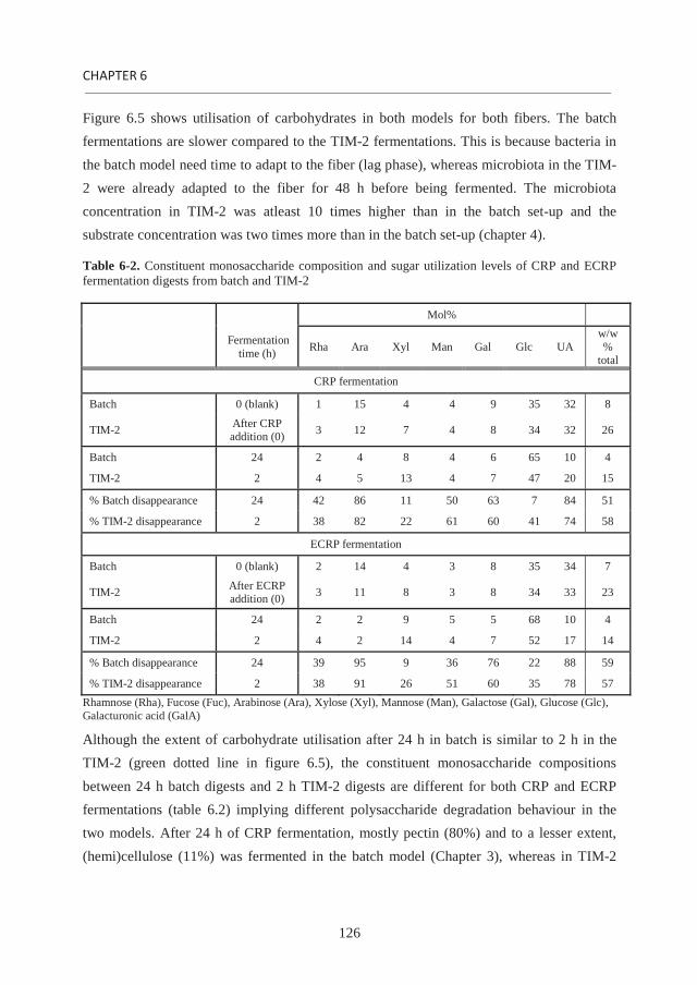
126
Figure 6.5 shows utilisation of carbohydrates in both models for both fibers. The batch fermentations are slower compared to the TIM-2 fermentations. This is because bacteria in the batch model need time to adapt to the fiber (lag phase), whereas microbiota in the TIM-2 were already adapted to the fiber for 48 h before being fermented. The microbiota concentration in TIM-2 was atleast 10 times higher than in the batch set-up and the substrate concentration was two times more than in the batch set-up (chapter 4).
Table 6-2. Constituent monosaccharide composition and sugar utilization levels of CRP and ECRP fermentation digests from batch and TIM-2
Mol%
Fermentation time (h) Rha Ara Xyl Man Gal Glc UA
w/w %
total
CRP fermentation
Batch 0 (blank) 1 15 4 4 9 35 32 8
TIM-2 After CRP addition (0) 3 12 7 4 8 34 32 26
Batch 24 2 4 8 4 6 65 10 4
TIM-2 2 4 5 13 4 7 47 20 15
% Batch disappearance 24 42 86 11 50 63 7 84 51
% TIM-2 disappearance 2 38 82 22 61 60 41 74 58
ECRP fermentation
Batch 0 (blank) 2 14 4 3 8 35 34 7
TIM-2 After ECRP addition (0) 3 11 8 3 8 34 33 23
Batch 24 2 2 9 5 5 68 10 4
TIM-2 2 4 2 14 4 7 52 17 14
% Batch disappearance 24 39 95 9 36 76 22 88 59
% TIM-2 disappearance 2 38 91 26 51 60 35 78 57 Rhamnose (Rha), Fucose (Fuc), Arabinose (Ara), Xylose (Xyl), Mannose (Man), Galactose (Gal), Glucose (Glc), Galacturonic acid (GalA)
Although the extent of carbohydrate utilisation after 24 h in batch is similar to 2 h in the TIM-2 (green dotted line in figure 6.5), the constituent monosaccharide compositions between 24 h batch digests and 2 h TIM-2 digests are different for both CRP and ECRP fermentations (table 6.2) implying different polysaccharide degradation behaviour in the two models. After 24 h of CRP fermentation, mostly pectin (80%) and to a lesser extent, (hemi)cellulose (11%) was fermented in the batch model (Chapter 3), whereas in TIM-2

General discussion
127
after 2 h, (hemi)cellulose was more extensively fermented (40%) next to pectin (72%) (Chapter 3). This indicates that fermentation in the batch model was selective towards pectin, whereas fermentation in TIM-2 involved bacteria that were also able to ferment (hemi)cellulose. Likewise, the same differences was observed for ECRP fermentation in the batch and the TIM-2.
Microbiota in CRP fermentation The microbiota composition of the 24 h fermentation digests obtained during CRP fermentation in the batch model (chapter 3) and TIM-2 (unpublished data) is expressed as % abundance of genera of the total microbiota in the fermentation digest (figure 6.6). The 24 h digest from TIM-2 is compared with the 24 h digest of the batch fermentation since the microbiota composition for the 2 h digest in TIM-2 was not determined. Still, the microbiota composition for the 24 h TIM-2 digest is expected to be representative for the 2 h TIM-2 digest since a constant ratio of acetate: propionate: butyrate (58:23:18) was seen in the adaptation phase and the fermentation phase. Shifts in activity in TIM-2 are reported to occur swiftly and are maintained throughout the experimental period (11). Analysis of the microbiota composition in both model fermentations showed that both the 24 h digests contain Bacteroides and unclassified bacteria from the Lachnospiraceae family. However, the ratio of Bacteroides : bacteria from the Lachnospiraceae family is much higher in the batch (12:1) than in TIM 2 (1:1) fermentation (figure 6.6). This difference in proportions of dominating bacteria may contribute to differences in polysaccharide utilisation between the batch and TIM-2 fermentations. This difference also arises from adaptation in TIM-2. Bacteroides is a diverse group comprising many different species, which are selective towards degrading different CWPs (12, 13). B. thetaiotaomicron, a dominant member of Bacteroides, has been suggested to utilize pectin (13), whereas Lachnospiraceae which also adhere to fibers, have been shown to utilize hemicellulose and cellulose (14, 15). This means that the lower proportion of Lachnospiraceae in the batch fermentation compared to the TIM-2 fermentation may be responsible for the poor utilisation of hemicellulose and cellulose. The lower presence of Lachnospiraceae could be due to conditions caused by metabolite accumulation during the batch fermentation. Bacteroides were also found to be the dominant bacteria (60% abundance) in other batch fermentations involving pectin, such as fermentation of sugar beet pulp oligosaccharides using human facecal inoculum (16).

128
Figure 6-6. Microbiota composition during fermentation of chicory root pulp (CRP) in the batch and TIM-2, expressed as % abundance of genera of the total microbiota in the fermentation digest. Numbers in the legend indicate operation taxonomic units (OTUs) which are highlighted in the graph.
Presence of soluble fibers affect insoluble fiber fermentation efficiency Both batch and TIM-2 fermentations showed a lower insoluble fiber utilization from ECRP compared to CRP (chapters 3 and 4). We hypothesized that in ECRP, containing equal proportions of soluble and insoluble fibers, the bacteria were programmed to utilize soluble fibers since soluble fibers are more fermentable than insoluble fibers (9) and the bacteria were not adapted to grow on insoluble fibers. It has been reported that different bacteria

General discussion
129
grow on soluble fibers and insoluble fibers (17). This is because bacteria degrading insoluble fiber need to adhere to the fiber (18) whereas bacteria degrading soluble fiber do not. In a fermentation that contains more insoluble fiber than soluble fiber as in CRP, there are more bacteria able to attach and colonize insoluble fibers compared to fermentation where soluble fiber and insoluble fiber are present in equal proportions. The ECRP fermentation is hypothesized to stimulate more soluble fiber degrading bacteria than CRP. The growth of soluble fiber degrading bacteria was further enhanced during adaptation of microbiota in TIM-2 when ECRP was fed continuously in the model as is the case for insoluble fiber in CRP. Fermentation of CRP and ECRP in both fermentations resulted in almost complete utilisation of arabinan and homogalacturonan (HG) (chapters 3 and 4) (table 6.1). As mentioned earlier, most of arabinan and HG was extractable by chelating agent. This implied that these polysaccharides were relatively easily accessible for bacterial enzymes. Although bacteria may not be able to enter the cell wall network and degrade fibers, it is advantageous for bacteria to have polysaccharide degrading enzymes in close association with the bacterial cell so that polysaccharide degradation products can be transported immediately across the cell wall (12). Although the mechanism of feeding is not clear, extracellular enzymes produced by bacteria may be endo-acting enzymes, which cleave polysaccharides to smaller fragments which are then available for the cell wall associated exo-enzymes to be degraded further to monomers and transported across the cell wall.
Enzyme activity in CWP degradation from chicory root pulp Analysis of enzyme activities from fermentation liquids of CRP batch fermentation (2 h, 12 h and 24 h)(chapter 5) revealed differences in the diversity and levels of enzymes. Enzyme activities were mostly exo compared to endo. The enzyme extract (EE) from 24 h CRP fermentation had increased activities of enzymes towards pectin substrates (sugar beet pectin, arabinan and galactan) and hemicellulose substrates (xyloglucan, xylan and manna) whereas the EE from 24 h fermentation had increased activities of only pectin degrading enzymes. The decrease in the activity of (hemi)cellulose degrading enzymes may be due to their instability or utilization by bacteria arising from inavailability of (hemi)cellulose (19). The no availability of (hemi)cellulose may be due to the inaccessibility of (hemi)cellulose in the architecture for enzymes after 12 h of fermentation. This explains why pectin was selectively utilized during CRP batch fermentation (chapter 3).

130
The time after 12 h of CRP fermentation was important because of increased levels of arabinofuranosidase and -galactosidase as well as the increased levels of other enzymes (exo-polygalacturonase, endo-arabinanase, endo-galactanase) and enzymes from circumstantial evidence (pectin de-esterifying enzymes, endo-polygalacturonase). These enzymes contributed to synergy in increasing the degradation of HG, arabinan and galactan. From 12 to 24 h, the increased degradation presumably caused increased porosity in the network leading to increased accessibility for enzymes. This combined with the increased levels of pectin degrading enzymes, contributed to an overall utilisation of GalA, Ara and Gal during CRP fermentation. Still, the architecture of CRP seemed to cause a difference between arabinan and galactan degradation despite higher activities of the 24 h EE on potato galactan compared to linear arabinan. Chapter 5 showed that arabinan was more accessible for enzymes than galactan in the network. This contributed to the higher utilisation of Ara (86%) than Gal (64%) after 24 h of CRP fermentation observed in an earlier study (9). Enzyme activities during ECRP fermentation were not measured. However, enzymes expressed during CRP fermentation may be the same during ECRP fermentation since both CRP and ECRP have similar constituent monosaccharide compositions. Due to the presence of soluble polysaccharides in ECRP, the enzyme expression is expected to happen earlier than in CRP fermentation since the rate of polysaccharide utilisation was faster in ECRP than CRP (chapter 3).
6.5. Future perspectives The aim of this thesis was to understand the relationship between CRP CWPs and CRP in vitro fermentability. Results of this thesis proved the hypothesis that a) pectin in CRP is fermentable b) increased pectin fermentability after 12 h of CRP fermentation is due to increased activities of pectin degrading enzymes. However, results of the TIM-2 study also disproved the hypothesis that a cell wall material containing more soluble fibers is more fermentable than a cell wall material containing less soluble fibers. Results of this thesis will be useful in understanding in vitro fermentations of cell wall materials. Quantification of sugar disappearance was used to determine the utilisation of sugars during fermentation. In addition, determination of the degree of esterification during fermentation will be useful in order to determine how esterfied pectin is utilized during fermentation. It was hypothesized in chapter 5 that pectin methyl esterase and pectin acetyl esterase were

General discussion
131
active during CRP fermentation in the batch model. It is also recommended to analyse activities of these enzymes for fermentations involving esterfied pectin. Combining fiber utilisation with analysis of the microbiota composition aided in predicting which bacteria are functional on fibers. As found elsewhere (16) and in chapter 2, batch fermentation of pectin rich fibers promoted growth of Bacteroides. The technique used to determine microbiota composition in this thesis allowed analysis up to the genus level. Use of other techniques such as, phylogenetic microarray analysis (20) would be useful to determining the microbiota composition till the species level. This is especially necessary for fermentations which promote the growth of Bacteroides since this group comprises a broad distribution of species. From chapters 2 and 5, a correlation was found between the WHC of CRP and the accessibility of residues derived sequentially from CRP. WUS and CHUS, having similar WHC, were both inaccessible towards fermentation enzymes in the 2h EE prepared from CRP fermentation. DAUS, having the highest WHC, was the most accessible for enzymes followed by CAUS. This indicates that the WHC (measure of a cell wall network to accommodate water) may be a parameter to be used to determine accessability of CWPs in residues. The use of two different models for fermenting CRP and ECRP pointed to clear differences in fermentation behaviour between the models. Since the batch fermentation is slow, changes in polysaccharide structures could be studied during batch fermentation. The TIM-2 can be used for studying the effect of microbiota adaptation on fiber utilization. Unlike TIM-2, batch fermentation of CRP did not involve adaptation of microbiota to the fiber substrate. Still, to be compared with TIM-2, an adaptation cannot be advocated for batch fermentation, due to metabolite accumulation and changes in the microbiota composition which may not be representative as the starting bacteria in the fermentation phase. Fermentation of CRP in TIM-2 to predict in vivo fermentation needs to be verified. The pH in TIM-2 is maintained at a constant pH (5.8) of the human proximal colon by alkali addition. In the human colon, the pH varies from 5.7 in the caecum to 6.7 in the rectum (21). Gastrointestinal movement and gut transit time in vivo may determine the site of fermentation of the fiber in the colon, especially if the fiber has a high WHC. CRP with a high WHC is expected to shorten the gut transit time. Consumption of 33g/day of beet fiber decreased transit time by 25% in healthy human subjects (22). The rate of CRP fermentation, as seen in the TIM-2, may not be the case in in vivo. The SCFA ratio in TIM-

132
2 fermentation may also be different in vivo since SCFA production is also affected by the gut pH and gut transit time (23). However, acetate will still be the dominant SCFA since fermentation of pectin is known to produce high levels of acetate (23). Based on these conclusions, it is recommended to compare TIM-2 fermentation of CRP to an in vivofermentation of CRP. Furthermore, the effect of adaptation in TIM-2 and adaptability in vivo should be compared to test if differences arise in fermentations in TIM-2 and in vivobecause of adaptation.
From this thesis, detailed information on different polysaccharide structures as present in CRP and their likely arrangement in the cell wall was revealed. The TIM-2 fermentation revealed that cell wall materials containing more soluble fibers need not be as fermentable as cell wall materials containing insoluble fibers. Although TIM-2 mimics the proximal colon, it would be beneficial to have the whole colon in the model so as to mimic fiber dependant colon transit time.

General discussion
133
6.6. References 1. Voragen, A. G. J.; Coenen, G. J.; Verhoef, R. P.; Schols, H. A., Pectin, a versatile polysaccharide present in plant cell walls. Structural Chemistry 2009, 20, 263-275. 2. Ramasamy, U. S.; Gruppen, H.; Schols, H. A., Structural and water-holding characteristics of untreated and ensiled chicory root pulp. Journal of Agricultural and Food Chemistry 2013, 61, 6077-6085. 3. Appeldoorn, M. M.; Kabel, M. A.; Van Eylen, D.; Gruppen, H.; Schols, H. A., Characterization of oligomeric xylan structures from corn fiber resistant to pretreatment and simultaneous saccharification and fermentation. Journal of Agricultural and Food Chemistry 2010, 58, 11294-11301. 4. Abdel-Massih, R. M.; Baydoun, E. A.; Brett, C. T., In vitro biosynthesis of 1,4-beta-galactan attached to a pectin-xyloglucan complex in pea. Planta 2003, 216, 502-511. 5. Popper, Z. A.; Fry, S. C., Xyloglucan-pectin linkages are formed intra-protoplasmically, contribute to wall-assembly, and remain stable in the cell wall. Planta 2008, 227, 781-794. 6. Jia, Z.; Cash, M.; Darvill, A. G.; York, W. S., NMR characterization of endogenously O-acetylated oligosaccharides isolated from tomato (Lycopersicon esculentum) xyloglucan. Carbohydrate Research 2005, 340, 1818-1825. 7. Zykwinska, A.; Thibault, J.-F.; Ralet, M.-C., Organization of pectic arabinan and galactan side chains in association with cellulose microfibrils in primary cell walls and related models envisaged. Journal of Experimental Botany 2007, 58, 1795-1802. 8. Ryden, P.; Selvendran, R. R., Cell-wall polysaccharides and glycoproteins of parenchymatous tissues of runner bean (Phaseolus coccineus). Journal of Biochemistry 1990, 269, 393-402. 9. Ramasamy U. S., V. K., Schols H. A. and Gruppen H., The effect of soluble and insoluble fibers on the fermentation of chicory root pulp 2014 Submitted for publication 10. Minekus, M.; Smeets-Peeters, M.; Bernalier, A.; Marol-Bonnin, S.; Havenaar, R.; Marteau, P.; Alric, M.; Fonty, G.; Huis in't Veld, J. H., A computer-controlled system to simulate conditions of the large intestine with peristaltic mixing, water absorption and absorption of fermentation products. Applied Microbiology and Biotechnology 1999, 53, 108-114. 11. Venema, K.; van Nuenen, M. H.; van den Heuvel, E. G.; Pool, W.; van der Vossen, J. M., The effect of lactulose on the composition of the intestinal microbiota and short-chain fatty acid production in human volunteers and a computer-controlled model of the proximal large intestine. Microbial Ecology in Health and Disease 2003, 15, 94-105. 12. Salyers, A. A., Energy sources of major intestinal fermentative anaerobes. The American Journal of Clinical Nutrition 1979, 32, 158-163. 13. Ravcheev, D.; Godzik, A.; Osterman, A.; Rodionov, D., Polysaccharides utilization in human gut bacterium Bacteroides thetaiotaomicron: comparative genomics reconstruction of metabolic and regulatory networks. BMC Genomics 2013, 14, 1-17. 14. Amy, B.; Lucy, S.; Jeffrey, B.; Susan, L., Untangling the genetic basis of fibrolytic specialization by Lachnospiraceae and Ruminococcaceae in diverse gut communities. Diversity 2013, 5, 627-640.

134
15. Brulc, J. M.; Antonopoulos, D. A.; Miller, M. E.; Wilson, M. K.; Yannarell, A. C.; Dinsdale, E. A.; Edwards, R. E.; Frank, E. D.; Emerson, J. B.; Wacklin, P.; Coutinho, P. M.; Henrissat, B.; Nelson, K. E.; White, B. A., Gene-centric metagenomics of the fiber-adherent bovine rumen microbiome reveals forage specific glycoside hydrolases. Procedings of the National Academy of Sciences of the United States of America 2009, 106, 1948-1953. 16. Leijdekkers A. G.M. , A. M., Venema K, Bosch G, Gruppen H. and Schols H. A. , In vitro fermentability of sugar beet pulp derived oligosaccharides using human and pig fecal inoculum. Journal of Agricultural and Food Chemistry 2014, 62, 1079-1087. 17. Maathuis, A.; Hoffman, A.; Evans, A.; Sanders, L.; Venema, K., The effect of the undigested fraction of maize products on the activity and composition of the microbiota determined in a dynamic in vitro model of the human proximal large intestine. Journal of the American College of Nutrition 2009, 28, 657-666. 18. Lynd, L. R.; Weimer, P. J.; Van Zyl, W. H.; Pretorius, I. S., Microbial cellulose utilization: Fundamentals and biotechnology. Microbiology and Molecular Biology Reviews 2002, 66, 506-577. 19. Jonathan, M. C.; Borne, J. J. G. C. v. d.; Wiechen, P. v.; Souza da Silva, C.; Schols, H. A.; Gruppen, H., In vitro fermentation of 12 dietary fibres by faecal inoculum from pigs and humans. Food Chemistry 2012, 133, 889-897. 20. Rajili -Stojanovi , M.; Maathuis, A.; Heilig, H. G. H. J.; Venema, K.; de Vos, W. M.; Smidt, H., Evaluating the microbial diversity of an in vitro model of the human large intestine by phylogenetic microarray analysis. Microbiology 2010, 156, 3270-3281. 21. Fallingborg, J., Intraluminal pH of the human gastrointestinal tract. Danish Medical Bulletin 1999, 46, 183-96. 22. Cherbut, C.; Salvador, V.; Barry, J. L.; Doulay, F.; Delort-Laval, J., Dietary fibre effects on intestinal transit in man: involvement of their physicochemical and fermentative properties. Food Hydrocolloids 1991, 5, 15-22. 23. Macfarlane, G. T.; Macfarlane, S., Fermentation in the human large intestine: its physiologic consequences and the potential contribution of prebiotics. Journal of Clinical Gastroenterology 2011, 45, 120-127.

135
Summary

136
Cell wall polysaccharides (CWPs) from agricultural by-products have the potential to be used as fiber supplements to food. Chicory root pulp (CRP) is an agricultural by-product obtained after the industrial extraction of inulin from the chicory root and is aimed to be used as a dietary fiber. Not much is known on the different CWP populations, interactions between CWPs in CRP as well as the fermentability of CRP. Furthermore, solubilisation of fibers from the cell wall by processing of the pulp may have an effect on the fermentability of the pulp. The aim of this thesis was to understand the effect of CRP CWPs present as soluble and insoluble fibers on their in vitro fermentability by human colonic bacteria.
Chapter 1 describes CWP structures and enzymes that can degrade different polysaccharide structures. The fermentation of polysaccharides and the bacteria involved in fermenting fibers are described. In addition, the chapter also describes different fermentation models such as the batch and TNO In vitro Model (TIM-2) that could be used to study fermentation since the fermentation of a fiber may also depend on the type of model used.
First, CWPs of CRP and processed (ensiled) CRP (ECRP) were characterised in Chapter 2. CWPs of CRP comprised 62 % pectin, 11% hemicellulose and 27% cellulose. Pectin in CRP is highly methyl esterfied whereas both pectin and xyloglucan were found to be acetylated. Xyloglucans were mainly of the XXXG type. Half of CRP pectin is extractable with chelating agents. CRP CWPs are almost (90%) insoluble, whereas ECRP having similar polysaccharide content as CRP has equal proportions of soluble and insoluble CWPs. Moreover, the insoluble cell wall network of ECRP was hypothesized to be more open due to degradation of residual pectin and modification of the (hemi)cellulose network.
Chapter 3 describes in vitro fermentability of CRP and ECRP in a batch model using human faecal inoculum. Both CRP and ECRP were fermentable in 24 h. 51% of all carbohydrates were utilised from CRP and 59% of all carbohydrates were utilised from ECRP. Bacteroides were found to be the dominant bacteria in fermenting CRP and are expected to directly utilise mono and oligomeric degradation products formed from the insoluble fiber. This was concluded from the absence of degradation products and decrease in the levels of CWPs. Ensiling of CRP had more effect on the rate of fiber utilization (11% more carbohydrate utilization in 12 h compared to CRP) than on the extent of fiber

137
utilization. It was concluded that this effect was due to the presence of soluble fibers and the hypothesized open cell wall network of ECRP.
Fermentation of CRP and ECRP in a dynamic model such as the TIM-2 was described in Chapter 4. After the adaptation phase of the human faecal inoculum to CRP and ECRP for 48 h, 57% of carbohydrates were rapidly utilised in 2 h. The fermentability of CRP and ECRP were rapid compared to the batch fermentation. ECRP carbohydrates (85%) were slightly less fermented in 24 h compared to CRP carbohydrates (92%). This was due to lower utilization of insoluble fibers in ECRP compared to CRP. It was hypothesized in this study that soluble fibers that are readily fermentable and dominantly present in ECRP favoured proliferation of soluble fiber degrading bacteria more than the insoluble fiber degrading bacteria and the inflexibility of the soluble fiber degrading bacteria to adapt to the insoluble fiber thereafter. The lower SCFA levels for ECRP were in line with the lower carbohydrate utilization in ECRP than CRP.
In order to understand pectin degradation in CRP during batch in vitro fermentation by human faecal inoculum, enzyme activities were studied over fermentation time as described in Chapter 5. Enzymes activities were identified towards PNP-glycosides and complex polysaccharides. Enzyme extracts (EEs) from CRP fermentation liquids (2, 12 and 24 h) were incubated also on polysaccharides and cell wall residues (CWRs) derived from CRP for 24 h to determine the mechanism and efficiency of enzyme action. The study showed an increase in levels of arabinofuranosidase, -galactosidase, endo-arabinanase, endo-galactanase and exo-polygalacturonase and enzymes suggested from the release of galacturonic acid (pectin de-esterifying enzymes and endo-polygalacturonase). Levels of these enzymes contributed to a synergy in degrading pectin in CRP from 12 to 24 h of fermentation. It was hypothesized that the increased degradation with time caused increased porosity in the network leading to an increased accessibility for the enzymes. This combined with the increased levels of pectin degrading enzymes, enhanced degradability of residual homogalacturonan, arabinan and galactan. The higher degradability of arabinan compared to galactan in the network was due to the architecture in CRP involving more accessible arabinan than galactan.
In Chapter 6, the distribution of CWPs in the cell wall is proposed for CRP and ECRP. Batch fermentation of CRP and ECRP were compared to TIM-2 fermentation of CRP and

138
ECRP. The influence of soluble fibers on the fermentability of insoluble fibers is discussed. The enzyme activities involved in fermenting CRP in the batch model are also discussed.

139
Samenvatting

140
Celwandpolysachariden (CWPs) uit agrarische bijproducten kunnen mogelijk gebruikt worden als vezel supplementen in levensmiddelen. Cichorei wortelpulp (CRP) is een agrarisch bijproduct dat verkregen wordt na industriële extractie van inuline uit de cichorei-wortel en wordt gebruikt als voedingsvezel. Er is nog niet veel bekend over de verschillende populaties van CRP polysachariden, hun interacties binnen de celwand en hun fermenteerbaarheid. Het in oplossing brengen van vezels uit de celwand tijdens de verwerking van de pulp kan mogelijk ook effect hebben op de fermenteerbaarheid van de vezels. Het doel van het in dit proefschrift beschreven onderzoek was dan ook het bestuderen van het effect van oplosbare en onoplosbare CRP CWPs op de in vitrofermenteerbaarheid door menselijke darmbacteriën.
Hoofdstuk 1 beschrijft CWP structuren en enzymen die verschillende polysacharide structuren kunnen afbreken. De fermentatie van polysachariden en de bacteriën die betrokken zijn bij de fermentatie van vezels worden besproken. Daarnaast worden verschillende fermentatiemodellen zoals het batchmodel en het TNO In vitro Model (TIM-2) behandeld. Hoewel beide modellen vaak gebruikt worden om fermentatie te bestuderen, is de waargenomen fermentatie mogelijk afhankelijk van het gebruikte model.
De karakterisering van de CWPs van CRP en ingekuild (ensiled) CRP (ECRP) wordt beschreven in Hoofdstuk 2. CWPs uit CRP bestaan voor 62% uit pectine, voor 11% uit hemicellulose en voor 27% uit cellulose. Pectine in CRP is hoog methylveresterd en zowel pectine als xyloglucaan zijn geacetyleerd. Xyloglucanen zijn vooral van het XXXG-type. De helft van de CRP pectine kan worden geëxtraheerd in de aanwezigheid van een sekwestreermiddel. De andere CRP CWPs (90%), zijn vrijwel onoplosbaar. ECRP echter bevat gelijke hoeveelheden oplosbare en onoplosbare CWPs. Het onoplosbare celwandnetwerk van ECRP wordt beschouwd als ‘meer open’ door de modificatie van het pectine en het hemicellulose-cellulose netwerk.
Hoofdstuk 3 beschrijft de in vitro fermenteerbaarheid van CRP en ECRP in een batchmodel, gebruik makend van humaan fecaal inoculum. Zowel CRP als ECRP werden binnen 24 uur gefermenteerd. Van CRP werd 51% van alle koolhydraten en van ECRP werd 59% van alle koolhydraten benut. Bacteroides bleken de meest dominant aanwezige bacteriën bij de fermentatie van CRP en worden geacht mono- en oligomere afbraakproducten vrij te maken uit oplosbare vezels en die meteen zelf te gebruiken. Dit

141
werd geconcludeerd uit de afwezigheid van afbraakproducten en afname in de hoeveelheden CWPs. Het inkuilen van CRP had vooral effect op de snelheid waarmee vezels benut werden (11% meer koolhydraten benut binnen 12 uur in vergelijking met CRP) en minder op de mate van vezel benutting. Dit effect werd mogelijk veroorzaakt door de aanwezigheid van oplosbare vezels en het geopende celwandnetwerk van ECRP.
Fermentatie van CRP en ECRP in een dynamisch fermentatiemodel, zoals TIM-2 wordt beschreven in Hoofdstuk 4. Na een aanpassingsfase van de fecale microbiota aan CRP en ECRP gedurende 48 uur, werd 57% van de koolhydraten binnen 2 uur benut. De fermentatie van CRP en ECRP in het TIM model was snel vergeleken met de batchfermentatie. Tijdens batchfermentatie werden ECRP koolhydraten (85%) in 24 uur iets minder gefermenteerd dan CRP koolhydraten (92%). Dit werd veroorzaakt door de lagere benutting van onoplosbare vezels in ECRP vergeleken met CRP. De lagere hoeveelheden SCFA voor ECRP komen overeen met de lagere koolhydraatbenutting van ECRP in vergelijking tot CRP. De resultaten suggereren dat oplosbare, vlot fermenteerbare vezels zoals aanwezig in ECRP, de groei van bacteriën die goed groeien op deze vezels zodanig stimuleren dat de microbiota zich vervolgens minder goed kan aanpassen aan het benutten van onoplosbare vezels. Om de afbraak van CRP pectine tijdens de in vitro batchfermentatie door humaan fecaal microbiota beter te begrijpen, werden enzymactiviteiten gevolgd in de tijd, zoals beschreven in Hoofdstuk 5. Enzymactiviteiten werden geïdentificeerd met behulp van PNP-glycosides en complexe polysachariden. Enzym extracten (EEs) uit CRP fermentatie vloeistoffen (verkregen na 2, 12 en 24 uur) werden gedurende 24 uur geïncubeerd met goed gekarakteriseerde polysachariden en met celwand residuen (CWRs), verkregen na sequentiële extractie uit CRP, om de werkingswijze en efficiëntie van het enzym te bepalen. De resultaten lieten een toename zien in de hoeveelheid arabinofuranosidase, -galactosidase, endo-arabinanase, endo-galactanase en exo-polygalacturonase en pectine-afbrekende enzymen (pectinemethylesterase, endo-polygalacturonase). De hoeveelheden van deze enzymen dragen bij aan een synergie in de pectine-afbraak uit CRP tussen 12 en 24 uur fermentatie. De hypothese wordt gesteld dat de toename in afbraak in de tijd werd veroorzaakt door een toegenomen porositeit van de celwanden en een daardoor toegenomen toegankelijkheid voor de enzymen. Gecombineerd met de toegenomen hoeveelheden

142
pectine-afbrekende enzymen leidde dit tot een verhoogde afbreekbaarheid van resterende homogalacturonaan, arabinaan en galactaan. De hogere afbreekbaarheid van arabinaan in vergelijking met galactaan aanwezig in het netwerk werd veroorzaakt door de architectuur in CRP waarbij het arabinaan meer toegankelijk was dan galactaan.
In Hoofdstuk 6 wordt de distributie van CWPs binnen de celwanden van CRP en ECRP voorgesteld. Verder wordt de batchfermentatie van CRP en ECRP vergeleken met TIM-2 fermentatie van CRP en ECRP. De invloed van oplosbare vezels op de fermenteerbaarheid van onoplosbare vezels wordt besproken. De enzymactiviteiten die betrokken zijn bij de fermentatie van CRP in het batch model worden bediscussieerd.

143
Acknowledgements

ACKNOWLEDGEMENTS
144
Like every PhD student, it is time for me to move on to a newer destination. For this once in a lifetime opportunity, I was preparing a PhD thesis for which I would like to express my gratitude to people who helped me and to those who were there to keep the nice moments special.
Firstly, I would like to express my gratitude to my promoters Harry and Henk. It is a great honour to be supervised by Harry and Henk who gave me their expert guidance and assisted me in accomplishing this thesis. Harry, it was a pleasure to have clear, detailed and inspiring discussions with you. I felt encouraged and motivated after our talks and am grateful to you for your support and care. Henk, I cannot express my thank you. Words are not enough. I am deeply indebted to you for your support, patience and for lending your arm. With every passing time, I learned to communicate more openly, thenks to you. I would also like to thank you for fermentative (fruitful) discussions especially because it was challenging for insoluble fibers.
Further, my research on fermentation, would not have been possible without Koen Venema, who was always there when we needed discussions and when preparing manuscripts for publication. I would also like to thank Gerdie Pullens and Diederick Meyer from Royal Cosun who made every project meeting pleasant and enjoyable. Besides, the meetings were also enlightening, many thanks to Leonie and Paul (UMCG) for discussions on immunomodulation and to Monica and Anne (WU-Human nutrition) for discussions on nutrition.
Next, I would like to give my special thanks to Jolanda who is there to take care of all the minor things in the laboratory! Dankuwel! A warm thank you to my students – Heleen, Valerio and Karen who contributed to research. Wish you all good luck and success in your endeavours.
And my extensive thanks to all my PhD student colleagues at the laboratory for adding spice and colour during my stay. It was great fun to be with you all during the lab trips, laboratory drinks, birthdays etc. and even during the making of stukjes for graduating PhDs. A thank you to my classic 509 roomies- Stephanie, Marijn, Anne, Jianwei, Rudy, Milou, Ja and Wieteke for the nice gezellig moments and not to forget… chocolates! My merci to Stephanie and terima kasih to Melliana for your time on discussing fermentation. My dankuwel to Annemieke too for talks on fermentation as well as for helping me with

145
Nederlands and the summary. I would also like to thank my present roomies (Surender, Wibke, Kok-phen and Belen) for the warm moments. Rianne, it was nice to work with you on freeze dryers. We were a great team and I enjoyed those moments with you.
And my warm and special thanks to Claire with whom I shared a lot, not only in the laboratory but also outside …baking, desserts, movies and travelling. I still have a list of your cake recipes which I have to try. Another thank you for being with me as my paranymph. I would also like to thank Christianne for being my paranymph on this special day for me. It was a great pleasure for me to share and discuss research on fermentation with you. Wish you too good luck with your fermentations and enzymes!
My special thanks to Anja Janssen. Anja, you were not only my MSc supervisor in the past, but our friendship grew afar. Thankyou for your time and pleasant moments at your home with your family. Karin, many thanks to you for your time, support and encouragement. Urmila and me enjoyed trips with you as well as listening to Dutch artists.
I would also like to thank my friends from India staying in Wageningen for their support, desi food and good times. Abhishek bhaiyya, many thanks to you too. Ashutosh, thank you for everything. And lastly, I would like to thank my sister and my family for giving me the opportunity to learn from challenges and to grow beyond…and most importantly my grandparents for their unsaid patience and strength.
Thank you all,

ACKNOWLEDGEMENTS
146

147
About the author

148
Curriculum Vitae
Uttara Ramasamy was born on the 2nd of Dec 1984 in Bangalore, India. She completed her bachelors in Microbiology from Abasaheb Garware College in Pune, Maharashtra, India. During her bachelors, she also worked part time on a project in Bioinformatics which involved the contruction of three dimensional phylogenetic trees. Towards the end of her bachelors, she undertook an internship at United Breweries in Mumbai, India. From here, she went on further, to pursue her masters in Food Technology from Wageningen University. Her master thesis dealt with kinetic modelling of enzymatic starch hydrolysis in the Food Process Engineering Group and her intership was in product development in baking in DMV-Friesland Campina. After completing her master degree, she was offered to work as a PhD student at the Labortaory of Food Chemistry under the supervision of Dr. Henk. A. Schols and Prof. Dr. Harry Gruppen. The outcome of her research during her PhD is presented in this thesis.
149
List of publications:
Ramasamy, U. S.; Gruppen, H.; Schols, H. A., Structural and water-holding characteristics of untreated and ensiled chicory root pulp. Journal of Agricultural and Food Chemistry2013, 61, 6077-6085.
Ramasamy U. S., Venema K., Schols H. A. and Gruppen H., The effect of soluble and insoluble fibers on the fermentation of chicory root pulp 2014. Submitted for publication
Ramasamy U. S., Venema K., Gruppen H. and Schols H. A., The fate of chicory root pulp polysaccharides during fermentation in the TNO In vitro model of the colon (TIM-2) 2014. Submitted for publication
Ramasamy U. S., Schols H. A. and Gruppen H., Characteristics of bacterial enzymes present during in vitro fermentation of chicory root pulp by human faecal microbiota 2014. Submitted for publication
Vogt, L., Ramasamy U., D. Meyer, G. Pullens, K. Venema, M. M. Faas, H. A. Schols and P. de Vos. Immune modulation by different types of 2 1-fructans is toll-like receptor dependent. PLoS ONE. 2013, 8 (7): e68367.
Vogt, L., Meyer D., Pullens G., Faas M., Smelt M., Venema K., Ramasamy U., Schols H. A. and Vos P. de. Immunological properties of inulin-type fructans. Critical Reviews in Food Science and Nutrition. 2013. (Just-accepted).

150
Overview of completed training activities
Discipline specific courses
Courses
Summercourse Glycosciences * (VLAG), Wageningen, The Netherlands, 2010 Advanced Food Analysis * (VLAG), Wageningen, The Netherlands, 2010 Food and biorefinery enzymology * (VLAG), Wageningen, The Netherlands, 2010
Conferences and meetings
CCC Scientific days *^ Groningen, The Netherlands, 2009-2013 Plant cell wall conference * Porto, Portugal, 2010 Cosun Workshop ^, Royal Cosun, The Netherlands, 2010 EPNOE (European Polysaccharide Network of Excellence)*, Wageningen, The Netherlands, 2011 Plant and Sea-weed Polysaccharide Workshop*, INRA, Nantes, France 2012
General courses
Information literacy including Endnote, WGS, 2009 VLAG PhD week, 2009 Techniques for writing and presenting a scientific paper, 2010 Interpersonal communication skills, 2013 Mobilising scientific network, 2013 Career perspectives, 2013
Additional activities at the Laboratory of Food Chemistry
Preparation of PhD research proposal PhD Trip to Switzerland and Italy, 2010 PhD Trip to Malaysia and Singapore, 2012 MSc thesis student presentations and colloquia (2009-2013) PhD presentations (2009-2013) Project meetings (2009-2013)
* poster presentation, ^ oral presentation

151
Abbreviations used:
VLAG: Graduate School for Nutrition, Food Technology, Agrobiotechnology and Health Science CCC: Carbohydrate Competence Centre WGS: Wageningen Graduate School

152
The research was jointly financed by the European Union, European Regional Development Fund and the Ministry of Economic Affairs, Agriculture and Innovation, Peaks in the Delta, the Muncipality of Groningen, the Province of Groningen, as well as the Dutch Carbohydrates Competence Centre (CCC WP2a) supported by Royal Cosun, TNO, UMCG and Wageningen University.
Financial support from Wageningen University for printing this thesis is greatfully acknowledged.
This thesis was printed by GildePrint drukkerijen, Enschede, The Netherlands
Edition: 350 copies
Uttara Ramasamy, 2014