the role of semaphorin6a in the formation of boundary cap ... filezurich open repository and archive...
TRANSCRIPT

Zurich Open Repository andArchiveUniversity of ZurichMain LibraryStrickhofstrasse 39CH-8057 Zurichwww.zora.uzh.ch
Year: 2008
The Role of Semaphorin6A in the Formation of Boundary Cap Cells
Mauti, O
Posted at the Zurich Open Repository and Archive, University of ZurichZORA URL: https://doi.org/10.5167/uzh-9749DissertationPublished Version
Originally published at:Mauti, O. The Role of Semaphorin6A in the Formation of Boundary Cap Cells. 2008, University ofZurich, Faculty of Science.
The Role of Semaphorin6A in the Formation of Boundary Cap Cells
Dissertation
zur
Erlangung der naturwissenschaftlichen Doktorwürde (Dr. sc. nat.)
vorgelegt der
Mathematisch-naturwissenschaftlichen Fakultät
der
Universität Zürich
von
Olivier Mauti
von
Niedererlinsbach SO
Promotionskomitee
Prof. Dr. Esther Stoeckli (Leitung der Dissertation)
Prof. Dr. Stephan Neuhauss
Prof. Dr. Martin Schwab
Zürich, 2009
- 2 -
INDEX
1. Summary ........................................................................................................................................3 2. Introduction.....................................................................................................................................5 3. Boundary Cap Cells......................................................................................................................12 4. Expression Pattern Analysis of Plexins and Neuropilins..............................................................25 5. Functional Analysis of Semaphorin6A..........................................................................................42 6. Additional Materials and Methods ................................................................................................61 7. Discussion ....................................................................................................................................76 8. Acknowledgments ........................................................................................................................83 9. Curriculum Vitae ...........................................................................................................................84 10. References ...................................................................................................................................85

- 3 -
1. Summary
The dorsal neural tube is the origin of neural crest cells which migrate in a stereotypic manner to form
a large variety of peripheral nervous system (PNS) structures, such as the dorsal root ganglia
(DRGs), the sympathetic ganglia, the enteric nervous system, and the boundary cap cell (BCC)
clusters. BCCs are located at the transition zone between central and peripheral nervous system and
can be identified by the expression of Krox20, Cadherin-7, protein zero (P0), and other molecules.
Ultrastructural analyses of the trunk spinal cord revealed that BCCs tightly ensheath axons passing
the transition zone and that these cells appear ventrally after the outgrowth of motor axons and
dorsally before the ingrowth of primary sensory axons. BCCs are transient and disappear postnatally
in rats after day 6 (P6). A few studies revealed interesting functions of these cells, mainly acting as
“border control” at the transition zone: the dorsal BCCs were shown to be necessary for the ingrowth
of primary sensory axons into the spinal cord and to contribute to nociceptive neurons and satellite
cells of the DRG. In contrast, ventral BCCs were shown to maintain the integrity of the motor column,
since in their absence motor neurons left the spinal cord and migrated along the ventral roots.
In my PhD thesis, I analyzed the function of Semaphorin6A, a transmembrane class-6 Semaphorin,
during embryogenesis. At early developmental stages Semaphorin6A shows a very restricted
expression pattern in the dorsal neural tube and in the transition zone between CNS and PNS of the
lumbar spinal cord of chicken embryos. Its expression overlaps spatio-temporally with Krox20,
Cadherin-7, and P0, indicating that Semaphorin6A is expressed in BCCs. Downregulation of
Semaphorin6A using in ovo RNAi resulted in emigration of motor neurons along the ventral roots as
previously observed after the ablation of BCCs. Additionally, the organization of the dorsal roots was
severely disturbed resulting in fused dorsal roots at various levels and irregular DRG shapes. Analysis
of BCC localization revealed disorganization or even absence of the BCC clusters.
Previously I had analyzed the expression patterns of the Semaphorin receptors, Plexins and
Neuropilins, throughout development. Based on these data, PlexinA1 was a good candidate for the
interaction with Semaphorin6A in the lumbar spinal cord due to its strong expression in the lateral
motor column. Downregulation of PlexinA1 phenocopied the motor neuron translocations, the
disorganization of the dorsal roots, and the malformation of the BCC clusters as seen in the absence
of Semaphorin6A function. Based on these results I propose the following working hypothesis for the
formation of the ventral BCC clusters: migrating neural crest cells express Semaphorin6A that
recognizes PlexinA1 present on motor axons. This interaction stops the migration of neural crest cells
resulting in their aggregation and finally the formation of BCC clusters. The absence of either
Semaphorin6A on neural crest cells or PlexinA1 on motor axons results in the failure of BCC
aggregation, and thus, in loose or even missing BCC clusters. As a consequence, the motor column is
destabilized leading to the previously reported motor neuron emigration. The formation of the dorsal
BCC clusters differs from the ventral ones due to their appearance at the prospective dorsal root entry
site before the ingrowth of sensory axons. Therefore their formation cannot be explained by the same
mechanism.

- 4 -
Zusammenfassung
Das dorsale Neuralrohr ist der Ursprung von Neuralleistenzellen, welche entlang von zwei
spezifischen Migrationswegen wandern um Bestandteile des peripheren Nervensystems zu bilden.
Die Spinalganglien, die sympathischen Ganglien, das enterische Nervensystem, sowie die Boundary
Cap Zellen (BCCs) zwischen dem peripheren und dem zentralen Nervensystem sind deren
Abkömmlinge. Die BCCs können durch die spezifische Expression von Krox20, Cadherin-7, Protein
null (P0), sowie weiteren Molekülen identifiziert werden. Ultrastrukturelle Analysen des Rückenmarks
haben ergeben, dass die BCCs die Axone, welche die Übergangszone passieren, eng umschlingen.
Die ventralen BCCs aggregieren erst nachdem die Motoraxone in die Peripherie wachsen, während
die dorsalen BCCs vor dem Einwachsen von sensorischen Axonen aggregieren. Die BCCs sind
transient und verschwinden in Ratten 6 Tage nach der Geburt. Einige Studien haben die Funktion von
BCCs analysiert und herausgefunden, dass sie hauptsächlich eine Kontrollfunktion an der
Übergangszone haben: die dorsalen BCCs sind für die Innervation von primären sensorischen
Axonen ins Rückenmark von grosser Bedeutung. Im Gegensatz dazu, halten die ventralen BCCs die
Integrität der Motorsäule aufrecht. In Abwesenheit der ventralen BCCs verlassen Motorneurone das
Rückenmark entlang der Vorderwurzeln.
In der vorliegenden Arbeit habe ich die Funktion von Semaphorin6A, einem transmembranären
Klasse 6 Semaphorin, während der Embryogenese analysiert. Während der frühen Embryogenese ist
die Expression von Semaphorin6A auf wenige Zellen im Neuralrohr sowie auf die Grenze zwischen
zentralem und peripheren Nervensystems in der Beinregion des Hühnerembryos begrenzt. Die
Expression überlappt zeitlich und örtlich mit Krox20, Cadherin-7 und P0, was darauf hinweist, dass
Semaphorin6A in den BCCs exprimiert ist. Die Herunterregulierung von Semaphorin6A mittels in ovo
RNS Interferenz führt zum Auswandern von Motorneuronen, was bereits schon in der Abwesenheit
von BCCs beobachtet wurde. Zusätzlich war die Organisation der Hinterwurzeln an unterschiedlichen
Orten beeinträchtigt. Die Spinalganglien wiesen zudem unregelmässige Formen auf. Die BCCs waren
entweder unorganisiert oder abwesend.
In einer vorangehenden Studie habe ich die Expression von Plexinen und Neuropilinen, beides
Rezeptoren für Semaphorin6A, analysiert. Die erhaltenen Expressionsmuster deuteten darauf hin,
dass PlexinA1 ein guter Interaktionspartner für Semaphorin6A ist. Die Herunterregulierung von
PlexinA1 hatte die gleichen Phenotypen zur Folge, welche in Abwesenheit von Semaphorin6A
beobachtet wurden. Aufgrund der erhaltenen Daten postuliere ich folgenden Mechanismus für die
Aggregation der BCCs: Migrierende Neuralleistenzellen exprimieren Semaphorin6A welches PlexinA1
auf der Oberfläche von Motoraxonen erkennt. Diese Interaktion führt zu einem Migrationsstopp der
Neuralleistenzellen. Dadurch aggregieren sie und formieren sich schlussendlich zu den BCC
Clustern. In Abwesenheit von entweder Semaphorin6A oder PlexinA1 ist die Aggregation von BCCs
beeinträchtigt, was eine Translokation von Motorneuronen zur Folge hat. Im Unterschied zu den
ventralen BCCs, erscheinen die dorsalen BCCs bevor die primären sensorischen Axone ins
Rückenmark einwandern. Der beschriebene ventrale Mechanismus kann daher die Aggregation der
dorsalen BCCs nicht erklären.

- 5 -
2. Introduction
In the adult nervous system, the transition zone where axons enter or exit the spinal cord, is
characterized by the presence of an impenetrable glial barrier formed by the apposition of CNS-
derived astrocytes and PNS-derived Schwann cells. However, during embryogenesis when the
neuronal circuits are formed, PNS axons have to interconnect with CNS neurons, and motor axons
extend out of the CNS into the periphery to innervate target muscles. During that specific period the
barrier has to be permissive for axons. The boundary cap cells (BCCs) which are neural crest (NC)
cell derivatives were shown to populate the transition zones in order to permit axonal elongation
across the embryonic CNS:PNS border (Golding et al., 1997; Golding and Cohen, 1997).
In this chapter, I give a general overview about the establishment of PNS structures. Chapter 3
focuses specifically on the current knowledge about BCCs and genes expressed by them. Chapter 4
describes my detailed analysis of the expression pattern of Plexins and Neuropilins which are known
receptors for Semaphorins. These findings are taken as a basis for the functional analysis of
Semaphorin6A in the chicken embryo described in chapter 5. The functional analysis of
Semaphorin6A and its interaction partner was done using the in ovo RNAi technique previously
established in our lab (Pekarik et al., 2003) and is summarized in chapter 6. Finally, I discuss the
importance and interpretation of my findings in chapter 7.

- 6 -
The neural crest
PNS structures are derived from a pluripotent and transient population of cells located in the dorsal
neural tube, the NC cells (Le Douarin and Kalcheim, 1999). During late gastrulation, the NC is
induced by a complex interaction of environmental cues acting at the non-neural ectoderm border
involving Wnt and fibroblast growth factor (FGF). In the presence of intermediate levels of bone
morphogenetic protein (BMP), Pax3/7, Zic1, and Msx1/2 are induced which act synergistically in the
presence of Wnt to upregulate NC specifiers in the dorsal neural tube, such as FoxD3, Sox9, and
Snail. The NC progenitor pool is kept in a multipotent and proliferative state through interaction of the
proto-oncogene c-Myc and the basic Helix-Loop-Helix (bHLH) gene Id3 (Light et al., 2005), while cell
death is prevented via Sox9 and Snail1/2 (Cheung et al., 2005). A complex interaction between Snail,
Sox9, and FoxD3 induces the epithelial to mesenchymal transition (EMT) which is characterized by a
loss of apical-basal cell polarity, a switch from tight to gap junctions, and downregulation of various
cell adhesion molecules (reviewed in Thiery and Sleeman, 2006). The switch from epithelial cell
specific type I N-Cadherin to type II Cadherin-7 results in a decreased NC cell adhesiveness (see
below; Chu et al., 2006). The subsequent NC cell delamination is facilitated by a reduction of Laminin
at the basal surface of the dorsal neural tube, most likely by proteolytic activity of NC cells, and
upregulation of Integrin-β1 (Valinsky and Le Douarin, 1985; Cheung et al., 2005). Delaminating and
subsequently migrating NC cells maintain their pluripotency by Sox10, which also prevents premature
neuronal differentiation (Kim et al., 2003).
Figure 1: Induction and delamination of neural crest cells. The concerted interaction of BMP, Wnt, and FGF at the non-neuronal ectoderm induces the neural plate during late
gastrulation. In the presence of Wnt, NC-specifiers are upregulated that in turn induce the maturation of NC cell progenitors.
Finally, NC cells undergo an epithelial to mesenchymal transition (EMT) and delaminate from the neural tube. (Modified from
Thiery and Sleeman, 2006)

- 7 -
NC cells of mice lacking Pax3 (Splotch; Serbedzija and McMahon, 1997), SoxE (Southard-Smith et
al., 1998; Cheung et al., 2005), or Slug/Snail (Jiang et al., 1998) fail to delaminate or undergo
apoptosis resulting in defects in PNS structures or even their absence. Similarly, surgical ablation of
the dorsal neural tube results in a loss of NC cells, and thus, in missing PNS structures (Vermeren et
al., 2003).
The extracellular matrix
The extracellular matrix (ECM) is a tight network of a large number of structural proteins giving unique
flexible and elastic properties to many tissues, e.g. the skin or the sclerotome. During embryogenesis,
the ECM plays an integral role in the formation of the nervous system by providing an appropriate
surrounding and support to migrating NC cells and axons. The structure of the ECM is primarily
formed by proteins that belong to the family of Collagens and Elastins (Duband and Thiery, 1987).
While Collagen fibers help to stabilize the ECM, Elastin fibers provide elasticity and both together are
important for maintaining the unique ECM structure. Cross-links between the main fibers of the ECM
are generated by Fibronectin and Laminin, and more importantly, they serve as adhesive substrates
for migrating cells and growing axons (Newgreen and Thiery, 1980; Bixby and Harris, 1991; Tucker
and McKay, 1991). Other components of the ECM are Tenascins, Aggrecan, Versicans,
Thrombospondin-repeat containing proteins (e.g. F-Spondin) that act mainly as repellents for
migrating NC cells and axons (Bixby and Harris, 1991; Perris, 1997; Debby-Brafman et al., 1999). The
aqueous environment of the ECM is maintained by Heparan Sulfate Proteoglycans (HSPG) which
form a gel-like mixture (reviewed in Iozzo, 1998). Furthermore, HSPGs have the ability to trap growth
factors and help to distribute morphogens across the ECM, such as Sonic Hedgehog which is
secreted from the notochord (Guerrero and Chiang, 2007). The balance of all these factors defines
and influences the permissiveness of the ECM for cell motility and axon guidance.
Neural crest cell migration
The vertebrate embryo has a segmental organization and each segment, the so-called somite, is
subdivided into anterior and posterior sclerotome. The delamination of NC cells takes place all along
the anterior-posterior axis of the embryo but their migration is restricted to the anterior sclerotome due
to the non-permissive posterior sclerotome (Keynes and Stern, 1984; Kalcheim and Teillet, 1989). In
the trunk of vertebrates, several migratory waves occur that form different NC derivatives during
embryogenesis. NC cells migrate in a stereotypic manner from their origin in the dorsal neural tube to
their prospective target sites where they condensate and form the PNS structures. The majority of
migrating NC cells can be traced by their specific expression of Sox10, the HNK-1 and the 1E8
antigens (Tucker et al., 1984; Bhattacharyya et al., 1991; Kuhlbrodt et al., 1998). During NC cell
migration various repulsive guidance molecules in the ECM form permissive and non-permissive
paths; examples are F-Spondin, secreted class 3 Semaphorins, and Ephrins (Bronner-Fraser, 1994;
Krull et al., 1997; Debby-Brafman et al., 1999; Eickholt et al., 1999). They are found in the posterior

- 8 -
sclerotome, the perinotochordal area, and (at early developmental stages) in the dermomyotome
which are normally avoided by migrating NC cells. ECM proteins such as Laminin and Fibronectin are
necessary substrates and perturbing their docking sites using function-blocking antibodies strongly
interferes with NC cell migration (Boucaut et al., 1984). Integrin-β1, a necessary component of the
receptor for Laminin and Fibronectin, is playing a major role in facilitating NC cell migration (Horwitz et
al., 1985; Lallier et al., 1994). The conditional deletion of Integrin-β1 in NC cells of mice showed
severe defects in the formation of the PNS, mainly resulting from a delayed migration of NC cells
(Pietri et al., 2004). The molecular mechanism underlying NC cell migration is not fully understood,
but the extension of NC cell filopodia is comparable to the motility of the growth cone: at the surface
of filopodia different receptors are expressed that transduce environmental cues into precise guidance
responses. The extension or retraction of NC cell filopodia requires active actin cytoskeleton
dynamics. One of the molecules implicated in actin filament dynamics belongs to the Ena/VASP
family (Drees and Gertler, 2008). Their function is to antagonize the capping protein that prevents
further polymerization of the actin filaments (Bear et al., 2002). Two recent studies discuss this protein
family as being downstream of repulsive receptors during NC cell migration: Ena/VASP proteins
mediate repulsion from Ephrin ligands (Evans et al., 2007) and from Sema6D during endocardiac
crest cell migration (Toyofuku et al., 2004).
Pathways of neural crest cell migration
Early migrating NC cells travel ventrally in the anterior sclerotome between the neural tube and the
dermomyotome and give rise to most PNS structures like DRGs, sympathetic ganglia, enteric nervous
system, Schwann cells, and BCCs (Figure 2; Rickmann et al., 1985; Bronner-Fraser, 1986; Le
Douarin et al., 1992; Niederländer and Lumsden, 1996). Conversely, the majority of late delaminating
NC cells travel dorsolaterally, i.e. between the ectoderm and the dermomyotome, to form melanocytes
of the skin, cartilage derivatives, and smooth muscles (Erickson et al., 1992; Le Douarin et al., 1992;
Bronner-Fraser, 1994).
For a long time the migratory pattern of NC cells was believed to be topographically defined in the NC,
since apically located NC cells migrate along the dorsolateral pathway and basally positioned cells
travel along the ventromedial pathway. However, different studies showed that a combination of
environmental cues and their time point of delamination define their respective pathways: all early
delaminating NC cells migrate along the ventromedial pathway in the anterior sclerotome. Eph-Ephrin
and Sema3A-Plexin/Neuropilin interactions were shown to restrict the migration of NC cells to the
anterior sclerotome (Krull et al., 1997; Eickholt et al., 1999). Late migrating NC cells travel in a non-
segmented manner dorsolaterally between the epidermis and the dermomyotome. The decision point
for the dorsolateral or ventromedial migration appears to be the dorsal edge of the dermomyotome
(Figure 2B). Recently, Jia and coworkers discovered Robo/Slit signaling that guides late emigrating
NC cells to the dorsolateral pathway (Jia et al., 2005): Robo-1 is expressed in early migrating NC cells
and its ligand Slit2 was shown to repel NC cells. Slit2 is expressed in the dermomyotome and thus,
restricts early NC cells to the ventromedial pathway. F-Spondin which was shown to repel NC cells is
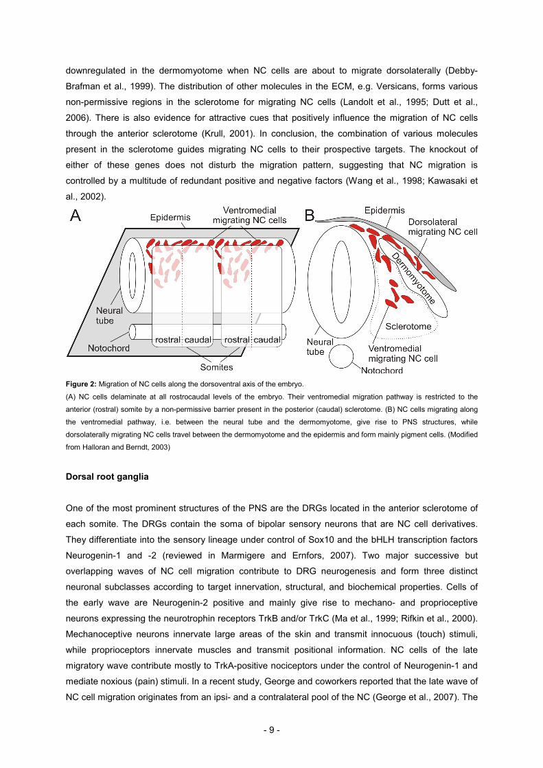
- 9 -
downregulated in the dermomyotome when NC cells are about to migrate dorsolaterally (Debby-
Brafman et al., 1999). The distribution of other molecules in the ECM, e.g. Versicans, forms various
non-permissive regions in the sclerotome for migrating NC cells (Landolt et al., 1995; Dutt et al.,
2006). There is also evidence for attractive cues that positively influence the migration of NC cells
through the anterior sclerotome (Krull, 2001). In conclusion, the combination of various molecules
present in the sclerotome guides migrating NC cells to their prospective targets. The knockout of
either of these genes does not disturb the migration pattern, suggesting that NC migration is
controlled by a multitude of redundant positive and negative factors (Wang et al., 1998; Kawasaki et
al., 2002).
Figure 2: Migration of NC cells along the dorsoventral axis of the embryo.
(A) NC cells delaminate at all rostrocaudal levels of the embryo. Their ventromedial migration pathway is restricted to the
anterior (rostral) somite by a non-permissive barrier present in the posterior (caudal) sclerotome. (B) NC cells migrating along
the ventromedial pathway, i.e. between the neural tube and the dermomyotome, give rise to PNS structures, while
dorsolaterally migrating NC cells travel between the dermomyotome and the epidermis and form mainly pigment cells. (Modified
from Halloran and Berndt, 2003)
Dorsal root ganglia
One of the most prominent structures of the PNS are the DRGs located in the anterior sclerotome of
each somite. The DRGs contain the soma of bipolar sensory neurons that are NC cell derivatives.
They differentiate into the sensory lineage under control of Sox10 and the bHLH transcription factors
Neurogenin-1 and -2 (reviewed in Marmigere and Ernfors, 2007). Two major successive but
overlapping waves of NC cell migration contribute to DRG neurogenesis and form three distinct
neuronal subclasses according to target innervation, structural, and biochemical properties. Cells of
the early wave are Neurogenin-2 positive and mainly give rise to mechano- and proprioceptive
neurons expressing the neurotrophin receptors TrkB and/or TrkC (Ma et al., 1999; Rifkin et al., 2000).
Mechanoceptive neurons innervate large areas of the skin and transmit innocuous (touch) stimuli,
while proprioceptors innervate muscles and transmit positional information. NC cells of the late
migratory wave contribute mostly to TrkA-positive nociceptors under the control of Neurogenin-1 and
mediate noxious (pain) stimuli. In a recent study, George and coworkers reported that the late wave of
NC cell migration originates from an ipsi- and a contralateral pool of the NC (George et al., 2007). The

- 10 -
ipsilateral NC cells populate the inner DRG core and become neurons while the contralateral NC cells
cross the dorsal midline and colonize the dorsal roots and dorsal parts of DRG. Preventing the
migration of the contralaterally emigrating NC cells resulted in a 50% reduction of TrkA-positive
neurons. Another source of TrkA-positive neurons appears to be the BCCs: a third wave of NC cell
migration was reported originating from the BCCs (Maro et al., 2004). Fate-mapping the progeny of
BCCs revealed that up to 5% of TrkA-positive DRG neurons and peripheral glia are derived from
BCCs. Surprisingly, in the absence of BCCs approximately 50% of TrkA-positive neurons were absent
in the DRGs. How can this difference be explained? A possibility would be that BCC-derived cells
either induce the differentiation of other nociceptive progenitors located in the DRG perimeter and/or
the lack of BCC-derived glial cells results in the atrophy of other TrkA-positive neurons. However, it is
unknown if BCC-derived glial cells preferentially associate with TrkA-positive nociceptors. In
summary, all TrkA-positive neurons originate directly and indirectly from the second NC cell migration
wave, some of which derive from a contralateral pool of the NC and some derive from BCCs.
Shortly after the formation of the sensory neurons, the first axons start to elongate in order to
interconnect with other neurons in the spinal cord and to innervate sensory targets in the periphery.
The initial pathway of sensory axons along the dorsoventral axis is supposed to be shaped by
surround repulsion rather than by attraction from intermediate targets. The overlying dermomyotome
and the notochord (see Figure 2) were shown to repel sensory axons in vitro (Keynes et al., 1997).
Once the sensory axons reach the dorsal root entry site (DRES) they innervate the spinal cord, where
they connect to distinct laminae: nociceptive neurons terminate in the dorsal horn, mechanoreceptors
in the deeper laminae, and proprioceptive neurons in the intermediate zone and the motor column of
the spinal cord.
Motor neurons
Motor neurons are located in the ventral neural tube and are distinct from interneurons because they
project axons into the periphery. They derive from a highly proliferative pool of motor neuron
progenitors (pMN). This domain is specified by a combinatorial interaction of various homeodomain
proteins induced by the floor plate-derived morphogen Sonic Hedgehog. The earliest determinant of
motor neuron identity is the homeodomain protein MNR2 (Tanabe et al., 1998). In turn, MNR2
induces the expression of downstream transcription factors such as Islet-1/2, Lim3, and HB9 that
further define motor neuron subpools within the motor column. The postmitotic neurons form
rostrocaudally oriented motor columns in the ventral spinal cord based on cell body position, axonal
projections, and gene expression (Figure 3). Roughly, the columns can be subdivided into medial
motor column (MMC) and lateral motor column (LMC). In comparison to the MMC, which is present all
along the rostrocaudal axis of the spinal cord, the LMC is only present at lumbar and brachial levels.
Motor neurons of the MMC project into epaxial (back) and hypaxial (body wall and limb) muscles. In
contrast, LMC neurons innervate muscles of the limbs and can be further divided into medial LMC
(LMCM) and lateral LMC (LMCL). In retrograde labeling experiments of limb muscles, neurons of the
LMCM and LMCL were shown to innervate ventral and dorsal limb muscles, respectively (Landmesser,

- 11 -
1978). Shortly after the migration of motor neuron cell bodies to their final locations in the ventral
neural tube, they start to send out axons. At that time point the basal lamina surrounding the spinal
cord is not dense and motor axons were shown to penetrate the basal lamina at the ventral motor
axon exit point (VMEP) (Fraher et al., 2007). They further bundle to form the ventral roots and
associate with sensory axons in the plexus region. Finally, they separate again in dorsal and ventral
nerves which innervate the corresponding limb muscles.
Figure 3: Organization of motor neurons and their prospective targets during embryogenesis.
Motor neurons are formed from a proliferating progenitor pool located in the ventral ventricular zone. Postmitotic motor neuron
precursors migrate to their prospective locations in the ventral spinal cord where they form rostrocaudal motor columns. The
LMC is only present at limb levels, while the MMC extends all along the anterior-posterior axis. The LMC can be further divided
into subpools innervating the dorsal (LMCL) and ventral (LMCM) limb muscles. The MMC innervates axial (body wall and back)
muscles. (Modified from Jessell, 2000)

- 12 -
3. Boundary Cap Cells
Summary
The boundary at the interface between the central and the peripheral nervous system consists of a
glial cell barrier. In adult vertebrates, the barrier is formed by the apposition of CNS-derived astrocytes
and PNS-derived Schwann cells which is impenetrable for regenerating axons. However, during
embryogenesis sensory and motor axons have to pass the transition zone in order to connect to
prospective targets in both, the CNS and in the periphery. The embryonic glial barrier differs from the
adult one in terms of axonal permissiveness. A transient NC-derived population of cells, the so-called
boundary cap cells (BCCs), occupy the transition zones and act as a gatekeeper. The BCCs allow
axons to enter and exit the spinal cord; they control the maturation of the adult glial barrier and
preserve the cellular integrity of the central and the peripheral nervous system. Furthermore, BCCs
were shown to contribute to the formation of other PNS structures, such as the DRG neurons and glial
cells. In the absence of BCCs the integrity of both the central and the peripheral nervous system is
seriously compromised resulting in the intermixing of CNS and PNS cells and loss of neurons. The
present chapter summarizes the different functions of BCCs and discusses BCC-specific genes.
BCCs populate the transition zones
The entry and exit points of the embryonic transition zone between the central and the peripheral
nervous system are characterized by the presence of a highly proliferating and transient cell
population that derives from the neural crest (NC) and are formed during a late wave of NC cell
migration (Altman and Bayer, 1982; Altman and Bayer, 1984; Niederländer and Lumsden, 1996;
Golding and Cohen, 1997). These cells, the so-called boundary cap cells (BCCs), were identified in
rodents and birds (Niederländer and Lumsden, 1996; Golding and Cohen, 1997). In rats, they appear
by E13 and persist until postnatal day 6 (P6) (Altman and Bayer, 1984; Golding and Cohen, 1997). In
chicken embryos, they appear in the lumbosacral spinal cord at Hamburger-Hamilton stage 18 (HH18;
Hamburger and Hamilton, 1951) and persist at least until HH36 (Mauti et al., 2007). In mice they were
shown to populate the transition zones between E10.5 and were found until after birth (Baron-Van
Evercooren et al., 2008).
In the trunk spinal cord, two distinct BCC clusters are formed, the ventral clusters located at the
ventral motor axon exit points (VMEP), where motor axons leave the spinal cord, and the dorsal
clusters located at the dorsal root entry zone (DREZ), where the primary sensory afferents enter the
spinal cord (Figure 4). The cervical spinal cord consists of a VMEP and a lateral axon exit and entry
point (LEP) that is shared by spinal accessory motor and sensory axons and both are occupied by
BCCs (Snider and Palavali, 1990). In contrast, only one common entry and exit point containing BCCs
is formed in the hindbrain (Niederländer and Lumsden, 1996).
BCCs express a diversity of genes, most of which exhibit unknown functions in this context (for
details, see below): Krox20, Cadherin-7, Lingo-1, Sox10, SSEA-1, Erythropoietin (EPO), Monoamine

- 13 -
oxidase B (MAOB), neurotrophin receptor TrkB, protein zero (P0), chemokine receptor Cxcr4, Lgi4,
Semaphorin3B, -3G, -6A, and others (Wilkinson et al., 1989; Bhattacharyya et al., 1991; Ernfors et al.,
1992; Nakagawa and Takeichi, 1995; Golding and Cohen, 1997; Vitalis et al., 2003; Belmadani et al.,
2005; Hjerling-Leffler et al., 2005; Knabe et al., 2005; Okafuji and Tanaka, 2005; Bermingham et al.,
2006; Bron et al., 2007; Mauti et al., 2007).
Figure 4: Boundary cap cells in the trunk spinal cord.
(A) The roof plate (RP) is the origin of neural crest cells that migrate along the ventromedial pathway to form dorsal root ganglia
(DRGs) and boundary cap cells (BCCs). DRG neurons extend their neurites, form the dorsal roots, and cross the dorsal BCCs
(dBCCs) at the dorsal root entry site (DRES) in order to innervate the spinal cord at the dorsal root entry zone (DREZ). The
motor neurons (MN) extend their axons ventrolaterally and exit the spinal cord at the ventral motor axon exit point (VMEP). The
VMEPs are populated by the ventral BCCs (vBCCs). (B) Axons passing the CNS:PNS boundary are covered by BCCs and
ensheath each single axon at the transition zone as seen in (C). (Adapted from Fraher, 1997)
Aggregation of BCCs
For a long time, the BCCs were thought to delineate the prospective entry and exit point at the border
of the CNS based on the observation that they appeared at the transition sites before axonal contact
(Niederländer and Lumsden, 1996; Golding and Cohen, 1997; Vermeren et al., 2003). However, a
detailed ultrastructural study during the formation of VMEP and DRES revealed for the first time that
the ventral BCCs appear after motor axons exit the spinal cord at all spinal and cranial levels (Fraher
et al., 2007). In contrast, the dorsal BCCs appear before the ingrowth of primary sensory afferents.
The mechanism that arrests NC cells at the prospective dorsal or ventral transition zone remained
unknown. A favorite candidate mediating this function was Cadherin-7 (Cad-7), a Ca2+-dependent
homophilic cell adhesion molecule. It is expressed in migrating NC cells and later in BCCs (see below;
Nakagawa and Takeichi, 1995). However, no Cad-7 expression was found at the prospective BCC
locations at the time point of aggregation that could define their prospective locations, i.e. on the
neuroepithelium or in the ECM (Golding and Cohen, 1997). Although motor neurons express Cad-7,
the onset of expression is after the formation of the ventral BCC clusters, and therefore, Cad-7 is
unlikely to be part of the migration arrest mechanism (Nakagawa and Takeichi, 1995). Rather the
interaction among Cad-7-positive NC cells and motor axons in the periphery is supposed to be
necessary for the aggregation of Schwann cells that populate the peripheral motor axons (Nakagawa
and Takeichi, 1995). Thus, the mechanism responsible for the aggregation of BCC remains elusive.

- 14 -
Why are dorsal but not ventral BCCs prefiguring the transition sites?
Initially, BCCs were thought to guide axons to the entry and exit points. However, the possibility that
the dorsal BCCs are a potential source of chemoattractants guiding primary sensory afferents to the
DREZ could not be confirmed (Keynes et al., 1997). Furthermore, sensory axons of embryos lacking
BCCs (knock-in of diphtheria toxin in the Krox20 locus; Krox20-DT) did not fail to grow towards the
prospective DREZ (Vermeren et al., 2003; Maro et al., 2004). Albeit the entry site into the spinal cord
was normal, the dorsal roots were shorter and often defasciculated. Keynes and coworkers suggested
that surround repulsion derived from the overlying dermomyotome and the notochord provides a
mechanism that guides sensory afferents to the dorsal neural tube (Keynes et al., 1997).
An interesting finding was that the basal lamina of the neural tube underlying the dorsal BCCs
disappeared shortly after their aggregation but before axonal contact (Niederländer and Lumsden,
1996; Fraher et al., 2007). Since the basal lamina of the dorsal neural tube displays an impenetrable
barrier for sensory axons, it is likely that the preoccupation of the DREZ by BCCs is necessary to
prepare the ingrowth for sensory axon. Evidence is provided by different studies that discovered
secretion of proteases and plasminogen activators from NC cells which are necessary for their
migration through the ECM and invasion of different tissues (Figure 5; Valinsky and Le Douarin, 1985;
Erickson et al., 1992; Carroll et al., 1994). The disappearance of the basal lamina shortly before the
first NC cells emerge from the dorsal neural tube further confirmed this observation (Cheung et al.,
2005).
In contrast, the basal lamina covering the ventral neural tube is not fully established when the first
motor axons exit the spinal cord. Motor axons were shown to penetrate the basal lamina. The basal
lamina does not seem to be degraded by this process because fragments can be detected on the
distal part of the motor axon (Fraher et al., 2007). In agreement with this observation, motor axon
outgrowth and location was not perturbed in the absence of ventral BCCs (Vermeren et al., 2003).
Dorsal BCCs control the maturation of the DREZ
Several studies performed during the past decades addressed the function of BCCs. In 1997, Golding
and coworkers investigated the function of dorsal BCCs (Golding et al., 1997; Golding and Cohen,
1997). In this study the authors propose two key aspects of dorsal BCCs: (1) they control and promote
the initial ingrowth of primary sensory afferents into the spinal cord, and (2) they are involved in the
maturation of the DREZ after birth. In cryoculture experiments they could show that embryonic
sensory neurites preferentially grew through dorsal BCCs while P6 sensory neurons did not. In a
second experiment they found that E18 and P6 neurons could extend neurites equally well when they
were in contact with CNS tissue, suggesting that the failure of neurite extension into the P6 spinal
cord is not due to growth-inhibitory molecules expressed in the CNS. Surprisingly, embryonic DRG
neurons were able to extend neurites and grow through the mature P6 DREZ, concluding that the
failure of P6 DRG neurites to innervate the CNS is based on the expression of receptors recognizing
the inhibitory ligands present in the mature (P6) DREZ. The authors found that the maturation of the
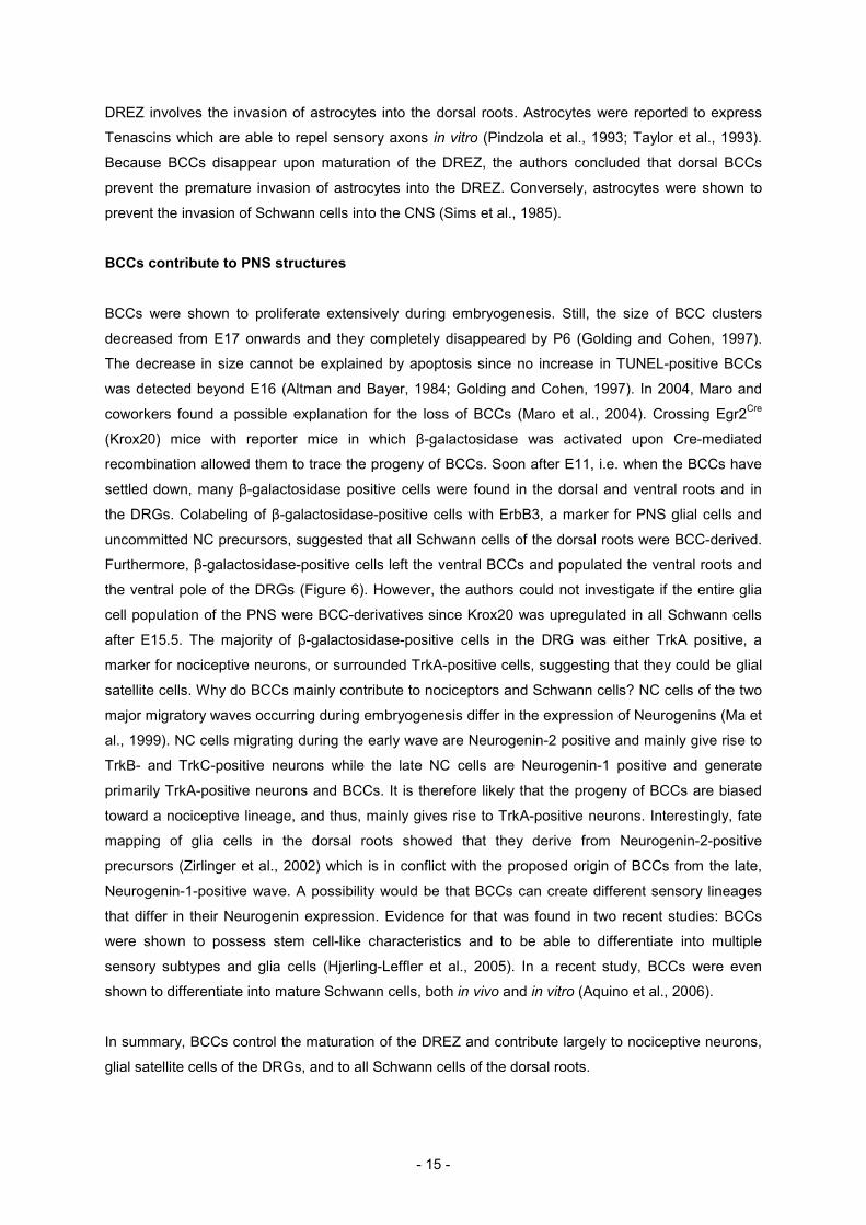
- 15 -
DREZ involves the invasion of astrocytes into the dorsal roots. Astrocytes were reported to express
Tenascins which are able to repel sensory axons in vitro (Pindzola et al., 1993; Taylor et al., 1993).
Because BCCs disappear upon maturation of the DREZ, the authors concluded that dorsal BCCs
prevent the premature invasion of astrocytes into the DREZ. Conversely, astrocytes were shown to
prevent the invasion of Schwann cells into the CNS (Sims et al., 1985).
BCCs contribute to PNS structures
BCCs were shown to proliferate extensively during embryogenesis. Still, the size of BCC clusters
decreased from E17 onwards and they completely disappeared by P6 (Golding and Cohen, 1997).
The decrease in size cannot be explained by apoptosis since no increase in TUNEL-positive BCCs
was detected beyond E16 (Altman and Bayer, 1984; Golding and Cohen, 1997). In 2004, Maro and
coworkers found a possible explanation for the loss of BCCs (Maro et al., 2004). Crossing Egr2Cre
(Krox20) mice with reporter mice in which β-galactosidase was activated upon Cre-mediated
recombination allowed them to trace the progeny of BCCs. Soon after E11, i.e. when the BCCs have
settled down, many β-galactosidase positive cells were found in the dorsal and ventral roots and in
the DRGs. Colabeling of β-galactosidase-positive cells with ErbB3, a marker for PNS glial cells and
uncommitted NC precursors, suggested that all Schwann cells of the dorsal roots were BCC-derived.
Furthermore, β-galactosidase-positive cells left the ventral BCCs and populated the ventral roots and
the ventral pole of the DRGs (Figure 6). However, the authors could not investigate if the entire glia
cell population of the PNS were BCC-derivatives since Krox20 was upregulated in all Schwann cells
after E15.5. The majority of β-galactosidase-positive cells in the DRG was either TrkA positive, a
marker for nociceptive neurons, or surrounded TrkA-positive cells, suggesting that they could be glial
satellite cells. Why do BCCs mainly contribute to nociceptors and Schwann cells? NC cells of the two
major migratory waves occurring during embryogenesis differ in the expression of Neurogenins (Ma et
al., 1999). NC cells migrating during the early wave are Neurogenin-2 positive and mainly give rise to
TrkB- and TrkC-positive neurons while the late NC cells are Neurogenin-1 positive and generate
primarily TrkA-positive neurons and BCCs. It is therefore likely that the progeny of BCCs are biased
toward a nociceptive lineage, and thus, mainly gives rise to TrkA-positive neurons. Interestingly, fate
mapping of glia cells in the dorsal roots showed that they derive from Neurogenin-2-positive
precursors (Zirlinger et al., 2002) which is in conflict with the proposed origin of BCCs from the late,
Neurogenin-1-positive wave. A possibility would be that BCCs can create different sensory lineages
that differ in their Neurogenin expression. Evidence for that was found in two recent studies: BCCs
were shown to possess stem cell-like characteristics and to be able to differentiate into multiple
sensory subtypes and glia cells (Hjerling-Leffler et al., 2005). In a recent study, BCCs were even
shown to differentiate into mature Schwann cells, both in vivo and in vitro (Aquino et al., 2006).
In summary, BCCs control the maturation of the DREZ and contribute largely to nociceptive neurons,
glial satellite cells of the DRGs, and to all Schwann cells of the dorsal roots.

- 16 -
Figure 5: Organization and maturation of the dorsal root entry site.
(A) Dorsal BCCs (dBCCs) prefigure the DREZ before the ingrowth of primary sensory axons into the CNS. Inset: dBCCs are
thought to degrade the basal lamina by the secretion of proteases and plasminogen activators. (B) dBCCs prevent the invasion
of astrocytes into the DRES and possibly control astrocytogenesis via erythropoietin (see below). dBCCs migrate away to give
rise to Schwann cells ensheathing the dorsal roots, to nociceptive neurons, and to satellite glia cells of the DRG. (C)
Postnatally, the dBCCs disappear and the mature CNS:PNS interface consisting of an apposition of CNS-derived astrocytes
and PNS-derived Schwann cells is formed.
Ventral BCCs prevent the emigration of motor neurons
To assess the function of ventral BCCs, Vermeren and colleagues used three experimental models:
Splotch mice which fail to form BCCs due to missing NC cells in the trunk, a targeted genetic ablation
of BCCs using the Krox20-DT mouse (see above), and surgical ablation of the dorsal neural tube
resulting in the absence of NC cells (Vermeren et al., 2003). In all three experiments, the absence of
BCCs led to a destabilization of the motor column resulting in the emigration of motor neuron somas
out of the CNS into the periphery along the ventral roots. However, the phenotype was more
prominent in Splotch mice and in chicken embryos after dorsal neural tube ablation compared to
Krox20-DT transgenic mice. One possibility could be that the death of BCCs happens progressively
and the short contact between motor axons and the “intact” BCCs is sufficient to partially stabilize
some motor neurons. Another explanation would be that the BCC pool is not homogenously
expressing Krox20 and therefore some Krox20-negative BCCs could survive that partially maintain
motor column integrity. It is even conceivable that BCCs stabilize only one part of the motor column.
In Sox10 mutant mice that fail to form BCCs (see below) ectopic motor neurons were also observed
(Bron et al., 2007). Vermeren and coworkers further confirmed the necessity of BCCs in maintaining
motor column integrity by grafting quail NC cells into the spinal cord of dorsal neural tube ablated
chicken embryos (Vermeren et al., 2003). The NC cell grafts migrated to the VMEP and appeared to
form BCCs which prevented motor neuron emigration. In embryos lacking BCCs the ventral roots
appeared much thicker and defasciculated. However, whether this is due to motor neuron somas
occupying more space or the failure of motor axons to fasciculate remains unknown. The study did
not address the organization of the DRG and DREZ. Maro and coworkers could not find misplaced
sensory neurons in the proximity of the DRG in mouse embryos lacking BCCs (Krox20-DT; Maro et
al., 2004).

- 17 -
Figure 6: Formation of the ventral BCC clusters.
(A) Motor axons emerge from the spinal cord before the presence of ventral BCCs (vBCCs). (B) Shortly after, vBCCs start to
aggregate around the motor axons at the VMEP. The proliferating vBCCs migrate along the ventral roots where they
differentiate into Schwann cells (orange oval). Migrating NC cells give also rise to Schwann cells (grey oval). (C) Postnatally,
the vBCCs disappear and the transition zone is formed by the direct apposition of astrocytes and Schwann cells.
Genes expressed in boundary cap cells
A number of genes were shown to be expressed in BCCs. In the majority of cases, their function in
the context of BCCs is unknown. The following paragraph summarizes genes identified either in BCCs
or in NC cells giving rise to BCCs (see also Table 1). Some of the genes play important roles during
neurogenesis and mouse mutants lacking these genes often suffer from severe peripheral
neuropathies.
Cadherin-7 and N-Cadherin
Cadherins form a large group of Ca2+-dependent cell adhesion molecules that have mostly been
implicated in cell-cell interactions and cell sorting (Nakagawa and Takeichi, 1995). They comprise an
extracellular, a transmembrane, and a highly conserved cytoplasmic domain. Two types of Cadherins
are distinguished based on sequence characteristics (type I and II) (Takeichi, 1995). Type I Cadherins
include E-Cadherin (Cdh1, L-CAM), N-Cadherin (Cdh2), P-Cadherin (Cdh3), R-Cadherin (Cdh4), and
U-Cadherin, while type II Cadherins comprise cad5-cad12 (Cdh5-12) (reviewed in Pla et al., 2001).
Cadherins mediate cell-cell adhesion by homophilic interaction and are anchored to the cytoskeleton
via Catenins that link the cytoplasmic domain of Cadherins to actin filaments (reviewed in Kemler,
1993). Generally, premature NC cells express type I but not type II Cadherins. Upon delamination
there is a switch from type I to type II Cadherins that is supposed to be important for the ability to
migrate, since the adherence of type II Cadherins is much lower than that of type I Cadherins (Chu et
al., 2006). Once NC cells home in at their prospective target sites, they switch back to type I
Cadherins. However, there are exceptions to this rule as observed for Cadherin-7 in BCCs (see
below; Nakagawa and Takeichi, 1995).
During the delamination of NC cells from the dorsal neural tube Cadherin-7 is upregulated in a
subpopulation of NC cells migrating ventrolaterally (Nakagawa and Takeichi, 1995; Takeichi, 1995;
Nakagawa and Takeichi, 1998). It is not clear, if NC cells differentiating to BCCs already express

- 18 -
Cadherin-7 or if its expression is upregulated once the BCC clusters are formed. However, the
function of Cadherin-7 in migrating NC cells is supposed to ensure a coherent migration through the
ECM rather than to be required for homing in at specific loci (Nakagawa and Takeichi, 1995; Golding
et al., 1997).
Several studies showed a major role for N-Cadherin in cell adhesion, axon growth and guidance. N-
Cadherin is expressed in the ventricular zone of the neural tube, in premigratory NC cells, in DRGs,
Schwann cells, and BCCs (Wanner et al., 2006). Upon arrest of NC cells at the prospective entry and
exit points of the neural tube, the switch from type II to type I Cadherin involves the upregulation of N-
Cadherin. N-Cadherin is a valuable candidate mediating cell-cell contacts between BCCs due to its
strong adhesive properties. Furthermore, N-Cadherin was reported to be a potent growth-promoting
molecule inducing neurite extension in cultured chick ciliary ganglion neurons (Bixby and Zhang,
1990).
Cxcr4
The chemokine receptor Cxcr4 and its ligand stromal cell-derived factor 1 (Sdf1) are mainly known for
their function in the hematopoietic stem cell system. However, there is growing evidence that the
interaction between Cxcr4 and Sdf1 directs chemotactic cell migration in various developmental
processes. The migration of the posterior lateral line (PLL) primordium in zebrafish was reported to
depend on the distribution of Sdf1 along the migratory path which is recognized by Cxcr4 on the
leading tip of the primordium (David et al., 2002). Similarly, Cxcr4 is expressed in migrating NC cells
while Sdf1 is present in the sclerotome, prominent in the region of the prospective DRGs and around
the ventral neural tube, suggesting that it could direct the migration of NC cells to their prospective
condensation points. Cxcr4 is also expressed in DRGs and motor neurons and seems to be present
in BCCs and Schwann cells (Belmadani et al., 2005). Additionally, Sdf1 transcripts were found in the
DRGs and dorsal roots (Lieberam et al., 2005; Odemis et al., 2005).
Egr2 (Krox20)
Krox20 is a well established marker for BCCs. It belongs to the group of zinc-finger transcription
factors. It is upregulated in a feed-forward loop involving Brn2 and Sox10. Krox20 was reported to
control the segmentation of the hindbrain and the myelination of the PNS (Schneider-Maunoury et al.,
1993; Topilko et al., 1994). In mice, the onset of Krox20 expression was detected by radioactive in
situ hybridization at E8.5 in the dorsal neural tube, possibly in precursors of BCCs (Wilkinson et al.,
1989). However, in the avian embryo, no Krox20 signal can be detected before the formation of the
ventral BCC clusters at HH18 (Mauti et al., 2007). The divergence in the onset of expression could be
species-specific or due to technical limitations, i.e. different detection thresholds for the chromogenic
in situ hybridization. Knockout mice or mice carrying a hypomorphic Krox20 allele do not fail to form
BCCs but have severe defects in the formation and maintenance of peripheral myelin (reviewed in

- 19 -
Svaren and Meijer, 2008). The specific expression of Krox20 in BCCs is transient, as it gets
upregulated by E15.5 in immature Schwann cells of the entire PNS (Topilko et al., 1994).
Erythropoietin
Erythropoietin (EPO, hematopoietic growth factor) is a glycoprotein hormone that is mainly known to
control erythropoiesis. In addition, it has also been identified as a candidate neuroprotective
compound in certain neurodegenerative diseases. EPO was shown to protect neurons from
undergoing apoptosis (Siren et al., 2001). Knabe and coworkers analyzed the expression of EPO and
its receptor, EPOR, in a detailed study in mice (Knabe et al., 2005). EPO is expressed during
embryogenesis in lateral motor neurons, floor plate, DRG neurons and BCCs. In contrast, EPOR is
expressed in precursors of astrocytes, astrocytes, and glial satellite cells. Between E14.5 and E19, all
DRG neurons and satellite cells express both, EPO and EPOR. Precursors of astrocytes that mature
express EPOR and there is evidence that the astrocytogenesis is controlled by the EPO system
(Shibata et al., 1997). It is therefore tempting to speculate that BCCs which are in contact with the
precursors of astrocytes induce and control the astrocytogenesis at the transition zone. Indeed,
GLAST, a marker that represents cells transforming to astrocytes, showed prominent
immunoreactivity at the DREZs and VMEPs (Shibata et al., 1997).
Gdf7
The growth differentiation factor 7 (Gdf7) is a BMP family member that is exclusively expressed in the
roof plate of the embryonic spinal cord. It was reported to induce the differentiation of dorsal
commissural neurons in vitro and the formation of sensory neurons (Lee et al., 1998; Lo et al., 2005).
Fate-mapping studies revealed that the progeny of Gdf7-positive NC cells are found in BCCs,
peripheral glia, and in nociceptive neurons (Lo et al., 2005). This is a further indication that the NC
does not consist of a homologous population of NC cells that acquire their identity once they emigrate
from the neural tube. Rather, premigratory NC cells are predefined while they mature in the dorsal
neural tube.
Gli3
The zinc-finger-containing transcription factors Gli1, Gli2, and Gli3 are mediators of the
Shh/Patched/Smoothened pathway. While Gli1 and Gli2 are activated upon Shh signaling, Gli3 is
repressed. The presence of Gli3 is often and indicator for low concentration of Shh. For example, the
dorsal neural tube expresses Gli3 due to the antagonizing effect of roof-plate derived BMP on floor-
plate derived Shh. Furthermore, BCCs were shown to express Gli3, presumably because of the low
concentration of notochord-derived Shh in the ECM (Luo et al., 2006).

- 20 -
Lgi4
Lgi4 encodes a secreted glycosylated leucine-rich repeat protein and is a part of the Schwann cell
signaling pathway that controls axon segregation and the formation of myelin. It is mutated in clp
(claw paw) mice that are characterized by an abnormal limb posture and peripheral hypomyelination.
At E14 in mice, Lgi4 is expressed in DRGs, BCCs and in Schwann cell precursors or immature
Schwann cells of the peripheral nerves (Bermingham et al., 2006). The function of Lgi4 in BCCs is
unknown.
Lingo-1
Lingo-1 is an evolutionary highly conserved protein being exclusively expressed in the nervous
system (Okafuji and Tanaka, 2005; Mi et al., 2008). It contains leucine-rich repeats, an
immunoglobulin (Ig) domain, a transmembrane domain, and a short cytoplasmic tail. Lingo-1 has
been shown to associate with the Nogo-66 receptor (NgR1) and the p75 neurotrophin receptor (p75),
and thus to function as an additional component of the NgR1/p75 receptor complex (Mi et al., 2004).
Additionally, associations with TrkA and the epidermal growth factor receptor (EGFR) were shown.
Interestingly, the expression of Lingo-1 in the DRG is controlled via TrkA (Lee et al., 2007; Mi et al.,
2008). Lingo-1 functions as a negative regulator of oligodendrocyte differentiation and myelination,
neuronal survival and axonal regeneration (Mi et al., 2008). Despite the fact that Lingo-1 is expressed
in BCCs none of the binding partners mentioned above colocalize with it during embryogenesis
(Okafuji and Tanaka, 2005), suggesting that it has to associate with other yet unknown molecules.
Monoamine oxidase B
Monoamine oxidases are membrane-bound mitochondrial enzymes that catalyze the oxidative
deamination of monoaminergic neurotransmitters. Two types of monoamine oxidases (MAO) exist
that have different substrate affinity. While MAOA has a higher affinity for Serotonin and Noradrenalin,
MAOB processes β-phenylethylamine and tele-methylhistamine. Vitalis and coworkers investigated
the distribution of MAOB during mouse embryogenesis (Vitalis et al., 2003). They found MAOB
expressed in cells of the hindbrain at the entry zone of the trigeminal nerve into the rhombencephalon
that colocalized with Krox20, presumably cranial BCCs. A particularly interesting observation was that
no monoaminergic marker colocalized with these BCCs suggesting that the role of MAOB in these
BCCs is not the processing of monoaminergic neurotransmitters. However, the authors speculate that
MAOB expressed in cranial BCCs could prevent cerebrospinal fluid (CSF)-derived amines from
entering the CNS.
The BCCs of the spinal cord express MAOB (Hjerling-Leffler et al., 2005). It is unlikely, that spinal
BCCs would exert the same function as suggested for the cranial BCCs since they are not in direct
contact with CSF that is present in the central canal of the spinal cord. Furthermore, BCCs seem to
retain MAOB expression but downregulate Krox20 once they leave the clusters.

- 21 -
MPZ (Protein zero, P0)
Protein zero (P0), an integral transmembrane glycoprotein, belongs to the Immunoglobulin-
superfamily of adhesion molecules and is encoded by the myelin protein zero gene (MPZ, CMT1B). It
is a major component of the peripheral myelin sheath and is necessary for the formation of compact
myelin. Mutations in the MPZ gene are linked to a significant number of peripheral neuropathies
resulting in hypomyelination as seen in the Charcot-Marie-Tooth disease (CMT). MBZ was shown to
be a direct regulatory target of Krox20 in cooperation with Sox10. Krox20 mutant mice exhibit defects
in the myelination of peripheral nerves and have reduced expression levels of MPZ (Topilko et al.,
1994). In contrast, the overexpression of Krox20 in Schwann cells induces the expression of MPZ in
vivo (Parkinson et al., 2003). However, P0 is already expressed in a subset of migrating NC cells
before the onset of Krox20 expression suggesting that additionally other regulatory mechanisms are
involved in the control of MPZ (Bhattacharyya et al., 1991). Homophilic binding of P0 was reported to
mediate cell-cell adhesion in both, neuronal and non-neuronal tissues (Filbin et al., 1990; Schneider-
Schaulies et al., 1990). Therefore the presence of P0 in BCCs could possibly mediate adhesion
among them.
Pax3 and Pax7
The paired-box-containing transcription factor Pax3 and Pax7 are class-I transcription factors that are
repressed by Shh (Briscoe et al., 2000). They are required for the patterning of the neural tube and
were shown to be necessary for the induction of NC cells. They are expressed in overlapping areas
such as the dorsal neural tube, NC cells, and BCCs. Splotch mice lacking Pax3, or Pax7 mutant mice
were shown to exhibit defects in the formation of NC cells (Mansouri et al., 1996; Serbedzija and
McMahon, 1997). However, their function in BCCs remains elusive.
Semaphorins
The Semaphorins form a large family of guidance molecules that are predominantly known for their
role in neuronal repulsion. More than 20 different Semaphorins have been identified and classified
into eight subfamilies (Semaphorin Nomenclature Committee, 1999). Classes 1 and 2 are exclusively
found in invertebrates, while classes 3, 4, 6, and 7 are expressed in vertebrates. Class 5
Semaphorins are expressed in both invertebrates and vertebrates, whereas class V is a virally
encoded Semaphorin. Secreted class-3 Semaphorins are the best described with respect to their
function. With a few exceptions they bind to a receptor complex consisting of a Plexin and a
Neuropilin. In contrast, transmembrane class-6 Semaphorins bind PlexinAs in a Neuropilin-
independent manner (Mauti et al., 2006) and can function as receptors mediating bidirectional
signaling (reviewed in Zhou et al., 2008). Semaphorin3B, -3G, and -6A are expressed in BCCs.
However, a function in the formation of BCC clusters and the subsequent stabilization of the motor
column was discovered only for Semaphorin6A (Bron et al., 2007; Mauti et al., 2007).

- 22 -
Sox8 and Sox10
The HMG-box transcription factors of the SoxE family play eminent roles during the development of
the peripheral nervous system. They are involved in the formation of NC cells and interact with other
genes to initiate developmental programs involved in the formation of the PNS. Sox8 is upregulated
during the formation of the NC and is later found in BCCs and oligodendrocyte precursors (Bell et al.,
2000; McKeown et al., 2005). The function of Sox8 in BCCs is unknown.
Sox10 is upregulated in NC cells once they emerge from the dorsal neural tube (Kuhlbrodt et al.,
1998) and it was reported to downregulate N-Cadherin which is a crucial step during the EMT of NC
cell precursors (Cheung et al., 2005). As soon as NC cells start to differentiate, Sox10 is
downregulated. Sox10 was reported to maintain the multipotency of NC cells (Kim et al., 2003).
However, BCCs express Sox10, most probably to keep their multipotent state which is necessary for
their contribution to DRG neurons and peripheral glial cells.
SSEA-1
Stage specific embryonic antigen 1 (SSEA-1) is a specific cell surface marker for sensory neuroblasts
and multipotent cells (Sieber-Blum, 1989). It is expressed in all sensory neuron lineages at all axial
levels of quail embryos. Interestingly, the expression pattern differs between quail and rat or chicken
embryos, where only a small subpopulation of sensory neurons expresses SSEA-1 (Ernsberger and
Rohrer, 1988). NC cells, DRGs, BCCs and their emigrating progeny express SSEA-1 (Oudega et al.,
1992).
TrkB
The neurotrophin tyrosine kinase receptor B (TrkB) is a high affinity receptor for BDNF, NT-3, and NT-
4. It is expressed in many parts of nervous system such as the NC, NC cells, DRGs, motor neurons,
and peripheral glia. The TrkB was reported to be involved in neuronal differentiation and cell survival.
Furthermore, the large diameter proprioceptive and mechanoceptive neurons can be distinguished
from nociceptors by the expression of TrkB and/or TrkC. However, the function of TrkB in BCCs
remains elusive.

- 23 -
Gene
BC
Cs
NC
NC
Cs
pGC
s
DR
Gs
MN
s Expression profile Knockout mice
Cadherin-7 + - + + (+) + (Nakagawa and Takeichi, 1995; Luo et al., 2006) No
Cxcr4 + - + + + + (Belmadani et al., 2005; Lieberam et al., 2005) (Zou et al., 1998)a
Egr2 (Krox20) + (+)b (+)b +c - - (Topilko et al., 1994; Maro et al.,
2004; Mauti et al., 2007) (Topilko et al., 1994)
EPO + - - +d + + (Knabe et al., 2005) (Tsai et al., 2006)
Gdf7 (-)e + (-)e (-)e (-)e - (Lee et al., 1998; Lo et al., 2005) (Lee et al., 1998)
Gli3 + - - - - - (Luo et al., 2006) No (but see Persson et al., 2002 and references therein)
Lgi4 + - - + + - (Bermingham et al., 2006) (Bermingham et al., 2006)
Lingo-1 + - - + + + (Okafuji and Tanaka, 2005) (Inoue et al., 2007)
MAOB + - - + + - (Vitalis et al., 2003; Hjerling-Leffler et al., 2005) (Grimsby et al., 1997)
MPZ (P0) + - + + - - (Golding and Cohen, 1997; Mauti et al., 2007) (Giese et al., 1992)
N-Cadherin + + - + + + (Wanner et al., 2006) (Luo et al., 2001)f
Pax3 + + + + - - (Goulding et al., 1994; Lacosta et al., 2005) (Serbedzija and McMahon, 1997)
Pax7 + + + - - - (Goulding et al., 1994; Lacosta et al., 2005; Luo et al., 2006) (Mansouri et al., 1996)
Sema3B + - - - - - (Bron et al., 2007) (Falk et al., 2005)
Sema3G + - - - + - (Bron et al., 2007) No
Sema6A + (+) + - - (+)g (Mauti et al., 2007) (Mitchell et al., 2001)
Sox8 + - - - - - (Bell et al., 2000; McKeown et al., 2005) (Sock et al., 2001)
Sox10 + - + + + - (Kuhlbrodt et al., 1998; Mauti et al., 2007)
(Herbarth et al., 1998; Southard-Smith et al., 1998; Kapur, 1999)
SSEA-1 + + + (+) + - (Sieber-Blum, 1989; Oudega et al., 1992) (Kudo et al., 2004)h
TrkB + + + + + + (Ernfors et al., 1992; Jungbluth et al., 1997; Straub et al., 2007) (Klein et al., 1993)
Table 1: Genes expressed in BCCs or their progenitors.
(BCCs) Boundary cap cells, (pGCs) peripheral glia cells, (NC) neural crest, (NCCs) neural crest cells, (DRGs) dorsal root
ganglia, (MNs) motor neurons. (a) Embryonic lethal. (b) Radioactive in situ hybridization showed a signal (Wilkinson et al.,
1989). (c) After E15.5 in all Schwann cells. (d) Starting at E14.5. (e) Gdf7 is exclusively expressed in the NC. Fate-mapping
studies revealed that the traced NC cells colonize the DRES (apparently BCCs), dorsal roots, and colocalize with TrkA-positive
neurons. (f) The N-Cadherin knockout embryo dies due to severe cardiovascular defects (Radice et al., 1997). Specific
expression of N- or E-Cadherin in the heart rescues the early lethality. (g) Transient upregulation at HH25. (h) Disruption of the
Fut9 gene results in absence of SSEA-1.

- 24 -
Aim of the thesis
The striking expression pattern of Semaphorin6A in BCCs prompted us to analyze its function during
nervous system development.
The aims of the thesis were:
1. Analysis of the expression patterns of Plexins and Neuropilins
2. Functional analysis of Semaphorin6A and PlexinA1

- 25 -
4. Expression Pattern Analysis of Plexins and Neuropilins
Expression patterns of plexins and neuropilins are consistent with cooperative and separate functions during neural development
Olivier Mauti, Rejina Sadhu, Joelle Gemayel, Matthias Gesemann, and Esther T. Stoeckli
Institute of Zoology, University of Zurich, Winterthurerstrasse 190, 8057 Zurich, Switzerland
Published in BMC Developmental Biology, 2006, 6:32
BioMed Central
Page 1 of 13(page number not for citation purposes)
BMC Developmental Biology
Open AccessResearch articleExpression patterns of plexins and neuropilins are consistent with cooperative and separate functions during neural developmentOlivier Mauti1, Rejina Sadhu1, Joelle Gemayel2, Matthias Gesemann2 and Esther T Stoeckli*1
Address: 1Institute of Zoology, University of Zurich, Winterthurerstrasse 190, 8057 Zurich, Switzerland and 2Brain Research Institute, University of Zurich, Winterthurerstrasse 190, 8057 Zurich, Switzerland
Email: Olivier Mauti - [email protected]; Rejina Sadhu - [email protected]; Joelle Gemayel - [email protected]; Matthias Gesemann - [email protected]; Esther T Stoeckli* - [email protected]
* Corresponding author
AbstractBackground: Plexins are a family of transmembrane proteins that were shown to act as receptorsfor Semaphorins either alone or in a complex together with Neuropilins. Based on structuralcriteria Plexins were subdivided into 4 classes, A through D. PlexinAs are mainly thought to act asmediators of repulsive signals in cell migration and axon guidance. Their functional role invertebrates has been studied almost exclusively in the context of Semaphorin signaling, i.e. as co-receptors for class 3 Semaphorins. Much less is known about Plexins of the other three classes.Despite the fact that Plexins are involved in the formation of neuronal circuits, the temporalchanges of their expression patterns during development of the nervous system have not beenanalyzed in detail.
Results: Only seven plexins are found in the chicken genome in contrast to mammals, where nineplexins have been identified. Here, we describe the dynamic expression patterns of all known plexinfamily members in comparison to the neuropilins in the developing chicken spinal cord.
Conclusion: Our in situ hybridization study revealed that the expression patterns of plexins andneuropilins are only partially overlapping, especially during early and intermediate stages of spinalcord development, supporting both cooperative and separate functions of plexins and neuropilinsin neural circuit formation.
BackgroundThe formation of neuronal circuits crucially depends onthe correct navigation of axons to their target areas, wherethey contact individual target cells to establish synapticcontacts. Axonal navigation is based on sequential growthfrom choice point to choice point. Pathfinding decisionsat choice points and along the axonal trajectory are theconsequence of molecular interactions between guidance
cues presented by the environment and guidance recep-tors expressed on the growth cones. A multitude of in vitroand in vivo approaches led to the identification of guid-ance cues that provide directional information for thenavigation of growth cones. The Semaphorins are a struc-turally diverse family of guidance cues. They are subdi-vided into eight subfamilies, two found in invertebrates,one in viruses, and five in vertebrates. Initially, Sema-
Published: 17 July 2006
BMC Developmental Biology 2006, 6:32 doi:10.1186/1471-213X-6-32
Received: 17 April 2006Accepted: 17 July 2006
This article is available from: http://www.biomedcentral.com/1471-213X/6/32
© 2006 Mauti et al; licensee BioMed Central Ltd.This is an Open Access article distributed under the terms of the Creative Commons Attribution License (http://creativecommons.org/licenses/by/2.0), which permits unrestricted use, distribution, and reproduction in any medium, provided the original work is properly cited.

BMC Developmental Biology 2006, 6:32 http://www.biomedcentral.com/1471-213X/6/32
Page 2 of 13(page number not for citation purposes)
phorins were found to be repellents for extending axons.In 1997, Neuropilins were identified as receptors for Sem-aphorins concurrently in two labs [1-3]; reviewed in [4]. Ashort time later, a role for Plexins as receptors for Sema-phorins was described [5-9]. However, Neuropilins andPlexins had been discovered many years earlier as anti-gens of monoclonal antibodies raised against proteinsfrom the optic tectum of Xenopus laevis [10-12]. In contrastto Neuropilins, which have only been found in verte-brates, Plexins are distributed widely in both vertebratesand invertebrates [13]. The nine Plexins found in verte-brates have been subdivided into four subclasses A-Ddepending on structural criteria. The largest subfamily isthe PlexinA subfamily with four members, followed bythe PlexinB subfamily with three members. Subfamilies Cand D contain only one member each.
By far the best-studied Plexins are class-A Plexins [14-16].Their function has been studied predominantly in contextof their role as co-receptors (together with Neuropilins)for secreted class-3 Semaphorins [14,16,17]. However,PlexinAs must have functions that are independent ofNeuropilins, because they are expressed much morewidely in the developing nervous system than Neuropilin-1 and -2. Consistent with this, Plexins were shown tomediate homophilic cell-cell adhesion in a calcium-dependent manner [11]. Furthermore, PlexinAs wereshown to mediate effects of membrane-bound class-6Semaphorins in a Neuropilin-independent manner[18,19].
Until recently, when Sema3E binding to PlexinD1 in aNeuropilin-independent manner was demonstrated dur-ing the development of the intersomitic vasculature [20],Neuropilins were thought to be required but not sufficientfor class-3 Semaphorin signaling [8,9,21-23]. No signal-ing component in the cytoplasmic part of Neuropilinscould be identified, suggesting that they confer ligand spe-cificity to the complex formed with Plexins, L1, or Off-track [24,25]. In contrast to the secreted class-3 Sema-phorins, membrane-associated Semaphorins were shownto bind to Plexins directly [18,19,26,27].
Much less is known about the function of other classes ofplexins. An interaction of PlexinB1 with Sema4D has been
described, but little is known about the role of PlexinBs invivo [26,28]. PlexinC1 was demonstrated to interact withSema7A [7], although the only functional study availableto date indicates Integrins rather than PlexinC1 as thefunction-mediating receptor for Sema7A [29]. PlexinD1finally was linked to the development of the heart and thevascular system consistent with its predominant expres-sion in endothelial cells [30-32].
As a step toward a better understanding of the diverseroles of Plexins in the developing nervous system, wedecided to assess the expression patterns of plexins. Here,we describe the expression of all plexins found in thechicken genome during spinal cord development in com-parison with neuropilins.
ResultsThe avian genome lacks homologues for two mammalian plexinsIn order to identify chicken plexins and neuropilins weperformed extensive databank searches using the com-bined information from the BBSRC ChickEST and thegenomic database. These searches indicate that thechicken genome encodes a reduced number of plexinscompared to mammals, i.e. only seven instead of nine.While homologous chicken genes for the two unique sub-family members plexinC1 and plexinD1 could readily beidentified, fewer chicken plexinAs and plexinBs arepresent in the chicken genome when compared to mam-mals (Table 1; Figure 1; [33]). The plexinA subfamily con-tains no matching chicken sequence for plexinA3.Furthermore, no counterpart for plexinB3 could beextracted from chicken databases (Figure 1 and Table 1).Interestingly, the mouse variants of the two missing plexingenes are located on the X chromosome, implying that ahomologous region of this chromosome is absent inchicken. Birds use a Z/W sex determination system insteadof the X/Y system found in mammals. Moreover, in con-trast to mammals, in chicken the female is the heteroga-metic sex (ZW), whereas the male is homogametic (ZZ),further suggesting that intense chromosomal rearrange-ments happened during evolutionary development [34].These changes are also reflected by the fact that the hap-loid chicken genome contains 38 autosomes compared toonly 19 autosomes in mouse [33,35]. Based on the align-
Table 1: Chromosomal localization and EST representation of different plexin genes
plexin A1 A2 A3 A4 B1 B2 B3 C1 D1
# ESTs 16 23 n.d. 4 4 23 n.d. 6 9Chick Chr. 12 26 n.d. 1 12 ?1 n.d. 1 12Mouse Chr. 6d1 1h6 Xa7.1 6a3.3 9f2 15e3 Xa7.1 10c2 6e3
The number of identified chicken ESTs for each gene and the chromosomal location of the chicken and its corresponding mouse gene are given. Genes that were not detected are abbreviated by 'n.d.' 1PlexinB2 has not yet been assigned to a chromosome.

BMC Developmental Biology 2006, 6:32 http://www.biomedcentral.com/1471-213X/6/32
Page 3 of 13(page number not for citation purposes)
ment of human, mouse, and chicken chromosomes,genes found on the mammalian X chromosomes wereassigned either to the chicken chromosome 1 or 4 [33].
The chicken genome with 1.2 × 10⁹ bp is only about 40%as long as the human genome, and with an estimated20'000 to 23'000 genes the chicken contains fewer genesthan the mouse or the human genome, although the dif-ference in gene number is not proportional to the reduc-tion in genome size. The chicken genome containsmarkedly less repetitive sequence and a reduced numberof degraded copies of gene sequence, but also fewer dupli-cated copies of genes overall [33]. Thus, our finding thattwo plexins are missing is most likely due to their actualabsence in the genome rather than our inability to detectthem in the databases. Moreover, all attempts to clone themissing chicken homologues using degenerate primersfor RT-PCR failed, again implying that the missing genesare indeed not represented in the chicken genome.
Interestingly, while conservation between differentchicken plexinAs was in the range of 70 to 90%, depend-ing on the plexin parts used for alignment, conservationbetween chicken plexinBs was only around 50% (Table2). This value is not much higher than the values obtainedwhen plexinBs were compared to plexins in other sub-classes, suggesting that although placed into the samesubclass, plexinB1 and B2 might actually be members ofdifferent subclasses.
The expression patterns of plexins are dynamically regulated during early spinal cord developmentWe started to analyze plexin expression in the lumbosac-ral spinal cord at stage 18 [36]. At this time, motor neu-
rons are born in the ventral spinal cord and start to extendaxons. The first plexins detected in motor neurons wereplexinA1 and A2 (Figure 2, compare [Additional file 1] forsense controls). In addition, motor neurons expressedneuropilin-1 (npn-1), but not npn-2. When comparedwith markers for precursors and mature neurons (Figure3), respectively, plexinA1 and A2 were clearly expressedalready in precursors of motor neurons, whereas npn-1was restricted to more lateral, mature motor neurons.
More dorsally, plexinA1, A2, and B1 were expressed inPax6-positive precursor cells (compare to Figure 3A). Inaddition, cells in the dorsal spinal cord expressedplexinA1, A2, B1, as well as npn-2 (Figure 2). These cellsexpressed Pax7 (Figure 3D) but did not express neurofila-ment proteins (Figure 3E) and therefore represent precur-sors of dorsal interneurons [37]. At the lumbosacral levelof the spinal cord, dorsal commissural neurons derivedfrom these Pax7-positive precursors and characterized bythe expression of the cell adhesion molecule Axonin-1start to extend axons at stage 19 (Figure 3F; [38]). Theseaxons grow toward the floor plate in response to chemoat-tractants derived from the floor plate, Netrin-1 (reviewedin [39]) and Sonic hedgehog [40]. The majority of theseaxons have reached the floor plate by stage 22. At thisstage, dorsal commissural neurons identified by axonin-1expression [38,41] expressed all three members of theplexinA class but neither npn-1 nor npn-2 (Figure 4; com-pare [Additional file 2] for sense controls). Interestingly,plexinA1 and A2 were now also expressed in the floorplate, although plexinA1 was found only in lateral but notmedial floor-plate cells (Figure 5). Furthermore, the floorplate was also the earliest site of plexinB1 and C1 expres-sion in the spinal cord (Figure 4 and 5). In contrast to allother plexins, plexinB1 was expressed strongly in theentire ventricular zone and at low levels in almost all cellsof the spinal cord at stage 22 and later.
Motor neurons innervating the hindlimb express a large variety of plexins and neuropilinsAfter stage 22, motor axons reach the plexus region wherethey have to sort out according to their target muscles.While the first decision is primarily a choice to grow eitherdorsally or ventrally, more refined pathways are chosen atstages 25/26, when individual nerves begin to form[42,43]. At that time, motor neurons can be separated intodifferent subpopulations based on the expression patternof transcription factors [44-46] or type-II cadherins [47].At stage 25, all plexins and both neuropilins wereexpressed in motor neurons (Figure 6). PlexinA1, A4, and,at low levels, B2 were expressed in a pattern overlappingwith Isl1-positive motor neurons (Panel C in Figure 6)that coincided with the expression of SC1 a surface markerfor motor neurons C1 (not shown). In contrast, theexpression of plexinD1 and npn-1 was strong in medial
Phylogenetic tree of the plexin superfamilyFigure 1Phylogenetic tree of the plexin superfamily. Plexins were aligned on the basis of their sema domain using the CLUSTAL W program. The scale bar represents a substitu-tion rate of 10 amino acids per 100 amino acid residues. For simplicity only mouse (m) and chicken (c) sequences are depicted but the alignment of rat or human sequences gave similar results (data not shown).

BMC Developmental Biology 2006, 6:32 http://www.biomedcentral.com/1471-213X/6/32
Page 4 of 13(page number not for citation purposes)
motor neurons labeled by MNR2 but not by Isl1 (Panel Din Figure 6). Npn-2 appeared to be expressed predomi-nantly in a dorsolateral subset of Isl1-positive cells.PlexinA2 and C1 were scattered throughout the ventralhorn without clear resemblance to either the Isl1 or theMNR2 pattern and expression levels were rather low. Thiswas also true for plexinB1 that was expressed throughoutthe neural tube with higher levels found in dorsal com-missural neurons, the floor plate, and the ventricularzone. In addition to plexinB1, the floor plate maintainedexpression of plexinA2, and C1 at stage 25. PlexinA1 wasstill detectable in lateral floor-plate cells, although atlower levels. Dorsolateral commissural neurons stillexpressed all plexinAs, plexinB1, and C1, but no neuropi-lin.
During intermediate stages of spinal cord development plexin expression is reducedThe early phase of spinal cord development that we ana-lyzed (stages 18 – 25) is characterized by the birth and dif-ferentiation of neuronal populations [48-50,37]. Thistwo-day period (E3-E5) revealed fundamental changes inthe expression patterns of plexins and neuropilins (com-pare Figures 2, 4, and 6). In contrast, the expression pat-terns changed much less during the next two to three days,
the intermediate phase of spinal cord development. Atstage 30 (Figure 7), the expression pattern of plexinAsremained the same as at stage 25, except for the decreaseof plexinA1 in the dorsal half of the spinal cord. PlexinB1persisted only in the ventral ventricular zone, andplexinB2 disappeared altogether. PlexinC1 was nowexpressed diffusely throughout the gray matter withhigher levels in the lateral motor column and the floorplate. After its transient expression in motor neurons atstages 25 (Figure 6) and 26 (not shown) plexinD1 was nolonger expressed in the spinal cord but was restricted toendothelial cells of the blood vessels as described for themouse [30]. The expression of npn-1 and npn-2 at stage30 (Figure 7) remained largely complementary in differ-ent populations of motor neurons as already indicated atstage 25 (Figure 6). The expression patterns of theneuropilins in motor neurons did not resemble those ofany of the plexins. A more detailed analysis of the individ-ual subpopulations expressing plexins and neuropilinswould exceed the scope of this study and would requiredouble and triple staining for transcription factors of theETS and LIM homeodomain families [45,51,52] or com-parison with the analysis of type-II cadherin expressionpatterns in motor neuron pools [47]. The analysis ofwhole-mount preparations of stage 26 spinal cords did
Table 2: Conservation of different domains between Plexin superfamily members
SEMA domainplexinA2 57/71plexinA4 56/70 59/76plexinB1 28/47 28/48 27/47plexinB2 28/47 30/49 30/48 36/55plexinC1 13/23 12/22 13/22 12/20 12/21plexinD1 24/38 23/40 23/40 27/43 24/40 15/28
plexinA1 plexinA2 plexinA4 plexinB1 plexinB2 plexinC1
PSI domainplexinA2 64/73plexinA4 74/87 62/77plexinB1 50/61 44/55 50/58plexinB2 44/65 39/56 39/60 48/66plexinC1 42/70 42/76 46/65 48/65 38/57plexinD1 37/53 41/61 37/55 40/51 39/53 39/56
plexinA1 plexinA2 plexinA4 plexinB1 plexinB2 plexinC1
SP domainplexinA2 88/95plexinA4 82/94 86/95plexinB1 56/72 56/73 53/73plexinB2 56/74 56/75 56/76 66/79plexinC1 49/69 48/68 49/70 45/65 48/66plexinD1 56/75 56/74 58/75 55/74 55/74 57/77
plexinA1 plexinA2 plexinA4 plexinB1 plexinB2 plexinC1
Pairwise alignment of individual domains of chicken Plexin proteins indicate that the intracellular SP (sex-plexin) domain is highly conserved between the different family members, whereas the two extracellular domains, SEMA and PSI (plexin/semaphorin/integrin), show far less conservation. Interestingly the conservation between individual domains of chicken plexinB1 and B2 is only slightly higher than the homology between class A and class B plexins, suggesting that chicken plexinBs are evolutionary quite distinct. Numbers indicate percentage of identical/conserved amino acids.
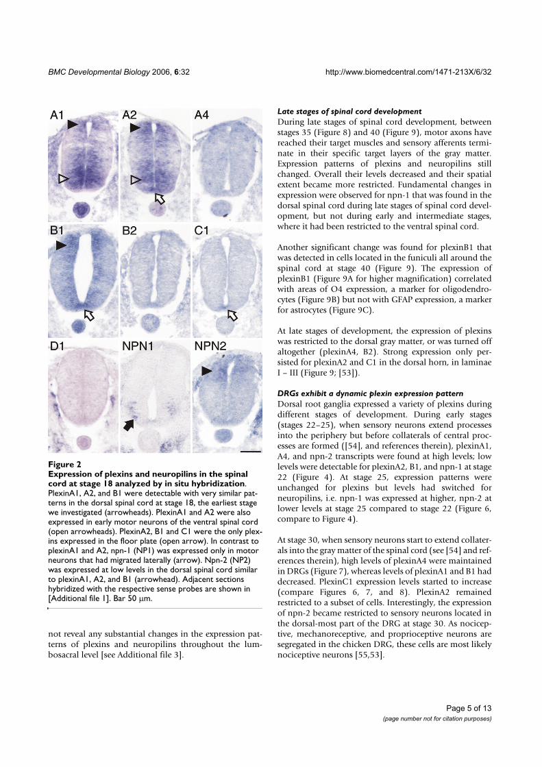
BMC Developmental Biology 2006, 6:32 http://www.biomedcentral.com/1471-213X/6/32
Page 5 of 13(page number not for citation purposes)
not reveal any substantial changes in the expression pat-terns of plexins and neuropilins throughout the lum-bosacral level [see Additional file 3].
Late stages of spinal cord developmentDuring late stages of spinal cord development, betweenstages 35 (Figure 8) and 40 (Figure 9), motor axons havereached their target muscles and sensory afferents termi-nate in their specific target layers of the gray matter.Expression patterns of plexins and neuropilins stillchanged. Overall their levels decreased and their spatialextent became more restricted. Fundamental changes inexpression were observed for npn-1 that was found in thedorsal spinal cord during late stages of spinal cord devel-opment, but not during early and intermediate stages,where it had been restricted to the ventral spinal cord.
Another significant change was found for plexinB1 thatwas detected in cells located in the funiculi all around thespinal cord at stage 40 (Figure 9). The expression ofplexinB1 (Figure 9A for higher magnification) correlatedwith areas of O4 expression, a marker for oligodendro-cytes (Figure 9B) but not with GFAP expression, a markerfor astrocytes (Figure 9C).
At late stages of development, the expression of plexinswas restricted to the dorsal gray matter, or was turned offaltogether (plexinA4, B2). Strong expression only per-sisted for plexinA2 and C1 in the dorsal horn, in laminaeI – III (Figure 9; [53]).
DRGs exhibit a dynamic plexin expression patternDorsal root ganglia expressed a variety of plexins duringdifferent stages of development. During early stages(stages 22–25), when sensory neurons extend processesinto the periphery but before collaterals of central proc-esses are formed ([54], and references therein), plexinA1,A4, and npn-2 transcripts were found at high levels; lowlevels were detectable for plexinA2, B1, and npn-1 at stage22 (Figure 4). At stage 25, expression patterns wereunchanged for plexins but levels had switched forneuropilins, i.e. npn-1 was expressed at higher, npn-2 atlower levels at stage 25 compared to stage 22 (Figure 6,compare to Figure 4).
At stage 30, when sensory neurons start to extend collater-als into the gray matter of the spinal cord (see [54] and ref-erences therein), high levels of plexinA4 were maintainedin DRGs (Figure 7), whereas levels of plexinA1 and B1 haddecreased. PlexinC1 expression levels started to increase(compare Figures 6, 7, and 8). PlexinA2 remainedrestricted to a subset of cells. Interestingly, the expressionof npn-2 became restricted to sensory neurons located inthe dorsal-most part of the DRG at stage 30. As nocicep-tive, mechanoreceptive, and proprioceptive neurons aresegregated in the chicken DRG, these cells are most likelynociceptive neurons [55,53].
Expression of plexins and neuropilins in the spinal cord at stage 18 analyzed by in situ hybridizationFigure 2Expression of plexins and neuropilins in the spinal cord at stage 18 analyzed by in situ hybridization. PlexinA1, A2, and B1 were detectable with very similar pat-terns in the dorsal spinal cord at stage 18, the earliest stage we investigated (arrowheads). PlexinA1 and A2 were also expressed in early motor neurons of the ventral spinal cord (open arrowheads). PlexinA2, B1 and C1 were the only plex-ins expressed in the floor plate (open arrow). In contrast to plexinA1 and A2, npn-1 (NP1) was expressed only in motor neurons that had migrated laterally (arrow). Npn-2 (NP2) was expressed at low levels in the dorsal spinal cord similar to plexinA1, A2, and B1 (arrowhead). Adjacent sections hybridized with the respective sense probes are shown in [Additional file 1]. Bar 50 �m.

BMC Developmental Biology 2006, 6:32 http://www.biomedcentral.com/1471-213X/6/32
Page 6 of 13(page number not for citation purposes)
PlexinA1 and A2 are already expressed in neural precursorsFigure 3PlexinA1 and A2 are already expressed in neural precursors. At stage 18, the homeodomain protein Pax6 labels pre-cursor cells in the intermediate zone of the developing spinal cord (A; [60]). Its ventral expression boundary that is defined by the morphogen Shh released from the floor plate reaches the area with the progenitors of motor neurons expressing MNR2 (B and G). Low levels of MNR2 proteins are seen in progenitor cells located medially (arrowhead in B), where motor neurons are born. MNR2 protein persists and accumulates in postmitotic motor neurons expressing Isl1 (C). In the dorsal spinal cord, Pax7 expression marks a population of precursor cells that give rise to dorsal interneuron subpopulations (D; [37]). Note the decline of Pax7 staining toward the periphery of the spinal cord. Even at stage 19, postmitotic neurons expressing neurofila-ment proteins are found only at the peripheral margin of the spinal cord (E). At this stage, the dorsolateral population of com-missural axons, characterized by the expression of Axonin-1 (arrow in F), starts to extend axons toward the floor plate [38]. At the same, more ventrally located interneurons expressing Axonin-1 also extend axons but their pathway has not been char-acterized in detail (arrowheads). A comparison of the expression of Pax6 (green hatched area), MNR2 (light and dark blue, characterizing low and high protein levels, respectively) and Isl1 (dark blue) is shown in G. For clarity the mRNA expression domains of plexinA1 and A2 have been added in H (red). Significant overlap was found between the plexin expression domain and early motor neurons characterized by MNR2 staining (H). In the dorsal spinal cord domains of Pax7 (green) and Axonin-1 protein expression (yellow) are compared to the domains expressing plexinA1 and A2 mRNA in I. Plexins do not extend to the periphery of the neural tube where postmitotic neurons expressing neurofilament or axonin-1 are found. Bar in A through F 50 �m.

BMC Developmental Biology 2006, 6:32 http://www.biomedcentral.com/1471-213X/6/32
Page 7 of 13(page number not for citation purposes)
At stage 35, plexinA1 and A2 were only found in a fewcells located in the periphery of the DRG, where proprio-ceptive neurons are located (Figure 8; [47,53,55]).
DiscussionPlexinAs have a more widespread distribution compared to neuropilinsThe distribution of class-A plexins has been studied inmore detail than other plexin subfamilies [15,56,57].Their function has been analyzed predominantly in con-text of their role as co-receptors (together with Neuropi-lins) for secreted class-3 Semaphorins [9,56,58,59] but see[18,19,27]. However, based on their much more dynamicand more widespread expression, a Neuropilin-independ-ent role of PlexinAs is evident. In particular, the observa-
tion that plexins are expressed in the floor plate is ratherunexpected, as the floor plate is the intermediate target ofcommissural axons, and therefore, the site where ligandsfor axonal guidance receptors should be expressed. Areceptor function of Plexins on floor-plate cells at stage 22is not obvious, as floor-plate cells do not migrate orundergo structural remodeling at this time. Floor-platedevelopment is terminated much earlier[41,49,50,60,61].
As mentioned above, the chicken genome contains onlythree members of the A class: plexinA1, A2, and A4. Theavailable expression patterns of class-A plexins[15,56,58,62] fail to reflect the dynamics of plexinA-expression changes shown in this study. At E12.5,plexinA1 is reported to be barely detectable in the mousespinal cord and DRGs [56,58]. PlexinA2 was found in theroof plate and in interneurons of the ventral spinal cordbut not in motor neurons of the trunk region [58]. Atmore rostral levels, plexinA2 was found in the dorsal spi-nal cord and in DRGs. Reports of plexinA3 expressionwere contradictory. Brown et al. [58] reported plexinA3expression in the ventricular zone and in the floor plate,whereas Cheng et al. [56] reported plexinA3 to beexpressed throughout the spinal cord (except for the ven-tricular zone). The reason for this discrepancy is unclear.Compared to the expression pattern in chick, the patternof plexinA3 reported by Brown et al. [58] is almost identi-cal to the one we found for plexinB1 at stage 25 (Figure 6).The pattern of plexinA4 in the E12.5 spinal cord of themouse was restricted to a single population of cells in thelateral spinal cord and to the DRGs [56,62]. With respectto the development of sensory afferents to the spinal cord,E12.5 in the mouse is comparable to stage 30 in the chick.The development of motor neurons at E12.5 is closer tostage 25. We therefore used both stage 25 and stage 30 spi-nal cords to compare the plexinA patterns between mouseand chick. While plexinA2 and A4 patterns did not changeconsiderably between stages 25 and 30, plexinA1 levelsdecreased strongly in the dorsal spinal cord and in theDRGs. Consistent with the pattern reported by Brown etal. [58] in mouse, plexinA2 was expressed in the roof plateat stage 25. However, in contrast to the chick, mouseplexinA2 was not expressed in the floor plate.
PlexinBs are only transiently expressed in neuronsThe expression of class-B plexins is difficult to link to anyspecific function. They are expressed in neurons, althoughonly transiently, and in glial cells. The most prominentand longest lasting expression for a class-B plexin is seenin the ventricular zone, where plexinB1 is expressed fromstage 22 to stage 35. This pattern is consistent with themouse, where plexinB1 was found in the ventricular zoneof the spinal cord at E13.5 [63]. At stage 40 in the chick,plexinB1 is restricted to cells in the white matter, presum-
Both neuropilins and all plexins, except D1, were expressed in the spinal cord at stage 22Figure 4Both neuropilins and all plexins, except D1, were expressed in the spinal cord at stage 22. Motor neu-rons expressed plexinA1, A2, A4, B2, and both neuropilins (open arrowheads). Levels of plexinB2 were very low and plexinA2 appeared to be expressed only in a subset of motor neurons. All class-A plexin were found in dorsal commissural neurons (arrowheads). PlexinA1 and A2, but not A4, were detected in the floor plate (open arrows). Furthermore, the floor plate expressed plexinB1 and C1. PlexinB1 was found throughout the spinal cord with higher levels in the ventricu-lar zone. PlexinD1 was not expressed in the spinal cord at all, but was restricted to endothelial cells and cells ensheathing the spinal cord. The DRGs expressed plexinA1, A2, A4, B1, npn-1 and npn-2 (asterisks). For a comparison sections hybridized with the respective sense probes are shown in [Additional file 2]. Bar 200 �m.

BMC Developmental Biology 2006, 6:32 http://www.biomedcentral.com/1471-213X/6/32
Page 8 of 13(page number not for citation purposes)
ably oligodendrocytes. This is in contrast to the expressionof plexinB1 in mouse [63]. In mouse, plexinB3 isexpressed in cells of the white matter resembling theexpression of plexinB1 in chick. Furthermore, the expres-sion of plexinB2, which has been reported to match theexpression of plexinB1 in the ventricular zone of E13.5mice [63], differs in chick, where levels of plexinB2 aregenerally low in the spinal cord and peripheral ganglia. Avery weak transient expression of plexinB1 and B2 isdetectable throughout the gray matter and in motor neu-rons between stages 22 and 25, respectively. During thattime expression is also found in the DRGs and along theventral roots, which would indicate that these cells areearly Schwann cells aligning with motor axons. In contrastto observations in the mouse [63] plexinBs are notexpressed during the time when sensory afferents targettheir specific layers in gray matter, as no expression isdetectable between stages 30 to 35. In the chick, collater-als of primary sensory axons do not form before stage 29[54]. Thus, a contribution of both plexinB1 and B2 to the
formation of central sensory connections seems ratherunlikely in the chick in contrast to mouse, where plexinB1was found in DRGs from E13.5 to E15.5 [63].
PlexinC1 is not expressed in early stages of neural developmentIn contrast to plexinAs, which are expressed during thetime when neurons extend their axons, plexinC1 isexpressed only during late stages of neural development.Neither commissural neurons nor motor neurons expressplexinC1 during the time when they approach their firstintermediate target, the floor plate and the plexus region,respectively. Interestingly, strong expression of plexinC1is seen in the floor plate at stage 22 (Figures 4 and 5C), i.e.when the majority of the axons from dorsolateral com-missural neurons are in the floor plate [38]. The expres-sion in the floor plate persists through stage 25, whencommissural axons have crossed the midline and turnedinto the longitudinal axis [41]. A massive increase inplexinC1 expression in DRGs is found at stage 35, a timewhen all other plexins are downregulated compared totheir earlier expression levels in DRGs. PlexinC1 expres-sion in DRGs persists at stage 40. Thus, plexinC1 starts tobe expressed in all neuronal subpopulations that we ana-lyzed when axons have completed the navigation to theirtargets suggesting that plexinC1 might be involved in tar-get recognition rather than pathfinding. This would beconsistent with the finding of Pasterkamp et al. [29] whoreported that the effect of Semaphorin7A, which binds toPlexinC1 with high affinity [7], on axon growth was inde-pendent of PlexinC1 but rather mediated bySemaphorin7A's interaction with Integrins.
PlexinD1 is expressed predominantly in endothelial cellsPlexinD1 is expressed transiently in motor neurons in asubpopulation-specific manner between stages 24 and 26.It is not expressed in the spinal cord during initial axongrowth of motor neurons. Only after motor axons havereached the plexus region, some medially located cellpopulations express plexinD1 at stage 25. As described inmouse [30,31], plexinD1 is predominantly expressed inendothelial cells of the developing intersomitic blood ves-sels during early stages of development and in blood ves-sels in general during later stages.
The expression of neuropilin-2 in chick is much more restricted than in mouseThe expression of npn-2, but not npn-1, in the developingchicken spinal cord differs considerably from the expres-sion pattern reported in mouse. Neuropilin-1 is expressedin DRGs and motor neurons in E10.5 and E12.5 mousespinal cord [1,58] in a pattern that is similar to the expres-sion in chick at stages 22–25 (Figures 4 and 6). The expres-sion seen in mouse spinal cord at E13.5 [1] is virtually thesame as seen in chick at stage 35 (Figure 8).
The expression of plexinA1 in the floor plate is not uniformFigure 5The expression of plexinA1 in the floor plate is not uniform. At stage 22, plexinA1 (A) is expressed strongly in lateral floor-plate cells but present only at very much reduced levels or absent altogether in medial floor-plate cells (arrowhead). In contrast to plexinA1, plexinA2 (B) and C1 (C) are found throughout the floor plate at high levels. The floor plate is identified by the expression of the transcription factor HNF3� (D, red in F; [41]). The area exhibiting reduced levels of plexinA1, A2, and C1 adjacent to the floor plate represents area p3, characterized by the expression of the transcription factor Nkx2.2 (E, green in F; [41,50]). Bar 100 �m.

BMC Developmental Biology 2006, 6:32 http://www.biomedcentral.com/1471-213X/6/32
Page 9 of 13(page number not for citation purposes)
In contrast to the widespread expression of npn-2 in themouse spinal cord between E10.5 and 12.5 [1,58,64,65],the expression in the embryonic chicken spinal cord ismuch more restricted at comparable stages. In mouse,npn-2 is expressed very strongly in dorsal commissuralneurons, in ventral populations of interneurons, in motorneurons, and in the floor plate. Functional studies haveidentified a role for npn-2 in commissural axon pathfind-
ing in the mouse [66]. In chick, npn-2 is not expressed inthe dorsal spinal cord at comparable stages. It is restrictedto some pools of motor neurons and becomes furtherrestricted with increasing embryonic age. Moreover, npn-2 is never expressed in the embryonic chicken floor plate.
ConclusionOur analysis of plexin and neuropilin expression duringdevelopment of the spinal cord reflects their dynamic reg-ulation that coincides with time windows of axon growthand guidance of different neuronal populations. PlexinAsare expressed more widely than any other class of plexinsboth temporally and spatially. Notably they are alsoexpressed more widely than the neuropilins, indicatingthat they must have functions that are Neuropilin-inde-pendent. This aspect is underestimated when globalexpression patterns are compared rather than the detailedsubpopulation-specific expression patterns. PlexinBs areexpressed during early and intermediate stages of spinalcord development but always at low levels and ratherubiquitously. PlexinC1 is expressed predominantly dur-ing late stages of development, whereas plexinD1 isexpressed only very transiently in the spinal cord.
MethodsAssembly of chicken plexin cDNAscDNA sequences for chicken plexins were assembledusing the combined information from the ChickEST [67]and the chicken genomic database [68]. Seven differentgenomic regions coding for plexins were identified usingthe tblastx alignment algorithm on available vertebrateplexins. The corresponding genomic fragments weredownloaded and analyzed using the Genscan gene predic-tion program [69]. Putative cDNA and protein sequenceswere compared to the corresponding mammalian homo-logues and Genscan prediction errors were corrected bymanual inspection of the intron/exon boundaries. Gapsin the assembled sequences, due to inaccurate or incom-plete genome sequencing, were filled by correspondingEST sequences where possible. A total of 85 chicken ESTscovering parts of 7 different plexins were identified.Among these, 65 contained parts of the coding sequence,whereas the rest covered only parts of the UTR. Sequencealignment of genomic and EST sequences was done usingthe SeqMan software (Lasergene, DNASTAR). Based onoverlapping EST sequences that were supplemented withgenomic sequences, 3'UTR sequences were added to thecoding sequence. UTR sequences were terminated at thefirst polyadenylation AATAAA/ATTAAA sequence that fol-lowed after verified chicken UTR EST sequences. Usingthis combined approach a total of 5 complete and 2 par-tial cDNA sequences for plexins could be assembled.
Stage 25 was the only stage when all plexins and neuropilins were expressed in the spinal cordFigure 6Stage 25 was the only stage when all plexins and neuropilins were expressed in the spinal cord. The most prominent mRNA levels were found for plexinAs. They were all detectable in dorsolateral commissural interneurons (arrowheads) and in motor neurons (open arrowheads). Expression of plexinA2 was restricted to some scattered cells in the ventral horn, but absent from lateral areas. Only subpopulations of motor neurons expressed plexinD1, npn-1, and npn-2. PlexinB1, B2, and C1 were found in all motor neurons although at low levels (open arrowheads). Panels A and B are higher magnifications of the sections hybridized with plexinA1 and npn-1, respectively, in comparison to Isl1 (C) and MNR2 (D) staining in adjacent sections. The expres-sion domains of plexinA1, A4, and npn-2 are more similar to the domains expressing Isl1, whereas those for plexinA2, D1, and npn-1 overlap with the more medial motor neurons expressing MNR2. Floor-plate expression was still found for plexinA1 (lateral floor-plate cells only), A2, B1, and C1 (open arrows). The presence of plexinA1, A2, A4, B1, npn-1, and npn-2 in DRGs is indicated by asterisks. Bar 200 �m.

BMC Developmental Biology 2006, 6:32 http://www.biomedcentral.com/1471-213X/6/32
Page 10 of 13(page number not for citation purposes)
Phylogenetic tree assembly and analysis of domain identityThe domain structure of representative members of thechicken and mouse plexin superfamily was obtainedusing the SMART program [70]. Individual domains wereextracted from the sequence using predicted domainboundaries. Conserved domains from the different plexinsubfamilies were aligned using the CLUSTAL W alignmentalgorithm [71,72] provided by the MegAlign software(Lasergene, DNASTAR). Obvious mistakes in domainboundary prediction were manually adjusted. For betterrepresentation alignment files were exported into theTREEVIEW software, enabling the graphical representa-tion of the unrooted tree [73]. Identical and conservedamino acids within individual domains were determinedby pairwise alignment using the bl2seq program [74].
Preparation of in situ probesThe chicken cDNA plasmids derived from the ESTsChEST53D13 (plexinA1), ChEST128L21 (plexinA2),ChEST1014M19 (plexinA4), ChEST890P9 (plexinB1),ChEST799I19 (plexinB2), ChEST860K1 (plexinC1),ChEST867E24 (plexinD1), ChEST110K21 (npn-1), andChEST675H12 (npn-2) were linearized using restrictionendonucleases NotI or EcoRI (Roche). The linearized plas-mids were used as templates to produce DIG-labeled in
situ probes. For this purpose 1 �g of each cDNA wasmixed with 3 �l DIG RNA labeling mix (Roche; 10 mMNTP). Three �l T3 or T7 RNA polymerase (Promega, 20 U/�l), 6 �l of 5× transcription buffer (Roche), 0.8 �l RNasin(Promega, 40 U/�l), 3 �l 100 mM DTT and DEPC-treatedH2O were added to a final volume of 30 �l. After incuba-tion at 37°C for 2 hours, 3 �l RNase-free DNaseI(Promega, 1 U/�l) were added to the mix, and incubatedat 37°C for 30 minutes. Nuclease treatment was stoppedby the addition of 2 �l 0.5 M EDTA (pH 8.0). The cRNAprobes were LiCl-precipitated and dissolved in 50 �l ofDEPC-treated H2O.
In situ hybridizationChicken embryos were collected at indicated embryonicstages, fixed for 2 hours in 4% paraformaldehyde (PFA)and cryoprotected with 25 % sucrose in 0.1 M sodiumphosphate buffer overnight. The embryos were embeddedin Tissue-Tek O.C.T compound (Sakura), cut transversallyinto 20–25 �m sections at a temperature of -20°C, andmounted on Superfrost Plus microscope slides (Menzel-Glaeser). After drying the slides for 30 minutes at 37°C,they were stored at -20°C until further use. For all stepsuntil hybridization we used diethyl pyrocarbonate(DEPC) treated H2O and stock solutions. Sections werewashed in PBS for 5 minutes and postfixed in 4% PFA for30 minutes. They were washed twice in PBS and once inH2O for 5 minutes each. Sections were then acetylated for10 minutes in 1% triethanolamine containing 0.25%(vol/vol) acetic anhydride. After two washes for 5 minutes
Expression of plexins and neuropilins in the spinal cord at stage 35Figure 8Expression of plexins and neuropilins in the spinal cord at stage 35. Plexins became more restricted during late stages of spinal cord development. Motor neurons expressed plexinA1, A2, npn-1, and npn-2 in a subpopula-tion-specific manner (open arrowhead). PlexinA2 expression levels were very high in the dorsal horn (arrowhead), whereas plexinA4 was found only in a restricted area of the lateral dorsal horn. PlexinC1 was still found throughout the gray matter. In contrast to earlier stages, npn-1 was now expressed in the dorsal spinal cord (arrowhead). Expression of plexinA1, A2, A4, C1, and npn-1 in DRGs is indicated by asterisks. Bar 500 �m.
Expression of plexins and neuropilins in the spinal cord at stage 30Figure 7Expression of plexins and neuropilins in the spinal cord at stage 30. The transition from early to late phases of spinal cord development was characterized by restriction of plexin-expressing areas. PlexinB2 and D1 were no longer expressed in the spinal cord, the expression levels of plexinB1 had declined considerably compared to stage 25. PlexinA1 was strongly reduced in the dorsal spinal cord, but remained expressed at high levels in motor neurons (open arrowhead). Motor neurons still expressed plexinA2, A4, C1, npn-1 and npn-2 in a subpopulation-specific manner. In DRGs plexinA1, A2, A4, C1, npn-1, and npn-2 were expressed (asterisk). Bar 200 �m.

BMC Developmental Biology 2006, 6:32 http://www.biomedcentral.com/1471-213X/6/32
Page 11 of 13(page number not for citation purposes)
each in PBS and a 5-minute wash in 2× SSC (0.3 M NaCl,0.03 M tri-sodium citrate, pH 7.0), sections were prehy-bridized for 3 hours with 300 �l prehybridization solu-tion per slide. To avoid evaporation of the solution, slideswere covered with parafilm (pretreated with 7% H2O2 inH2O) for prehybridization and subsequent hybridization.Hybridization was for at least 16 hours. The prehybridiza-tion solution comprised of 5× Denhardt's, 250 �g/mlyeast tRNA, 500 �g/ml herring sperm DNA, 5× SSC, and50% formamide. For hybridization, 200–500 ng/ml ofeach cRNA probe was added to the prehybridization solu-tion, 300 �l placed on each slide and covered with para-film. Slides were hybridized at 56°C in a humidifiedchamber containing 50% formamide/5× SSC. Afterhybridization, sections were washed for 5 minutes in 5×
SSC at 56°C, 5 minutes in 2× SSC at 56°C, 5 minutes in0.2× SSC at 56°C, 20 minutes in 50% formamide/0.2×SSC at 56°C, 5 minutes in 0.2× SSC at room temperature,and finally washed two times in detection buffer (0.1 MTris-base, 0.15 M NaCl, pH 7.5) for 5 minutes each atroom temperature. Sections were then blocked for 1 hourin blocking buffer (3% milkpowder in detection buffer)and incubated for 1 hour in anti-DIG-AP antibodies(Roche) diluted 1:5000 in blocking buffer (300 �l/slide)in a humidified chamber. After two 15-minutes washes indetection buffer, sections were washed for 5 minutes inalkaline phosphatase (AP) buffer (0.1 M Tris-base, 0.1 MNaCl, 50 mM MgCl2, pH 9.5). For the color reaction, sec-tions were incubated 12–24 hours in a dark humidifiedchamber in AP buffer (500 �l/slide) containing 337.5 �g/ml nitroblue tetrazolium (NBT; Roche), 175 �g/ml 5-bromo-4-chloro-3-indoyl phosphate (BCIP; Roche), and240 �g/ml levamisole (Sigma). The reaction was stoppedby washing sections 2 times for 10 minutes each in TEbuffer (10 mM Tris-base, 1 mM EDTA, pH 8.0), followedby a short dip in H2O. Finally, the sections were cover-slipped with an aqueous mounting medium (Celvol).Reaction times were the same for all stages for a givenprobe, except for plexinA1 and A2. For these two probesreaction times for stage 18 were longer than those forolder stages to optimize signal to noise rations. Thus,expression levels at st18 cannot be directly compared toolder stages.
ImmunohistochemistryAntibodies recognizing Pax6, Pax7, MNR2, Isl1, and SC1/Ben were obtained from the Developmental StudiesHybridoma Bank. The monoclonal antibody RMO270,recognizing neurofilament protein was purchased fromZymed, rabbit anti-axonin-1 was described earlier [75].Secondary antibodies goat anti-mouse Cy3 (Jackson Lab-oratories) and goat anti-rabbit Alexa488 (MolecularProbes) were used to visualize primary antibody bindingsites. Sections were prepared as detailed above. The stain-ing protocol was as detailed earlier [54].
Authors' contributionsOM carried out all the in situ hybridizations and dataanalyses, RS contributed to probe preparation, tissue sec-tioning, and immunohistochemistry. JG and MG carriedout the bioinformatics analysis, ES conceived and coordi-nated the study, and wrote the manuscript. Figures wereprepared by OM, MG, and ES. All authors read andapproved the final manuscript.
Expression of plexins and neuropilins in late stages of spinal cord developmentFigure 9Expression of plexins and neuropilins in late stages of spinal cord development. At stage 40, the latest stage we analyzed, plexin expression still changed. The most promi-nent change was found for plexinB1, which was now restricted to the white matter (arrow; high magnification shown in A). Strong expression in the dorsal spinal cord was still found for plexinA2, C1, and npn-1 (arrowhead). Few positive motor neurons were still found for plexinA1, A2, npn-1, and npn-2 (open arrowheads). DRGs maintained expression of plexinA4, C1, and npn-1 (asterisk). Single scat-tered cells were still expressing plexinA1 and A2. Panels A – C show high magnifications of a section processed by in situ hybridization to detect plexinB1 (A). Adjacent sections were stained with antibodies recognizing the oligodendrocyte marker O4 (B) and the astrocyte marker GFAP (C). Bar 500 �m for in situ hybridizations of plexins and neuropilins and 200 �m for panels A-C.

BMC Developmental Biology 2006, 6:32 http://www.biomedcentral.com/1471-213X/6/32
Page 12 of 13(page number not for citation purposes)
Additional material
AcknowledgementsWe thank Maja Hess for excellent technical help. Research in the lab of ES is supported by the Swiss National Science Foundation. Research in the lab of MG is supported by grants from the Roche Research Foundation, the Hartmann Müller Stiftung, and the Swiss National Science Foundation.
References1. Chen H, Chedotal A, He Z, Goodman CS, Tessier-Lavigne M:
Neuropilin-2, a novel member of the neuropilin family, is ahigh affinity receptor for the semaphorins Sema E and SemaIV but not Sema III. Neuron 1997, 19:547-59.
2. He Z, Tessier-Lavigne M: Neuropilin is a receptor for the axonalchemorepellent Semaphorin III. Cell 1997, 90:739-51.
3. Kolodkin AL, Levengood DV, Rowe EG, Tai YT, Giger RJ, Ginty DD:Neuropilin is a semaphorin III receptor. Cell 1997, 90:753-62.
4. Fujisawa H, Kitsukawa T: Receptors for collapsin/semaphorins.Curr Opin Neurobiol 1998, 8:587-92.
5. Comeau MR, Johnson R, DuBose RF, Petersen M, Gearing P, Vanden-Bos T, Park L, Farrah T, Buller RM, Cohen JI, et al.: A poxvirus-encoded semaphorin induces cytokine production frommonocytes and binds to a novel cellular semaphorin recep-tor, VESPR. Immunity 1998, 8:473-82.
6. Winberg ML, Noordermeer JN, Tamagnone L, Comoglio PM, SpriggsMK, Tessier-Lavigne M, Goodman CS: Plexin A is a neuronal sem-aphorin receptor that controls axon guidance. Cell 1998,95:903-16.
7. Tamagnone L, Artigiani S, Chen H, He Z, Ming GI, Song H, ChedotalA, Winberg ML, Goodman CS, Poo M, et al.: Plexins are a largefamily of receptors for transmembrane, secreted, and GPI-anchored semaphorins in vertebrates. Cell 1999, 99:71-80.
8. Takahashi T, Fournier A, Nakamura F, Wang LH, Murakami Y, KalbRG, Fujisawa H, Strittmatter SM: Plexin-neuropilin-1 complexesform functional semaphorin-3A receptors. Cell 1999, 99:59-69.
9. Rohm B, Ottemeyer A, Lohrum M, Puschel AW: Plexin/neuropilincomplexes mediate repulsion by the axonal guidance signalsemaphorin 3A. Mech Dev 2000, 93:95-104.
10. Takagi S, Tsuji T, Amagai T, Takamatsu T, Fujisawa H: Specific cellsurface labels in the visual centers of Xenopus laevis tadpoleidentified using monoclonal antibodies. Dev Biol 1987,122:90-100.
11. Ohta K, Mizutani A, Kawakami A, Murakami Y, Kasuya Y, Takagi S,Tanaka H, Fujisawa H: Plexin: a novel neuronal cell surface mol-ecule that mediates cell adhesion via a homophilic bindingmechanism in the presence of calcium ions. Neuron 1995,14:1189-99.
12. Satoda M, Takagi S, Ohta K, Hirata T, Fujisawa H: Differentialexpression of two cell surface proteins, neuropilin andplexin, in Xenopus olfactory axon subclasses. J Neurosci 1995,15:942-55.
13. Fujisawa H: Discovery of semaphorin receptors, neuropilinand plexin, and their functions in neural development. J Neu-robiol 2004, 59:24-33.
14. Tamagnone L, Comoglio PM: Signalling by semaphorin recep-tors: cell guidance and beyond. Trends Cell Biol 2000, 10:377-83.
15. Murakami Y, Suto F, Shimizu M, Shinoda T, Kameyama T, Fujisawa H:Differential expression of plexin-A subfamily members in themouse nervous system. Dev Dyn 2001, 220:246-58.
16. Fiore R, Puschel AW: The function of semaphorins during nerv-ous system development. Front Biosci 2003, 8:s484-99.
17. Huber AB, Kolodkin AL, Ginty DD, Cloutier JF: Signaling at thegrowth cone: ligand-receptor complexes and the control ofaxon growth and guidance. Annu Rev Neurosci 2003, 26:509-63.
18. Toyofuku T, Zhang H, Kumanogoh A, Takegahara N, Suto F, Kamei J,Aoki K, Yabuki M, Hori M, Fujisawa H, et al.: Dual roles of Sema6Din cardiac morphogenesis through region-specific associa-tion of its receptor, Plexin-A1, with off-track and vascularendothelial growth factor receptor type 2. Genes Dev 2004,18:435-47.
19. Suto F, Ito K, Uemura M, Shimizu M, Shinkawa Y, Sanbo M, ShinodaT, Tsuboi M, Takashima S, Yagi T, et al.: Plexin-a4 mediates axon-repulsive activities of both secreted and transmembranesemaphorins and plays roles in nerve fiber guidance. J Neuro-sci 2005, 25:3628-37.
20. Gu C, Yoshida Y, Livet J, Reimert DV, Mann F, Merte J, HendersonCE, Jessell TM, Kolodkin AL, Ginty DD: Semaphorin 3E andplexin-D1 control vascular pattern independently ofneuropilins. Science 2005, 307:265-8.
21. Kolodkin AL, Ginty DD: Steering clear of semaphorins:neuropilins sound the retreat. Neuron 1997, 19:1159-62.
22. Nakamura F, Kalb RG, Strittmatter SM: Molecular basis of sema-phorin-mediated axon guidance. J Neurobiol 2000, 44:219-29.
23. Raper JA: Semaphorins and their receptors in vertebrates andinvertebrates. Curr Opin Neurobiol 2000, 10:88-94.
24. Castellani V, Rougon G: Control of semaphorin signaling. CurrOpin Neurobiol 2002, 12:532-41.
25. Pasterkamp RJ, Kolodkin AL: Semaphorin junction: makingtracks toward neural connectivity. Curr Opin Neurobiol 2003,13:79-89.
26. Giordano S, Corso S, Conrotto P, Artigiani S, Gilestro G, Barberis D,Tamagnone L, Comoglio PM: The semaphorin 4D receptor con-trols invasive growth by coupling with Met. Nat Cell Biol 2002,4:720-4.
27. Toyofuku T, Zhang H, Kumanogoh A, Takegahara N, Yabuki M,Harada K, Hori M, Kikutani H: Guidance of myocardial pattern-ing in cardiac development by Sema6D reverse signalling.Nat Cell Biol 2004, 6:1204-11.
28. Neufeld G, Shraga-Heled N, Lange T, Guttmann-Raviv N, Herzog Y,Kessler O: Semaphorins in cancer. Front Biosci 2005, 10:751-60.
29. Pasterkamp RJ, Peschon JJ, Spriggs MK, Kolodkin AL: Semaphorin7A promotes axon outgrowth through integrins and MAPKs.Nature 2003, 424:398-405.
30. van der Zwaag B, Hellemons AJ, Leenders WP, Burbach JP, BrunnerHG, Padberg GW, Van Bokhoven H: PLEXIN-D1, a novel plexinfamily member, is expressed in vascular endothelium andthe central nervous system during mouse embryogenesis.Dev Dyn 2002, 225:336-43.
Additional File 1Sense controls of stage-18 spinal cord sections. Adjacent transverse sec-tions of stage-18 spinal cords were hybridized with the respective sense probes for a comparison with the antisense probes shown in Figure 2. Bar 50 �m.Click here for file[http://www.biomedcentral.com/content/supplementary/1471-213X-6-32-S1.pdf]
Additional File 2Sense controls of stage-22 spinal cord sections. Transverse sections of stage-22 spinal cords adjacent to the ones shown in Figure 4 were hybrid-ized with the respective sense probes as a negative control4. Bar 50 �m.Click here for file[http://www.biomedcentral.com/content/supplementary/1471-213X-6-32-S2.pdf]
Additional File 3The expression patterns of plexins and neuropilins do not change sig-nificantly within the lumbosacral region of the spinal cord. Whole-mount preparations of stage 26 spinal cord were used for in situ hybridi-zation to detect plexin and neuropilin mRNAs. At the resolution of whole-mounts no changes were detectable throughout the lumbosacral region of the spinal cord. The only exception being expression levels of npn-2 mRNA that seemed to decrease in some segments of the lumbosacral spinal cord (arrowhead). Bar 500 �m, 200 �m in A-C.Click here for file[http://www.biomedcentral.com/content/supplementary/1471-213X-6-32-S3.pdf]
BMC Developmental Biology 2006, 6:32 http://www.biomedcentral.com/1471-213X/6/32
Page 13 of 13(page number not for citation purposes)
31. Gitler AD, Lu MM, Epstein JA: PlexinD1 and semaphorin signal-ing are required in endothelial cells for cardiovascular devel-opment. Dev Cell 2004, 7:107-16.
32. Torres-Vazquez J, Gitler AD, Fraser SD, Berk JD, Van NP, FishmanMC, Childs S, Epstein JA, Weinstein BM: Semaphorin-plexin sign-aling guides patterning of the developing vasculature. Dev Cell2004, 7:117-23.
33. Hillier LW, Miller W, Birney E, Warren W, Hardison RC, Ponting CP,Bork P, Burt DW, Groenen MA, Delany ME, et al.: Sequence andcomparative analysis of the chicken genome provide uniqueperspectives on vertebrate evolution. Nature 2004,432:695-716.
34. Smith CA, Sinclair AH: Sex determination: insights from thechicken. Bioessays 2004, 26:120-32.
35. Vinogradov AE: Intron-genome size relationship on a largeevolutionary scale. J Mol Evol 1999, 49:376-84.
36. Hamburger V, Hamilton HL: A series of normal stages in thedevelopment of the chick embryo. J Morphol 1951, 88:49-92.
37. Helms AW, Johnson JE: Specification of dorsal spinal cordinterneurons. Curr Opin Neurobiol 2003, 13:42-9.
38. Stoeckli ET, Landmesser LT: Axonin-1, Nr-CAM, and Ng-CAMplay different roles in the in vivo guidance of chick commis-sural neurons. Neuron 1995, 14:1165-79.
39. Leonardo ED, Hinck L, Masu M, Keino-Masu K, Fazeli A, Stoeckli ET,Ackerman SL, Weinberg RA, Tessier-Lavigne M: Guidance ofdeveloping axons by netrin-1 and its receptors. Cold SpringHarb Symp Quant Biol 1997, 62:467-78.
40. Charron F, Stein E, Jeong J, McMahon AP, Tessier-Lavigne M: Themorphogen sonic hedgehog is an axonal chemoattractantthat collaborates with netrin-1 in midline axon guidance. Cell2003, 113:11-23.
41. Bourikas D, Pekarik V, Baeriswyl T, Grunditz A, Sadhu R, Nardo M,Stoeckli ET: Sonic hedgehog guides commissural axons alongthe longitudinal axis of the spinal cord. Nat Neurosci 2005,8:297-304.
42. Landmesser L: The development of motor projection patternsin the chick hind limb. J Physiol 1978, 284:391-414.
43. Landmesser LT: The acquisition of motoneuron subtype iden-tity and motor circuit formation. Int J Dev Neurosci 2001,19:175-82.
44. Tsuchida T, Ensini M, Morton SB, Baldassare M, Edlund T, Jessell TM,Pfaff SL: Topographic organization of embryonic motor neu-rons defined by expression of LIM homeobox genes. Cell1994, 79:957-70.
45. Lin JH, Saito T, Anderson DJ, Lance-Jones C, Jessell TM, Arber S:Functionally related motor neuron pool and muscle sensoryafferent subtypes defined by coordinate ETS gene expres-sion. Cell 1998, 95:393-407.
46. Sharma K, Belmonte JC: Development of the limb neuromuscu-lar system. Curr Opin Cell Biol 2001, 13:204-10.
47. Price SR, De Marco Garcia NV, Ranscht B, Jessell TM: Regulation ofmotor neuron pool sorting by differential expression of typeII cadherins. Cell 2002, 109:205-16.
48. Tanabe Y, Jessell TM: Diversity and pattern in the developingspinal cord. Science 1996, 274:1115-23.
49. Briscoe J, Ericson J: The specification of neuronal identity bygraded Sonic Hedgehog signalling. Semin Cell Dev Biol 1999,10:353-62.
50. Jessell TM: Neuronal specification in the spinal cord: inductivesignals and transcriptional codes. Nat Rev Genet 2000, 1:20-9.
51. Arber S, Ladle DR, Lin JH, Frank E, Jessell TM: ETS gene Er81 con-trols the formation of functional connections between groupIa sensory afferents and motor neurons. Cell 2000, 101:485-98.
52. Livet J, Sigrist M, Stroebel S, De Paola V, Price SR, Henderson CE, Jes-sell TM, Arber S: ETS gene Pea3 controls the central positionand terminal arborization of specific motor neuron pools.Neuron 2002, 35:877-92.
53. Eide AL, Glover JC: Developmental dynamics of functionallyspecific primary sensory afferent projections in the chickenembryo. Anat Embryol (Berl) 1997, 195:237-50.
54. Perrin FE, Rathjen FG, Stoeckli ET: Distinct subpopulations ofsensory afferents require F11 or axonin-1 for growth to theirtarget layers within the spinal cord of the chick. Neuron 2001,30:707-23.
55. Scott S: Sensory Neurons: Diversity, Development, and Plas-ticity. New York: Oxford University Press; 1992.
56. Cheng HJ, Bagri A, Yaron A, Stein E, Pleasure SJ, Tessier-Lavigne M:Plexin-A3 mediates semaphorin signaling and regulates thedevelopment of hippocampal axonal projections. Neuron2001, 32:249-63.
57. Perala NM, Immonen T, Sariola H: The expression of plexins dur-ing mouse embryogenesis. Gene Expr Patterns 2005, 5:355-62.
58. Brown CB, Feiner L, Lu MM, Li J, Ma X, Webber AL, Jia L, Raper JA,Epstein JA: PlexinA2 and semaphorin signaling during cardiacneural crest development. Development 2001, 128:3071-80.
59. Yaron A, Huang PH, Cheng HJ, Tessier-Lavigne M: Differentialrequirement for Plexin-A3 and -A4 in mediating responsesof sensory and sympathetic neurons to distinct class 3 Sem-aphorins. Neuron 2005, 45:513-23.
60. Briscoe J, Pierani A, Jessell TM, Ericson J: A homeodomain proteincode specifies progenitor cell identity and neuronal fate inthe ventral neural tube. Cell 2000, 101:435-45.
61. Wijgerde M, McMahon JA, Rule M, McMahon AP: A direct require-ment for Hedgehog signaling for normal specification of allventral progenitor domains in the presumptive mammalianspinal cord. Genes Dev 2002, 16:2849-64.
62. Suto F, Murakami Y, Nakamura F, Goshima Y, Fujisawa H: Identifi-cation and characterization of a novel mouse plexin, plexin-A4. Mech Dev 2003, 120:385-96.
63. Worzfeld T, Puschel AW, Offermanns S, Kuner R: Plexin-B familymembers demonstrate non-redundant expression patternsin the developing mouse nervous system: an anatomicalbasis for morphogenetic effects of Sema4D during develop-ment. Eur J Neurosci 2004, 19:2622-32.
64. Kawakami A, Kitsukawa T, Takagi S, Fujisawa H: Developmentallyregulated expression of a cell surface protein, neuropilin, inthe mouse nervous system. J Neurobiol 1996, 29:1-17.
65. Chen H, Bagri A, Zupicich JA, Zou Y, Stoeckli E, Pleasure SJ, Lowen-stein DH, Skarnes WC, Chedotal A, Tessier-Lavigne M: Neuropilin-2 regulates the development of selective cranial and sensorynerves and hippocampal mossy fiber projections. Neuron2000, 25:43-56.
66. Zou Y, Stoeckli E, Chen H, Tessier-Lavigne M: Squeezing axonsout of the gray matter: a role for slit and semaphorin pro-teins from midline and ventral spinal cord. Cell 2000,102:363-75.
67. BBSRC ChickEST database [http://www.chick.umist.ac.uk]68. Ensembl chicken genome database [http://www.ensembl.org/
Multi/blastview?species=Gallus_gallus]69. Genscan gene prediction program [http://genes.mit.edu/GEN
SCAN.html]70. SMART program [http://smart.embl-heidelberg.de]71. Higgins DG, Sharp PM: Fast and sensitive multiple sequence
alignments on a microcomputer. Comput Appl Biosci 1989,5:151-3.
72. Thompson JD, Higgins DG, Gibson TJ: CLUSTAL W: improvingthe sensitivity of progressive multiple sequence alignmentthrough sequence weighting, position-specific gap penaltiesand weight matrix choice. Nucleic Acids Res 1994, 22:4673-80.
73. Page RD: TreeView: an application to display phylogenetictrees on personal computers. Comput Appl Biosci 1996, 12:357-8.
74. NCBI-BLAST program [http://www.ncbi.nlm.nih.gov/BLAST/bl2seq/wblast2.cgi]
75. Stoeckli ET, Kuhn TB, Duc CO, Ruegg MA, Sonderegger P: Theaxonally secreted protein axonin-1 is a potent substratumfor neurite growth. J Cell Biol 1991, 112:449-55.

- 39 -
Supplementary Figure 1

- 40 -
Supplementary Figure 2

- 41 -
Supplementary Figure 3

- 42 -
5. Functional Analysis of Semaphorin6A
Semaphorin6A acts as a gate keeper between the central and the peripheral nervous system
Olivier Mauti, Elena Domanitskaya, Irwin Andermatt, Rejina Sadhu, and Esther T. Stoeckli
Institute of Zoology, University of Zurich, Winterthurerstrasse 190, 8057 Zurich, Switzerland
Published in Neural Development, 2007, 2:28
This paper was evaluated by the Faculty of 1000 Biology.

BioMed Central
Page 1 of 17(page number not for citation purposes)
Neural Development
Open AccessResearch articleSemaphorin6A acts as a gate keeper between the central and the peripheral nervous systemOlivier Mauti1, Elena Domanitskaya1, Irwin Andermatt1, Rejina Sadhu1,2 and Esther T Stoeckli*1
Address: 1Developmental Neuroscience, Institute of Zoology, University of Zurich, Winterthurerstrasse 190, 8057 Zurich, Switzerland and 2Novartis, DOC, Lichtstrasse 35, CH-4002 Basel, Switzerland
Email: Olivier Mauti - [email protected]; Elena Domanitskaya - [email protected]; Irwin Andermatt - [email protected]; Rejina Sadhu - [email protected]; Esther T Stoeckli* - [email protected]
* Corresponding author
AbstractBackground: During spinal cord development, expression of chicken SEMAPHORIN6A(SEMA6A) is almost exclusively found in the boundary caps at the ventral motor axon exit pointand at the dorsal root entry site. The boundary cap cells are derived from a population of latemigrating neural crest cells. They form a transient structure at the transition zone between theperipheral nervous system (PNS) and the central nervous system (CNS). Ablation of the boundarycap resulted in emigration of motoneurons from the ventral spinal cord along the ventral roots.Based on its very restricted expression in boundary cap cells, we tested for a role of Sema6A as agate keeper between the CNS and the PNS.
Results: Downregulation of Sema6A in boundary cap cells by in ovo RNA interference resulted inmotoneurons streaming out of the spinal cord along the ventral roots, and in the failure of dorsalroots to form and segregate properly. PlexinAs interact with class 6 semaphorins and areexpressed by both motoneurons and sensory neurons. Knockdown of PlexinA1 reproduced thephenotype seen after loss of Sema6A function both at the ventral motor exit point and at the dorsalroot entry site of the lumbosacral spinal cord. Loss of either PlexinA4 or Sema6D function had aneffect only at the dorsal root entry site but not at the ventral motor axon exit point.
Conclusion: Sema6A acts as a gate keeper between the PNS and the CNS both ventrally anddorsally. It is required for the clustering of boundary cap cells at the PNS/CNS interface and, thus,prevents motoneurons from streaming out of the ventral spinal cord. At the dorsal root entry siteit organizes the segregation of dorsal roots.
BackgroundDuring development of the nervous system, axons navi-gate long distances to connect to their targets. Along theirtrajectories they encounter a large variety of guidance cuesthat support their navigation [1,2]. Axon guidance cues
are subdivided into long-range and short-range guidancecues. They belong to a relatively small number of proteinfamilies, the immunoglobulin superfamily of cell adhe-sion molecules [3], the Eph/ephrins [4], the netrins [5,6],the semaphorins [7,8] and their receptors, plexins and
Published: 18 December 2007
Neural Development 2007, 2:28 doi:10.1186/1749-8104-2-28
Received: 2 August 2007Accepted: 18 December 2007
This article is available from: http://www.neuraldevelopment.com/content/2/1/28
© 2007 Mauti et al.; licensee BioMed Central Ltd. This is an open access article distributed under the terms of the Creative Commons Attribution License (http://creativecommons.org/licenses/by/2.0), which permits unrestricted use, distribution, and reproduction in any medium, provided the original work is properly cited.

Neural Development 2007, 2:28 http://www.neuraldevelopment.com/content/2/1/28
Page 2 of 17(page number not for citation purposes)
neuropilins [9,10]. More recently, morphogens such asWnts and Shh have also been implicated in axon guidance[11-13].
The semaphorins comprise a large family subdivided intoeight subclasses based on structural criteria and theirexpression in vertebrates or non-vertebrate organisms[7,14,15]. Class 1 and 2 semaphorins are expressed onlyin invertebrates, classes 3, 4, 6, and 7 are expressed only invertebrates, class 5 semaphorins are expressed in bothinvertebrates and vertebrates, whereas class V consists of aviral semaphorin. With respect to their function, solubleclass 3 semaphorins are the best characterized. They havebeen shown to act mainly as repellents but, in some cases,also as attractants for extending axons. Class 3 sema-phorins bind to a receptor complex composed ofNeuropilin-1 or -2 and a member of the class A plexins[10,15], although there is at least one exception to thisrule [16].
Plexins are expressed in a highly dynamic pattern duringdevelopment of the nervous system [17-20]. They are sub-divided into four classes comprising a total of nine mem-bers in mammals and seven members in chicken [20].Plexins of class A and B were shown to bind to transmem-brane semaphorins in the absence of neuropilins [21,22].PlexinBs are receptors for class 4 semaphorins, whereasPlexinAs were shown to be receptors for class 6 sema-phorins [22-25]. Interestingly, transmembrane sema-phorins have functions in axon guidance and synapseformation that are independent of neuropilins [22]. Thelong cytoplasmic tail of Sema6A contains binding sites forEna/VASP-like protein EVL and may, therefore, directlyregulate cytoskeletal dynamics [26]. Consistent with thesestructural features, Sema6A was suggested to act as a recep-tor [27], similar to findings for Sema1a, the closestortholog of Sema6A in invertebrates [28]. Sema1a wasshown to act both as a repellent [29,30] and as an attract-ant [31]. A receptor function for Sema1a was reported inthe visual system of Drosophila, where photoreceptor cellsdepended on Sema1a for their targeting to the optic lobe[32].
In mammals, Sema6A was shown to affect pathfinding ofthalamocortical axons [27] and to be required for cellmigration in the cerebellum [33]. The mode of action hasnot been determined in these studies but, based on theexpression pattern and the analysis of the phenotypes, arepulsive mechanism has been suggested in the latter. Thiswould be consistent with in vitro studies that demon-strated a repulsive role of Sema6A on sympathetic axons[22,34]. More recently, a repellent activity of Sema6D onproprioceptive sensory afferents has been shown in bothmouse and chicken [25]. The targeting of proprioceptiveaxons was dependent on PlexinA1 mediating the repul-
sive activity of Sema6C/D. PlexinA1 was also shown to bethe binding partner of Sema6D in neural crest cell migra-tion during heart development [23]. In these studies areceptor function of Sema6D was demonstrated [24].Thus, transmembrane class 6 semaphorins are bifunc-tional molecules in axon guidance and cell migration.They can act as a ligand for PlexinAs but also transmit asignal themselves.
In vertebrates, the receptors for Sema6A in cerebellardevelopment have not been identified. However, in vitrobinding studies have indicated that Sema6A can bind toPlexinA2 and A4 [22], whereas Sema6B binds toPlexinA1and A4 [22], Sema6C was suggested to bind toPlexinD1 [35], and finally Sema6D was shown to bind toPlexinA1 in neural crest cell migration [23,24].
Analysis of SEMA6A expression during chicken spinalcord development revealed its restriction to the ventralventricular zone, the origin of oligodendrocytes, and,most strikingly, to cells at the ventral motor axon exitpoint (VMEP) and the dorsal sensory axons entry point.Cells located at the transition zone between the PNS andthe CNS were shown to have gate keeper function [36,37].In analogy to their function they are called boundary capcells (BCCs). BCCs are derived from a late migrating pop-ulation of neural crest cells [38]. They express Krox20 andthe 1E8 antigen in addition to the more general neuralcrest marker Sox10. BCCs are necessary to prevent emigra-tion of motoneurons from the ventral spinal cord [37].More recently, the boundary cap was identified as a sourceof neural crest stem cells that give rise to glia and sensoryneurons of the dorsal root ganglion (DRG) [39-41].
Here, we show that Sema6A is required for the gate keeperfunction of BCCs, as in the absence of Sema6A BCCs failto cluster properly at the CNS/PNS interface and, thus,cannot prevent the emigration of motoneurons in aPlexinA1-dependent manner. At the dorsal root entry siteSema6A is required for the appropriate segregation of dor-sal roots.
ResultsSEMA6A is expressed in boundary cap cellsSEMA6A shows a much more restricted expression patternduring development of the chicken spinal cord comparedto the mouse. In contrast to the mouse, where SEMA6Awas found throughout the ventral spinal cord and inDRGs [22], it was expressed only transiently in the ventralspinal cord but never in DRGs in the chick. Most striking,however, was its expression in cells at the boundarybetween the CNS and the PNS (Figure 1). SEMA6A wasdetectable in a ventral stream of neural crest cells at stage19 (HH19; Figure 1a) [42]. At that time, small clusters ofBCCs identified by KROX20 [43] were seen only at the
Neural Development 2007, 2:28 http://www.neuraldevelopment.com/content/2/1/28
Page 3 of 17(page number not for citation purposes)
SEMA6A is expressed in neural crest cells that give rise to boundary cap cellsFigure 1SEMA6A is expressed in neural crest cells that give rise to boundary cap cells. (a) Neural crest cells that give rise to boundary cap cells express SEMA6A while they are still migrating ventrally (HH19; arrow). Boundary cap cells start to cluster first at the VMEP. (b) Only those cells that have formed clusters at the VMEP express the BCC marker KROX20 (arrowhead). The neural crest markers (c) SOX10 and the (d) 1E8 epitope are expressed by many neural crest-derived cell populations and are not restricted to BCCs at HH19. Note that neither SOX10 (open arrow in (c)), nor 1E8 (open arrow in (d)) are expressed in BCCs while they are still migrating. First clusters of BCCs next to the DREZ marked by (e) SEMA6A expression (open arrowhead) or (f) KROX20 (open arrowhead) are detectable at lum-bosacral levels by HH21. At this stage, many SEMA6A-expressing cells are still migrating along the neural tube to reach the ventral BCC cluster (arrow in (e)). (i-l) A similar situation is found at HH24. SEMA6A expression is clearly detectable in dorsal BCCs (open arrow-head in (i); compare with (j)). (n, o) After HH30, SEMA6A expression in dorsal (open arrowhead in (n)) and ventral (arrowhead in (n)) BCCs decreased but was still visible by HH36 (o). In situ hybridizations on adjacent transverse sections of the lumbosacral spinal cord are shown for SEMA6A (a, e, i, m-p), KROX20 (b, f, j), and SOX10 (c, g, k) at HH19 (a-d), HH21 (e-h), HH24 (i-l) as indicated. Sections shown in (d, h, l) were stained for 1E8 (red) and neurofilament (green). Bars are 100 �m in (a-n), 200 �m in (o), and 500 �m in (p).

Neural Development 2007, 2:28 http://www.neuraldevelopment.com/content/2/1/28
Page 4 of 17(page number not for citation purposes)
VMEP (Figure 1b). Motor axons start to leave the spinalcord shortly before the cluster of BCCs is detectable by1E8 staining (data not shown) [37,44]. Clustering ofBCCs at the dorsal root entry site started at HH21, as vis-ualized by KROX20 (Figure 1f). By HH24, BCC clusterswere very prominent both ventrally at the VMEP and dor-sally at the dorsal root entry zone (DREZ; Figure 1j). BCCsexpressed SEMA6A (Figure 1a,e,i,m–o), KROX20 (Figure1b,f,j), SOX10 (Figure 1c,g,k) [45], the 1E8 antigen (anepitope of P0; Figure 1d,h,l) [46], and Cadherin-7 (datanot shown) [47]. The SEMA6A-expressing cells were notSOX10 or 1E8 positive while they migrated toward theVMEP. Similarly, the expression of KROX20 was visibleonly after cells had clustered. After clustering, boundarycap cells were positive for SOX10 (Figure 1c,g,k) and 1E8(Figure 1d,h,l). SOX10 and 1E8 were not restricted toBCCs but were also expressed by Schwann cells associatedwith the ventral roots and in DRGs. Thus, SEMA6A is theearliest marker for cells that end up in clusters at theboundary between the CNS and the PNS.
Sema6A is required to keep motoneurons from migrating out of the ventral spinal cordBCCs at the VMEP were shown to prevent the emigrationof motoneurons from the ventral spinal cord [37]. Thefailure in BCC cluster formation after ablation of neuralcrest cells resulted in streams of motoneurons migratingalong the axons of the ventral root. Because of therestricted expression of SEMA6A in BCCs, we set out totest whether Sema6A would be required for the role ofBCCs as gate keepers between the CNS and the PNS. Tothis end, we used in ovo RNA interference (RNAi), our pre-viously established method to induce loss-of-function
phenotypes [48]. In ovo RNAi at HH12-14 efficiently tar-geted neural crest cells and resulted in downregulation ofSema6A but did not interfere with the expression of otherfamily members of class 6 semaphorins (data not shown).Downregulation of Sema6A did indeed reproduce thephenotype seen after ablation of the BCCs (Figure 2) [37].Groups of motoneurons identified by Isl-1 staining werefound along the ventral roots in all HH25 embryos lack-ing Sema6A function (Figure 2a). Motoneurons exitingthe spinal cord were seen, on average, in 40% of the sec-tions from the lumbosacral spinal cord (range 25–54%).Single motoneurons leaving the ventral spinal cord wereoccasionally detected in control embryos (Figure 2b).However, cells did not emigrate in clusters as seen afterdownregulation of Sema6A, and the number of sectionsthat contained motoneurons along the ventral roots wasmuch smaller in control-treated embryos compared toembryos lacking Sema6A function. Downregulation ofthe other class 6 semaphorins, Sema6B (12%) andSema6D (10%), did not significantly enhance emigrationof motoneurons compared to control embryos expressingenhanced green fluorescent protein (EGFP; 8%). SEMA6Dbut not SEMA6B was found to be expressed in BCCs (IAand ES, unpublished observation). However, as seen forKROX20, expression started only after clustering of BCCs.SEMA6D was not found in BCCs that were still migrating(data not shown).
Sema6A is required for appropriate entry of sensory afferents into the dorsal spinal cordThe strong effect on motoneurons and the fact thatSEMA6A was expressed also in BCCs at the DREZprompted us to analyze the effect of Sema6A downregula-
Downregulation of Sema6A in BCCs results in translocation of motoneurons out of the spinal cordFigure 2Downregulation of Sema6A in BCCs results in translocation of motoneurons out of the spinal cord. (a) In the absence of Sema6A from BCCs, motoneurons stream out of the ventral spinal cord and migrate along the ventral roots (arrows). The open arrow points to a motoneuron that is located in the ventral funiculus. (b) In control-treated embryos motoneurons along ventral roots or in the ventral funiculus were rarely seen. Motoneurons were identified by Isl-1 (red). An EGFP plasmid was co-injected with the dsRNA derived from SEMA6A. Axons were stained with an antibody against neurofilament (blue). Note that sensory neurons in the DRG (asterisk in (a, b)) are also stained by Isl-1. (c) Perturbation of Sema6B or Sema6D did not enhance the number of motoneurons in the periphery compared to control-treated embryos injected only with the plasmid encoding EGFP. Three asterisks indicate P < 0.0001 for the comparison between dsS6A and all other treatment groups. Values are given as mean ± standard error of mean. Bar: 50 �m.

Neural Development 2007, 2:28 http://www.neuraldevelopment.com/content/2/1/28
Page 5 of 17(page number not for citation purposes)
tion on sensory afferents. Loss of Sema6A in dorsal BCCshad a severe effect on the arrangement of dorsal roots (Fig-ure 3). In control embryos analyzed at HH25/26, fibersemanating from a single DRG formed, on average, four tofive well separated fiber bundles or roots that entered thedorsal spinal cord via the DREZ. Roots derived fromneighboring DRGs were clearly segregated (Figure 3a).This was not the case after downregulation of Sema6A indorsal BCCs (Figure 3b). In 71% of these embryos thearrangement of dorsal roots and their number wereseverely perturbed (Figure 3d). Furthermore, the shape ofthe DRGs was more variable than in control embryos,including many DRGs with a bell shape; that is, with a dis-tance between the most anterior and the most posteriorfiber entering the spinal cord that was larger than theanteroposterior size of the DRG (Figure 3e). In controlembryos these two lengths were identical, resulting in anarc-like shape of the DRG. In addition to the embryosexhibiting a strong phenotype, we found 18% with a weakphenotype (Figure 3d). In these embryos no bell-shapedDRGs were found despite the fact that the number andarrangement of roots varied. In more than 70% of theembryos lacking Sema6A in BCCs, we found no segrega-tion between adjacent DRGs; that is, roots were formed byfibers emanating from two adjacent DRGs. Only 12% ofthe embryos treated with double-stranded RNA (dsRNA)derived from SEMA6A were normal. In 58% of the con-trol-treated embryos, DRGs and their roots were normal(Figure 3a,d). Only 13% of them exhibited a strong phe-notype.
Interestingly, in contrast to our findings at the VMEP,downregulation of Sema6D resulted in a dorsal pheno-type (Figure 3c,d). Embryos lacking Sema6D were, over-all, not much different from embryos lacking Sema6A. Inonly 9% of the embryos were arrangement and number ofdorsal roots normal. Sixty-eight percent of the embryosexhibited a strong phenotype, and 23% a weak pheno-type. Downregulation of Sema6B resulted in a qualita-tively different phenotype. Despite the fact that DRGsexhibited a mushroom-like shape (Figure 3e), the numberand the arrangement of the roots were much less affected(data not shown; Figure 3d).
PlexinAs, known receptors for Sema6A, are expressed by motoneurons and sensory neuronsPlexinAs were shown to act both as ligands and as recep-tors for class 6 semaphorins [23-25]. Previously, we hadshown that the expression patterns of chicken PLEXINAswere highly dynamic both in motoneurons and sensoryneurons [20]. Based on these analyses, all PlexinAs werepotential binding partners for Sema6A. PLEXINA1 wasexpressed at high levels in the ventral spinal cord and inDRGs at HH18 to HH22 [20] (Figure 4). PLEXINA2 wasexpressed in the lumbosacral spinal cord at HH18 but was
subsequently downregulated in motoneurons duringdevelopment. Expression in DRGs was weak betweenHH20 and HH30. In contrast, PLEXINA4 was virtually notexpressed in the spinal cord at HH18 but was stronglyupregulated in motoneurons at HH22. PLEXINA4 wasalso expressed in DRGs at HH22 and later stages [20].Based on the temporal and spatial expression pattern,none of the PlexinAs could be ruled out as a binding part-ner for Sema6A at the VMEP and at the dorsal root entrysite. PLEXINA2 was the least likely candidate because wefocused our analysis on the lumbosacral level of the spinalcord, where PLEXINA2 was already below detection levelsby HH20, in contrast to the thoracic level wherePLEXINA2 remained expressed.
We first knocked down PlexinAs in motoneurons using inovo RNAi. For each PlexinA we used two independent longdsRNAs (see Materials and methods). Downregulationwas specific for the targeted gene (Additional file 1). Con-sistent with its strong expression in motoneurons at thetime when they extend their axons out of the VMEP, wefound pronounced effects after downregulation ofPlexinA1. In 34% of the sections from the lumbosacralregion that we analyzed we found groups of motoneuronsalong the ventral root (Figure 5). All embryos lackingPlexinA1 were affected and had motoneurons outside thespinal cord in 13–52% of the sections taken from the lum-bosacral spinal cord. Thus, the phenotype observed afterRNAi for PLEXINA1 was qualitatively and quantitativelycomparable to the phenotype observed after RNAi forSEMA6A (compare Figures 5a,b,e and 2a,c). Downregula-tion of PlexinA2 and A4 had no effect on the migratorybehavior of motoneurons. The number of motoneuronsoutside the spinal cord was not different from control(Figure 5e). We counted ectopic motoneurons in 9% ofthe lumbosacral sections from embryos lacking PlexinA2or PlexinA4 compared to 8% for control-treated embryos.
Next we analyzed the effect of PlexinA downregulation atthe dorsal root entry site. In the absence of PlexinA1 andPlexinA4 (Figure 5c,d), we found phenotypes that resem-bled those seen after downregulation of Sema6A andSema6D (Figure 3b,c). Downregulation of PlexinA1 per-turbed dorsal root formation and segregation in the vastmajority of the embryos. Only 17% of the embryos hadnormal DRGs (Figure 5f). Seventy percent of them exhib-ited a strong phenotype. Detailed analysis of the embryoslacking PlexinA1 revealed that the phenotype was qualita-tively different from the phenotype seen in the absence ofSema6A. In addition to fusions of adjacent DRGs, wefound a different type of DRG shape to predominate inembryos lacking PlexinA1. DRGs were narrower than nor-mal and had a reduced number of roots. The distancebetween the most anterior and the most posterior fiberemanating from a single DRG was much shorter than the

Neural Development 2007, 2:28 http://www.neuraldevelopment.com/content/2/1/28
Page 6 of 17(page number not for citation purposes)
Lack of Sema6A and Sema6D in dorsal BCCs results in aberrant segregation of dorsal rootsFigure 3Lack of Sema6A and Sema6D in dorsal BCCs results in aberrant segregation of dorsal roots. (a) In control embryos axon bundles from each dorsal root ganglion extend to the DREZ in a well organized manner. Roots from adjacent DRGs are segregated and they are all of the same length (dashed bars). (b) In contrast, in embryos lacking Sema6A, roots from adjacent DRGs are no longer segregated (arrow-heads). The arrangement of roots arising from individual DRGs is strongly disorganized and roots are often formed by fibers from two adjacent DRGs (arrowheads in (b)). (c) Similarly, roots are disorganized in embryos lacking Sema6D (arrowheads). In addition the length of the roots varied more in the absence of Sema6D (compare dashed bars in (c)). (d) Strong phenotypes were seen in 71% of the embryos lacking Sema6A and in 68% of the embryos lacking Sema6D. Only 13% of the embryos injected with an EGFP plasmid had a com-parable phenotype. Downregulation of Sema6B resulted in aberrant DRG shapes and root arrangement in 30% of the embryos. (e) The shapes of DRGs were classified as arc-like when the distance between the most anterior and the most posterior fiber emanating from the DRG was the same as the anteroposterior diameter of the DRG; as bell-shaped when the fibers spread an anteroposterior length that was bigger than the diameter of the DRG; and as mushroom-like when the fibers entered the dorsal spinal cord in a segment that was shorter than the diameter of the DRG. Note that the diameter of the mushroom-like DRGs was smaller than the diameter of arc-like or bell-shaped DRGs. Bar: 200 �m.

Neural Development 2007, 2:28 http://www.neuraldevelopment.com/content/2/1/28
Page 7 of 17(page number not for citation purposes)
The expression of PLEXINA1 (PA1) and PLEXINA2 (PA2) differs between the thoracic and the lumbosacral levels of the spinal cordFigure 4The expression of PLEXINA1 (PA1) and PLEXINA2 (PA2) differs between the thoracic and the lumbosacral levels of the spinal cord. Based on their expression pattern, none of the PlexinAs can be excluded as a binding partner for Sema6A [20]. In addition to the dynamic changes over time, the expression of PLEXINA1 and PLEXINA2 differs strongly between thoracic and lumbosacral levels of the spinal cord. PLEXINA1 is strongly expressed in the ventral spinal cord at HH20, but remains to be expressed strongly only at the lumbosacral but not the thoracic level at HH24 and HH25. Even more pronounced are the changes of PLEXINA2 expression. At HH20, expression is detectable in lateral motoneurons only at the thoracic but not at the lumbosacral level of the spinal cord. This difference is even more pronounced at older stages. AS, antisense probe; TH, thoracic level; LS lumbosacral level. Arrowheads indicate expression of either PA1 or PA2; open arrowheads indicate no or very weak expression. Asterisks label the hind limb to indicate that sections were taken from the lumbosacral level of the spinal cord. Bar: 200 �m.

Neural Development 2007, 2:28 http://www.neuraldevelopment.com/content/2/1/28
Page 8 of 17(page number not for citation purposes)
width of the DRG. Therefore, we qualified these DRGs asmushroom-like (Figure 3e). Variable shapes of DRGs werefound after loss of PlexinA4 function, where 48% of theembryos exhibited a strong phenotype. In both cases itwas sometimes not possible to identify individual DRGs,as they were fused across spinal cord segments. In theabsence of PlexinA1, only 17% of the embryos had nor-mal DRGs, and in the absence of PlexinA4, only 33% hadnormal DRGs. Downregulation of PlexinA2 did not showan effect on dorsal root arrangement; 60% of the embryoswere normal. Aberrant root arrangement and mushroom-shaped DRGs were only found in 13% of the embryos.
Sensory but not motor axons are repelled by Sema6ATo get a lead on the mechanism of Sema6A function inboundary control, we turned to an in vitro assay (Figure 6).We wanted to assess whether Sema6A in BCCs had an
attractive or a repulsive effect on sensory and motoraxons, respectively. For this purpose, we transfected COScells with SEMA6A and used them as a substrate for DRGneurons and motoneurons. We also used sympatheticneurons as they were shown to react to Sema6A contactwith growth cone collapse [34]. Axonin-1 was used as acontrol protein. We scored the behavior of axons encoun-tering transfected COS cells as 'repulsion' when axonfailed to grow onto a transfected cell by either stopping orturning away. The score was 'attraction' when axons read-ily crossed from a non-transfected to a transfected COScell but did not cross back from the transfected to a non-transfected cell. Axons that readily crossed from a non-transfected to a transfected cell and back to a non-trans-fected cell were scored as 'crossing', or, in other words,were considered not to be affected by the proteinexpressed on COS cells. COS cells expressing EGFP were
Downregulation of PlexinA1 results in the same phenotype as seen in the absence of Sema6AFigure 5Downregulation of PlexinA1 results in the same phenotype as seen in the absence of Sema6A. (a, b) Motoneurons streaming out of the ventral spinal cord identified by Isl-1 staining were only found after downregulation of PlexinA1 (arrows). The open arrow points to a motoneuron that is located in the ventral funiculus. Note that sensory neurons in the DRG (asterisk) are also stained by Isl-1. (e) Lack of none of the other PlexinAs enhanced the number of motoneurons found along the ventral roots compared to control-treated embryos (p = 0.0001 for the comparison between dsPA1 and all other treatment groups (indicated by three asterisks); values are given as mean ± standard error of the mean; see Figure 2b). (c, d) The phenotype seen after downregulation of Sema6A in dorsal BCCs was mimicked by both lack of PlexinA1 (c) and PlexinA4 (d). The effects of PlexinA downregulation were qualitatively different, however. In the absence of PlexinAs, the arrangement of DRGs, and not only the arrangement of their roots, was disorganized. (f) A phenotype was seen in 83% of embryos lacking PlexinA1 and in 67% of the embryos lacking PlexinA4. Bar 50 �m in (a, b); 200 �m in (c, d).

Neural Development 2007, 2:28 http://www.neuraldevelopment.com/content/2/1/28
Page 9 of 17(page number not for citation purposes)
used as an additional control to measure the 'baselinebehavior' of axons growing on COS cells. As expected, fewcells reacted with repulsion or attraction to COS cellsexpressing EGFP. For all types of neurons, we found thatmore than 70% of the axons crossed EGFP-transfectedCOS cells readily (Table 1). The behavior was different inresponse to COS cells expressing Sema6A. Both DRG (Fig-ure 6a) and sympathetic axons (Figure 6c) avoidedSema6A-positive cells. The effect was stronger for sympa-thetic neurons, where avoidance was found for 68% of theaxons compared to 53% of the DRG axons (Table 1).Axons of motoneurons did not show a reaction to eitherSema6A or Axonin-1 that differed from the behavior onEGFP-expressing cells (Figure 6b). Interestingly, we foundthat significantly more sympathetic axons reacted withattraction to Axonin-1 than to control COS cells express-ing EGFP (Figure 6c). In conclusion, axons of DRG neu-
rons were repelled by Sema6A, whereas motor axons didnot react at all to Sema6A.
The effect of Sema6A in PNS/CNS border control is caused by a defect in BCC clusteringTo gain insight into the mechanism of Sema6A functionas a gate keeper, we analyzed the formation of BCC clus-ters in the absence of Sema6A from migrating neural crestcells (Figure 7). BCC clusters were reduced in size or miss-ing altogether in the absence of Sema6A and their locali-zation along the anteroposterior and the dorsoventralaxes of the spinal cord was perturbed. The aberrantarrangement of BCC clusters was detectable both dorsallyat the DREZ (Figure 7c) and ventrally at the VMEP (Figure7f). At the dorsal root entry site 1E8-positive cells were nolonger found in regular, dense clusters, as seen in control-treated embryos (Figure 7b). Many axons were not inclose contact with BCCs in the absence of Sema6A (Figure
Axons of DRG and sympathetic neurons but not motor axons are repelled by Sema6AFigure 6Axons of DRG and sympathetic neurons but not motor axons are repelled by Sema6A. (a) Upon encountering a COS cell expressing Sema6A, 53% of all DRG axons were found to react with avoidance, that is, they turned away from the cell or stopped rather than grow-ing onto the Sema6A-positive COS cell (p = 0.0002 for the comparison between S6A/Ax-1 and S6A/EGFP (indicated by three asterisks)). COS cells expressing Axonin-1 (Ax-1) were perceived as slightly more attractive than control COS cells expressing only EGFP (p = 0.006 for S6A/Ax-1 and 0.02 for S6A/EGFP (indicated by asterisk)). (b) Motor axons were found to be indifferent to all types of COS cells. The majority showed neither attraction nor repulsion when encountering Sema6A or Axonin-1 compared to EGFP-expressing COS cells. (c) The majority of sympathetic axons (68%) were avoiding Sema6A-expressing cells (p < 0.0001 (three asterisks)). Compared to DRG axons and motor axons, sympathetic axons were more strongly attracted by Axonin-1-expressing COS cells (p < 0.0001 for Ax-1/S6A and Ax-1/EGFP). This is reflected by the fact that avoidance of Axonin-1-expressing cells was significantly lower compared to EGFP-expressing cells (p = 0.003 (two asterisks)). Similarly, axons of sympathetic neurons (SG) were significantly less attracted to Sema6A-expressing compared to EGFP-expressing cells (p < 0.0001 (three asterisks)). Values are given as mean ± standard error of the mean.
Table 1: DRG and sympathetic axons avoid Sema6A-expressing COS cells
Sema6A Axonin-1 EGFP
Avoid (%) Stay (%) Cross (%) Avoid (%) Stay (%) Cross (%) Avoid (%) Stay (%) Cross (%)
DRG 53.3 ± 4.7 5.9 ± 1.6 40.8 ± 4.7 15.5 ± 2.5 18.3 ± 3.0 66.1 ± 4.2 15.1 ± 2.2 9.0 ± 1.2 75.9 ± 1.7MN 17.6 ± 4.6 9.5 ± 2.1 72.9 ± 4.0 14.6 ± 1.3 14.9 ± 2.1 70.5 ± 3.0 18.5 ± 3.6 10.2 ± 2.2 71.3 ± 1.4SG 68.0 ± 2.6 3.1 ± 1.1 28.9 ± 3.4 2.9 ± 0.4 31.7 ± 1.1 65.4 ± 1.2 14.5 ± 1.7 14.9 ± 1.4 70.6 ± 3.1
COS cells transfected with Sema6A, Axonin-1, or EGFP were used as substrate for DRG, motor, and sympathetic neurons. For each condition at least 200 axons from 2 (sympathetic) or 3 (sensory and motoneurons) independent experiments were counted. Values represent mean ± standard error of the mean. MN, motoneuron; SG sympathetic neuron.

Neural Development 2007, 2:28 http://www.neuraldevelopment.com/content/2/1/28
Page 10 of 17(page number not for citation purposes)
7c). At the VMEP BCC clusters were smaller than their dor-sal counterparts (compare Figure 7e and 7b). The down-regulation of Sema6A in ventral BCCs resulted in theiraberrant clustering along both the anteroposterior and thedorsoventral axes.
Similarly, downregulation of PlexinA1 in sensory (Figure7d) and motoneurons (Figure 7g) resulted in the sameaberrant arrangement of BCC clusters as seen after inter-ference with Sema6A expression. As none of the PlexinAswas expressed in BCCs [20] and no homophilic interac-tion of Sema6A was found in vitro (data not shown), weconcluded that Sema6A on BCCs was necessary to recog-nize a stop signal on sensory and motor axons. This signalwas likely provided by PlexinA1, as axons were not deco-rated with 1E8-positive BCCs in the absence of it (Figure7d). In support of this hypothesis, we found binding ofAP-tagged Sema6A to both commissural and motor axonsbut not to BCCs, in accordance with the expectation thatSema6A would bind only to PlexinA-expressing cells andnot to Sema6A-expressing cells (Figure 8a).
As an alternative approach to block the interactionbetween PlexinAs on motor axons and Sema6A on BCCs,we expressed the ectodomain or full-length Sema6A inmotone1475-2875-6-162-6urons, where normally no
Sema6A is found in chick (except for a transient expres-sion at HH26; Figure 1m). Providing Sema6A on motoraxons would prevent PlexinA1 from interacting withSema6A on BCCs because it would compete with BCC-derived Sema6A. BCC clusters would thus fail to formproperly due to the absence of the stop signal (Figure 8e)and motoneurons would stream out of the spinal cord atthe VMEP just as found after either Sema6A downregula-tion in BCCs or PlexinA1 downregulation in motoneu-rons. This is indeed what we observed (Figure 8c).
In summary, our results support the hypothesis thatSema6A on BCCs interacts with PlexinA1 on motor axonsto recognize the VMEP, where BCCs aggregate and clusterto form a barrier for motor neurons but not motor axons.If the BCC clusters fail to form properly, they cannot fulfillthis barrier function and motoneurons stream out of thespinal cord along the ventral roots.
DiscussionEntry and exit sites of the CNS are well controlled transi-tion areas that are permissive for axons but not for cellbodies during development due to the presence of theBCCs. The boundary cap is a transient structure that disap-pears at postnatal day 6 in the rat [49]. In chicken, BCCclusters labeled by KROX20 or SEMA6A disappear
Clustering of BCCs is perturbed in the absence of Sema6A and PlexinA1Figure 7Clustering of BCCs is perturbed in the absence of Sema6A and PlexinA1. (a-g) Longitudinal sections of HH25 spinal cords (as indicated by the dashed line in (a)) were stained with 1E8 (red) and anti-neurofilament antibodies (green) to analyze dorsal (b-d) and ventral (e-g) BCCs from untreated embryos (b, e) or embryos treated with dsRNA derived from SEMA6A (c, f) and PLEXINA1 (d, g), respectively. Dorsal BCC clusters in control embryos (b) were relatively homogenous in size, closely aligned with the roots, and regularly spaced. In contrast, in the absence of either Sema6A (c) or PlexinA1 (d), the size of BCC clusters was very variable and their arrangement was highly disorganized. Many axons were not in contact with BCCs at all or only with individual cells or microclusters (open arrowheads in (c, d)). Ventral BCC clusters were smaller than their dorsal counterparts even in control embryos (e). Therefore, the effect of Sema6A (f) or PlexinA1 (g) perturbation on cluster size was less obvious. However, the absence of Sema6A and PlexinA1 clearly disrupted the align-ment of ventral BCC clusters (compare dashed lines in (e) with (f, g)). The color of the axons stained with anti-neurofilament antibodies and visualized with an Alexa350-coupled secondary antibody was changed to green using Adobe Photoshop CS2 to get better contrast. EGFP used to select the appropriate sections is not shown. MN, motoneurons; N, notochord. Bar 100 �m.

Neural Development 2007, 2:28 http://www.neuraldevelopment.com/content/2/1/28
Page 11 of 17(page number not for citation purposes)
between HH36 and HH40 (Figure 1). They are replacedby a non-permissive barrier at the CNS/PNS interface con-sisting of astrocytes and Schwann cells [36,49]. BCCs orig-inate from a late-migrating population of neural crest cells[38]. So far, they had been identified only after clusteringby their expression of KROX20 and Cadherin-7. A timecourse of SEMA6A expression analyzed in transverse sec-tions from the lumbosacral region of the embryonicchicken spinal cord suggests that BCCs express SEMA6Awhile they still migrate toward and cluster at the entry and
exit sites of the spinal cord (Figure 1). The confinedexpression of SEMA6A in boundary cap cells together withthe striking observation by Vermeren and colleagues [37]that ablation of BCC clusters resulted in the emigration ofmotoneurons from the ventral spinal cord into theperiphery motivated us to test for a role of Sema6A inBCCs as a gate keeper between the CNS and the PNS.Indeed, we found that knock-down of Sema6A resulted inthe same phenotype as ablation of the boundary cap(compare Figure 2 to [37]). In the absence of Sema6A
Ectopic expression of the Sema6A ectodomain or full-length Sema6A in motoneurons competes with BCC-derived Sema6A binding to motoneuronsFigure 8Ectopic expression of the Sema6A ectodomain or full-length Sema6A in motoneurons competes with BCC-derived Sema6A binding to motoneurons. The AP-tagged ectodomain of Sema6A binds to axons expressing PlexinAs. (a) Both commissural axons (open arrowhead) and motor axons (arrowhead) express PlexinAs [20] and bind the Sema6A ectodomain. (b) No binding of the AP-tag alone was detecta-ble. (c) Ectopic expression of both the ectodomain of Sema6A (not shown) and the full-length myc-tagged form resulted in motoneurons streaming out of the spinal cord along the ventral roots (arrows). (d) Staining of the myc tag demonstrates expression of Sema6A in motor axons (arrowhead), consistent with a competitive role of motor axon-derived Sema6A with BCC-derived Sema6A in the periph-ery. (e) As seen after downregulation of either Sema6A in BCCs (compare to Figure 7f) or PlexinA1 in motoneurons (compare to Figure 7g), ectopic expression of Sema6A resulted in the aberrant formation of BCC clusters. Bar: 100 �m.
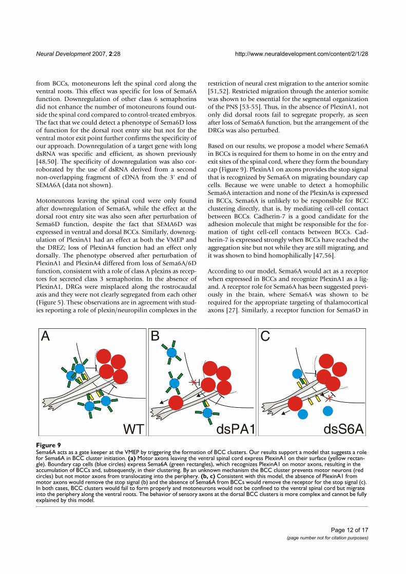
Neural Development 2007, 2:28 http://www.neuraldevelopment.com/content/2/1/28
Page 12 of 17(page number not for citation purposes)
from BCCs, motoneurons left the spinal cord along theventral roots. This effect was specific for loss of Sema6Afunction. Downregulation of other class 6 semaphorinsdid not enhance the number of motoneurons found out-side the spinal cord compared to control-treated embryos.The fact that we could detect a phenotype of Sema6D lossof function for the dorsal root entry site but not for theventral motor exit point further confirms the specificity ofour approach. Downregulation of a target gene with longdsRNA was specific and efficient, as shown previously[48,50]. The specificity of downregulation was also cor-roborated by the use of dsRNA derived from a secondnon-overlapping fragment of cDNA from the 3' end ofSEMA6A (data not shown).
Motoneurons leaving the spinal cord were only foundafter downregulation of Sema6A, while the effect at thedorsal root entry site was also seen after perturbation ofSema6D function, despite the fact that SEMA6D wasexpressed in ventral and dorsal BCCs. Similarly, downreg-ulation of PlexinA1 had an effect at both the VMEP andthe DREZ; loss of PlexinA4 function had an effect onlydorsally. The phenotype observed after perturbation ofPlexinA1 and PlexinA4 differed from loss of Sema6A/6Dfunction, consistent with a role of class A plexins as recep-tors for secreted class 3 semaphorins. In the absence ofPlexinA1, DRGs were misplaced along the rostrocaudalaxis and they were not clearly segregated from each other(Figure 5). These observations are in agreement with stud-ies reporting a role of plexin/neuropilin complexes in the
restriction of neural crest migration to the anterior somite[51,52]. Restricted migration through the anterior somitewas shown to be essential for the segmental organizationof the PNS [53-55]. Thus, in the absence of PlexinA1, notonly did dorsal roots fail to segregate properly, as seenafter loss of Sema6A function, but the arrangement of theDRGs was also perturbed.
Based on our results, we propose a model where Sema6Ain BCCs is required for them to home in on the entry andexit sites of the spinal cord, where they form the boundarycap (Figure 9). PlexinA1 on axons provides the stop signalthat is recognized by Sema6A on migrating boundary capcells. Because we were unable to detect a homophilicSema6A interaction and none of the PlexinAs is expressedin BCCs, Sema6A is unlikely to be responsible for BCCclustering directly, that is, by mediating cell-cell contactbetween BCCs. Cadherin-7 is a good candidate for theadhesion molecule that might be responsible for the for-mation of tight cell-cell contacts between BCCs. Cad-herin-7 is expressed strongly when BCCs have reached theaggregation site but not while they are still migrating, andit was shown to bind homophilically [47,56].
According to our model, Sema6A would act as a receptorwhen expressed in BCCs and recognize PlexinA1 as a lig-and. A receptor role for Sema6A has been suggested previ-ously in the brain, where Sema6A was shown to berequired for the appropriate targeting of thalamocorticalaxons [27]. Similarly, a receptor function for Sema6D in
Sema6A acts as a gate keeper at the VMEP by triggering the formation of BCC clustersFigure 9Sema6A acts as a gate keeper at the VMEP by triggering the formation of BCC clusters. Our results support a model that suggests a role for Sema6A in BCC cluster initiation. (a) Motor axons leaving the ventral spinal cord express PlexinA1 on their surface (yellow rectan-gle). Boundary cap cells (blue circles) express Sema6A (green rectangles), which recognizes PlexinA1 on motor axons, resulting in the accumulation of BCCs and, subsequently, in their clustering. By an unknown mechanism the BCC cluster prevents motor neurons (red circles) but not motor axons from translocating into the periphery. (b, c) Consistent with this model, the absence of PlexinA1 from motor axons would remove the stop signal (b) and the absence of Sema6A from BCCs would remove the receptor for the stop signal (c). In both cases, BCC clusters would fail to form properly and motoneurons would not be confined to the ventral spinal cord but migrate into the periphery along the ventral roots. The behavior of sensory axons at the dorsal BCC clusters is more complex and cannot be fully explained by this model.

Neural Development 2007, 2:28 http://www.neuraldevelopment.com/content/2/1/28
Page 13 of 17(page number not for citation purposes)
neural crest cell migration in heart development wasdescribed [23,24]. A receptor function for class 6 sema-phorins is also consistent with structural features [26].
Our model is supported by the aberrant clustering ofBCCs in the absence of either Sema6A from BCCs or theabsence of PlexinA1 from motoneurons (Figure 7). In theabsence of Sema6A, BCCs fail to recognize the exit sitemarked by the first motor axons extending into theperiphery, the boundary cap fails to form correctly and, asa consequence, motoneurons are no longer confined tothe ventral spinal cord and migrate into the periphery fol-lowing their axons (Figure 9). The same effect is achievedwhen PlexinA1 is downregulated in motoneurons. In thiscase motor axons are unable to provide a stop signal formigrating BCCs. Similarly, the PlexinA1 stop signal can bemasked by expression of soluble Sema6A ectodomain orfull-length Sema6A in motoneurons. In both cases motoraxon-derived Sema6A would compete with Sema6A onthe surface of BCCs and result in the aberrant formationof BCC clusters.
In addition to its function as a stop signal for Sema6A-expressing BCCs, PlexinA1 serves as a co-receptor togetherwith neuropilins for class 3 semaphorins. Sema3A waspostulated to act as a surround repellent and, thus, topolarize growth of sensory axons during initial stages ofdevelopment [57]. Later, class 3 semaphorins were shownto interfere with motor and sensory axon pathfinding[2,58-64]. Their effects were mediated by binding to eitherNeuropilin-1 or Neuropilin-2 associated with one of theclass A plexins as the signal transducing part of the recep-tor.
Chicken embryos express only three PlexinAs, as the geneencoding PlexinA3 is missing from the chicken genome[20]. Similarly, chickens express only three class 6 sema-phorins; an ortholog of Sema6C is not found. Therefore,a direct comparison of PlexinA/Sema6 interactionsbetween mouse/human and chicken proteins is not possi-ble. This may explain why, so far, a direct interactionbetween Sema6A and PlexinA1 has not been demon-strated [25]. The repulsive activity of Sema6A was foundto be mediated by PlexinA4 [22,65]. In our in vivo assaysPlexinA4 had an effect only at the dorsal root entry butnot at the ventral motor axon exit site. In our in vitro assay,sensory but not motor axons were repelled by Sema6A,despite the fact that all PlexinAs were expressed by sensoryand motor neurons [20]. Sema6D was expressed in bothdorsal and ventral BCCs but had an effect only at the dor-sal root entry zone.
Future experiments will have to elucidate the differencebetween Sema6A and Sema6D in BCCs and, thus, theirdifferent roles in gate keeping between the PNS and the
CNS. Obviously, the mechanism differs between the ven-tral and the dorsal transition zone. Motor axons were notrepelled by Sema6A but sensory axons were (Figure 6 andTable 1). The reason for this discrepancy is unknown.
ConclusionSema6A expression by BCCs acts as a gate keeper betweenthe PNS and the CNS by organizing the segregation ofdorsal root entry and ventral motor axon exit sites. In bothcases Sema6A on BCCs appears to act as a receptor recog-nizing the stop signal provided by PlexinA1 on axons. Asa consequence, BCCs aggregate at the dorsal root entry siteand the VMEP. BCCs then form clusters, possibly medi-ated by Cadherin-7, resulting in a tight barrier that pre-vents motor neurons from streaming out of the ventralspinal cord along the ventral roots. At the dorsal root entrysite the BCCs segregate and organize dorsal roots. Consist-ent with these observations, Sema6A was found to be arepellent for sensory but not for motor axons.
Materials and methodsCloning of the chicken SEMA6A cDNAA 728 base-pair fragment of chicken SEMAPHORIN6Aobtained in a screen for axon guidance cues [50] was usedto screen a � ZAP library prepared from E14 chickenbrains [66]. Two fragments encoding the entire open read-ing frame (ORF) were ligated and cloned into pBluescript.For the preparation of in situ probes and dsRNA we usedmainly a fragment spanning the 5' untranslated regionand the first 300 base-pairs from the ORF. In addition, weverified the specificity of the phenotype using a fragmentfrom the 3' untranslated region. The alignment of thesefragments with SEMA6B and SEMA6D did not result inany significant similarity.
To obtain a soluble AP-tagged ectodomain of Sema6A, thesequence corresponding to the ectodomain of chickenSema6A (amino acids 1–604) was amplified and insertedinto the APtag-2 vector [67]. COS7 cells were transientlytransfected with the Sema6A ectodomain-containing plas-mid using Lipofectamine 2000 (Invitrogen, Carlsbad,CA). After transfection cells were washed with phosphate-buffered saline (PBS) and grown for 4 days in MEM and1% fetal calf serum. The supernatant was collected andcentrifuged as described in [68]. Binding of AP-taggedSema6A to cryosections was carried out as described in[69]. Full-length Sema6A with a myc tag was expressedunder the control of the �-actin promoter. The plasmidwas injected at a concentration of 1 �g/�l into the centralcanal of the spinal cord of E2.5 embryos followed by elec-troporation of the ventral spinal cord. The embryos weresacrificed at E5 and analyzed.

Neural Development 2007, 2:28 http://www.neuraldevelopment.com/content/2/1/28
Page 14 of 17(page number not for citation purposes)
Preparation of in situ probes and dsRNAProbes for in situ hybridization and dsRNA were producedfrom expressed sequence tags (ESTs) obtained from Gen-eservice Ltd [70]. The ESTs used were: ChEST225N10(SEMAPHORIN6D), ChEST53D13 and 666O16(PLEXINA1), ChEST128L21 and 297D11 (PLEXINA2),ChEST1014M19 and 202O14 (PLEXINA4). ForSEMAPHORIN6A the cDNA fragment mentioned abovewas used. For SEMA6B a cDNA fragment was cloned usingRT-PCR because no ESTs were available. Total RNA wasprepared from HH30 (stage 30 chicken embryos accord-ing to Hamburger and Hamilton [42]) spinal cords. Ran-dom and oligo dT-primed first-strand cDNAs weregenerated using Superscript II reverse transcriptase accord-ing to the manufacturer's instructions (Invitrogen). A 656base-pair fragment for SEMA6B was amplified using theantisense primer 5'-CCCATGTCGTTCTTGCAC-3' and thesense primer 5'-ATCCAGCGCATCCTCAAG-3'. The result-ing PCR fragments were cloned into the TOPO TA cloningvector (Invitrogen) using EcoRI restriction sites (Gemayelet al., in preparation). We carefully compared and selectedsequences to avoid overlapping stretches that couldpotentially interfere with RNAi specificity. In fact, off-tar-get effects or unspecific knock-down of related familymembers were never detected in our approach with longdsRNA, most likely because the concentration of eachsmall interfering RNA produced in a given cell by Dicer isextremely low, with a theoretical maximal concentrationof about 1 nM or less [48,50].
In situ probes for the detection of KROX20 and SOX10mRNA were derived from ESTs 738N7 and 477F10,respectively. Plasmid DNA was linearized using restrictionenzymes NotI, EcoRI, XbaI, HindIII, or Asp718 (all fromRoche, Basel, Switzerland) to prepare either digoxigenin-labeled in situ probes [20] or dsRNA [48] by in vitro tran-scription as described previously.
In ovo RNAiIn ovo RNAi was used to knock down genes of interest asdescribed previously [48]. In brief, fertilized eggs werewindowed on the second day of incubation to get accessto the embryo. Embryos were staged according to Ham-burger and Hamilton [42] at the time of injection. A solu-tion containing the dsRNA (200–300 ng/�l) and aplasmid encoding EGFP under the control of the �-actinpromoter (50 ng/�l) was injected into the central canal ofthe spinal cord of HH12-14 embryos to efficiently trans-fect neural crest cells and motoneurons [71]. The lum-bosacral region of the spinal cord was electroporated with5 pulses of 18 Volts and 50 ms length with a 1 s interpulseinterval. Eggs were sealed and put back into the incubatoruntil embryos reached the desired stage. Embryos weresacrificed at HH25 for the analysis of motoneurons and atHH25/26 for the analysis of dorsal roots.
Tissue preparationFor analysis of phenotypes, embryos were sacrificed, evis-cerated and fixed in 4% paraformaldehyde in PBS for 60'to 120' depending on the age. Embryos were rinsed in PBSand subjected to cryoprotection or used directly forwhole-mount staining as detailed below. For immunohis-tochemistry and in situ hybridization, the cryoprotectedtissue was frozen in isopentane on dry ice and cut into 25�m thick sections. In situ hybridization was carried out asdetailed previously [20]. For immunohistochemistry, thestaining protocol described earlier [72] was used. Anti-bodies were diluted in blocking buffer (10% fetal calfserum in PBS). For permeabilization of the tissue, sectionswere incubated for 1' in 0.1% Triton-X-100. The antibod-ies used were: monoclonal antibodies 1E8 recognizing P0,40.2D6 recognizing Isl-1, and 9E10 recognizing the myctag (all from the Developmental Studies HybridomaBank, University of Iowa, Iowa City, IA) Furthermore, weused rabbit anti-neurofilament (Millipore, Billerica, MA),and a FITC-labeled goat anti-GFP antibody (Rockland,Gilbertsville, PA). Secondary antibodies were: goat anti-mouse IgG Cy3 (Jackson ImmunoResearch Newmarket,Suffolk, UK), goat anti-rabbit Alexa350, and goat anti-rab-bit Alexa488 (both Invitrogen/Molecular Probes,Carlsbad, CA).
Neurofilament staining of whole-mount embryosFor whole-mount staining, embryos were sacrificed atHH25/26, fixed as described above and transferred to 24-well plates. Tissue was permeabilized in 1% Triton/PBSfor 1 h at room temperature, rinsed in PBS, and incubatedin 20 mM lysine in 0.1 M sodium phosphate (pH 7.3) foranother hour. After rinsing thoroughly in PBS, embryoswere incubated in blocking buffer (10% fetal calf serum inPBS) for at least two hours before the anti-neurofilamentantibody (RMO270 from Zymed/Invitrogen, Carlsbad,CA, diluted 1:1,500) was added for 48 h at 4°C. Incuba-tion with the secondary antibody (goat anti-mouse IgGCy3, 1:250) was for 12 h. EGFP was visualized with aFITC-labeled goat anti-GFP antibody. Embryos wererinsed thoroughly and dehydrated in a graded series ofmethanol before transfer to benzyl benzoate/benzyl alco-hol (2:1).
Quantification of the phenotypesExperimental embryos and control-treated embryos thatwere injected and electroporated with the plasmid encod-ing EGFP only were sacrificed at HH25/26. Tissue prepa-ration, cutting and staining was as detailed above. Fromeach embryo lumbosacral sections were analyzed by anobserver who was blind to the treatment group. Sectionswere classified into groups containing either 0–1, or morethan one Isl-1-positive cell along the root. All sections thatcontained EGFP and the ventral roots were analyzed andscored. The percentage of sections per embryo containing
Neural Development 2007, 2:28 http://www.neuraldevelopment.com/content/2/1/28
Page 15 of 17(page number not for citation purposes)
more than one motoneuron outside the spinal cord wascalculated.
For the analysis of the phenotype at the dorsal root entrysite, embryos were sacrificed at HH25/26 and stained withRMO270 and goat anti-mouse IgG Cy3 as whole-mountsas detailed above. For the analysis of the segregation ofDRGs and dorsal roots, a dissection microscope equippedwith fluorescence optics (Olympus SZX12) was used. Sin-gle fibers crossing to the adjacent DRG or irregular spacingwas considered a weak phenotype. When roots wereformed by sensory axons emanating from two DRGs orwhen the DRGs were fused, the embryo was scored as hav-ing a severe phenotype. For statistical analysis, we usedtwo-way ANOVA with Bonferroni correction. Values rep-resent mean ± standard error of the mean.
In vitro assayCOS7 cells grown on 8-well LabTek slides were trans-fected with pcDNA3.1 vectors containing myc-taggedSEMA6A, myc-tagged AXONIN-1, or farnesylated EGFP(Invitrogen) as a control using Lipofectamine 2000 (Inv-itrogen). Sensory and sympathetic ganglia were dissectedfrom HH26 or HH35 embryos. Motoneurons wereobtained from the ventral halves of spinal cords dissectedfrom HH26-28 embryos. Single-cell suspensions wereobtained by digestion of ganglia and ventral spinal cordjunks with trypsin followed by trituration. Per well,25,000 DRG or sympathetic neurons or twice as manymotoneurons were plated. DRG and sympathetic neuronswere cultured in serum-free medium containing 20 ng/mlnerve growth factor (NGF) (see [73] for details). Motone-urons were cultured in MEM containing 5% fetal calfserum, N3, and 1 mM sodium pyruvate. Neurons weregrown on transfected COS cells for one (DRG, sympa-thetic neurons) or two days (motoneurons) before fixa-tion in 4% paraformaldehyde for 30 minutes at roomtemperature and staining with the 9E10 antibody (Devel-opmental Studies Hybridoma Bank) to detect successfullytransfected cells and rabbit anti-neurofilament to stainaxons.
Cultures were analyzed and the behavior of axonsencountering a transfected COS cell was classified asavoidance if an axon stopped or turned away from a trans-fected cell, or as attraction if an axon failed to leave thesurface of a transfected cell. We chose Axonin-1 as a con-trol protein because it was shown to promote axon out-growth of sensory neurons [73,74]. In addition, Axonin-1was shown to be required for pathfinding of nociceptiveafferents [75] and axons of dorsolateral commissural neu-rons [48,76] but not for extension of commissural axons[77].
Competing interestsThe author(s) declare that they have no competing inter-ests.
Authors' contributionsOM, ED, IA, RS, and ES carried out experiments and ana-lyzed the data. OM prepared the figures. ES conceived thestudy and wrote the manuscript.
Additional material
AcknowledgementsWe thank Maja Hess for excellent technical assistance, members of the lab for suggestions and reading the manuscript. This work was supported by the Swiss National Science Foundation and the NCCR Brain Plasticity and Repair.
References1. Dickson BJ: Molecular mechanisms of axon guidance. Science
2002, 298:1959-1964.2. Huber AB, Kolodkin AL, Ginty DD, Cloutier JF: Signaling at the
growth cone: ligand-receptor complexes and the control ofaxon growth and guidance. Annu Rev Neurosci 2003, 26:509-563.
3. Rougon G, Hobert O: New insights into the diversity and func-tion of neuronal immunoglobulin superfamily molecules.Annu Rev Neurosci 2003, 26:207-238.
4. Egea J, Klein R: Bidirectional Eph-ephrin signaling during axonguidance. Trends Cell Biol 2007, 17:230-238.
5. Baker KA, Moore SW, Jarjour AA, Kennedy TE: When a diffusibleaxon guidance cue stops diffusing: roles for netrins in adhe-sion and morphogenesis. Curr Opin Neurobiol 2006, 16:529-534.
6. Cirulli V, Yebra M: Netrins: beyond the brain. Nat Rev Mol Cell Biol2007, 8:296-306.
7. Yazdani U, Terman JR: The semaphorins. Genome Biol 2006, 7:211.
Additional file 1Downregulation of the targeted PlexinA was specific. For each PlexinA we used two different cDNA fragments to produce long dsRNAs for gene silencing (see Materials and methods for details). As shown qualitatively by in situ hybridization (a-c, e-g, i-k) and quantitatively by analyzing intensity levels for the three PlexinAs with ImageJ 1.38× (m) downregu-lation of the targeted PlexinA was specific. Downregulation of PLEXINA1 (PA1; a-d) resulted in a reduction of PA1 expression in motoneurons (arrowhead in (a)). Pattern and expression levels of PA2 (b) and PA4 (c) were not changed. EGFP expression from a co-injected plasmid indicates the electroporated half of the spinal cord (d). Similarly, silencing of PA2 (e-h) resulted in changes of the PA2 expression pattern in motoneurons (arrow in (f)) on the electroporated side (see (h)) but the expression of PA1 (e) and PA4 (g) were not altered. Targeting PA4, which is very weak at HH25, further reduced expression of PA4 in motoneurons (arrowhead in (k)) on the electroporated side (l) but had no effect on the expression of PA1 (i) and PA2 (j). The quantification of the signal intensity is shown in (m). Similar levels were obtained for all PlexinAs. Three or four differ-ent embryos were analyzed per condition using at least ten sections. Spe-cific downregulation was 24.9 ± 5.7% for PA1, 26.8 ± 5.5% for PA2, and 25.9 ± 2.1% for PA4. Values are shown ± standard deviation. P < 0.0001 indicated by three asterisks. Note that the section shown in (f) was taken from the thoracic level. AS, antisense probe.Click here for file[http://www.biomedcentral.com/content/supplementary/1749-8104-2-28-S1.jpeg]

Neural Development 2007, 2:28 http://www.neuraldevelopment.com/content/2/1/28
Page 16 of 17(page number not for citation purposes)
8. Tran TS, Kolodkin AL, Bharadwaj R: Semaphorin regulation ofcellular morphology. Annu Rev Cell Dev Biol 2007, 23:263-292.
9. Negishi M, Oinuma I, Katoh H: Plexins: axon guidance and signaltransduction. Cell Mol Life Sci 2005, 62:1363-1371.
10. Fujisawa H: Discovery of semaphorin receptors, neuropilinand plexin, and their functions in neural development. J Neu-robiol 2004, 59:24-33.
11. Bovolenta P: Morphogen signaling at the vertebrate growthcone: A few cases or a general strategy? J Neurobiol 2005,64:405-416.
12. Stoeckli ET: Longitudinal axon guidance. Curr Opin Neurobiol2006, 16:35-39.
13. Zou Y, Lyuksyutova AI: Morphogens as conserved axon guid-ance cues. Curr Opin Neurobiol 2007, 17:22-28.
14. de Wit J, Verhaagen J: Role of semaphorins in the adult nervoussystem. Prog Neurobiol 2003, 71:249-267.
15. Fiore R, Puschel AW: The function of semaphorins during nerv-ous system development. Front Biosci 2003, 8:s484-499.
16. Gu C, Yoshida Y, Livet J, Reimert DV, Mann F, Merte J, HendersonCE, Jessell TM, Kolodkin AL, Ginty DD: Semaphorin 3E andplexin-D1 control vascular pattern independently ofneuropilins. Science 2005, 307:265-268.
17. Worzfeld T, Puschel AW, Offermanns S, Kuner R: Plexin-B familymembers demonstrate non-redundant expression patternsin the developing mouse nervous system: an anatomicalbasis for morphogenetic effects of Sema4D during develop-ment. Eur J Neurosci 2004, 19:2622-2632.
18. Cohen S, Funkelstein L, Livet J, Rougon G, Henderson CE, CastellaniV, Mann F: A semaphorin code defines subpopulations of spi-nal motor neurons during mouse development. Eur J Neurosci2005, 21:1767-1776.
19. Perala NM, Immonen T, Sariola H: The expression of plexins dur-ing mouse embryogenesis. Gene Expr Patterns 2005, 5:355-362.
20. Mauti O, Sadhu R, Gemayel J, Gesemann M, Stoeckli ET: Expressionpatterns of plexins and neuropilins are consistent with coop-erative and separate functions during neural development.BMC Dev Biol 2006, 6:32.
21. Kruger RP, Aurandt J, Guan KL: Semaphorins command cells tomove. Nat Rev Mol Cell Biol 2005, 6:789-800.
22. Suto F, Ito K, Uemura M, Shimizu M, Shinkawa Y, Sanbo M, ShinodaT, Tsuboi M, Takashima S, Yagi T, et al.: Plexin-a4 mediates axon-repulsive activities of both secreted and transmembranesemaphorins and plays roles in nerve fiber guidance. J Neuro-sci 2005, 25:3628-3637.
23. Toyofuku T, Zhang H, Kumanogoh A, Takegahara N, Suto F, Kamei J,Aoki K, Yabuki M, Hori M, Fujisawa H, Kikutani H: Dual roles ofSema6D in cardiac morphogenesis through region-specificassociation of its receptor, Plexin-A1, with off-track and vas-cular endothelial growth factor receptor type 2. Genes Dev2004, 18:435-447.
24. Toyofuku T, Zhang H, Kumanogoh A, Takegahara N, Yabuki M,Harada K, Hori M, Kikutani H: Guidance of myocardial pattern-ing in cardiac development by Sema6D reverse signalling.Nat Cell Biol 2004, 6:1204-1211.
25. Yoshida Y, Han B, Mendelsohn M, Jessell TM: PlexinA1 signalingdirects the segregation of proprioceptive sensory axons inthe developing spinal cord. Neuron 2006, 52:775-788.
26. Klostermann A, Lutz B, Gertler F, Behl C: The orthologous humanand murine semaphorin 6A-1 proteins (SEMA6A-1/Sema6A-1) bind to the enabled/vasodilator-stimulated phosphopro-tein-like protein (EVL) via a novel carboxyl-terminal zyxin-like domain. J Biol Chem 2000, 275:39647-39653.
27. Leighton PA, Mitchell KJ, Goodrich LV, Lu X, Pinson K, Scherz P,Skarnes WC, Tessier-Lavigne M: Defining brain wiring patternsand mechanisms through gene trapping in mice. Nature 2001,410:174-179.
28. Godenschwege TA, Hu H, Shan-Crofts X, Goodman CS, MurpheyRK: Bi-directional signaling by Semaphorin 1a during centralsynapse formation in Drosophila. Nat Neurosci 2002,5:1294-1301.
29. Yu HH, Araj HH, Ralls SA, Kolodkin AL: The transmembraneSemaphorin Sema I is required in Drosophila for embryonicmotor and CNS axon guidance. Neuron 1998, 20:207-220.
30. Sweeney LB, Couto A, Chou YH, Berdnik D, Dickson BJ, Luo L,Komiyama T: Temporal target restriction of olfactory recep-
tor neurons by Semaphorin-1a/PlexinA-mediated axon-axoninteractions. Neuron 2007, 53:185-200.
31. Wong JT, Wong ST, O'Connor TP: Ectopic semaphorin-1a func-tions as an attractive guidance cue for developing peripheralneurons. Nat Neurosci 1999, 2:798-803.
32. Cafferty P, Yu L, Long H, Rao Y: Semaphorin-1a functions as aguidance receptor in the Drosophila visual system. J Neurosci2006, 26:3999-4003.
33. Kerjan G, Dolan J, Haumaitre C, Schneider-Maunoury S, Fujisawa H,Mitchell KJ, Chedotal A: The transmembrane semaphorinSema6A controls cerebellar granule cell migration. Nat Neu-rosci 2005, 8:1516-1524.
34. Xu XM, Fisher DA, Zhou L, White FA, Ng S, Snider WD, Luo Y: Thetransmembrane protein semaphorin 6A repels embryonicsympathetic axons. J Neurosci 2000, 20:2638-2648.
35. Burgaya F, Fontana X, Martinez A, Montolio M, Mingorance A, SimoS, del Rio JA, Soriano E: Semaphorin 6C leads to GSK-3-depend-ent growth cone collapse and redistributes after entorhino-hippocampal axotomy. Mol Cell Neurosci 2006, 33:321-334.
36. Golding JP, Cohen J: Border controls at the mammalian spinalcord: late-surviving neural crest boundary cap cells at dorsalroot entry sites may regulate sensory afferent ingrowth andentry zone morphogenesis. Mol Cell Neurosci 1997, 9:381-396.
37. Vermeren M, Maro GS, Bron R, McGonnell IM, Charnay P, Topilko P,Cohen J: Integrity of developing spinal motor columns is reg-ulated by neural crest derivatives at motor exit points. Neu-ron 2003, 37:403-415.
38. Niederlander C, Lumsden A: Late emigrating neural crest cellsmigrate specifically to the exit points of cranial branchiomo-tor nerves. Development 1996, 122:2367-2374.
39. Maro GS, Vermeren M, Voiculescu O, Melton L, Cohen J, Charnay P,Topilko P: Neural crest boundary cap cells constitute a sourceof neuronal and glial cells of the PNS. Nat Neurosci 2004,7:930-938.
40. Hjerling-Leffler J, Marmigere F, Heglind M, Cederberg A, KoltzenburgM, Enerback S, Ernfors P: The boundary cap: a source of neuralcrest stem cells that generate multiple sensory neuron sub-types. Development 2005, 132:2623-2632.
41. Marmigere F, Ernfors P: Specification and connectivity of neuro-nal subtypes in the sensory lineage. Nat Rev Neurosci 2007,8:114-127.
42. Hamburger V, Hamilton HL: A series of normal stages in thedevelopment of the chick embryo. J Morphol 1951, 88:49-92.
43. Wilkinson DG, Bhatt S, Chavrier P, Bravo R, Charnay P: Segment-specific expression of a zinc-finger gene in the developingnervous system of the mouse. Nature 1989, 337:461-464.
44. Bhattacharyya A, Brackenbury R, Ratner N: Axons arrest themigration of Schwann cell precursors. Development 1994,120:1411-1420.
45. Cheng Y, Cheung M, Abu-Elmagd MM, Orme A, Scotting PJ: Chicksox10, a transcription factor expressed in both early neuralcrest cells and central nervous system. Brain Res Dev Brain Res2000, 121:233-241.
46. Bhattacharyya A, Frank E, Ratner N, Brackenbury R: P0 is an earlymarker of the Schwann cell lineage in chickens. Neuron 1991,7:831-844.
47. Nakagawa S, Takeichi M: Neural crest cell-cell adhesion control-led by sequential and subpopulation-specific expression ofnovel cadherins. Development 1995, 121:1321-1332.
48. Pekarik V, Bourikas D, Miglino N, Joset P, Preiswerk S, Stoeckli ET:Screening for gene function in chicken embryo using RNAiand electroporation. Nat Biotechnol 2003, 21:93-96.
49. Golding J, Shewan D, Cohen J: Maturation of the mammaliandorsal root entry zone – from entry to no entry. Trends Neu-rosci 1997, 20:303-308.
50. Bourikas D, Pekarik V, Baeriswyl T, Grunditz A, Sadhu R, Nardo M,Stoeckli ET: Sonic hedgehog guides commissural axons alongthe longitudinal axis of the spinal cord. Nat Neurosci 2005,8:297-304.
51. Eickholt BJ, Mackenzie SL, Graham A, Walsh FS, Doherty P: Evi-dence for collapsin-1 functioning in the control of neuralcrest migration in both trunk and hindbrain regions. Develop-ment 1999, 126:2181-2189.
52. Gammill LS, Gonzalez C, Gu C, Bronner-Fraser M: Guidance oftrunk neural crest migration requires neuropilin 2/sema-phorin 3F signaling. Development 2006, 133:99-106.

Publish with BioMed Central and every scientist can read your work free of charge
"BioMed Central will be the most significant development for disseminating the results of biomedical research in our lifetime."
Sir Paul Nurse, Cancer Research UK
Your research papers will be:
available free of charge to the entire biomedical community
peer reviewed and published immediately upon acceptance
cited in PubMed and archived on PubMed Central
yours — you keep the copyright
Submit your manuscript here:http://www.biomedcentral.com/info/publishing_adv.asp
BioMedcentral
Neural Development 2007, 2:28 http://www.neuraldevelopment.com/content/2/1/28
Page 17 of 17(page number not for citation purposes)
53. Krull CE: Segmental organization of neural crest migration.Mech Dev 2001, 105:37-45.
54. Kuan CY, Tannahill D, Cook GM, Keynes RJ: Somite polarity andsegmental patterning of the peripheral nervous system.Mech Dev 2004, 121:1055-1068.
55. Kasemeier-Kulesa JC, Kulesa PM, Lefcort F: Imaging neural crestcell dynamics during formation of dorsal root ganglia andsympathetic ganglia. Development 2005, 132:235-245.
56. Nakagawa S, Takeichi M: Neural crest emigration from the neu-ral tube depends on regulated cadherin expression. Develop-ment 1998, 125:2963-2971.
57. Masuda T, Shiga T: Chemorepulsion and cell adhesion mole-cules in patterning initial trajectories of sensory axons. Neu-rosci Res 2005, 51:337-347.
58. Chen H, Bagri A, Zupicich JA, Zou Y, Stoeckli E, Pleasure SJ, Lowen-stein DH, Skarnes WC, Chedotal A, Tessier-Lavigne M: Neuropilin-2 regulates the development of selective cranial and sensorynerves and hippocampal mossy fiber projections. Neuron2000, 25:43-56.
59. Giger RJ, Cloutier JF, Sahay A, Prinjha RK, Levengood DV, Moore SE,Pickering S, Simmons D, Rastan S, Walsh FS, et al.: Neuropilin-2 isrequired in vivo for selective axon guidance responses tosecreted semaphorins. Neuron 2000, 25:29-41.
60. Kitsukawa T, Shimizu M, Sanbo M, Hirata T, Taniguchi M, Bekku Y,Yagi T, Fujisawa H: Neuropilin-semaphorin III/D-mediatedchemorepulsive signals play a crucial role in peripheral nerveprojection in mice. Neuron 1997, 19:995-1005.
61. Taniguchi M, Yuasa S, Fujisawa H, Naruse I, Saga S, Mishina M, Yagi T:Disruption of semaphorin III/D gene causes severe abnor-mality in peripheral nerve projection. Neuron 1997,19:519-530.
62. Varela-Echavarria A, Tucker A, Puschel AW, Guthrie S: Motor axonsubpopulations respond differentially to the chemorepel-lents netrin-1 and semaphorin D. Neuron 1997, 18:193-207.
63. Zou Y, Stoeckli E, Chen H, Tessier-Lavigne M: Squeezing axonsout of the gray matter: a role for slit and semaphorin pro-teins from midline and ventral spinal cord. Cell 2000,102:363-375.
64. Yaron A, Huang PH, Cheng HJ, Tessier-Lavigne M: Differentialrequirement for Plexin-A3 and -A4 in mediating responsesof sensory and sympathetic neurons to distinct class 3 Sem-aphorins. Neuron 2005, 45:513-523.
65. Suto F, Tsuboi M, Kamiya H, Mizuno H, Kiyama Y, Komai S, ShimizuM, Sanbo M, Yagi T, Hiromi Y, Chédotal A, et al.: Interactionsbetween Plexin-A2, Plexin-A4, and Semaphorin 6A controllamina-restricted projection of hippocampal mossy fibers.Neuron 2007, 53:535-547.
66. Zuellig RA, Rader C, Schroeder A, Kalousek MB, Von Bohlen und Hal-bach F, Osterwalder T, Inan C, Stoeckli ET, Affolter HU, Fritz A, etal.: The axonally secreted cell adhesion molecule, axonin-1.Primary structure, immunoglobulin-like and fibronectin-type-III-like domains and glycosyl-phosphatidylinositolanchorage. Eur J Biochem 1992, 204:453-463.
67. Flanagan JG, Cheng HJ, Feldheim DA, Hattori M, Lu Q, Vander-haeghen P: Alkaline phosphatase fusions of ligands or recep-tors as in situ probes for staining of cells, tissues, andembryos. Methods Enzymol 2000, 327:19-35.
68. Flanagan JG, Leder P: The kit ligand: a cell surface moleculealtered in steel mutant fibroblasts. Cell 1990, 63:185-194.
69. Brennan C, Fabes J: Alkaline phosphatase fusion proteins asaffinity probes for protein localization studies. Sci STKE 2003,2003:PL2.
70. Geneservice Ltd [http://www.geneservice.co.uk/]71. Bourikas D, Baeriswyl T, Sadhu R, Stoeckli ET: In ovo RNAi opens
new possibilities for functional genomics in vertebrates. InRNA Interference Technology – From Basic Research to Drug DevelopmentEdited by: Appasani K. Cambridge UK: Cambridge University Press;2005:220-232.
72. Perrin FE, Stoeckli ET: Use of lipophilic dyes in studies of axonalpathfinding in vivo. Microsc Res Tech 2000, 48:25-31.
73. Stoeckli ET, Kuhn TB, Duc CO, Ruegg MA, Sonderegger P: Theaxonally secreted protein axonin-1 is a potent substratumfor neurite growth. J Cell Biol 1991, 112:449-455.
74. Stoeckli ET, Ziegler U, Bleiker AJ, Groscurth P, Sonderegger P: Clus-tering and functional cooperation of Ng-CAM and axonin-1
in the substratum-contact area of growth cones. Dev Biol1996, 177:15-29.
75. Perrin FE, Rathjen FG, Stoeckli ET: Distinct subpopulations ofsensory afferents require F11 or axonin-1 for growth to theirtarget layers within the spinal cord of the chick. Neuron 2001,30:707-723.
76. Stoeckli ET, Landmesser LT: Axonin-1, Nr-CAM, and Ng-CAMplay different roles in the in vivo guidance of chick commis-sural neurons. Neuron 1995, 14:1165-1179.
77. Fitzli D, Stoeckli ET, Kunz S, Siribour K, Rader C, Kunz B, Kozlov SV,Buchstaller A, Lane RP, Suter DM, et al.: A direct interaction ofaxonin-1 with NgCAM-related cell adhesion molecule(NrCAM) results in guidance, but not growth of commis-sural axons. J Cell Biol 2000, 149:951-968.

- 60 -
Supplementary Figure 1

- 61 -
6. Additional Materials and Methods
Temporal Control of Gene Silencing by in ovo Electroporation
Thomas Baeriswyl*, Olivier Mauti*, and Esther T. Stoeckli
(*equal contribution)
Institute of Zoology, University of Zurich, Winterthurerstrasse 190, 8057 Zurich, Switzerland
Published in Methods in Molecular Biology, 2008, 442:231-244

16
Temporal Control of Gene Silencingby in ovo Electroporation
Thomas Baeriswyl∗, Olivier Mauti∗, and Esther T. Stoeckli
SummaryThe analysis of gene function during embryonic development asks for tight temporal
control of gene expression. Classic genetic tools do not allow for this, as the absence
of a gene during the early stages of development will preclude its functional analysis
during later stages. In contrast, RNAi technology allows one to achieve temporal control
of gene silencing especially when used with oviparous animal models. In contrast to
mammals, reptiles and birds are easily accessible during embryonic development. We
have developed approaches to use RNAi for the analysis of gene function during nervous
system development in the chicken embryo. Although the protocol given here describes a
method for gene silencing in the developing spinal cord, it can easily be adapted to other
parts of the developing nervous system. The combination of the easy accessibility of the
chicken embryo and RNAi provides a unique opportunity for temporal and spatial control
of gene silencing during development.
Key Words: in ovo RNAi; in ovo electroporation; long dsRNA; chicken embryo;
development; nervous system.
1. IntroductionNo matter whether you want to analyze the function of a number of candidate
genes that you identified in a screen or whether you want to assess the function
of your favorite gene, you may need an in vivo system that allows for the rapid
∗ These authors contributed equally.
From: Methods in Molecular Biology, vol. 442: RNAi: Design and ApplicationEdited by: S. Barik © Humana Press, Totowa, NJ
231

232 Baeriswyl et al.
detection of a possible phenotype during development. The analysis of gene
function during development requires tight temporal control of gene silencing.
Classic genetic tools will only allow for an assessment of gene function during
the initial phase of gene activity. Additional activities during later develop-
mental stages will not be within reach, as the lack of gene function during
the early stages will preclude the analysis of all subsequent stages. For this
reason, specific gene silencing by RNA interference (RNAi) provides an excep-
tional tool for loss-of-function approaches during development in vertebrates.
Until now, different RNAi strategies have been established for mouse, rat, and
chicken embryos (1–4; reviewed in 5–7). However, due to the limited acces-
sibility of mouse and rat embryos during development, RNAi in combination
with in utero electroporation is very difficult and requires special equipment
and expert training. Therefore, the use of mammals as a model organism for
developmental studies is limited. In contrast to mammals, the chicken is easily
accessible for in vivo manipulations during embryonic development. With the
establishment of in ovo electroporation, as an efficient method for nucleic acid
transfer, and in ovo RNAi, as a method for gene silencing, the chicken embryo
has been turned into a unique model organism for the efficient functional charac-
terization of genes involved in developmental processes (4,8–12; reviewed in
6). In ovo RNAi using long dsRNA, shRNA, or siRNA has been used for a
variety of functional studies in different parts of the CNS but also in other
embryonic tissues (4,13–17; reviewed in 5).Here we provide a detailed protocol for the silencing of a candidate gene
during early development of the spinal cord by in ovo RNAi. A particular
advantage of in ovo RNAi is the fact that long dsRNA can be used for the
induction of loss-of-function phenotypes. Unlike adult tissue or cell lines,
embryos do not respond to long dsRNA with unspecific inhibition of protein
synthesis and apoptosis (18,19). Therefore, there is no need for lengthy selectionof an efficient siRNA or shRNA. Any cDNA fragment or expressed sequence
tag (EST) can be used to produce dsRNA by in vitro transcription. Since the
chicken genome was fully sequenced in 2004, it can be directly compared with
the human, mouse, or rat genome, significantly facilitating the identification of
orthologs (20). Therefore, in ovo RNAi offers the possibility to study candidate
genes identified in other species using commercially available chicken ESTs
for the synthesis of the dsRNA.
In the protocol reported here, long dsRNA is injected into the central canal
of the developing spinal cord (Fig. 1a). Subsequently, the embryo is exposed to
an electric field for efficient transfection of selected cell populations (Fig. 1b).Depending on the time point and the position of the electrodes, different
neuronal populations within the spinal cord (Figs. 1c, 1d) but also of the
peripheral nervous system can be targeted. Furthermore, this method allows
for knockdown of several genes by injecting a mixture of different dsRNAs.

In ovo RNAi 233
Fig. 1. In ovo electroporation. (a) The chicken embryo is made directly accessible
through a window in the eggshell for the injection of nucleic acids into the central
canal of the spinal cord. (b) The electroporation permeabilizes the cell membrane and
therefore allows for the efficient uptake of RNA or DNA. Depending on the position of
the electrodes with respect to the embryonic body axis, different tissues can be targeted:
A parallel position of the electrode to the spinal cord results in a unilateral transfection
[checkered area in (c)]. Within the applied electric field, the injected nucleic acids
migrate toward the anode due to the negative charge of RNA and DNA. Therefore,
the untransfected side of the spinal cord [left side in (c)] can be used as an internal
control. The capillary should be kept at a 45° angle for injection. Placing the electrodes
over the dorsal (cathode) and the ventral (anode) midline of the spinal cord results in
efficient targeting of floor-plate cells [checkered area in (d)].
Thus, in ovo RNAi represents an efficient and inexpensive method to alter the
expression of specific genes in a temporally and spatially controlled manner.
2. Materials2.1. Preparation of dsRNA by in vitro Transcription
1. Heating block at 95 °C.
2. Equipment for gel electrophoresis.

234 Baeriswyl et al.
3. BamHI restriction endonuclease (10 U/μL; Roche, Basel, Switzerland).
4. SacI restriction endonuclease (10 U/μL; Roche).
5. RNasin (40 U/μL; Promega, Madison, WI).
6. SP6 and T7 RNA polymerases (15 U/μL; Promega).
7. RNase-free DNaseI (10 U/μL; Roche).
8. 5X transcription buffer (Promega).
9. 100 mM rNTPs (25 mM of each rNTP; Roche).
10. 100 mM DTT (Promega).
11. 0.5 M EDTA (pH 8.0).
12. 7.5 M ammonium acetate.
13. Phenol–chloroform–isoamylalcohol (25:24:1 vol/vol/vol; pH 7.6–8.0).
14. Acidic phenol–chloroform–isoamylalcohol (25:24:1 vol/vol/vol; pH 4.0).
15. Chloroform–isoamylalcohol (24:1 vol/vol).
16. 100% ethanol.
17. DEPC-treated double-distilled water (ddH2O) (1:1,000 vol/vol).
18. 70% ethanol in DEPC-treated ddH2O.
19. Phosphate-buffered saline (PBS, DEPC-treated, 1:1,000 vol/vol): 137 mM NaCl,
2.7 mM KCl (see Note 1), 8 mM Na2HPO4, 1.5 mM NaH2PO4 (pH 7.4).
20. RNaseZAP (Sigma-Aldrich, St. Louis, MO).
2.2. Windowing the Eggs
1. Fertilized Hisex eggs were obtained from a local hatchery.
2. Preincubator: 38–39 °C, 45% humidity (JUPPITER 576 SETTER+HATCHER;
F.I.E.M., Guanzate, Italy; see Note 2).3. Incubator: 38–39 °C, 45% humidity (Heraeus/Kendro Model B12, Kendro
Laboratory Products, Hanau, Germany; see Note 2).4. Egg-Lume Candler (Brinsea Products Ltd., Sandford, UK).
5. Heating plate, 80 °C, to melt paraffin.
6. Soldering iron.
7. Scalpel for drilling holes.
8. 10-mL syringe with needle (Sterican 100, Ø 18G, B. Braun Melsungen AG,
Melsungen, Germany).
9. Small scissors for cutting the eggshell (Fine Science Tools Inc., Foster City, CA).
10. Paraffin (Paraplast Tissue EmbeddingMedium, Oxford Labware, St. Louis, MO).
11. Coverslips, 24 × 24 mm (VWR International AG, Dietikon, Switzerland).
12. Kleenex.
13. Scotch tape.
14. 70% ethanol.
2.3. In ovo Injection and Electroporation
1. Spring scissors and forceps (Fine Science Tools Inc.).
2. Electroporator (Electro Square Porator ECM830, BTX Instrument Division,
Harvard Apparatus Inc., Holliston, MA; see Note 3).

In ovo RNAi 235
3. Platinum electrodes (4-mm length, 4 mm between anode and cathode; BTX
Instrument Division, Harvard Apparatus Inc.; see Note 3).4. Needle puller (PC-10, Narishige Co., Ltd., Tokyo, Japan).
5. Borosilicate glass capillaries (outer Ø/inner Ø: 1.2 mm/0.68 mm; World
Precision Instruments, Sarasota, FL).
6. Polyethylene tubing (Ø 1.24 mm).
7. 0.2-μm filter (Sarstedt, Sevelen, Switzerland).
8. Reporter plasmid: EGFP under the control of the chicken �-actin promoter.
9. Trypan Blue solution 0.4% (Invitrogen, Carlsbad, CA).
3. Methods3.1. Synthesis of Long dsRNA
A cDNA fragment of a candidate gene cloned into a standard plasmid
containing SP6 (or T3) and T7 promoters flanking the insert can be used for the
synthesis of long dsRNA by in vitro transcription. Here we synthesized dsRNA
from a 678-bp cDNA fragment (1,620–2,298 bp) encoding Axonin-1 cloned
in the pSP72 vector using SP6 and T7 promoters flanking the insert (seeNote 4).
1. Linearize 10 μg of the plasmid with 20 U BamHI and SacI restriction endonu-
clease, respectively, for 1 h at 37 °C.
2. For in vitro transcription, mix 2 μg of the linearized plasmid with 0.8 μL of 100
mM rNTPs, 0.5 μL of RNasin, 2 μL of SP6 or T7 RNA polymerase, 4 μL of 5X
transcription buffer, and 2 μL of 100 mM DTT, and add DEPC-treated ddH2O
to a total volume of 20 μL (see Note 5).3. Incubate the in vitro transcription mixture for 2 h at 37 °C (T7 RNA polymerase)
and 40 °C (SP6 RNA polymerase), respectively (see Note 6).4. Remove the DNA template from the in vitro transcription mixture by adding
2 μL RNase-free DNaseI, and incubate at 37 °C for 1 h (see Note 6).5. Add 20 μL of DEPC-treated ddH2O and mix well.
6. Add a mixture of 2 μL 0.5 M EDTA and 22 μL 7.5 M ammonium acetate. Mix
well.
7. Purify the synthesized ssRNA with 1 vol of acidic phenol–chloroform–
isoamylalcohol; subsequently, extract 1 vol of chloroform–isoamylalcohol.
8. Precipitate with 2.5 vol of 100% ethanol for at least 1 h at –80 °C.
9. Centrifuge for 30 min at 4 °C and 20,000 x g.
10. Wash the RNA pellet with 70% ethanol and spin down.
11. Air-dry the pellet.
12. Dissolve the ssRNA in 20 μL of DEPC-treated PBS (see Notes 6 and 7).13. Mix equal ng amounts of antisense and sense ssRNAs (see Note 8).14. Heat the mixture for 5 min at 95 °C, and allow for it to cool down slowly to
room temperature by switching off the heating block (see Note 6).15. Confirm the proper annealing by gel electrophoresis (see Note 6).16. Store the dsRNA at –80 °C until further use (see Note 17).

236 Baeriswyl et al.
3.2. Windowing the Eggs
For access to the embryo, the eggs are windowed on the third day of
incubation (Figs. 1a and 2).
1. Incubate the fertilized eggs in a preincubator at 39 °C (see Notes 9 and 10).2. After three days of incubation, place the egg on the side for at least 30 min
before opening, to allow the embryo to reposition on top of the yolk.
3. Mark the position of the embryo on the egg shell with a pencil using an Egg-
Lume Candler held against the blunt end of the egg.
4. Wipe the eggshell with 70% ethanol to avoid contamination.
5. Make small holes at the blunt end of the egg and at the corners, outlining the
planned window using a scalpel (Fig. 2a).6. Carefully remove 3 mL of albumin through the hole at the blunt end of the egg
using a syringe (Fig. 2a; see Note 11).7. Seal the hole at the blunt end and any possible cracks in the shell by applying
melted paraffin.
8. Put a piece of Scotch tape onto the shell to prevent small pieces of the eggshell
from falling into the egg (Fig. 2a).9. Cut the outlined window into the eggshell (see Note 12).
10. Seal the egg by applying melted paraffin to the edges of the window using a
brush and a coverslip (Fig. 2b; see Note 13).
Fig. 2. Windowing the egg. (a) Before egg windowing, the position of the embryo
is marked with a pencil on the shell. Subsequently, small holes are drilled at the blunt
end of the egg and at the corners outlining the window. Albumin (3 mL) is removed
through the hole at the blunt end (see Note 11). The syringe is kept at a 45º angle
to avoid damage to the embryo and the yolk. A piece of Scotch tape prevents pieces
of the shell from falling inside the egg while cutting the window with small scissors.
(b) For further incubation, the window is sealed with melted paraffin and a coverslip.

In ovo RNAi 237
11. Put the egg back in the incubator. Make sure that the position of the egg is the
same as before windowing to keep the embryo accessible through the window
(see Note 17).
3.3. In ovo Injection and Electroporation
1. Autoclave the tools and wipe the working space with 70% ethanol.
2. Reopen the sealed egg by pressing the soldering iron briefly onto the coverslip
and removing it carefully.
3. Stage the embryo according to Hamburger and Hamilton (21). Embryos should
be between stages 17 and 19.
4. Remove the extraembryonic membranes covering the embryo with forceps and
scissors in order to have direct access to the embryo (Fig. 3a).5. Break off the needle tip to obtain a tip diameter of approximately 5 μm.
6. Sterile PBS containing the dsRNA derived from AXONIN-1 (200–400 ng/μL)
and an EGFP reporter plasmid (20 ng/μL) with 0.04 % (vol/vol) Trypan Blue
are injected into the central canal of the spinal cord at the level of the hind limbs
using a glass capillary connected to a piece of tubing (Figs. 1c and 3b). Theinjection is controlled by mouth, and the maximal injection volume is achieved
when the blue dye reaches the brain vesicle (arrowhead in Fig. 3b).7. Add a few drops of PBS before electroporation to lower the electric resistance
and to prevent overheating of the embryo.
8. The electrodes are placed in a parallel manner along the anterior-posterior axis
of the spinal cord (Fig. 3c).9. Electroporate the embryo by applying five pulses of 26 V and 50-ms duration
each (see Note 14).10. After the electroporation, add a few drops of PBS to cool the embryo.
11. Rinse the electrodes with plenty of distilled water to remove denaturated proteins
from the surface (see Note 15).12. Reseal the egg with a glass coverslip and a soldering iron (see Note 13).13. For further incubation, the egg is returned to 39 °C until stage 25 is reached,
i.e., approximately two additional days of incubation (21) (see Note 17).
3.4. Analysis of the Phenotype and Electroporation Efficiency
For beginners we recommend assessing the efficiency and reproducibility
of the in ovo electroporation by analyzing EGFP expression directly in ovounder the stereomicroscope (Fig. 3d). Thus, embryos for further analyses can
be preselected (see Note 16).The efficiency as well as the specificity of gene silencing by in ovo RNAi
can be demonstrated by a variety of approaches. Immunohistochemistry on
cryostat sections (4,13,22) and Western blot analysis (23,24) are common ways
to show downregulation of the targeted protein. If antibodies against the targeted

238 Baeriswyl et al.
Fig. 3. Injection and electroporation of the embryo. (a) The extraembryonic
membranes covering the embryo are carefully removed before injection using forceps
and spring scissors. The injection mixture containing dsRNA derived from the gene of
interest, an EGFP plasmid for transfection control, and Trypan Blue is injected into the
central canal with a glass capillary. The maximal injection volume is achieved when
the Trypan Blue has reached the brain vesicle [arrowhead in (b)]. (c) After retraction of
the injection needle, the electrodes are placed in a parallel manner along the embryonic
axis. For stage 18 embryos, five pulses of 26 V with 50-ms duration are applied for
efficient transfection (see Note 14). (d) The successful transfection can be verified by
the expression of EGFP (indicated by arrowheads) under a stereomicroscope equipped
with fluorescence optics two days after electroporation.
protein are not available, a decrease in the mRNA can be assessed by in situhybridization using either whole-mount embryos (16,25) or cryostat sections(13). Alternatively, semi-quantitative RT-PCR can be used to detect a decrease
in the mRNA (26).Loss-of-function phenotypes can be analyzed in a variety of ways. To
study cell differentiation or cell migration, immunohistochemistry for known

In ovo RNAi 239
markers may be a good start (15). Changes in morphology and cell positions,
expression patterns, etc. can easily be detected. To visualize axonal trajec-
tories, staining and/or dye tracing in slices or whole-mount preparations are
used to account for their three-dimensionality. For an initial assessment and
to localize a specific phenotype within the peripheral nervous system, we
recommend a neurofilament staining of whole-mount preparations (24). Alter-natively or subsequently, mechanisms involved in wiring the nervous system
can be analyzed in vibratome slices by dye tracing or immunohistochemistry
(4,14). For example, we studied molecular mechanisms underlying the path-
finding behavior of commissural axons in the spinal cord using dye tracing in
open-book preparations (4,13,27).
4. Notes1. Different recipes exist for PBS, and the addition of KCl turned out to be crucial
for optimal survival rates.
2. The incubation time and developmental progress of the embryo are dependent
on the temperature and humidity. Any incubator set at a temperature of 38–39 °C
can be used, as long as high humidity (at least 45%) and good air circulation
can be achieved. The use of two incubators, one for preincubation and one
for treated embryos, is recommended to minimize contamination and to reduce
detrimental effects on the treated embryos due to frequent opening and closing
of the incubator.
3. Alternatively, any other electroporator that generates square wave pulses can be
used (for a comparison of the different electroporators that are commercially
available, see Ref. 8). For different target tissues, different electrodes have to
be chosen to get best results: For in ovo electroporations of the developing
spinal cord, we use wire electrodes (4,13). Commercially available electrodes
can be found at http://www.btxonline.com/products/electrodes/inovo. Alterna-
tively, for a widespread transfection, platelet electrodes can be used (28). For asmall transfection area, a needle electrode can be placed directly into the tissue
(17,29,30).4. In addition to long dsRNA, short interfering RNAs (siRNA) and short hairpin
RNAs (shRNA) have been applied successfully for RNAi in chicken embryos
(14–17,22,31). In contrast to siRNAs selected by various available algorithms,
long dsRNA always effectively silenced target genes in our hands. Long dsRNA
is processed by Dicer to give rise to a large number of siRNAs and therefore will
always produce many effective ones, making lengthy (and expensive) selection
processes unnecessary. Furthermore, long dsRNA can easily be produced by
in vitro transcription from a cDNA fragment or EST without further cloning steps
or expensive synthesis of siRNAs. Chicken ESTs can be obtained from Geneser-
vices Ltd. at http://www.geneservice.co.uk. To exclude any off-target effects

240 Baeriswyl et al.
(silencing of nontarget genes), we recommend using two different nonover-
lapping dsRNA fragments, as it is highly unlikely that they would both have
the same off-target effects. In contrast to mammalian cell lines and nonem-
bryonic tissue, long dsRNA can be applied to embryonic tissue without induction
of unspecific effects (4,18,19). No general inhibition of protein synthesis or
induction of apoptosis has been reported in mouse oocytes, embryo-derived cell
lines, and chicken embryos (4,15,32,33).5. For in vitro transcription, RNase-free tips, tubes, and DEPC-treated ddH2O have
to be used. Before starting, clean the workspace with RNaseZAP.
6. Collect 1-μL samples after each step and keep them to control the quality of
ssRNA and dsRNA. For this purpose, load the ssRNAs collected after each
step and the dsRNA on a 1% agarose gel. Lanes 1 and 2 (one sense and one
antisense) contain the linearized plasmids and the ssRNA synthesized by in vitrotranscription. Lanes 3/4 and 5/6 are sense and antisense ssRNA after DNaseI
treatment and purification, respectively. In lane 7 the resulting dsRNA after
annealing is loaded. When lanes 5 and 6 are compared to lane 7, the band
shift due to the difference in migration between ssRNA and dsRNA should be
detected. Furthermore, both ssRNA and dsRNA should give distinct bands. If a
smear indicating degradation of the RNA is obtained, the dsRNA should not be
used for in ovo RNAi.
7. Make sure that the pH and salt concentration of the buffer used to dissolve
the ssRNA are in the physiological range and do not have any effect on the
development and survival rate of the embryo. Do not use any buffers containing
Tris or glycerol.
8. The concentration of the dsRNA for in vivo injections should be in the range of
200–400 ng/μL.
9. Eggs should be stored at 15 °C for a maximum of one week before incubation.
When the eggs are stored for longer periods, normal development of the embryo
is unlikely.
10. To reach 45% humidity, it is usually sufficient to place a tray of distilled water
containing 0.1 g/L of copper sulfate at the bottom of the incubator. Copper
sulfate decreases the risk of contamination.
11. The holes at the corners are required to allow entry of air and detachment of the
embryo from the shell during removal of albumin. Insert the syringe at a steep
angle (about 45°; Fig. 2a) to avoid damage to the embryo and the yolk that is
not compatible with survival.
12. Keep the scissors as horizontally as possible to prevent any damage to the
embryo.
13. Make sure that the window is properly closed to prevent dehydration during
further incubation. Dehydration will cause the embryo’s death. If the paraffin
is cooled down too quickly, heat the coverslip briefly with the soldering iron
while pressing it down so that the coverslip seals properly along all the edges.
Alternatively, the window can be sealed with Scotch tape. Although sealing with
coverslips instead of tape is more time-consuming, it facilitates reopening the

In ovo RNAi 241
window and the development of the embryo can be directly observed through the
coverslip. The window can easily be reopened by brief heating of the coverslip
using a soldering iron.
14. The electroporation settings should be chosen according to the embryonic stage
(also see Ref. 12): For stage 18 embryos, 5 pulses of 26 V and 50 ms are
applied. The voltage should be adjusted to the embryonic stage and the tissue
that is electroporated:
Day of Incubation Embryonic Stage Target Tissue Settings
2 12–14∗ Spinal cord and neural crest 18 V
derivates (for example,
dorsal root ganglia)
3 18–20∗∗ Spinal cord and floor plate 26 V
* Red ink is applied to visualize the embryo. Blue ink should not be used,
because it interferes with detection of the Trypan Blue that is used to control
injection volume and injection site.
** Electroporation at stage 20 or later with the settings mentioned here will
prevent transfection of lateral motor neurons.
During electroporation, contact between the electrodes and the major blood
vessels as well as with the embryo has to be avoided to prevent severe damage
to, or the death of, the embryo. Keep the electrodes away from the heart.
15. Remaining proteins on the electrodes interfere with efficient electroporation.
16. The transfection efficiency depends on the time point of the injection, the
concentration of the injected nucleic acids, and the electroporation settings. Inovo electroporation with the given settings at embryonic stage 18 resulted in a
transfection efficiency of approximately 60% of cells within the electroporated
area (4).17. Troubleshooting list:
Troubleshooting:
Protocol Step Potential Problem See Note
Synthesis of long dsRNA Degradation or bad quality
of dsRNA
5, 6
Windowing the eggs Low survival rate 9, 11, 12, 13
Contamination 2, 10
Delay in development 2, 9, 13
In ovo injection and
electroporation
Low survival rate 1, 3, 7, 8, 13, 14
Contamination 2, 10
Inefficient transfection 14, 15, 16

242 Baeriswyl et al.
References1. Calegari, F., Marzesco, A. M., Kittler, R., Buchholz, F., and Huttner, W. B.
(2004). Tissue-specific RNA interference in post-implantation mouse embryos
using directional electroporation and whole embryo culture. Differentiation 72,92–102.
2. Takahashi, M., Sato, K., Nomura, T., and Osumi, N. (2002). Manipulating gene
expressions by electroporation in the developing brain of mammalian embryos.
Differentiation 70, 155–162.3. Bai, J., Ramos, R. L., Ackman, J. B., Thomas, A. M., Lee, R. V., and LoTurco, J. J.
(2003). RNAi reveals doublecortin is required for radial migration in rat neocortex.
Nat. Neurosci. 6, 1277–1283.4. Pekarik, V., Bourikas, D., Miglino, N., Joset, P., Preiswerk, S., and Stoeckli, E. T.
(2003). Screening for gene function in chicken embryo using RNAi and electro-
poration. Nat. Biotechnol. 21, 93–96.5. Baeriswyl, T., and Stoeckli, E. T. (2006). In ovo RNAi opens new possibilities for
temporal and spatial control of gene silencing during development of the vertebrate
nervous system. J. RNAi Gene Silencing 2, 126–135.6. Bourikas, D., and Stoeckli, E. T. (2003). New tools for gene manipulation in
chicken embryos. Oligonucleotides 13, 411–419.7. Prawitt, D., Brixel, L., Spangenberg, C., Eshkind, L., Heck, R., Oesch, F., Zabel, B.,
and Bockamp, E. (2004). RNAi knock-down mice: An emerging technology for
post-genomic functional genetics. Cytogenet. Genome Res. 105, 412–421.8. Krull, C. E. (2004). A primer on using in ovo electroporation to analyze gene
function. Dev. Dyn. 229, 433–439.9. Momose, T., Tonegawa, A., Takeuchi, J., Ogawa, H., Umesono, K., and Yasuda, K.
(1999). Efficient targeting of gene expression in chick embryos by microelectro-
poration. Dev. Growth Differ. 41, 335–344.10. Muramatsu, T., Mizutani, Y., Ohmori, Y., and Okumura, J. (1997). Comparison of
three nonviral transfection methods for foreign gene expression in early chicken
embryos in ovo. Biochem. Biophys. Res. Commun. 230, 376–380.11. Nakamura, H., and Funahashi, J. (2001). Introduction of DNA into chick embryos
by in ovo electroporation. Methods 24, 43–48.12. Itasaki, N., Bel-Vialar, S., and Krumlauf, R. (1999). “Shocking” developments in
chick embryology: Electroporation and in ovo gene expression. Nat. Cell Biol. 1,E203–E207.
13. Bourikas, D., Pekarik, V., Baeriswyl, T., et al. (2005). Sonic hedgehog guides
commissural axons along the longitudinal axis of the spinal cord. Nat. Neurosci.8, 297–304.
14. Bron, R., Eickholt, B. J., Vermeren, M., Fragale, N., and Cohen, J. (2004).
Functional knockdown of neuropilin-1 in the developing chick nervous system
by siRNA hairpins phenocopies genetic ablation in the mouse. Dev. Dyn. 230,299–308.
15. Chesnutt, C., and Niswander, L. (2004). Plasmid-based short-hairpin RNA inter-
ference in the chicken embryo. Genesis 39, 73–78.

In ovo RNAi 243
16. Katahira, T., and Nakamura, H. (2003). Gene silencing in chick embryos with a
vector-based small interfering RNA system. Dev. Growth Differ. 45, 361–367.17. Toyofuku, T., Zhang, H., Kumanogoh, A., et al. (2004). Guidance of myocardial
patterning in cardiac development by Sema6D reverse signalling. Nat. Cell Biol.6, 1204–1211.
18. Svoboda, P. (2004). Long dsRNA and silent genes strike back: RNAi in mouse
oocytes and early embryos. Cytogenet. Genome Res. 105, 422–434.19. Gan, L., Anton, K. E., Masterson, B. A., Vincent, V. A., Ye, S., and Gonzalez-
Zulueta, M. (2002). Specific interference with gene expression and gene function
mediated by long dsRNA in neural cells. J. Neurosci. Meth. 121, 151–157.20. Hillier, L. W., Miller, W., Birney, E., et al. (2004). Sequence and compar-
ative analysis of the chicken genome provide unique perspectives on vertebrate
evolution. Nature 432, 695–716.21. Hamburger, V., and Hamilton, H. L. (1951). A series of normal stages in the
development of the chick embryo. J. Morphol. 88, 49–92. Reprinted as Sanes, J. R.(1992). On the republication of the Hamburger–Hamilton stage series. Dev. Dyn.195, 229–272.
22. Rao, M., Baraban, J. H., Rajaii, F., and Sockanathan, S. (2004). In vivo comparative
study of RNAi methodologies by in ovo electroporation in the chick embryo. Dev.Dyn. 231, 592–600.
23. Bourikas, D., Baeriswyl, T., Sadhu, R., and Stoeckli, E. T. (2005). In ovo RNAi
opens new possibilities for functional genomics in vertebrates. In RNA Inter-ference Technology—From Basic Research to Drug Development (K. Appasani,ed.), Cambridge University Press, Cambridge, UK, pp. 220–232.
24. Stepanek, L., Stoker, A. W., Stoeckli, E., and Bixby, J. L. (2005). Receptor tyrosine
phosphatases guide vertebrate motor axons during development. J. Neurosci. 25,3813–3823.
25. Dai, F., Yusuf, F., Farjah, G. H., and Brand-Saberi, B. (2005). RNAi-induced
targeted silencing of developmental control genes during chicken embryogenesis.
Dev. Biol. 285, 80–90.26. Sato, F., Nakagawa, T., Ito, M., Kitagawa, Y., and Hattori, M. A. (2004). Appli-
cation of RNA interference to chicken embryos using small interfering RNA.
J. Exp. Zoolog. A Comp. Exp. Biol. 301, 820–827.27. Perrin, F. E., and Stoeckli, E. T. (2000). Use of lipophilic dyes in studies of axonal
pathfinding in vivo. Microsc. Res. Tech. 48, 25–31.28. Matsuda, T., and Cepko, C. L. (2004). Electroporation and RNA interference
in the rodent retina in vivo and in vitro. Proc. Natl. Acad. Sci. USA 101,16–22.
29. Oberg, K. C., Pira, C. U., Revelli, J. P., Ratz, B., Aguilar-Cordova, E., and
Eichele, G. (2002). Efficient ectopic gene expression targeting chick mesoderm.
Dev. Dyn. 224, 291–302.30. Luo, J., and Redies, C. (2004). Overexpression of genes in Purkinje cells in the
embryonic chicken cerebellum by in vivo electroporation. J. Neurosci. Meth. 139,241–245.

244 Baeriswyl et al.
31. Nakamura, H., Katahira, T., Sato, T., Watanabe, Y., and Funahashi, J. (2004).
Gain- and loss-of-function in chick embryos by electroporation. Mech. Dev. 121,1137–1143.
32. Billy, E., Brondani, V., Zhang, H., Muller, U., and Filipowicz, W. (2001). Specific
interference with gene expression induced by long, double-stranded RNA in mouse
embryonal teratocarcinoma cell lines. Proc. Natl. Acad. Sci. USA 98, 14428–14433.33. Stein, P., Zeng, F., Pan, H., and Schultz, R. M. (2005). Absence of non-specific
effects of RNA interference triggered by long double-stranded RNA in mouse
oocytes. Dev. Biol. 286, 464–471.

- 76 -
7. Discussion
The aggregation and formation of BCC clusters are crucial steps during the establishment of the
nervous system. They maintain the integrity of the CNS and the PNS and control the maturation of the
DREZ. Furthermore, BCCs possess stem cell-like characteristics that are important for the
contribution to PNS structures, such as nociceptive neurons and peripheral glia. In my thesis I
propose an interaction between Semaphorin6A and PlexinA1 that mediates the aggregation of ventral
BCCs at the VMEP. Furthermore, in the absence of either Semaphorin6A or PlexinA1 the dorsal BCC
clusters were malformed or even absent. However, since the dorsal BCCs aggregate before the
ingrowth of primary sensory axons into the spinal cord, the aggregation mechanism of dorsal clusters
cannot be compared to the ventral ones. Further studies will have to elucidate whether other extrinsic
factors present in the ECM or in the neuroepithelium are responsible for the aggregation of dorsal
BCCs. In the absence of ventral BCCs, motor neurons are leaving the spinal cord along the ventral
roots. It is unknown, however, whether they are capable of surviving outside of the motor column. This
may explain why translocated motor neurons were mainly reported in the proximal ventral root and
never at a greater distance from their origin (Vermeren et al., 2003). Similarly, in the absence of
dorsal BCCs up to 50% of nociceptive neurons were missing in the DRGs and the peripheral glia was
markedly reduced (Maro et al., 2004).
Even though many aspects of BCCs are unknown, understanding their formation, contribution, and
persistence in the nervous system might provide crucial information for future therapeutic treatments.
Sox8 and Semaphorin6A appear to be in the same pathway
The homeobox gene Sox8 is involved in the induction of NC cells and was shown to play a role during
the late phase of oligodendrocyte differentiation (Stolt et al., 2004). Interestingly, Semaphorin6A
shows a very similar expression pattern during early embryogenesis: both are initially expressed in
the dorsal neural tube, later in BCCs, and in the ventral ventricular zone, the region where
oligodendrocyte precursors are formed (Bell et al., 2000; Cheung and Briscoe, 2003; Mauti et al.,
2007). Sox8 was shown to bind to the same response elements as Sox10 does, and thus, Sox10
mutant mice carrying Sox8 in the Sox10 locus can partially compensate for the loss of Sox10 (Stolt et
al., 2004; Kellerer et al., 2006). In a yeast-two hybrid screen Sox8 and Sox10 were shown to interact
with many transcription factors such as Krox20, Olig2, Pax3, and others (Wissmüller et al., 2006).
However, no direct link between Sox8 and Semaphorin6A was reported so far, but the precise overlap
among both genes disserves further attention.
A Semaphorin-Plexin interaction is responsible for the aggregation of ventral BCCs
Based on our results we propose the following mechanism for the aggregation of BCCs at the VMEP
of the lumbar spinal cord: Semaphorin6A, which is expressed by NC cells that give rise to BCCs,
binds to PlexinA1 present on motor axons. This interaction arrests the migration of NC cells via

- 77 -
reverse Semaphorin6A signaling and initiates the formation of the ventral BCC clusters. This
hypothesis is supported by several findings: (1) A recent ultrastructural study revealed that the ventral
BCCs appear at the VMEP after the outgrowth of the first motor axons (Fraher et al., 2007). This
study shows clearly that no BCCs are present at the VMEP at any axial level during the initial motor
axon outgrowth, and thus, that they do not define the VMEP (see Vermeren et al., 2003). (2) The
intracellular domain of Semaphorin6A associates with Ena/VASP proteins that were shown to
modulate actin cytoskeleton dynamics of migrating cells (Klostermann et al., 2000; Drees and Gertler,
2008). (3) Class-6 Semaphorins were already shown to control the migration of crest cells: the
migration of endocardiac crest cells is controlled by Semaphorin6D (Toyofuku et al., 2004). (4)
Chicken Semaphorin6A and PlexinA1 where shown to interact (Shanthini Sockanathan, personal
communication). (5) Blocking PlexinA1 on motor axons by ectopic expression of the transmembrane
and soluble form of Semaphorin6A results in malformed BCC clusters and thus, to motor neuron
translocations (Mauti et al., 2007). (6) Overexpression of Shh in the spinal cord enlarges the motor
column resulting in an extended motor neuron pool with multiple ventral roots (data not shown). In
such embryos, all roots were populated by BCCs expressing Semaphorin6A and Krox20 (data not
shown). This finding supports the hypothesis that motor axons initiate the aggregation of BCCs.
It is noteworthy, that HB9-mutant mice exhibit a similar motor neuron translocation phenotype as
observed in the absence of BCCs or Semaphorin6A (Arber et al., 1999). HB9, a homeobox gene
required for the specification of motor neurons, is likely to induce and regulate the expression of
PlexinA1 which is needed for the aggregation of BCCs. The authors did not investigate whether the
BCC clusters were affected as one would predict according to our model, where the failure to express
PlexinA1 on motor neurons would result in malformed BCC clusters, and thus, to motor neuron
emigration.
Why do ventral boundary cap cells have to stabilize the motor column?
Motor neurons derive from a highly proliferative zone (pMN) in the ventral ventricular zone of the
neural tube. The postmitotic motor neuron precursors migrate and mature on the way to their final
position in the ventral spinal cord. During the migration process, they start to send out axons into the
periphery (Bron et al., 2007). One important question arises from this observation: why are the first
motor neurons not translocating into the periphery prior to the aggregation of ventral BCCs? Three
possible assumptions could explain this finding. First, the basal lamina could form an initial (weak)
physical barrier that prevents the first motor neuron cell bodies, but not the motor axons, to exit the
spinal cord. However, the progressive maturation of motor neurons and thus, the increased
perforation of the initial barrier by axons makes it more permissive for somal translocations. This is the
time point when the ventral BCCs are formed and take over the primary barrier function of the basal
lamina. Second, the motor axon elongation is much faster compared to the motor neuron migration.
Thus, when the motor neuron somas reach the outer border of the spinal cord, the BCCs are already
aggregated and stabilize the motor column. Third, cell adhesion molecules expressed on early born
motor neurons could initially prevent their translocation. Indeed, Price and coworkers found that MN-

- 78 -
Cadherin is present in postmitotic motor neurons and is involved in sorting of motor neurons within the
motor column (Price et al., 2002). However, the authors did not report ectopic motor neurons after
inactivation of MN-Cadherin. It is therefore likely that the basal lamina can initially stabilize the early
born motor neurons but this weak barrier needs to be replaced by a stronger one during the
progressive development of motor neurons.
Stabilization of the motor column
Recently, Bron and coworkers discovered a mechanism that possibly arrests the migration of motor
neurons in the prospective motor column (Bron et al., 2007). The interaction of BCC-derived
Semaphorin6A with PlexinA2 on motor axons was shown to prevent motor neuron translocations, as
in the absence of either Semaphorin6A (see above) or PlexinA2, motor neuron somas were leaving
the spinal cord. The authors further linked MICAL3 (molecule interacting with CasL, type 3), which
was shown to bind intracellularly to PlexinAs (Terman et al., 2002), to the Semaphorin-Plexin
interaction that possibly arrests somal migration of motor neurons. However, the suggested
hypothesis of the Semaphorin-Plexin-MICAL3 stabilization of the motor column contradicts our finding
in a very interesting point: we showed that ectopic expression of Semaphorin6A (soluble and
transmembrane form) in motor neurons resulted in the translocation of motor neurons by inhibiting the
aggregation of BCCs. According to Bron and coworkers, the exposure of motor neurons to
Semaphorin6A should arrest the migration which is in disagreement with our findings (compare Bron
et al., 2007; Mauti et al., 2007). Furthermore, the authors claim that the interaction among
Semaphorin6A and PlexinA2 occurs in the lumbosacral region of the chicken embryo, and as a
consequence, the Semaphorin6A knockout mouse preferentially shows ectopic motor neurons in the
hind limb region. An important point which the authors might have missed is that the expression
patterns of Plexins are very dynamic along the anterior-posterior axis and during embryogenesis
(Mauti et al., 2006). We were unable to detect significant expression levels of PlexinA2 in the
lumbosacral motor column, but instead, very strong expression of PlexinA1. Conversely, the thoracic
motor column expressed PlexinA2 whereas PlexinA1 was present at moderate levels. PlexinA4 was
not present in motor neurons during the initial motor axon outgrowth. Thus, the discrepancies
between both studies appear to be due to different anterior-posterior levels of the analyzed embryos.
This further suggests that other PlexinAs and/or Semaphorins are involved in the establishment and
the function of BCCs along the anterior-posterior axis. Indeed, in contrast to mice (Yoshida et al.,
2006), all PlexinAs were shown to bind to Semaphorin6A (Shanthini Sockanathan, personal
communication) and it will be worthwhile to analyze whether all PlexinAs could induce the aggregation
of BCCs.
Do ventral boundary cap cells organize the motor column?
Motor neurons derive from a common progenitor cell in the ventral ventricular zone and differentiate
into two distinct pools; the MMC and the LMC (see Figure 3). For example, early born motor neurons

- 79 -
form the LMCM, whereas late born motor neurons acquire LMCL identity (Jessell, 2000). The LMCM
motor neurons were shown to secrete retinoic acid which induces the LMCL identity of late born motor
neurons (Sockanathan and Jessell, 1998). The position of the motor neuron soma within the motor
column was shown to be linked to target muscle innervation (Livet et al., 2002). Thus, the emigration
of motor neurons in the absence of BCCs could even potentiate motor neuron defects: initial loss of
LMCM motor neurons leads to a failure to induce and specify the late born motor neurons of the LMCL
pool and hence to target innervation failures due to a lack of neurons in both LMC compartments.
Considering this, it is likely that ventral BCCs are necessary to organize the motor column.
Alternatively, the motor column could compensate for the loss of certain motor neurons, and thus
prevent severe innervation defects. Further analyses on the organization of the motor column and
target muscle innervations in absence of ventral BCCs will have to address these questions.
Evidence for multiple mechanisms in the stabilization of the motor column
Motor neurons were shown to leave the spinal cord in the absence of ventral BCCs. It is unknown
whether the emigration is subpool specific or whether all motor neurons could translocate.
Interestingly, absence of Semaphorin6A (in mice and chicken), genetic ablation of BCCs (Krox20-DT
mice) or lack of BCCs in Splotch mice resulted in a less severe translocation of motor neurons
compared to the surgical ablation of the NC (compare Vermeren et al., 2003; Bron et al., 2007; Mauti
et al., 2007). How can these different intensities of the motor neuron translocation phenotype be
explained? The ablation of the NC results in a complete absence of PNS structures, including BCCs
and peripheral glia. Similarly, Splotch mice have a strongly reduced number of NC cells in the trunk,
but importantly, the NC cells are not completely absent, their formation is markedly delayed (Moase
and Trasler, 1990). This implies that the formation of some PNS cell types, i.e. peripheral glia, takes
place to a certain extent. Indeed, glial cells were occasionally observed along ventral roots of Splotch
mice (Grim et al., 1992). In contrast to previous reports, these cells must be NC-derived and cannot
emigrate directly from the ventral neural tube (compare Le Douarin et al., 1991; Vermeren et al.,
2003). Krox20-DT mice lack BCCs and partially ventral root glial cells whereas the absence of
Semaphorin6A results in the failure to form BCCs and as a consequence, the motor column is
destabilized (Vermeren et al., 2003; Maro et al., 2004; Mauti et al., 2007). In both cases, a similar
intensity of the motor neuron translocation phenotype was observed. Analyzing Semaphorin6A-
deficient chicken embryos at older stages revealed a similarly restricted loss of motor neurons as
found at earlier stages (data not shown). Interestingly, MICAL3 is present in the entire motor column
and its downregulation results in a comparably strong motor neuron translocation phenotype as seen
after the ablation of the NC (compare Vermeren et al., 2003; Bron et al., 2007). Despite these findings
the stabilization of the motor column could be not solely dependent on BCCs, but also on peripheral
glia. Since peripheral glial cells aggregate after BCCs, I propose the following refined working
hypothesis (see Figure 7): Initially, the first motor neurons are confined to the spinal cord by a (weak)
basal lamina-derived barrier. Subsequently, the BCCs are formed that stabilize the early motor

- 80 -
neurons, possibly LMCM neurons. The later born motor neurons get induced by LMCM neurons to
acquire LMCL identity and are stabilized by a peripheral glial cell-derived mechanism.
Figure 7: Multiple mechanisms could contribute to the motor column stabilization.
(1) The basal lamina retains early born motor neurons in the spinal cord. (2) Subsequently, more postmitotic motor neurons
arrive at the motor column and extend axons into the periphery. This is the time point when the ventral BCCs aggregate and
stabilize a larger number of motor neurons. The Schwann cells of the ventral roots (oval) derive partially from BCCs (orange)
but also from NC cells (grey). (3) Finally, the latest born motor neurons are stabilized by peripheral glia cells-derived factors.
Further evidence for a peripheral glia cell-derived stabilization mechanism is provided by Vermeren
and coworkers (Vermeren et al., 2003): quail NC cell grafts in NC-ablated avian embryos appear to
aggregate preferentially along the roots and not specifically at the VMEP, suggesting that they rather
form peripheral glial cells than BCCs. Unfortunately, the authors did not analyze the identity of the
aggregated NC cells. In these embryos the integrity of the motor column is largely rescued, since only
a few ectopic motor neurons could be detected. It is possible that the emigrated motor neurons are
the early born LMCM neurons that failed to be arrested by BCCs. However, the identity of the
translocated motor neurons was never investigated in more details, and thus, disserves further
attention in future studies.
Why would there be a need for such multiple mechanisms? The simplest explanation is a redundancy
to ensure that the stabilization of the motor column is not dependent on the function of only one gene
(Semaphorin6A). Interestingly, the innervation pattern of motor axons in Splotch mice was reported to
be mainly unaffected, even though the axons grew slower (Grim et al., 1992). This could possibly
indicate a compensation mechanism for the loss of neurons in the motor column (see above).
Clustering of ventral BCCs
Shortly after BCCs aggregate at the transition zones they express various cell adhesion molecules
such as N-Cadherin, Cadherin-7, and P0 (Nakagawa and Takeichi, 1995; Golding and Cohen, 1997;
Wanner et al., 2006). These molecules have often multiple functions, as seen for N-Cadherin which is
a cell adhesion molecule but can also promote neurite outgrowth (Bixby and Zhang, 1990). P0 has
also been implicated in cell adhesion and neurite outgrowth but is also a component of peripheral
myelin (Filbin et al., 1990; Schneider-Schaulies et al., 1990). Among these molecules, the Cadherins
are the most likely candidates for mediating the adherence of BCCs to axons and themselves.

- 81 -
Cadherin-7 is expressed in various regions of the spinal cord such as motor neurons, intermediate
ventricular zone and migrating NC cells (Nakagawa and Takeichi, 1995; Luo et al., 2006).
Interestingly, the Semaphorin6A-positive NC cells (or BCC precursors) do not colocalize with
Cadherin-7-positive cells, suggesting that Cadherin-7 is upregulated once they aggregate (compare
Nakagawa and Takeichi, 1995; Mauti et al., 2007). In the spinal cord, Cadherin-7 was reported to be
regulated by both, Shh and the homeobox gene Pax7 (Luo et al., 2006): low levels of Shh activate
Cadherin-7, while high levels repress it. An indicator for low Shh concentration is the upregulation of
Gli3, a mediator of the Patched/Smoothened pathway, which is activated in the dorsal neural tube.
Furthermore, Pax7 as a negative regulator of Cadherin-7 restricts its expression to the intermediate
zone. BCCs were shown to express Gli3 and Pax7 but in contrast to the dorsal neural tube, they
express Cadherin-7 (Luo et al., 2006). The presence of Gli3 in BCCs probably indicates a low Shh
concentration derived from the notochord which spreads throughout the ECM. It is likely that the
expression level of Cadherin-7 in BCCs is dynamically regulated by a mechanism involving Shh and
Pax7, ensuring constant expression levels of Cadherin-7.
Figure 8: Proposed model for the clustering of ventral BCCs.
NC cells derive from a heterogeneous pool of cells in the dorsal neural tube. The precursors of BCCs (light blue) are supposed
to be distinct in the expression of certain genes compared to the remaining migrating NC cells (dark blue). However, both pools
express Integrin-β1 which is necessary for the migration through the ECM or along the basal lamina of the spinal cord. Sonic
Hedgehog (Shh, red) is secreted from the notochord (N) and the floor plate (FP) and is supposed to be distributed in the ECM
with the help of HSPGs. BCCs were shown to express Gli3 and there is evidence for the presence of Patched (data not shown).
Shh is supposed to control the expression of Cadherin-7 and N-Cadherin via the Patched/Smoothened/Gli pathway, and at
least for Cadherin-7, Pax7 is a known repressor. High Shh concentrations were shown to block Integrin-β1 resulting in a NC
cell migration arrest (Testaz et al., 2001).
The initial clustering of BCCs could be mediated by Cadherin-7. Later, when motor neurons
upregulate Cadherin-7, it likely mediates the adherence between motor axons and BCCs. The
adherence of BCCs is then supported by N-Cadherin that is strongly upregulated once the BCC
clusters are formed. This is further supported by the fact that N-Cadherin preferentially binds
homophilically and is not expressed in the surrounding of BCCs or in motor neurons. Recently it was

- 82 -
suggested that Shh could control N-Cadherin since their expression patterns overlap spatiotemporally
in the neural tube (Jarov et al., 2003). In that context it is interesting to note that Patched and Gli3 are
expressed in BCCs (data not shown; Luo et al., 2006). Thus, N-Cadherin in BCCs could possibly be
upregulated via notochord-derived Shh through the Patched/Smoothened/Gli pathway and “glue”
BCCs to form a tight BCC cluster due to its strong adhesive force (see Figure 8).
Conclusion
The present data indicate that Semaphorin6A triggers the formation of the ventral BCC clusters at the
VMEP by binding to PlexinA1 expressed on motor axons. The subsequent upregulation of cell
adhesion molecules is necessary to interconnect single BCCs to a cluster and to adhere to motor
axons. Different studies provide evidence of multiple mechanisms stabilizing the motor column,
indicating a certain redundancy of the system. However, future studies will have to address the open
questions in order to better understand the exact mechanism of BCC cluster formation and motor
column stabilization.

- 83 -
8. Acknowledgments
I would like to thank Prof. Dr. Esther Stoeckli for giving me the chance to do my PhD on a fascinating
research topic in her laboratory. Her constant support and competent guidance were valuable for my
work. I always appreciated that she had an open ear for everything even though her time schedule
was very busy.
Thanks to all former and present group members of the Stoeckli lab for the wonderful time in- and
outside of the lab.
I would also like to thank the members of my PhD committee, Prof. Dr. Stephan Neuhauss and Prof.
Dr. Martin Schwab for taking their time and providing constructive feedbacks.
I want to express my deepest gratitude to my family for their encouragements and support.
Last but not least, I want to thank Elena Domanitskaya for proofreading my thesis and supporting me
in everything I’m doing. I will never forget the evenings and nights working in the lab in order to meet
the deadline. Oh yeah, we were zombies the days after!

- 84 -
9. Curriculum Vitae
Surname: MAUTI First name: Olivier
Date of birth: 20th of February 1980 Nationality: Swiss
Education
2005 – 2008 PhD Thesis in Developmental Neurobiology, Institute of Zoology, University of Zurich
2004 – 2005 Diploma Thesis in Developmental Neurobiology, Institute of Zoology, University of
Zurich
Topic: “The characterization of plexins and neuropilins in the developing chicken
embryo”. Supervisor: Prof. Dr. Esther Stoeckli
2002 – 2004 Graduate studies in Developmental Biology and Developmental Neurobiology,
University of Zurich
2000 – 2002 Undergraduate studies in Biology, University of Zurich
1996 – 2000 Gymnasium, Alte Kantonsschule Aarau, Typus C
List of publications
Mauti, O.*, Baeriswyl, T.* and Stoeckli E. T. (2008). Gene Silencing by Injection and Electroporation of
dsRNA in Avian Embryos. Cold Spring Harbor Protocols. In press.
Baeriswyl, T.*, Mauti, O.* and Stoeckli, E. T. (2008). Temporal control of gene silencing by in ovo
electroporation. Methods Mol Biol 442, 231-44.
Mauti, O., Domanitskaya, E., Andermatt, I., Sadhu, R. and Stoeckli, E. T. (2007). Semaphorin6A acts
as a gate keeper between the central and the peripheral nervous system. Neural Develop 2, 28.
Deng, S., Hirschberg, A., Worzfeld, T., Penachioni, J. Y., Korostylev, A., Swiercz, J. M., Vodrazka, P.,
Mauti, O., Stoeckli, E. T., Tamagnone, L. Offermanns, S. and Kuner, R. (2007). Plexin-B2, but
not Plexin-B1, critically modulates neuronal migration and patterning of the developing nervous
system in vivo. J Neurosci 27, 6333-47.
Mauti, O., Sadhu, R., Gemayel, J., Gesemann, M. and Stoeckli, E. T. (2006). Expression patterns of
plexins and neuropilins are consistent with cooperative and separate functions during neural
development. BMC Dev Biol 6, 32.
Domanitskaya, E., Wacker, A., Mauti, O., Baeriswyl, T., Pilar, E., Bovolenta, P. and Stoeckli. E. T.
Sonic hedgehog guides postcommissural axons both directly and indirectly by regulating Wnt
activity. In revision.
Gemayel, J., Joset, P., Mauti, O., Sadhu, R., Baeriswyl, T., Stoeckli, E. T. and Gesemann, M.
Combinatorial expression of semaphorins in chicken motor and sensory neurons suggests
alternative semaphorin functions in axon growth and guidance. In revision.
Mauti, O. and Stoeckli E. T. Boundary Cap Cells – an underestimated population of cells required
during neural development. In preparation.

- 85 -
10. References
Altman J, Bayer SA. 1982. Development of the cranial nerve ganglia and related nuclei in the rat. Adv Anat Embryol Cell Biol 74:1-90.
Altman J, Bayer SA. 1984. The development of the rat spinal cord. Adv Anat Embryol Cell Biol 85:1-164.
Aquino JB, Hjerling-Leffler J, Koltzenburg M, Edlund T, Villar MJ, Ernfors P. 2006. In vitro and in vivo differentiation of boundary cap neural crest stem cells into mature Schwann cells. Exp Neurol 198:438-449.
Arber S, Han B, Mendelsohn M, Smith M, Jessell TM, Sockanathan S. 1999. Requirement for the homeobox gene Hb9 in the consolidation of motor neuron identity. Neuron 23:659-674.
Baron-Van Evercooren A, Zujovic V, Thibaud J, Deboux C, Topilko P, Charnay P. 2008. Abstracts of the American Society for Neurochemistry 39th Annual Meeting. March 1-5, 2008. San Antonio, Texas, USA. J Neurochem 104 Suppl 1:1-152.
Bear JE, Svitkina TM, Krause M, Schafer DA, Loureiro JJ, Strasser GA, Maly IV, Chaga OY, Cooper JA, Borisy GG, Gertler FB. 2002. Antagonism between Ena/VASP proteins and actin filament capping regulates fibroblast motility. Cell 109:509-521.
Bell KM, Western PS, Sinclair AH. 2000. SOX8 expression during chick embryogenesis. Mech Dev 94:257-260.
Belmadani A, Tran PB, Ren D, Assimacopoulos S, Grove EA, Miller RJ. 2005. The chemokine stromal cell-derived factor-1 regulates the migration of sensory neuron progenitors. J Neurosci 25:3995-4003.
Bermingham JR, Jr., Shearin H, Pennington J, O'Moore J, Jaegle M, Driegen S, van Zon A, Darbas A, Ozkaynak E, Ryu EJ, Milbrandt J, Meijer D. 2006. The claw paw mutation reveals a role for Lgi4 in peripheral nerve development. Nat Neurosci 9:76-84.
Bhattacharyya A, Frank E, Ratner N, Brackenbury R. 1991. P0 is an early marker of the Schwann cell lineage in chickens. Neuron 7:831-844.
Bixby JL, Harris WA. 1991. Molecular mechanisms of axon growth and guidance. Annu Rev Cell Biol 7:117-159.
Bixby JL, Zhang R. 1990. Purified N-cadherin is a potent substrate for the rapid induction of neurite outgrowth. J Cell Biol 110:1253-1260.
Boucaut JC, Darribere T, Poole TJ, Aoyama H, Yamada KM, Thiery JP. 1984. Biologically active synthetic peptides as probes of embryonic development: a competitive peptide inhibitor of fibronectin function inhibits gastrulation in amphibian embryos and neural crest cell migration in avian embryos. J Cell Biol 99:1822-1830.
Briscoe J, Pierani A, Jessell TM, Ericson J. 2000. A homeodomain protein code specifies progenitor cell identity and neuronal fate in the ventral neural tube. Cell 101:435-445.
Bron R, Vermeren M, Kokot N, Andrews W, Little GE, Mitchell KJ, Cohen J. 2007. Boundary cap cells constrain spinal motor neuron somal migration at motor exit points by a semaphorin-plexin mechanism. Neural Develop 2:21.
Bronner-Fraser M. 1986. Analysis of the early stages of trunk neural crest migration in avian embryos using monoclonal antibody HNK-1. Dev Biol 115:44-55.
Bronner-Fraser M. 1994. Neural crest cell formation and migration in the developing embryo. FASEB J 8:699-706.
Carroll PM, Tsirka SE, Richards WG, Frohman MA, Strickland S. 1994. The mouse tissue plasminogen activator gene 5' flanking region directs appropriate expression in development and a seizure-enhanced response in the CNS. Development 120:3173-3183.
Cheung M, Briscoe J. 2003. Neural crest development is regulated by the transcription factor Sox9. Development 130:5681-5693.
Cheung M, Chaboissier MC, Mynett A, Hirst E, Schedl A, Briscoe J. 2005. The transcriptional control of trunk neural crest induction, survival, and delamination. Dev Cell 8:179-192.
Chu YS, Eder O, Thomas WA, Simcha I, Pincet F, Ben-Ze'ev A, Perez E, Thiery JP, Dufour S. 2006. Prototypical type I E-cadherin and type II cadherin-7 mediate very distinct adhesiveness through their extracellular domains. J Biol Chem 281:2901-2910.
David NB, Sapede D, Saint-Etienne L, Thisse C, Thisse B, Dambly-Chaudiere C, Rosa FM, Ghysen A. 2002. Molecular basis of cell migration in the fish lateral line: role of the chemokine receptor CXCR4 and of its ligand, SDF1. Proc Natl Acad Sci U S A 99:16297-16302.

- 86 -
Debby-Brafman A, Burstyn-Cohen T, Klar A, Kalcheim C. 1999. F-Spondin, expressed in somite regions avoided by neural crest cells, mediates inhibition of distinct somite domains to neural crest migration. Neuron 22:475-488.
Drees F, Gertler FB. 2008. Ena/VASP: proteins at the tip of the nervous system. Curr Opin Neurobiol 18:53-59.
Duband JL, Thiery JP. 1987. Distribution of laminin and collagens during avian neural crest development. Development 101:461-478.
Dutt S, Kleber M, Matasci M, Sommer L, Zimmermann DR. 2006. Versican V0 and V1 guide migratory neural crest cells. J Biol Chem 281:12123-12131.
Eickholt BJ, Mackenzie SL, Graham A, Walsh FS, Doherty P. 1999. Evidence for collapsin-1 functioning in the control of neural crest migration in both trunk and hindbrain regions. Development 126:2181-2189.
Erickson CA, Duong TD, Tosney KW. 1992. Descriptive and experimental analysis of the dispersion of neural crest cells along the dorsolateral path and their entry into ectoderm in the chick embryo. Dev Biol 151:251-272.
Ernfors P, Merlio JP, Persson H. 1992. Cells Expressing mRNA for Neurotrophins and their Receptors During Embryonic Rat Development. Eur J Neurosci 4:1140-1158.
Ernsberger U, Rohrer H. 1988. Neuronal precursor cells in chick dorsal root ganglia: differentiation and survival in vitro. Dev Biol 126:420-432.
Evans IR, Renne T, Gertler FB, Nobes CD. 2007. Ena/VASP proteins mediate repulsion from ephrin ligands. J Cell Sci 120:289-298.
Falk J, Bechara A, Fiore R, Nawabi H, Zhou H, Hoyo-Becerra C, Bozon M, Rougon G, Grumet M, Puschel AW, Sanes JR, Castellani V. 2005. Dual functional activity of semaphorin 3B is required for positioning the anterior commissure. Neuron 48:63-75.
Filbin MT, Walsh FS, Trapp BD, Pizzey JA, Tennekoon GI. 1990. Role of myelin P0 protein as a homophilic adhesion molecule. Nature 344:871-872.
Fraher JP. 1997. Axon-glial relationships in early CNS-PNS transitional zone development: an ultrastructural study. J Neurocytol 26:41-52.
Fraher JP, Dockery P, O'Donoghue O, Riedewald B, O'Leary D. 2007. Initial motor axon outgrowth from the developing central nervous system. J Anat 211:600-611.
George L, Chaverra M, Todd V, Lansford R, Lefcort F. 2007. Nociceptive sensory neurons derive from contralaterally migrating, fate-restricted neural crest cells. Nat Neurosci 10:1287-1293.
Giese KP, Martini R, Lemke G, Soriano P, Schachner M. 1992. Mouse P0 gene disruption leads to hypomyelination, abnormal expression of recognition molecules, and degeneration of myelin and axons. Cell 71:565-576.
Golding J, Shewan D, Cohen J. 1997. Maturation of the mammalian dorsal root entry zone--from entry to no entry. Trends Neurosci 20:303-308.
Golding JP, Cohen J. 1997. Border controls at the mammalian spinal cord: late-surviving neural crest boundary cap cells at dorsal root entry sites may regulate sensory afferent ingrowth and entry zone morphogenesis. Mol Cell Neurosci 9:381-396.
Goulding M, Lumsden A, Paquette AJ. 1994. Regulation of Pax-3 expression in the dermomyotome and its role in muscle development. Development 120:957-971.
Grim M, Halata Z, Franz T. 1992. Schwann cells are not required for guidance of motor nerves in the hindlimb in Splotch mutant mouse embryos. Anat Embryol (Berl) 186:311-318.
Grimsby J, Toth M, Chen K, Kumazawa T, Klaidman L, Adams JD, Karoum F, Gal J, Shih JC. 1997. Increased stress response and beta-phenylethylamine in MAOB-deficient mice. Nat Genet 17:206-210.
Guerrero I, Chiang C. 2007. A conserved mechanism of Hedgehog gradient formation by lipid modifications. Trends Cell Biol 17:1-5.
Halloran MC, Berndt JD. 2003. Current progress in neural crest cell motility and migration and future prospects for the zebrafish model system. Dev Dyn 228:497-513.
Hamburger V, Hamilton HL. 1951. A series of normal stages in the development of the chick embryo. J Morphol 88:49-92.
Herbarth B, Pingault V, Bondurand N, Kuhlbrodt K, Hermans-Borgmeyer I, Puliti A, Lemort N, Goossens M, Wegner M. 1998. Mutation of the Sry-related Sox10 gene in Dominant megacolon, a mouse model for human Hirschsprung disease. Proc Natl Acad Sci U S A 95:5161-5165.
Hjerling-Leffler J, Marmigere F, Heglind M, Cederberg A, Koltzenburg M, Enerback S, Ernfors P. 2005. The boundary cap: a source of neural crest stem cells that generate multiple sensory neuron subtypes. Development 132:2623-2632.

- 87 -
Horwitz A, Duggan K, Greggs R, Decker C, Buck C. 1985. The cell substrate attachment (CSAT) antigen has properties of a receptor for laminin and fibronectin. J Cell Biol 101:2134-2144.
Inoue H, Lin L, Lee X, Shao Z, Mendes S, Snodgrass-Belt P, Sweigard H, Engber T, Pepinsky B, Yang L, Beal MF, Mi S, Isacson O. 2007. Inhibition of the leucine-rich repeat protein LINGO-1 enhances survival, structure, and function of dopaminergic neurons in Parkinson's disease models. Proc Natl Acad Sci U S A 104:14430-14435.
Iozzo RV. 1998. Matrix proteoglycans: from molecular design to cellular function. Annu Rev Biochem 67:609-652.
Jarov A, Williams KP, Ling LE, Koteliansky VE, Duband JL, Fournier-Thibault C. 2003. A dual role for Sonic hedgehog in regulating adhesion and differentiation of neuroepithelial cells. Dev Biol 261:520-536.
Jessell TM. 2000. Neuronal specification in the spinal cord: inductive signals and transcriptional codes. Nat Rev Genet 1:20-29.
Jia L, Cheng L, Raper J. 2005. Slit/Robo signaling is necessary to confine early neural crest cells to the ventral migratory pathway in the trunk. Dev Biol 282:411-421.
Jiang R, Lan Y, Norton CR, Sundberg JP, Gridley T. 1998. The Slug gene is not essential for mesoderm or neural crest development in mice. Dev Biol 198:277-285.
Jungbluth S, Koentges G, Lumsden A. 1997. Coordination of early neural tube development by BDNF/trkB. Development 124:1877-1885.
Kalcheim C, Teillet MA. 1989. Consequences of somite manipulation on the pattern of dorsal root ganglion development. Development 106:85-93.
Kapur RP. 1999. Early death of neural crest cells is responsible for total enteric aganglionosis in Sox10(Dom)/Sox10(Dom) mouse embryos. Pediatr Dev Pathol 2:559-569.
Kawasaki T, Bekku Y, Suto F, Kitsukawa T, Taniguchi M, Nagatsu I, Nagatsu T, Itoh K, Yagi T, Fujisawa H. 2002. Requirement of neuropilin 1-mediated Sema3A signals in patterning of the sympathetic nervous system. Development 129:671-680.
Kellerer S, Schreiner S, Stolt CC, Scholz S, Bosl MR, Wegner M. 2006. Replacement of the Sox10 transcription factor by Sox8 reveals incomplete functional equivalence. Development 133:2875-2886.
Kemler R. 1993. From cadherins to catenins: cytoplasmic protein interactions and regulation of cell adhesion. Trends Genet 9:317-321.
Keynes R, Tannahill D, Morgenstern DA, Johnson AR, Cook GM, Pini A. 1997. Surround repulsion of spinal sensory axons in higher vertebrate embryos. Neuron 18:889-897.
Keynes RJ, Stern CD. 1984. Segmentation in the vertebrate nervous system. Nature 310:786-789. Kim J, Lo L, Dormand E, Anderson DJ. 2003. SOX10 maintains multipotency and inhibits neuronal
differentiation of neural crest stem cells. Neuron 38:17-31. Klein R, Smeyne RJ, Wurst W, Long LK, Auerbach BA, Joyner AL, Barbacid M. 1993. Targeted
disruption of the trkB neurotrophin receptor gene results in nervous system lesions and neonatal death. Cell 75:113-122.
Klostermann A, Lutz B, Gertler F, Behl C. 2000. The orthologous human and murine semaphorin 6A-1 proteins (SEMA6A-1/Sema6A-1) bind to the enabled/vasodilator-stimulated phosphoprotein-like protein (EVL) via a novel carboxyl-terminal zyxin-like domain. J Biol Chem 275:39647-39653.
Knabe W, Siren AL, Ehrenreich H, Kuhn HJ. 2005. Expression patterns of erythropoietin and its receptor in the developing spinal cord and dorsal root ganglia. Anat Embryol (Berl) 210:209-219.
Krull CE. 2001. Segmental organization of neural crest migration. Mech Dev 105:37-45. Krull CE, Lansford R, Gale NW, Collazo A, Marcelle C, Yancopoulos GD, Fraser SE, Bronner-Fraser
M. 1997. Interactions of Eph-related receptors and ligands confer rostrocaudal pattern to trunk neural crest migration. Curr Biol 7:571-580.
Kudo T, Kaneko M, Iwasaki H, Togayachi A, Nishihara S, Abe K, Narimatsu H. 2004. Normal embryonic and germ cell development in mice lacking alpha 1,3-fucosyltransferase IX (Fut9) which show disappearance of stage-specific embryonic antigen 1. Mol Cell Biol 24:4221-4228.
Kuhlbrodt K, Herbarth B, Sock E, Hermans-Borgmeyer I, Wegner M. 1998. Sox10, a novel transcriptional modulator in glial cells. J Neurosci 18:237-250.
Lacosta AM, Muniesa P, Ruberte J, Sarasa M, Dominguez L. 2005. Novel expression patterns of Pax3/Pax7 in early trunk neural crest and its melanocyte and non-melanocyte lineages in amniote embryos. Pigment Cell Res 18:243-251.
- 88 -
Lallier T, Deutzmann R, Perris R, Bronner-Fraser M. 1994. Neural crest cell interactions with laminin: structural requirements and localization of the binding site for alpha 1 beta 1 integrin. Dev Biol 162:451-464.
Landmesser L. 1978. The distribution of motoneurones supplying chick hind limb muscles. J Physiol 284:371-389.
Landolt RM, Vaughan L, Winterhalter KH, Zimmermann DR. 1995. Versican is selectively expressed in embryonic tissues that act as barriers to neural crest cell migration and axon outgrowth. Development 121:2303-2312.
Le Douarin N, Dulac C, Dupin E, Cameron-Curry P. 1991. Glial cell lineages in the neural crest. Glia 4:175-184.
Le Douarin N, Kalcheim C. 1999. The Neural Crest. Cambridge, UK: Cambridge Univ Press. Le Douarin NM, Dupin E, Baroffio A, Dulac C. 1992. New insights into the development of neural crest
derivatives. Int Rev Cytol 138:269-314. Lee KJ, Mendelsohn M, Jessell TM. 1998. Neuronal patterning by BMPs: a requirement for GDF7 in
the generation of a discrete class of commissural interneurons in the mouse spinal cord. Genes Dev 12:3394-3407.
Lee X, Yang Z, Shao Z, Rosenberg SS, Levesque M, Pepinsky RB, Qiu M, Miller RH, Chan JR, Mi S. 2007. NGF regulates the expression of axonal LINGO-1 to inhibit oligodendrocyte differentiation and myelination. J Neurosci 27:220-225.
Lieberam I, Agalliu D, Nagasawa T, Ericson J, Jessell TM. 2005. A Cxcl12-CXCR4 chemokine signaling pathway defines the initial trajectory of mammalian motor axons. Neuron 47:667-679.
Light W, Vernon AE, Lasorella A, Iavarone A, LaBonne C. 2005. Xenopus Id3 is required downstream of Myc for the formation of multipotent neural crest progenitor cells. Development 132:1831-1841.
Livet J, Sigrist M, Stroebel S, De Paola V, Price SR, Henderson CE, Jessell TM, Arber S. 2002. ETS gene Pea3 controls the central position and terminal arborization of specific motor neuron pools. Neuron 35:877-892.
Lo L, Dormand EL, Anderson DJ. 2005. Late-emigrating neural crest cells in the roof plate are restricted to a sensory fate by GDF7. Proc Natl Acad Sci U S A 102:7192-7197.
Luo J, Ju MJ, Redies C. 2006. Regionalized cadherin-7 expression by radial glia is regulated by Shh and Pax7 during chicken spinal cord development. Neuroscience 142:1133-1143.
Luo Y, Ferreira-Cornwell M, Baldwin H, Kostetskii I, Lenox J, Lieberman M, Radice G. 2001. Rescuing the N-cadherin knockout by cardiac-specific expression of N- or E-cadherin. Development 128:459-469.
Ma Q, Fode C, Guillemot F, Anderson DJ. 1999. Neurogenin1 and neurogenin2 control two distinct waves of neurogenesis in developing dorsal root ganglia. Genes Dev 13:1717-1728.
Mansouri A, Stoykova A, Torres M, Gruss P. 1996. Dysgenesis of cephalic neural crest derivatives in Pax7-/- mutant mice. Development 122:831-838.
Marmigere F, Ernfors P. 2007. Specification and connectivity of neuronal subtypes in the sensory lineage. Nat Rev Neurosci 8:114-127.
Maro GS, Vermeren M, Voiculescu O, Melton L, Cohen J, Charnay P, Topilko P. 2004. Neural crest boundary cap cells constitute a source of neuronal and glial cells of the PNS. Nat Neurosci 7:930-938.
Mauti O, Domanitskaya E, Andermatt I, Sadhu R, Stoeckli ET. 2007. Semaphorin6A acts as a gate keeper between the central and the peripheral nervous system. Neural Develop 2:28.
Mauti O, Sadhu R, Gemayel J, Gesemann M, Stoeckli ET. 2006. Expression patterns of plexins and neuropilins are consistent with cooperative and separate functions during neural development. BMC Dev Biol 6:32.
McKeown SJ, Lee VM, Bronner-Fraser M, Newgreen DF, Farlie PG. 2005. Sox10 overexpression induces neural crest-like cells from all dorsoventral levels of the neural tube but inhibits differentiation. Dev Dyn 233:430-444.
Mi S, Lee X, Shao Z, Thill G, Ji B, Relton J, Levesque M, Allaire N, Perrin S, Sands B, Crowell T, Cate RL, McCoy JM, Pepinsky RB. 2004. LINGO-1 is a component of the Nogo-66 receptor/p75 signaling complex. Nat Neurosci 7:221-228.
Mi S, Sandrock A, Miller RH. 2008. LINGO-1 and its role in CNS repair. Int J Biochem Cell Biol 40:1971-1978.
Mitchell KJ, Pinson KI, Kelly OG, Brennan J, Zupicich J, Scherz P, Leighton PA, Goodrich LV, Lu X, Avery BJ, Tate P, Dill K, Pangilinan E, Wakenight P, Tessier-Lavigne M, Skarnes WC. 2001.

- 89 -
Functional analysis of secreted and transmembrane proteins critical to mouse development. Nat Genet 28:241-249.
Moase CE, Trasler DG. 1990. Delayed neural crest cell emigration from Sp and Spd mouse neural tube explants. Teratology 42:171-182.
Nakagawa S, Takeichi M. 1995. Neural crest cell-cell adhesion controlled by sequential and subpopulation-specific expression of novel cadherins. Development 121:1321-1332.
Nakagawa S, Takeichi M. 1998. Neural crest emigration from the neural tube depends on regulated cadherin expression. Development 125:2963-2971.
Newgreen D, Thiery JP. 1980. Fibronectin in early avian embryos: synthesis and distribution along the migration pathways of neural crest cells. Cell Tissue Res 211:269-291.
Niederländer C, Lumsden A. 1996. Late emigrating neural crest cells migrate specifically to the exit points of cranial branchiomotor nerves. Development 122:2367-2374.
Odemis V, Lamp E, Pezeshki G, Moepps B, Schilling K, Gierschik P, Littman DR, Engele J. 2005. Mice deficient in the chemokine receptor CXCR4 exhibit impaired limb innervation and myogenesis. Mol Cell Neurosci 30:494-505.
Okafuji T, Tanaka H. 2005. Expression pattern of LINGO-1 in the developing nervous system of the chick embryo. Gene Expr Patterns 6:57-62.
Oudega M, Marani E, Thomeer RT. 1992. Transient expression of stage-specific embryonic antigen-1 (CD15) in the developing dorsal rat spinal cord. Histochem J 24:869-877.
Parkinson DB, Dickinson S, Bhaskaran A, Kinsella MT, Brophy PJ, Sherman DL, Sharghi-Namini S, Duran Alonso MB, Mirsky R, Jessen KR. 2003. Regulation of the myelin gene periaxin provides evidence for Krox-20-independent myelin-related signalling in Schwann cells. Mol Cell Neurosci 23:13-27.
Pekarik V, Bourikas D, Miglino N, Joset P, Preiswerk S, Stoeckli ET. 2003. Screening for gene function in chicken embryo using RNAi and electroporation. Nat Biotechnol 21:93-96.
Perris R. 1997. The extracellular matrix in neural crest-cell migration. Trends Neurosci 20:23-31. Persson M, Stamataki D, te Welscher P, Andersson E, Bose J, Ruther U, Ericson J, Briscoe J. 2002.
Dorsal-ventral patterning of the spinal cord requires Gli3 transcriptional repressor activity. Genes Dev 16:2865-2878.
Pietri T, Eder O, Breau MA, Topilko P, Blanche M, Brakebusch C, Fassler R, Thiery JP, Dufour S. 2004. Conditional beta1-integrin gene deletion in neural crest cells causes severe developmental alterations of the peripheral nervous system. Development 131:3871-3883.
Pindzola RR, Doller C, Silver J. 1993. Putative inhibitory extracellular matrix molecules at the dorsal root entry zone of the spinal cord during development and after root and sciatic nerve lesions. Dev Biol 156:34-48.
Pla P, Moore R, Morali OG, Grille S, Martinozzi S, Delmas V, Larue L. 2001. Cadherins in neural crest cell development and transformation. J Cell Physiol 189:121-132.
Price SR, De Marco Garcia NV, Ranscht B, Jessell TM. 2002. Regulation of motor neuron pool sorting by differential expression of type II cadherins. Cell 109:205-216.
Radice GL, Rayburn H, Matsunami H, Knudsen KA, Takeichi M, Hynes RO. 1997. Developmental defects in mouse embryos lacking N-cadherin. Dev Biol 181:64-78.
Rickmann M, Fawcett JW, Keynes RJ. 1985. The migration of neural crest cells and the growth of motor axons through the rostral half of the chick somite. J Embryol Exp Morphol 90:437-455.
Rifkin JT, Todd VJ, Anderson LW, Lefcort F. 2000. Dynamic expression of neurotrophin receptors during sensory neuron genesis and differentiation. Dev Biol 227:465-480.
Schneider-Maunoury S, Topilko P, Seitandou T, Levi G, Cohen-Tannoudji M, Pournin S, Babinet C, Charnay P. 1993. Disruption of Krox-20 results in alteration of rhombomeres 3 and 5 in the developing hindbrain. Cell 75:1199-1214.
Schneider-Schaulies J, von Brunn A, Schachner M. 1990. Recombinant peripheral myelin protein P0 confers both adhesion and neurite outgrowth-promoting properties. J Neurosci Res 27:286-297.
Semaphorin Nomenclature Committee. 1999. Unified nomenclature for the semaphorins/collapsins. Semaphorin Nomenclature Committee. Cell 97:551-552.
Serbedzija GN, McMahon AP. 1997. Analysis of neural crest cell migration in Splotch mice using a neural crest-specific LacZ reporter. Dev Biol 185:139-147.
Shibata T, Yamada K, Watanabe M, Ikenaka K, Wada K, Tanaka K, Inoue Y. 1997. Glutamate transporter GLAST is expressed in the radial glia-astrocyte lineage of developing mouse spinal cord. J Neurosci 17:9212-9219.
Sieber-Blum M. 1989. SSEA-1 is a specific marker for the spinal sensory neuron lineage in the quail embryo and in neural crest cell cultures. Dev Biol 134:362-375.

- 90 -
Sims TJ, Gilmore SA, Waxman SG, Klinge E. 1985. Dorsal-ventral differences in the glia limitans of the spinal cord: an ultrastructural study in developing normal and irradiated rats. J Neuropathol Exp Neurol 44:415-429.
Siren AL, Fratelli M, Brines M, Goemans C, Casagrande S, Lewczuk P, Keenan S, Gleiter C, Pasquali C, Capobianco A, Mennini T, Heumann R, Cerami A, Ehrenreich H, Ghezzi P. 2001. Erythropoietin prevents neuronal apoptosis after cerebral ischemia and metabolic stress. Proc Natl Acad Sci U S A 98:4044-4049.
Snider WD, Palavali V. 1990. Early axon and dendritic outgrowth of spinal accessory motor neurons studied with DiI in fixed tissues. J Comp Neurol 297:227-238.
Sock E, Schmidt K, Hermanns-Borgmeyer I, Bosl MR, Wegner M. 2001. Idiopathic weight reduction in mice deficient in the high-mobility-group transcription factor Sox8. Mol Cell Biol 21:6951-6959.
Sockanathan S, Jessell TM. 1998. Motor neuron-derived retinoid signaling specifies the subtype identity of spinal motor neurons. Cell 94:503-514.
Southard-Smith EM, Kos L, Pavan WJ. 1998. Sox10 mutation disrupts neural crest development in Dom Hirschsprung mouse model. Nat Genet 18:60-64.
Stolt CC, Lommes P, Friedrich RP, Wegner M. 2004. Transcription factors Sox8 and Sox10 perform non-equivalent roles during oligodendrocyte development despite functional redundancy. Development 131:2349-2358.
Straub JA, Sholler GL, Nishi R. 2007. Embryonic sympathoblasts transiently express TrkB in vivo and proliferate in response to brain-derived neurotrophic factor in vitro. BMC Dev Biol 7:10.
Svaren J, Meijer D. 2008. The molecular machinery of myelin gene transcription in Schwann cells. Glia 56:1541-1551.
Takeichi M. 1995. Morphogenetic roles of classic cadherins. Curr Opin Cell Biol 7:619-627. Tanabe Y, William C, Jessell TM. 1998. Specification of motor neuron identity by the MNR2
homeodomain protein. Cell 95:67-80. Taylor J, Pesheva P, Schachner M. 1993. Influence of janusin and tenascin on growth cone behavior
in vitro. J Neurosci Res 35:347-362. Terman JR, Mao T, Pasterkamp RJ, Yu HH, Kolodkin AL. 2002. MICALs, a family of conserved
flavoprotein oxidoreductases, function in plexin-mediated axonal repulsion. Cell 109:887-900. Testaz S, Jarov A, Williams KP, Ling LE, Koteliansky VE, Fournier-Thibault C, Duband JL. 2001.
Sonic hedgehog restricts adhesion and migration of neural crest cells independently of the Patched- Smoothened-Gli signaling pathway. Proc Natl Acad Sci U S A 98:12521-12526.
Thiery JP, Sleeman JP. 2006. Complex networks orchestrate epithelial-mesenchymal transitions. Nat Rev Mol Cell Biol 7:131-142.
Topilko P, Schneider-Maunoury S, Levi G, Baron-Van Evercooren A, Chennoufi AB, Seitanidou T, Babinet C, Charnay P. 1994. Krox-20 controls myelination in the peripheral nervous system. Nature 371:796-799.
Toyofuku T, Zhang H, Kumanogoh A, Takegahara N, Yabuki M, Harada K, Hori M, Kikutani H. 2004. Guidance of myocardial patterning in cardiac development by Sema6D reverse signalling. Nat Cell Biol 6:1204-1211.
Tsai PT, Ohab JJ, Kertesz N, Groszer M, Matter C, Gao J, Liu X, Wu H, Carmichael ST. 2006. A critical role of erythropoietin receptor in neurogenesis and post-stroke recovery. J Neurosci 26:1269-1274.
Tucker GC, Aoyama H, Lipinski M, Tursz T, Thiery JP. 1984. Identical reactivity of monoclonal antibodies HNK-1 and NC-1: conservation in vertebrates on cells derived from the neural primordium and on some leukocytes. Cell Differ 14:223-230.
Tucker RP, McKay SE. 1991. The expression of tenascin by neural crest cells and glia. Development 112:1031-1039.
Valinsky JE, Le Douarin NM. 1985. Production of plasminogen activator by migrating cephalic neural crest cells. EMBO J 4:1403-1406.
Vermeren M, Maro GS, Bron R, McGonnell IM, Charnay P, Topilko P, Cohen J. 2003. Integrity of developing spinal motor columns is regulated by neural crest derivatives at motor exit points. Neuron 37:403-415.
Vitalis T, Alvarez C, Chen K, Shih JC, Gaspar P, Cases O. 2003. Developmental expression pattern of monoamine oxidases in sensory organs and neural crest derivatives. J Comp Neurol 464:392-403.
Wang HU, Chen ZF, Anderson DJ. 1998. Molecular distinction and angiogenic interaction between embryonic arteries and veins revealed by ephrin-B2 and its receptor Eph-B4. Cell 93:741-753.
Wanner IB, Guerra NK, Mahoney J, Kumar A, Wood PM, Mirsky R, Jessen KR. 2006. Role of N-cadherin in Schwann cell precursors of growing nerves. Glia 54:439-459.
- 91 -
Wilkinson DG, Bhatt S, Chavrier P, Bravo R, Charnay P. 1989. Segment-specific expression of a zinc-finger gene in the developing nervous system of the mouse. Nature 337:461-464.
Wissmüller S, Kosian T, Wolf M, Finzsch M, Wegner M. 2006. The high-mobility-group domain of Sox proteins interacts with DNA-binding domains of many transcription factors. Nucleic Acids Res 34:1735-1744.
Yoshida Y, Han B, Mendelsohn M, Jessell TM. 2006. PlexinA1 signaling directs the segregation of proprioceptive sensory axons in the developing spinal cord. Neuron 52:775-788.
Zhou Y, Gunput RA, Pasterkamp RJ. 2008. Semaphorin signaling: progress made and promises ahead. Trends Biochem Sci 33:161-170.
Zirlinger M, Lo L, McMahon J, McMahon AP, Anderson DJ. 2002. Transient expression of the bHLH factor neurogenin-2 marks a subpopulation of neural crest cells biased for a sensory but not a neuronal fate. Proc Natl Acad Sci U S A 99:8084-8089.
Zou YR, Kottmann AH, Kuroda M, Taniuchi I, Littman DR. 1998. Function of the chemokine receptor CXCR4 in haematopoiesis and in cerebellar development. Nature 393:595-599.