the role of pyrimidine and water as underlying molecular
TRANSCRIPT
University of Wollongong University of Wollongong
Research Online Research Online
Faculty of Engineering and Information Sciences - Papers: Part A
Faculty of Engineering and Information Sciences
1-1-2015
The role of pyrimidine and water as underlying molecular constituents for The role of pyrimidine and water as underlying molecular constituents for
describing radiation damage in living tissue: A comparative study describing radiation damage in living tissue: A comparative study
Martina C. Fuss Consejo Superior De Investigaciones Científicas
L Ellis-Gibbings Consejo Superior de Investigaciones Cientificas
Darryl B. Jones Flinders University
M J. Brunger Flinders University, University of Malaya
Francisco Blanco Universidad Complutense de Madrid
See next page for additional authors
Follow this and additional works at: https://ro.uow.edu.au/eispapers
Part of the Engineering Commons, and the Science and Technology Studies Commons
Recommended Citation Recommended Citation Fuss, Martina C.; Ellis-Gibbings, L; Jones, Darryl B.; Brunger, M J.; Blanco, Francisco; Muñoz, A; Limao-Vieira, Paulo; and Garcia, Gustavo, "The role of pyrimidine and water as underlying molecular constituents for describing radiation damage in living tissue: A comparative study" (2015). Faculty of Engineering and Information Sciences - Papers: Part A. 4637. https://ro.uow.edu.au/eispapers/4637
Research Online is the open access institutional repository for the University of Wollongong. For further information contact the UOW Library: [email protected]
The role of pyrimidine and water as underlying molecular constituents for The role of pyrimidine and water as underlying molecular constituents for describing radiation damage in living tissue: A comparative study describing radiation damage in living tissue: A comparative study
Abstract Abstract Water is often used as the medium for characterizing the effects of radiation on living tissue. However, in this study, charged-particle track simulations are employed to quantify the induced physicochemical and potential biological implications when a primary ionising particle with energy 10 keV strikes a medium made up entirely of water or pyrimidine. Note that pyrimidine was chosen as the DNA/RNA bases cytosine, thymine, and uracil can be considered pyrimidine derivatives. This study aims to assess the influence of the choice of medium on the charged-particle transport, and identify how appropriate it is to use water as the default medium to describe the effects of ionising radiation on living tissue. Based on the respective electron interaction cross sections, we provide a model, which allows the study of radiation effects not only in terms of energy deposition (absorbed dose and stopping power) but also in terms of the number of induced molecular processes. Results of these parameters for water and pyrimidine are presented and compared.
Keywords Keywords study, describing, comparative, constituents, molecular, underlying, water, pyrimidine, role, tissue, living, damage, radiation
Disciplines Disciplines Engineering | Science and Technology Studies
Publication Details Publication Details Fuss, M. C., Ellis-Gibbings, L., Jones, D. B., Brunger, M. J., Blanco, F., Muñoz, A., Limao-Vieira, P. & García, G. (2015). The role of pyrimidine and water as underlying molecular constituents for describing radiation damage in living tissue: A comparative study. Journal of Applied Physics, 117 (21), 214701-1-214701-8.
Authors Authors Martina C. Fuss, L Ellis-Gibbings, Darryl B. Jones, M J. Brunger, Francisco Blanco, A Muñoz, Paulo Limao-Vieira, and Gustavo Garcia
This journal article is available at Research Online: https://ro.uow.edu.au/eispapers/4637

The role of pyrimidine and water as underlying molecular constituents for describingradiation damage in living tissue: A comparative studyM. C. Fuss, L. Ellis-Gibbings, D. B. Jones, M. J. Brunger, F. Blanco, A. Muñoz, P. Limão-Vieira, and G. García Citation: Journal of Applied Physics 117, 214701 (2015); doi: 10.1063/1.4921810 View online: http://dx.doi.org/10.1063/1.4921810 View Table of Contents: http://scitation.aip.org/content/aip/journal/jap/117/21?ver=pdfcov Published by the AIP Publishing Articles you may be interested in Correlation between energy deposition and molecular damage from Auger electrons: A case study of ultra-lowenergy (5–18 eV) electron interactions with DNA Med. Phys. 41, 072502 (2014); 10.1118/1.4881329 Investigations of DNA damage induction and repair resulting from cellular exposure to high dose-rate pulsedproton beams AIP Conf. Proc. 1546, 96 (2013); 10.1063/1.4816615 Influence of organic ions on DNA damage induced by 1 eV to 60 keV electrons J. Chem. Phys. 133, 155102 (2010); 10.1063/1.3505046 Protection by organic ions against DNA damage induced by low energy electrons J. Chem. Phys. 132, 045102 (2010); 10.1063/1.3298895 Radiation Protection Dosimetry: A Radical Reappraisal Med. Phys. 26, 2047 (1999); 10.1118/1.598829
[This article is copyrighted as indicated in the article. Reuse of AIP content is subject to the terms at: http://scitation.aip.org/termsconditions. Downloaded to ] IP:
130.130.37.84 On: Tue, 16 Jun 2015 00:08:22

The role of pyrimidine and water as underlying molecular constituentsfor describing radiation damage in living tissue: A comparative study
M. C. Fuss,1 L. Ellis-Gibbings,1 D. B. Jones,2 M. J. Brunger,2,3 F. Blanco,4 A. Mu~noz,5
P. Lim~ao-Vieira,2,6 and G. Garc�ıa1,7,a)
1Instituto de F�ısica Fundamental, Consejo Superior de Investigaciones Cient�ıficas (CSIC),Serrano 113-bis, 28006 Madrid, Spain2School of Chemical and Physical Sciences, Flinders University, GPO Box 2100, Adelaide,South Australia 5001, Australia3Institute of Mathematical Sciences, University of Malaya, 50603 Kuala Lumpur, Malaysia4Departamento de F�ısica At�omica, Molecular y Nuclear, Universidad Complutense de Madrid,Avenida Complutense, 28040 Madrid, Spain5Centro de Investigaciones Energ�eticas Medioambientales y Tecnol�ogicas, Avenida Complutense 22,28040 Madrid, Spain6Laborat�orio de Colis~oes At�omicas e Moleculares, CEFITEC, Departamento de F�ısica, Faculdadede Ciencias e Tecnologia, Universidade Nova de Lisboa, 2829-516 Caparica, Portugal7Centre for Medical Radiation Physics, University of Wollongong, Wollongong, NSW 2522, Australia
(Received 6 February 2015; accepted 16 May 2015; published online 1 June 2015)
Water is often used as the medium for characterizing the effects of radiation on living tissue.
However, in this study, charged-particle track simulations are employed to quantify the induced
physicochemical and potential biological implications when a primary ionising particle with energy
10 keV strikes a medium made up entirely of water or pyrimidine. Note that pyrimidine was chosen
as the DNA/RNA bases cytosine, thymine, and uracil can be considered pyrimidine derivatives.
This study aims to assess the influence of the choice of medium on the charged-particle transport,
and identify how appropriate it is to use water as the default medium to describe the effects of ion-
ising radiation on living tissue. Based on the respective electron interaction cross sections, we pro-
vide a model, which allows the study of radiation effects not only in terms of energy deposition
(absorbed dose and stopping power) but also in terms of the number of induced molecular proc-
esses. Results of these parameters for water and pyrimidine are presented and compared. VC 2015AIP Publishing LLC. [http://dx.doi.org/10.1063/1.4921810]
I. INTRODUCTION
Exposing living tissue to ionising radiation has become
routine practice in clinical medicine for diagnostic purposes,
and in particular for cancer therapeutic treatments. The biolog-
ical effects of such radiation are known to be essentially pro-
duced by the secondary species generated along the radiation
tracks and their subsequent reactions within the irradiated bio-
logical environment.1 These species can cause mutagenic, gen-
otoxic, and other potentially lethal DNA lesions.2 Secondary
electrons are the most abundant of the secondary species pro-
duced by the primary interaction, typically 5� 104/MeV of
incident radiation,3,4 and can efficiently induce damage at the
molecular level, i.e., breaking chemical bonds that leads to mo-
lecular dissociations.1 This produces large quantities of highly
reactive radicals, cations, and anions. The vast majority of
these secondary electrons are created with energies typically
below 30 eV.5,6 The ballistic electrons, before reaching ther-
malization, lose their kinetic energies through successive
inelastic interactions (e.g., ionisation, excitation, dissociation,
etc.) or can alternatively, attach to atoms and molecules in the
medium, or hydrate in the medium itself to induce further
chemical changes.7 Greater control of radiation-induced chem-
ical processes within the biological system will therefore facili-
tate desirable improvements in radiotherapeutic treatments,
such as site-specific damage and minimizing damage to sur-
rounding healthy tissue. This can only be achieved through a
more detailed understanding of the processes occurring
between the primary and secondary particles, and the mole-
cules constituting biological systems.8
In order to quantitatively assess radiation dose and
radiation-induced damage, there has been a concerted effort
to develop Monte-Carlo simulations for describing charged-
particle interactions within living tissue. Some of the popular
simulation codes include the Geometry and Tracking 4
(GEANT4),9,10 Penetration and Energy Loss of Positrons
and Electrons (PENELOPE),11 Electron and Positron
Transport (EPOTRAN),12 and the Low-Energy Particle
Track Simulations (LEPTS).13 The inherent limitations in
these current models are two-fold, as we now discuss below.
First, the simulations are largely carried out using gase-
ous or liquid water as a representation of the biological me-
dium.14 Recently, the modelling of water in gas or liquid
phases has been demonstrated to influence charged-particle
transport.15 Here, the only difference between the cross sec-
tions (CSs) employed in the two models was a structure fac-
tor that “converted” gas-phase data into data suitable for the
liquid phase. More recently, charged-particle transport in
a)Author to whom correspondence should be addressed. Electronic mail:
[email protected]. Tel.: þ34 91 5616800 ext. 943214. Fax: þ34 91
5854894.
0021-8979/2015/117(21)/214701/8/$30.00 VC 2015 AIP Publishing LLC117, 214701-1
JOURNAL OF APPLIED PHYSICS 117, 214701 (2015)
[This article is copyrighted as indicated in the article. Reuse of AIP content is subject to the terms at: http://scitation.aip.org/termsconditions. Downloaded to ] IP:
130.130.37.84 On: Tue, 16 Jun 2015 00:08:22
different He:H2O16 and THF:H2O17 gas mixtures has been
observed to produce a behaviour that is different from that of
either of the pure gases. This highlights the sensitivity of
charged-particle transport phenomena to the “make-up” of
the medium. Here, we note that work has begun aiming to
quantify radiation damage through prediction of single and
double strand breaks in DNA through collaborative projects,
like GEANT4-DNA.18 However, those simulations are still
currently only available for liquid water.
The second limitation is that the relevant codes often
have somewhat inadequate descriptions for how low-energy
electrons interact with the constituent particles. In particular,
some codes actually stop modelling secondary electrons if
their energies drop below 50 eV.13 This may reflect a histori-
cal deficiency in the availability of reliable fundamental
data, which can be incorporated into these models, but it
nonetheless needs to be addressed. In particular, in recent
years, substantial experimental and theoretical progress has
been made, through a combined effort, to provide the essen-
tial data that describes how low-energy electrons interact
with the key molecular building blocks of living tissue, i.e.,
water and molecular analogues to the structural components
of DNA. In this paper, we carry out charged-particle track
simulations in pyrimidine (C4H4N2) using the LEPTS code,
and compare these results against those for water.
Pyrimidine is a prototypical structure for the RNA/DNA
bases thymine, cytosine, and uracil. This has made pyrimi-
dine the subject of numerous experimental and theoretical
investigations19–35 that provide a near complete characteriza-
tion of pyrimidine’s structure and scattering cross sections.
These previous findings are presented here to form a near-
complete database for pyrimidine, including theoretical and
experimental cross section data for elastic and discrete
inelastic scattering and ionization and attachment processes.
As such, it is now possible to study pyrimidine in charged-
particle transport simulations. Pyrimidine, being a larger
molecule that contains many more atoms than water, pos-
sesses many additional energy deposition channels than
those available for water. Further, key molecular properties
will also influence the charged-particle transport. Here, the
LEPTS package describes low-energy electron interactions
down to thermalisation (of the order of milli-electron-volts).
Our pyrimidine results are compared to identical simulations
performed when water is used as the transport medium. In
this way, we can begin to assess the limitations and influence
of selecting water as a default transport medium in simula-
tions of radiation damage, and perhaps choose other back-
ground species or mixture of species that are more attuned to
represent generic molecular systems.
In Sec. II, we present the details on our LEPTS pro-
gramme, whereas in Sec. III we briefly describe the method-
ology and data sources. This is followed by the presentation
and discussion of our results in Sec. IV. Finally, in Sec. V,
the conclusions from this study are drawn.
II. LEPTS PROGRAMME
The LEPTS code has been described in detail else-
where36 and so only a brief description is given here. Briefly,
the programme is based on critically selected experimental
and theoretical input data previously compiled for each tar-
get molecule, in this particular case for water and pyrimi-
dine. The LEPTS physics model is designed to give a
comprehensive description of the underlying mechanisms of
electron (and positron) interactions with the relevant target
molecules, i.e., it provides detailed information on each
particle-molecule interaction event. Such interactions are
typically described in an energy region below 10 keV, where
standard approximations such as the Born–Bethe theory
become unsuitable, down to thermal energies.37 Therefore,
the Monte Carlo code LEPTS38 is an event-by-event simula-
tion procedure that runs in a Cþþ environment, which is
compatible, and can be combined, with other general pur-
pose Monte Carlo codes like GEANT48 and PENELOPE.10
As such, it can be used for expanding the energy range
upwards, or for simulating other types of primary radiation
(such as happens with photons). Other related tools, like the
GEANT4 code, are used to define the target materials and
geometries. While tracking an incident particle throughout
the energy deposition process, elastic collisions and different
types of inelastic interactions are distinguished and modelled
according to the underlying input data. As such, for each col-
lisional event, the programme samples the scattering angle
and the particle’s energy loss specifically according to the
corresponding distribution functions,36 which are the differ-
ential cross section (DCS) data and the energy loss spectra.
The programme is also capable of providing an efficient
response if an ionisation event takes place. A secondary elec-
tron is immediately generated and will enter the simulation
process with its energy and direction given by applying
energy conservation and linear momentum conservation. All
the particles, including the secondary ones, are followed
until thermalisation is reached. After the simulation is com-
pleted, 3D maps of all the collisions produced in the course
of the particles’ thermalisation, together with information
about the corresponding incident energy (immediately before
the collision), energy deposition, particle type, and type of
interaction, are provided with the possibility to obtain suffi-
cient detail in particular, nanovolumes so as to investigate
the nanodosimetry response.
III. METHODOLOGY AND DATA SOURCES
We have followed a similar methodology13 to that
employed in our previous studies on other molecular targets,
when modelling single electron tracks in biologically relevant
media in order to assess molecular-level dosimetry. The infor-
mation required to perform those simulations includes a con-
siderable amount of scattering data in regards to integral and
differential cross sections, and energy loss distribution func-
tions. Briefly, these establish a self-consistent and complete
set of interaction data and are compiled from both new and
existing experimental and/or theoretical data for electron
impact energies in the range of 1–10 000 eV. For a compre-
hensive description, see Ref. 36 and references therein. When
comparing the different studies published on electron scatter-
ing from pyrimidine, with regard to the compilation of a scat-
tering data base, the most significant data sources are the
214701-2 Fuss et al. J. Appl. Phys. 117, 214701 (2015)
[This article is copyrighted as indicated in the article. Reuse of AIP content is subject to the terms at: http://scitation.aip.org/termsconditions. Downloaded to ] IP:
130.130.37.84 On: Tue, 16 Jun 2015 00:08:22

vibrational and electronic excitation measurements on con-
densed pyrimidine by Levesque et al.,31 the electronic-state
integral cross sections and differential cross sections in Jones
and colleagues,20–22 the differential elastic scattering experi-
ments of Maljkovic et al.28 and Palihawadana et al.,25 and the
theoretical independent atom model with screening corrected
additivity rule (IAM-SCAR) results for total scattering27 and
integral elastic, inelastic, and rotational excitation.39 Other
studies include the Schwinger multichannel (SMC) approach
on elastic differential scattering as published in Palihawadana
et al.,25 and the R-matrix theory predictions of elastic scatter-
ing and electronic excitation, as well as the experimental dif-
ferential electronic excitation CSs given in Masin et al.22
Regarding ionisation, such measurements have been reported
by Linert et al.40 and, most recently, we note the total CS
measurements by Baek et al.41 and by Fuss et al.26 Finally,
we note the calculation of Ferraz et al.,42 based on the scaled
quasi-free-scattering model (SQFSM), and the experimental
ionization results of Wolff et al.43 The cross sections are col-
lated to give the recommended set plotted in Fig. 1, while a
sample of our IAM-SCAR elastic DCS is given in Fig. 2.
Input data and cross sections are summarised in Tables I and
II, respectively, with a clarification regarding the nature of the
original data (experimental, theoretical, or combination) they
are based on.
IV. RESULTS AND DISCUSSION
The database we employed for our water simulations
has been described previously,36,37,44 and so we do not repeat
that detail again here. Nonetheless, for completeness, the
FIG. 1. Integral electron scattering cross sections for (a) pyrimidine (see text
for details) and (b) water36 used for the simulations.
FIG. 2. Elastic differential cross sections used for the LEPTS simulation of
electron interactions with pyrimidine.
TABLE I. Overview of the input parameters available for simulation with
LEPTS. The origin of each dataset is indicated as experimental (“exp”), the-
oretical (“th”), approximation or derivation by other principles (“approx”),
or a combination of any of these. The datasets listed cover at least the energy
range of 1–10 keV.
Scattering targets
Collision type e-H2O e-pyrimidine
Integral scattering cross sections
Total exp/th exp/th
Elastic th th
Ionisation exp exp
Auger electron approx approx
Electronic excitation exp exp
Vibration exp exp
Rotation exp exp/th
Neutral dissociation approx approx
DEA exp exp
EELS
Angular differential
scattering cross sections
Elastic th exp/th
Inelastic approx approx
Electronic structure Water49 Pyrimidine24
Main vibrational modes �1; �2; �3 �1; �4; �6a/b; �9a; �16
Electronic excitation
nature and thresholds (eV)
(nO!c*) (nN!p*); (p!p*)
�6.60 �3.85
�8.66 �4.90
�5.70
�6.25
�7.10
Ionisation energies
(eV) (VIE/AIE)a
12.621/… 9.73/9.32
14.730/… 10.41/10.41
11.23/11.10
11.39/…
aVIE—vertical ionisation energy; AIE—adiabatic ionisation energy.
214701-3 Fuss et al. J. Appl. Phys. 117, 214701 (2015)
[This article is copyrighted as indicated in the article. Reuse of AIP content is subject to the terms at: http://scitation.aip.org/termsconditions. Downloaded to ] IP:
130.130.37.84 On: Tue, 16 Jun 2015 00:08:22
relevant cross sections are also reproduced in Fig. 1(b). The
LEPTS procedure for a given particle-target arrangement
requires a proper database, which the present authors mainly
prefer to use from available experimental sources rather than
the calculations. Nonetheless, data from theoretical work are
used if no conclusive experimental results are available.
Here, the methodology used to select the most reliable stud-
ies is to compare data we have generated against all other
available work, with the ultimate recommended set then
being chosen. Another procedure pertains to results from dis-
tinct groups in different energy ranges. Here, we follow, as
smoothly as possible, transitions from one source to another
by taking average values, when necessary, of two data points
in the overlapping region. However, if no data are available
for an energy range and scattering process, in particular, in
the high-energy domain (as it happens for most cases in the
keV range), an interpolation is carried out using the closest
recommended data points.
The first and initially most relevant set of data is the
total CS, which equals the sum of all the other integral CSs,
and serves therefore for checking the level of self-
consistence of the various integral CSs. For the majority of
cases, the total cross section is the most accurate (typically
within 5%–10%) parameter due to the availability of several
experimental and/or theoretical studies. Integral CSs for indi-
vidual interaction processes are then examined roughly in
the order of importance (i.e., in terms of the relative magni-
tude of the CS). The final recommended CS set for all the
scattering processes is required to sum up exactly to the total
CS, for the entire energy range, in order to form an adequate
input database for the track structure simulation. Fig. 1(a)
gives an overview of the available and different pyrimidine
total, elastic, and inelastic integral cross sections. In the in-
termediate energy range, the experimental CS values typi-
cally show good agreement with each other and with the
IAM-SCAR theory and the sum of the different partial inte-
gral cross sections is well compatible with the total CS. For
high energies, a less significant number of data are available
but the total cross sections from all sources26,39,41 do nicely
agree. In addition, the highest-energy experimental points on
ionisation43 are very compatible with the IAM-SCAR elec-
tronically inelastic CSs.26 However, the picture drawn from
the total22,39,41,42 and integral elastic CSs22,25 from different
sources is rather inhomogeneous in the lower energy range
for both theory and the experiments. It should be noted that
due to the considerable permanent dipole moment of pyrimi-
dine (�2.33 D (Ref. 45)), rotational excitation processes,
causing scattering of the electrons into forward directions,
gain a large weight compared to the other collisional interac-
tions, particularly for the much lower incident energies.
Unfortunately, experiments for determining total cross sec-
tions do not attain a sufficient energy resolution for discrimi-
nating against the small energy losses caused by rotational
excitations, nor do they reach a geometrical acceptance angle
low enough to effectively reject extremely forward-scattered
electrons, introducing therefore systematic uncertainties in
the measured values. Elastic CS measurements can in turn
include some contributions from rotational excitations due to
the energy resolution the spectrometer can attain. On the
other hand, theoretical methods address this issue with dif-
ferent dipole “corrections” in order to obtain numerically
convergent CS values, sometimes even in the case of elastic
scattering. Summarizing, the observed discrepancies encoun-
tered between total and elastic cross section data from differ-
ent sources at lower incident energies are closely related to
the difficulties in accurately determining the contribution of
rotational excitations in electron scattering from pyrimidine.
In order to obtain an accurate and self-consistent set of
input data for the electron-pyrimidine interaction simulation,
the most reliable partial cross sections (for the different proc-
esses) were first identified as the primary reference. In the
case of the integral elastic CS, the purely elastic (non-dipole-
corrected) CS obtained with a SMC method by Paliwahadana
et al.25 was used in the energy range <50 eV and completed
with the IAM-SCAR results of Zecca et al.27 for energies
�50 eV. The rotational excitation cross section was included
as calculated in Ref. 39 for a free electric dipole. Vibrational
excitations are taken from the values found by Levesque
et al.31 in the solid state. The integral CS values for electronic
excitation utilized in the simulation are those derived by Jones
et al.,20 based on their differential measurements, and their
extrapolation to higher incident energies. Electron-impact ion-
isation is incorporated using the experimental values of Linert
et al.,40 from threshold up to 100 eV, and the results of Wolff
et al.43 for energies �150 eV. For still higher energies, data
were extrapolated with the help of semi-experimental results
gained from energy loss spectra measured in the Madrid labo-
ratory.46 Finally, experimental integral cross sections for dis-
sociative electron attachment (DEA) were supplied by Field.47
In the energy range noticeably affected by the dipole-
induced enhancement of rotational scattering (0–20 eV), the
total scattering CS was then obtained by summing up the
aforementioned partial CSs. The resulting values are consid-
erably higher than the experimental data26,41 and the
SQFSM theory,42 but come to lie below the R-matrix and
IAM-SCAR predictions which include dipole-induced rota-
tions.26 Around 30 eV, the sum of the partial CSs comes into
very good agreement with the IAM-SCAR calculation, so
that the latter is used for energies �30 eV. The small differ-
ences between the total CS and the sum of partial CSs arising
for energies �30 eV are ascribed to neutral dissociation. A
compilation of all the resulting integral cross sections is pre-
sented in Table II.
Regarding the elastic DCSs, we observe a situation very
similar to the one found for tetrahydrofuran.48 The experi-
mental angular distributions23,28 for low energies are very
well reproduced by the un-Born-corrected SMC results25
(not shown), although some particular features in the inter-
mediate angular range become less apparent with increasing
energy. As the input for the simulation database, angular dis-
tributions are therefore derived mainly on the basis of the ex-
perimental data from Palihawadana et al.25 and Maljkovic
et al.28 The theoretical distributions are used as a guide to
extrapolate data towards the near-zero and extreme back-
ward angles not covered by the experiments. For higher inci-
dent energies, the experimental DCSs reported by Maljkovic
et al.28 agree very well with the IAM-SCAR calculation;
however, the calculated values offer the advantage of
214701-4 Fuss et al. J. Appl. Phys. 117, 214701 (2015)
[This article is copyrighted as indicated in the article. Reuse of AIP content is subject to the terms at: http://scitation.aip.org/termsconditions. Downloaded to ] IP:
130.130.37.84 On: Tue, 16 Jun 2015 00:08:22

covering the complete angular range on a 1� grid, as is nec-
essary for the simulation input. These theoretical results are
thus used for incident energies �70 eV. The resulting angu-
lar distributions for some of the energies included in the
database are presented in Fig. 2.
In addition to integral and differential scattering cross
sections, energy loss distributions are also required for an
accurate simulation of the energy deposition caused in a mo-
lecular material. Experimental electron energy loss spectra
for pyrimidine have been presented by Colmenares et al.46
for a range of incident energies (30–2000 eV). Examining
these spectra, one notes a significant increase in the average
energy loss as the incident energy increases. This affects the
electronic excitation region of the spectra, where the main
distinguishing discrete inelastic peak is more prominent for
lower incident energies, and the part of the spectrum near
and after the ionization maximum, which is shifted towards
higher energy losses and presents a slower decay for the
higher incident energies. Consequently, for the simulation
we use two representative electron energy loss distributions
that are based on the low-energy and high-energy measure-
ments, depending on the incident electron’s energy (�50 eV
or >50 eV). The average energy loss of a charged particle
per unit path length, when passing through an absorber, to-
gether with the cross section for inelastic scattering, can now
be used to derive the stopping power. We assume that the
total stopping power S equals the collisional stopping power
Scol, since radiative energy losses are negligible in the energy
range studied in the present work. For a detailed description
and justification, see Ref. 46. Since here we are aiming at a
comparative study of the behaviour in pyrimidine against
that in water, Fig. 3 shows the stopping power data for both
molecular targets. Note that in the case of pyrimidine we are
extending the energy range being studied up to 10 000 eV, a
higher energy than that considered in our preliminary
work.46 For both targets, the overall uncertainty on the
results in Fig. 3 amounts to about 20% (E� 50 eV) or 15%
(E> 300 eV), when taking into account the uncertainties
associated with the inelastic CSs and the mean energy losses.
Although the trend of both curves seems to qualitatively con-
verge at higher energies (>100 eV), with mutual differences
in magnitude of �16%, in the low energy range (<20 eV)
TABLE II. Integral cross sections (A2) selected for our LEPTS simulation for electron scattering from pyrimidine.
Energy (eV) Total Elastic Rotation Vibration Electronic excitation Ionisation Neutral dissociation Dissociative electron attachment
1.0 253.9 32.48 221.2 0.23 … … … 0.000192
1.5 185.1 30.69 154.0 0.35 … … … 0.000168
2.0 149.4 29.41 119.6 0.46 … … … 0.000144
3.0 117.1 31.84 83.17 2.09 … … … 0.000288
4.0 101.5 33.76 64.41 3.32 … … … 0.00106
5.0 93.0 37.47 52.65 2.7 0.205 … … 0.02161
7.0 85.4 42.58 38.92 2.20 1.72 … … 0.00738
10 76.9 45.78 28.28 1.10 1.69 0.03 … 0.00286
15 65.7 42.97 19.60 0.038 1.43 1.63 … …
20 53.8 33.25 15.12 0.013 1.46 3.93 … …
30 46.8 27.6 10.36 … 1.49 6.56 0.754 …
40 42.3 23.6 8.121 … 1.45 7.96 1.150 …
50 38.6 18.3 6.441 … 1.34 8.51 4.091 …
70 33.9 15.2 4.760 … 1.14 8.61 4.163 …
100 29.1 12.6 3.500 … 0.92 8.57 3.555 …
150 24.1 10.11 2.380 … 0.70 8.14 2.727 …
200 20.8 8.65 1.820 … 0.56 7.36 2.387 …
300 16.6 6.83 1.232 … 0.41 5.71 2.392 …
400 13.9 5.74 0.952 … 0.32 4.82 2.116 …
500 12.1 4.96 0.784 … 0.26 4.28 1.811 …
700 9.60 3.95 0.560 … 0.20 3.49 1.408 …
1000 7.42 3.05 0.420 … 0.14 2.75 1.062 …
2000 4.26 1.75 0.224 … 0.07 1.63 0.584 …
3000 3.02 1.24 0.140 … 0.05 1.17 0.426 …
5000 1.93 0.80 0.090 … 0.03 0.75 0.262 …
10 000 1.04 0.431 0.048 … 0.02 0.41 0.137 …
FIG. 3. Comparison of the pyrimidine (red line) and water (blue line) mass
stopping powers as a function of the electron impact energy. Note the x- and
y-log scales. See text for further details.
214701-5 Fuss et al. J. Appl. Phys. 117, 214701 (2015)
[This article is copyrighted as indicated in the article. Reuse of AIP content is subject to the terms at: http://scitation.aip.org/termsconditions. Downloaded to ] IP:
130.130.37.84 On: Tue, 16 Jun 2015 00:08:22
the differences are more noticeable and are certainly due to
the different CSs of both species at lower energies (see Fig.
1) and to the role of the molecular structure where the excita-
tion/ionisation thresholds differ from pyrimidine23 to water49
(see Table I bottom). Note that the changes of slope in the
stopping power curves for water and pyrimidine shown in
Fig. 3 are due to changes in the corresponding cross section
values (due to resonances or to the opening of inelastic
channels).
As mentioned earlier, cross section data and energy loss
distribution functions for both water and pyrimidine have
been used in the LEPTS code to simulate single electron
tracks.36 As indicated in Ref. 36, we use, as input parame-
ters, data for single molecules (in the gas phase) but electron
tracks are modelled in the liquid phase by considering the
liquid density and correcting the cross section values in order
to introduce screening effects from the surrounding mole-
cules (see Ref. 36 for details). In order to show their charac-
teristics, in particular, for possible applications and
differences of this modelling procedure, for electrons in py-
rimidine and water with initial energies of 10 keV, the num-
ber of interactions as a function of the depth (lm) for each
scattering process or group of common processes (all inelas-
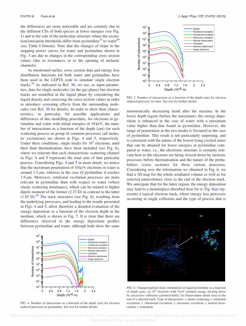
tic excitations) are shown in Figs. 4 and 5, respectively.
Under these conditions, single tracks for 105 electrons, until
their final thermalisation, have been included (see Fig. 6),
where we reiterate that each characteristic scattering channel
in Figs. 4 and 5 represents the total sum of that particular
process. Considering Figs. 4 and 5 in more detail, we notice
that the maximum penetration of 10 keV electrons in water is
around 1.5 lm, whereas in the case of pyrimidine it reaches
1.9 lm. Moreover, rotational excitation processes are more
relevant in pyrimidine than with respect to water (where
elastic scattering dominates), which can be related to higher
dipole moment of the former (2.33 D) in contrast to the latter
(1.85 D).45 The track structures (see Fig. 6), resulting from
the underlying processes, and leading to the results presented
in Figs. 4 and 5, allow therefore a detailed evaluation of the
energy deposition as a function of the electron depth in the
medium, which is shown in Fig. 7. It is clear that there are
differences observed in the energy deposition profiles
between pyrimidine and water, although both show the same
monotonically decreasing trend after the maxima. In the
lower depth region (before the maximum), the energy depo-
sition is enhanced in the case of water with a maximum
value higher than that found in pyrimidine. However, the
range of penetration in the two media is favoured in the case
of pyrimidine. This result is not particularly surprising, and
is consistent with the nature of the lowest-lying excited states
that can be attained for lower energies in pyrimidine com-
pared to water, i.e., the electronic structure is certainly rele-
vant here as the electrons are being slowed down by inelastic
processes before thermalisation and the nature of the proba-
bilities (cross sections) for those various processes.
Considering now the information we obtained in Fig. 6, we
find a 3D map for the whole irradiated volume as well as for
selected nanovolumes close to the end of the electron track.
We anticipate that for the latter region, the energy deposition
may lead to a meaningless absorbed dose for it. Fig. 6(a) rep-
resents a typical electron track, where energy loss processes
occurring in single collisions and the type of process that is
FIG. 4. Number of interactions as a function of the depth (lm) for electron
induced processes in pyrimidine. See text for further details.
FIG. 5. Number of interactions as a function of the depth (lm) for electron
induced processes in water. See text for further details.
FIG. 6. Charged-particle track simulations in liquid pyrimidine as a function
of depth (lm). (a) 105 electrons with 5 keV incident energy slowing down
by successive collisions (coloured balls). (b) Nanovolume detail close to the
end of a selected track. Type of interactions: •, elastic scattering; •, rotational
excitation; •, vibrational excitation; •, electronic excitation; •, neutral disso-
ciation; •, ionisation.
214701-6 Fuss et al. J. Appl. Phys. 117, 214701 (2015)
[This article is copyrighted as indicated in the article. Reuse of AIP content is subject to the terms at: http://scitation.aip.org/termsconditions. Downloaded to ] IP:
130.130.37.84 On: Tue, 16 Jun 2015 00:08:22
taking place, respectively, are represented. For the charged-
particle track simulations presented here, electrons are
released into liquid pyrimidine with an initial energy of
5 keV and are then tracked until thermalisation occurs.
While the corresponding water results, under identical initial
conditions, are not shown here, we note that the determined
track structure is very different than that seen for pyrimidine
in Fig. 6(a). This observation, at least in part, reflects the
very different cross sections for both species (see Fig. 1). In
Fig. 6(b), the final portion of a single track ending can be
seen. Due to the molecular-level description of all collisional
events that is achieved by the LEPTS programme, a wide
range of detailed information on the underlying processes,
such as the type of interaction and exact energy loss at each
collision point, becomes available in addition to the more
common transport information (not shown here). Of particu-
lar relevance is the presence of secondary electrons being
produced as additional tracks emerging from the main trajec-
tory, which are clearly identified from the large density of
tracks pertaining to the elastic scattering process.
Notwithstanding this point, several tens of dissociative proc-
esses in the volume are also seen to be inducing damage in
regard to bond breaking and structural modifications of the
medium molecular constituents. In such a description, we
define nano-dosimetry as the result of dissociation induced
damage at the molecular level rather than the absorbed dose.
Therefore, the simulation procedure implemented in this
methodology can be considered as a useful nano-dosimetric
tool for applications requiring such a detailed level of
description, i.e., at the molecular level. Whether pyrimidine
can actually be considered as a better mimic of a physiologi-
cal environment, to the detriment of water, or whether both
and yet further species are needed for a quantitative repre-
sentation of such environment, still requires further extensive
studies with particular relevance for experimental data. As
far as the authors’ are aware, no previous results for pyrimi-
dine from simulation codes in the energy range of
1–10 000 eV, as considered here, have been reported in the
literature. We believe this reflects a hitherto lack of proper
experimental data being available.
V. CONCLUSION
We have provided a complete set of differential and in-
tegral electron scattering cross section data for pyrimidine,
based on our previous experiments and together with calcu-
lations and other data available in the literature. Quantitative
and detailed knowledge of the underlying processes (integral
and differential cross sections and energy loss spectra)
resulting from the interaction of electrons (1–10 000 eV)
with pyrimidine, and water molecules, have been used as
input parameters to describe the interaction model in the
LEPTS code. Electrons as incoming particles were found to
have a higher penetration depth for pyrimidine (1.9 lm) than
for water (1.5 lm). This may have considerable biological
consequences within the physiological environment regard-
ing how effective dose is evaluated within nanovolumes in
targeted radiation therapies. It certainly indicates that the
behaviour of electrons traversing through pyrimidine is dif-
ferent than when they traverse through water. Therefore, the
present results strongly suggest that using water as the
default medium for biological systems might not be quantita-
tively correct. We believe that the two main reasons which
explain our different observations for electron tracks in py-
rimidine vis a vis water relate to both their very different
electronic structures and their different electron scattering
and interaction probabilities (i.e., the cross sections). The
present results therefore provide a transparent example for
how the behaviour at the nanoscale drives the macroscopic
behaviour of the system, which is described by terms such as
the stopping power or energy deposition. Using a complete
set of realistic cross section data, we have shown that radia-
tion effects can be described not only in terms of energy dep-
osition but also in terms of induced molecular processes. At
this stage, the present modeling procedure provides informa-
tion on the number and type of molecular dissociations
induced by low energy electrons but additional information
on further interactions of the created radicals with the con-
stituent of the medium would be desirable and will be the
subject of future investigations.
ACKNOWLEDGMENTS
This research was supported by the Australian Research
Council (ARC) through its Centres of Excellence Program.
D.B.J. thanks the ARC for provision of a Discovery Early
Career Researcher Award. We also acknowledge the support
of the Spanish Ministerio de Economia y Competitivad under
Project No. FIS 2012-31230 and the European Union COST
Actions (MP1002 and CM1301). P.L.V. acknowledges the
Portuguese Foundation for Science and Technology (FCT-
MEC) through research grants PTDC/FIS-ATO/1832/2012,
UID/FIS/00068/2013, and SFRH/BSAB/105792/2014. P.L.V.
also acknowledges his Visiting Professor position at Flinders
University, Adelaide, South Australia.
1B. Boudaiffa, P. Cloutier, D. Hunting, M. A. Huels, and L. Sanche,
Science 287, 1658 (2000).2C. von Sonntag, Free-Radical-Induced DNA Damage and its Repair(Springer, New York, 2005).
FIG. 7. Energy deposition of 105 incident electrons each with 10 keV energy
in liquid water and pyrimidine (1.000 and 1.016 g/cm3 density, respectively),
as a function of depth (lm).
214701-7 Fuss et al. J. Appl. Phys. 117, 214701 (2015)
[This article is copyrighted as indicated in the article. Reuse of AIP content is subject to the terms at: http://scitation.aip.org/termsconditions. Downloaded to ] IP:
130.130.37.84 On: Tue, 16 Jun 2015 00:08:22

3L. Sanche, in Radiation Induced Molecular Phenomena in Nucleic Acids,
edited by M. Shukla and J. Leszczynski (Springer, Netherlands, 2008),
Vol. 5, pp. 531–575.4S. M. Pimblott and J. A. LaVerne, Radiat. Phys. Chem. 76, 1244 (2007).5V. Cobut, Y. Frongillo, J. P. Patau, T. Goulet, M.-J. Fraser, and J.-P. Jay-
Gerin, Radiat. Phys. Chem. 51, 229 (1998).6J. A. LaVerne and S. Pimblott, Radiat. Res. 141, 208 (1995).7L. Turi and P. J. Rossky, Chem. Rev. 112, 5641–5674 (2012).8L. Sanche, Nature 461, 358–359 (2009).9S. Agostinelli, J. Allison, K. Amako, J. Apostolakis, H. Araujo, P. Arce,
M. Asai, D. Axen, S. Banerjee, G. Barrand, F. Behner, L. Bellagamba, J.
Boudreau, L. Broglia, A. Brunengo, H. Burkhardt, S. Chauvie, J. Chuma,
R. Chytracek, G. Cooperman, G. Cosmo, P. Degtyarenko, A. Dell’Acqua,
G. Depaola, D. Dietrich, R. Enami, A. Feliciello, C. Ferguson, H.
Fesefeldt, G. Folger, F. Foppiano, A. Forti, S. Garelli, S. Giani, R.
Giannitrapani, D. Gibin, J. J. G�omez Cadenas, I. Gonz�alez, G. Gracia
Abril, G. Greeniaus, W. Greiner, V. Grichine, A. Grossheim, S. Guatelli,
P. Gumplinger, R. Hamatsu, K. Hashimoto, H. Hasui, A. Heikkinen, A.
Howard, V. Ivanchenko, A. Johnson, F. W. Jones, J. Kallenbach, N.
Kanaya, M. Kawabata, Y. Kawabata, M. Kawaguti, S. Kelner, P. Kent, A.
Kimura, T. Kodama, R. Kokoulin, M. Kossov, H. Kurashige, E. Lamanna,
T. Lamp�en, V. Lara, V. Lefebure, F. Lei, M. Liendl, W. Lockman, F.
Longo, S. Magni, M. Maire, E. Medernach, K. Minamimoto, P. Mora de
Freitas, Y. Morita, K. Murakami, M. Nagamatu, R. Nartallo, P. Nieminen,
T. Nishimura, K. Ohtsubo, M. Okamura, S. O’Neale, Y. Oohata, K. Paech,
J. Perl, A. Pfeiffer, M. G. Pia, F. Ranjard, A. Rybin, S. Sadilov, E. Di
Salvo, G. Santin, T. Sasaki, N. Savvas, Y. Sawada, S. Scherer, S. Sei, V.
Sirotenko, D. Smith, N. Starkov, H. Stoecker, J. Sulkimo, M. Takahata, S.
Tanaka, E. Tcherniaev, E. Safai Tehrani, M. Tropeano, P. Truscott, H.
Uno, L. Urban, P. Urban, M. Verderi, A. Walkden, W. Wander, H. Weber,
J. P. Wellisch, T. Wenaus, D. C. Williams, D. Wright, T. Yamada, H.
Yoshida, and D. Zschiesche, Nucl. Instrum. Methods Phys. Res., Sect. A
506, 250–303 (2003).10J. Allison, K. Amako, J. Apostolakis, H. Araujo, P. A. Dubois, M. Asai, G.
Barrand, R. Capra, S. Chauvie, R. Chytracek, G. A. P. Cirrone, G.
Cooperman, G. Cosmo, G. Cuttone, G. G. Daquino, M. Donszelmann, M.
Dressel, G. Folger, F. Foppiano, J. Generowicz, V. Grichine, S. Guatelli,
P. Gumplinger, A. Heikkinen, I. Hrivnacova, A. Howard, S. Incerti, V.
Ivanchenko, T. Johnson, F. Jones, T. Koi, R. Kokoulin, M. Kossov, H.
Kurashige, V. Lara, S. Larsson, F. Lei, O. Link, F. Longo, M. Maire, A.
Mantero, B. Mascialino, I. McLaren, P. M. Lorenzo, K. Minamimoto, K.
Murakami, P. Nieminen, L. Pandola, S. Parlati, L. Peralta, J. Perl, A.
Pfeiffer, M. G. Pia, A. Ribon, P. Rodrigues, G. Russo, S. Sadilov, G.
Santin, T. Sasaki, D. Smith, N. Starkov, S. Tanaka, E. Tcherniaev, B.
Tome, A. Trindade, P. Truscott, L. Urban, M. Verderi, A. Walkden, J. P.
Wellisch, D. C. Williams, D. Wright, and H. Yoshida, IEEE Trans. Nucl.
Sci. 53, 270–278 (2006).11F. Salvat, J. M. Fernandez-Varea, and J. Sempau, in PENELOPE2011: A
Code System for Monte-Carlo Simulation of Electron and Photon
Transport, OECD—Nuclear Energy Agency, 2011.12C. Champion, C. Le Loirec, and B. Stosic, Int. J. Radiat. Biol. 88, 54–61
(2012).13A. Mu~noz, F. Blanco, J. C. Oller, J. M. P�erez, and G. Garc�ıa, in Advances
in Quantum Chemistry, edited by J. R. Sabin and E. Br€andas (Academic
Press, 2007), Vol. 52, pp. 21–57.14A. Mu~noz, M. Fuss, M. A. Cort�es-Giraldo, S. Incerti, V. Ivanchenko, A.
Ivanchenko, J. M. Quesada, F. Salvat, C. Champion, and G. G�omez-
Tejedor, in Radiation Damage in Biomolecular Systems, edited by G.
Garc�ıa G�omez-Tejedor and M. C. Fuss (Springer, Netherlands, 2012), pp.
203–225.15R. D. White, W. Tattersall, G. Boyle, R. E. Robson, S. Dujko, Z. L.
Petrovic, A. Bankovic, M. J. Brunger, J. P. Sullivan, S. J. Buckman, and
G. Garcia, Appl. Radiat. Isot. 83(Pt. B), 77–85 (2014).16J. de Urquijo, E. Basurto, A. M. Juarez, K. F. Ness, R. E. Robson, M. J.
Brunger, and R. D. White, J. Chem. Phys. 141, 014308 (2014).17R. D. White, M. J. Brunger, N. A. Garland, R. E. Robson, K. F. Ness, G.
Garcia, J. de Urquijo, S. Dujko, and Z. L. Petrovic, Eur. Phys. J. D 68, 1–6
(2014).18S. Incerti, G. Baldacchino, M. Bernal, R. Capra, C. Champion, Z. Francis,
P. Gueye, A. Mantero, B. Mascialino, P. Moretto, P. Nieminen, C.
Villagrasa, and C. Zacharatou, Int. J. Model. Simul. Sci. Comput. 1,
157–178 (2010).19J. Builth-Williams, S. M. Bellm, D. B. Jones, H. Chaluvadi, D. Madison,
C. G. Ning, B. Lohmann, and M. J. Brunger, J. Chem. Phys. 136, 024304
(2012).20D. B. Jones, S. M. Bellm, F. Blanco, M. Fuss, G. Garcia, P. Lim~ao-Vieira,
and M. J. Brunger, J. Chem. Phys. 137, 074304 (2012).21D. B. Jones, S. M. Bellm, P. Lim~ao-Vieira, and M. J. Brunger, Chem.
Phys. Lett. 535, 30–34 (2012).22Z. Masin, J. D. Gorfinkiel, D. B. Jones, S. M. Bellm, and M. J. Brunger,
J. Chem. Phys. 136, 144310 (2012).23M. Stener, P. Decleva, D. M. P. Holland, and D. A. Shaw, J. Phys. B: At.
Mol. Phys. 44, 075203 (2011).24F. Ferreira da Silva, D. Almeida, G. Martins, A. R. Milosavljevic, B. P.
Marinkovic, S. V. Hoffmann, N. J. Mason, Y. Nunes, G. Garcia, and P.
Limao-Vieira, Phys. Chem. Chem. Phys. 12, 6717–6731 (2010).25P. Palihawadana, J. Sullivan, M. Brunger, C. Winstead, V. McKoy, G.
Garcia, F. Blanco, and S. Buckman, Phys. Rev. A 84, 062702 (2011).26M. C. Fuss, A. G. Sanz, F. Blanco, J. C. Oller, P. Lim~ao-Vieira, M. J.
Brunger, and G. Garc�ıa, Phys. Rev. A 88, 042702 (2013).27A. Zecca, L. Chiari, G. Garcia, F. Blanco, E. Trainotti, and M. J. Brunger,
J. Phys. B: At. Mol. Phys. 43, 215204 (2010).28J. B. Maljkovic, A. R. Milosavljevic, F. Blanco, D. Sevic, G. Garcia, and
B. P. Marinkovic, Phys. Rev. A 79, 052706 (2009).29C. G. Ning, K. Liu, Z. H. Luo, S. F. Zhang, and J. K. Deng, Chem. Phys.
Lett. 476, 157–162 (2009).30S. H. R. Shojaei, B. Hajgat�o, and M. S. Deleuze, Chem. Phys. Lett. 498,
45–51 (2010).31P. L. Levesque, M. Michaud, and L. Sanche, J. Chem. Phys. 122, 094701
(2005).32M. H. Palmer, I. C. Walker, M. F. Guest, and A. Hopkirk, Chem. Phys.
147, 19–33 (1990).33I. Nenner and G. J. Schulz, J. Chem. Phys. 62, 1747–1758 (1975).34D. Almeida, D. Kinzel, F. Ferreira da Silva, B. Puschnigg, D. Gschliesser,
P. Scheier, S. Denifl, G. Garc�ıa, L. Gonz�alez, and P. Lim~ao-Vieira, Phys.
Chem. Chem. Phys. 15, 11431–11440 (2013).35D. Almeida, F. Ferreira da Silva, G. Garc�ıa, and P. Lim~ao-Vieira, Phys.
Rev. Lett. 110, 023201 (2013).36F. Blanco, A. Mu~noz, D. Almeida, F. Ferreira da Silva, P. Lim~ao-Vieira,
M. C. Fuss, A. G. Sanz, and G. Garc�ıa, Eur. Phys. J. D 67, 199 (2013).37A. G. Sanz, M. C. Fuss, A. Mu~noz, F. Blanco, P. Lim~ao-Vieira, M. J.
Brunger, S. J. Buckman, and G. Garc�ıa, Int. J. Radiat. Biol. 88, 71–76
(2012).38A. Mu~noz, J. M. P�erez, G. Garc�ıa, and F. Blanco, Nucl. Instrum. Methods
Phys. Res., Sect. A 536, 176–188 (2005).39A. G. Sanz, M. C. Fuss, F. Blanco, Z. Ma�s�ın, J. D. Gorfinkiel, F. Carelli, F.
Sebastianelli, F. A. Gianturco, and G. Garc�ıa, Appl. Radiat. Isot. 83(Pt. B),
57–67 (2014).40I. Linert, M. Dampc, B. Mielewska, and M. Zubek, Eur. Phys. J. D 66, 1–9
(2012).41W. Y. Baek, A. Arndt, M. U. Bug, H. Rabus, and M. Wang, Phys. Rev. A
88, 032702 (2013).42J. R. Ferraz, A. S. dos Santos, G. L. C. de Souza, A. I. Zanelato, T. R. M.
Alves, M. T. Lee, L. M. Brescansin, R. R. Lucchese, and L. E. Machado,
Phys. Rev. A 87, 032717 (2013).43W. Wolff, H. Luna, L. Sigaud, A. C. Tavares, and E. C. Montenegro,
J. Chem. Phys. 140, 064309 (2014).44A. Mu~noz, F. Blanco, G. Garc�ıa, P. A. Thorn, M. J. Brunger, J. P.
Sullivan, and S. J. Buckman, Int. J. Mass Spectrom. 277, 175–179 (2008).45CRC Handbook of Chemistry and Physics, edited by D. R. Lide (CRC
Press, Boca Raton, FL, 2005), Internet Version 2005, http://
www.hbcpnetbase.com.46R. Colmenares, A. G. Sanz, M. C. Fuss, F. Blanco, and G. Garc�ıa, Appl.
Radiat. Isot. 83(Pt. B), 91–94 (2014).47T. Field, private communication (2008).48M. C. Fuss, A. G. Sanz, F. Blanco, P. Lim~ao-Vieira, M. J. Brunger, and G.
Garc�ıa, Eur. Phys. J. D 68, 161 (2014).49R. Mota, R. Parafita, A. Giuliani, M.-J. Hubin-Franskin, J. M. C.
Lourenco, G. Garcia, S. V. Hoffmann, N. J. Mason, P. A. Ribeiro, M.
Raposo, and P. Lim~ao-Vieira, Chem. Phys. Lett. 416, 152–159 (2005).
214701-8 Fuss et al. J. Appl. Phys. 117, 214701 (2015)
[This article is copyrighted as indicated in the article. Reuse of AIP content is subject to the terms at: http://scitation.aip.org/termsconditions. Downloaded to ] IP:
130.130.37.84 On: Tue, 16 Jun 2015 00:08:22