the role of proprioception in the management and rehabilitation of
TRANSCRIPT

0363-5465/97/2525-0130$02.00/0THE &'dERICAN JOURNAL OF SPORTS MEDICINE. Vol. 25. No.1@ 1997 American Orthopaedic Society for Sports Medicine
The Role of Proprioception in theManagement and Rehabilitation of
InjuriesScott M. Lephart,* PhD, ATC, Danny M. Pincivero, MEd, Jorge L. Giraldo, MD, alnd
Freddie H. Fu, MO
From the Neuromuscular Research Laboratory, University of Pittsburf7h,Pittsburgh, Pennsylvania
ABSTRACT
Rehabilitation continues to evolve with the increasedemphasis on patient management and proprioceptivetraining. Proprioception can be defined as a special-ized variation of the sensory modality of touch thatencompasses the sensation of joint movement (kines-thesia) and joint position Goint position sense). Numer-ous investigators have observed that afferent feedbackto the brain and spinal pathways is mediated by skin,articular, and muscle mechanoreceptors. Examiningthe effects of ligamentous injury, surgical intervention,and proprioceptively mediated activities in the rehabil-itation program provides an understanding of the com-plexity of this system responsible for motor control. Itappears that this neuromuscular feedback mechanismbecomes interrupted with injury and abnormalities, andapproaches restoration after surgical intervention andrehabilitation. Rehabilitation programs should be de-signed to include a proprioceptive component that ad-dresses the following three levels of motor control:spinal reflexes, cognitive programming, and brainstemactivity. Such a program is highly recommended topromote dynamic joint and functional stability. Thus far,current knowledge regarding ~he basic science andclinical application of proprioception has led the pro-:fession of sports medicine one':-$tep closer to its ulti-
mate goal of restoring function.
The proper management of athletic-related injuries alndorthopaedic lesions can be complex in the sports medic:inesetting. One of the most challenging aspects to the clini-cian is understanding the role of proprioceptively me,di-ated neuromuscular control after joint injury and itsrestoration through rehabilitation. Proprioception con-tributes to the motor programming for neuromuscularcontrol required for precision movements and also contzib-utes to muscle reflex, providing d)ffiamic joint stabili,ty.The coupling effect of ligamentous trauma resulting inmechanical instability and proprioceptive deficits contzib-utes to functional instability, which could ultimately leadto further microt~auma and reinjury (Fig. 1). AchieV:lngfunctional and sport-specific activities after musculos~:el-etal trauma and rehabilitation can be enhanced signifi-cantly if proprioception is addressed and instituted earlyin the treatment program.
In addition to the mechanical restraint provided by ar-ticular structures, it has been observed that ligame:ntsprovide neurologic feedback that directly mediates reflexmuscular stabilization about the joint.2o The inclusion ofproprioception in the rehabilitation program should bebased on the preceding finding and not on anecdotal E!vi-dence without an understanding of the neuromuscularmechanism. This understanding, coupled with a basEl ofknowledge regarding the current research on propriocep-tion, is necessary for sports medicine practitioners to op-timize treatment programs for athletes.
THE ROLE OF PROPRIOCEPTION
Numerous investigators have provided definitions regard-ing the terminology of joint sensation, or proprioceptionand kinesthesia.3.26 Most contemporary authorities defineproprioception as a specialized variation of the sensory
.Address correspondence and reprint requests to Scott M. Lephart. PhD.ATC. Neuromuscular Research Laboratory. 127 Trees Hall. University of
Pittsburgh. Pittsburgh. PA 15261.No author or related institution has received financial benefit from research
in this study.
130

Role of Proprioception in Athletic Injuries 131Vol. 25, No.1, 1997
ganglia, and the cerebellum) refers to voluntary m.ove-ments that are repeated and stored as central commands.This awareness of body position and movement allowsvarious skills to be performed without continuous rlefer-ence to consciousness.33 As defined earlier, proprioceptivefeedback plays a major role in the conscious and UIJ,con-scious awareness of a joint or limb in motion.
iLigamentous Inju;Y I
--~"",II'ropriuceptive II Deficits I
+I Decreased Neuromuscular I
I Control I
~I Repetitive Injuryj
\.I Functional I
I Instability I
tQ~§]
Figure 1. Functional stability paradigm depicting the pro-gression of functional instability of the shoulder joint due to
the interaction between mechanical instability and decreasedneuromuscular control. (Reprinted with permission from Lep-
hart and Henry.23)
PERIPHERAL AFFERENTS
The concept of proprioception is based on the fact thatneural feedback to the CNS is mediated by cutaneous,muscle, and joint mechanoreceptors (Fig. 3). When eJcam-ining the neural composition of joints, Hilton's law s1;atesthat joints are innervated by articular branches oj' thenerves supplying the muscles that cross the joint.1.9 Inaddition to proprioceptive mechanoreceptors, arti<:ularstructures also include nociceptive free nerve endings.
Activation of joint mechanoreceptors is triggered b:'r thedeformation and loading of the soft tissues that com.posethe joint. This neural stimulation travels to the CN:S forintegration via cortical and reflex pathways. These mech-anoreceptors demonstrate adaptive properties depeIJldingon a particular stimulus 15 (Table 1).
Quick-adapting joint mechanoreceptors, such as th,~ Pa-cinian corpuscles, decrease their discharge rate to eJ!:tinc-tion within milliseconds of the onset of a continuous :,tim-ulUs.7 The Ruffini ending, Ruffini corpuscles, and theGolgi tendon-like organs that are referred to as the :,low-adapting mechanorec~ptors. continue their discharge inresponse to a continuous stimulUS.15 The properties of the
modality of touch that encompasses the sensation of jointmovement (kinesthesia) and joint position (joint positionsense). The sensory receptors for proprioception that arefound in the skin, muscles, and joints as well as in liga-ments and tendons all provide input to the central nervoussystem (CNS) regarding tissue deformation.15 Visual andvestibular centers also contribute afferent information tothe CNS regarding body position and balance.33
Trauma to tissues that contain mechanor~ceptors mayresult in partial deafferentation, which can lead to propri-oceptive deficits. Susceptibility to reinjury, therefore, be-comes a realistic possibility because of this decrease inproprioceptive feedback. However, studies have shown atleast partial restoration of kinesthesia and joint positionsense in surgically reconstructed shoulders and knees af-ter rehabilitation.24.25 Regaining neuromuscular controlafter injury or surgery is a necessary prerequisite forathletes wishing to return to competition.
The neural input that is provided by the peripheralmechanoreceptors as well as the visual and vestibularreceptors are all integrated by the CNS to generate amotor response. These responses generally fall underthree levels of motor control: spinal reflexes, cognitiveprogramming, and brainstem activity (Fig. 2). In a situa-tion ..vhere a joint is placed under mechanical loading,reflex muscular stabilization is stimulated through thespinal reflexes.17 Cognitive programming that involvesthe highest level of CNS function (motor cortex, basal
I.EVELS Of MOTORCONTROl.
~-r;f~""'-c:- ,_,Cosniti..ll ~AT ,crT i: j
~roST.mmi~ -I ~USCLE J
" /r-s;;in 51;;;;""1~nc. ~
/~ribw.t t.C.pl~
Figure 3. Schematic representation of proprioceptive mech-
anoreceptors: a) Ruffini ending, b) Pacinian corpusl:le, c)muscle spindle receptors. and d) Golgi tendon organs.
(Adapted from Willis and Grossman.34)Figure 2. Neuromuscular control pathways. (Reprinted with
permission from Lephart and Henry .23)

132 Lephart et at.American Journal of Sports IYJedicine
TABLE 1Articular Mechanoreceptors and Articular Nociceptorso
Receptor type Location Adaptation rate Function
Joint pressureHigh frequency vibrationJoint pain
I, Ruffini endingsII, Pacinian corpusculeN (a), Unmyelinated free nerve endings-a Modified from Freeman and Wyke.ll
Joint capsule and ligamentsJoint capsuleLigaments (and related muscles)
from descending pathways, thereby increasing i[OrCeproduction. 27
With respect to changes in muscle and tendon tension,the Golgi tendon organs function as a protective mecha-nism. The Golgi tendon organs are located within thetendons of muscles and are recruited when muscle con-traction pulls on the tendon, which straightens the colla-gen bundles and distorts the receptor endings of the affer-ent neurons.27 This distortion increases the discharge rateof action potentials of these receptors that travel andsynapse on spinal interneurons that project to motor neu-rons. Increased activity of the Golgi tendon organ ajfer-ents result in the inhibition of the motor neurons inJler-vating the muscles that were stretched while exciting themotor nerves of the antagonistic muscles.
Studies have demonstrated the detrimental effect onreflex joint stabilization as a result of joint injury.30.31 'rhecontribution of musculotendinous receptors over joint re-ceptors to the proprioceptive reflex remains a contro\'er-sial issue in the literature. The ability.to quantify pro]~ri-oceptive deficits is a vital component to the evaluation ofjoint injury that may attempt to answer a number ofclinical research questions.
quick-adapting mechanoreceptors lead to the notion thatthey mediate the sensation of joint motion because theyare very sensitive to changes in position. Muscular mech-anoreceptors and Ruffini ending joint receptors are slow-adapting mechanoreceptors and are thought to mediatethe sensation of joint position and changes in positionbecause they are maximally stimulated at specific jointangles. One fonn of the slow-adapting receptors are thecomplex, fusifonn muscle spindle receptors found withinskeletal muscle. The muscle spindle receptor functions tomeasure muscle tension over a large range of extrafusalmuscle length (Table 2). It has been suggested that muscleand joint mechanoreceptors are complementary to eachother in providing afferent input in regard to limbposition.4
This relationship between muscle and joint mechanore-ceptors has been supported by the identification of theneural components necessary for the sensation of motion(rapidly adapting receptors, e.g., Pacinian corpuscles),joint position and acceleration (slow-adapting receptors,e.g., Ruffini endings and Ruffini corpuscles), and pain(free nerve endings) within ligamentous, cartilaginous,and muscular structures of the joints.
The spindle receptors found within the muscle are com-posed of a small bundle of modified muscle fibers calledintrafusal fibers, to which the endings of several sensorynerves are attached. The extrafusal fibers that fonn thebulk of the muscle are responsible for generating force andare innervated by the alpha-motor neurons whereas theintrafusal fibers are innervated by the gamma-motor neu-rons. In the case of muscle contraction, alpha-gamma co-activation is believed to be the mechanism through whichmuscle length and tension are monitored,27 Activation ofgamma-motor neurons allow the readjustment of spindlesensitivity in the case where extrafusal fibers are short-ened. This allows the spindles to be functional at all timesduring a contraction. When a muscle is loaded beyond ananticipated level, intrafusal fiber shortening occurs to agreater degree than extrafusal shortening. Stretching ofthe spindles in the central region causes a burst of exci-tatory postsynaptic potentials from spindle afferents.These signals summate with the alpha-motor neurons
TABLE 2 -.::.Muscular MechanoreceptorsG
Location Adaptationrate
Receptor type
CUNICAL QUESTIONS
Clinical application of proprioception research finmrLgsmust be achieved at various levels of care for the mana!~e-ment of orthopaedic lesions. A thorough understanding ofproprioception assessment techniques will aid the cli:m-cian and orthopaedic surgeon to apply what is currentlyknown concerning the nontraumatized joint to muscullir,ligamentous, and cartilaginous injury. Establishing theeffects of musculoskeletal trauma on joint position sen:si-bility and neuromuscular control assists the need for crit-ical decision-making regarding the appropriateness ofvarious forms of treatment. From this point, it is knovmthat surgical intervention plays an important role in therestoration of mechanical joint stability but its effects onproprioception pathways require further clarification. F'i-nally, rehabilitation activities incorporating principlesthat stimulate the different levels of motor control to fa-cilitate the return to function should be examined not onlyfor its theoretical basis but also for its practic:al
applicability.The assessment of neuromuscular control includes the
measurement of cortical, spinal reflex, and brainstempathways. The evaluation of this complex neuromusculfLrsystem as different components allows a more detailedexplanation of afferent control mechanisms. As definedpreviously, kinesthesia and joint position sense are
Function
Tendons Slow Reflex
Muscle Slow Reflex (stretch reflex)a Modified from Freeman and Wyke.ll

Vol. 25. No.1. 1997 Role of Proprioception in Athletic Injuries 133
tion of the reflex arc stimulated by mechanoreceptors andmuscle spindle receptors occurs at a faster rate than sig-nals induced by nociceptors (70 to 100 m/sec versus 1m/sec),15 This suggests that proprioception may i;>lay amore significant role than pain impulses in preventinginjury in the acute setting, However, the incidence ofreinjury and the cause of chronic injuries may be :3.ttrib-uted, to a greater extent, to proprioceptive deficits. Thesedeficits may be induced by partial deafferentation as aresult of initial knee injury and may also contribute tochronic joint disease through a decrease in joint afferents.This phenomenon has been observed by Beard et :3.1.5 insubjects with arthroscopically confirmed ACL deficiency.A significant deficit in reflex activation of the hamstringmuscles after a 109 N anterior shear force in a single-legged closed kinetic chain position was identified, as com-pared with the contralateral uninjured limb.5 Further-more, Solomonow et al.3o found that a direct stress appliedto the ACL resulted in reflex hamstring activity, therebycontributing to the maintenance of joint integrity.
Although Barrack et al.1 demonstrated a propriocleptivedeficit after ACL disruption, it appears that kinestheticawareness may be partially restored after ACL recon-struction. Kinesthesia has been reported to be re~;toredafter surgery as detected by the threshold to the detectionof passive motion in the midrange of motion (45°).1 How-ever, a longer threshold to the detection of passive motionwas observed in the ACL reconstructed knee comparedwith the contralateral uninvolved knee when tested at 15°of flexion. Lephart et al.24 found similar results in patientsafter either arthroscopically assisted patellar-tendon au-tograft or allograft ACL reconstruction. This evidence sug-gests that kinesthesia may have returned in the midrangeof motion after ACL reconstruction and appears to bemore sensitive in the near-terminal range of motion.
The importance of incorporating a proprioceptive ele-ment in any comprehensive rehabilitation program ilS jus-tified based on the results of these studies. Proprioceptivedeficits may predispose an athlete to reinjury th:roughdecrements in the neuromuscular pathways resulti.ng inthe inhibition of complete rehabilitation.
components of proprioception. Functionally, kinesthesia isassessed by measuring threshold to detection of passivemotion while joint position sense is assessed by measuringreproduction of passive positioning and reproduction ofactive positioning. When tested at a slow angular velocity(0.5 to 2 deg/sec), threshold to detection of passive motionas well as the reproduction of passive positioning isthought to selectively stimulate Ruffini or Golgi-typemechanoreceptors. Because the test is perfonned pas-sively, it is believed to maximally stimulate joint recep-tors, thereby relying on the cortical pathway in the neu-romuscular control system. After ligament lesions, passivejoint sensibility testing is often chosen to assess afferentactivity because muscle activity is negated. Stimulation ofboth joint and muscle receptors is done by the reproduc-tion of active positioning, which provides a more func-tional assessment of the afferent pathways.
The evaluation of reflex capabilities is often assessed bymeasuring the latency of muscular activation to involun-tary perturbation via electromyographic interpretation.The ability to quantify the sequencing of muscle firing canprovide a valuable tool for the assessment of asynchronousneuromuscular activation patterns that may predisposean articulation to overuse trauma.
Functional assessment of the combined peripheral, ves-tibular, and visual contributions to neuromuscular controlis best accomplished through the use of balance and pos-tural sway measurements for the lower extremity. Theavailability of stabilometric methods and instrumenta-tion can provide a relatively accurate index for thesemeasures.
To reiterate this understanding of the proprioceptionmechanism, the clinician must apply the available knowl-edge to try to delineate the effects of orthopaedic injury,surgical reconstruction, and rehabilitation on the variousafferent pathways. Specifically, clinical research aimed atdetermining the effects of injury, surgery, and rehabilita-tion on joint position sensibility, neuromuscular control aswell as balance and postural sway can provide a solidfoundation for the development of a testing model for theknee, ankle, and shoulder that attempt to address theseissues.
KNEE PROPRIOCEPTIONANKLE PROPRIOCEPTION
Chronic ankle instability as a result of partial deafferen-tation of articular mechanoreceptors with joint injury wasfirst postulated by Freeman et apo They observed j;hat adecrease in the ability to maintain a one-legged stanceoccurred in the sprained ankle versus the contralateraluninjured ankle.1O The effect of unilateral ankle sflrainson cortical pathway measures of proprioception havEi beeninvestigated by Garn and Newton,12 who measured theability of a subject to properly sense a passive movementor no movement state in the sagittal plane. Deficits In theability to actively replicate passive ankle and foot posi-tioning in this plane was reported by Glencross andThornton 13 while testing the sprained ankle verS1;LS thecontralateral uninjured ankle. Gross 16 recently reflorted
that an increased probability of reinjury occurs as a re-sult of a decrease in sensory input from joint
Numerous studies have been performed that examinedthe role of proprioception in the knee joint. It has beenfound that damage to articular structures, such as theACL and meniscus, in addition to osteoarthritic changesdisrupts articular structures containing mechanorecep-tors. The disruption in the cortical pathway, therefore,
~results in an alteration in joinlposition sense and kines-thesia. Barrack et al.1 and Skinner et al.28 observed de-creased kinesthesia with increasing age and ACL disrup-tion. Joint position sensibility decrements have also beendocumented as a result of osteoarthritic changes in the
knee.2Deficits in the neuromuscular reflex pathway may have
a detrimental effect on this motor control system's role asa protective mechanism in acute knee injury. The initia-

134 Lephart et at. American Journal of Sports l\lledicine
ity through a decrease in the normal internal rotationforce required for this motion.14 Compensatory increasesin biceps and supraspinatis muscle activity were also dis-covered in an attempt to restore anterior stability. Thisloss in the normal synchronization of neuromuscular fir-ing patterns in the unstable shoulder has, therefore, beenattributed to altered joint kinematics resulting in repeti-tive microtrauma.14
Surgical intervention has been shown to partially re-store joint proprioception through the repair of trauma-tized tissue. Partial restoration of kinesthesia has beenobserved in patients undergoing capsulolabral reconstruc-tion.25 This observation of enhanced proprioception cen-ters around the procedural techniques used that promotedmodification of joint sensation. Because this proceduremodifies soft tissue dissection, there was a minimal loss ofintact mechanoreceptors and a promotion of repopulation.In addition, the use of the capsular shift in these shoulderinstability cases, which tightens the capsule, "retensions"the soft tissue and most likely facilitated proprioceptionfunction. It may be through this procedure ofretensioningthat mechanoreceptor-containing shoulder capsuloliga-mentous structures transmit afferent information at amore functional level regarding joint position sensibility.
Regaining dynamic neuromuscular control of the unsta-ble or postoperative shoulder is of primary importancefor the return of an athlete to functional activity. Reha-bilitation exercises should focus on the importance of in-corporating joint position sensibility .and retJexive-typecontractions into the therapy program. The inclusion ofreflexive-mediated activities is based on the recent find-ings of Guanche and coworkers17 who have observed threedifferent articular branches of the a.wlary nerve innervat-'fng the shoulder capsule that provide a primary reflex arcto the biceps, supraspinatis, infraspinatis, deltoid, andsubscapularis muscles in the feline model. This reflexivecontraction has been attributed to providing a dynamicmuscular restraint to the intact shoulder capsule.17 Be-cause the shoulder model in this study was that of aweightbearing joint, the closed kinetic chain principle isneeded in upper extremity rehabilitation.
receptors, leading to abnormal body positioning and di-minished postural-reflex responses. It was also found byKonradsen and Ravn 21 that chronic ankle instability re-
sulted in a prolonged peroneal reaction time in response toa sudden inversion stress when compared with age-matched controls. Partial deafferentation resulting in di-minished reflex joint stabilization may contribute to thesefindings.
The development of high technologic systems to assessthe effects of musculoskeletal injury on balance has oc-curred in an attempt to quantify both static and dynamiccomponents of proprioception.1s The method of evaluationis based on the notion that damage to joint proprioceptorsafter injury to the lateral ligamentous complex of theankle dinlinishes afferent feedback from the injured joint,thereby resulting in increases in postural sway. 10 To date,
however, documented evidence exists concerning the al-terations in postural sway after ankle injury using sub-jective evaluation (i.e., Romberg test). No increases inpostural sway were observed by Tropp and Odenrick 32
when comparing a group of soccer players with previousankle sprains to a control group of uninjured soccer play-ers. Furthermore, no differences in postural sway werefound between the involved and uninvolved ankles in agroup of soccer players with a history of unilateral, recur-rent ankle sprains.32 However, significant increases inpostural sway were observed by Cornwall and ~furrellswhen comparing patients with acute ankle sprains withuninjured controls as long as 2 years after their injuries.
The effects of surgical reconstruction for functional an-kle instability on proprioception pathways as measuredthrough joint position sensibility or balance and posturalsway assessments have not yet been thoroughly investi-gated. Empirical evidence exists suggesting that proprio-ceptive training techniques after acute and chronic ankleinjuries are highly effective. In addition, ankle wrappingand bracing have also been suggested to have a proprio-ceptive benefit. However, this notion remains untestedand, therefore, unproven.
SHOULDER PROPRIOCEPTION
Placement of the hand is a necessary task during activi-ties of daily living in addition to sport-specific patterns.Joint position sensibility has not only played a role in themaintenance of dynamic shoulder stability but has alsobeen shown to demonstrate alterations after injury. Smithand Brunolli 29 have observed deficits in shoulder kines-
thesia and joint position sense in male subjects with uni-lateral, traumatic, recurrent anterior shoulder instabili-ties. In a similar group of patients, Lephart andcoworkers25- also demonstrated pr:oP.rioceptive.deficits inthe -pathologic shoulder as compared with the contralat-eral normal shoulder.
In addition to alterations in the cortical pathway, Glous-mann and coworkers 14 observed changes in the electro-
myographic pattern in baseball pitchers demonstratingshoulder instability. Reduction in neuromuscular activa-tion of the pectoralis major, subscapularis, and latissimusdorsi muscles was found to contribute to anterior instabil-
REHABILITATION
The objectives of proprioceptive rehabilitation are to re-train altered afferent pathways to enhance the sensationof joint movement. Proprioceptively mediated neuromus-cular control of joints takes into account three distinctlevels of motor activation within the CNS. Reflexes at thespinal level mediate movement patterns that are receivedfrom higher levels of the nervous system. This actionprovides for reflex joint stabilization during conditions ofabnormal stress about the articulation and has significantimplications for rehabilitation.2o The use of exercises thatfacilitate dynamic joint stabilization may result in theimprovement of this neuromuscular mechanism.
The second level of motor control, located withinthe brainstem, receives input from joint mechanorecep-tors, vestibular centers, and visual input from the eyes tomaintain posture and balance of the body. Reacti'.e

Vol. 25. No.1, 1997 Role of Proprioception in Athletic Injuries 135
mary sport-specific activity of the upper extremity is thethrowing motion, refined joint positioning and reposition-ing of the shoulder is vital. Therefore, mechanoreceptoractivity plays an important role in both performance anddynamic shoulder stabilization. To maximally restore pro-prioception and neuromuscular control, it is recommendedthat the following progression of activities be conducted toallow the return of an athlete to functional levels: 1) jointposition sense and kinesthesia, 2) dynamic joint stabiliza-tion, 3) reactive neuromuscular control, and 4) function-ally specific activities. Such a progression allows the re-habilitation program to address the integration of spinalreflex, cognitive, and brainstem pathways to focus onscapular stabilization, glenohumeral stabilization, hu-meral motion, and neuromuscular control.23
Position sensibility activities are designed to restorejoint position sense and kinesthesia (Fig. 4). These ,exer-cises stimulate cognitive level processing through the useof such an exercise as glenohumeral repositioning bothwith and without visual input and proprioceptive ru~uro-muscular facilitation patterns performed with manualresistance.23
Dynamic stabilization activities are designed to stimu-late muscular coactivation. In the shoulder, such activitiesinclude axial loading of the glenohumeral joint promotecoactivation of the glenohumeral and scapulothoracicforce couples (Fig. 5).6 The use of such activities as upperextremity balance training results in muscular
...'coactlvatlon.
ultimately, the integration of both spinal and cognitivelevels can be accomplished by the use of neuromus(:ularcontrol exercises such as plyometrics. Shoulder plyometricexercises stimulate reflexive activity through the facilita-tion of the myotatic reflex via the release of stored elasticenergy .23 Such activities stimulate reflex joint stabiliza-tion, which are critical to the overhead athlete (Fig. 6).
Once joint sensibility and dynamic muscle joint stabili-zation are restored, progression to functionally specificactivities can be accomplished. Functionally specific activ-
neuromuscular activities that allow this pathway to pro-cess input from the aforementioned forms of afferent stim-uli can be used to enhance brainstem function.
The highest level of CNS function (motor cortex, basalganglia, and cerebellum) provides cognitive awareness ofbody position and movement in which motor commandsare initiated for voluntary movements. Use of the corticalpathway allows movements that are repeated and storedas central commands to be performed without continuousreference to consciousness. Kinesthetic and proprioceptiontraining are such types of activity that can enhance thisfunction.
Incorporating the three levels of motor control into ac-tivities to address proprioceptive deficiencies should beinitiated early during the rehabilitation process.23 En-couraging maximum afferent discharge to the respectiveCNS level must be the goal in stimulating joint and mus-cle receptors.33 To stimulate reflex joint stabilization,which emanates from the spinal cord, activities shouldfocus on sudden alterations in joint positioning that ne-cessitate reflex neuromuscular control. Enhancing motorfunction at the brainstem level can be achieved by per-forming balance and postural activities, both with andwithout visual input. Maximally stimulating the conver-sion of conscious to unconscious motor programming canbe achieved by performing joint positioning activities, es-pecially at joint end ranges.33 Simple tasks such as bal-ance training and joint repositioning should begin early inthe rehabilitation program and should become increas-ingly more difficult as the patient progresses. Regainingjoint sense awareness to initiate muscular reflex stabili-zation to prevent reinjury should be the primary objectiveonce the final stage of rehabilitation is reached.
Some contemporary authors believe that adaptationsthat occur during rehabilitation are related to (mediatedby) feed-forward processing and are less a function ofenhanced afferent pathways.22 This theory suggests thatfast movements are controlled by advance informationknown about the task, while concurrent proprioceptivefeedback is relatively less important. Feedback is usedprimarily at the cortical level to determine the success orfailure of that movement and to a lesser extent at thesubcortical level for directing the movement. With repeti-tion, the cerebral cortex can determine the most effectivemotor pattern for a given task, based on the proprioceptiveinformation of previous attempts.22 Biofeedback trainingappears to use the feed-forward learning process.9 How-ever, there is still controversy regarding the contributionof afferent feedback in feed-forward processing.
Following is an example of a shoulder rehabilitationprotocol that has been designed .using the principles out-!ined in this paper for reestablishing proprioception and
neuromuscular control.
Reestablishing Proprioception and Neuromuscular Control
in the Shoulder Figure 4. Joint position sense and kinesthetic exercise:Shoulder kinesthetic training through functional arcs of ab-
duction and external rotation. (Reprinted with permi:5sion
from Lephart and Henry .23)
Proprioception training of the upper extremity has beenincorporated into the rehabilitation program to a lesserextent than that of the lower extremity. Because the pri-

136 Lephart et at. American Journal of Sports L"ledicine
.-ities are designed to restore functional motor patternsnecessary for successful performance of the overhead ath-lete (Fig. 7).
The integration of these levels is a necessary componentof the rehabilitation program to provide proper neuromus-cular control and functional stability of the joint. Comple-tion of the progressive neuromuscular control rehabilita-tion program minimizes the risk of reinjury and promotesa greater chance of successful return to competition.23
With respect to the lower extremity, mechanoreceptorslocated within the joints are most functionally stimulatedwhen the extremity is positioned in a closed-kinetic chainorientation and perpendicular axial loading of the joint ispermitted. These exercises should be performed at variouspositions throughout the full range of motion because ofthe difference in the afferent response that has been ob-served at different joint positions.
i~-.."..;, '-I"~,,' ';\l' \.\\
'\ ~, --I"
,.:.~
-;:"
Figure 5. Dynamic joint stabilization exercise: Exercise per-formed with a wobble board to stimulate coactivation ofshoulder muscle force couples. (Reprinted with permissionfrom Lephart and Henry ,23) FUTURE DIRECTIONS
The numerous investigations cited in this review haveattempted to present an understanding and a rationalebehind the use of proprioception exercises in rehabilita-tion. However, this pool of knowledge has opened the doorto many more questions concerning proprioceptively me-diated neuromuscular control. Evidence regarding the ef.fects of rehabilitation on proprioception has yet to be ver-ified. In addition. the effects of tissue regeneration onneuromuscular pathways is an area that also needs to beinvestigated. The establishment of this relationship be-tween proprioceptive deficits and motor control attem]~tsto incorporate basic science and clinical findings into apractical rehabilitation exercise prescription. Finally, pro-spectively evaluating the effects of ligamentous injury,surgical reconstruction, and rehabilitation will help tojustify the means through which articular lesions aremanaged. The research presented thus far puts the clini.cian one step closer to optimizing clinical decision-making
Figure 6. Reactive neuromuscular. -exercises: Shoulderplyometric exercise performed in the~ open kinematic chainposition for stimulating reflex muscle activation necessary fordynamic joint stabilization. (Reprinted with permission from
Lephart and Henry.23)Figure 7. Functionally specific exercise: Isokinetic dyna-mometry for the overhead throwing athlete using functionalmotor patterns. (Reprinted with permission from Lephart and
Henry .23)
Vol. 25, No.1, 1997 Role of Proprioception in Athletic Injuries 137
of the rehabilitation protocol. This process of understand-ing leads to the ultimate goal of restored function.
REFERENCES
1 Barrack RL. Skinner HB. Buckley SL: Proprioception in the anterior cru-ciate deficient knee. Am J Sports Med 17: 1-6.1989
2. Barrett DS. Cobb AG. Bentley G: Joint proprioception in normal. osteoar-thritic and replaced knees. J 80ne Joint Surg 738: 53-56. 1991
3. Bastian HC: The "muscular sense"; its nature and cortical localization.8rain 10: 1-137, 1988
4. Baxendale RH. Ferrell WR. Wood L: Responses of quadriceps motor unitsto mechanical stimuiation of knee joint receptors in the decerebrate goat.8rain Res 453: 150-156. 1988
5. Beard DJ. Kyberd PJ. O'Connor JJ. et al: Reflex hamstring contractionlatency in anterior cruciate ligament deficiency. J Orthop Res 12: 219-228. 1994
6. Borsa PA. Lephart SM. Kocher MS. et al: Functional assessment andrehabililation of shoulder proprioception for glenohumeral instability.J Sport Rehab 3: 84-104, 1994
7. Boyd IA: The histological structure of the receptors in the knee joint of thecat correlated with their physiological response. J Physio/124: 476-488.1954
8. Cornwall MW. Murrell P: Postural sway following inversion sprain of theankle. JAm Podiatr Med Assoc 81: 243-247, 1991
9. Dunn TG. Gillig SE. Ponsor SE. et al: The learning process in biofeed-back: Is it feed-forward or feedback? Biofeedback Self-Regu/ 11: 143-156. 1986
10. Freeman MAR. Dean M. Hanham I: The etiology and prevention of func-tional instability of the foot. J 8one Joint Surg 478: 669-677. 1965
11. Freeman MAR. Wyke B: The innervation of the knee joint. An anatomicaland histological study in the cat. J Anal 101: 505-532. 1964
12. Gam SN. Newton RA; Kinesthetic awareness in subjects with multipleankle sprains. Phys Ther 68: 1667-1671, 1988
13. Glencross D, Thornton E; Position sense following joint injury. J SportsMed Phys Fitness 21: 23-27, 1981
14. Glousmann R. Jobe FW. Tibone JE. et al: Dynamic electromyographicanalysis of the throwing shoulder with glenohumeral instability. J 8oneJoint Surg 70A: 220-226, 1988
15. Grigg P: Peripheral neural mechanisms in proprioception. J Sport Rehab3: 2-17, 1994
16. Gross MT: Effects of recurrent lateral ankle sprains on active and ~)assivejudgements of joint position. Phys Ther 67: 1505-1509. 1987
17. Guanche C. Knatt T, Solomonow M. et al: The synergistic action of thecapsule and the shoulder muscles. Am J Sport Med 23: 301-306, 1995
18. Guskiewicz KM, Perrin OH: Research and clinical applications of assess-ing balance. J Sport Rehabil5: 45-63, 1996
19. Hilton J: On the Influence of Mechanical and Physiological Rest in theTreatment of Accidents and Surgical Diseases and the Diagnostic' Valueof Pain. A Course of Lectures. London. Bell & Oaldy. 1863
20. Kennedy JC. Alexander IJ, Hayes KC: Nerve supply of the huma'1 kneeand its functional importance. Am J Sports Med 10: 329-335, 1982
21, Konradsen L. Ravn JB: Ankle instability caused by prolonged pe,ronealreaction time. Acta Orthop Scand 61: 388-390. 1990
22. Leisman G: Cybemetic model of psychophysiologic pathways: II. Con-sciousness of tension and kinesthesia. J Manipulative PhysiOI Ther 12:174-191,1989
23. Lephart SM, Henry T J: The physiological basis for open and closed kineticchain rehabilitation for the upper extremity. J Sport Rehab 5: 71-87, 1996
24. Lephart SM. Kocher MS, Fu FH. et al: Proprioception followin!~ ACLreconstruction. J Sport Rehab 1: 186-196. 1992
25. Lephart SM. Warner JP, Borsa PA, et al: Proprioception of the shol-lider innormal. unstable and post-surgical individuals. J Shoulder Elbow o;urg 3:371-380. 1994
26. McCloskey 01: Kinesthetic sensibility. Physiol Rev 58: 763-820, 197827. Moffett OF. Moffett SB. Schauf CL: Human Physiology: Foundatiol1s and
Frontiers. St. Louis, Mosby-Year Book, Inc.. 199328. Skinner HB. Barrack RL. Cook SO: Age-related decline in proprioception.
ClinOrthop 184:208-211, 198429. Smith RL. Brunolli J: Shoulder kinesthesia after shoulder dislocation. Phys
!her 69: 106-112, 198930. Solomonow M. Baratta R. Zhou BH. et al: The synergistic action of the
anterior cruciate ligament and thigh muscles in maintaining joint stability.Am J Sports Med 15: 207-213, 1987
31. Tibone JE, Antich T J. Fanton GS. et al: Functional analysis of anteriorcruciate ligament instability. Am J Sports Med 14: 276-284. 1986
32. Tropp H, Odenrick P: Postural control in single-limb stance. J Orthop Res6: 833-839. 1988
33. Tyldesling B, Greve JI: Muscles. Nerves anti Movement: Kinesiology inDaily Living. Boston, Blackwell Scientific Publications. 1989. pp 26,~-284
34. Willis WO, Grossman RG: iWedicali'leurooiology: Neuroanatomical andNeurophysiological ,crinciples Basic to Clinical Neuroscience. Third edi-tion. St. Louis. CV ,VICS:JY Co., 1981, pp 123-128

162
Gender Differences in Strength and LowerExtremity Kinematics During Landing
Scott M. Lephart, PhD, ATC; Cheryl M. Ferris, MEd, ATC; Bryan L. Riemann, PhD, ATC; Joseph B. Myers, PhD, ATC;
and Freddie H. Fu, MD, ScD
CLINICAL ORTHOPAEDICS AND RELATED RESEARCHNumber 401, pp. 162–169© 2002 Lippincott Williams & Wilkins, Inc.
This study evaluated kinematic, vertical groundreaction forces, and strength variables in healthycollegiate female basketball, volleyball, and soc-cer players compared with matched male sub-jects. Thirty athletes did single-leg landing andforward hop tasks. An electromagnetic trackingdevice synchronized with a force plate providedkinematic data and vertical ground reactionforce data, respectively. Maximum angular dis-placement and time to maximum angular dis-placement kinematic variables were calculatedfor hip flexion, abduction, rotation, knee flexion,and lower leg rotation. Vertical ground reactionforce data normalized to body mass provided im-pulse, maximum force, time to maximum force,and stabilization time variables. An isokinetic de-vice measured quadriceps and hamstring peaktorque to body mass at 60�/second. With bothtasks, females had significantly less knee flexionand lower leg internal rotation maximum angu-
lar displacement, and less knee flexion time tomaximum angular displacement than males. Forthe single-leg land, females had significantlymore hip internal rotation maximum angulardisplacement, and less lower leg internal rotationtime to maximum angular displacement thanmales. For the forward hop, females had signifi-cantly more hip rotation time to maximum angu-lar displacement than males. Females also hadsignificantly less peak torque to body mass for thequadriceps and hamstrings than males. Weakerthigh musculature may be related to the abruptstiffening of the knee and lower leg on landing infemales.
The rate of injury to the anterior cruciate liga-ment in the population in the United States ex-ceeds one in every 3000 persons.25 Of thesephysically active individuals, females sustainanterior cruciate ligament ruptures two-to-eighttimes more frequently than their male counter-parts with risk of injury increasing with partic-ipation in soccer and basketball.1,10,11,18,20,25
Many noncontact mechanisms of injury havebeen proposed to be responsible for this dispro-portionate injury rate.3,12
Numerous studies have focused on neuro-muscular and biomechanical variables.4–7,13,
15,17,22,26,31,33 Although these studies have showngender-related differences, there is a lack ofconsistency in study designs and variables re-
From the Neuromuscular Research Laboratory, Muscu-loskeletal Research Center, Department of OrthopedicSurgery, University of Pittsburgh Medical Center, Pitts-burgh, Pennsylvania. ATC denotes certified athletictrainer.Reprint requests to Scott M. Lephart, PhD, ATC, Neuro-muscular Research Laboratory, Musculoskeletal Re-search Center, Department of Orthopedic Surgery, Uni-versity of Pittsburgh Medical Center, 3200 S. WaterStreet, Pittsburgh, PA 15203. E-mail: [email protected]: May 16, 2001.Revised: September 18, 2001; October 23, 2001.Accepted: November 16, 2001.

ported. From a neuromuscular perspective, fe-males have been identified as being quadri-ceps dominant, where the quadriceps are thefirst muscle to activate in response to injurymechanism perturbations15 and selective ath-letic maneuvers.4 This tendency may result inexcessive stress placed on the anterior cruciateligament because an unopposed quadricepscontraction will displace the tibia anteriorly.34
Additionally, one study found females to havereduced proprioception,29 which may allowexcessive joint movement before dynamic sta-bilizers can effectively protect the joint. Twobiomechanical studies focused on knee flexionangles at ground contact, suggesting that thereis a relationship between this angle and therisk for anterior cruciate ligament injury.4,19
Another study found that high ground reactionforces are gender-specific accompanying land-ing in female athletes.13
Although the above studies reported valu-able findings and began to establish a gender-related profile, they lack data concerning mo-tion of the lower extremity that occurs afterground contact that may provide additional in-formation about fundamental mechanisms con-tributing to the risk of anterior cruciate ligamentinjuries. Of particular concern is the effective-ness of the lower extremity to dissipate theforces generated during landing. If impact forceat the knee is applied during a short time, andwithout accommodating joint movement, thebody has less of an opportunity to attenuateforces. To date, a few studies7,8,31 have focusedon the maximum angular displacement or thedifference between the ground contact and peakangles; however, a gender comparison was notconducted. If the amount of time to maximumangular displacement is maximal, and if themaximum angular displacement is large, thenimpact forces will be attenuated. Theoretically,this biomechanical pattern should allow opti-mal conditions to prevent injury.
The current study evaluated lower extremitykinematic patterns, vertical ground reactionforces, and muscle strength in collegiate femalebasketball, volleyball, and soccer players com-pared with matched recreational male athletes.
MATERIALS AND METHODS
Subjects and Research DesignFifteen female Division I basketball, volleyball, andsoccer athletes (age, 19.3 � 1.2 years; height, 174.5� 6.8 cm; weight, 68.0 � 9.0 kg) and 15 matched,according to age and activity level, male recreationalathletes who previously had played organized bas-ketball or soccer (age, 21.26 � 1.55 years; height,177.62 � 6.34 cm; weight, 75.45 � 8.53 kg) partic-ipated in this study. All subjects were injury-free,signed informed consent, and attended one testingsession. During this session, each subject completedtwo landing tasks and a strength assessment.
Kinematic data were collected using the Mo-tion Monitor Motion Analysis System (InnovativeSports Training, Chicago, IL) electromagnetic track-ing device. Four electromagnetic motion analysissensors were secured to subjects using prefabri-cated neoprene cuffs with clips to the desiredlimbs. Sensors were placed over the upper thorax,sacrum, lateral thigh, and lateral lower leg to eval-uate hip flexion, rotation and abduction, knee flex-ion, and lower leg rotation at 100 Hz. Maximumangular displacement and time to maximum angulardisplacement were calculated for the aforementionedjoint motions. Maximum angular displacement wasdefined as the difference between the ground contactangle and the peak angle attained after ground con-tact. The time to maximum angular displacementwas defined as the time to achieve maximum angu-lar displacement from ground contact.
Vertical ground reaction forces were assessedusing a Bertec force plate (Bertec Corporation,Columbus, OH) that was synchronized with themotion monitor. Vertical ground reaction forceswere sampled at 1000 Hz. Landing forces, specif-ically maximum vertical force and time to maxi-mum vertical force, were evaluated at ground con-tact, which was defined as 1% of body mass. Thevertical ground reaction force also was used tomeasure the time to maximum force and impulse.Impulse was calculated and defined as the area un-der the curve in a time interval from which the ver-tical ground reaction force exceeds 10% of thesubjects’ body mass to 0.1 second after ground con-tact. A sequential estimation using an algorithmdefined by Colby et al5 was used to determine thestabilization time of the vertical ground reactionforce (Fz), mediolateral force (Fx), and anteropos-terior force (Fy). Stabilization time is calculatedby the interval from ground contact to when the
Number 401August, 2002 Gender Differences, Kinematics, and Strength 163

vertical reaction force is reduced to 5% of the sub-ject’s body weight.
The landing task order was counterbalanced be-tween subjects. The first subject was assigned ran-domly to first do single-leg landing, followed bythe forward hop. The order of performance of thelanding tasks then was alternated between subjects.Both landing tasks began with subjects standingwith their hands on their hips and balancing on thedominant leg. The dominant leg was defined as theleg with which subjects prefer to kick a ball. Theverbal cue of jump signaled the subjects to hop ontothe X marked on the force plate. For single-leglanding (Fig 1), subjects hopped off a 20 cm plat-form. The platform was placed 11 cm from the backedge of a force plate.5,23 During the forward hoptask (Fig 2), subjects started at a distance of 45% oftheir height away from the X marked on the forceplate. An obstacle was placed equidistant betweenthe starting line and X.5 Subjects did three practicetrials followed by four test trials of each task. Aninvestigator was present at all times to preventfalling or other potential adverse events.
Isokinetic AssessmentIsokinetic strength data were recorded with theBiodex System III Dynamometer (Biodex MedicalInc, Shirley, NY) to assess peak torque to bodyweight of the quadriceps and hamstrings. Torquevalues were adjusted automatically for gravity by theBiodex Advantage Software v. 3.2 (Biodex MedicalInc). Calibration of the Biodex dynamometer wasdone according to the specifications outlined in themanufacturer’s service manual.
For knee testing, subjects sat in a comfortable up-right position on the Biodex dynamometer chair andwere secured using thigh, pelvic, and torso straps tominimize extraneous body movements and momen-tum. The lateral femoral condyle was used as thebony landmark for aligning the axis of rotation of theknee with the axis of rotation of the dynamometer.Practice trials of three submaximal and three maxi-mal repetitions preceded the test to ensure unre-stricted movement through the range of motion andsubject familiarization. Before the test trials, sub-jects were instructed to fold their arms across their
Clinical Orthopaedics164 Lephart et al and Related Research
Fig 1. The subject is showing the single-leg landing task.
Fig 2. The subject is showing the forward hoplanding task.
Number 401August, 2002 Gender Differences, Kinematics, and Strength 165
chest and give maximal effort. Subjects did five iso-kinetic concentric knee flexion and extension repeti-tions at 60�/second of their dominant limb.
RESULTS
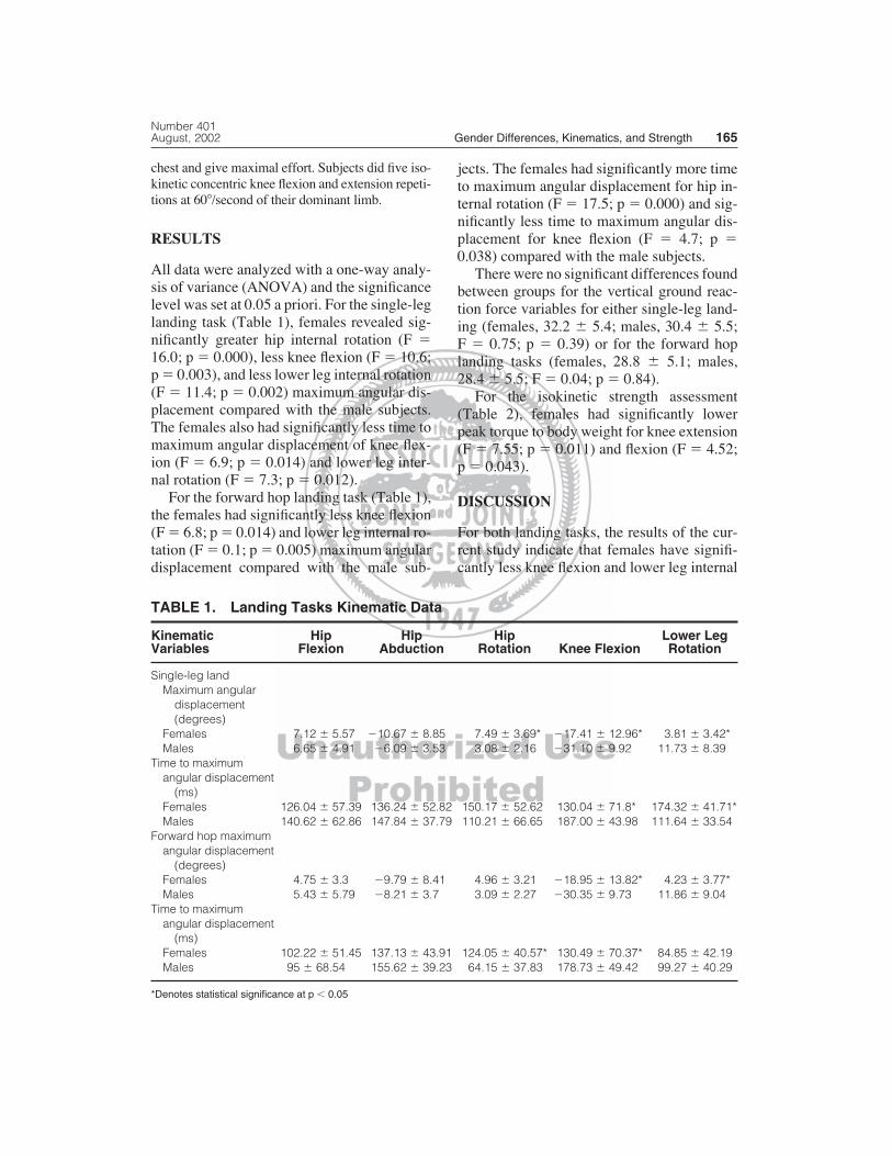
All data were analyzed with a one-way analy-sis of variance (ANOVA) and the significancelevel was set at 0.05 a priori. For the single-leglanding task (Table 1), females revealed sig-nificantly greater hip internal rotation (F �16.0; p � 0.000), less knee flexion (F � 10.6;p � 0.003), and less lower leg internal rotation(F � 11.4; p � 0.002) maximum angular dis-placement compared with the male subjects.The females also had significantly less time tomaximum angular displacement of knee flex-ion (F � 6.9; p � 0.014) and lower leg inter-nal rotation (F � 7.3; p � 0.012).
For the forward hop landing task (Table 1),the females had significantly less knee flexion(F � 6.8; p � 0.014) and lower leg internal ro-tation (F � 0.1; p � 0.005) maximum angulardisplacement compared with the male sub-
jects. The females had significantly more timeto maximum angular displacement for hip in-ternal rotation (F � 17.5; p � 0.000) and sig-nificantly less time to maximum angular dis-placement for knee flexion (F � 4.7; p �0.038) compared with the male subjects.
There were no significant differences foundbetween groups for the vertical ground reac-tion force variables for either single-leg land-ing (females, 32.2 � 5.4; males, 30.4 � 5.5; F � 0.75; p � 0.39) or for the forward hoplanding tasks (females, 28.8 � 5.1; males,28.4 � 5.5; F � 0.04; p � 0.84).
For the isokinetic strength assessment(Table 2), females had significantly lowerpeak torque to body weight for knee extension(F � 7.55; p � 0.011) and flexion (F � 4.52;p � 0.043).
DISCUSSION
For both landing tasks, the results of the cur-rent study indicate that females have signifi-cantly less knee flexion and lower leg internal
TABLE 1. Landing Tasks Kinematic Data
Kinematic Hip Hip Hip Lower Leg Variables Flexion Abduction Rotation Knee Flexion Rotation
Single-leg land Maximum angular
displacement (degrees)
Females 7.12 � 5.57 �10.67 � 8.85 7.49 � 3.69* �17.41 � 12.96* 3.81 � 3.42*Males 6.65 � 4.91 �6.09 � 3.53 3.08 � 2.16 �31.10 � 9.92 11.73 � 8.39
Time to maximum angular displacement
(ms)Females 126.04 � 57.39 136.24 � 52.82 150.17 � 52.62 130.04 � 71.8* 174.32 � 41.71*Males 140.62 � 62.86 147.84 � 37.79 110.21 � 66.65 187.00 � 43.98 111.64 � 33.54
Forward hop maximum angular displacement
(degrees)Females 4.75 � 3.3 �9.79 � 8.41 4.96 � 3.21 �18.95 � 13.82* 4.23 � 3.77*Males 5.43 � 5.79 �8.21 � 3.7 3.09 � 2.27 �30.35 � 9.73 11.86 � 9.04
Time to maximum angular displacement
(ms)Females 102.22 � 51.45 137.13 � 43.91 124.05 � 40.57* 130.49 � 70.37* 84.85 � 42.19Males 95 � 68.54 155.62 � 39.23 64.15 � 37.83 178.73 � 49.42 99.27 � 40.29
*Denotes statistical significance at p � 0.05

rotation after impact than males. The resultsalso revealed that females took significantlyless time to reach maximum knee flexion sub-sequent to impact. Because females had lessmaximum angular displacement than males, itdid not take as long to reach their maximumknee flexion angle resulting in a more abruptabsorption of the impact forces of landing(Figs 3,4).
The kinematic pattern of the females in re-lation to the males during these landing tasksincluded more hip internal rotation with lowerleg external rotation from impact to the maxi-mum rotation point of the maneuver with kneeflexion relatively limited. The kinematics dur-ing landing of the females in the current studywere consistent with those often observed in
the noncontact anterior cruciate ligament in-jury.3,16 The relative lack of knee flexion,combined with a tibial rotary force, muscle re-flex, or a combination of both in response to anunexpected perturbation may result in injuryto the anterior cruciate ligament.4,9,14,28,30,32,34
The current results are consistent with re-sults from other studies which showed therewere gender differences when doing athleticmaneuvers, such as cutting4,19 and landing froma jump.13 These studies showed that femalestend to land with the knee in a more extendedposition4,19 and therefore subject themselvesto higher forces per body weight during theimpact of landing.13 Some reports attributelanding characteristics to training experienceof the athlete.2,8,21,24,27,31 In general, skilled,well-trained, or experienced athletes have beenreported to have increased ankle plantar flex-ion,21,24 knee flexion,8,24,31 and lowered verti-cal ground reaction forces during landing.27,31
Thus this theoretically would permit more timeto distribute the impact forces and allow theopportunity for the musculature to absorb theseforces.24,31 However, to date, no studies haveinvestigated a potential relationship of genderby skill level for these landing characteristics.
Clinical Orthopaedics166 Lephart et al and Related Research
TABLE 2. Isokinetic Assessment: PeakTorque to Body Weight (N-m) at60�/Second
Group Quadriceps Hamstrings
Females 222.93 � 30.86* 113.74 � 23.66*Males 271.68 � 59.27 131.72 � 21.89
*Denotes statistical significance at p � 0.05
Fig 3. Points A and B represent the maximum angular displacement of knee flexion and the time toachieve this position after ground contact. The deficit represents a significant (p � 0.05) difference inknee position after ground contact between females and males.

The other significant result of the currentstudy is related to the relative weakness of thefemale quadriceps and hamstrings when nor-malized to body mass compared with themales (Fig 5). This finding may play a funda-mental role in the landing position observed inthe females during landing.
The role of the quadriceps landing seems tobe critical to the distribution and absorption ofthe impact forces resulting from landing. Al-though the vertical ground reaction forces didnot differ between genders in the current study,the relative lack of knee flexion subsequent toimpact in females has significant implications
Number 401August, 2002 Gender Differences, Kinematics, and Strength 167
Fig 4. Points A and B represent the maximum angular displacement of knee flexion and the time toachieve this position after ground contact. The deficit represents a significant (p � 0.05) difference inknee position after ground contact between females and males.
Fig 5. The quadriceps isokinetic peak torque to body mass at 60�/second for the male group was sig-nificantly greater (p � 0.05) than that of the female group. The hamstring isokinetic peak torque to bodymass at 60�/second for the male group was significantly greater (p � 0.05) than that of the female group.

for the manner in which force transmission upthe kinetic chain occurs. The authors suspectthat the lack of vertical ground reaction forcedifference was attributed to other force ab-sorbing compensatory mechanisms that werenot studied, such as ankle kinematics or mus-cle activity.
Subsequent to impact, the quadriceps mus-cle eccentrically contracts to control knee flex-ion and decelerate the land. The minimal kneeflexion at impact observed and the lack of con-trolled knee flexion deceleration in the femaleathletes may be related to the relatively weakleg musculature, especially the quadriceps.Without sufficient strength available to de-celerate the body by the eccentric quadricepsmechanism, it seems that the females land in amore extended knee position and tend to main-tain this extended position subsequent to groundcontact rather than absorbing the impact withcontrolled knee flexion. This knee extendedposition, combined with internal hip rotation,makes females vulnerable for anterior cruciateligament loading.
Physicians, athletic trainers, and others whoare concerned with the care of athletes, need toevaluate the biomechanics of the female ath-letes to ensure proper technique is being usedduring landing activities and continue to edu-cate coaches to implement training practicesusing proper techniques. Maximizing joint an-gles, specifically knee flexion, subsequent toimpact will aid in attenuating potentially harm-ful forces and ensure protective biomechani-cal patterns, which may promote more appro-priate muscle firing patterns to protect the knee.Additionally, awkward or poor landing skillsmay identify a specific muscle weakness. Ad-ditional research is needed to explore if a rela-tionship exists between the weakness of mus-cles and poor landing tasks as center of gravityand trunk angle differences may alter hip andknee stability.
The data from this study suggest that bio-mechanical and neuromuscular variables differbetween genders during impact on landing.Males had a greater amount of knee flexionsubsequent to impact. The larger flexion dis-
placement serves to attenuate impact forces re-ducing loads imposed on the joint. The absenceof this controlled knee flexion in females maybe related to the weaker quadriceps and ham-strings, resulting in an abrupt stiffening of theknee. These factors need to be considered re-lated to the pathoetiology of anterior cruciateligament injuries in the female athlete.
References1. Arendt E, Dick R: Knee injury patterns among men
and women in collegiate basketball and soccer. AmJ Sports Med 23:694–701, 1995.
2. Bobbert MF: Drop landings as a training method forjumping ability. Sports Med 9:7–22, 1990.
3. Boden BP, Dean GS, Feagin JA, et al: Mechanismsof injury to the anterior cruciate ligament. Orthope-dics 23:573–578, 2000.
4. Colby S, Francisco A, Yu B, et al: Electromyo-graphic and kinematic analysis of cutting maneu-vers: Implications for anterior cruciate ligament in-jury. Am J Sports Med 28:234–240, 2000.
5. Colby SM, Hintermeister RA, Torry MR, et al:Lower limb stability with ACL impairment. J OrthopSports Phys Ther 29:444–454, 1999.
6. DeVita P, Skelly WA: Effect of landing stiffness onjoint kinetics and energetics in the lower extremity.Med Sci Sports Exerc 24:108–115, 1992.
7. Dufek JS, Bates BT: Biomechanical factors associ-ated with injury during landing in jump sports.Sports Med 12:326–337, 1991.
8. Dufek JS, Zhang S. Landing models for volleyballplayers: A longitudinal evaluation. J Sports MedPhys Fitness 36:35–42, 1996.
9. Durselen L, Claes L, Keifer H: The influence of mus-cle forces and external loads on cruciate ligamentstrain. Am J Sports Med 23:129–136, 1995.
10. Ferretti A, Papandrea P, Conteduca F, et al: Knee lig-ament injuries in volleyball players. Am J SportsMed 20:203–207, 1992.
11. Gray J, Taunton JE, McKensie DC, et al: A surveyof injuries to the anterior cruciate ligament of theknee in female basketball players. Int J Sports Med6:314–316, 1985.
12. Griffin LY, Agel J, Albohm MJ, et al: Noncontact an-terior cruciate ligament injuries: Risk factors and pre-vention strategies. J Am Acad Orthop Surg 8:141–150,2000.
13. Hewett TE, Stroupe AL, Nance TA, et al: Plymetrictraining in female athletes: Decreased impact forcesand increased hamstring torques. Am J Sports Med24:765–773, 1996.
14. Hirokawa S, Solomonow M, Lu Y, et al: Anterior-posterior and rotational displacement of the tibiaelicited by quadriceps contractions. Am J SportsMed 20:229–306, 1992.
15. Huston LJ, Wojtys EM: Neuromuscular perfor-mance characteristics in elite female athletes. Am JSports Med 24:427–436, 1996.
16. Ireland ML, Gaudette M, Crook S: ACL injuries inthe female athlete. J Sport Rehabil 6:97–110, 1997.
Clinical Orthopaedics168 Lephart et al and Related Research

17. Knapik JJ, Bauman CL, Jones BH, et al: Preseasonstrength and flexibility imbalances associated withathletic injuries in female collegiate athletes. Am JSports Med 19:76–81, 1991.
18. Lindenfeld TN, Schmitt DJ, Hendy MP, et al: Inci-dence of injury in indoor soccer. Am J Sports Med22:364–371, 1994.
19. Malinzak RA, Colby SM, Kirkendall DT, Yu B, Gar-rett WE: A comparison of knee joint motion patternsbetween men and women in selected athletic tasks.Clin Biomech 16:438–445, 2001.
20. Malone TR, Hardaker WT, Garrett WE, et al: Rela-tionship of gender to anterior cruciate ligament in-juries in intercollegiate basketball players. J SouthOrthop Assoc 2:36–39, 1993.
21. McKinley P, Pedotti A: Motor strategies in landingfrom a jump: The role of skill in task execution. ExpBrain Res 90:427–440, 1992.
22. McLean SG, Neal RJ, Myers PT, et al: Knee jointkinematics during the sidestep cutting maneuver: Po-tential for injury in women. Med Sci Sports Exerc31:959–968, 1999.
23. McNair PJ, Marshall RN: Landing characteristics insubjects with normal and anterior cruciate liga-ment deficient knee joints. Arch Phys Med Rehabil75:584–589, 1994.
24. McNitt-Gray JL: Kinetics of the lower extremitiesduring drop landings from three heights. J Biomech26:1037–1046, 1993.
25. Miyasaka KC, Daniel DM, Stone ML, et al: The in-cidence of knee ligament injuries in the general pop-ulation. Am J Knee Surg 4:3–8, 1991.
26. Nyland JA, Shapiro R, Caborn DNM, et al: The ef-
fect of quadriceps femoris, hamstring, and placeboeccentric fatique on knee and ankle dynamics dur-ing crossover cutting. J Orthop Sports Phys Ther25:171–184, 1997.
27. Prapavessis H, McNair PJ: Effects of instruction injumping technique and experience jumping onground reaction forces. J Orthop Sports Phys Ther29:352–356, 1999.
28. Renstrom P, Arms SW, Stanwyck TS, et al: Strainwithin the anterior cruciate ligament during hamstringand quadriceps activity. Am J Sports Med 14:83–87,1986.
29. Rozzi SL, Lephart SM, Gear WS, et al: Knee jointlaxity and neuromuscular characteristics of male andfemale soccer and basketball players. Am J SportsMed 27:312–319, 1999.
30. Smidt JG: Biomechanical analysis of knee flexionand extension. J Biomech 6:79–92, 1973.
31. Viitasalo JT, Salo A, Lahtinen J: Neuromuscularfunctioning of athletes and non-athletes in the dropjump. Eur J Appl Physiol 78:432–440, 1998.
32. Wilk KE, Escamilla RF, Fleisig GS, et al: A com-parison of tibiofemoral joint forces and electromyo-graphic activity during open and closed kinetic chainexercises. Am J Sports Med 24:518–527, 1996.
33. Wojtys EM, Huston LJ, Taylor PD, et al: Neuromus-cular adaptations in isokinetic, isotonic, and agilitytraining programs. Am J Sports Med 24:187–192,1996.
34. Woo SL, Hollis M, Adams D, et al: Tensile proper-ties of the human femur-anterior cruciate ligament-tibia complex: The effects of specimen age and ori-entation. Am J Sports Med 9:217–225, 1991.
Number 401August, 2002 Gender Differences, Kinematics, and Strength 169