the role of melatonin in diabetes: therapeutic implications
TRANSCRIPT

Copy
right
© A
E&M
all r
ight
s res
erve
d.
391
review
Arch Endocrinol Metab. 2015;59/5
The role of melatonin in diabetes: therapeutic implications
Shweta Sharma1, Hemant Singh1, Nabeel Ahmad2, Priyanka Mishra1, Archana Tiwari1
ABSTRACTMelatonin referred as the hormone of darkness is mainly secreted by pineal gland, its levels being elevated during night and low during the day. The effects of melatonin on insulin secretion are me-diated through the melatonin receptors (MT1 and MT2). It decreases insulin secretion by inhibiting cAMP and cGMP pathways but activates the phospholipaseC/IP3 pathway, which mobilizes Ca2+ from organelles and, consequently increases insulin secretion. Both in vivo and in vitro, insulin secretion by the pancreatic islets in a circadian manner, is due to the melatonin action on the melatonin recep-tors inducing a phase shift in the cells. Melatonin may be involved in the genesis of diabetes as a reduction in melatonin levels and a functional interrelationship between melatonin and insulin was observed in diabetic patients. Evidences from experimental studies proved that melatonin induces production of insulin growth factor and promotes insulin receptor tyrosine phosphorylation. The dis-turbance of internal circadian system induces glucose intolerance and insulin resistance, which could be restored by melatonin supplementation. Therefore, the presence of melatonin receptors on hu-man pancreatic islets may have an impact on pharmacotherapy of type 2 diabetes. Arch Endocrinol Metab. 2015;59(5):391-9
KeywordsMelatonin; diabetes; insulin; beta cells; calcium; circadian rhythm
1 School of Biotechnology, Rajiv Gandhi Technical University, Gandhi Nagar, Bhopal, Madhya Pradesh 2 School of Biotechnology, IFTM University, Lodhipur Rajput, Uttar Pradesh
Correspondence to:Shweta SharmaSchool of BiotechnologyRajiv Gandhi Technical UniversityAirport Bypass Road, Gandhi Nagar462036 – Bhopal, Madhya Pradesh [email protected]
Received on June/8/2015Accepted on July/6/2015
DOI: 10.1590/2359-3997000000098
INTRODUCTION
D iabetes is an endocrine disease, consist of insu-lin resistance, a diminished pancreatic beta-cell
function, abnormally high glucagon levels and a re-duced incretin effect (1). Diabetes is classified into two main categories: type 1 (an autoimmune disease of younger patients with a lack of insulin production caus-ing hyperglycemia) and type 2 (a metabolic disorder resulting from the body’s inability to produce enough or properly utilize insulin hence patients have hypergly-cemia). Changing lifestyle trends such as a tendency to nocturnality and intake of excessively rich diets, cause disturbance of the sleep/wake cycle along with other circadian rhythms (2). Deviation in circadian patterns favors the occurrence of diabetes (3).
Inconsistent data have been reported concerning the effect of pineal hormone on the secretion of in-sulin, on blood glucose and carbohydrate metabolism. Melatonin (N-acetyl-5-methoxytryptamine), a trypto-phan derived small indolic molecule, is mainly secreted by the pineal gland locally in several other tissues (4,5). Experimental evidences proposed the diurnal profiles of blood glucose due to melatonin and increased insu-lin levels in diabetic animals and humans (6). Pinealec-
tomy of rodents causes hyperinsulinemia (7). Moreo-ver, diabetes is coupled with lower melatonin levels as reduction in serum melatonin and higher insulin level is observed in type 2 diabetic Goto Kakizaki rats (6). Genome-wide association studies has shown that spe-cific single-nucleotide polymorphisms of the melatonin receptor 2 (MTNR1B) locus is linked with an increased blood glucose concentration and type 2 diabetes (8-13). Melatonin can be able to bring anti-hyperglycemic effect either by improving insulin sensitization or by improvement of insulin secretion, or both.
MELATONIN AND ITS FUNCTIONMelatonin is known as the hormone of darkness, is an indoleamine with the chemical name N-acetyl-5-me-thoxytryptamine. Circulating plasma concentrations are secreted by the pineal gland. In mammals, the con-centration in plasma during night was found to be (80–100 pg/mL) and low levels during the day (10–20 pg/mL) (14). It maintains homeostasis in the body, helps adjust the timing or reinforces oscillations of the bio-logical clock (15). Its synthesis comprised of two steps, initially the conversion of amino acid tryptophan into serotonin (5-hydroxytryptamine, 5-HT), further acety-
Copy
right
© A
E&M
all r
ight
s res
erve
d.
392
Melatonin and type 2 diabetes
Arch Endocrinol Metab. 2015;59/5
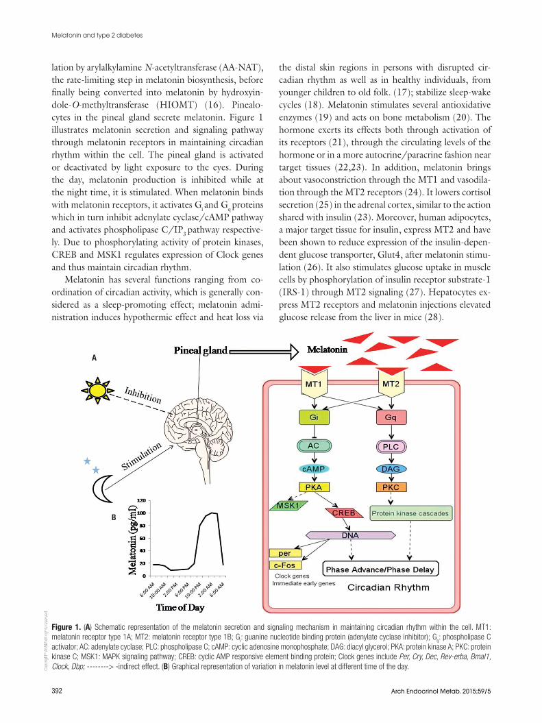
lation by arylalkylamine N-acetyltransferase (AA-NAT), the rate-limiting step in melatonin biosynthesis, before finally being converted into melatonin by hydroxyin-dole-O-methyltransferase (HIOMT) (16). Pinealo-cytes in the pineal gland secrete melatonin. Figure 1 illustrates melatonin secretion and signaling pathway through melatonin receptors in maintaining circadian rhythm within the cell. The pineal gland is activated or deactivated by light exposure to the eyes. During the day, melatonin production is inhibited while at the night time, it is stimulated. When melatonin binds with melatonin receptors, it activates Gi and Gq proteins which in turn inhibit adenylate cyclase/cAMP pathway and activates phospholipase C/IP3 pathway respective-ly. Due to phosphorylating activity of protein kinases, CREB and MSK1 regulates expression of Clock genes and thus maintain circadian rhythm.
Melatonin has several functions ranging from co-ordination of circadian activity, which is generally con-sidered as a sleep-promoting effect; melatonin admi-nistration induces hypothermic effect and heat loss via
the distal skin regions in persons with disrupted cir-cadian rhythm as well as in healthy individuals, from younger children to old folk. (17); stabilize sleep-wake cycles (18). Melatonin stimulates several antioxidative enzymes (19) and acts on bone metabolism (20). The hormone exerts its effects both through activation of its receptors (21), through the circulating levels of the hormone or in a more autocrine/paracrine fashion near target tissues (22,23). In addition, melatonin brings about vasoconstriction through the MT1 and vasodila-tion through the MT2 receptors (24). It lowers cortisol secretion (25) in the adrenal cortex, similar to the action shared with insulin (23). Moreover, human adipocytes, a major target tissue for insulin, express MT2 and have been shown to reduce expression of the insulin-depen-dent glucose transporter, Glut4, after melatonin stimu-lation (26). It also stimulates glucose uptake in muscle cells by phosphorylation of insulin receptor substrate-1 (IRS-1) through MT2 signaling (27). Hepatocytes ex-press MT2 receptors and melatonin injections elevated glucose release from the liver in mice (28).
Figure 1. (A) Schematic representation of the melatonin secretion and signaling mechanism in maintaining circadian rhythm within the cell. MT1: melatonin receptor type 1A; MT2: melatonin receptor type 1B; G
i: guanine nucleotide binding protein (adenylate cyclase inhibitor); G
q: phospholipase C
activator; AC: adenylate cyclase; PLC: phospholipase C; cAMP: cyclic adenosine monophosphate; DAG: diacyl glycerol; PKA: protein kinase A; PKC: protein kinase C; MSK1: MAPK signaling pathway; CREB: cyclic AMP responsive element binding protein; Clock genes include Per, Cry, Dec, Rev-erba, Bmal1, Clock, Dbp; --------> -indirect effect. (B) Graphical representation of variation in melatonin level at different time of the day.
A
B
Copy
right
© A
E&M
all r
ight
s res
erve
d.
393
Melatonin and type 2 diabetes
Arch Endocrinol Metab. 2015;59/5
MELATONIN RECEPTORS AND ITS CLASSIFICATION
Melatonin receptors belong to a family of receptors re-ferred to as G protein coupled receptors (GPCR) (29). Melatonin mediates circadian rhythms and other physi-ological functions via membrane receptors on the cell surface. Melatonin is considered as membrane-perme-able substance owing to its chemical structure so it has both receptor-independent and receptor-dependent effects. All of its cellular actions and effects are likely transmitted via two known GPCR isoforms, denoted MT1 and MT2 previously known as Mel1a and Mel1b (30,31). The two receptors exhibit a high degree of homology (31). There are mainly two types of mela-tonin receptors found in humans, melatonin receptor 1 (MT1; MTNR1A) and melatonin receptor 2 (MT2; MTNR1B), a third melatonin receptor supposed to ex-ist and has been identified that belong to the family of quinone reductases (32).
The melatonin influences exocytosis of insulin by β-cells as concluded from experiments via non-hydroly-sable guanosine-5’-trisphosphate (GTP) analogue gua-nosine 5’-O-(3-thiotrisphosphate) and the melatonin antagonist luzindole (33), both of which inhibit the melatonin action on secretion of insulin from neonatal rat islets. The existence of MT2 receptor on the pancre-atic β-cell is accomplished by application of melatonin as isolated islets of rats has phase-shifting effects on the insulin rhythm (34). Moreover, molecular and immu-nocytochemical studies confirmed the presence of the melatonin receptors MT1 and MT2 in the islets of Lan-gerhans and also in human pancreatic tissue (35).
MELATONIN AND CIRCADIAN RHYTHM
Deregulation of the circadian system is associated with the instance of metabolic syndrome, including diabetes and obesity (36). The transcription factors that main-tains rhythmic functions consist of the clock regulators including the clock circadian regulator (Clock) and Aryl hydrocarbon receptor nuclear translocator-like (Arntl, also known as Bmal1) that heterodimerize and activate transcription of target genes, including Period (Per1, 2, and 3) and Cryptochrome (Cry 1 and 2) (37). Pan-creatic circadian insulin oscillations were analyzed by changing the expression of clock genes on the transcrip-tional level (38).During a 24-hr period, Tim, Bmal1, Per1, Per2, Clock and Cry1 as well as clock-controlled output genes, Dbp and Rev-erbα were examined by by
real-time RT-PCR (39,40). The results established the role of a circadian pacemaker in the rat pancreas and the existence of a circadian oscillator within islets. In addi-tion to the SCN, clock activities have been identified in numerous peripheral tissues including the liver, white adipose tissue, and pancreas that control metabolic pro-cesses (41,42).
Melatonin exerts its effect through melatonin recep-tors of different peripheral tissues, thus maintaining cir-cadian rhythms. Likewise other physiological functions, glucose metabolism is regulated by circadian system (43,44). In an experiment, study Clock mutant mice showed lack of rhythmicity in the action of insulin, a condition which was reversible once the clock protein was reintroduced (43). The removal of pancreatic Clock and Bmal1 in mice resulted in functional defects in in-sulin secretion and decreases in islet size and survival, signifies a key role of peripheral clocks in the regulation of glucose homeostasis (45). In this manner, melatonin could either directly influence the clock machinery in the pancreas or indirectly via the SCN.
MELATONIN SIGNALING IN PANCREATIC β-CELLS
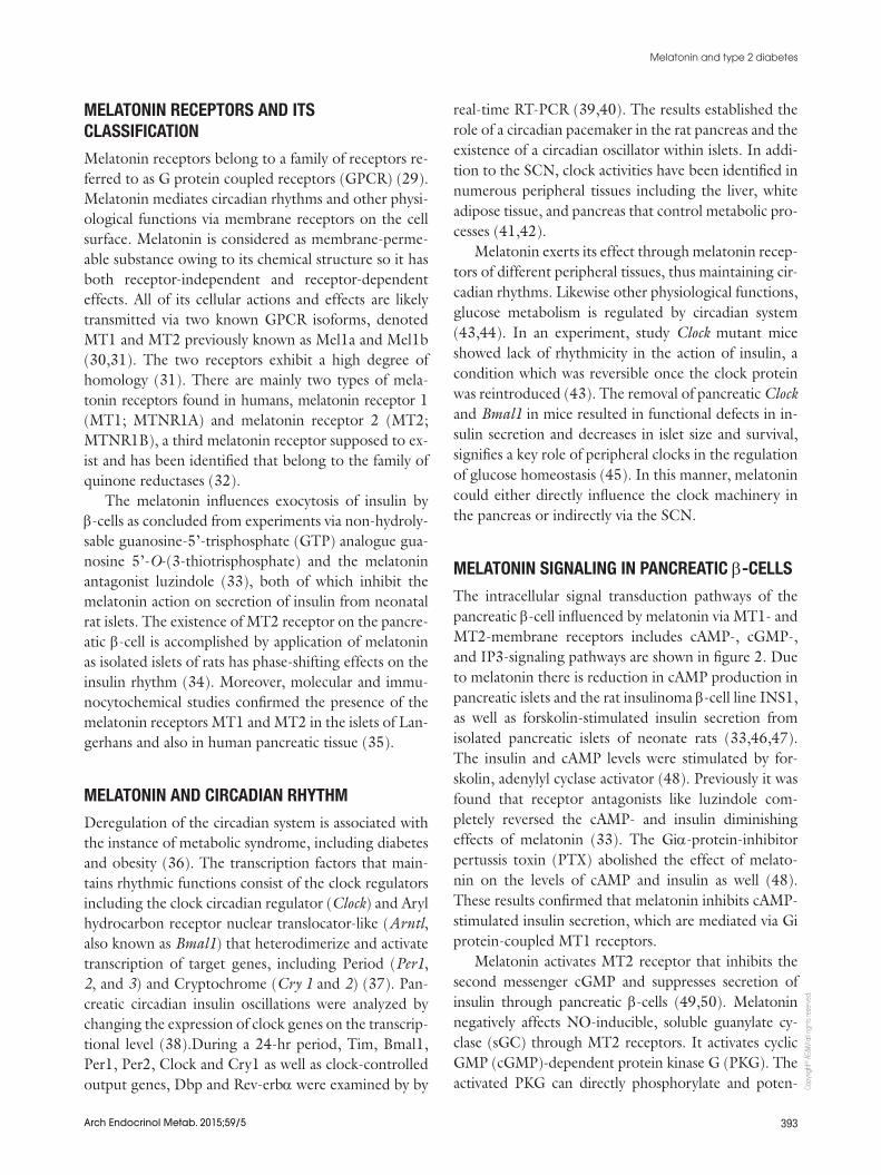
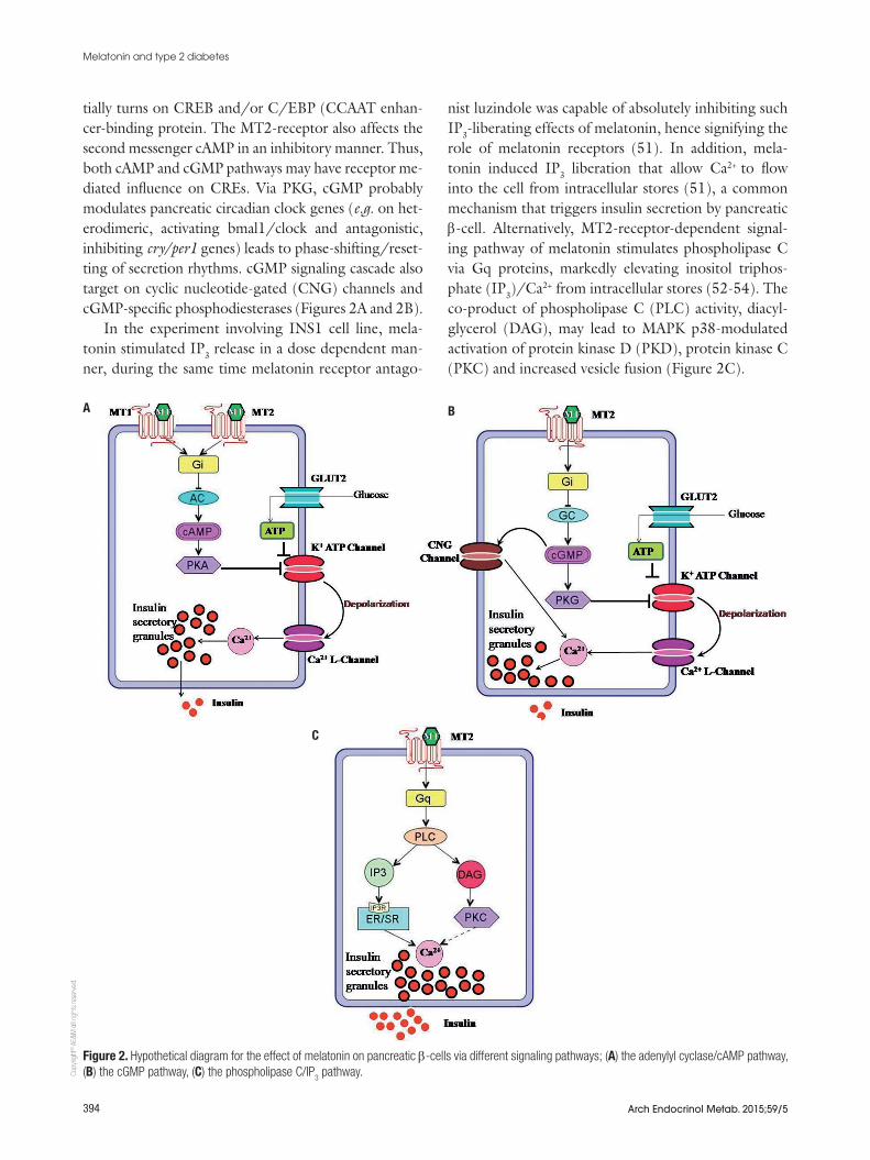
The intracellular signal transduction pathways of the pancreatic β-cell influenced by melatonin via MT1- and MT2-membrane receptors includes cAMP-, cGMP-, and IP3-signaling pathways are shown in figure 2. Due to melatonin there is reduction in cAMP production in pancreatic islets and the rat insulinoma β-cell line INS1, as well as forskolin-stimulated insulin secretion from isolated pancreatic islets of neonate rats (33,46,47). The insulin and cAMP levels were stimulated by for-skolin, adenylyl cyclase activator (48). Previously it was found that receptor antagonists like luzindole com-pletely reversed the cAMP- and insulin diminishing effects of melatonin (33). The Giα-protein-inhibitor pertussis toxin (PTX) abolished the effect of melato-nin on the levels of cAMP and insulin as well (48).These results confirmed that melatonin inhibits cAMP-stimulated insulin secretion, which are mediated via Gi protein-coupled MT1 receptors.
Melatonin activates MT2 receptor that inhibits the second messenger cGMP and suppresses secretion of insulin through pancreatic β-cells (49,50). Melatonin negatively affects NO-inducible, soluble guanylate cy-clase (sGC) through MT2 receptors. It activates cyclic GMP (cGMP)-dependent protein kinase G (PKG). The activated PKG can directly phosphorylate and poten-
Copy
right
© A
E&M
all r
ight
s res
erve
d.
394
Melatonin and type 2 diabetes
Arch Endocrinol Metab. 2015;59/5
tially turns on CREB and/or C/EBP (CCAAT enhan-cer-binding protein. The MT2-receptor also affects the second messenger cAMP in an inhibitory manner. Thus, both cAMP and cGMP pathways may have receptor me-diated influence on CREs. Via PKG, cGMP probably modulates pancreatic circadian clock genes (e.g. on het-erodimeric, activating bmal1/clock and antagonistic, inhibiting cry/per1 genes) leads to phase-shifting/reset-ting of secretion rhythms. cGMP signaling cascade also target on cyclic nucleotide-gated (CNG) channels and cGMP-specific phosphodiesterases (Figures 2A and 2B).
In the experiment involving INS1 cell line, mela-tonin stimulated IP3 release in a dose dependent man-ner, during the same time melatonin receptor antago-
nist luzindole was capable of absolutely inhibiting such IP3-liberating effects of melatonin, hence signifying the role of melatonin receptors (51). In addition, mela-tonin induced IP3 liberation that allow Ca2+ to flow into the cell from intracellular stores (51), a common mechanism that triggers insulin secretion by pancreatic β-cell. Alternatively, MT2-receptor-dependent signal-ing pathway of melatonin stimulates phospholipase C via Gq proteins, markedly elevating inositol triphos-phate (IP3)/Ca2+ from intracellular stores (52-54). The co-product of phospholipase C (PLC) activity, diacyl-glycerol (DAG), may lead to MAPK p38-modulated activation of protein kinase D (PKD), protein kinase C (PKC) and increased vesicle fusion (Figure 2C).
Figure 2. Hypothetical diagram for the effect of melatonin on pancreatic β-cells via different signaling pathways; (A) the adenylyl cyclase/cAMP pathway, (B) the cGMP pathway, (C) the phospholipase C/IP
3 pathway.
A B
C

Copy
right
© A
E&M
all r
ight
s res
erve
d.
395
Melatonin and type 2 diabetes
Arch Endocrinol Metab. 2015;59/5
MELATONIN AND GLUCOSE HOMEOSTASIS
Various studies have shown that melatonin may influ-ence insulin secretion and glucose homeostasis. A low quantity of circulating melatonin occur in patients with type 2 diabetes (11), at the same time upregulated mRNA expression of melatonin membrane receptor was observed (55). Furthermore, polymorphisms in the melatonin receptor gene were linked with fasting blood glucose level and susceptibility to the occurrence of type 2 diabetes (56). These clinical results indicate that melatonin improves glycemic control in blood and the insufficiency of melatonin might be associated with the development of type 2 diabetes. The study investi-gating the effects of melatonin on glucose homeostasis in young male Zucker diabetic fatty (ZDF) rats, an ex-perimental model of metabolic syndrome and type 2 diabetes, show that oral melatonin administration exert anti-hyperglycemic effect in young ZDF rats as insulin sensitizer and by improvement in β-cell function (57). Reverse transcription polymerase chain reaction estab-lished that melatonin receptor deficiency have an effect on transcript levels of pancreatic islet hormones in ad-dition to pancreatic and hepatic glucose transporters (Glut1 and 2) (58). In a group of type 2 diabetic pa-tients, when melatonin and zinc acetate supplemented with and without metformin improved glycemic control through decreasing FPG (fasting plasma glucose) but the mechanism not related to increase in insulin secre-tion (59). Clinical implications of melatonin were pre-sented by the data obtained from selected population of postmenopausal women as administration of melatonin reduced glucose tolerance and insulin sensitivity (60).
MELATONIN AND INSULIN SECRETION
Human and rodent pancreatic tissues and islets and rodent cell lines has been found to express MT1 and MT2 receptors (10,35,53,55,61-65). The occurrence of melatonin receptors in the pancreatic islets proposes that their activation by melatonin might directly influ-ence insulin or glucagon production and provides a biochemical basis to explain how decreased melatonin levels of diabetic patients could affect the function of the pancreas (6,66). The basis for glucose homeostasis is the secretion of insulin and glucagon by the pancre-atic islet cells. High level of glucose observed at the start of the active phase shows circadian variation in the concentration of plasma glucose. Since the intake of
food induces insulin secretion, the amount of insulin in plasma follow the daily rhythm in feeding and may exhibit a daily rhythm as well. In contrast, mouse and human pancreatic islet cells have been found to pos-sess circadian activity as well (34,67,68). This concept gives emphasis to the presence of a circadian regulation over pancreatic function. The analysis of melatonin on pancreatic islet by immunoprecipitation and immu-noblotting shown that melatonin regulate growth and differentiation of pancreatic cells by stimulating insulin growth factor receptor (IGF-R) and insulin receptor (IR) tyrosine phosphorylation (69). It activates two intracellular signaling pathways: PI3K/AKT (involved with cell metabolism) and MEK/ERKs (involved in cell proliferation, growth and differentiation) (69). Moreover, the decrease in melatonin levels augmented insulin secretion in rats during the day while at the time of night, low levels of insulin along with high glucose levels are measured when melatonin levels are elevated (70,71).
There is prospect that there might be an associa-tion between melatonin and type 2 diabetes based on the findings that insulin secretion is inversely propor-tional to plasma melatonin concentration (72). Mela-tonin inhibits glucose mediated release of insulin from pancreatic cells emphasizing its activity in the function of insulin (72). Suppression of melatonin secretion by nocturnal light exposure could be a critical factor for type 2 diabetes development (44). Furthermore, MT1 receptors are involved in the modulation of glucose ho-meostasis in mice and might stimulate insulin to induce glucose uptake (73). Therefore, the available literature proposed that presence of melatonin have direct or in-direct effect on insulin secretion both in vivo and in vitro, and night-time melatonin levels are associated to night-time insulin concentrations in patients with dia-betes.
MELATONIN RECEPTOR POLYMORPHISM
The effect of melatonin is exerted by the two G-pro-tein coupled receptors, melatonin receptor type 1A and melatonin receptor type 1B. The two distinct recep-tors have been found to be expressed in human pan-creatic islets (23,64). Recent genome-wide association studies (GWAS) identified common genetic variants within MTNR1B were associated with higher fasting glucose levels or the increased risk of type 2 diabetes (11,61). The two common genetic variants: rs1387153
Copy
right
© A
E&M
all r
ight
s res
erve
d.
396
Melatonin and type 2 diabetes
Arch Endocrinol Metab. 2015;59/5
and rs10830963 are located near the gene MTNR1B that encodes the MT2 receptor of melatonin. The vari-ant with the strongest association signal was the single nucleotide polymorphism (SNP) rs10830963 (74). A meta-analysis revealed that rs10830963 is strongly associated with fasting glucose levels and moderately associated with an increased risk to develop diabetes (12). The risk allele was also related to impairment of early insulin secretion and beta cell dysfunction that might represent the pathomechanism for the increased risk of type 2 diabetes by the rs10830963 risk allele (11,74). Recently, the associations of rs10830963 with the elevated fasting glucose and risk of type 2 diabe-tes were reported in Asian adults, including Chinese (75-80), Japanese and Sri Lankan populations (81). In the pregnant Chinese women, the MTNR1B variant rs10830963, rs1387153, rs2166706 and rs1447352 were shown to be associated with gestational glucose in-tolerance so MTNR1B is probably involved in the regu-lation of glucose homeostasis during pregnancy (82).
The expression of MT2 receptor in the β-cells im-plies that MTNR1B gene variant might affect pancre-atic glucose sensing and insulin secretion and thereby hyperglycemia (61). One study suggested that IGR (Impaired glucose regulation) might have similar background of susceptible genetic variations as well as indicated significantly increased risk of MTNR1B rs10830963 polymorphism for IGF (impaired fasting glucose) but not for IGT (impaired glucose tolerance) when stratified by IGR outcome (83). Another study on a Czech cohort of women confirms that allele G of rs10830963 in MTNR1B gene is associated with in-creased risk of developing GDM (Gestational diabetes mellitus) and, in non-diabetic normoglycemic subjects, with FPG (Fasting plasma glucose) levels and glucose processing during the oral glucose-tolerance test (84). Furthermore, a polymorphic allele was identified in CRY2 associated with type 2 diabetes (85). A group of researchers reported that the MTNR1B-associated Single Nucleotide Polymorphisms rs10830962, rs4753426 and the aforementioned rs10830963 were all significantly associated with higher fasting glucose concentrations in the blood and decreased insulin se-cretion in German cohorts (86).
MELATONIN AND DIABETES
In diabetic patients, a reduction in melatonin levels and a functional inter-relationship between melatonin
and insulin was observed. On this basis melatonin may perhaps be involved in the genesis of diabetes (6). In humans, melatonin administration reduced glucose tol-erance mainly by decreasing insulin release at the time of morning while decline in insulin sensitivity was ob-served in the evening (87). In addition, various studies established a correlation between sleep disorders and a greater risk for a decreased glucose tolerance and type 2 diabetes (88-90). The existence of an association be-tween glucose and time keeping mechanism has been proved by the alteration of 24 h rhythmic expression of clock genes as a result of high fat diet intake in rats (91). Genome-wide association studies have proposed that allelic variations of the melatonin receptor MT2 affect glycemic traits such as elevated fasting glucose levels in plasma, impaired insulin secretion, and risk of type 2 diabetes (11,92,93). Both melatonin and insulin exhibit a circadian rhythm but there is negative correla-tion between melatonin and insulin i.e. insulin levels alters in a reverse fashion to melatonin (94). Decreased melatonin level in irregular manner has been related with diabetes (6,55), which suggests that the melato-nin signal is critical for glucose regulation in blood and maintaining homeostasis (95). In patients with type 2 diabetes, gluconeogenesis and endogenous glucose pro-duction exhibit circadian rhythm that impel fasting high blood glucose and do not exist in healthy humans (96).
Significant changes in behavioral activity in control rats were observed on reversing the LD (light/dark) conditions whereas no shift was observed in rats with diabetes (97). There were larger variations in blood glucose levels of rats suggesting that the changes in behavior and insulin levels are due to misalignment of clock functioning as a result of LD changes (97). Hence, the decline in melatonin levels during exposure to light at night and aging, may lead to the occurrence or development of type 2 diabetes.
CONCLUSION
Circadian system may be a tractable target for decreas-ing the prevalence of hyperglycemia and insulin resis-tance. The loss of glycemic control and substantial el-evations of fasting glucose are complications that arise from type 2 diabetes and typically result from progres-sive loss of pancreatic beta-cell function and decline in insulin. Different animal studies suggest that melatonin supplementation may have beneficial effects on glucose homeostasis and body weight regulation under certain
Copy
right
© A
E&M
all r
ight
s res
erve
d.
397
Melatonin and type 2 diabetes
Arch Endocrinol Metab. 2015;59/5
circumstances, which should encourage clinical trials in humans to evaluate the therapeutic potential of this hormone in diabetes. Diabetes is a prevalent disease in middle-aged and older adults and maintenance of opti-mal levels of blood sugar in diabetes patients is a major clinical issue. The present evidence that melatonin in-duces insulin secretion by IP3- signaling pathway and can improve β-cell function, so melatonin supplemen-tation may have beneficial effects on glucose homeosta-sis. It would advance the current therapeutic strategy to overcome the diabetes effects which is currently pre-scribed for sleep and circadian rhythm.
REFERENCES1. Bergenstal R, Kendall D, Franz M, Rubenstein A. Management of
type 2 diabetes: a systematic approach to meeting the standards of care. II: Oral agents, insulin, and management of complica-tions. Endocrinology 4th ed Philadelphia, Pa: WB Saunders Co. 2001. 822 p.
2. Bixler E. Sleep and society: an epidemiological perspective. Sleep Med. 2009;10:S3-6.
3. Scheer FA, Hilton MF, Mantzoros CS, Shea SA. Adverse metabolic and cardiovascular consequences of circadian misalignment. Proc Natl Acad Sci U S A. 2009;106(11):4453-8.
4. Reiter RJ. Pineal melatonin: cell biology of its synthesis and of its physiological interactions. Endocr Rev. 1991;12(2):151-80.
5. Stefulj J, Hörtner M, Ghosh M, Schauenstein K, Rinner I, Wölfler A, et al. Gene expression of the key enzymes of melatonin synthe-sis in extrapineal tissues of the rat. J Pineal Res. 2001;30(4):243-7.
6. Peschke E, Frese T, Chankiewitz E, Peschke D, Preiss U, Schneyer U, et al. Diabetic Goto Kakizaki rats as well as type 2 diabetic pa-tients show a decreased diurnal serum melatonin level and an increased pancreatic melatonin‐receptor status. J Pineal Res. 2006;40(2):135-43.
7. Nishida S, Segawa T, Murai I, Nakagawa S. Long-term melatonin administration reduces hyperinsulinemia and improves the al-tered fatty-acid compositions in type 2 diabetic rats via the resto-ration of Delta-5 desaturase activity. J Pineal Res. 2002;32(1):26-33.
8. Staiger H, Machicao F, Schäfer SA, Kirchhoff K, Kantartzis K, Guthoff M, et al. Polymorphisms within the novel type 2 dia-betes risk locus MTNR1B determine β-cell function. PLoS One. 2008;3(12):e3962.
9. Bouatia-Naji N, Bonnefond A, Cavalcanti-Proença C, Sparsø T, Holmkvist J, Marchand M, et al. A variant near MTNR1B is as-sociated with increased fasting plasma glucose levels and type 2 diabetes risk. Nat Genet. 2008;41(1):89-94.
10. Lyssenko V, Nagorny CL, Erdos MR, Wierup N, Jonsson A, Spégel P, et al. Common variant in MTNR1B associated with increased risk of type 2 diabetes and impaired early insulin secretion. Nat Genet. 2008;41(1):82-8.
11. Prokopenko I, Langenberg C, Florez JC, Saxena R, Soranzo N, Thorleifsson G, et al. Variants in MTNR1B influence fasting glu-cose levels. Nat Genet. 2008;41(1):77-81.
12. Sparsø T, Bonnefond A, Andersson E, Bouatia-Naji N, Holmkvist J, Wegner L, et al. G-allele of Intronic rs10830963 in MTNR1B Con-fers Increased Risk of Impaired Fasting Glycemia and Type 2 Dia-betes Through an Impaired Glucose-Stimulated Insulin Release Studies Involving 19,605 Europeans. Diabetes. 2009;58(6):1450-6.
13. Rönn T, Wen J, Yang Z, Lu B, Du Y, Groop L, et al. A common vari-ant in MTNR1B, encoding melatonin receptor 1B, is associated
with type 2 diabetes and fasting plasma glucose in Han Chinese individuals. Diabetologia. 2009;52(5):830-3.
14. Simonneaux V, Ribelayga C. Generation of the melatonin endo-crine message in mammals: a review of the complex regulation of melatonin synthesis by norepinephrine, peptides, and other pineal transmitters. Pharmacol Rev. 2003;55(2):325-95.
15. Arendt J. Melatonin and the mammalian pineal gland: Springer; 1995.
16. Axelrod J, Weissbach H. Enzymatic O-methylation of N-acetylse-rotonin to melatonin. Science. 1960;131(3409):1312.
17. Kräuchi K, Cajochen C, Wirz-Justice A. A relationship between heat loss and sleepiness: effects of postural change and melato-nin administration. J Appl Physiol (1985). 1997;83(1):134-9.
18. McArthur AJ, Lewy AJ, Sack RL. Non-24-hour sleep-wake syn-drome in a sighted man: circadian rhythm studies and efficacy of melatonin treatment. Sleep. 1996;19(7):544-53.
19. Reiter RJ, Tan D-x, Osuna C, Gitto E. Actions of melatonin in the reduction of oxidative stress. J Biomed Sci. 2000;7(6):444-58.
20. Suzuki N, Somei M, Seki A, Reiter RJ, Hattori A. Novel bromomel-atonin derivatives as potentially effective drugs to treat bone dis-eases. J Pineal Res. 2008;45(3):229-34.
21. Boutin JA, Audinot V, Ferry G, Delagrange P. Molecular tools to study melatonin pathways and actions. Trends Pharmacol Sci. 2005;26(8):412-9.
22. Kvetnoy I, Sandvik A, Waldum H. The diffuse neuroendocrine sys-tem and extrapineal melatonin. J Mol Endocrinol. 1997;18(1):1-3.
23. Peschke E. Melatonin, endocrine pancreas and diabetes. J Pineal Res. 2008;44(1):26-40.
24. Masana MI, Doolen S, Ersahin C, Al-Ghoul WM, Duckles SP, Dubo-covich ML, et al. MT2 melatonin receptors are present and function-al in rat caudal artery. J Pharmacol Exp Ther. 2002;302(3):1295-302.
25. Weitzman ED, Fukushima D, Nogeire C, Roffwarg H, Gallagher T, Hellman L. Twenty-four hour pattern of the episodic secre-tion of cortisol in normal subjects. J Clin Endocrinol Metab. 1971;33(1):14-22.
26. Brydon L, Petit L, Delagrange P, Strosberg AD, Jockers R. Func-tional expression of MT2 (Mel1b) melatonin receptors in human PAZ6 adipocytes. Endocrinology. 2001;142(10):4264-71.
27. Ha E, Yim SV, Chung JH, Yoon KS, Kang I, Cho YH, et al. Melato-nin stimulates glucose transport via insulin receptor substrate‐1/phosphatidylinositol 3‐kinase pathway in C2C12 murine skeletal muscle cells. J Pineal Res. 2006;41(1):67-72.
28. Poon A, Choy E, Pang S. Modulation of blood glucose by melato-nin: a direct action on melatonin receptors in mouse hepatocytes. Neurosignals. 2001;10(6):367-79.
29. von Gall C, Stehle JH, Weaver DR. Mammalian melatonin recep-tors: molecular biology and signal transduction. Cell Tissue Res. 2002;309(1):151-62.
30. Reppert SM, Weaver DR, Ebisawa T. Cloning and characterization of a mammalian melatonin receptor that mediates reproductive and circadian responses. Neuron. 1994;13(5):1177-85.
31. Reppert SM, Godson C, Mahle CD, Weaver DR, Slaugenhaupt SA, Gusella JF. Molecular characterization of a second melatonin re-ceptor expressed in human retina and brain: the Mel1b melatonin receptor. Proc Natl Acad Sci U S A. 1995;92(19):8734-8.
32. Dubocovich ML, Markowska M. Functional MT1 and MT2 melato-nin receptors in mammals. Endocrine. 2005;27(2):101-10.
33. Peschke E, Fauteck JD, Mußhoff U, Schmidt F, Beckmann A, Pe-schke D. Evidence for a melatonin receptor within pancreatic is-lets of neonate rats: functional, autoradiographic, and molecular investigations. J Pineal Res. 2000;28(3):156-64.
34. Peschke E, Peschke D. Evidence for a circadian rhythm of insu-lin release from perifused rat pancreatic islets. Diabetologia. 1998;41(9):1085-92.

Copy
right
© A
E&M
all r
ight
s res
erve
d.
398
Melatonin and type 2 diabetes
Arch Endocrinol Metab. 2015;59/5
35. Muhlbauer E, Peschke E. Evidence for the expression of both the MT1- and in addition, the MT2-melatonin receptor, in the rat pan-creas, islet and beta-cell. J Pineal Res. 2007;42(1):105-6.
36. Pulimeno P, Mannic T, Sage D, Giovannoni L, Salmon P, Lemeille S, et al. Autonomous and self-sustained circadian oscillators dis-played in human islet cells. Diabetologia. 2013;56(3):497-507.
37. Ko CH, Takahashi JS. Molecular components of the mammalian circadian clock. Hum Mol Genet. 2006;15(suppl 2):R271-R7.
38. Mühlbauer E, Wolgast S, Finckh U, Peschke D, Peschke E. Indi-cation of circadian oscillations in the rat pancreas. FEBS letters. 2004;564(1):91-6.
39. Lopez‐Molina L, Conquet F, Dubois‐Dauphin M, Schibler U. The DBP gene is expressed according to a circadian rhythm in the su-prachiasmatic nucleus and influences circadian behavior. EMBO J. 1997;16(22):6762-71.
40. Preitner N, Damiola F, Lopez-Molina L, Zakany J, Duboule D, Al-brecht U, et al. The orphan nuclear receptor REV-ERBalpha con-trols circadian transcription within the positive limb of the mam-malian circadian oscillator. Cell. 2002;110(2):251-60.
41. Yoshino J, Imai S. A clock ticks in pancreatic beta cells. Cell me-tabolism. 2010;12(2):107-8.
42. Bass J, Takahashi JS. Circadian integration of metabolism and energetics. Science. 2010;330(6009):1349-54.
43. Shi SQ, Ansari TS, McGuinness OP, Wasserman DH, Johnson CH. Circadian disruption leads to insulin resistance and obesity. Curr Biol. 2013;23(5):372-81.
44. Fonken LK, Nelson RJ. The effects of light at night on circadian clocks and metabolism. Endocr Rev. 2014;35(4):648-70.
45. Marcheva B, Ramsey KM, Buhr ED, Kobayashi Y, Su H, Ko CH, et al. Disruption of the clock components CLOCK and BMAL1 leads to hypoinsulinaemia and diabetes. Nature. 2010;466(7306):627-31.
46. Peschke E, Mühlbauer E, Mußhoff U, Csernus VJ, Chankiewitz E, Peschke D. Receptor (MT1) mediated influence of melatonin on cAMP concentration and insulin secretion of rat insulinoma cells INS‐1. J Pineal Res. 2002;33(2):63-71.
47. Kemp DM, Ubeda M, Habener JF. Identification and functional characterization of melatonin Mel 1a receptors in pancreatic beta cells: potential role in incretin-mediated cell function by sensitiza-tion of cAMP signaling. Mol Cell Endocrinol. 2002;191(2):157-66.
48. Peschke E, Muhlbauer E, Musshoff U, Csernus VJ, Chankiewitz E, Peschke D. Receptor (MT(1)) mediated influence of melatonin on cAMP concentration and insulin secretion of rat insulinoma cells INS-1. J Pineal Res. 2002;33(2):63-71.
49. Stumpf I, Muhlbauer E, Peschke E. Involvement of the cGMP pathway in mediating the insulin-inhibitory effect of melatonin in pancreatic beta-cells. J Pineal Res. 2008;45(3):318-27.
50. Stumpf I, Bazwinsky I, Peschke E. Modulation of the cGMP signal-ing pathway by melatonin in pancreatic beta-cells. J Pineal Res. 2009;46(2):140-7.
51. Bach AG, Wolgast S, Muhlbauer E, Peschke E. Melatonin stimu-lates inositol-1,4,5-trisphosphate and Ca2+ release from INS1 in-sulinoma cells. J Pineal Res. 2005;39(3):316-23.
52. Brydon L, Roka F, Petit L, de Coppet P, Tissot M, Barrett P, et al. Dual signaling of human Mel1a melatonin receptors via G(i2), G(i3), and G(q/11) proteins. Mol Endocrinol. 1999;13(12):2025-38.
53. Peschke E, Bach AG, Muhlbauer E. Parallel signaling pathways of melatonin in the pancreatic beta-cell. J Pineal Res. 2006;40(2):184-91.
54. Godson C, Reppert SM. The Mel1a melatonin receptor is cou-pled to parallel signal transduction pathways. Endocrinology. 1997;138(1):397-404.
55. Peschke E, Stumpf I, Bazwinsky I, Litvak L, Dralle H, Muhlbauer E. Melatonin and type 2 diabetes - a possible link? J Pineal Res. 2007;42(4):350-8.
56. Sakotnik A, Liebmann PM, Stoschitzky K, Lercher P, Schauenstein K, Klein W, et al. Decreased melatonin synthesis in patients with coronary artery disease. Eur Heart J. 1999;20(18):1314-7.
57. Agil A, Rosado I, Ruiz R, Figueroa A, Zen N, Fernandez-Vazquez G. Melatonin improves glucose homeostasis in young Zucker dia-betic fatty rats. J Pineal Res. 2012;52(2):203-10.
58. Bazwinsky-Wutschke I, Bieseke L, Muhlbauer E, Peschke E. Influ-ence of melatonin receptor signalling on parameters involved in blood glucose regulation. J Pineal Res. 2014;56(1):82-96.
59. Hussain SA, Khadim HM, Khalaf BH, Ismail SH, Hussein KI, Sahib AS. Effects of melatonin and zinc on glycemic control in type 2 diabetic patients poorly controlled with metformin. Saudi Med J. 2006;27(10):1483-8.
60. Cagnacci A, Arangino S, Renzi A, Paoletti AM, Melis GB, Cagnacci P, et al. Influence of melatonin administration on glucose toler-ance and insulin sensitivity of postmenopausal women. Clin En-docrinol (Oxf). 2001;54(3):339-46.
61. Bouatia-Naji N, Bonnefond A, Cavalcanti-Proenca C, Sparso T, Holmkvist J, Marchand M, et al. A variant near MTNR1B is as-sociated with increased fasting plasma glucose levels and type 2 diabetes risk. Nat Genet. 2009;41(1):89-94.
62. Bahr I, Muhlbauer E, Schucht H, Peschke E. Melatonin stimu-lates glucagon secretion in vitro and in vivo. J Pineal Res. 2011;50(3):336-44.
63. Kemp DM, Ubeda M, Habener JF. Identification and functional characterization of melatonin Mel 1a receptors in pancreatic beta cells: potential role in incretin-mediated cell function by sensitiza-tion of cAMP signaling. Mol Cell Endocrinol. 2002;191(2):157-66.
64. Ramracheya RD, Muller DS, Squires PE, Brereton H, Sugden D, Huang GC, et al. Function and expression of melatonin receptors on human pancreatic islets. J Pineal Res. 2008;44(3):273-9.
65. Nagorny CL, Sathanoori R, Voss U, Mulder H, Wierup N. Distribu-tion of melatonin receptors in murine pancreatic islets. J Pineal Res. 2011;50(4):412-7.
66. O’Brien IA, Lewin IG, O’Hare JP, Arendt J, Corrall RJ. Abnormal circadian rhythm of melatonin in diabetic autonomic neuropathy. Clin Endocrinol (Oxf). 1986;24(4):359-64.
67. Delattre E, Cipolla-Neto J, Boschero AC. Diurnal variations in in-sulin secretion and K+ permeability in isolated rat islets. Clin Exp Pharmacol Physiol. 1999;26(7):505-10.
68. Allaman-Pillet N, Roduit R, Oberson A, Abdelli S, Ruiz J, Beckmann JS, et al. Circadian regulation of islet genes involved in insulin pro-duction and secretion. Mol Cell Endocrinol. 2004;226(1-2):59-66.
69. Picinato MC, Hirata AE, Cipolla-Neto J, Curi R, Carvalho CR, Anhe GF, et al. Activation of insulin and IGF-1 signaling pathways by melatonin through MT1 receptor in isolated rat pancreatic islets. J Pineal Res. 2008;44(1):88-94.
70. Bizot-Espiard JG, Double A, Guardiola-Lemaitre B, Delagrange P, Ktorza A, Penicaud L. Diurnal rhythms in plasma glucose, insulin, growth hormone and melatonin levels in fasted and hyperglycae-mic rats. Diabetes Metab. 1998;24(3):235-40.
71. la Fleur SE, Kalsbeek A, Wortel J, van der Vliet J, Buijs RM. Role for the pineal and melatonin in glucose homeostasis: pinealec-tomy increases night-time glucose concentrations. J Neuroendo-crinol. 2001;13(12):1025-32.
72. Peschke E, Bahr I, Muhlbauer E. Melatonin and Pancreatic Islets: Interrelationships between Melatonin, Insulin and Glucagon. Int J Mol Sci. 2013;14(4):6981-7015.
73. Contreras-Alcantara S, Baba K, Tosini G. Removal of melatonin receptor type 1 induces insulin resistance in the mouse. Obesity (Silver Spring). 2010;18(9):1861-3.
74. Lyssenko V, Nagorny CL, Erdos MR, Wierup N, Jonsson A, Spegel P, et al. Common variant in MTNR1B associated with increased risk of type 2 diabetes and impaired early insulin secretion. Nat Genet. 2009;41(1):82-8.

Copy
right
© A
E&M
all r
ight
s res
erve
d.
399
Melatonin and type 2 diabetes
Arch Endocrinol Metab. 2015;59/5
75. Ronn T, Wen J, Yang Z, Lu B, Du Y, Groop L, et al. A common vari-ant in MTNR1B, encoding melatonin receptor 1B, is associated with type 2 diabetes and fasting plasma glucose in Han Chinese individuals. Diabetologia. 2009;52(5):830-3.
76. Liu C, Wu Y, Li H, Qi Q, Langenberg C, Loos RJ, et al. MTNR1B rs10830963 is associated with fasting plasma glucose, HbA1C and impaired beta-cell function in Chinese Hans from Shanghai. BMC Med Genet. 2010;11:59.
77. Tam CH, Ho JS, Wang Y, Lee HM, Lam VK, Germer S, et al. Com-mon polymorphisms in MTNR1B, G6PC2 and GCK are associated with increased fasting plasma glucose and impaired beta-cell function in Chinese subjects. PLoS One. 2010;5(7):e11428.
78. Hu C, Zhang R, Wang C, Yu W, Lu J, Ma X, et al. Effects of GCK, GCKR, G6PC2 and MTNR1B variants on glucose metabolism and insulin secretion. PLoS One. 2010;5(7):e11761.
79. Kan MY, Zhou DZ, Zhang D, Zhang Z, Chen Z, Yang YF, et al. Two susceptible diabetogenic variants near/in MTNR1B are associ-ated with fasting plasma glucose in a Han Chinese cohort. Diabet Med. 2010;27(5):598-602.
80. Li C, Shi Y, You L, Wang L, Chen ZJ. Association of rs10830963 and rs10830962 SNPs in the melatonin receptor (MTNR1B) gene among Han Chinese women with polycystic ovary syndrome. Mol Hum Reprod. 2011;17(3):193-8.
81. Takeuchi F, Katsuya T, Chakrewarthy S, Yamamoto K, Fujioka A, Serizawa M, et al. Common variants at the GCK, GCKR, G6PC2-ABCB11 and MTNR1B loci are associated with fasting glucose in two Asian populations. Diabetologia. 2010;53(2):299-308.
82. Liao S, Liu Y, Tan Y, Gan L, Mei J, Song W, et al. Association of ge-netic variants of melatonin receptor 1B with gestational plasma glucose level and risk of glucose intolerance in pregnant Chinese women. PLoS One. 2012;7(7):e40113.
83. Xia Q, Chen ZX, Wang YC, Ma YS, Zhang F, Che W, et al. Associa-tion between the melatonin receptor 1B gene polymorphism on the risk of type 2 diabetes, impaired glucose regulation: a meta-analysis. PLoS One. 2012;7(11):e50107.
84. Vejrazkova D, Lukasova P. MTNR1B Genetic Variability Is Associ-ated with Gestational Diabetes in Czech Women. Int J Endocrinol. 2014;2014:508923.
85. Kelly MA, Rees SD, Hydrie MZ, Shera AS, Bellary S, O’Hare JP, et al. Circadian gene variants and susceptibility to type 2 diabetes: a pilot study. PLoS One. 2012;7(4):e32670.
86. Staiger H, Machicao F, Schafer SA, Kirchhoff K, Kantartzis K, Guthoff M, et al. Polymorphisms within the novel type 2 diabe-tes risk locus MTNR1B determine beta-cell function. PLoS One. 2008;3(12):e3962.
87. Rubio-Sastre P, Scheer FA, Gomez-Abellan P, Madrid JA, Ga-raulet M. Acute melatonin administration in humans impairs glucose tolerance in both the morning and evening. Sleep. 2014;37(10):1715-9.
88. Donga E, van Dijk M, van Dijk JG, Biermasz NR, Lammers GJ, van Kralingen KW, et al. A single night of partial sleep depriva-tion induces insulin resistance in multiple metabolic pathways in healthy subjects. J Clin Endocrinol Metab. 2010;95(6):2963-8.
89. Yaggi HK, Araujo AB, McKinlay JB. Sleep duration as a risk factor for the development of type 2 diabetes. Diabetes Care. 2006;29(3):657-61.
90. Gottlieb DJ, Punjabi NM, Newman AB, Resnick HE, Redline S, Bald-win CM, et al. Association of sleep time with diabetes mellitus and impaired glucose tolerance. Arch Intern Med. 2005;165(8):863-7.
91. Cardinali DP, Cano P, Jimenez-Ortega V, Esquifino AI. Melatonin and the metabolic syndrome: physiopathologic and therapeutical implications. Neuroendocrinology. 2011;93(3):133-42.
92. Andersson EA, Holst B, Sparso T, Grarup N, Banasik K, Holmkvist J, et al. MTNR1B G24E variant associates With BMI and fasting plasma glucose in the general population in studies of 22,142 Eu-ropeans. Diabetes. 2010;59(6):1539-48.
93. Sparso T, Bonnefond A, Andersson E, Bouatia-Naji N, Holmkvist J, Wegner L, et al. G-allele of intronic rs10830963 in MTNR1B confers increased risk of impaired fasting glycemia and type 2 diabetes through an impaired glucose-stimulated insulin release: studies involving 19,605 Europeans. Diabetes. 2009;58(6):1450-6.
94. Boden G, Ruiz J, Urbain JL, Chen X. Evidence for a circadian rhythm of insulin secretion. Am J Physiol. 1996;271(2 Pt 1):E246-52.
95. Claustrat B, Brun J, Chazot G. The basic physiology and patho-physiology of melatonin. Sleep Med Rev. 2005;9(1):11-24.
96. Radziuk J, Pye S. Diurnal rhythm in endogenous glucose produc-tion is a major contributor to fasting hyperglycaemia in type 2 diabetes. Suprachiasmatic deficit or limit cycle behaviour? Diabe-tologia. 2006;49(7):1619-28.
97. Wu T, ZhuGe F, Zhu Y, Wang N, Jiang Q, Fu H, et al. Effects of light on the circadian rhythm of diabetic rats under restricted feeding. J Physiol Biochem. 2014;70(1):61-71.