the role of heparin in the activation of mast cell tryptase
TRANSCRIPT
The Role of Heparin in the Activationof Mast Cell Tryptase
Jenny HallgrenDepartment of Molecular Biosciences
Uppsala
Doctoral thesis
Swedish University of Agricultural Sciences
Uppsala 2004

Acta Universitatis Agriculturae Sueciae
Veterinaria 179
ISSN 1401-6257
ISBN 91-576-6676-8
© 2004 Jenny Hallgren, Uppsala
Tryck: SLU Service/Repro, Uppsala 2004

Abstract
Hallgren, J., 2004. The role of heparin in the activation of mast cell tryptase. Doctor’sdissertation.ISSN 1401-6257, ISBN 91-576-6676-8
Mast cells play an important role in our immune defense against bacteria and parasitesbut are also key effector cells in various inflammatory diseases. They act by releasinginflammatory mediators from intracellular granules. Tryptase, one of the mostabundant mast cell proteases, is stored in its active form and may therefore actimmediately after mast cell degranulation. In this thesis, the activation mechanism ofmast cell tryptase has been addressed. Further, the interaction between heparin andtryptase has been thoroughly investigated.
We found that the mouse tryptase, mMCP-6, is critically dependent on heparin andacidic pH for its activation. The critical role of heparin for tryptase activation indicatedthat displacement of heparin might inactivate tryptase. Indeed, we proved that heparinantagonists, protamine and Polybrene, were potent inhibitors of mMCP-6 and purifiedhuman lung tryptase. A closer study of the structural requirements of heparin revealedthat its capacity to activate tryptase is dependent on size and high anionic chargedensity. Further, these studies led to a novel finding in the demonstration of an activetryptase monomer.
The dependence of mMCP-6 activation on acidic pH suggested that histidines wereinvolved in heparin binding. Site-directed mutagenesis of four selected histidines(H35, H106, H108 and H238) demonstrated that H106, positioned closest to theinterface, contributed most to heparin binding, indicating that this region may beparticularly important. Generally, the single mutants displayed subtle defectscompared to when several mutations were combined, which produced large defects inactivation, tetramerization and heparin binding. The heparin-induced activation ofhuman -tryptase was dependent on the size and high anionic charge density of theactivator and closely resembled the structural requirements of mMCP-6 for itsinteraction with heparin. Altogether, we showed that the mechanism for activation ofhuman -tryptase was very similar to that of mMCP-6. This indicates that the mousesystem is a highly relevant model for the analysis of the biological role of tryptase inhuman mast cell-related diseases.
Keywords: mast cell mediator, serine protease, carbohydrate-protein interactions,oligomerization, inflammation, allergy.
Authors address: Jenny Hallgren, Department of Molecular Biosciences, SLU, Box 575,S-751 23 UPPSALA, Sweden.

To Mum and Dad
Success consists of going from failure to
failure without the loss of enthusiasm
-Winston Churchill

Contents
INTRODUCTION
General overview, 9
Mast cells, 10Subtypes and heterogeneity, 10Mechanisms of mast cell activation, 11
IgE-mediated activation
IgE-independent mechanisms
Function, 13Role of mast cells in the immune response
Role of mast cells in diseases
Inflammatory mediators, 16Leukotrienes and prostaglandins
Cytokines
Histamine
Proteoglycans
Proteases, 21Chymase
Carboxypeptidase A
Tryptase, 23Human tryptase, 23
-tryptase
-tryptase
-tryptase
-tryptase
Mouse tryptase, 25mMCP-6
mMCP-7
mTMT
mMCP-11
Structure and stability, 26Biological function, 28
Proinflammatory properties
Biological substrates
Tryptase inhibitors, 30Heparin antagonists
Processing and activation, 32
SUMMARY OF PRESENT INVESTIGATIONAim, 33Results and discussion, 33Future perspectives, 40Populärvetenskaplig sammanfattning, 41
Acknowledgements, 43
References, 44

Appendix
Papers I-V
The present thesis is based on the following papers, which will be referred to by
their Roman numerals:
I. Hallgren J., Karlson U., Poorafshar M., Hellman L., and Pejler G.
”Mechanism for activation of mouse mast cell tryptase: Dependence on heparin
and acidic pH for formation of active tetramers of mouse mast cell protease 6.”
Biochemistry. (2000) 39:13068-77.
II. Hallgren J., Estrada S., Karlson U., Alving K., and Pejler G.
”Heparin antagonists are potent inhibitors of mast cell tryptase.”
Biochemistry. (2001) 40:7342-9.
III. Hallgren J., Spillman D., and Pejler G.
”Structural requirements and mechanism for heparin-induced activation of a
recombinant mouse mast cell tryptase, mouse mast cell protease-6.”
J. Biol. Chem. (2001) 276:42774-81.
IV. Hallgren J., Bäckström S., Estrada S., Thuvesson M., and Pejler G.
“Histidines are critical for heparin-dependent activation of mast cell tryptase.”
J. Immunol. (2004) 173:1868-75.
V. Hallgren J., Lindahl S., and Pejler G.
“Structural requirements and mechanism for heparin-dependent activation and
tetramerization of human I- and II-tryptase”
J. Mol. Biol. (2004). In press.
Reprints are published with the permission of the journals concerned.

Abbreviations
APCs Antigen presenting cells
BMMC Bone marrow derived mast cell
CGRP Calcitonin gene related peptide
CPA Carboxy peptidase A
CS Chondroitin sulfate
CSPG Chondroitin sulfate proteoglycan
DPPI Dipeptidyl peptidase
ECM Extracellular matrix
Heparin PG Heparin proteoglycan
Ig Immunoglobulin
IFN- Interferon-
LTs Leukotrienes
LPS Lipopolysaccharide
mMCP Mouse mast cell protease
MC Mast cell
MCT Mast cell type containing only tryptase
MCTC Mast cell type containing tryptase and chymase
MCP-1 Monocyte chemoattractant peptide
MIP-1 Macrophage inflammatory protein
MMP Matrix metallo protease
PAR-2 Proteinase activated receptor -2
PCA Passive cutaneous anaphylaxis
TLR Toll-like receptor
TNF- Tumor necrosis factor-
VIP Vasoactive intestinal peptide


9
INTRODUCTION
General overview
The immune system protects us from a variety of microbes ranging from viruses
and bacteria to parasites. The first line of defense is our skin, which protects us
from most potentially dangerous organisms. If a microbe succeeds in entering the
body, an immune response is necessary. The innate immune response (or non-
adaptive), predominant in early immune responses, is a non-specific way of
eliminating pathogens. If the innate immune system fails to eliminate the
pathogen, adaptive immunity takes over. This response is highly specific towards
one particular pathogen and after repeated encounters, the immune response
improves further- a memory of how to respond is created.
Mast cells (MCs) are cells of the immune system that are responsible for
attracting phagocytes and lymphocytes to a site of infection. Moreover, MCs have
a role in the initiation of adaptive immune responses and play an active role in
defense against certain pathogens. MCs and basophils are commonly referred to as
granulocytes. Basophils are cells of the immune system that share some functions
with MCs. Importantly, they share a common distinctive feature: their cytoplasms
are filled with granules packed with inflammatory mediators. They differ,
however, in that basophils circulate in the blood while MCs reside in mucosal
areas and in connective tissues. Although MCs are mostly beneficial by
participating in our defense against for example bacteria and parasites, under
certain circumstances they can cause considerable damage. Allergies, asthma and
autoimmune diseases such as rheumatoid arthritis and multiple sclerosis are MC-
mediated diseases that may be initiated if the immune system is activated due to
false recognition of either endogenous molecules or harmless exogenous
substances as threats. Irrespective of whether the role of the MC is beneficial or
harmful to the host, MCs are activated through different pathways, degranulate and
release inflammatory mediators, which cause the physiological effect. One of the
inflammatory mediators is an enzyme, which due to its trypsin-like activity is
named tryptase.
In the present study, we have characterized tryptase in terms of its mechanism of
activation and its interaction with heparin proteoglycan (heparin PG), another MC
mediator stored in the granules. We have mainly focused on the mouse tryptase,
mouse MC protease -6 (mMCP-6), but we have also investigated the
corresponding human -tryptase. An understanding of the fundamental
biochemical events leading to activation may be crucial in the fight against MC-
related diseases where tryptase is involved.

10
Mast cells
Knowledge of MCs has greatly increased in recent years. However, they were first
described by Ehrlich in the late 19th century. Using aniline dyes, he saw how
certain cells were filled with granules. Ehrlich called them “mastzellen”, meaning
well fed cells (Ehrlich, 1878).
Figure 1. An intact MC and a degranulating MC stained with May-Grünwald Giemsa.The negatively charged proteoglycans bind the dye and make the MC granules denselycolored.
Subtypes and heterogeneity
MCs originate in the bone marrow but mature in peripheral tissues (Galli, 1993).
Circulating human MC precursors are defined as being CD34+, c-kit
+and CD13
+
cells (Kirshenbaum et al., 1999). Mouse MC precursors are poorly granulated and
are defined as Thy-1lo and c-kit
hi cells (Rodewald et al., 1996). Intestinal mucosa
in adult mice have been found to constitute a peripheral pool of precursor MCs
(Guy-Grand et al., 1984). Mature MCs are distributed throughout the body, often
located strategically in tissues that interface the outside world. Different types of
MCs arise due to the influence of different microenvironments in various tissues.
In mice, two types of mature MCs have been described based on location and
granule content. The connective tissue type MC resides in connective tissues in
the skin and peritoneum, whereas the mucosal type is typically found in the
gastrointestinal mucosa. The connective tissue type MCs contain heparin
proteoglycan (heparin PG), high amounts of histamine and, in addition, the
proteases tryptase, chymase and carboxypeptidase A (CPA). In contrast, mucosal
MCs contain chondroitin sulfate proteoglycans (CSPG) and other types of
chymases but lack tryptase and CPA. Human MCs are classified according to their
granule contents. MCTs contain only tryptase and mostly resemble mucosal MCs
in their distribution pattern whereas MCTCs contain tryptase, chymase and CPA
and predominate in skin (Metcalfe, Baram & Mekori, 1997; Miller & Pemberton,
2002; Schwartz, 1994a).

11
Table 1. MC heterogeneity in mouse and human.
Mouse Human
*Recent findings have suggested that mMCP-5 has elastase-like substrate specificity(Karlson et al., 2003; Kunori et al., 2002).
Mechanisms of mast cell activation
IgE-mediated activation
The classical route of MC activation is through the adaptive immune response via
antibodies that bind to receptors on the MC surface. This is how MCs act both in
our immune defense towards parasites and in mediating hypersensitivity reactions
such as allergies and asthma. The response is initiated when an antigen e.g. a
pollen or a parasite enters the body. Firstly, parts of the antigen are taken up and
degraded by antigen presenting cells (APCs). These cells present antigenic
peptides on special cell-surface molecules referred to as MHC (major
histocompatibility complex) class II. In the presence of TH2 cytokines, the APCs
interact with CD4+ T cells and thereby induce them to proliferate. The newly
formed TH2 cells interact with B cells, which proliferate into plasma cells and
secrete specific antibodies of the immunoglobulin E (IgE) isotype. The MCs
become sensitized when IgE molecules bind to the high affinity Fc R1 receptors
on the MC membrane. Upon a second encounter, the antigen can bind directly to
the IgE-Fc R1 receptor complex. The binding of a multivalent antigen induces
cross-linking of the Fc RI receptor, which, via a signaling cascade involving
tyrosine phosphorylation and Ca2+
influx, causes MC degranulation. Besides the
release of pre-formed mediators, MC activation also induces production of de novo
synthesized lipid mediators and various cytokines that are released within hours of
activation.
IgE-independent mechanisms
Besides the common IgE-dependent mechanism there are several other pathways
that lead to MC activation. IgG antibodies can mediate MC activation through
low affinity IgG receptors. Mouse MCs express two isoforms of IgG receptors,
Fc RIIb and Fc RIII, while human MCs express the two isoforms, Fc RI and
Fc RII (Tkaczyk et al., 2004). Stimulation of Fc RI and Fc RIII induce MC
degranulation. However, simultaneous ligand binding of Fc RII and Fc RI result
in down-regulation of the degranulation initiated by Fc RI aggregation (Daeron &
Vivier, 1999). The biological significance of IgG receptors was demonstrated by
Connective tissuetype
Mucosal type MCT MCTC
Proteoglycan Heparin Chondroitin sulfate Heparin,Chondroitin
sulfate
Heparin,Chondroitin
sulfateTryptase mMCP-6, mMCP-7 + +Chymase mMCP-4, mMCP-5* mMCP-1, mMCP-2 - +
CPA + - - +

12
Plasma cell
Mast cell degranulation
PollenParasite
Sensitized mast cell
IgE
T cell B cellH2
APC
CD4 T cell+
Figure 2. Mechanism for IgE-mediated MC activation.
IgE-/-
mice that despite a complete lack of IgE, show an anaphylactic reaction in
response to sensitization and allergen challenge (Oettgen et al., 1994).
MCs can be activated in a direct fashion in several other ways. For example,
recent studies have demonstrated MC activation by Toll-like receptors (TLRs).
The family of TLRs comprises cell-surface molecules that directly recognize
different pathogens. TLRs were first found in drosophila and TLR4 was identified
as the first mammalian TLR (Medzhitov, Preston-Hurlburt & Janeway, 1997).
MCs become activated when TLRs on the MC surface bind to pathogens. MCs
express TLR2, 4, 6, and 8 (Takeda, Kaisho & Akira, 2003). The identification of
the responsible gene of two mouse strains that failed to respond to
lipopolysaccharide (LPS) demonstrated that TLR4 recognizes LPS (Poltorak et
al., 1998; Qureshi et al., 1999). These results were later verified in a TLR4-/-
strain (Hoshino et al., 1999). Bacteria-derived lipopeptides, peptidoglycan and the
yeast cell wall component, zymosan, are potent activators of TLR2 (Aliprantis et
al., 1999; Brightbill et al., 1999; Means et al., 1999; Schwandner et al., 1999).
However, TLR2 seems to require cooperation of other TLR family members e.g.
TLR1 and TLR6 for ligand recognition. Accordingly, heterodimers of
TLR2/TLR6 are suggested to mediate responses to peptidoglycan and zymosan
(Ozinsky et al., 2000). The natural activator(s) of TLR8 remain unknown.

13
Complement factors such as C3a and C5a have long been known to induce MC
activation and were subsequently referred to as anaphylatoxins (Johnson, Hugli &
Muller-Eberhard, 1975). However, mucosal MCs do not express receptors for C3a
and C5a and fail to respond to complement factors (Mousli et al., 1994). In a
model of acute septic peritonitis, complement-mediated MC activation was
demonstrated to be crucial for bacterial clearance in vivo (Echtenacher, Mannel &
Hultner, 1996; Prodeus et al., 1997). Different cytokines and chemokines, e.g.
MIP-1 (macrophage inflammatory protein-1 ) and MCP-1 (monocyte
chemoattractant peptide-1), can also directly cause MC activation (Alam et al.,
1994). Moreover, MCs can be activated by cell-cell contact with activated T cells
(Baram et al., 2001). This cell-cell contact is mediated at least partly by ICAM-1
(intercellular adhesion molecule-1) and its ligand LFA-1 (leukocyte function-
associated antigen-1) (Inamura et al., 1998). Furthermore, the co-localization of
MCs with nerve terminals, the ability of neuropeptides to stimulate MC activation
and evidence that MC tryptase stimulates release of neuropeptides from neurons
has suggested a neurogenic control of MC activation (Bauer & Razin, 2000;
Steinhoff et al., 2000). The MC activating neuropeptides include substance P,
CGRP (calcitonin gene related peptide), VIP (vasoactive intestinal peptide) and
neurotensin (Church et al., 1989). In addition, it has been known for a long time
that MC degranulation can be induced by various basic compounds such as
compound 48/80, which has been extensively used as a research tool (Metcalfe,
Baram & Mekori, 1997).
Function
For many years, MCs were considered effector cells of anaphylactic reactions.
Recent evidence, however, suggests that MCs may also have a significant
beneficial role.
Role of Mast Cells in the immune response
Host defense against parasites
Parasites such as nematodes, which colonize the gastrointestinal tract, are highly
prevalent in the human population, particularly in tropical and sub-tropical areas of
the world. MC mastocytosis i.e. MC accumulation and proliferation, can be
triggered by nematode infection and is accompanied by eosinophilia and IgE
production (Love, Ogilvie & Mclaren, 1976; Maizels & Holland, 1998; Negrao-
Correa, 2001). These hallmarks of nematode infection are regulated by cytokines
derived from TH2 cells. TH2 cytokines include IL-4, IL-5, IL-9, IL-10 and IL-13 and
promote growth and differentiation of MCs and eosinophils as well as promoting
B-cells to produce Ig-E antibodies (Abbas, Murphy & Sher, 1996). MC-dependent
immune responses to parasites have been demonstrated by the use of IL-3-/-
mice,
which lack mature MCs. These mice show delayed expulsion of Strongyloides
venezuelensis (Lantz et al., 1998). Further, another study identified the mucosal
MC-specific chymase, mMCP-1, to be involved in immune responses to parasites.
mMCP-1-/-
mice show delayed expulsion of Trichinella spiralis compared to wild
type mice (Knight et al., 2000). Many studies have shown the importance of IgE

14
in the response towards gastrointestinal nematode infection (Negrao-Correa, 2001).
Recently, IgE-/-
mice were used to demonstrate that IgE regulates MC responses to
Trichinella Spiralis. Interestingly, these mice have delayed worm expulsion in
combination with markedly diminished MC numbers and reduced serum levels of
mMCP-1 (Gurish et al., 2004).
Host defense against bacterial infections
MCs have a critical role in host defenses against certain bacteria. This was
demonstrated in vivo using MC-deficient mice (KitW
/KitW-v
), which showed
impaired clearance and survival to enterobacterial infections compared to wild type
or MC-reconstituted (KitW
/KitW-v
) mice (Malaviya et al., 1996), as well as in a
model of acute septic peritonitis (Echtenacher, Mannel & Hultner, 1996). The key
MC mediator that initiates the host response to bacterial infection is thought to be
preformed TNF- (Tumor necrosis factor- ), which recruits neutrophils to the site
of infection. However, MC tryptase can also induce recruitment of neutrophils
(Huang et al., 2001; Huang et al., 1998). Furthermore, MCs have an important
role in the adaptive immune response to bacteria through MC-derived TNF- that
goes to the lymph nodes and induces recruitment of circulating T cells (Mclachlan
et al., 2003). MCs also act directly in the immune defense through their capacity
to phagocytose and eliminate bacteria (Malaviya et al., 1994; Sher et al., 1979).
Role of mast cells in diseases
MCs contribute to the pathology of many diseases. It has been known for a long
time that MCs play a key role in inflammatory conditions such as asthma and
allergies. However, knowledge of MC involvement in other severe diseases has
emerged. Lately, MCs have been demonstrated to play a role in autoimmune
diseases such as multiple sclerosis and they have also been proposed to participate
in some types of cancers.
Allergies and asthma
Inflammatory conditions such as asthma and allergies are typically divided into
three effector phases: the early response or acute reaction that occurs within
minutes of allergen exposure, the late phase reaction that occurs within a few hours
of allergen exposure, and chronic allergic inflammation that is ongoing for days or
years. MCs are considered the primary cells responsible for acute allergic reactions
such as type I hypersensitivity reactions. For example, MC involvement was
shown in a mouse model of passive cutaneous anaphylaxis (PCA). In this study,
MC-deficient (KitW
/KitW-v
) mice were unable to express detectable PCA reactions
(Wershil et al., 1987). The MC mediators, histamine and leukotrienes are thought
to play a part because both antihistamines and antagonists to leukotrienes block
the early reaction (Roquet et al., 1997). There are conflicting data on the
significance of the role that MCs play in late phase reactions and chronic
inflammatory conditions. For example, when MC-deficient mice are sensitized
with ovalbumin (OVA) together with adjuvant they become “asthmatic”, however
when the same mice are sensitized with OVA without adjuvant they remain

15
healthy (Williams & Galli, 2000). These results suggest that MCs may have the
key role or a non-essential role depending on the asthma model chosen.
Many different MC mediators may contribute to inflammatory conditions.
Histamine stimulates smooth muscle contraction and increases vascular
permeability but also increases mucus secretion in the lower airway (Hart, 2001).
Leukotrienes and prostaglandins mediate bronchoconstriction and vasodilatation.
In a mouse model of cutaneous late phase reactions, MCs were responsible for
essentially all the leukocyte infiltration after challenge with IgE and specific
antigen. TNF- clearly is important to these reactions because approximately 50%
of the leukocyte infiltration was blocked using a neutralizing antibody to
recombinant TNF- (Wershil et al., 1991). Other cytokines that MCs secrete,
such as IL-4, IL-5 and IL-13 participate in the inflammatory response (Brightling
et al., 2003). In addition, MC tryptase has been demonstrated to contribute to the
late phase reaction in atopic asthmatics (Krishna et al., 2001).
Autoimmune diseases
Recent studies have indicated that MCs are important for the onset of several
autoimmune diseases such as multiple sclerosis (MS) and rheumatoid arthritis
(RA). In a mouse model of MS named experimental allergic encephalomyelitis
(EAE), MC-deficient (KitW
/KitW-v
) mice were shown to have significantly reduced
symptoms of disease compared to wild type mice (Secor et a l . , 2000).
Subsequently, using MC-deficient mice reconstituted with Fc-/-
, Fc RIII-/-
or
Fc RIIB-/-
bone marrow derived MCs (BMMCs) it was demonstrated that the
activating Fc receptors (Fc RI and Fc RIII) and the inhibitory receptor Fc RIIB
regulate EAE (Robbie-Ryan et al., 2003). Other indirect evidence has also
suggested MC involvement in MS. For example, MCs accumulate in sites of
demyelination in the brain and spinal cord (Ibrahim et al., 1996) and myelin can
be degraded by MC proteases (Johnson, Seeldrayers & Weiner, 1988). Increased
tryptase levels are found in the cerebrospinal fluid of MS patients (Rozniecki et
al., 1995). Further, analysis of MS lesions by microarray techniques showed a
high contribution of transcripts derived from MCs including genes for tryptase,
the TNF receptor and the high affinity IgE receptor (Lock et al., 2002).
Lately, the importance of MCs in the pathology of RA was demonstrated using
two strains of MC-deficient mice (KitlSl/ Kitl
Sl-d and Kit
W/Kit
W-v). In this study,
mice were injected with serum from the K/BxN mice, which caused wild type
mice to develop symptoms similar to RA. However, the MC deficient mice were
rescued from disease (Lee et al., 2002a). In patients suffering from RA, MCs have
been shown to accumulate in synovial tissues and fluid in response to a number of
MC chemoattractants e.g. SCF (stem cell factor) and TGF- (transforming growth
factor - ) (Olsson, Ulfgren & Nilsson, 2001) . MCs have also been implicated in
other autoimmune diseases such as bullous pemphigoid (Chen et al., 2001) and
lupus nephritis (Lin, Gerth & Peng, 2004).

16
Cancer
There is also some evidence for the involvement of MCs in cancer. MCs
accumulate around tumors such as basal-cell carcinoma lesions (Grimbaldeston et
al., 2000), invasive melanoma (Reed et al., 1996) and breast cancer (Kankkunen,
Harvima & Naukkarinen, 1997). In addition, MC mediators such as histamine,
tryptase, heparin and different cytokines/chemokines, particularly VEGF (vascular
endothelial growth factor), are implicated as either beneficial to the tumor, or in
some cases, detrimental (Theoharides & Conti, 2004).
Inflammatory mediators
MC mediators encompass both preformed mediators stored inside the granules in
their active forms and de novo synthesized mediators. The preformed mediators
include histamine, proteoglycans and proteases whereas leukotrienes and
prostaglandins are synthesized upon MC activation. Cytokines may be stored in
the granules as well as synthesized upon MC activation.
Leukotrienes and prostaglandins
Leukotrienes (LTs) and prostaglandins (PGs) are lipid mediators derived from
arachidonic acid. The LTs include LTA4, LTB4 and the cysteinyl LTs, LTC4,
LTD4 and LTE4. However, MCs predominately express cysteinyl LTs. Other cell
types such as basophils, eosinophils and macrophages are also important sources
of cysteinyl LTs. These act through two G-protein coupled receptors called
CysLT1 and CysLT2 (Kanaoka & Boyce, 2004). Originally, the cysteinyl LTs
were recognized for their broncho constricting effects (Dahlen et al., 1980) and
induction of increased venular permeability (Peck, Piper & Williams, 1981).
Recently, a number of additional LT functions have been proposed such as
leukocyte recruitment (Medeiros et al., 1999) and migration of dendritic cells
(Robbiani et al., 2000). LTs are also suggested to play a role in allergic diseases.
This was demonstrated in mice lacking cytosolic PLA2 (phospholipase A2), a key
enzyme for the biosynthesis of LTs. PLA2-/-
mice showed reduced bronchiolar
hyperreactivity after allergen challenge (Uozumi et al., 1997). Further studies have
suggested a role for LTs in asthmatic airway remodeling (Henderson et al., 2002)
and pulmonary inflammation and fibrosis (Beller et al., 2004; Nagase et al.,
2002). Recently, the FLAP (5-lipoxygenase-activating protein) gene, an early
enzyme in leukotriene biosynthesis, was identified as the first common gene
associated with a greater risk of stroke and heart attack (Helgadottir et al., 2004).
Most cell types express prostaglandins (PGD2, PGE2, PGF2 and PGI2).
However, MCs express predominantly PGD2, which can also be produced by
macrophages and dendritic cells. PGD2 exerts its effect through two cell surface
receptors, DP (prostaglandin receptor D) and CRTH2 (chemoattractant receptor-
homologous molecule expressed on TH2) (Kabashima & Narumiya, 2003). The
importance of PGD2 in inflammatory conditions was shown using DP-/-
in a
mouse model of asthma (Matsuoka et a l . , 2000). DP-deficient mice had
significantly reduced levels of TH2 cytokines such as IL-4, IL-5 and IL-13, and less
infiltration of lymphocytes and eosinophils. However, similar serum

17
concentrations of total and specific IgE were detected, indicating that the primary
response was not affected. PGD2 is also associated with other inflammatory
diseases such as atopic dermatitis, allergic rhinitis and allergic conjunctivitis.
Cytokines
MCs are a source of many different cytokines. Preformed MC-derived cytokines,
e.g. TNF- , can be stored in the granules. However, most studies indicate up-
regulation of cytokine production after MC activation. The MC cytokines include
IL-3, IL-4, IL-5, IL-6, IL-10, IL-13 and TNF- (Hart, 2001). Briefly, IL-3 plays
an important role for growth and differentiation of CD34+ progenitor cells into
MCs, basophils and dendritic cells (Martinez-Moczygemba & Huston, 2003).
Interestingly, IL-3 is needed for protective immunity in mice infected with the
nematode, Stronglyoides venezuelensis, through the induction of increased
numbers of tissue MCs and basophils (Lantz et al., 1998). IL-4 is a well-known
mediator of allergic asthma and belongs to the TH2 cytokines. The discovery of IL-
4 antagonists that prevent the development of allergic reactivity in mice
(Grunewald et al., 1998) has stimulated a search for new IL-4 antagonists to be
used as a treatment of allergic asthma (Mueller et al., 2002).
IL-5 is a typical TH2 cytokine, which stimulates eosinophil production and
activation (Martinez-Moczygemba & Huston, 2003). The significant role of IL-5
in asthma was demonstrated in IL-5-deficient mice which were rescued from
airway eosinophilia and airway hyper-reactivity after allergen challenge (Foster et
al., 1996). IL-6 has a wide range of different biological activities such as its role
as a growth factor for T cells and its capacity to induce the differentiation of
cytotoxic T cells, macrophages and osteoclasts. Moreover, IL-6 works in synergy
with IL-3 to induce proliferation of hematopoetic stem cells (Naka, Nishimoto &
Kishimoto, 2002). IL-6 overproduction may be responsible for the clinical
symptoms of RA, and antibodies towards IL-6 are currently being evaluated as a
new therapeutic strategy (Naka, Nishimoto & Kishimoto, 2002). IL-10 has both
pro-inflammatory and anti-inflammatory effects. The pro-inflammatory effects of
IL-10 predominate in innate immune reactions whereas the anti-inflammatory
effects are features of the adaptive immune response (Mocellin et al., 2003). IL-10
is also a maturation factor for human MC progenitors and posseses anti-tumor
properties. IL-13 is a key mediator of allergic asthma (Grunig et al., 1998; Wills-
Karp et al., 1998), but also plays a key role in host immunity to gastrointestinal
parasites (Mckenzie et al., 1998; Wynn, 2003). TNF- is a multifunctional
cytokine, which mediates key roles in all stages of inflammation, infection and
anti-tumor responses (Palladino et al., 2003). For example, mice that overexpress
TNF- develop RA-like symptoms (Douni et al., 1995). Since TNF- induces
other pro-inflammatory cytokines and chemokines it is likely that TNF- acts
both directly and indirectly in the development of RA (Van Den Berg, 2001). In
addition, preformed TNF- is thought to initiate the host response to bacterial
infections (Echtenacher, Mannel & Hultner, 1996; Malaviya et al., 1996).
Accordingly, anti-TNF- therapies for a variety of diseases are currently under
development (Palladino et al., 2003).

18
MCs also express a variety of chemotactic cytokines or chemokines such as
MCP-1 (monocyte chemoattractant protein -1), RANTES and IL-8 (Ono et al.,
2003). MCP-1 and RANTES recruit monocytes/macrophages whereas IL-8 is a
neutrophil chemoattractant (Hart, 2001; Mukaida, Harada & Matsushima, 1998).
MCP-1 also attracts T cells and RANTES recruits eosinophils.
Histamine
Histamine is one of the most well studied MC mediators. It is stored in the MC
granules and is recognized as a central mediator of allergic diseases. Although
MCs are the main source, other cell types such as basophils, gastric
enterochromafin-like cells, and histaminergic nerves in the brain also produce
histamine. Besides these cell types, lymphocytes and monocytes may produce
histamine in minute quantities (Macglashan, 2003). The physiological effects of
histamine include bronchoconstriction, stimulation of smooth muscle contraction,
increased vascular permeability and increased mucus secretion in the lower airway
(Bachert, 2002). Histamine mediates its effect through at least four different G-
linked receptors (H1-H4). H1 and H2 are widely distributed while H3 expression
is restricted to the brain. H4 is found in the intestines and in hematopoetic tissues.
Because of the expression of H receptors on almost every cell, the role of
histamine at the cellular level is extremely complicated.
Histidine decarboxylase (HDC) catalyzes the synthesis of histamine from the
amino acid histidine. A knockout of HDC therefore produces an almost histamine-
free mouse although histamine may be taken up from food. MCs from HDC-/-
mice have altered morphology and reduced granular content (Ohtsu et al., 2001).
Further, IL-3 differentiated BMMC from HDC-/-
mice show impaired
differentiation compared to those from wild type mice (Wiener et al., 2002). HDC-
/- mice have been used in several studies to prove the effect of histamine. For
example, in a model of asthma, HDC-/-
mice exhibit strongly reduced antigen-
induced airway responses as well as reduced eosinophil infiltration and IgE levels
(Kozma et al., 2003). However, in this and other studies of different disease
models using the HDC knockouts, it is unclear if it is lack of histamine or the
presence of a reduced number of MCs, which contain less amounts of other
granule constituents, that causes the effect.
Proteoglycans
MCs express two types of proteoglycans, heparin PG and CSPG, in their
granules.
Heparin proteoglycan
Heparin PG is exclusively expressed by MCs. However, it closely resembles the
broadly expressed heparan sulfate proteoglycan (HSPG). Heparin PG consists of
glycosaminoglycans (GAGs) that are linked to the serglycin protein core. The
GAG chains consist of repeating disaccharide units of glucuronic acid or iduronic
acid and glucosamine. The GAG chain is O-linked to serglycin through a
tetrasaccharide linker (Xyl-Gal-Gal-GlcA). A key enzyme in the biosynthetic

19
pathway leading to the production of the GAG chain is the N-deacetylase/N-
sulfotransferase (NDST), which is necessary for subsequent modifications such as
C5-epimerization, 2-O-sulfation, 6-O-sulfation and 3-O-sulfation. A heparin
disaccharide contains on average 2.7 sulfate groups that give rise to the unusually
high negative charge density as well as much of the heterogeneity of the GAG
structure (Capila & Linhardt, 2002). There are four isoforms of NDST. NDST-1 is
expressed ubiquitously whereas NDST-2 is expressed exclusively in MCs. NDST-
3 and -4 are expressed during embryonic development.
Heparin was first discovered in liver extracts on account of its anti-coagulant
properties in the beginning of the 20th century (heparin; from hepatic origin). It is
primarily known for its use in anti-thrombosis therapy. Later, it was found that
the anti-coagulant activity was due to a specific highly sulfated pentasaccharide in
the heparin GAGs that binds to antithrombin and thereby induces an allosteric
change that increases binding of thrombin and factor Xa (Olson, Bjork & Bock,
2002). As most heparin is located in MC granules and antithrombin is a serum
protein, it is likely that HSPGs, which are found at the plasma membrane of
endothelial cells lining blood vessels, bind to anti-thrombin in vivo (Marcum et
al., 1986). A number of other heparin-binding proteins have been described, such
as FGFs (fibroblast growth factors), annexins, chemokines and adhesion proteins.
For many of these, HSPG is thought to be the endogenous ligand (Capila &
Linhardt, 2002).
CH2OSO3-
OH
NSO3-
COO-
OSO3-
OH
NSO3-OSO3
-
COO-
OH
OSO3-
OHOH
NAc
CH2OSO3- CH2OSO3
-
COO-
OSO3-
Figure 3. Heparin proteoglycan. The close up shows the structure of the heparin chains.

20
The main function of heparin PGs in MC granules is to work as a storage
scaffold for other MC granule components. This was demonstrated in two separate
studies using NDST-2 knockout mice (Forsberg et al., 1999; Humphries et al.,
1999). The NDST-2-/-
mice showed a drastic reduction in various granule proteases
as well as histamine although the mRNA levels were unchanged. Further,
morphology of the MCs was distorted, with the cells developing large empty
vacuoles. Heparin PG, with its high negative charge, possibly binds the positively
charged granule compounds, neutralizes their charge and packs them efficiently in
the MC granules. As proteases are stored in MC granules in an active form,
packing with heparin PG may also prevent undesired proteolytic cleavage of the
granule components. Further, heparin PG may also play a protective role after
degranulation. For example, MC chymase remains in complex with heparin PG
after degranulation and is protected from plasma protease inhibitors (Pejler &
Berg, 1995). Moreover, heparin PG also helps chymase in a more sophisticated
way by binding to other heparin-binding proteins, thereby potentiating recruitment
of substrate (Pejler & Sadler, 1999). Other granule components, for example
histamine, bind to heparin PG in the acidic granule microenvironment (~ pH 5.5)
because of the positively charged histidine residues (pKa ~ 6.5). After exocytosis,
the higher pH in the extracellular milieu causes deprotonation and dissociation
from heparin PG. Furthermore, heparin PG may also be involved in the
activation/processing of MC proteases such as CPA (Henningsson et al., 2002)
and tryptase (Sakai, Ren & Schwartz, 1996).
Chondroitin sulfate PG
Murine mucosal MCs contain exclusively CSPG whereas human MCs contain
both heparin PG and CSPG at a ratio of about 2:1 (Stevens et al. , 1988).
Chondroitin sulfate (CS) is linked to the same core protein (serglycin) as heparin.
Further, CS consists of repeating units of glucuronic acid (GlcUA) and
galactosamine (GalNAc) where the GalNAc can be 4-O or 6-O sulfated (Kolset,
Prydz & Pejler, 2004).The CS type found in MCs is referred to as CS-E and can
be sulfated at both positions. However, CSPG is normally not as negatively
charged as heparin PG. It has been demonstrated that CSPG may compensate for
the lack of heparin PG under certain circumstances. In fact, BMMCs from NDST-
2-/-
mice synthesize CSPGs that are as equally negatively charged as heparin PGs
(Henningsson et al., 2002).

21
Proteases
All MC proteases are stored in their active form inside MC granules and have a
variably functional relationship to heparin PG. The MC proteases include
chymase, CPA and tryptase.
Chymase
Chymases are chymotrypsin-like serine proteases uniquely expressed by MCs.
They are further categorized into - and -chymases based on structure. In
humans, there is only one -chymase whereas in mice there are five: one -
chymase, mMCP-5 and four -chymases, mMCP-1, mMCP-2, mMCP-4 and the
newly discovered mMCP-9. mMCP-9 is implicated in inflammation of the
jejunum during helminth infections and tissue remodeling of the uterus during
pregnancy (Friend et al., 2000; Hunt et al., 1997). As mentioned earlier, different
MC populations selectively express the different chymases. Thus, mucosal MCs
preferentially express mMCP-1 and -2 whereas connective tissue MCs
predominantly express mMCP-4 and -5. Recently, it was found that mMCP-5 has
elastase-like activity rather than chymotrypsin-like activity (Karlson et al., 2003;
Kunori et al., 2002). This implies that the functional homologue of human
chymase must be found among the -chymases. Accordingly, it was demonstrated
that the -chymase, mMCP-4, is responsible for the chymotrypsin-like activity in
peritoneum and ear-tissue, whereas human chymase is widely distributed
(Tchougounova, Pejler & Abrink, 2003). An important feature of the connective
tissue MC chymases is their interactions with heparin PG. In the MC granules,
they are stored in complex with each other and even after MC degranulation the
chymases remain associated with heparin PG. Outside the MC, heparin PG binds
potential substrates for chymase and thereby facilitates the cleavage of these
substrates (Pejler & Sadler, 1999). Heparin PG also protects extracellular chymase
from protease inhibitors (Pejler & Berg, 1995). However, CSPG-containing
mucosal MCs contain chymases that may be constitutively secreted (Brown et al.,
2003) and less dependent on negatively charged PGs.
Chymases play an important role in various inflammatory conditions. For
example, chymase attracts neutrophils and eosinophils (He & Walls, 1998;
Watanabe, Miura & Fukuda, 2002) and may have a role in lung fibrosis
(Tomimori et al., 2003). Moreover, chymase has been demonstrated to activate
TGF- , which is a profibrotic cytokine (Lindstedt et al., 2001). Chymases are also
involved in atherosclerotic diseases through several different mechanisms. These
include inhibition of smooth muscle cell (SMC) mediated collagen synthesis
(Leskinen, Kovanen & Lindstedt, 2003), degradation of fibronectin which is
necessary for SMC adhesion and survival (Leskinen et al., 2003), activation of
MMP-1 (matrix metallo protease –1) and MMP-9, which degrade the collagen
matrix (Suzuki et al., 1995), and inactivation of TIMP (tissue inhibitor of
metalloprotease) (Frank et al., 2001). Further, chymases ( and ) can convert
angiotensin I (AngI) to angiotensin II (AngII), the latter being a peptide with
important physiological effects such as vasoconstriction and increased blood
pressure, although chymase may also function in degrading angiotensins
(Dell'italia & Husain, 2002). The most important enzyme for AngII formation in

22
the blood is angiotensin-converting enzyme (ACE). In contrast, chymase may be
the predominant AngI converter in tissues (Wei et al., 2002). In a recent study, it
was found that mMCP-4 and CPA cooperate in the formation and degradation of
AngII (Lundequist et al., 2004). In addition, the mucosal MC type chymase
mMCP-1 is involved in defense against gastrointestinal nematode infections
(Knight et al., 2000).
Carboxypeptidase A
CPA is a monomeric Zn2+
-dependent exoprotease exclusively produced by MCs.
CPA is only distantly related to the other MC proteases. However, it is highly
similar to pancreatic carboxypeptidases (Reynolds et al., 1989). CPA is stored in
the MC granules in complex with heparin PG. Human CPA is found in the class
of human MCs denoted MCCT whereas mouse CPA seems to be restricted to the
expression by connective tissue type MCs (Irani et al., 1991; Mcneil et al., 1992).
CPA is transported into the MC granules with its 94 amino acid long activation
peptide attached. In the MC granule, pro-CPA is processed into mature CPA
(Rath-Wolfson, 2001). The processing of pro-CPA was demonstrated to be
critically dependent on heparin (Henningsson et al., 2002). A recent study has
suggested that cathepsin E may process pro-CPA inside the MC granules (F.
Henningsson; personal communication). There is also some evidence that mCPA
may be physically associated to mMCP-5 in the granules. It was demonstrated
that mMCP-5-/-
mice cannot store CPA in their granules (Stevens et al., 1996).
Accordingly, it was shown that mCPA and mMCP-5 levels were equally increased
in cathepsin -C and -S knockout mice (Henningsson et al., 2003). Further,
chymase and CPA have been shown to be located in the same macromolecular
complex with heparin and are located separately from tryptase in the MC granules
(Goldstein et al., 1992). The biological function of CPA has remained largely
unknown but recently it was demonstrated that CPA may have a role in
extravascular formation of AngII (Lundequist et al., 2004).

23
Tryptase
In 1960, a trypsin-like activity was found in MCs (Glenner & Cohen, 1960).
Since then, much knowledge about MC tryptase has been gathered but little is
still known about its true biological function. The predominant form of tryptase is
a granular protease stored in its active form and therefore able to act immediately
after MC degranulation. Besides the tryptases found in human and mice, several
different species such as dog, rat, sheep, cow and gerbil have been shown to
produce different types of functional tryptases.
Tryptase-betaIII (NP_077078)Tryptase-betaII (P20231)Tryptase-betaI (Q15661)Tryptase-alphaII (AAG35695)Tryptase-alphaI (P15157)Tryptase-deltaI (AAH69143) Tryptase-deltaII (AAG35694)mMCP-6 (P21845)mMCP-7 (Q02844)mTMT (AAF03698)
Tryptase-gammaII (AF7658)Tryptase-gammaI (Q9NRR2)
mMCP-1 (P11034)mMCP-4 (P21812)mMCP-9 (NP_034912)mMCP-2 (NP_032597)Human chymase (P23946)mMCP-5 (P21844)
Figure 4. A phylogenetic tree showing the similarity between human and mousetryptases and chymases. The clustal method with pairwise alignment (DNASTAR) was
used to obtain the phylogenetic tree. NCBI protein accession numbers are indicated
within brackets.
Human tryptase
Several human MC tryptases are known today. They include -, -, -, -tryptase
and TMT (transmembrane tryptase). Tryptase is also expressed by human
basophils. However, mean levels of tryptase in basophils are less than 1% of those
found in MCs (Jogie-Brahim et al., 2004).
-tryptase
There are two very similar -tryptases identified, I (Miller, Westin & Schwartz,
1989) and II (Pallaoro et al., 1999). Human -tryptase was previously
considered unable to be processed into its mature form (Sakai, Ren & Schwartz,
1996). In contrast, recombinant -tryptase was shown to be assembled into an
active tetramer, although the activity was extremely low compared to -tryptase
(Huang et al., 1999). Site-directed mutagenesis of Asp216 into Gly, which is the
corresponding amino acid in -tryptase, demonstrated that the difference in activity
was partly attributed to this amino acid substitution (Huang et al., 1999). Further,
the crystal structure of -tryptase revealed that the substrate binding region

24
(Ser214-Gly219) is kinked in the -tryptase tetramer, which makes substrate
binding and processing unproductive (Marquardt et al., 2002). -tryptase seems to
be the predominant form of tryptase in serum under normal conditions (Schwartz
et al., 1995). It was suggested that due to the differences in the signal peptide, -
tryptase is continuously secreted rather than directed to the MC granules (Sakai,
Ren & Schwartz, 1996). Later, it was found that precursor forms of both - and -
tryptase are secreted spontaneously (Schwartz et al., 2003). The discovery that the
and I alleles compete at one locus suggested that there may be individuals with
a complete lack of -tryptase (Caughey, 2002). Surprisingly, -tryptase deficiency
is very common; about 29% of the human population lack -tryptase (Soto et al.,
2002).
-tryptase
The three -tryptases identified are almost identical. These are I, II and III
(Miller, Moxley & Schwartz, 1990; Vanderslice et al., 1990). I and III differ
from II in that Asn104 is substituted to a Lys in II. As a result, I and III are
glycosylated whereas II is unglycosylated at this position. I and II differ in
only this amino acid. However, III is more significantly different from I and II
in that positions 21-23 consist of RDR in contrast to HGP (Fiorucci & Ascoli,
2004). The -tryptases preferentially cleave substrates with Lys or Arg in the P1
and P3 positions. For P2 and P4 positions, they have a much broader specificity
with some preference for proline (Harris et al., 2001; Huang et al., 2001).
Increased -tryptase can be found in serum during extreme inflammatory
conditions such as systemic anaphylaxis (Schwartz et al., 1995).
-tryptase
There are two different -tryptases, I and II (Caughey et al., 2000). In contrast
to - and - tryptases, -tryptases contain an extended hydrophobic C-terminal
domain followed by a small cytoplasmic tail, that makes anchoring to plasma
membranes possible. Another transmembrane tryptase (TMT) may be identical to
I-tryptase or at least very similar (98-99%) (Wong et al., 2002; Wong et al.,
1999). Interestingly, it was demonstrated that TMT migrates to the plasma
membrane upon MC degranulation (Wong et a l . , 2002). When TMT is
enzymatically activated it retains its propeptide and forms a disulfide bond linking
two TMT chains together. Further, when recombinant TMT is injected into mice
trachea, airway hyperresponsiveness (AHR) is induced in combination with
increased levels of IL-13 (Wong et al., 2002).
-tryptase
Finally, there are two -tryptases, I and II (Wang et al., 2002). These were
previously referred to as mMCP-7-like (I and II), due to homology between their
fifth exon and mMCP-7 (Pallaoro et al., 1999). The I- and II-tryptases differ in
only one amino acid. -tryptase contains a premature stop-codon that results in a
shorter mature protein that is likely to alter the substrate specificity significantly,
although the catalytic triad is intact (Wang et al., 2002). Immunohistochemical

25
analysis has shown that -tryptase is expressed in MCs from tissues such as
colon, lung and heart (Wang et al., 2002).
Mouse tryptase
Four murine MC tryptases have been identified to date. These are mMCP-6,
mMCP-7, mTMT (mouse transmembrane tryptase) and mMCP-11. All MC
tryptases have been localized to mouse chromosome 17A3.3 (Wong et al., 2004).
mMCP-6
mMCP-6 is exclusively expressed in connective tissue type MCs (Reynolds et al.,
1990). It is the mouse tryptase that is most closely related to human -tryptase,
with 78% sequence identity. mMCP-6 and -7 are homologous enzymes with 71%
sequence identity. Phage-display experiments to define the substrate specificity
revealed that mMCP-6 prefers Lys to Arg in the P1 position and has some
preference for Pro in the P4 position, closely resembling the substrate specificity
of human -tryptase (Huang et al., 1998). Up to an hour after MC degranulation,
mMCP-6 can be found in the adjacent ECM but not in circulation (Ghildyal et
al., 1996). This indicates that mMCP-6 exerts its effect locally.
mMCP-7
mMCP-7 was first discovered in early stages of BMMC cultures (Mcneil et al.,
1992). Later, expression was found in ear and skin connective tissues of adult
mice (Stevens et al., 1994). mMCP-7 was demonstrated to preferentially cleave
substrates with Arg in the P1 position and Ser or Thr in the P2 position. Further,
mMCP-7 shows an unusually high negative net charge at neutral pH (-10). In
contrast to mMCP-6, mMCP-7 can be detected in plasma as early as 20 minutes
after MC degranulation, probably due to lack of serglycin proteoglycan-mediated
retention (Ghildyal et al., 1996). This may be explained by histidines in mMCP-7
that become neutral extracellularly and no longer mediate heparin PG binding
(Matsumoto et al., 1995).
mTMT
Mouse transmembrane tryptase (mTMT), similar to human I-tryptase/human
TMT, was identified by mapping the mouse tryptase locus to chromosome 17
(Wong et al., 1999). mTMT has a C-terminal hydrophobic extension similar to
I-tryptase and probably has similar properties.
mMCP-11
mMCP-11 was recently discovered in BMMCs and in the V3 and C57.1 cell lines
(Wong et al., 2004). As the level of mMCP-11 transcripts in BMMCs decrease
dramatically after 3 weeks of culture, this protease has long remained unidentified.
mMCP-11 has 52% and 54% sequence identity to mMCP-6 and -7, respectively.

26
Structure and stability
It was early discovered that tryptase is active as a tetramer (Schwartz, Lewis &
Austen, 1981). Gel electrophoresis and gelfiltration studies showed that the
tryptase tetramer has an apparent molecular mass of approximately 140 kDa, built
from four identical subunits of 30-36 kDa. The active tryptase tetramer is
stabilized by heparin PG and other polymers with high anionic charge density
(Alter et al., 1987; Schwartz & Bradford, 1986). In the absence of heparin, the
tryptase tetramer is dissociated into inactive monomers. However, the stability of
free tryptase tetramers can be increased at high NaCl concentrations. On the other
hand, increasing NaCl concentrations in the presence of heparin-stabilized tryptase
has the opposite effect due to dissociation of the tetramer (Alter et al., 1987).
Spontaneous tryptase inactivation was discovered to be associated with
structural changes that could be reversed by heparin or dextran sulfate (Schechter et
al., 1995). Further, an inactive tetramer intermediate was shown to be re-activable
by the addition of heparin (Addington & Johnson, 1996). In a subsequent study,
the dissociation of the tetramer was suggested to occur in three steps (Selwood,
Mccaslin & Schechter, 1998). The first reversible step involved conformational
changes into an inactive destabilized tetramer followed by a second reversible step
in which dissociation of the destabilized tetramer occurred. In a third and final
slow, irreversible step, the inactive monomers were unable to be reactivated. The
same authors later demonstrated that recombinant human II-tryptase displays
stability properties similar to the purified skin tryptase described above (Selwood
et al., 2002). In contrast, another study concluded that the dissociation from active
tetramer into inactive monomers occurs immediately at the beginning of the
inactivation process (Kozik, Potempa & Travis, 1998). In yet another study, it
was demonstrated that when dissociation of tryptase into inactive monomers has
occurred, addition of heparin at neutral pH failed to reverse the process. However,
complete reactivation occurred at acidic pH even without addition of heparin (Ren,
Sakai & Schwartz, 1998). It should be noted that almost all of these studies were
performed with purified lung or skin tryptase. The occurrence of several different
tryptases and the possibility of heterotypic formation of tetramers may explain
some of the discrepancies between the investigations (Huang et al., 2000; Pallaoro
et al., 1999).
Several attempts have been made to predict the tetramer structure. One model,
based on a crystal structure of bovine trypsin (~ 40% identity to -tryptase)
suggested that a group of conserved tryptophans and a proline-rich region could be
responsible for tetramer formation and it was speculated that 10-13 histidines on
the model surface might be involved in heparin-binding (Johnson & Barton,
1992). In 1998, the crystal structure of human II-tryptase revealed a fascinating
tetramer structure were the monomer units are positioned at the corners of a flat
rectangular frame (Pereira et al., 1998). Each monomer has its active site facing a
continuous pore in the middle of the tetramer. Access to the wider central cavity is
limited due to a loop that projects from each of the monomers. The monomer
units have two different interfaces with its neighbors, one consisting of
hydrophobic and polar interactions and the other with only hydrophobic
interactions. The unique tetramer structure can explain earlier observations such as

27
the inability of endogenous protease inhibitors to inhibit tryptase and the
relatively limited number of protein substrates (Alter et al., 1990).
Figure 5. The structure of human -tryptase. Adapted from Sommerhoff et al. (Pereira etal., 1998).
Besides the active tryptase tetramer, the existence of an active tryptase monomer
has been suggested (Addington & Johnson, 1996). An active tryptase monomer
would explain the observations that tryptase cleaves large substrates that cannot fit
into the small central pore of the tetramer. Recently, the formation of an active -
tryptase monomer has been verified (Fajardo & Pejler, 2003a; Fukuoka &
Schwartz, 2004). The first study demonstrated that active monomers could be
obtained from human -tryptase tetramers. It was shown that this process occurred
at neutral pH and low heparin concentrations at body temperature, suggesting that
active monomers are formed in vivo after MC degranulation (Fajardo & Pejler,
2003a). In the second study, the formation of active -tryptase from inactive
monomers was demonstrated to occur at acidic pH in the presence of heparin,
suggesting an intermediate step in the formation of active tetramers (Fukuoka &
Schwartz, 2004). Accordingly, the active -tryptase monomers represent short-
lived states that may occur both inside the MC granule before tetramer formation
and extracellularly before inactivation.
28
Biological function
Tryptase is the most abundant mediator stored in MC granules and plays a pivotal
role in several inflammatory conditions.
Proinflammatory properties
Tryptase levels in bronchiolar lavage fluid increase with clinical conditions such as
anaphylaxis and bronchial asthma (Jarjour et al., 1991; Schwartz, 1994b). Further,
tryptase injected into mouse peritoneum or the tracheas of the lung induces
neutrophil infiltration in mouse (He, Peng & Walls, 1997; Huang et al., 1998)
and human (Huang et al., 2001). The proinflammatory properties of tryptase may
be explained by its role in regulating endothelial cell proliferation by inducing IL-
1 and IL-8 mRNA expression and selective IL-8 release (Compton et al., 1998)
that can promote neutrophil accumulation (Smart & Casale, 1994). Moreover,
inhalation of tryptase causes bronchoconstriction in sheep (Molinari et al., 1996).
Biological substrates
Tryptase has been suggested to be involved in a variety of biological processes
through cleavage of different substrates. Fibrinogen was one of the first recognized
tryptase substrates, suggesting anticoagulant activity (Schwartz et al., 1985). It
has also been demonstrated that tryptase increases vascular permeability through
activation of prekallikrin and production of bradykinin from kininogens (Imamura
et al., 1996). In addition, tryptase may have a role in artherosclerosis by degrading
HDL (high density lipoprotein) and thereby hindering the removal of cholesterol
by HDLs (Lee et al., 2002b). Other studies describe the ability of tryptase to
degrade neuropeptides such as VIP (vasoactive intestinal peptide) (Caughey et al.,
1988), PHM (peptide histidine-methionine) and CGRP (calcitonine gene-related
peptide) (Tam & Caughey, 1990). The degradation of these mediators of
bronchodilation may lead to increased bronchial responsiveness and contribute to
the involvement of tryptase in asthma. Another important feature of asthma is
subepithelial fibrosis. Tryptase may contribute to this process through its role as a
mitogen for smooth muscle cells and lung fibroblasts. The mechanism behind this
effect is debatable; one study shows that the mitogenic effects of tryptase are via
non-proteolytic actions, because irreversible inhibition of the activity of tryptase
did not abolish the mitogenic effect (Brown et al., 2002). Others have suggested
that the ability of tryptase to cleave and activate proteinase activated receptor-2
(PAR-2) is responsible for the mitogenic effect (Akers et al., 2000; Berger et al.,
2001).
PAR-2 belongs to a family of four G-protein coupled receptors (PAR-(1-4))
involved in cell signaling. PAR activation occurs when a part of its extracellular
N-terminal is cleaved, exposing a new N-terminal that auto-activates the receptor
(Dery et al., 1998; Schmidlin & Bunnett, 2001). PAR-2 activation may have a
broncho-protective role in the lungs, mediated by epithelial trypsin (Cocks et al.,
1999). However, most reports demonstrate a pro-inflammatory response after
PAR-2 activation. For example, tryptase was demonstrated to induce
inflammation by activation of PAR-2 on sensory nerves, which, upon stimulation,
29
release proinflammatory neuropeptides such as CGRP and substance P (Steinhoff
et al., 2000). The localization of PAR-2 on MCs suggests an amplification of MC
degranulation (D'andrea, Rogahn & Andrade-Gordon, 2000). Moreover,
neuropeptides can also stimulate MC degranulation (Church et al., 1989). Another
indication of the role of PAR-2 in inflammation is that LPS and pro-inflammatory
cytokines, including IL-1 and TNF- , upregulate PAR-2 mRNA (Nystedt,
Ramakrishnan & Sundelin, 1996). Further, PAR-2-/-
mice demonstrate a delayed
onset of inflammation with a defect in P-selectin-mediated leukocyte rolling
(Lindner et al., 2000). Another study using PAR2-/-
mice demonstrates that
stimulation of PAR-2 contributes to allergic inflammation of the airways by
mediating hyperreactivity and infiltration of eosinophils (Schmidlin et al., 2002).
Furthermore, increased tryptase levels are found in inflammatory bowel diseases
such as Crohn’s disease (He, 2004). Accordingly, PAR-2 activation was found to
induce intestinal inflammation, as indicated by granulocyte infiltration, thickening
of the bowel wall and tissue damage (Cenac et a l . , 2002). Lately, PAR-2
activation by tryptase has been suggested to play a role in a number of different
diseases such as contact dermatitis (Seeliger et al., 2003) and arthritis (Ferrell et
al., 2003).
Activation of PAR-2 also involves release of MMP-9 from airway epithelial
cells, which can have a role in tissue-remodeling in asthma (Vliagoftis et al.,
2000). However, tryptase can also activate pro-MMP-3 (Gruber et al., 1989). Once
activated, MMP-3 can degrade different ECM components such as proteoglycans,
fibronectin and laminin, a feature of inflammatory diseases such as rheumatoid
arthritis. In addition, tryptase can directly cleave fibronectin (Lohi, Harvima &
Keski-Oja, 1992) and gelatin (Fajardo & Pejler, 2003b). Thus, tryptase has a role
in tissue remodeling and promotes angiogenesis (Blair et al., 1997). Angiogenesis
is crucial to many pathological conditions including tumor growth. Tryptase may
therefore play a role in these conditions. Recently, a correlation between the extent
of angiogenesis and tryptase-positive neurons and microvessels was found in a
mouse model of duchenne muscular dystrophy, an X-linked genetic disorder
characterized by muscle degeneration and brain damage (Nico et al., 2004).

30
Tryptase inhibitors
The search for selective, high affinity tryptase inhibitors has been intense in recent
years. The primary goal has been the discovery of drugs for treatment of tryptase-
mediated diseases but also for the ability of selective inhibitors to determine the
true physiological role of tryptase.
One of the first tryptase inhibitors was APC-366, a peptide-based inhibitor,
which, despite problems of specificity underwent clinical trials as a treatment for
asthma. It was shown that APC-366 administered to allergic sheep significantly
inhibited late-phase responses (Clark et al., 1995). Immediate cutaneous responses
could be partly inhibited with APC-366 (Molinari et al., 1995). Lately, APC-366
was demonstrated to significantly reduce acute airway obstruction in a pig model
of asthma (Sylvin et al., 2002). Another early inhibitor was BABIM, a
benzamidine derivative that showed effects similar to APC-366 when administered
to allergic sheep (Clark et al., 1995). Later it was found that BABIM acts via a
Zn2+
that is tetrahedrally coordinated between two chelating nitrogens of BABIM
and with the catalytic Ser195 and His57 residues in tryptase (Katz et al., 1998).
The crystal structure of human II-tryptase revealed the unique organization of
the tryptase tetramer and the presence of an acidic surface loop in the active site
region suggested that fairly simple dibasic inhibitors could be designed (Pereira et
al., 1998; Rice et al., 1998). AMG-126737 is a selective dibasic tryptase inhibitor
(Ki = 90nM). It blocked the development of airway hyperresponsiveness in
allergen-challenged guinea pigs as well as inhibiting both early and late phase
bronchoconstriction in a sheep model of asthma (Wright et al., 1999). MOL 6131
is another selective dibasic tryptase inhibitor (Ki = 45 nM), which has anti-
inflammatory effects in a mouse model of asthma (Oh et al., 2002). Furthermore,
BMS-262084 is a guanidine-based dibasic tryptase inhibitor (IC50 = 4 nM),
which was demonstrated to efficiently prevent allergen-induced
bronchoconstriction and infiltration of inflammatory cells to the lung in guinea
pig models (Sutton et al., 2002). Since then, the same group has developed the
guanidine concept further to find compounds that are more selective (Slusarchyk et
al., 2002). Recently, they reported very potent (IC50 down to 1.8 nM) and highly
selective non-guanidine azetidinone based inhibitors of tryptase (Bisacchi et al.,
2004). The use of tryptase inhibitors has been adapted to the treatment of
inflammatory bowel diseases. APC 2059, another dibasic tryptase inhibitor that is
highly specific and selective, was used in a phase II study of ulcerative colitis
(Tremaine et al., 2002). Fifty-six adults received APC 2059 daily for 28 days. It
was concluded that APC 2059 was safe and half of the patients showed clinical
improvement. In addition, some of them showed complete remission. A
monobasic inhibitor called gabexate mesylate, which is therapeutically used in
pancreatitis, has also been demonstrated to selectively inhibit human MC tryptase
with high potency (Ki=3.4 nM) (Erbaa et al., 2001). In a recent study, gabexate
mesylate and nafamostat mesilate, which is a structurally related compound, were
shown to suppress pulmonary dysfunction in a rat model (Sendo et al., 2003).

31
LDTI (leech derived tryptase inhibitor), a small protein of 46 amino acids
isolated from the leech Hirudo medicinalis, was until recently considered the only
protein active site inhibitor of tetrameric -tryptase (Sommerhoff et al., 1994).
LDTI interacts with the same acidic loop as dibasic inhibitors but is non-selective
and inhibits other related proteases such as trypsin. Interestingly, SERPINB6 is a
newly discovered -tryptase protein inhibitor that belongs to a family of
intracellular serpins (serine protease inhibitors). Serpins have a so-called reactive
loop region (RSL) that determines the specificity of the serpin. This RSL is
cleaved by the protease and thereby the serpin becomes covalently linked to the
protease. Meanwhile, the protease becomes inactivated. SERPINB6 was suggested
to inhibit trypsin-like proteases such as tryptase because of an Arg in the P1
position in its RSL. Apparently, SERPINB6 is abundantly expressed in human
MCs and is thought to regulate intracellular -tryptase (Strik et al., 2004).
SERPINB6 is relatively large (42kDa) and would not fit into the central cavity of
a tryptase tetramer. It was therefore suggested that SERPINB6 inhibits monomeric
active -tryptase. This is possible because active tryptase monomers may occur in
the granules as an intermediate in the formation of active tryptase tetramers
(Fukuoka & Schwartz, 2004). Moreover, several protein inhibitors that are unable
to inhibit active tryptase tetramers have been shown to inhibit active tryptase
monomers (Fajardo & Pejler, 2003a; Fukuoka & Schwartz, 2004). These include
BPTI (bovine pancreatic trypsin inhibitor), STI (soybean trypsin inhibitor),
antithrombin, and 2-macroglobulin.
Heparin antagonists
Heparin antagonists use a completely different mode of inhibition. This class of
inhibitors exploits the requirement of heparin for stabilization of the active
tryptase tetramer. Heparin antagonists may block tryptase irreversibly because
when the tetramer has been destabilized to dissociate in the absence of heparin, it
will not associate again unless more heparin is added. Antithrombin was the first
compound reported to partly inhibit tryptase by binding to heparin (Alter et al.,
1990). Later, lactoferrin, a cationic protein released by neutrophils, was shown to
potently inhibit tryptase (IC50 = 24 nM) (Elrod et al., 1997). In the same study,
lactoferrin was also demonstrated to block late-phase responses and airway hyper-
responsiveness in a sheep model of asthma. Recently, another study reported that
lactoferrin could be taken up into MCs and inhibit degranulation (He et al., 2003).
In this study, lactoferrin was a less potent inhibitor of tryptase; the IC50 value was
demonstrated to increase with increasing heparin concentrations and ranged from
69 nM to 3.1 mM. This indicates that lactoferrin is not acting as a tryptase
inhibitor in vivo but might instead have a role in regulating MC activation.
Myeloperoxidase, another cationic protein secreted from neutrophils, also inhibits
tryptase by displacement of heparin (Cregar et al., 1999). Further, protamine,
which is widely used in cardiovascular surgery to neutralize the anticoagulant
effect of heparin, has also been suggested to inhibit tryptase by the same
mechanism (Rice et al., 1998). In a recent study, synthetic polycationic peptides
were shown to have excellent inhibiting potency for recombinant I-tryptase (IC50
down to 1 nM) (Lundequist et al., 2003).

32
Processing and activation
Active -like tryptases are stored in complex with heparin PG in the MC granule.
Like the other MC proteases, tryptase is synthesized as a precursor protein with an
N-terminal signal peptide followed by an activation peptide. Human protryptase
contains a 12 amino acid long activation peptide whereas the activation peptide of
mouse protryptase contains only 10 amino acids (Lutzelschwab et al. , 1997;
Vanderslice et al., 1990). In vitro studies of recombinant -tryptase produced in a
baculovirus/insect cell system have suggested a two-step process (Sakai, Ren &
Schwartz, 1996). The first step consists of an intermolecular autocatalytic cleavage
at Arg-3/Val-2, which is dependent on acidic pH and heparin. Apparently,
monomeric protryptase may have separate substrate specificity and cleaves other
protryptases but is unable to cleave small peptide substrates. In the second step,
dipeptidyl peptidase I (DPPI; also referred to as cathepsin C) completes the
processing to generate the mature monomer form of tryptase. Cleavage by DPPI
did not require heparin. However, heparin was found to be required to produce
enzymatically active tryptase (Sakai, Ren & Schwartz, 1996). In contrast,
recombinant mMCP-6 produced in insect cells seemed not to require heparin for
activation (Huang et al., 1998) whereas recombinant human tryptase produced
using the same insect cell system was found to require heparin for activation
(Huang et al., 2001). In vivo studies using mice lacking DPPI demonstrated that
tryptase is not solely dependent on DPPI for processing (Wolters et al., 2001).
The levels of active tryptase in the DPPI-/-
mice were diminished by 75%. This
indicates that another enzyme can compensate for DPPI or that an unknown
processing enzyme is affected by the lack of DPPI.

33
SUMMARY OF PRESENT INVESTIGATION
Aim
The aim of this thesis was to define the activation mechanisms of MC tryptase.
Two closely related tryptases, mMCP-6 and human -tryptase, were studied.
Further, we wanted establish whether heparin has a role in tryptase activation and
how heparin and tryptase interact.
Results and discussion
Paper I. The activation mechanism of mMCP-6.
The fate of tryptase is determined in several ways by heparin, its companion in the
MC granule. Tryptase depends on heparin for stabilization of its tetrameric form
and its catalytic activity (Schwartz & Bradford, 1986) and heparin is crucial for the
storage of tryptase in the MC granule (Forsberg et al., 1999; Humphries et al.,
1999). Tryptase activation is divided into two parts. The first part is characterized
by proteolytic cleavage of the protryptase into the mature form. In vitro studies
using recombinant -tryptase suggest a two-step process consisting of heparin-
dependent autocatalytic cleavage followed by cleavage by DPPI (Sakai, Ren &
Schwartz, 1996). The assembly of inactive monomers into an active tetramer
characterizes the second part of tryptase activation. Although one study suggested
that mMCP-6 could be activated in the absence of heparin (Huang et al., 1998),
the role of heparin and the actual activation mechanism was not investigated in
detail. To address this issue, we decided to study the mouse tryptase, mMCP-6,
considered the murine counterpart of human -tryptase. A recombinant form of
mMCP-6 was produced, with an N-terminal histidine tag (for purification)
followed by an enterokinase cleavage site replacing the natural activation peptide.
The mammalian expression system, human 293 EBNA cells, provided high yields
of mMCP-6 protein. Efficient purification was obtained using Ni-NTA agarose
and after digestion with enterokinase, we obtained the mature monomeric form of
mMCP-6.
34
Inactive monomer Active tetramer
??
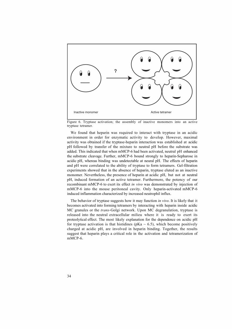
Figure 6. Tryptase activation; the assembly of inactive monomers into an activetryptase tetramer.
We found that heparin was required to interact with tryptase in an acidic
environment in order for enzymatic activity to develop. However, maximal
activity was obtained if the tryptase-heparin interaction was established at acidic
pH followed by transfer of the mixture to neutral pH before the substrate was
added. This indicated that when mMCP-6 had been activated, neutral pH enhanced
the substrate cleavage. Further, mMCP-6 bound strongly to heparin-Sepharose in
acidic pH, whereas binding was undetectable at neural pH. The effects of heparin
and pH were correlated to the ability of tryptase to form tetramers. Gel-filtration
experiments showed that in the absence of heparin, tryptase eluted as an inactive
monomer. Nevertheless, the presence of heparin at acidic pH, but not at neutral
pH, induced formation of an active tetramer. Furthermore, the potency of our
recombinant mMCP-6 to exert its effect in vivo was demonstrated by injection of
mMCP-6 into the mouse peritoneal cavity. Only heparin-activated mMCP-6
induced inflammation characterized by increased neutrophil influx.
The behavior of tryptase suggests how it may function in vivo. It is likely that it
becomes activated into forming tetramers by interacting with heparin inside acidic
MC granules or the trans-Golgi network. Upon MC degranulation, tryptase is
released into the neutral extracellular milieu where it is ready to exert its
proteolytical effect. The most likely explanation for the dependence on acidic pH
for tryptase activation is that histidines (pKa ~ 6.5), which become positively
charged at acidic pH, are involved in heparin binding. Together, the results
suggest that heparin plays a critical role in the activation and tetramerization of
mMCP-6.

35
Paper II: Heparin antagonists are potent inhibitors of mast cell
tryptase.
The critical role of heparin for tryptase activation indicates that displacement of
heparin may inactivate tryptase. In paper II, we investigated whether the
polycationic compounds, protamine and Polybrene, could be used as tryptase
inhibitors. Polybrene is a synthetic heparin antagonist that is used in the clinic for
the reversal of heparin therapy, whereas protamine is an Arg-rich protein involved
in packing of DNA in sperm and certain viruses (Brewer, Corzett & Balhorn,
1999). Kinetic studies showed that both heparin antagonists were potently
inhibiting mMCP-6 and human lung tryptase (IC50 values in the nM range). The
most likely cause of this effect is that the heparin antagonists compete with
tryptase for binding to heparin. When tryptase loses heparin, the tetramer may
destabilize, rapidly monomerize and lose its activity, resembling the events
occurring during spontaneous inactivation in the absence of an inhibitor (Schechter
et al., 1995). Accordingly, when active tryptase in complex with heparin is co-
injected with a heparin antagonist on a gel chromatography column, tryptase elutes
as an inactive monomer. This suggests a non-competitive mode of inhibition,
because heparin-antagonists would bind heparin rather than occupying the active
site. Correspondingly, Polybrene inhibited tryptase with non-competitive kinetics.
In contrast, protamine displayed competitive inhibition kinetics. This may be due
to the high content of arginine in protamine, and that arginine side-chains may
interact with the active sites. However, both Polybrene and protamine were
sensitive to increasing heparin concentrations, which reduced their potency. The
reason for the sensitivity of protamine, despite competitive kinetics, may be that
protamine binds free heparin but fails to challenge tryptase for its associated
heparin and instead interacts with the active site of tryptase at low heparin
concentration. Further, tryptase inactivated with Polybrene seemed to be mostly
resistant to reactivation with heparin. In contrast, when tryptase was inhibited by
protamine, all activity could be regained by addition of excess heparin.
Spontaneous inactivation is proposed to take place through several reversible steps
ending with reactivable monomers and proceeding to an irreversible step to
monomers that cannot be reactivated (Selwood, Mccaslin & Schechter, 1998). The
reason for the irreversibility of Polybrene may be that its high potency makes the
tryptase tetramers monomerize to a stage were they can’t be reactivated.
Consequently, protamine-inactivated tryptase can be reactivated since protamine is
unable to compete with the tryptase-bound heparin and tryptase therefore never
dissociates completely from heparin.
In comparison, both lactoferrin, a previously reported heparin antagonist and
inhibitor of human tryptase (Elrod et al., 1997), and APC 366, an active site-
directed inhibitor (Clark et al., 1995), displayed no or only moderate inhibition of
mMCP-6 and human lung tryptase. Interestingly, APC-366 displayed different Ki
values over time for human tryptase, but not for mMCP-6. After 40 minutes
incubation, the Ki was in the millimolar range whereas after four hours the Ki had
decreased 500 times to approximately 0.5 µM. The reason for this behavior might
be explained by subtle differences in the active sites of mouse and human tryptase
that make human tryptase more susceptible for the reshaped structure of APC-366
appearing after four hours of incubation. However, APC-366 also displayed an

36
inhibitory effect on MC chymase. This may be exlained by the interaction of an
aromatic 1-hydroxy-2-naphthoyl group in the active site of chymase, in contrast to
the inhibition of trypsin-like proteases, which involve interaction of an arginine-
like structure in APC-366 with the Asp189 of the S1 pocket. Thus, that APC-366
can reduce asthmatic symptoms in sheep and pig models (Clark et al., 1995;
Sylvin et al., 2002) may be related to inhibition of chymase as well as tryptase.
Paper III: Structural requirements for heparin-induced activation and
identification of active tryptase monomers.
In paper III, we studied what the structures that determine the capacity of heparin
to activate mMCP-6. We found that most other structurally related, but less
sulfated compounds e.g. heparan sulfate, were unable to substitute for heparin.
However, the highly negatively charged synthetic compound, dextran sulfate,
efficiently activated tryptase. Further, structurally modified heparins, in which the
N-sulfate, 2-O sulfate or 6-O sulfate groups were selectively removed, were all less
effective than unmodified heparin. These results suggest that heparin interacts
through its high negative charge density rather than through any specific structural
motif. Moreover, experiments using heparin oligosaccharides of defined sizes (up
to 26 saccharide units) showed that the heparin-tryptase interaction was highly
size-dependent. Generally, the longer the heparin chain, the better its ability to
induce tetramerization, however, intact heparin remained superior. Although short
oligosaccharides consisting of 8-10 units were able to bind to tryptase, they were
highly inefficient in inducing tetramerization. Thus, heparin binding does not by
itself induce tetramerization. Furthermore, tryptase activation displayed a bell-
shaped dose-response curve. Together, these results suggest a model for tetramer
formation that involves bridging of tryptase monomers by heparin or other highly
sulfated polysaccharides of sufficient chain length.
A completely novel finding was the identification of an active tryptase
monomer. Short heparin oligosaccharides that bound to heparin did not produce
tetramers but instead induced activation of monomeric tryptase. To ensure that the
activities found in the monomer elution position really were due to active
monomers and not tetramers that were formed after the chromatography step, the
fractions were pooled and re-injected in the same column. The monomer fractions
were again recovered in the monomer position. The presence of an active monomer
was further proven by BPTI, which inhibited the active monomers but not the
active tetramers. Moreover, the ECM component, fibronectin, which was
recognized as a substrate for tryptase in earlier studies (Kaminska et al., 1999;
Lohi, Harvima & Keski-Oja, 1992), was found to be a substrate for active
monomers but not for active tetramers.
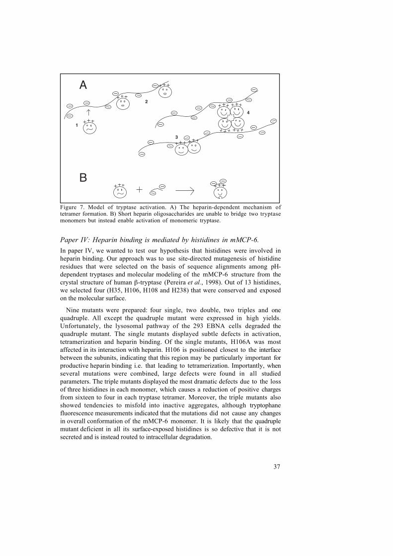
37
A
B
1
2
3
4
Figure 7. Model of tryptase activation. A) The heparin-dependent mechanism oftetramer formation. B) Short heparin oligosaccharides are unable to bridge two tryptasemonomers but instead enable activation of monomeric tryptase.
Paper IV: Heparin binding is mediated by histidines in mMCP-6.
In paper IV, we wanted to test our hypothesis that histidines were involved in
heparin binding. Our approach was to use site-directed mutagenesis of histidine
residues that were selected on the basis of sequence alignments among pH-
dependent tryptases and molecular modeling of the mMCP-6 structure from the
crystal structure of human -tryptase (Pereira et al., 1998). Out of 13 histidines,
we selected four (H35, H106, H108 and H238) that were conserved and exposed
on the molecular surface.
Nine mutants were prepared: four single, two double, two triples and one
quadruple. All except the quadruple mutant were expressed in high yields.
Unfortunately, the lysosomal pathway of the 293 EBNA cells degraded the
quadruple mutant. The single mutants displayed subtle defects in activation,
tetramerization and heparin binding. Of the single mutants, H106A was most
affected in its interaction with heparin. H106 is positioned closest to the interface
between the subunits, indicating that this region may be particularly important for
productive heparin binding i.e. that leading to tetramerization. Importantly, when
several mutations were combined, large defects were found in all studied
parameters. The triple mutants displayed the most dramatic defects due to the loss
of three histidines in each monomer, which causes a reduction of positive charges
from sixteen to four in each tryptase tetramer. Moreover, the triple mutants also
showed tendencies to misfold into inactive aggregates, although tryptophane
fluorescence measurements indicated that the mutations did not cause any changes
in overall conformation of the mMCP-6 monomer. It is likely that the quadruple
mutant deficient in all its surface-exposed histidines is so defective that it is not
secreted and is instead routed to intracellular degradation.

38
Figure 8. Sequence alignment and model of the mMCP-6 tetramer. A) Sequencealignment of mature tryptases from various species. Mutated His residues are labeled inblue and nonmutated His residues are shown in magenta. B) A surface representation ofthe mMCP-6 model. C, D) Electrostatic potential surface of the mMCP-6 model atneutral pH (C) and acidic pH (D).
Paper V. Activation mechanisms for human I- and II-tryptase.
In order to confidently use mouse models of human MC-related diseases and to
assess the potential role of tryptase, it is important to ensure that the basic
biochemical behavior of mouse tryptase and its human counterpart, -tryptase, are
similar. We therefore addressed the mechanisms of activation for human I- and
II- tryptase. These tryptases differ in only one amino acid: Asn102 in I-tryptase
versus Lys102 in II-tryptase. Asn102 is part of an N-glycosylation site and as a
consequence, I-tryptase is glycosylated at Asn102 whereas II-tryptase lacks the
corresponding glycosylation. We could therefore assess whether glycosylation at
Asn102 was involved in the activation mechanism. Although an earlier study did
show that recombinant human -tryptase needed heparin for its activation (Huang
et al., 2001), a detailed study of the mechanism of activation and the structural
requirement was lacking. We constructed recombinant human I- and II-tryptase
using the same system for expression and purification as previously used for
mMCP-6.

39
We found that heparin was crucial for the activation of I- and II-tryptase. A
preference for acidic pH for activation of I-tryptase, closely resembling the
requirements for activation of mMCP-6 was also demonstrated. However, unlike
mMCP-6, heparin-dependent tryptase activation was also detectable at neutral pH.
This indicates that histidines are important for heparin binding in human I-
tryptase, but not as crucial as for mMCP-6. In contrast, II-tryptase was much less
dependent on acidic pH than I-tryptase. Nevertheless, both -tryptases showed
similar bell-shaped dose-response curves and approximately equally high affinity
for heparin. This means that the reason for the lower degree pH dependence of II-
tryptase is unclear and cannot be explained by lower affinity for heparin.
Moreover, gel filtration analysis demonstrated that when II-tryptase was activated
with heparin it showed tendencies to misfold into inactive aggregates, whereas
activation of I-tryptase resulted in only active tetramers. This indicated that
glycosylation may play a role in the assembly of tryptase tetramers.
The heparin-induced activation of the human -tryptases was dependent on the
size and high anionic charge density of the activator, and closely resembled the
structural requirements in terms of heparin for mMCP-6. We also found evidence
that the tryptases formed active monomers in the presence of low molecular
weight heparin. Together, we found that the mechanism for activation of human -
tryptase was highly similar to that of mMCP-6. Thus, results obtained from
studies of mouse tryptase appear to be highly relevant for the situation in humans
and vice versa. This indicates that the mouse system is a good system for
analyzing the biological role of tryptase and that mouse models of human MC-
related diseases might be highly relevant.
In conclusion, we have proven the significance and nature of the heparin-tryptase
interaction, shown a new mode of tryptase inhibition, constructed a model for
formation of the active tryptase tetramer, demonstrated the presence of an active
tryptase monomer and shown that mMCP-6 and human -tryptase are very similar
proteases as regarding their modes of activation.

40
Future perspectives
Our studies have revealed that the mouse tryptase, mMCP-6, and human -
tryptase show similar properties and that mouse models may be used for searching
for the biological role of tryptase. Clearly, if a knockout mouse for mMCP-6 were
to be constructed, it would be very interesting to employ various inflammation
models to study the biological effects of tryptase in vivo. However, in recent years,
many new human MC tryptases have been discovered. Some, such as /TMT
tryptase, have a corresponding mouse variant (mTMT) whereas the mouse variants
of other human tryptases such as - and -tryptase may be missing. The near
future will be important to judge which of these new tryptases are functional and
account for the effects that have been discovered using purified tryptase. To do
these studies, it will be important to make recombinant tryptases and characterize
their basic biochemical behavior.
In a recent study, the possibility of the existence of active monomers in vivo
was addressed (Fajardo & Pejler, 2003a). It was demonstrated that active tryptase
monomers could be formed from dissociated tetramers under conditions that
resemble the in vivo conditions to which tryptase is exposed after MC
degranulation. This means that the discovery of the active tryptase monomer,
recently confirmed by another major group in the field (Fukuoka & Schwartz,
2004), will encourage further characterization of the biological functions that can
be attributed specifically to the active monomer, the active tetramer and to both in
combination. Interestingly, both active forms of mMCP-6 mediated activation of
PAR-2 (J. Hallgren, R. Pike, G. Pejler; unpublished data) and both active forms
of human -tryptase mediated cleavage of fibrinogen (Fajardo & Pejler, 2003a),
whereas fibronectin is specifically cleaved by the active monomer (paper III)
(Fajardo & Pejler, 2003a). The possibility of active tryptase monomers in vivo is
of consequence not only to scientists wishing to dissect the true biological role(s)
of MC tryptases, but also to those who want to find potent tryptase inhibitors to
be used as medical treatment. For some clinical conditions, it may be important to
inhibit only one active form, whereas in most cases it may be critical that the
inhibitors can potently inhibit both forms. As the heparin antagonist, Polybrene,
inhibits both forms of active tryptase (Fajardo & Pejler, 2003a), it may be
important to further investigate the potential use of heparin antagonists.

41
Populärvetenskaplig sammanfattning
Mastcellen är en typ av cell som ingår i kroppens immunförsvar. Mastceller
hjälper till att försvara oss mot bakterier och parasiter. Tyvärr är mastceller inte
bara till hjälp för oss, utan det har visat sig att de är involverade i flera olika
sjukdomar t.ex. allergier och astma. Inuti mastcellen finns en mängd små kapslar,
s.k. granula, som innehåller en hel arsenal av olika inflammatoriska ämnen. Det är
dessa ämnen som ibland försvarar oss och ibland gör oss sjuka. När mastcellen
aktiveras töms innehållet i granula, de inflammatoriska substanserna, ut i kroppen.
Ett av dessa inflammatoriska ämnen är det proteolytiska enzymet tryptas. Ett
proteolytiskt enzym är ett protein som klyva andra proteiner i mindre bitar utan att
själv påverkas. Tryptas är aktiv som en tetramer. Det betyder att för att tryptas ska
vara aktivt så måste fyra stycken identiska tryptasmolekyler sitta ihop. Eftersom
tryptas medverkar i vissa mastcells-relaterade sjukdomar är det många som forskar
för hitta bra tryptasinhibitorer. En inhibitor är en molekyl som blockerar ett
enzyms aktivitet.
Den här avhandlingen visar att för att tryptas ska kunna fungera som enzym
behöver tryptas aktiveras av en annan molekyl, nämligen heparin. Heparin finns
också inne i mastcellens granula och är ett sorts negativt laddat socker. Vi har
visat att för att tryptas ska bli aktivt måste det interagera med heparin i en sur
miljö. Eftersom vi fann att tryptasets aktivitet var kritiskt beroende av heparin,
ville vi testa om man kunde blockera tryptasets aktivitet genom att förhindra att
heparin binder till tryptas. Heparinantagonister är positivt laddade molekyler och
binder själva starkt till heparin. Vi visade att heparinantagonister är effektiva
tryptasinhibitorer. Vad är det då som gör heparin till en så bra tryptasaktivator? Vi
kom fram till att det är längden på heparinkedjan och dess mycket negativa
laddning som avgör. Vi fann att heparinkedjorna måste vara tillräckligt långa för
att kunna binda till två tryptasmolekyler samtidigt och på det sättet binda ihop
tetrameren och aktivera tryptaset. Dessutom hittade vi en ny aktiv form av tryptas,
en aktiv tryptasmonomer, d v s en molekyl tryptas som blivit aktiv på egen hand.
Vi visade att aktiva tryptasmonomerer uppstår när man aktiverar tryptas med korta
kedjor av heparin (som inte förmår att brygga över två tryptasmolekyler).
Vilka delar av tryptas binder till heparin? Liksom andra proteiner är tryptas
uppbyggd av olika sorters aminosyror. Eftersom vi visste att heparinbindningen
måste ske vid lågt pH och att den var av elektrostatisk natur så misstänkte vi att
en viss sorts aminosyror som kallas histidiner var inblandade. Det som
kännetecknar aminosyran histidin är att det är den enda aminosyran som är positivt
laddad vid lågt pH men oladdad vid neutralt pH. Därför kan histidiner bara binda
till den negativt laddade heparinkedjan vid lågt pH. För att studera detta
konstruerade vi tryptasmutanter, d v s varianter av tryptas där någon eller några
aminosyror är förändrade. Våra tryptasmutanter förändrades så att en eller flera
histidiner var muterade till den neutrala aminosyran alanin. Vi fann fyra histidiner
som var speciellt viktiga för tryptasets förmåga att binda till heparin. I de första
fyra studierna använde vi oss av ett rekombinant mustryptas, d v s vi tillverkade
mustryptaset utifrån dess DNA-sekvens. För att kunna studera olika mänskliga
sjukdomar i möss är det viktigt att undersöka att de grundläggande biokemiska

42
mekanismerna är lika för motsvarande enzym i mus och människa. I den femte
studien visade vi att humant tryptas blir aktivt på samma sätt som mustryptas.
I denna avhandling har vi visat hur och varför tryptas blir aktiverat och hittat ett
nytt sätt att inhibera tryptas, samt att tryptas även kan vara aktivt som monomer.
Dessutom har vi visat att att heparin via sin negativa laddning binder till positivt
laddade histidiner i tryptas och att mus- och humantryptas aktiveras på liknande
sätt.

43
Acknowledgements
First and foremost
I wish to thank Gunnar for being an excellent supervisor; you are inspiring,
supportive and a true role model. Many thanks for daring to take on an organic
chemist that never had heard of a mast cell before. It has been a great pleasure to
work with you and the expanding group over the years.
Thanks to past and present members of the mast cell group
Frida – my roommate, friend and weekly punching bag. For good companionship
and for being cheerful and always ready to help. Anders – my friend and “Sancho
Panza” since 10 years! For being caring, fun and never judgmental. Sergio – my
co-supervisor and friend, for warm welcoming and excellent answers to all those
millions questions especially in the beginning, and for continuing to be a support
even after leaving the group.
Elena, Ignacio, Magnus and Mirjana – for valuable discussions and pleasant
times in the lab! Ulrika – adopted member of the group, for good collaboration!
Hanna and Susanne for doing your diploma work within my project in such an
excellent fashion and for being such nice persons!
Thanks to all other past and present colleagues of our department
Especially to:
Kerstin – you have answers to all sorts of questions and is such a nice and happy
person!
Piotr – for all computer help and your good sense of humor!
Cecilia, Ingemar, Maud, Randi, Ashraf, Gunnar F, Liya, Charlotte, Staffan
E, Elena U and Mia amongst others.
Thanks to past and present c olleagues in “the other corridor”
Most especially to Jenny and Stina, for all support, discussions (scientific,
personal and lately babies...) and for being great friends!
For all your help and always being so nice to me:
Lena, Inger, Staffan J, Maria T, Gunbjørg, Birgitta, Johan, Sophie, Anne,
Pernilla, Fredrik and Maria R just to mention a few.
GLIBS – Ulf Lindahl and Barbro Lowisin and all others in this inspiring
network that has continued to arrange many interesting courses and scientific
meetings over the years!
Uppsala Mast Cell Society – Thanks to the Lasse Hellman group and the
Gunnar Nilsson group for interesting discussions as well as great soccer games
and fun.
Great thanks to all my Friends and Family for making my life joyful.
The greatest thanks to Carl, my husband and life companion for your love and
support. I love you, Calle-totten!
44
References
Abbas, A.K., Murphy, K.M. & Sher, A. (1996). Functional diversity of helper Tlymphocytes. Nature 383, 787-93.
Addington, A.K. & Johnson, D.A. (1996). Inactivation of human lung tryptase:evidence for a re-activatable tetrameric intermediate and active monomers.Biochemistry 35, 13511-8.
Akers, I.A. et a l . (2000). Mast cell tryptase stimulates human lung fibroblastproliferation via protease-activated receptor-2. Am J Physiol Lung Cell Mol Physiol278, L193-201.
Alam, R. et al. (1994). Macrophage inflammatory protein-1 alpha and monocytechemoattractant peptide-1 elicit immediate and late cutaneous reactions and activatemurine mast cells in vivo. J Immunol 152, 1298-303.
Aliprantis, A.O. et al. (1999). Cell activation and apoptosis by bacterial lipoproteinsthrough toll-like receptor-2. Science 285, 736-9.
Alter, S.C. et al. (1990). Interactions of human mast cell tryptase with biologicalprotease inhibitors. Arch Biochem Biophys 276, 26-31.
Alter, S.C. et al. (1987). Regulation of human mast cell tryptase. Effects of enzymeconcentration, ionic strength and the structure and negative charge density ofpolysaccharides. Biochem J 248, 821-7.
Bachert, C. (2002). The role of histamine in allergic disease: re-appraisal of itsinflammatory potential. Allergy 57, 287-96.
Baram, D. et al. (2001). Human mast cells release metalloproteinase-9 on contact withactivated T cells: juxtacrine regulation by TNF-alpha. J Immunol 167, 4008-16.
Bauer, O. & Razin, E. (2000). Mast Cell-Nerve Interactions. News Physiol Sci 15, 213-18.
Beller, T.C. et al. (2004). Cysteinyl leukotriene 1 receptor controls the severity ofchronic pulmonary inflammation and fibrosis. Proc Natl Acad Sci U S A 101, 3047-52.
Berger, P. et al. (2001). Tryptase and agonists of PAR-2 induce the proliferation ofhuman airway smooth muscle cells. J Appl Physiol 91, 1372-9.
Bisacchi, G.S. et al. (2004). Synthesis of potent and highly selective nonguanidineazetidinone inhibitors of human tryptase. Bioorg Med Chem Lett 14, 2227-31.
Blair, R.J. et al. (1997). Human mast cells stimulate vascular tube formation. Tryptaseis a novel, potent angiogenic factor. J Clin Invest 99, 2691-700.
Brewer, L.R., Corzett, M. & Balhorn, R. (1999). Protamine-induced condensation anddecondensation of the same DNA molecule. Science 286, 120-3.
Brightbill, H.D. et al. (1999). Host defense mechanisms triggered by microbiallipoproteins through toll-like receptors. Science 285, 732-6.
Brightling, C.E. et al. (2003). New insights into the role of the mast cell in asthma.Clin Exp Allergy 33, 550-6.
Brown, J.K. et al. (2002). Tryptase's potent mitogenic effects in human airway smoothmuscle cells are via nonproteolytic actions. Am J Physiol Lung Cell Mol Physiol 282,L197-206.
Brown, J.K. et al. (2003). Constitutive secretion of the granule chymase mouse mastcell protease-1 and the chemokine, CCL2, by mucosal mast cell homologues. ClinExp Allergy 33, 132-46.
Capila, I. & Linhardt, R.J. (2002). Heparin-protein interactions. Angew Chem Int EdEngl 41, 391-412.
Caughey, G.H. (2002). New developments in the genetics and activation of mast cellproteases. Mol Immunol 38, 1353.
Caughey, G.H. et al. (1988). Substance P and vasoactive intestinal peptide degradationby mast cell tryptase and chymase. J Pharmacol Exp Ther 244, 133-7.
Caughey, G.H. et al. (2000). Characterization of human gamma-tryptases, novelmembers of the chromosome 16p mast cell tryptase and prostasin gene families. JImmunol 164, 6566-75.
45
Cenac, N. et al. (2002). Induction of intestinal inflammation in mouse by activation ofproteinase-activated receptor-2. Am J Pathol 161, 1903-15.
Chen, R. et al. (2001). Mast cells play a key role in neutrophil recruitment inexperimental bullous pemphigoid. J Clin Invest 108, 1151-8.
Church, M.K. et al. (1989). Mast cells, neuropeptides and inflammation. AgentsActions 27, 8-16.
Clark, J.M. et al. (1995). Tryptase inhibitors block allergen-induced airway andinflammatory responses in allergic sheep. Am J Respir Crit Care Med 152, 2076-83.
Cocks, T.M. et al. (1999). A protective role for protease-activated receptors in theairways. Nature 398, 156-60.
Compton, S.J. et al. (1998). The role of mast cell tryptase in regulating endothelial cellproliferation, cytokine release, and adhesion molecule expression: tryptase inducesexpression of mRNA for IL-1 beta and IL-8 and stimulates the selective release of IL-8 from human umbilical vein endothelial cells. J Immunol 161, 1939-46.
Cregar, L. et al. (1999). Neutrophil myeloperoxidase is a potent and selectiveinhibitor of mast cell tryptase. Arch Biochem Biophys 366, 125-30.
D'Andrea, M.R., Rogahn, C.J. & Andrade-Gordon, P. (2000). Localization of protease-activated receptors-1 and -2 in human mast cells: indications for an amplified mastcell degranulation cascade. Biotech Histochem 75, 85-90.
Daeron, M. & Vivier, E. (1999). Biology of immunoreceptor tyrosine-based inhibitionmotif-bearing molecules. Curr Top Microbiol Immunol 244, 1-12.
Dahlen, S.E. et al. (1980). Leukotrienes are potent constrictors of human bronchi.Nature 288, 484-6.
Dell'Italia, L.J. & Husain, A. (2002). Dissecting the role of chymase in angiotensin IIformation and heart and blood vessel diseases. Curr Opin Cardiol 17, 374-9.
Dery, O. et al. (1998). Proteinase-activated receptors: novel mechanisms of signalingby serine proteases. Am J Physiol 274, C1429-52.
Douni, E. et al. (1995). Transgenic and knockout analyses of the role of TNF in immuneregulation and disease pathogenesis. J Inflamm 47, 27-38.
Echtenacher, B., Mannel, D.N. & Hultner, L. (1996). Critical protective role of mastcells in a model of acute septic peritonitis. Nature 381, 75-7.
Ehrlich, P. (1878). Beiträge zur Theorie und Praxis der histologischer Färbung. Ph.D.thesis. Leipzig, Germany: University of Leipzig.
Elrod, K.C. et al. (1997). Lactoferrin, a potent tryptase inhibitor, abolishes late-phaseairway responses in allergic sheep. Am J Respir Crit Care Med 156, 375-81.
Erbaa, F. et al. (2001). Selective inhibition of human mast cell tryptase by gabexatemesylate, an antiproteinase drug. Biochem Pharmacol 61, 271-76.
Fajardo, I. & Pejler, G. (2003a). Formation of active monomers from tetrameric humanbeta-tryptase. Biochem J 369, 603-10.
Fajardo, I. & Pejler, G. (2003b). Human mast cell beta-tryptase is a gelatinase. JImmunol 171, 1493-9.
Ferrell, W.R. et al. (2003). Essential role for proteinase-activated receptor-2 inarthritis. J Clin Invest 111, 35-41.
Fiorucci, L. & Ascoli, F. (2004). Mast cell tryptase, a still enigmatic enzyme. Cell MolLife Sci 61, 1278-95.
Forsberg, E. et al. (1999). Abnormal mast cells in mice deficient in a heparin-synthesizing enzyme [see comments]. Nature 400, 773-6.
Foster, P.S. et al. (1996). Interleukin 5 deficiency abolishes eosinophilia, airwayshyperreactivity, and lung damage in a mouse asthma model. J Exp Med 183, 195-201.
Frank, B.T. et al. (2001). Mast Cell Tissue Inhibitor of Metalloproteinase-1 Is Cleavedand Inactivated Extracellularly by alpha-Chymase. J Immunol 166, 2783-92.
Friend, D.S. et al. (2000). Senescent jejunal mast cells and eosinophils in the mousepreferentially translocate to the spleen and draining lymph node, respectively,during the recovery phase of helminth infection. J Immunol 165, 344-52.
Fukuoka, Y. & Schwartz, L.B. (2004). Human beta-Tryptase: Detection andCharacterization of the Active Monomer and Prevention of Tetramer Reconstitutionby Protease Inhibitors. Biochemistry 43, 10757-64.

46
Galli, S.J. (1993). New concepts about the mast cell. N Engl J Med 328, 257-65.Ghildyal, N. et al. (1996). Fate of two mast cell tryptases in V3 mastocytosis and
normal BALB/c mice undergoing passive systemic anaphylaxis: prolonged retentionof exocytosed mMCP-6 in connective tissues, and rapid accumulation ofenzymatically active mMCP-7 in the blood. J Exp Med 184, 1061-73.
Glenner, G.G. & Cohen, L.A. (1960). Histochemical demonstration of a species-specific trypsin-like enzyme in mast cells. Nature 185, 846-7.
Goldstein, S.M. et al. (1992). Protease composition of exocytosed human skin mastcell protease-proteoglycan complexes. Tryptase resides in a complex distinct fromchymase and carboxypeptidase. J Immunol 148, 2475-82.
Grimbaldeston, M.A. et al. (2000). Communications: high dermal mast cell prevalenceis a predisposing factor for basal cell carcinoma in humans. J Invest Dermatol 115,317-20.
Gruber, B.L. et al. (1989). Synovial procollagenase activation by human mast celltryptase dependence upon matrix metalloproteinase 3 activation. J Clin Invest 84,1657-62.
Grunewald, S.M. et al. (1998). An antagonistic IL-4 mutant prevents type I allergy inthe mouse: inhibition of the IL-4/IL-13 receptor system completely abrogateshumoral immune response to allergen and development of allergic symptoms invivo. J Immunol 160, 4004-9.
Grunig, G. et al. (1998). Requirement for IL-13 independently of IL-4 in experimentalasthma. Science 282, 2261-3.
Gurish, M.F. et al. (2004). IgE enhances parasite clearance and regulates mast cellresponses in mice infected with Trichinella spiralis. J Immunol 172, 1139-45.
Guy-Grand, D. et al. (1984). Gut mucosal mast cells. Origin, traffic, and differentiation.J Exp Med 160, 12-28.
Harris, J.L. et al. (2001). Definition of the extended substrate specificity determinantsfor beta- tryptases I and II. J Biol Chem 276, 34941-7.
Hart, P.H. (2001). Regulation of the inflammatory response in asthma by mast cellproducts. Immunol Cell Biol 79, 149-53.
He, S. et al. (2003). The inhibition of mast cell activation by neutrophil lactoferrin:uptake by mast cells and interaction with tryptase, chymase and cathepsin G.Biochem Pharmacol 65, 1007-15.
He, S., Peng, Q. & Walls, A.F. (1997). Potent induction of a neutrophil and eosinophil-rich infiltrate in vivo by human mast cell tryptase: selective enhancement ofeosinophil recruitment by histamine. J Immunol 159, 6216-25.
He, S. & Walls, A.F. (1998). Human mast cell chymase induces the accumulation ofneutrophils, eosinophils and other inflammatory cells in vivo. Br J Pharmacol 125,1491-500.
He, S.H. (2004). Key role of mast cells and their major secretory products ininflammatory bowel disease. World J Gastroenterol 10, 309-18.
Helgadottir, A. et al. (2004). The gene encoding 5-lipoxygenase activating proteinconfers risk of myocardial infarction and stroke. Nat Genet 36, 233-9.
Henderson, W.R., Jr. et al. (2002). A role for cysteinyl leukotrienes in airwayremodeling in a mouse asthma model. Am J Respir Crit Care Med 165, 108-16.
Henningsson, F. et al. (2002). Altered storage of proteases in mast cells from micelacking heparin: a possible role for heparin in carboxypeptidase A processing. BiolChem 383, 793-801.
Henningsson, F. et al. (2003). Mast cell cathepsins C and S control levels ofcarboxypeptidase A and the chymase, mouse mast cell protease 5. Biol Chem 384,1527-31.
Hoshino, K. et al. (1999). Cutting edge: Toll-like receptor 4 (TLR4)-deficient mice arehyporesponsive to lipopolysaccharide: evidence for TLR4 as the Lps gene product. JImmunol 162, 3749-52.
Huang, C. et al. (2001). Evaluation of the substrate specificity of human mast celltryptase beta I and demonstration of its importance in bacterial infections of thelung. J Biol Chem 276, 26276-84.

47
Huang, C. et al. (1998). Induction of a selective and persistent extravasation ofneutrophils into the peritoneal cavity by tryptase mouse mast cell protease 6. JImmunol 160, 1910-9.
Huang, C. et al. (1999). Human tryptases alpha and beta/II are functionally distinctdue, in part, to a single amino acid difference in one of the surface loops that formsthe substrate-binding cleft. J Biol Chem 274, 19670-6.
Huang, C. et al. (2000). Formation of enzymatically active, homotypic, and heterotypictetramers of mouse mast cell tryptases. Dependence on a conserved Trp-rich domainon the surface. J Biol Chem 275, 351-8.
Humphries, D.E. et al. (1999). Heparin is essential for the storage of specific granuleproteases in mast cells [see comments]. Nature 400, 769-72.
Hunt, J.E. et al. (1997). Mouse mast cell protease 9, a novel member of thechromosome 14 family of serine proteases that is selectively expressed in uterinemast cells. J Biol Chem 272, 29158-66.
Ibrahim, M.Z. et al. (1996). The mast cells of the multiple sclerosis brain. JNeuroimmunol 70, 131-8.
Imamura, T. et al. (1996). Induction of vascular permeability enhancement by humantryptase: dependence on activation of prekallikrein and direct release of bradykininfrom kininogens. Lab Invest 74, 861-70.
Inamura, N. et al. (1998). Induction and enhancement of Fc(epsilon)RI-dependentmast cell degranulation following coculture with activated T cells: dependency onICAM-1- and leukocyte function-associated antigen (LFA)-1-mediated heterotypicaggregation. J Immunol 160, 4026-33.
Irani, A.M. et al. (1991). Human mast cell carboxypeptidase. Selective localization toMCTC cells. J Immunol 147, 247-53.
Jarjour, N.N. et al. (1991). Elevated bronchoalveolar lavage fluid histamine levels inallergic asthmatics are associated with increased airway obstruction. Am Rev RespirDis 144, 83-7.
Jogie-Brahim, S. et al. (2004). Expression of alpha-tryptase and beta-tryptase byhuman basophils. J Allergy Clin Immunol 113, 1086-92.
Johnson, A.R., Hugli, T.E. & Muller-Eberhard, H.J. (1975). Release of histamine fromrat mast cells by the complement peptides C3a and C5a. Immunology 28, 1067.
Johnson, D., Seeldrayers, P.A. & Weiner, H.L. (1988). The role of mast cells indemyelination. 1. Myelin proteins are degraded by mast cell proteases and myelinbasic protein and P2 can stimulate mast cell degranulation. Brain Res 444, 195-8.
Johnson, D.A. & Barton, G.J. (1992). Mast cell tryptases: examination of unusualcharacteristics by multiple sequence alignment and molecular modeling. Protein Sci1, 370-7.
Kabashima, K. & Narumiya, S. (2003). The DP receptor, allergic inflammation andasthma. Prostaglandins Leukot Essent Fatty Acids 69, 187-94.
Kaminska, R. et a l . (1999). Focal dermal-epidermal separation and fibronectincleavage in basement membrane by human mast cell tryptase. J Invest Dermatol 113,567-73.
Kanaoka, Y. & Boyce, J.A. (2004). Cysteinyl leukotrienes and their receptors: cellulardistribution and function in immune and inflammatory responses. J Immunol 173,1503-10.
Kankkunen, J.P., Harvima, I.T. & Naukkarinen, A. (1997). Quantitative analysis oftryptase and chymase containing mast cells in benign and malignant breast lesions.Int J Cancer 72, 385-8.
Karlson, U. et al. (2003). Extended substrate specificity of rat mast cell protease 5, arodent alpha-chymase with elastase-like primary specificity. J Biol Chem 278,39625-31.
Katz, B.A. et al. (1998). Design of potent selective zinc-mediated serine proteaseinhibitors. Nature 391, 608-12.
Kirshenbaum, A.S. et al. (1999). Demonstration that human mast cells arise from aprogenitor cell population that is CD34(+), c-kit(+), and expresses aminopeptidase N(CD13). Blood 94, 2333-42.

48
Knight, P.A. et al. (2000). Delayed expulsion of the nematode Trichinella spiralis inmice lacking the mucosal mast cell-specific granule chymase, mouse mast cellprotease-1. J Exp Med 192, 1849-56.
Kolset, S.O., Prydz, K. & Pejler, G. (2004). Intracellular proteoglycans. Biochem J 379,217-27.
Kozik, A., Potempa, J. & Travis, J. (1998). Spontaneous inactivation of human lungtryptase as probed by size- exclusion chromatography and chemical cross-linking:dissociation of active tetrameric enzyme into inactive monomers is the primaryevent of the entire process. Biochim Biophys Acta 1385, 139-48.
Kozma, G.T. et al. (2003). Histamine deficiency in gene-targeted mice strongly reducesantigen-induced airway hyper-responsiveness, eosinophilia and allergen-specificIgE. Int Immunol 15, 963-73.
Krishna, M.T. et al. (2001). Inhibition of mast cell tryptase by inhaled APC 366attenuates allergen- induced late-phase airway obstruction in asthma. J Allergy ClinImmunol 107, 1039-45. t&artType=abs&id=a115631&target=.
Kunori, Y. et al. (2002). Rodent alpha-chymases are elastase-like proteases. Eur JBiochem 269, 5921-30.
Lantz, C.S. et al. (1998). Role for interleukin-3 in mast-cell and basophil developmentand in immunity to parasites. Nature 392, 90-3.
Lee, D.M. et al. (2002a). Mast cells: a cellular link between autoantibodies andinflammatory arthritis. Science 297, 1689-92.
Lee, M. et al. (2002b). Mast cell tryptase degrades HDL and blocks its function as anacceptor of cellular cholesterol. Arterioscler Thromb Vasc Biol 22, 2086-91.
Leskinen, M.J., Kovanen, P.T. & Lindstedt, K.A. (2003). Regulation of smooth musclecell growth, function and death in vitro by activated mast cells--a potentialmechanism for the weakening and rupture of atherosclerotic plaques. BiochemPharmacol 66, 1493-8.
Leskinen, M.J. et al. (2003). Mast cell chymase induces smooth muscle cell apoptosisby a mechanism involving fibronectin degradation and disruption of focaladhesions. Arterioscler Thromb Vasc Biol 23, 238-43.
Lin, L., Gerth, A.J. & Peng, S.L. (2004). Susceptibility of mast cell-deficient W/Wvmice to pristane-induced experimental lupus nephritis. Immunol Lett 91, 93-7.
Lindner, J.R. et al. (2000). Delayed onset of inflammation in protease-activatedreceptor-2- deficient mice. J Immunol 165, 6504-10.
Lindstedt, K.A. et al. (2001). Activation of paracrine TGF-beta1 signaling uponstimulation and degranulation of rat serosal mast cells: a novel function forchymase. Faseb J 15, 1377-88.
Lock, C. et al. (2002). Gene-microarray analysis of multiple sclerosis lesions yieldsnew targets validated in autoimmune encephalomyelitis. Nat Med 8, 500-8.
Lohi, J., Harvima, I. & Keski-Oja, J. (1992). Pericellular substrates of human mastcell tryptase: 72,000 dalton gelatinase and fibronectin. J Cell Biochem 50, 337-49.
Love, R.J., Ogilvie, B.M. & McLaren, D.J. (1976). The immune mechanism whichexpels the intestinal stage of Trichinella spiralis from rats. Immunology 30, 7-15.
Lundequist, A. et al. (2003). Polycationic peptides as inhibitors of mast cell serineproteases. Biochem Pharmacol 65, 1171-80.
Lundequist, A. et al. (2004). Cooperation between mast cell carboxypeptidase A andthe chymase mouse mast cell protease 4 in the formation and degradation ofangiotensin II. J Biol Chem 279, 32339-44.
Lutzelschwab, C. et al. (1997). Secretory granule proteases in rat mast cells. Cloning of10 different serine proteases and a carboxypeptidase A from various rat mast cellpopulations. J Exp Med 185, 13-29.
MacGlashan, D., Jr. (2003). Histamine: A mediator of inflammation. J Allergy ClinImmunol 112, S53-9.
Maizels, R.M. & Holland, M.J. (1998). Parasite immunology: pathways for expellingintestinal helminths. Curr Biol 8, R711-4.
Malaviya, R. et al. (1996). Mast cell modulation of neutrophil influx and bacterialclearance at sites of infection through TNF-alpha. Nature 381, 77-80.

49
Malaviya, R. et al. (1994). Mast cell phagocytosis of FimH-expressing enterobacteria.J Immunol 152, 1907-14.
Marcum, J.A. et al. (1986). Anticoagulantly active heparin-like molecules from mastcell-deficient mice. Am J Physiol 250, H879-88.
Marquardt, U. et al. (2002). The crystal structure of human alpha1-tryptase reveals ablocked substrate-binding region. J Mol Biol 321, 491-502.
Martinez-Moczygemba, M. & Huston, D.P. (2003). Biology of common beta receptor-signaling cytokines: IL-3, IL-5, and GM-CSF. J Allergy Clin Immunol 112, 653-65;quiz 66.
Matsumoto, R. et al. (1995). Packaging of proteases and proteoglycans in the granulesof mast cells and other hematopoietic cells. A cluster of histidines on mouse mastcell protease 7 regulates its binding to heparin serglycin proteoglycans. J Biol Chem270, 19524-31.
Matsuoka, T. et al. (2000). Prostaglandin D2 as a mediator of allergic asthma. Science287, 2013-7.
McKenzie, G.J. et al. (1998). A distinct role for interleukin-13 in Th2-cell-mediatedimmune responses. Curr Biol 8, 339-42.
McLachlan, J.B. et al. (2003). Mast cell-derived tumor necrosis factor induceshypertrophy of draining lymph nodes during infection. Nat Immunol 4, 1199-205.
McNeil, H.P. et al. (1992). Isolation, characterization, and transcription of the geneencoding mouse mast cell protease 7. Proc Natl Acad Sci U S A 89, 11174-8.
Means, T.K. et al. (1999). Human toll-like receptors mediate cellular activation byMycobacterium tuberculosis. J Immunol 163, 3920-7.
Medeiros, A.I. et al. (1999). Leukotrienes are involved in leukocyte recruitmentinduced by live Histoplasma capsulatum or by the beta-glucan present in their cellwall. Br J Pharmacol 128, 1529-37.
Medzhitov, R., Preston-Hurlburt, P. & Janeway, C.A., Jr. (1997). A human homologueof the Drosophila Toll protein signals activation of adaptive immunity. Nature 388,394-7.
Metcalfe, D.D., Baram, D. & Mekori, Y.A. (1997). Mast cells. Physiol Rev 77, 1033-79.Miller, H.R. & Pemberton, A.D. (2002). Tissue-specific expression of mast cell granule
serine proteinases and their role in inflammation in the lung and gut. Immunology105, 375-90.
Miller, J.S., Moxley, G. & Schwartz, L.B. (1990). Cloning and characterization of asecond complementary DNA for human tryptase. J Clin Invest 86, 864-70.
Miller, J.S., Westin, E.H. & Schwartz, L.B. (1989). Cloning and characterization ofcomplementary DNA for human tryptase. J Clin Invest 84, 1188-95.
Mocellin, S. et al. (2003). The dual role of IL-10. Trends Immunol 24, 36-43.Molinari, J.F. et al. (1995). Role of tryptase in immediate cutaneous responses in
allergic sheep. J Appl Physiol 79, 1966-70.Molinari, J.F. et al. (1996). Inhaled tryptase causes bronchoconstriction in sheep via
histamine release. Am J Respir Crit Care Med 154, 649-53.Mousli, M. et al. (1994). Peptidergic pathway in human skin and rat peritoneal mast
cell activation. Immunopharmacology 27, 1-11.Mueller, T.D. et al. (2002). Structure, binding, and antagonists in the IL-4/IL-13
receptor system. Biochim Biophys Acta 1592, 237-50.Mukaida, N., Harada, A. & Matsushima, K. (1998). Interleukin-8 (IL-8) and monocyte
chemotactic and activating factor (MCAF/MCP-1), chemokines essentially involvedin inflammatory and immune reactions. Cytokine Growth Factor Rev 9, 9-23.
Nagase, T. et al. (2002). A pivotal role of cytosolic phospholipase A(2) in bleomycin-induced pulmonary fibrosis. Nat Med 8, 480-4.
Naka, T., Nishimoto, N. & Kishimoto, T. (2002). The paradigm of IL-6: from basicscience to medicine. Arthritis Res 4 Suppl 3, S233-42.
Negrao-Correa, D. (2001). Importance of immunoglobulin E (IgE) in the protectivemechanism against gastrointestinal nematode infection: looking at the intestinalmucosae. Rev Inst Med Trop Sao Paulo 43, 291-9.

50
Nico, B. et al. (2004). A possible role of tryptase in angiogenesis in the brain of mdxmouse, a model of Duchenne muscular dystrophy. Neuroscience 123, 585-8.
Nystedt, S., Ramakrishnan, V. & Sundelin, J. (1996). The proteinase-activated receptor2 is induced by inflammatory mediators in human endothelial cells. Comparisonwith the thrombin receptor. J Biol Chem 271, 14910-5.
Oettgen, H.C. et al. (1994). Active anaphylaxis in IgE-deficient mice. Nature 370, 367-70.
Oh, S.W. et al. (2002). Tryptase inhibition blocks airway inflammation in a mouseasthma model. J Immunol 168, 1992-2000.
Ohtsu, H. et al. (2001). Mice lacking histidine decarboxylase exhibit abnormal mastcells. FEBS Lett 502, 53-6.
Olson, S.T., Bjork, I. & Bock, S.C. (2002). Identification of critical molecularinteractions mediating heparin activation of antithrombin: implications for thedesign of improved heparin anticoagulants. Trends Cardiovasc Med 12, 198-205.
Olsson, N., Ulfgren, A.K. & Nilsson, G. (2001). Demonstration of mast cell chemotacticactivity in synovial fluid from rheumatoid patients. Ann Rheum Dis 60, 187-93.
Ono, S.J. et al. (2003). Chemokines: roles in leukocyte development, trafficking, andeffector function. J Allergy Clin Immunol 111, 1185-99; quiz 200.
Ozinsky, A. et al. (2000). The repertoire for pattern recognition of pathogens by theinnate immune system is defined by cooperation between toll-like receptors. ProcNatl Acad Sci U S A 97, 13766-71.
Palladino, M.A. et al. (2003). Anti-TNF-alpha therapies: the next generation. Nat RevDrug Discov 2, 736-46.
Pallaoro, M. et al. (1999). Characterization of genes encoding known and novel humanmast cell tryptases on chromosome 16p13.3. J Biol Chem 274, 3355-62.
Peck, M.J., Piper, P.J. & Williams, T.J. (1981). The effect of leukotrienes C4 and D4 onthe microvasculature of guinea-pig skin. Prostaglandins 21, 315-21.
Pejler, G. & Berg, L. (1995). Regulation of rat mast cell protease 1 activity. Proteaseinhibition is prevented by heparin proteoglycan. Eur J Biochem 233, 192-9.
Pejler, G. & Sadler, J.E. (1999). Mechanism by which heparin proteoglycan modulatesmast cell chymase activity. Biochemistry 38, 12187-95.
Pereira, P.J. et al. (1998). Human beta-tryptase is a ring-like tetramer with active sitesfacing a central pore. Nature 392, 306-11.
Poltorak, A. et al. (1998). Defective LPS signaling in C3H/HeJ and C57BL/10ScCrmice: mutations in Tlr4 gene. Science 282, 2085-8.
Prodeus, A.P. et a l . (1997). Impaired mast cell-dependent natural immunity incomplement C3-deficient mice. Nature 390, 172-5.
Qureshi, S.T. et al. (1999). Endotoxin-tolerant mice have mutations in Toll-likereceptor 4 (Tlr4). J Exp Med 189, 615-25.
Rath-Wolfson, L. (2001). An immunocytochemical approach to the demonstration ofintracellular processing of mast cell carboxypeptidase. Appl Immunohistochem MolMorphol 9, 81-5.
Reed, J.A. et al. (1996). Expression of the mast cell growth factor interleukin-3 inmelanocytic lesions correlates with an increased number of mast cells in theperilesional stroma: implications for melanoma progression. J Cutan Pathol 23,495-505.
Ren, S., Sakai, K. & Schwartz, L.B. (1998). Regulation of human mast cell beta-tryptase: conversion of inactive monomer to active tetramer at acid pH. J Immunol160, 4561-9.
Reynolds, D.S. et al. (1989). Isolation and molecular cloning of mast cellcarboxypeptidase A. A novel member of the carboxypeptidase gene family. J BiolChem 264, 20094-9.
Reynolds, D.S. et al. (1990). Different mouse mast cell populations express variouscombinations of at least six distinct mast cell serine proteases. Proc Natl Acad Sci US A 87, 3230-4.
Rice, K.D. et al. (1998). Inhibitors of tryptase for the treatment of mast cell-mediateddiseases. Curr Pharm Des 4, 381-96.
51
Robbiani, D.F. et al. (2000). The leukotriene C(4) transporter MRP1 regulates CCL19(MIP-3beta, ELC)-dependent mobilization of dendritic cells to lymph nodes. Cell103, 757-68.
Robbie-Ryan, M. et al. (2003). Cutting edge: both activating and inhibitory Fcreceptors expressed on mast cells regulate experimental allergic encephalomyelitisdisease severity. J Immunol 170, 1630-4.
Rodewald, H.R. et al. (1996). Identification of a committed precursor for the mast celllineage. Science 271, 818-22.
Roquet, A. et al. (1997). Combined antagonism of leukotrienes and histamine producespredominant inhibition of allergen-induced early and late phase airway obstructionin asthmatics. Am J Respir Crit Care Med 155, 1856-63.
Rozniecki, J.J. et al. (1995). Elevated mast cell tryptase in cerebrospinal fluid ofmultiple sclerosis patients. Ann Neurol 37, 63-6.
Sakai, K., Ren, S. & Schwartz, L.B. (1996). A novel heparin-dependent processingpathway for human tryptase. Autocatalysis followed by activation with dipeptidylpeptidase I [see comments]. J Clin Invest 97, 988-95.
Schechter, N.M. et al. (1995). Structural changes associated with the spontaneousinactivation of the serine proteinase human tryptase. Biochemistry 34, 10628-38.
Schmidlin, F. et al. (2002). Protease-activated receptor 2 mediates eosinophilinfiltration and hyperreactivity in allergic inflammation of the airway. J Immunol169, 5315-21.
Schmidlin, F. & Bunnett, N.W. (2001). Protease-activated receptors: how proteasessignal to cells. Curr Opin Pharmacol 1, 575-82.
Schwandner, R. et al. (1999). Peptidoglycan- and lipoteichoic acid-induced cellactivation is mediated by toll-like receptor 2. J Biol Chem 274, 17406-9.
Schwartz, L.B. (1994a). Mast cells: function and contents. Curr Opin Immunol 6, 91-7.Schwartz, L.B. (1994b). Tryptase: a clinical indicator of mast cell-dependent events.
Allergy Proc 15, 119-23.Schwartz, L.B. & Bradford, T.R. (1986). Regulation of tryptase from human lung mast
cells by heparin. Stabilization of the active tetramer. J Biol Chem 261, 7372-9.Schwartz, L.B. et al. (1985). The fibrinogenolytic activity of purified tryptase from
human lung mast cells. J Immunol 135, 2762-7.Schwartz, L.B., Lewis, R.A. & Austen, K.F. (1981). Tryptase from human pulmonary
mast cells. Purification and characterization. J Biol Chem 256, 11939-43.Schwartz, L.B. et al. (2003). Tryptase Precursors Are Preferentially and Spontaneously
Released, Whereas Mature Tryptase Is Retained by HMC-1 Cells, Mono-Mac-6 Cells,and Human Skin-Derived Mast Cells. J Immunol 170, 5667-73.
Schwartz, L.B. et al. (1995). The alpha form of human tryptase is the predominant typepresent in blood at baseline in normal subjects and is elevated in those withsystemic mastocytosis. J Clin Invest 96, 2702-10.
Secor, V.H. et al. (2000). Mast cells are essential for early onset and severe disease in amurine model of multiple sclerosis. J Exp Med 191, 813-22.
Seeliger, S. et al. (2003). Proinflammatory role of proteinase-activated receptor-2 inhumans and mice during cutaneous inflammation in vivo. Faseb J 17, 1871-85.
Selwood, T., McCaslin, D.R. & Schechter, N.M. (1998). Spontaneous inactivation ofhuman tryptase involves conformational changes consistent with conversion of theactive site to a zymogen-like structure. Biochemistry 37, 13174-83.
Selwood, T. et al. (2002). Diverse stability and catalytic properties of human tryptasealpha and beta isoforms are mediated by residue differences at the S1 pocket.Biochemistry 41, 3329-40.
Sendo, T. et al. (2003). A potent tryptase inhibitor nafamostat mesilate dramaticallysuppressed pulmonary dysfunction induced in rats by a radiographic contrastmedium. Br J Pharmacol 138, 959-67.
Sher, A. et al. (1979). Complement receptors promote the phagocytosis of bacteria byrat peritoneal mast cells. Lab Invest 41, 490-9.
Slusarchyk, W.A. et al. (2002). Synthesis of potent and highly selective inhibitors ofhuman tryptase. Bioorg Med Chem Lett 12, 3235-8.

52
Smart, S.J. & Casale, T.B. (1994). TNF-alpha-induced transendothelial neutrophilmigration is IL-8 dependent. Am J Physiol 266, L238-45.
Sommerhoff, C.P. et al. (1994). A Kazal-type inhibitor of human mast cell tryptase:isolation from the medical leech Hirudo medicinalis, characterization, and sequenceanalysis. Biol Chem Hoppe Seyler 375, 685-94.
Soto, D. et al. (2002). Genetic deficiency of human mast cell alpha-tryptase. Clin ExpAllergy 32, 1000-6.
Steinhoff, M. et a l . (2000). Agonists of proteinase-activated receptor 2 induceinflammation by a neurogenic mechanism. Nat Med 6, 151-8.
Stevens, R.L. et al. (1988). Identification of chondroitin sulfate E proteoglycans andheparin proteoglycans in the secretory granules of human lung mast cells. Proc NatlAcad Sci U S A 85, 2284-7.
Stevens, R.L. et al. (1994). Strain-specific and tissue-specific expression of mousemast cell secretory granule proteases. Proc Natl Acad Sci U S A 91, 128-32.
Stevens, R.L. et al. (1996). Transgenic mice that possess a disrupted mast cell protease5 (mMCP-5) gene can not store carboxypeptidase A (mMC-CPA) protein in theirgranules. Faseb J 10, 17772.
Strik, M.C. et al. (2004). Intracellular serpin SERPINB6 (PI6) is abundantly expressedby human mast cells and forms complexes with beta-tryptase monomers. Blood 103,2710-7.
Sutton, J.C. et al. (2002). Synthesis and SAR of 4-carboxy-2-azetidinone mechanism-based tryptase inhibitors. Bioorg Med Chem Lett 12, 3229-33.
Suzuki, K. et al. (1995). Activation of precursors for matrix metalloproteinases 1(interstitial collagenase) and 3 (stromelysin) by rat mast-cell proteinases I and II.Biochem J 305 ( Pt 1), 301-6.
Sylvin, H. et al. (2002). The tryptase inhibitor APC-366 reduces the acute airwayresponse to allergen in pigs sensitized to Ascaris suum. Clin Exp Allergy 32, 967-71.
Takeda, K., Kaisho, T. & Akira, S. (2003). Toll-like receptors. Annu Rev Immunol 21,335-76.
Tam, E.K. & Caughey, G.H. (1990). Degradation of airway neuropeptides by humanlung tryptase. Am J Respir Cell Mol Biol 3, 27-32.
Tchougounova, E., Pejler, G. & Abrink, M. (2003). The chymase, mouse mast cellprotease 4, constitutes the major chymotrypsin-like activity in peritoneum and eartissue. A role for mouse mast cell protease 4 in thrombin regulation and fibronectinturnover. J Exp Med 198, 423-31.
Theoharides, T.C. & Conti, P. (2004). Mast cells: the JEKYLL and HYDE of tumorgrowth. Trends Immunol 25, 235-41.
Tkaczyk, C. et al. (2004). Fcgamma receptors on mast cells: activatory and inhibitoryregulation of mediator release. Int Arch Allergy Immunol 133, 305-15.
Tomimori, Y. et al. (2003). Involvement of mast cell chymase in bleomycin-inducedpulmonary fibrosis in mice. Eur J Pharmacol 478, 179-85.
Tremaine, W.J. et al. (2002). Treatment of mildly to moderately active ulcerativecolitis with a tryptase inhibitor (APC 2059): an open-label pilot study. AlimentPharmacol Ther 16, 407-13.
Uozumi, N. et al. (1997). Role of cytosolic phospholipase A2 in allergic response andparturition. Nature 390, 618-22.
van den Berg, W.B. (2001). Uncoupling of inflammatory and destructive mechanismsin arthritis. Semin Arthritis Rheum 30, 7-16.
Vanderslice, P. et al. (1990). Human mast cell tryptase: multiple cDNAs and genesreveal a multigene serine protease family. Proc Natl Acad Sci U S A 87, 3811-5.
Vliagoftis, H. et al. (2000). Proteinase-activated receptor-2-mediated matrixmetalloproteinase-9 release from airway epithelial cells. J Allergy Clin Immunol 106,537-45.
Wang, H.W. et al. (2002). Delta tryptase is expressed in multiple human tissues, and arecombinant form has proteolytic activity. J Immunol 169, 5145-52.

53
Watanabe, N., Miura, K. & Fukuda, Y. (2002). Chymase inhibitor ameliorateseosinophilia in mice infected with Nippostrongylus brasiliensis. Int Arch AllergyImmunol 128, 235-9.
Wei, C.C. et al. (2002). Differential ANG II generation in plasma and tissue of mice withdecreased expression of the ACE gene. Am J Physiol Heart Circ Physiol 282, H2254-8.
Wershil, B.K. et al. (1987). 125I-fibrin deposition in IgE-dependent immediatehypersensitivity reactions in mouse skin. Demonstration of the role of mast cellsusing genetically mast cell-deficient mice locally reconstituted with cultured mastcells. J Immunol 139, 2605-14.
Wershil, B.K. et al. (1991). Recruitment of neutrophils during IgE-dependentcutaneous late phase reactions in the mouse is mast cell-dependent. Partialinhibition of the reaction with antiserum against tumor necrosis factor-alpha. J ClinInvest 87, 446-53.
Wiener, Z. et al. (2002). Bone marrow-derived mast cell differentiation is stronglyreduced in histidine decarboxylase knockout, histamine-free mice. Int Immunol 14,381-7.
Williams, C.M. & Galli, S.J. (2000). Mast cells can amplify airway reactivity andfeatures of chronic inflammation in an asthma model in mice. J Exp Med 192, 455-62.
Wills-Karp, M. et al. (1998). Interleukin-13: central mediator of allergic asthma.Science 282, 2258-61.
Wolters, P.J. et al. (2001). Dipeptidyl Peptidase I Is Essential for Activation of MastCell Chymases, but Not Tryptases, in Mice. J Biol Chem 276, 18551-56.
Wong, G.W. et al. (2002). Biochemical and functional characterization of humantransmembrane tryptase (TMT)/tryptase gamma. TMT is an exocytosed mast cellprotease that induces airway hyperresponsiveness in vivo via an interleukin-13/interleukin-4 receptor alpha/signal transducer and activator of transcription(STAT) 6-dependent pathway. J Biol Chem 277, 41906-15.
Wong, G.W. et al. (1999). Identification of a new member of the tryptase family ofmouse and human mast cell proteases which possesses a novel COOH-terminalhydrophobic extension. J Biol Chem 274, 30784-93.
Wong, G.W. et al. (2004). Mouse chromosome 17A3.3 contains 13 genes that encodefunctional tryptic-like serine proteases with distinct tissue and cell expressionpatterns. J Biol Chem 279, 2438-52.
Wright, C.D. et al. (1999). Inhibition of allergen-induced pulmonary responses by theselective tryptase inhibitor 1,5-bis-[4-[(3-carbamimidoyl-benzenesulfonylamino)-methyl]-phenoxy]-pen tane (AMG-126737). Biochem Pharmacol 58, 1989-96.
Wynn, T.A. (2003). IL-13 effector functions. Annu Rev Immunol 21, 425-56.