the role of eriophyoids in fungal pathogen epidemiology ... role of eriophyoids in... · the role...
TRANSCRIPT

The role of eriophyoids in fungal pathogen epidemiology,mere association or true interaction?
Efrat Gamliel-Atinsky Æ Stanley Freeman Æ Marcel Maymon ÆEduard Belausov Æ Ronald Ochoa Æ Gary Bauchan Æ Anna Skoracka ÆJorge Pena Æ Eric Palevsky
Received: 30 March 2009 / Accepted: 26 August 2009 / Published online: 23 September 2009� Springer Science+Business Media B.V. 2009
Abstract A considerable number of plant feeding mites representing different families
such as Acaridae, Siteroptidae, Tydeidae, and Tarsonemidae interact with plant pathogenic
fungi. While species within the Eriophyoidea appear to be the most common phytophagous
mites vectoring virus diseases, little is known of their role in fungal pathogen epidemi-
ology. In the present article, we present two studies on eriophyoid-fungal relationships.
E. Gamliel-AtinskyDepartment of Plant Pathology, University of Georgia, 2311 Plant Sciences, 120 Carlton St., Athens,GA 30602, USA
S. Freeman � M. MaymonDepartment of Plant Pathology and Weed Research, The Volcani Center, Agricultural ResearchOrganization (ARO), P. O. Box 6, 50250 Bet Dagan, Israel
E. BelausovMicroscopy Unit, The Volcani Center, ARO, Bet Dagan, Israel
R. OchoaSystematic Entomology Laboratory, Agriculture Research Service, US Department of Agriculture,Henry A. Wallace Beltsville Agricultural Research Center, Beltsville, MD 20705, USA
G. BauchanElectron and Confocal Microscopy Unit, Agriculture Research Service, US Department of Agriculture,Henry A. Wallace Beltsville Agricultural Research Center, Beltsville, MD 20705, USA
A. SkorackaDepartment of Animal Taxonomy and Ecology, Institute of Environmental Biology,Faculty of Biology, Adam Mickiewicz University, Umultowska 89, 61-614 Poznan, Poland
J. PenaDepartment of Entomology and Nematology, Tropical Research and Education Center,University of Florida, Homestead, FL 33031, USA
E. Palevsky (&)Department of Entomology, Newe-Ya’ar Research Center, (ARO), P. O. Box 1021,30095 Ramat Yishay, Israele-mail: [email protected]
123
Exp Appl Acarol (2010) 51:191–204DOI 10.1007/s10493-009-9302-y

The first focusing on the association between Aceria mangiferae and the fungal pathogen
Fusarium mangiferae in mango is presented as a case study. The second, as the research is
still in a preliminary phase, reports on quantitative and descriptive associations between
the cereal rust mite Abacarus hystrix and rusts caused by Puccinia spp. Mango bud tissue
colonized with F. mangiferae, and wheat and quackgrass leaves colonized with Pucciniaspp., supported significantly higher populations of eriophyoid mites. Both mite species
were observed bearing the spores of the respective pathogens on their body integument.
Aceria mangiferae vectored the pathogen’s spores into the bud, the sole port of entry for
the fungal pathogen and the frequency and severity of fungal infection increased in the
presence of A. mangiferae. While it appears that eriophyoids are playing a role in fungal
epidemiology, clearly further research is needed to enhance our understanding of direct and
indirect (plant mediated) interactions between plant pathogens and eriophyoid mites in
different plant-pathogen systems.
Keywords Eriophyoidea � Fungi � Mite-fungus-plant interactions � Aceria mangiferae �Abacarus hystrix � Fusarium mangiferae � Puccinia spp.
Introduction
The evolution of mycophagy in the Acari has been well reviewed by Krantz and Lindquist
(1979) based on the premise that the ability to feed on vascular tissues arose through
feeding on fungi in some mite groups and also on the adaptive strategies and ecological
relationships associated with the mycophagous habits that provide parallels to those related
to phytophagy. A considerable number of mites representing many families in different
suborders interact with plant pathogenic fungi. Herbivores may facilitate fungal infection
by two main mechanisms: either by vectoring pathogen spores or by creating wound sites
for fungal penetration (Agrios 1980; Hatcher and Paul 2001).
Abdel-Sater and Eraky (2001) reported that the acarid mites, Tyrophagus putrescentiaeShrank and Rhizoglyphus robini Claparede, when living on bulbs, transfer the fungal
pathogens Aspergillus niger, Nectria haematococca, Rhyzopus stolonifer and Penicilliumchrysogenum attached to the outside of their bodies. Conversely the transfer of Aspergillusflavus and Aspergillus ochraceus is performed through their digestive tracts along with
their foods. Rhizoglyphus robini are attracted to and penetrate Fusarium infested bulbs
more rapidly than healthy ones (Okabe and Amano 1990, 1991).
Fusarium poae is associated with the siteroptid mite, Siteroptes avenae (Muller).
Microscopic examination of S. avenae feeding on F. poae cultures revealed the presence of
sporothecae containing microsporidia of the fungus. This close association between the
mite and fungus is considered responsible for causing Fusarium glume spot on wheat in
South Africa (Kemp et al. 1996).
The relationship of tydeid mites and mycophagy was emphasized by Krantz and
Lindquist (1979). For instance, the tydeid Parapronematus acaciae Baker feeds and breeds
on fungi that commonly occur on the leaves of citrus (McCoy et al. 1969). Lorryia formosaCooreman was an effective sanitizing agent in citrus groves because it reduced levels of
sooty mould fungi (Mendel and Gerson 1982). Tydeus caudatus (Duges) densities posi-
tively correlated with levels of the grapevine downy mildew, Plasmopara viticola, in north
eastern Italy (Duso et al. 2005). The tydeid, Orthotydeus lambi (Baker) plays a role in
reducing the severity of powdery mildew on grapes caused by the fungus Uncinulanecator, suggesting the important role of mycophagous mites as potential biological
192 Exp Appl Acarol (2010) 51:191–204
123

control agents of plant pathogenic fungi (English-Loeb et al. 1999). Vitus viparia (Vita-
ceae), with closed off domatia, harbored lowered populations of O. lambi and higher levels
of U. necator (Norton et al. 2001).
The tarsonemid, Steneotarsonemus spinki Smiley, appears to open ports of entry for the
fungi, Helmintosporium and Sarocladium oryzae (Cardenas et al. 2003), which are
responsible for grain discoloration of rice, Oryza sativa. Steneotarsonemus ananas(Tryon), the pineapple leathery pocket mite or pineapple fruit mite, is host specific to
pineapples throughout the world (Mourichon 1991; Petty et al. 2002). Trichome cells fed
upon by S. ananas serve as a favorable substrate for development of the fungus Penicilliumfuniculosum, the causal agent of inter fruitlet corking, leathery pocket, and fruitlet core rot/
black spot (Petty et al. 2002).
While several species within the Eriophyoidea are apparently the most common phy-
tophagous mites vectoring viral diseases (Oldfield and Proeseler 1996), little is known of
their role in fungal pathogen epidemiology. In the present article, we present two studies on
eriophyoid-fungal relationships. The first focuses on the association between Aceriamangiferae Sayed and the fungal pathogen Fusarium mangiferae Britz, Wingfield &
Marasas in mango and is presented as a case study where we describe some of the
underlying mechanisms clarifying the role of the mite in mango malformation epidemi-
ology. In the second study, as the research is still in a preliminary phase, we report on
quantitative and descriptive associations between the cereal rust mite Abacarus hystrix(Nalepa) and rusts caused by Puccinia spp.
The role of the mango bud mite in mango malformation: a case study
Mango malformation is a severe disease, widely distributed in almost all mango-growing
regions worldwide (Crookes and Rijkenberg 1985; Kumar et al. 1993; Ploetz 2001; Ploetz
et al. 2002; Kvas et al. 2008). Symptoms of disease are associated with hormonal
imbalance in the host that results in misshapen growth of both vegetative and reproductive
parts of the tree (Majumder and Sinha 1972; Prasad et al. 1972; Kumar et al. 1993; Ploetz
2001, 2003). Vegetative malformation includes hypertrophy of young shoots, shorter in-
ternodes, dwarfed malformed leaves and an overall tightly bunched appearance of the
shoot. Inflorescence malformation includes short, thick and branched axes of the floral
panicles, larger flowers containing increased numbers of male and hermaphroditic flowers
that are either sterile or eventually abort. Malformed inflorescences do not bear any fruit,
resulting in great economic losses.
The etiology of mango malformation disease was controversial for many years and
many factors have been suggested as causal agents of disease such as: nutritional
deficiencies, hormonal imbalance, viruses, phytoplasmas, fungi and mites (Narasimhan
1954; Nariani and Seth 1962; Zaher and Osman 1970; Denmark 1983; Ochoa et al.
1994; Ploetz 2001). Aceria mangiferae, the mango bud mite, was hypothesized as the
causal agent of mango malformation for over 40 years mainly due to high numbers of
mites observed in malformed trees, and also because other members of the Eri-
ophyoidea are known to cause proliferation, ‘‘witches broom’’ and gall symptoms of
inflorescences in other plants (Westphal and Manson 1996). Despite the fact that the
fungal theory was well established following Koch’s postulates with several fungi,
certain members of the genus Fusarium have been shown to cause the disease
(Summanwar et al. 1966; Varma et al. 1974; Chakrabarti and Ghosal 1989; Manicom
1989; Ploetz and Gregory 1993; Freeman et al. 1999; Noriega-Cantu et al. 1999; Britz
Exp Appl Acarol (2010) 51:191–204 193
123

et al. 2002; Ploetz 2003; Marasas et al. 2006; Kvas et al. 2008; Lima et al. 2009;
Rodrıguez-Alvarado et al. 2008), It is also clear now that A. mangiferae is not the
causal agent of mango malformation, however, various studies suggest that the mite
interacts with the fungal pathogen resulting in increased severity of disease (Prasad
et al. 1972; Sternlicht and Goldenberg 1976; Gamliel-Atinsky et al. 2009a).
Our study was aimed at revealing the possible associations between A. mangiferae and
the fungal pathogen Fusarium mangiferae. The specific objectives of the study were to:
(1) assess whether A. mangiferae can carry conidia of F. mangiferae on or within its body;
(2) determine the role of the mite in vectoring fungal conidia into the infection site;
(3) determine the role of the bud mite in promoting the fungal infection process, and
(4) evaluate the possible role of the bud mite in long-distance aerial dissemination of the
fungal conidia. A green fluorescent protein (gfp)-marked strain of F. mangiferae was used
throughout this study which distinguished it from that used in previous studies (Nariani and
Seth 1962; Summanwar and Raychaudhuri 1968; Manicom 1989; Labuschagne et al. 1993)
and helped distinguish the pathogen from other contaminants and opportunistic fungi.
Below we will briefly describe the major results and implications from this study; a
detailed account of this research can be found in Gamliel-Atinsky et al. (2009a).
Bearing conidia on or within the body of Aceria mangiferae
In order to test whether A. mangiferae can bear F. mangiferae conidia, two methods were
used: the first one was aimed at determining whether the conidia attach to the outer
integument of the mite and if this attachment is specific to a certain part of the body. Mites
were exposed to the gfp-marked isolate of F. mangiferae and then inspected under a
confocal microscope. In the second method, the possibility that the mite carries conidia
within its body was assessed using a low-temperature scanning electron microscope
(LT-SEM), taking measurements of both conidia and mite stylets (measured directly and
indirectly according to the diameter of the feeding holes in the plant). We hypothesized
that the diameter of the smallest conidia is larger than the diameter of the mite’s stylets.
Results from these observations demonstrated that external rather than internal bearing of
conidia is feasible since mites were observed with conidia clinging to their body (Fig. 1)
and LT-SEM measurements indicated that the width of the mite mouthparts was sub-
stantially smaller than the diameter of the smallest conidium (Fig. 2). Moreover, since
there is a lack of continuity between the midgut and the hindgut of eriophyoid mites
(Nuzzaci and Alberti 1996), fungal spores cannot be transferred and secreted in feces.
Internal bearing of pathogens by eriophyoid mites is a known phenomenon with viral
pathogens due to their minute size (Oldfield and Proeseler 1996). Associations with fungi
is apparently only possible via external-body attachment, such as in the case of acaro-
pathogenic fungi (McCoy 1996).
Vectoring conidia to penetration site
One possible benefit for F. mangiferae from this bipartite association is reaching the
infection site. Results from our recent study demonstrated that the apical bud is the
exclusive penetration site for the fungal pathogen (Gamliel-Atinsky et al. 2009b) which
also serves as the exclusive living habitat of A. mangiferae. Most conidia of F. mangiferaedisseminate in the air and randomly fall on the tree canopy which takes up most of the
surface area of the orchard. One possible route of reaching the hidden, small apical bud is
194 Exp Appl Acarol (2010) 51:191–204
123

via the bud mite which posseses the ability of actively seeking out and reaching the apical
bud from any location in the tree. In order to test the role of the mite in vectoring conidia
into apical buds, an experiment on potted plants was performed. The experiment consisted
of four treatments of different combinations of mites and/or conidia that were placed on
leaves 5 cm away from apical buds. Our results showed that conidia reached the apical bud
only in the treatment where both mites and conidia were co-inoculated on the leaves,
demonstrating the potential of eriophyoid mites as vectors of fungal pathogens (Table 1).
Assisting conidial penetration
Two quantitative evaluations of the possible role of A. mangiferae in fungal penetration, a
process taking place inside apical buds, were assessed. When apical buds of potted mango
plants were inoculated with F. mangiferae in the presence and absence of bud mites, the
frequency and severity of fungal colonization were significantly higher in buds inoculated
with both fungus and mites (Table 2). This result clearly demonstrated that the presence of
mites within the buds enhanced fungal colonization.
Association between fungal infestation and mite population levels
Another quantitative evaluation was designed in order to determine whether the presence
of the fungus affected mite populations within the bud. Numbers of bud mites were
compared within colonized and non-colonized buds by the fungal pathogen during 2006
and 2007. Higher numbers of mites were detected in buds colonized by the fungus rather
Fig. 1 Mango bud mite, Aceria mangiferae, bearing gfp-marked conidia (arrows) of Fusarium mangiferae,the causal agent of mango malformation disease
Exp Appl Acarol (2010) 51:191–204 195
123

Table 1 Total number of Aceria mangiferae and green fluorescent protein (gfp)-marked conidia ofFusarium mangiferae detected within 20 apical mango buds
Inoculation with Bud mites Conidia
Mites with gfp conidia 28a 9
Mites alone 35 0
Gfp alone 0 0
Untreated control 0 0
a All numbers different from zero were found significant (P \ 0.05), according to a t-test analysis of theaverage number of mites and conidia per apical bud, (modified from Gamliel-Atinsky et al. 2009a)
Fig. 2 a SEM image of mango bud mites, Aceria mangiferae, feeding on bud bracts; b SEM image ofmango bud mite anterior ventral view of stylet. c SEM image of respective feeding puncture holes;d confocal image of gfp-marked conidia and micro-conidia of Fusarium mangiferae
Table 2 Frequency (%) and severity (%) of green fluorescent protein (gfp)-marked Fusarium mangiferaecolonization in buds in the presence or absence of Aceria mangiferae over a 2 year period
Treatments Frequency of colonizationa Severity of colonizationb
2006 2007 2006 2007
Mites with gfp conidia 81.6 78.4 60.8 44.7
Gfp conidia alone 50.0 57.4 29.5 28.9
Pc 0.004 0.043 \0.0001 0.009
a Statistical analysis was calculated using a chi-square testb Statistical analysis was calculated using a t testc Significance refers to each pair of means per year separately
196 Exp Appl Acarol (2010) 51:191–204
123

than in non-colonized buds (Fig. 3). When a bi-annual average was calculated for the ten
sampling periods and analyzed by t-test, a significant difference was detected between 50
mites per bud found in colonized buds vs. 33.6 mites per bud found in non-colonized buds
(P = 0.0002; t586 = 3.731).
Qualitative associations were assessed with both confocal and LT-SEM observations of
apical buds that were inoculated with both the pathogen and the mites. Microscopic
observations supported our finding by demonstrating the physical proximity and the actual
contact between the two organisms within infected mango buds. Bud mites were observed
in close proximity with gfp-marked hyphae and conidia, using a confocal microscope
(Fig. 4a). LT-SEM observations also illustrated bud mites bearing hyphae and conidia on
their body (Fig. 4b, c, d).
Assisting aerial conidial dissemination
Since previous attempts to trap conidia in the air failed (Varma et al. 1971) and the role
of A. mangiferae in aerial dissemination of conidia was proposed (Ploetz 2001) we
attempted to monitor both fungal conidia and mango bud mites using several trapping
methods in order to shed light on the mode of conidial dissemination in the field.
Conidia of F. mangiferae were trapped successfully using two trapping methods and an
annual peak of dissemination was found in the spring/early summer months (Gamliel-
Atinsky et al. 2009a). Aceria mangiferae was trapped throughout the season, but no
fungal growth was detected after placing these mites on selective media for the fungus F.mangiferae. Our findings imply that conidia can reach the mango tree independently of
the bud mite and thus the latter does not seem to play a role in the windborne dis-
semination of the pathogen.
Fig. 3 Mean density of Aceria mangiferae within colonized and non-colonized buds by Fusariummangiferae over a 2-year-survey in the Volcani experimental orchard, Bet Dagan, Israel. Bars representstandard errors of the mean (±SE). Biannual means were calculated and subjected to t-test analysis (resultspresented in text)
Exp Appl Acarol (2010) 51:191–204 197
123

Associations between the cereal rust mite Abacarus hystrix and rusts causedby Puccinia spp.
The cereal rust mite A. hystrix is notable among eriophyoid mites in causing losses to
cultivated grasslands. It has been found to infest wheat, oats, barley, rye, rice, quackgrass,
timothy, orchardgrass and many other (ca. 60–70) cultivated and wild grass species
throughout the world (Amrine and De Lillo 2003). Abacarus hystrix feeding causes leaf
discoloration and inhibition of seed production, and at high densities the mite causes
withering of plants and retards their growth (Frost and Ridland 1996). The cereal rust mite
is also known to transmit ryegrass mosaic virus (RMV), a serious disease of temperate
grasslands, and agropyron mosaic virus (AMV), a minor disease of wheat and quackgrass
(Oldfield and Proeseler 1996). The mite lives freely on the upper leaf surfaces and dis-
perses passively on air currents (Nault and Styer 1969).
Rusts caused by Puccinia spp. are significant diseases affecting cereal and other grasses
worldwide. Infections may occur on stems, leaf sheaths, or leaf blades and glumes. A few
days post infection elongated pustules develop, subsequently rupturing the plant epidermis
and exposing a mass of reddish brown urediniospores (Fig. 5). Urediniospores are released
from the uredinia and may be transported over long distances by wind and infect other
plants. In areas with mild climates, infected grass foliage can serve as the overwintering
Fig. 4 Microscopic images of the mango bud mite Aceria mangiferae and Fusarium mangiferae withininoculated apical buds. a Confocal microscope image of three A. mangiferae mites and gfp-markedF. mangiferae hyphae with germinating conidia surrounding them; b SEM image of A. mangiferae withfungal conidia on their bodies and eggs in close proximity of fungal hyphae; c SEM image of a colony ofA. mangiferae mites and fungal hyphae around and surrounding them; d SEM image of A. mangiferae nearplant trichomes with fungal hyphae proximate to them
198 Exp Appl Acarol (2010) 51:191–204
123

site for the mycelium and urediniospores. Another type of rust spore that may overwinter is
the teliospore, from which basidiospores are produced. The latter infect non-grass hosts,
e.g., barberries Berberis spp., and after germination two more spore types (i.e., pycni-
ospores and aeciospores) are formed. Aeciospores can infect only grasses, giving rise to
urediniospores and thus completing the life cycle (Cummings 1971; Leonard and Szabo
2005).
During a survey of the eriophyoid fauna on grasses in Poland carried out from 1998
(Skoracka 2004), the concurrent presence of A. hystrix and rusts caused by Puccinia spp.
have been reported on quackgrass and wheat. Moreover, mite specimens bearing rust
spores on their bodies have been observed. Up to now, no reports have suggested the
existence of relationships between eriophyoid mites and rust fungi. Here we present results
of observations regarding the associations between rust fungi and the cereal rust mite.
Association between fungal infestation and mite population levels
Quackgrass Elymus repens (L.) Gould and wheat Triticum aestivum L. plants were col-
lected in 1998–2001 in west Poland from 23 locations. The material included 143 samples,
among them 99 samples contained quackgrass and 44 samples contained wheat. Each
sample consisted of 5–10 individual shoots of a given grass species, collected randomly
from the sampling location. Shoots were cut just above ground level and put into a plastic
bag. The grasses were examined in the laboratory with a stereomicroscope. The presence
of rust fungi per leaf was recorded. Mites were counted and mounted on slides, and
subsequently identified with a phase-contrast microscope. Mean density of mites on leaves
infected with rust [DR], and mean density of mites on leaves with no symptoms of rust
(clear) [DC] were calculated. To test the differences between means two sample t-tests was
applied. For computations, the S-PLUS software was used (S-PLUS 2005).
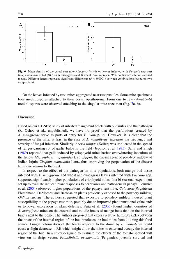
The density of the cereal rust mite was significantly higher on leaves infected by rust
fungi (DR) compared to leaves with no symptoms of rust (DC). This result is true for both
host species, quackgrass (P \ 0.0001; t3477 = -14.87; Fig. 6A), and wheat (P \ 0.0001;
t1212 = -8.86; Fig. 6B).
Fig. 5 SEM of a rust pustule caused by Puccinia spp. releasing urediniospores on the underside of aquackgrass leaf
Exp Appl Acarol (2010) 51:191–204 199
123
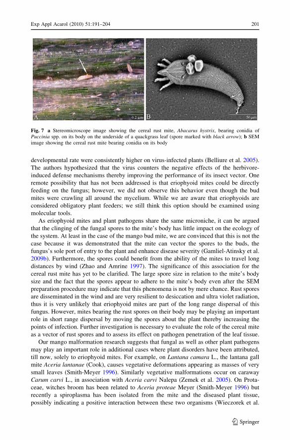
On the leaves infected by rust, mites aggregated near rust pustules. Some mite specimens
bore urediniospores attached to their dorsal opisthosoma. From one to few (about 5–6)
urediniospores were observed attaching to the singular mite specimen (Fig. 7a, b).
Discussion
Based on our LT-SEM study of infested mango bud bracts with bud mites and the pathogen
(R. Ochoa et al., unpublished), we have no proof that the perforations created by
A. mangiferae serve as ports of entry for F. mangiferae. However, it is clear that the
presence of the mite, at least in the case of A. mangiferae, increases the frequency and
severity of fungal infection. Similarly, Aceria tulipae (Keifer) was implicated in the spread
of fungus-causing rot of garlic bulbs in the field (Jeppson et al. 1975). Saini and Singh
(1989) reported that galls induced by eriophyoid mites harbor overwintering inoculum of
the fungus Microsphaera alphitoides f. sp. ziziphi, the causal agent of powdery mildew of
Indian Jujube Ziziphus mauritania Lam., thus improving the perpetuation of the disease
from one season to the next.
In respect to the effect of the pathogen on mite populations, both mango bud tissue
infected with F. mangiferae and wheat and quackgrass leaves infected with Puccinia spp.
supported significantly higher populations of eriophyoid mites. In a bi-seasonal experiment
set up to evaluate induced plant responses to herbivores and pathogens in papaya, Fournier
et al. (2004) observed higher populations of the papaya rust mite, Calacarus flagellisetaFletchmann, DeMoraes, and Barbosa on plants previously exposed to the powdery mildew,
Oidium caricae. The authors suggested that exposure to powdery mildew induced plant
susceptibility to the papaya rust mite, possibly due to improved plant nutritional value and/
or to lower expression of plant defenses. Pena et al. (2005) found higher densities of
A. mangiferae mites on the external and middle bracts of mango buds than on the internal
bracts next to the dome. The authors proposed that excess relative humidity (RH) between
the bracts of the internal region of the bud precludes the bud mites from utilizing this food
source. Fungal colonization of the bracts adjacent to the dome by F. mangiferae could
cause a slight decrease in RH which might allow the mites to enter and occupy the internal
region of the bud. In a study designed to evaluate the effects of the tomato spotted wilt
virus on its thrips vector, Frankliniella occidentalis (Pergande), juvenile survival and
Fig. 6 Mean density of the cereal rust mite Abacarus hystrix on leaves infected with Puccinia spp. rust(DR) and non-infected (DC) on A quackgrass and B wheat. Bars represent 95% confidence intervals aroundmeans. Different letters represent significant differences (P \ 0.0001) between combinations based on twosample t-test
200 Exp Appl Acarol (2010) 51:191–204
123
developmental rate were consistently higher on virus-infected plants (Belliure et al. 2005).
The authors hypothesized that the virus counters the negative effects of the herbivore-
induced defense mechanisms thereby improving the performance of its insect vector. One
remote possibility that has not been addressed is that eriophyoid mites could be directly
feeding on the fungus; however, we did not observe this behavior even though the bud
mites were crawling all around the mycelium. While we are aware that eriophyoids are
considered obligatory plant feeders; we still think this option should be examined using
molecular tools.
As eriophyoid mites and plant pathogens share the same microniche, it can be argued
that the clinging of the fungal spores to the mite’s body has little impact on the ecology of
the system. At least in the case of the mango bud mite, we are convinced that this is not the
case because it was demonstrated that the mite can vector the spores to the buds, the
fungus’s sole port of entry to the plant and enhance disease severity (Gamliel-Atinsky et al.
2009b). Furthermore, the spores could benefit from the ability of the mites to travel long
distances by wind (Zhao and Amrine 1997). The significance of this association for the
cereal rust mite has yet to be clarified. The large spore size in relation to the mite’s body
size and the fact that the spores appear to adhere to the mite’s body even after the SEM
preparation procedure may indicate that this phenomena is not by mere chance. Rust spores
are disseminated in the wind and are very resilient to desiccation and ultra violet radiation,
thus it is very unlikely that eriophyoid mites are part of the long range dispersal of this
fungus. However, mites bearing the rust spores on their body may be playing an important
role in short range dispersal by moving the spores about the plant thereby increasing the
points of infection. Further investigation is necessary to evaluate the role of the cereal mite
as a vector of rust spores and to assess its effect on pathogen penetration of the leaf tissue.
Our mango malformation research suggests that fungal as well as other plant pathogens
may play an important role in additional cases where plant disorders have been attributed,
till now, solely to eriophyoid mites. For example, on Lantana camara L., the lantana gall
mite Aceria lantanae (Cook), causes vegetative deformations appearing as masses of very
small leaves (Smith-Meyer 1996). Similarly vegetative malformations occur on caraway
Carum carvi L., in association with Aceria carvi Nalepa (Zemek et al. 2005). On Prota-
ceae, witches broom has been related to Aceria proteae Meyer (Smith-Meyer 1996) but
recently a spiroplasma has been isolated from the mite and the diseased plant tissue,
possibly indicating a positive interaction between these two organisms (Wieczorek et al.
Fig. 7 a Stereomicroscope image showing the cereal rust mite, Abacarus hystrix, bearing conidia ofPuccinia spp. on its body on the underside of a quackgrass leaf (spore marked with black arrow); b SEMimage showing the cereal rust mite bearing conidia on its body
Exp Appl Acarol (2010) 51:191–204 201
123

2003). Even though these examples of plant disorders do not possess the same symptoms
as those of mango malformation, there do seem to be marked similarities. We propose that
future research on eriophyoid related plant disorders, especially those without typical
eriophyoid galls and erinea (Westphal and Manson 1996), should consider and evaluate
additional pathogens as causal agents.
In summary, while it appears that eriophyoids are playing a role in fungal epidemiology,
clearly further research is needed to broaden our understanding of direct and indirect (plant
mediated) interactions between plant pathogens and eriophyoid mites in different plant-
pathogen systems.
Acknowledgments This research was supported in part by grant no. 132-0972 from the Chief Scientist ofthe Israeli Ministry of Agriculture, and by the Bureau for Economic Growth, Agriculture, and Trade, USAgency for International Development, under the terms of the Middle East Regional Cooperation ProgramAward No. TA-MOU-02-M21-030, awarded to SF. We would like to express our gratitude to Drs. H. Voet(Hebrew University, Israel) and Lechosław Kuczynski (Adam Mickiewicz University, Poznan, Poland) foradvice in the statistical analyses, Prof. D. Shtienberg (ARO) for advice on epidemiological studies,A. Zveibel (ARO), Y. Denisov (ARO) and M. Sharon (ARO) for technical assistance, Drs. G. Miller (SEL-ARS-USDA), Matt Buffington, G. Evans (APHIS-USDA) and Prof. Uri Gerson (Hebrew University) fortheir assistance in the preparation and review of the manuscript, and Magdalena Gawlak (IOR, Poznan,Poland) for taking SEM photos of A. hystrix and Puccinia sp.
References
Abdel-Sater MA, Eraky SA (2001) Bulbs micoflora and their relation with three stored product mites.Mycopathologia 153:33–39
Agrios GN (1980) Insect involvement in the transmission of fungal pathogens. In: Harris KF, MaramoroschK (eds) Vectors of plant pathogens. Academic Press, New York, pp 293–324
Amrine JW Jr, De Lillo E (2003) Catalog of the eriophyoidea. A working catalog of the eriophyoidea of theworld. Version 1.0 http://insects.tamu.edu/research/collection/hallan/acari/eriophyidae. Accessed 5 Jan2008
Belliure B, Janssen A, Maris PC, Peters D, Sabelis MW (2005) Herbivore arthropods benefit from vectoringplant viruses. Ecol Lett 8:70–79
Britz H, Steenkamp ET, Coutinho TA, Wingfield BD, Marasas WFO, Wingfield MJ (2002) Two newspecies of Fusarium section liseola associated with mango malformation. Mycologia 94:722–730
Cardenas RM, Gonzalez L, Parra Y, Rivero D, Cruz A (2003) Influence of rice grain discoloration (Oryzasativa Lin.) on variety J-104, harmfulness and genera of fungi present. Rev Prot Veg 18:124–128
Chakrabarti DK, Ghosal S (1989) The disease cycle of mango malformation induced by Fusarium moniliformevar. subglutinans and the curative effects of mangiferin-metal chelates. J Phytopathol 125:238–246
Crookes CA, Rijkenberg FHJ (1985) A literature review of the distribution, symptomology, cause andcontrol of mango blossom malformation. S Afr Mango Grow Assoc Res Rep 5:15–24
Cummings GB (1971) The rust fungi of cereals, grasses and bamboos. Springer, New YorkDenmark HA (1983) Eriophyes mangiferae (Sayed) a pest of mango (Acarina: Eriophyidae). Fla Dep Agric
Consum Serv Div Plant Ind Entomol Circ 254Duso C, Pozzebon A, Capuzzo C, Malagnini V, Otto S, Borgo M (2005) Grape downy mildew spread and
mite seasonal abundance in vineyards: effects on Tydeus caudatus and its predators. Biol Control32:143–154
English-Loeb G, Norton A, Gadoury D, Seem R, Wilcox W (1999) Control of powdery mildew in wild andcultivated grapes by a Tydeid mite. Biol Control 14:97–103
Fournier V, Rosenheim J, Brodeur J, Johnson M (2004) Inducible responses in papaya: impact on populationgrowth rates of herbivorous mites and powdery mildew under field conditions. Environ Entomol 33:1088–1094
Freeman S, Maimon M, Pinkas Y (1999) Use of GUS transformants of Fusarium subglutinans for deter-mining aetiology of mango malformation disease. Phytopathology 89:456–461
Frost WE, Ridland PM (1996) Grasses. In: Lindquist EE, Sabelis MW, Bruin J (eds) Eriophyoid mites—their biology, natural enemies and control. Elsevier, Amsterdam, pp 619–629
202 Exp Appl Acarol (2010) 51:191–204
123

Gamliel-Atinsky E, Freeman S, Sztejnberg A, Maymon M, Ochoa R, Belausov E, Palevsky E (2009a)Interaction of the mite Aceria mangiferae with Fusarium mangiferae, the causal agent of mangomalformation disease. Phytopathology 99:152–159
Gamliel-Atinsky E, Sztejnberg A, Maymon M, Vintal H, Shtienberg D, Freeman S (2009b) Infectiondynamics of Fusarium mangiferae, causal agent of mango malformation disease. Phytopathology 99:775–781
Hatcher PE, Paul ND (2001) Plant pathogen—herbivore interactions and their effects on weeds. In: JegerMJ, Spence NJ (eds) Biotic interactions in plant-pathogen associations. CAB International, Walling-ford, pp 193–218
Jeppson LR, Keiffer H, Baker EW (1975) Mites injurious to economic plants. University of California Press,Berkeley, 614 pp
Kemp G, Pretorius Z, Wingfield M (1996) Fusarium glume spot of wheat: a newly recorded mite associateddisease in South Africa. Plant Dis 80:48–51
Krantz GW, Lindquist E (1979) Evolution of phytophagous mites (Acari). Ann Rev Entomol 24:121–158Kumar J, Singh US, Beniwal SPS (1993) Mango malformation: one hundred years of research. Annu Rev
Phytopathol 31:217–232Kvas M, Steenkamp ET, Al Adawi AO, Deadman ML, Al Jahwari AA, Marasas WFO, Wingfield BD,
Ploetz RC, Wingfield MJ (2008) Fusarium mangiferae associated with mango malformation in theSultanate of Oman. Eur J Plant Pathol 121:195–199
Labuschagne TI, Joubert MH, Steyn A (1993) Role of the mango bud mite, Aceria mangiferae (Sayed) inmango malformation. NISSV Inligtingsbull 246:19–24
Leonard KJ, Szabo LJ (2005) Stem rust of small grains and grasses caused by Puccinia graminis. Mol PlantPathol 6:99–111
Lima CS, Pfenning LH, Costa SS, Campos MA, Leslie JF (2009) A new Fusarium lineage within theGibberella fujikuroi species complex is the main causal agent of mango malformation disease inBrazil. Plant Pathol 58:33–42
Majumder PK, Sinha GC (1972) Studies on the effect of malformation on growth, sex ratio, fruit set andyield of mango. Acta Hortic 24:230–234
Manicom BQ (1989) Blossom malformation of mango. S Afr Mango Grow Assoc Yearb 10:11–12Marasas WFO, Ploetz RC, Wingfield MJ, Wingfield BD, Steenkamp ET (2006) Mango malformation
disease and the associated Fusarium species. Phytopathology 96:667–672McCoy CW (1996) Pathogens of eriophyoid mites. In: Lindquist EE, Sabelis MW, Bruin J (eds) Eriophyoid
mites—their biology, natural enemies and control. Elsevier, Amsterdam, pp 481–490McCoy CW, Selhime A, Kanavel R (1969) The feeding behavior and biology of Parapromenatus acacia
(Acarina: Tydeidae). Fla Entomol 52:13–19Mendel Z, Gerson U (1982) Is the mite Lorryia formosa Cooreman (Prostigmata: Tydeidae) a sanitizing
agent in citrus groves? Acta Oecol Appl 3:47–51Mourichon X (1991) Etude sur les maladies du fruit: les taches noires et leathery pocket. Fruits 46:390–394Narasimhan MJ (1954) Malformation of panicles in mango incited by a species of Eriophyes. Curr Sci
23:297–298Nariani TK, Seth ML (1962) Role of eriophyid mites in causing malformation disease in mango. Indian
Phytopathol 15:231–234Nault LR, Styer WE (1969) The dispersal of Aceria tulipae and three other grass-infesting eriophyid mites in
Ohio. Ann Entomol Soc Am 62:1446–1455Noriega-Cantu DH, Teliz D, Mora-Aguilera G, Rodriguez-Alcazar J, Zavaleta Mejıa E, Otero-Colinas G,
Campbell CL (1999) Epidemiology of mango malformation in Guerrero, Mexico, with traditional andintegrated management. Plant Dis 83:223–228
Norton AP, English-Loeb G, Belden E (2001) Host plant manipulation of natural enemies: leaf domatiaprotect beneficial mites from insect predators. Oecologia 126:535–542
Nuzzaci G, Alberti G (1996) Internal anatomy and physiology. In: Lindquist EE, Sabelis MW, Bruin J (eds)Eriophyoid mites—their biology, natural enemies and control. Elsevier, Amsterdam, pp 101–150
Ochoa R, Aguilar H, Vargas C (1994) Phytophagous mites of central America: an illustrated guide. CATIE,Turrialba, Costa Rica, 234 pp
Okabe K, Amano H (1990) Attractancy of alcohols isolated from culture filtrates of Fusarium fungi for therobine bulb mite, Rhizoglyphus robini Claparede (Acari: Acaridae), in sand. Appl Entomol Zool 25:397–404
Okabe K, Amano H (1991) Penetration and population growth of the robine bulb mite, Rhizoglyphus robiniClaparede (Acari: acaridae), on healthy and Fusarium-infected rakkyo bulbs. Appl Entomol Zool 26:129–136
Exp Appl Acarol (2010) 51:191–204 203
123

Oldfield GN, Proeseler G (1996) Eriophyoid mites as vectors of plant pathogens. In: Lindquist EE, SabelisMW, Bruin J (eds) Eriophyoid mites—their biology, natural enemies and control. Elsevier, Amster-dam, pp 259–273
Pena JE, Palevsky E, Otero Colinas G, Ochoa R, Meister CW (2005) Mango bud mite, Aceria mangiferaebionomics and control under Florida conditions. P Fl St Hortic Soc 118:228–234
Petty G, Stirling G, Bartholomew D (2002) Pests of pineapple. In: Pena JE, Sharp J, Wysoki M (eds)Tropical fruit pests and pollinators: their economic importance, natural enemies and control. CABInternational, Wallingford, pp 157–196
Ploetz RC (2001) Malformation: a unique and important disease of mango, Mangifera indica L. In: Sum-merell BA, Leslie JF, Backhouse D, Bryden WL, Burgess WL (eds) Fusarium: Paul E. Nelsonmemorial symposium. The American Phytopathological Society, St. Paul, MN, pp 233–247
Ploetz RC (2003) Diseases of mango. In: Ploetz RC (ed) Diseases of tropical fruit crops. CABI Publishing,Oxford, pp 327–363
Ploetz RC, Gregory NF (1993) Mango malformation in Florida: distribution of Fusarium subglutinans inaffected trees, and relationships among strains within and among different orchards. Acta Hortic341:388–394
Ploetz RC, Zheng QI, Vazquez A, Abdel Sattar MA (2002) Current status and impact of mango malfor-mation in Egypt. Int J Pest Manage 48:279–285
Prasad A, Singh N, Singh S (1972) Mango malformation—a review of work done at the horticulturalresearch institute, Saharanpur, India. Acta Hortic 24:227–229
Rodrıguez-Alvarado G, Fernandez-Pavıa SP, Ploetz RC, Valenzuela-Vazquez M (2008) A Fusarium sp.different from Fusarium oxysporum and F. mangiferae is associated with mango malformation inMichoacan, Mexico. Plant Pathol 57:781
Saini LC, Singh S (1989) Perpetuation of powdery mildew (Microsphaera alphitoides f. sp. ziziphi) in mitegall tissues on Ziziphus mauritana in India. FAO Plant Prot Bull 37:175–177
Skoracka A (2004) Eriophyid mites from grasses in Poland (Acari: Eriophyoidea). Genus 13:1–205Smith-Meyer MKP (1996) Ornamental flowering plants. In: Lindquist EE, Sabelis MW, Bruin J (eds)
Eriophyoid mites—their biology, natural enemies and control. Elsevier, Amsterdam, pp 641–650S-PLUS 7.0 for Windows (2005) Professional edition. Insightful, SeattleSternlicht M, Goldenberg S (1976) Mango eriophyid mites in relation to inflorescence. Phytoparasitica
4:45–50Summanwar AS, Raychaudhuri SP (1968) The role of eriophyid mite (Aceria mangiferae) in the causation
of mango malformation. Indian Phytopathol 21:463–464Summanwar AS, Raychaudhuri SP, Phatak SC (1966) Association of the fungus Fusarium moniliforme
Sheld. with the malformation in mango (Mangifera indica L.). Indian Phytopathol 19:227–228Varma A, Raychaldhuri SP, Lele VC, Ram A (1971) Preliminary investigations on epidemiology and
control of mango malformation. Proc Indian Nat Sci Acad 37:291–300Varma A, Lele VC, Raychoudhuri SP, Ram A, Sang A (1974) Mango malformation: a fungal disease.
Phytopathol Z 79:254–257Westphal E, Manson DCM (1996) Feeding effects on host plants: gall formation and other distortions. In:
Lindquist EE, Sabelis MW, Bruin J (eds) Eriophyoid mites—their biology, natural enemies andcontrol. Elsevier, Amsterdam, pp 231–242
Wieczorek AM, Wright MG, Leonhardt KW (2003) PCR detection of phytoplasma from witches’ broomdisease on Protea spp. (Proteaceae) and associated arthropods. Acta Hortic 602:161–166
Zaher MA, Osman AA (1970) Population studies on mites associated with mango trees in Egypt. Bull SocEntomol Egypt 54:141–148
Zemek R, Kurowska M, Kamenıkova L, Zemkova Rovenska G, Havel J, Reindl F (2005) Studies onphenology and harmfulness of Aceria carvi Nal. (Acari: Eriophyidae) on caraway, Carum carvi L., inthe Czech Republic. J Pest Sci 78:115–116
Zhao S, Amrine JW Jr (1997) Investigation of snowborne mites (Acari) and relevancy to dispersal. J InternAcarol 23:209–213
204 Exp Appl Acarol (2010) 51:191–204
123