the role of epigenetic modifications in retinoic acid...
TRANSCRIPT

The Role of Epigenetic Modifications in Retinoic AcidReceptor b2 Gene Expression in Human
Prostate CancersTsuyoshi Nakayama, Masatoshi Watanabe, Mikio Yamanaka, Yoshifumi Hirokawa,Hiroyoshi Suzuki, Haruo Ito, Ryuichi Yatani, and Taizo Shiraishi
Second Department of Pathology (TN, MW, MY, YH, RY, TS), Mie University School of Medicine, Mie; and
Department of Urology (HS, HI), Chiba University School of Medicine, Chiba, Japan
SUMMARY: The retinoic acid receptor (RAR) b gene is a putative tumor suppressor gene on chromosome 3p24, where a highincidence of loss of heterozygosity is detected in many types of tumors. Retinoic acid suppresses cancer cell growth throughbinding to RARs, especially RARb, indicating a critical role in mediating anticancer effects. Selective loss or down-regulation ofRARb mRNA and protein has been reported in prostate cancers (PCas), although the mechanisms remain unclear. Weinvestigated the role of epigenetic modification in RARb2 gene silencing. Aberrant methylation was detected in 11 of 14 (79%)primary PCas, 9 of 10 (90%) hormone-refractory PCas, and 2 of 4 (50%) PCa cell lines, but not in any normal prostate samples.Chromatin immunoprecipitation assay showed that all RARb2-negative cells (LNCaP, PC3, and DU145) were hypoacetylated atboth histones H3 and H4. After exposure to 5-aza-2'-deoxycytidine treatment, Trichostatin A and all-trans retinoic acid inducedpartial demethylation, increased accumulation of acetylated histones, and markedly restored the expression of RARb2 inRARb2-negative cells. These data suggest that the RARb2 gene may be one of the frequently silenced genes by epigeneticmodifications in PCa. (Lab Invest 2001, 81:1049–1057).
R etinoids are inhibitors of tumorigenesis, with ef-fects mediated by binding to nuclear retinoid
receptors. Nuclear retinoid receptors comprise twodifferent families: the retinoic acid receptors (RARs)and the retinoid X receptors (RXRs), with three sub-types for each (a, b, and g) (Chambon, 1996). Eachsubtype has several isoforms resulting from differentpromoter usage and alternative splicing. Among thesereceptors, RARb, or more specifically, the isoform b2,is decreased or down-regulated in a number of tu-mors, including lung, breast, and esophageal cancers,and squamous cell carcinomas of the head and neck(Lotan et al, 1995; Picard et al, 1999; Qiu et al, 1999;Xu et al, 1997). Additionally, RARb mRNA and proteinare selectively lost in prostate cancer (PCa) tissues(Lotan et al, 2000). The level of RARb transcriptsincreases in many cell types in response to all-transretinoic acid (ATRA). This is caused by the presence ofseveral retinoic acid responsive elements (RAREs)within the RARb promoter region, where the nuclearhormone receptor heterodimer RAR/RXR binds (deThe et al, 1990). Exogenous expression of the RARbgene in RARb-negative cancer cells increases theirresponsiveness to growth inhibition and induction of
apoptosis by retinoic acid (RA) (Sun et al, 2000).However, little is known about the mechanisms under-lying the silencing of RARb expression in tumor cells.
The RARb gene is characterized by two differentpromoters and transcripts, which are produced byalternative splicing. Most human cells express RARb2as the major isoform. The RARb2 promoter is charac-terized by a CpG (cytidine phosphate guanosine)-richregion, the CpG island (Gardiner-Garden and From-mer, 1987), which is located in the 5'-untranslatedregion, along with several motifs that are potentialbinding sites for transcription factors such as AP-1,AP-2, and Sp1. Additionally, RAREs, bRAREs, and aTATA box are located near the transcription initiationsite (Baust et al, 1996; van der Leede et al, 1992).
DNA methylation, especially in the CpG-rich pro-moter regions, inhibits transcription by interfering withinitiation or by reducing the binding affinity ofsequence-specific transcription factors (reviewed inBird and Wolffe, 1999). Recently, it was demonstratedthat methyl-CpG binding proteins recruit transcriptionrepressors such as histone deacetylase (Jones et al,1998; Nan et al, 1998; Ng et al, 1999; Wade et al,1999).
To clarify the epigenetic mechanism of RARb2 generegulation in PCas, we detailed the methylation statusof the RARb2 promoter region using the bisulfite PCRmethod and histone acetylation status associated withpromoter region by chromatin immunoprecipitation(ChIP) assay.
Received April 18, 2001.Address reprint requests to: Dr. Masatoshi Watanabe, Second Departmentof Pathology, Mie University Faculty of Medicine, 2-174 Edobashi, Tsu-shi, Mie 514, Japan. E-mail: [email protected]
0023-6837/01/8107-1049$03.00/0LABORATORY INVESTIGATION Vol. 81, No. 7, p. 1049, 2001Copyright © 2001 by The United States and Canadian Academy of Pathology, Inc. Printed in U.S.A.
Laboratory Investigation • July 2001 • Volume 81 • Number 7 1049

Results
Methylation Analysis of the RARb2 Promoter Region
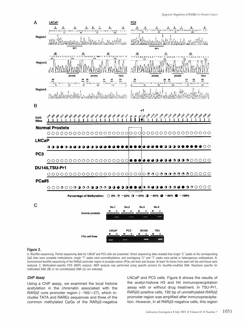
Using bisulfite PCR direct sequencing andmethylation-specific PCR (MSP) methods, approxi-mately 1 kb of the RARb2 promoter region (Fig. 1)(2477/1392, GenBank accession numbers S82362and M96016) containing 38 CpGs was examined forits methylation status. In four PCa cell lines, LNCaPcells were densely methylated in the entire regionaround the CpG island. PC3 cells were heteroge-neously methylated in regions 2 and 3, but not inregion 1 (Fig. 2, A and B). The most commonlymethylated CpG sites were numbers 21, 22, and 23 inregion 2 (Fig. 2B). We constructed specific MSPprimers to detect the methylation status of these CpGsites. In DU145, TSU-Pr1, and all normal prostatesamples, aberrant methylation was not detected bybisulfite direct sequencing in these regions (Fig. 2B).MSP analysis indicated hypermethylation in LNCaPand PC3, and no methylation in normal prostatesamples, DU145, or TSU-Pr1 (Fig. 2C). These resultswere consistent with bisulfite sequencing data.
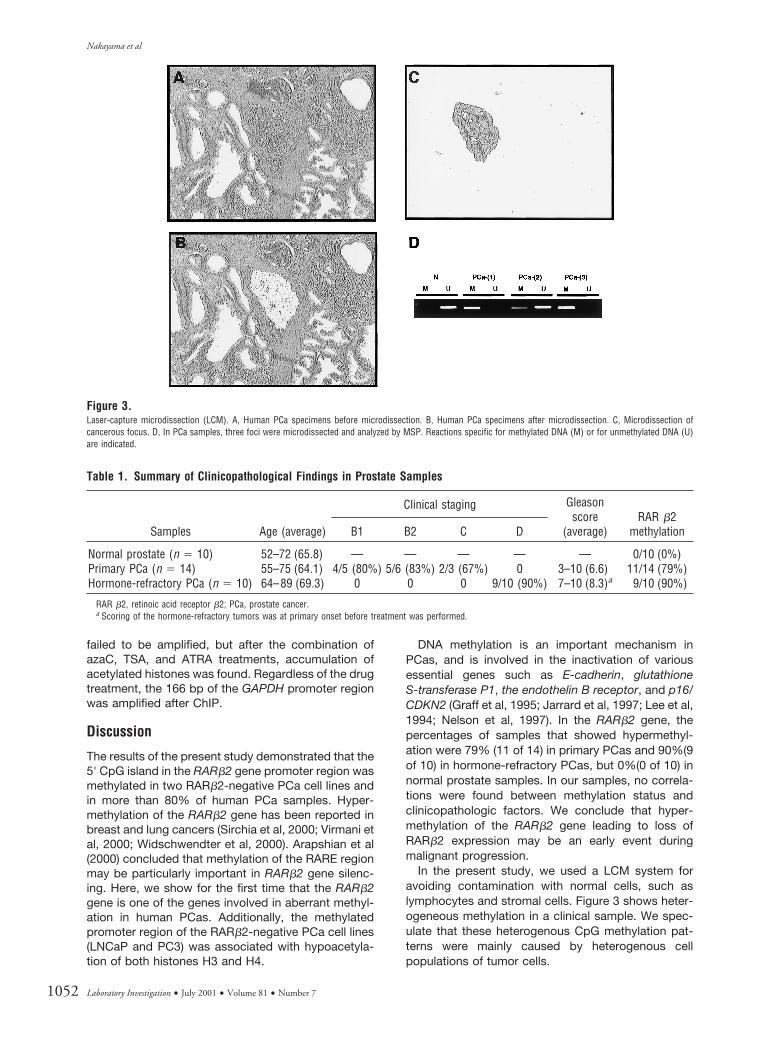
Using a laser-capture microdissection (LCM) sys-tem, three foci were microdissected in each clinicalsample and DNA was extracted from each focus.When methylation was detected in more than onefocus by MSP, the clinical specimen was classified asmethylation-positive (Fig. 3). In tumor specimens, 11of 14 primary PCas (79%) and 9 of 10 refractory PCas(90%) had hypermethylation of the RARb2 promoterregion. All samples were confirmed by bisulfite se-quencing (Fig. 2B). The relationships between methyl-ation and clinicopathologic factors are summarized inTable 1.
Expression of RARb2 in Normal Prostate Samples, PCaSamples, and PCa Cell Lines
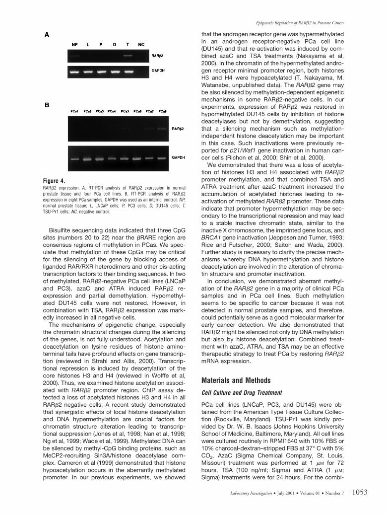
Only one of four PCa cell lines (TSU-Prl) expressedRARb2 mRNA, the other three cell lines (LNCaP, PC3,and DU145) were essentially negative for RARb2mRNA (Fig. 4A). In normal prostate tissues, RARb2was expressed at various levels. In primary PCas, lossor down-regulation of RARb2 expression was ob-served in five of eight cases (Fig. 4B). Regarding themethylation status of the RARb2 promoter, positivePCa samples (PC5, 7, and 8) were not methylated. Allnormal prostate samples expressed RARb2 and werenot methylated.
RARb2 Re-expression with Demethylating Agent andHistone Deacetylase Inhibitor Treatment
To clarify the role of epigenetic suppression of theRARb2 gene, we treated PCa cell lines with 5-aza-2'-deoxycitidine (azaC), Trichostatin A (TSA), and ATRA.With single agents, RARb2 re-expression was notdetected in RARb2-negative cells (data not shown).However, combined treatment with azaC and ATRAinduced slight RARb2 gene re-expression in LNCaPand PC3 cells, and after azaC treatment, the combi-nation of TSA and ATRA caused a marked increase inRARb2 gene re-expression. In DU145 cells, re-expression of RARb2 was not detected with azaC andATRA treatments, but was observed with addition ofTSA and ATRA after azaC treatment (Fig. 5A). Todetermine the effects of the demethylating agent, weexamined the methylation status in LNCaP and PC3cells after azaC treatment. In both cell lines, partialdemethylation was detected by MSP (Fig. 5B).
Figure 1.5'-untranslated region of the retinoic acid receptor (RAR) b2 gene. Top, The cytidine phosphate guanosine (CpG) island covers approximately 1 kb of the RARb2gene 5'-untranslated region, containing 38 CpGs (numbered 1 to 38). Several transcription factor binding sites are also indicated. Bottom, Sequence of b retinoicacid responsive element (RARE) region including three CpG dinucleotides.
Nakayama et al
1050 Laboratory Investigation • July 2001 • Volume 81 • Number 7
ChIP AssayUsing a ChIP assay, we examined the local histoneacetylation in the chromatin associated with theRARb2 core promoter region (2165/127), which in-cludes TATA and RAREs sequences and three of thecommon methylated CpGs of the RARb2-negative
LNCaP and PC3 cells. Figure 6 shows the results ofthe acetyl-histone H3 and H4 immunoprecipitationassay with or without drug treatment. In TSU-Pr1,RARb2-positive cells, 192 bp of unmethylated RARb2promoter region was amplified after immunoprecipita-tion. However, in all RARb2-negative cells, this region
Figure 2.A, Bisulfite sequencing. Partial sequencing data for LNCaP and PC3 cells are presented. Direct sequencing data revealed that single “C” peaks at the correspondingCpG sites were complete methylations, single “T” peaks were nonmethylations, and overlapping “C” and “T” peaks were partial or heterogenous methylations. B,Summarized bisulfite sequencing of the RARb2 promoter region in prostate cancer (PCa) cell lines and tissues. At least 10 clones from each cell line and tissue wereanalyzed. C, Methylation-specific PCR (MSP) analysis. MSP analysis was performed using specific primers for bisulfite-modified DNA. Reactions specific formethylated DNA (M) or for unmethylated DNA (U) are indicated.
Epigenetic Regulation of RARb2 in Prostate Cancer
Laboratory Investigation • July 2001 • Volume 81 • Number 7 1051
failed to be amplified, but after the combination ofazaC, TSA, and ATRA treatments, accumulation ofacetylated histones was found. Regardless of the drugtreatment, the 166 bp of the GAPDH promoter regionwas amplified after ChIP.
Discussion
The results of the present study demonstrated that the5' CpG island in the RARb2 gene promoter region wasmethylated in two RARb2-negative PCa cell lines andin more than 80% of human PCa samples. Hyper-methylation of the RARb2 gene has been reported inbreast and lung cancers (Sirchia et al, 2000; Virmani etal, 2000; Widschwendter et al, 2000). Arapshian et al(2000) concluded that methylation of the RARE regionmay be particularly important in RARb2 gene silenc-ing. Here, we show for the first time that the RARb2gene is one of the genes involved in aberrant methyl-ation in human PCas. Additionally, the methylatedpromoter region of the RARb2-negative PCa cell lines(LNCaP and PC3) was associated with hypoacetyla-tion of both histones H3 and H4.
DNA methylation is an important mechanism inPCas, and is involved in the inactivation of variousessential genes such as E-cadherin, glutathioneS-transferase P1, the endothelin B receptor, and p16/CDKN2 (Graff et al, 1995; Jarrard et al, 1997; Lee et al,1994; Nelson et al, 1997). In the RARb2 gene, thepercentages of samples that showed hypermethyl-ation were 79% (11 of 14) in primary PCas and 90%(9of 10) in hormone-refractory PCas, but 0%(0 of 10) innormal prostate samples. In our samples, no correla-tions were found between methylation status andclinicopathologic factors. We conclude that hyper-methylation of the RARb2 gene leading to loss ofRARb2 expression may be an early event duringmalignant progression.
In the present study, we used a LCM system foravoiding contamination with normal cells, such aslymphocytes and stromal cells. Figure 3 shows heter-ogeneous methylation in a clinical sample. We spec-ulate that these heterogenous CpG methylation pat-terns were mainly caused by heterogenous cellpopulations of tumor cells.
Figure 3.Laser-capture microdissection (LCM). A, Human PCa specimens before microdissection. B, Human PCa specimens after microdissection. C, Microdissection ofcancerous focus. D, In PCa samples, three foci were microdissected and analyzed by MSP. Reactions specific for methylated DNA (M) or for unmethylated DNA (U)are indicated.
Table 1. Summary of Clinicopathological Findings in Prostate Samples
Samples Age (average)
Clinical staging Gleasonscore
(average)RAR b2
methylationB1 B2 C D
Normal prostate (n 5 10) 52–72 (65.8) — — — — — 0/10 (0%)Primary PCa (n 5 14) 55–75 (64.1) 4/5 (80%) 5/6 (83%) 2/3 (67%) 0 3–10 (6.6) 11/14 (79%)Hormone-refractory PCa (n 5 10) 64–89 (69.3) 0 0 0 9/10 (90%) 7–10 (8.3)a 9/10 (90%)
RAR b2, retinoic acid receptor b2; PCa, prostate cancer.a Scoring of the hormone-refractory tumors was at primary onset before treatment was performed.
Nakayama et al
1052 Laboratory Investigation • July 2001 • Volume 81 • Number 7
Bisulfite sequencing data indicated that three CpGsites (numbers 20 to 22) near the bRARE region areconsensus regions of methylation in PCas. We spec-ulate that methylation of these CpGs may be criticalfor the silencing of the gene by blocking access ofliganded RAR/RXR heterodimers and other cis-actingtranscription factors to their binding sequences. In twoof methylated, RARb2-negative PCa cell lines (LNCaPand PC3), azaC and ATRA induced RARb2 re-expression and partial demethylation. Hypomethyl-ated DU145 cells were not restored. However, incombination with TSA, RARb2 expression was mark-edly increased in all negative cells.
The mechanisms of epigenetic change, especiallythe chromatin structural changes during the silencingof the genes, is not fully understood. Acetylation anddeacetylation on lysine residues of histone amino-terminal tails have profound effects on gene transcrip-tion (reviewed in Strahl and Allis, 2000). Transcrip-tional repression is induced by deacetylation of thecore histones H3 and H4 (reviewed in Wolffe et al,2000). Thus, we examined histone acetylation associ-ated with RARb2 promoter region. ChIP assay de-tected a loss of acetylated histones H3 and H4 in allRARb2-negative cells. A recent study demonstratedthat synergistic effects of local histone deacetylationand DNA hypermethylation are crucial factors forchromatin structure alteration leading to transcrip-tional suppression (Jones et al, 1998; Nan et al, 1998;Ng et al, 1999; Wade et al, 1999). Methylated DNA canbe silenced by methyl-CpG binding proteins, such asMeCP2-recruiting Sin3A/histone deacetylase com-plex. Cameron et al (1999) demonstrated that histonehypoacetylation occurs in the aberrantly methylatedpromoter. In our previous experiments, we showed
that the androgen receptor gene was hypermethylatedin an androgen receptor-negative PCa cell line(DU145) and that re-activation was induced by com-bined azaC and TSA treatments (Nakayama et al,2000). In the chromatin of the hypermethylated andro-gen receptor minimal promoter region, both histonesH3 and H4 were hypoacetylated (T. Nakayama, M.Watanabe, unpublished data). The RARb2 gene maybe also silenced by methylation-dependent epigeneticmechanisms in some RARb2-negative cells. In ourexperiments, expression of RARb2 was restored inhypomethylated DU145 cells by inhibition of histonedeacetylases but not by demethylation, suggestingthat a silencing mechanism such as methylation-independent histone deacetylation may be importantin this case. Such inactivations were previously re-ported for p21/Waf1 gene inactivation in human can-cer cells (Richon et al, 2000; Shin et al, 2000).
We demonstrated that there was a loss of acetyla-tion of histones H3 and H4 associated with RARb2promoter methylation, and that combined TSA andATRA treatment after azaC treatment increased theaccumulation of acetylated histones leading to re-activation of methylated RARb2 promoter. These dataindicate that promoter hypermethylation may be sec-ondary to the transcriptional repression and may leadto a stable inactive chromatin state, similar to theinactive X chromosome, the imprinted gene locus, andBRCA1 gene inactivation (Jeppesen and Turner, 1993;Rice and Futscher, 2000; Saitoh and Wada, 2000).Further study is necessary to clarify the precise mech-anisms whereby DNA hypermethylation and histonedeacetylation are involved in the alteration of chroma-tin structure and promoter inactivation.
In conclusion, we demonstrated aberrant methyl-ation of the RARb2 gene in a majority of clinical PCasamples and in PCa cell lines. Such methylationseems to be specific to cancer because it was notdetected in normal prostate samples, and therefore,could potentially serve as a good molecular marker forearly cancer detection. We also demonstrated thatRARb2 might be silenced not only by DNA methylationbut also by histone deacetylation. Combined treat-ment with azaC, ATRA, and TSA may be an effectivetherapeutic strategy to treat PCa by restoring RARb2mRNA expression.
Materials and Methods
Cell Culture and Drug Treatment
PCa cell lines (LNCaP, PC3, and DU145) were ob-tained from the American Type Tissue Culture Collec-tion (Rockville, Maryland). TSU-Pr1 was kindly pro-vided by Dr. W. B. Isaacs (Johns Hopkins UniversitySchool of Medicine, Baltimore, Maryland). All cell lineswere cultured routinely in RPMI1640 with 10% FBS or10% charcoal-dextran–stripped FBS at 37° C with 5%CO2. AzaC (Sigma Chemical Company, St. Louis,Missouri) treatment was performed at 1 mM for 72hours, TSA (100 ng/ml; Sigma) and ATRA (1 mM;Sigma) treatments were for 24 hours. For the combi-
Figure 4.RARb2 expression. A, RT-PCR analysis of RARb2 expression in normalprostate tissue and four PCa cell lines. B, RT-PCR analysis of RARb2expression in eight PCa samples. GAPDH was used as an internal control. NP,normal prostate tissue; L, LNCaP cells; P, PC3 cells; D, DU145 cells; T,TSU-Pr1 cells; NC, negative control.
Epigenetic Regulation of RARb2 in Prostate Cancer
Laboratory Investigation • July 2001 • Volume 81 • Number 7 1053

nation of azaC, TSA, and ATRA treatments, azaC wasintroduced for an initial incubation of 48 hours, andATRA or TSA/ATRA were added for an additional 24hours.
Tissue Samples
Ten samples of normal prostate tissue were obtainedat autopsy. All samples were examined by histopa-thology and determined to have no evidence of can-cerous lesions. Fourteen primary PCa specimens wereobtained at radical prostatectomy at Mie UniversityHospital, Mie, Japan, snap-frozen and stored at280° C. Additionally, ten hormone-refractory tumorswere obtained from distant organ site metastases atautopsy at Chiba University Hospital, Chiba, Japan,and genomic DNA was immediately extracted by astandard protocol. All ten of the hormone-refractorytumor patients had experienced a new onset of cancerunder hormonal therapy and died. For RNA extraction,we selected eight of the primary PCa samples andmicrodissected parts of the specimens composed ofmore than 80% tumor cells. The clinicopathologic
findings for the examined samples are summarized inTable 1.
Tissue Preparation and Sampling by LCM
Frozen tissues were cut into 4 to 6 mm-thick sectionswith a cryostat. Sections were placed onto glassslides that had been baked at 230° C for 4 hours. Thesections were immediately fixed with 70% ethanol for10 minutes and then washed with diethylpyrocarbonate-treated water for 5 seconds. Sectionswere stained with hematoxylin for 15 seconds,washed with diethyl pyrocarbonate-treated water for10 seconds, dehydrated with an ethanol gradient, andcounterstained with an alcoholic Eosin Y solution for30 seconds. Sections were washed three times with100% ethanol and three times with xylenes. Thesections were air dried with a fan for 20 minutes andstored in a plastic container with silica gel at 280° Cuntil use.
Frozen tumor/or adjacent normal tissues were mi-crodissected using a Pixcell LCM system (LM200;Arcturus Engineering Inc., Mountain View, California).
Figure 5.RARb2 re-expression. A, RARb2 re-expression by the treatment of chromatin remodeling drugs. The treatment of 5-aza-2'-deoxycitidine (azaC), Trichostatin A (TSA),and all-trans retinoic acid (ATRA) is described in “Materials and Methods”. B, Demethylation after combined drug treatment. MSP analysis showed partialdemethylation of the RARb2 promoter region after azaC and ATRA treatment. These experiments were performed three times, each with similar results.
Figure 6.Chromatin immunoprecipitation (ChIP) assay. ChIP, with the use of antibodies to acetylated histone H3 and H4, detected both histones acetylations of the RARb2promoter in the RARb2-positive TSU-Pr1 cell line and of the GAPDH promoter in all cell lines. After the combined treatment with TSA and ATRA after azaC exposure,histone H3 and H4 acetylation was increased in all RARb2-negative cell lines. These experiments were performed three times, each with similar results.
Nakayama et al
1054 Laboratory Investigation • July 2001 • Volume 81 • Number 7
Sections were covered with LCM transfer film (Cap-ture TF-100; Arcturus Engineering Inc.), and specificportions of the histologic section were affixed to thecapture film by brief laser pulses. DNA and RNA wereextracted as described previously (Goldsworthy et al,1999; Hayes et al, 2000).
Bisulfite Modification
Genomic DNA (approximately 0.5 mg) was treated withsodium bisulfite as described previously (Frommer etal, 1992). After denaturation in 0.3 M NaOH at 37° Cfor 15 minutes, sodium bisulfite was added to a finalconcentration of 3.1 M, and hydroquinone was addedto a final concentration of 0.5 mM. The reaction wasperformed at 55° C for 16 hours, and desalted usingthe Wizard DNA purification resin (Promega, Madison,Wisconsin) according to the manufacturer’s instruc-tions. Bisulfite modification was completed by 0.3 MNaOH treatment at 37° C for 15 minutes. ModifiedDNA was precipitated with ethanol, washed in 70%ethanol, dried, and resuspended in 50 ml of distilledwater.
Bisulfite Sequencing and Methylation-Specific PCR(MSP)
The methylation status of the 5'-regulatory region ofRARb2 in PCa cell lines was analyzed by bisulfite PCRsequencing and MSP as described previously (From-mer et al, 1992; Herman et al, 1996). Bisulfite genomicsequencing was performed with the following primers:Region 1 (product size, 355 bp): forward, 5'-GTA TGTGTT TTT TTT GGA GTG G-3', reverse, 5'-AAC TTAAAA ACT CCC AAC AAC C-3'; Region 2 (product size,154 bp): forward, 5'-TGG GAG TTG GTG ATG TTAGA-3', reverse, 5'-ACC CTC CTA ACC TCT AAACA-3'; Region 3 (product size, 391 bp): forward,5'-TGT TTA GAG GTT AGG GTT TAT T-3', reverse,5'-AAC TCC ATC AAA CTC TAC CCC TT-3'. No CpGdinucleotide motifs were contained in these primersequences. PCR conditions were as follows: 95° C for10 minutes, 30 cycles of 95° C for 30 seconds, 57° Cfor 30 seconds, 72° C for 30 seconds, and 72° C for 10minutes. For bisulfite genomic sequencing, total PCRproducts were gel-purified and directly sequencedusing the ABI 310 automated sequencing system(Perkin Elmer, Foster City, California).
For MSP analysis, modified DNA was amplified withspecific-primers: 5'-GGG TTT ATC GAA AGT TTATTC-3' (forward-methylated) and 5'-TTC CGA ATACGT TCC GAA T-3' (reverse-methylated); 5'-GGTAGG GTT TAT TGA AAG TTT ATT T-3' (forward-unmethylated) and 5'-AAA CCT TCC AAA TAC ATTCCA AAT-3' (reverse-unmethylated). PCR was carriedout under the following conditions: 95° C for 10 min-utes then 30 amplification cycles (95° C for 30 sec-onds, 59° C for 30 seconds, 72° C for 30 seconds) anda final extension incubation of 10 minutes at 72° C.PCR products were directly loaded on 2.0% agarosegels and analyzed after ethidium-bromide staining.
RT-PCR for RARb2
Total RNA was prepared using Isogen (Nippon Gene,Tokyo, Japan), according to the manufacturer’s in-structions. Aliquots of 2 mg of total RNA were used forgeneration of cDNAs using Superscript reverse tran-scriptase (GIBCO BRL, Gaithersburg, Maryland). Thespecific primers applied to detect RARb2 transcripts(GenBank accession number X07282) were as follows:forward (located in exon 3), 5'-GCA TGG CAG AGTGCC CTA TC-3'; reverse (located in exon 6), 5'-TCCCAG AGT CAT CCC TGC TTC AT-3'. PCR amplifica-tion was performed for 30 cycles at 95° C for 30seconds, 62° C for 30 seconds, and 72° C for 60seconds. Human GAPDH was used as an internalcontrol. The PCR products were subjected to electro-phoresis in 2.0% agarose gels and visualized byethidium bromide staining.
ChIP Assay
ChIP assays using antibodies to acetyl-histone H3 andH4 were performed according to the manufacturer’sinstructions (Upstate Biotechnology, Lake Placid, NewYork). Cells were cultured and treated with 100 ng/mlof TSA and/or 1 mM ATRA for 24 hours after 1 mM azaCtreatment for 48 hours. Formaldehyde was then addedto the cells to a final concentration of 1% and incu-bated at 37° C for 10 minutes. The cells were washedin 1 ml of ice-cold PBS with proteinase inhibitors,scraped, resuspended in 200 ml of SDS lysis buffer,and incubated on ice for 10 minutes. Lysates weresonicated for 10 seconds three times on ice andcentrifuged at 15,000 rpm for 10 minutes at 4° C.Supernatants were loaded on 1% agarose gels anddetermined to have reduced DNA lengths to between200 and 1000 bp. Sonicated samples were diluted10-fold with immunoprecipitation buffer and dividedequally to prepare negative control (no antibody) im-munoprecipitation samples. The samples were pre-cleaned with a salmon sperm DNA/protein A agaroseslurry and incubated overnight at 4° C with or withoutantibodies to histone H3 or H4. Chromatin-antibodycomplexes were collected using a salmon spermDNA/protein A agarose slurry and washed accordingto the manufacturer’s protocol. Immunocomplexeswere eluted twice with 250 ml of elution buffer (1%SDS, 0.1 M NaHCO3) for 15 minutes at room temper-ature. To reverse crosslinks, 20 ml of 5 M NaCl wereadded with incubation for 4 hours at 65° C. Tenmicroliters of 0.5 M EDTA, 20 ml of 1 M Tris-HCl pH6.5, and 2 ml of 10 mg/ml Proteinase K were added,and the samples were incubated at 45° C for 1 hour.Immunoprecipitated DNA was recovered by phenol/chloroform extraction and ethanol precipitation andanalyzed by PCR. The primer pairs used for ChIPanalysis of the RARb2 promoter region (GenBankaccession numbers S82362 and M96016, PCR prod-uct length 192 bp) were 5'-CTC TGG CTG TCT GCTTTT GC-3' (forward), 5'-CAG CTC ACT TCC TAC TACTTC-3' (reverse). The primers used for the GAPDHpromoter region (GenBank accession number J04038,
Epigenetic Regulation of RARb2 in Prostate Cancer
Laboratory Investigation • July 2001 • Volume 81 • Number 7 1055
PCR product length 166 bp) were 5'-TAC TAG CGGTTT TAC GGG CG-3' (forward), 5'-TCG AAC AGGAGG AGC AGA GA-3' (reverse). PCR was performedfor 25 to 30 cycles of 95° C for 30 seconds, 58° C for30 seconds, and 72° C for 30 seconds. PCR productswere analyzed on 2.0% agarose gels and visualized byUV illumination.
Acknowledgement
We are grateful to Dr. M. Toyota (Department ofMolecular Biology, Cancer Research Institute, Sap-poro Medical University School of Medicine, Hok-kaido, Japan) for constructive comments during thepreparation of the manuscript.
ReferencesArapshian A, Kuppumbatti YS, and Mira-y-Lopez R (2000).Methylation of conserved CpG sites neighboring the betaretinoic acid response element may mediate retinoic acidreceptor beta gene silencing in MCF-7 breast cancer cells.Oncogene 19:4066–4070.
Baust C, Redpath L, and Schwarz E (1996). Different ligandresponsiveness of human retinoic-acid-receptor beta-genetranscription in tumorigenic and non-tumorigenic cervical-carcinoma-derived cell lines is mediated through a largeretinoic-acid-response domain. Int J Cancer 67:409–416.
Bird AP and Wolffe AP (1999). Methylation-inducedrepression: Belts, braces, and chromatin. Cell 99:451–454.
Cameron EE, Bachman KE, Myohanen S, Herman JG, andBaylin SB (1999). Synergy of demethylation and histonedeacetylase inhibition in the re-expression of genes silencedin cancer. Nat Genet 21:103–107.
Chambon P (1996). A decade of molecular biology of retinoicacid receptors. FASEB J 10:940–954.
de The H, Vivanco-Ruiz MM, Tiollais P, Stunnenberg H, andDejean A (1990). Identification of a retinoic acid responsiveelement in the retinoic acid receptor beta gene. Nature343:177–180.
Frommer M, McDonald LE, Millar DS, Collis CM, Watt F,Grigg GW, Molloy PL, and Paul CL (1992). A genomicsequencing protocol that yields a positive display of5-methylcytosine residues in individual DNA strands. ProcNatl Acad Sci USA 89:1827–1831.
Gardiner-Garden M and Frommer M (1987). CpG islands invertebrate genomes. J Mol Biol 196:261–282.
Graff JR, Herman JG, Lapidus RG, Chopra H, Xu R, JarrardDF, Isaacs WB, Pitha PM, Davidson NE, and Baylin SB (1995)E-cadherin expression is silenced by DNA hypermethylationin human breast and prostate carcinomas. Cancer Res55:5195–5199.
Goldsworthy SM, Stockton PS, Trempus CS, Foley JF, andMaronpot RR (1999). Effects of fixation on RNA extractionand amplification from laser capture microdissected tissue.Mol Carcinog 25:86–91.
Hayes AJ, Huang W-Q, Yu J, Maisonpierre PC, Liu A, KernFG, Lippman ME, McLeskey SW, and Li L-Y (2000) Expres-sion and function of angiopoietin-1 in breast cancer. Br JCancer 83:1154–1160.
Herman JG, Graff JR, Myohanen S, Nelkin BD, and Baylin SB(1996). Methylation-specific PCR: A novel PCR assay formethylation status of CpG islands. Proc Natl Acad Sci USA93:9821–9826.
Jarrard DF, Bova GS, Ewing CM, Pin SS, Nguyen SH, BaylinSB, Cairns P, Sidransky D, Herman JG, and Isaacs WB(1997). Deletional, mutational, and methylation analyses ofCDKN2 (p16/MTS1) in primary and metastatic prostate can-cer. Genes Chromosomes Cancer 19:90–96.
Jeppesen P and Turner BM (1993). The inactive X chromo-some in female mammals is distinguished by a lack ofhistone H4 acetylation, a cytogenetic marker for gene ex-pression. Cell 74:281–289
Jones PL, Veenstra GJ, Wade PA, Vermaak D, Kass SU,Landsberger N, Strouboulis J, and Wolffe AP (1998). Meth-ylated DNA and MeCP2 recruit histone deacetylase to re-press transcription. Nat Genet 19:187–191.
Lee WH, Morton RA, Epstein JI, Brooks JD, Campbell PA,Bova GS, Hsieh WS, Isaacs WB, and Nelson WG (1994).Cytidine methylation of regulatory sequences near the pi-class glutathione S-transferase gene accompanies humanprostatic carcinogenesis. Proc Natl Acad Sci USA 91:11733–11737.
Lotan R, Xu XC, Lippman SM, Ro JY, Lee JS, Lee JJ, andHong WK (1995). Suppression of retinoic acid receptor-betain premalignant oral lesions and its up-regulation by isotreti-noin. N Engl J Med 332:1405–1410.
Lotan Y, Xu XC, Shalev M, Lotan R, Williams R, Wheeler TM,Thompson TC, and Kadmon D (2000). Differential expressionof nuclear retinoid receptors in normal and malignant pros-tates. J Clin Oncol 18:116–121.
Nan X, Ng HH, Johnson CA, Laherty CD, Turner BM, Eisen-man RN, and Bird A (1998). Transcriptional repression by themethyl-CpG-binding protein MeCP2 involves a histonedeacetylase complex. Nature 393:386–389.
Nakayama T, Watanabe M, Suzuki H, Toyota M, Sekita N,Hirokawa Y, Mizokami A, Ito H, Yatani R, and Shiraishi T(2000). Epigenetic regulation of androgen receptor geneexpression in human prostate cancers. Lab Invest 80:1789–1796.
Nelson JB, Lee WH, Nguyen SH, Jarrard DF, Brooks JD,Magnuson SR, Opgenorth TJ, Nelson WG, and Bova GS(1997). Methylation of the 5' CpG island of the endothelin Breceptor gene is common in human prostate cancer. CancerRes 57:35–37.
Ng HH, Zhang Y, Hendrich B, Johnson CA, Turner BM,Erdjument-Bromage H, Tempst P, Reinberg D, and Bird A(1999). MBD2 is a transcriptional repressor belonging to theMeCP1 histone deacetylase complex. Nat Genet 23:58–61.
Picard E, Seguin C, Monhoven N, Rochette-Egly C, Siat J,Borrelly J, Martinet Y, Martinet N, and Vignaud JM (1999).Expression of retinoid receptor genes and proteins in non-small-cell lung cancer. J Natl Cancer Inst 91:1059–1066.
Qiu H, Zhang W, El-Naggar AK, Lippman SM, Lin P, Lotan R,and Xu XC (1999). Loss of retinoic acid receptor-beta expres-sion is an early event during esophageal carcinogenesis.Am J Pathol 155:1519–1523.
Rice JC and Futscher BW (2000). Transcriptional repressionof BRCA1 by aberrant cytosine methylation, histone hy-poacetylation and chromatin condensation of the BRCA1promoter. Nucleic Acids Res 28:3233–3239.
Nakayama et al
1056 Laboratory Investigation • July 2001 • Volume 81 • Number 7

Richon VM, Sandhoff TW, Rifkind RA, and Marks PA (2000).Histone deacetylase inhibitor selectively induces p21WAF1expression and gene-associated histone acetylation. ProcNatl Acad Sci USA 97:10014–10019.
Saitoh S and Wada T (2000). Parent-of-origin specific histoneacetylation and reactivation of a key imprinted gene locus inPrader-Willi syndrome. Am J Hum Genet 66:1958–1962.
Shin JY, Kim HS, Park J, Park JB, and Lee JY (2000).Mechanism for inactivation of the KIP family cyclin-dependent kinase inhibitor genes in gastric cancer cells.Cancer Res 60:262–265.
Sirchia SM, Ferguson AT, Sironi E, Subramanyan S, OrlandiR, Sukumar S, and Sacchi N (2000). Evidence of epigeneticchanges affecting the chromatin state of the retinoic acidreceptor b2 promoter in breast cancer cells. Oncogene19:1556–1563.
Strahl BD and Allis CD (2000). The language of covalenthistone modifications. Nature 403:41–45.
Sun SY, Wan H, Yue P, Hong WK, and Lotan R (2000).Evidence that retinoic acid receptor beta induction by retin-oids is important for tumor cell growth inhibition. J Biol Chem275:17149–17153.
van der Leede BJ, Folkers GE, Kruyt FA, and van der Saag PT(1992). Genomic organization of the human retinoic acidreceptor b2. Biochem Biophys Res Commun 188:695–702.
Virmani AK, Rathi A, Zochbauer-Muller S, Sacchi N,Fukuyama Y, Bryant D, Maitra A, Heda S, Fong KM, Thun-nissen F, Minna JD, and Gazdar AF (2000). Promoter meth-ylation and silencing of the retinoic acid receptor-beta genein lung carcinomas. J Natl Cancer Inst 92:1303–1307.
Wade PA, Gegonne A, Jones PL, Ballestar E, Aubry F, andWolffe AP (1999). Mi-2 complex couples DNA methylation tochromatin remodelling and histone. Nat Genet 23:62–66.
Widschwendter M, Berger J, Hermann M, Muller HM, Am-berger A, Zeschnigk M, Widschwendter A, Abendstein B,Zeimet AG, Daxenbichler G, and Marth C (2000). Methylationand silencing of the retinoic acid receptor-b2 gene in breastcancer. J Natl Cancer Inst 92:826–832.
Wolffe AP, Urnov FD, and Guschin D (2000). Co-repressorcomplexes and remodelling chromatin for repression. Bio-chem Soc Trans 28:379–386.
Xu XC, Sneige N, Liu X, Nandagiri R, Lee JJ, Lukmanji F,Hortobagyi G, Lippman SM, Dhingra K, and Lotan R (1997).Progressive decrease in nuclear retinoic acid receptor betamessenger RNA level during breast carcinogenesis. CancerRes 57:4992–4996.
Epigenetic Regulation of RARb2 in Prostate Cancer
Laboratory Investigation • July 2001 • Volume 81 • Number 7 1057