the role of early experience in infant development
TRANSCRIPT

Pediatric Round Table
The Role of EarlyExperience in Infant Development
Sponsored by:
Edited by: Nathan A. Fox, PhDLewis A. Leavitt, MDJohn G. Warhol, PhD

THE ROLE OF EARLY EXPERIENCE IN INFANT
DEVELOPMENT

ii
Series includes continuing education programs and patient education materials.
For more information,call 1-877-JNJ-LINK or fax 1-877-656-3299
or visit us @ jnjPediatricInstitute.com.
Summary Publications in the Johnson & Johnson Pediatric Round Table Series:
1. New Perspectives in Early Emotional DevelopmentEdited by Steven P. Shelov, MD and John G. Warhol, PhD
2. The Role of Early Experience in Infant DevelopmentEdited by Nathan Fox, PhD, Lewis A. Leavitt, MD,and John G. Warhol, PhD

THE ROLE OF EARLY EXPERIENCE IN INFANT
DEVELOPMENT
Chaired by Nathan A. Fox, PhD
Edited by Nathan A. Fox, PhD, Lewis A. Leavitt MD,
and John G. Warhol, PhD
Sponsored by

Library of Congress Catalog Card Number: 99-96523
Main entry under title:
The Role of Early Experience in Infant Development
Johnson & Johnson Pediatric Institute Pediatric Round Table Series: 1999
Includes bibliography
Summary of a conference held in January 1999.
ISBN 0-93-1562-20-1
Copyright © 1999 by Johnson & Johnson Consumer Companies, Inc.
Printed in the United States of America. All rights reserved. Except as permit-ted under the Copyright Act of 1976, no part of this publication may be repro-duced or distributed in any form or by any means or stored in a data base orretrieval system, without prior written permission of the publisher.
The opinions and data presented by the Pediatric Round Table faculty are theirown, and are not necessarily those of Johnson & Johnson or the editors.
Cover photo: Frank Peluso

iv
Table of Contents
Page
Participants . . . . . . . . . . . . . . . . . . . . . . . . . . . . . . . . . . . . . . . . . . . . . . . .vii
Preface . . . . . . . . . . . . . . . . . . . . . . . . . . . . . . . . . . . . . . . . . . . . . . . . . . . .xiJulia A. Freedman
Introduction . . . . . . . . . . . . . . . . . . . . . . . . . . . . . . . . . . . . . . . . . . . . . . .xiiiNathan A. Fox, PhD, Lewis A. Leavitt, MD
Section 1. Brain Development . . . . . . . . . . . . . . . . . . . . . . . . . . . . . . . . . .1
Neuroanatomy and Development Overview . . . . . . . . . . . . . . . . . . . . . . . . .5Bryan Kolb, PhD
Synaptogenesis in Human Cerebral Cortex and the Concept of Critical Periods . . . . . . . . . . . . . . . . . . . . . . . . . . . . . . . . .15Peter R. Huttenlocher, MD
Experience, Neural Plasticity, and Psychological Development . . . . . . . . . .29William T. Greenough, PhD, James E. Black, MD, PhD
Early Experience, Behavior, and the Changing Brain . . . . . . . . . . . . . . . . .41Bryan Kolb, PhD, Robbin Gibb, Agnes Dallison
Section 2. Auditory/Language Development . . . . . . . . . . . . . . . . . . . . . .65
Language Input and Language Growth . . . . . . . . . . . . . . . . . . . . . . . . . . . .69Janellen Huttenlocher, PhD
One Step at a Time . . . . . . . . . . . . . . . . . . . . . . . . . . . . . . . . . . . . . . . . . .83Peter W. Jusczyk, PhD
The Role of Experience in Early Language Development: LinguisticExperience Alters the Perception and Production of Speech . . . . . . . . . . .101Patricia K. Kuhl, PhD
Section 3. Cognitive Development . . . . . . . . . . . . . . . . . . . . . . . . . . . .127
Development of Cognitive Functions is Linked to the Prefrontal Cortex . . . . . . . . . . . . . . . . . . . . . . . . . . . . . . . . . . . . . .131Adele Diamond, PhD
Born to Learn: What Infants Learn from Watching Us . . . . . . . . . . . . . .145Andrew N. Meltzoff, PhD

v
Early Experience Matters for Spatial Development (But Different Kinds at Different Times) . . . . . . . . . . . . . . . . . . . . . . . . .165Nora S. Newcombe, PhD
Section 4. Perceptual Development . . . . . . . . . . . . . . . . . . . . . . . . . . . .187
About Functional Brain Specialization: the Development of Face Recognition . . . . . . . . . . . . . . . . . . . . . . . . . . .191Scania de Schonen, PhD
The Development of Representations for Perception and Action . . . . . . .209Rick O. Gilmore, PhD
The Emergence of Future-oriented Thinking in the Early Years . . . . . . . .225Marshall M. Haith, PhD
Section 5. Biosocial Development . . . . . . . . . . . . . . . . . . . . . . . . . . . . .249
Hormones May Influence Both Social Attachment and Reactivity to Stress . . . . . . . . . . . . . . . . . . . . . . . . . . . . . . . . . . . . . . .253C. Sue Carter, PhD
The Role of Early Experience in Infant Development – Enhancing Outcome After Extremely Preterm Birth . . . . . . . . . . . . . . . .267Neena Modi, MB, ChB, MD, FRCP, FRCPCH
Effects on Mother and Infant of Oxytocin Released in the Postpartum Period . . . . . . . . . . . . . . . . . . . . . . . . . . . . . . . . . . . . .283Kerstin Uvnäs-Moberg, MD, PhD
Section 6. Discussion . . . . . . . . . . . . . . . . . . . . . . . . . . . . . . . . . . . . . . .291Nathan A. Fox, PhD, Betsy Lozoff, MD, Lewis A. Leavitt, MD, Ronald G. Barr, MD, Ann C. Stadtler, MSN, CPNP, Rosemary White-Traut, DNSc, RN
Table of Contents

vi
Ronald G. Barr, MDProfessor of Pediatrics and PsychiatryMcGill UniversityHead, Child Development ProgramMontreal Children’s Hospital2300 Rue TupperMontreal, QC, Canada H3H 1P3
C. Sue Carter, PhDDistinguished University Professor of
BiologyDepartment of BiologyUniversity of MarylandRoom 4222 Zoology-PsychologyBuilding 144College Park, MD 20742
Adele Diamond, PhDDirector, Center for Developmental
Cognitive NeuroscienceEunice Kennedy Shriver Center200 Trapelo RoadWaltham, MA 02452
Nathan A. Fox, PhDProfessor, Institute for Child StudyDepartment of Human DevelopmentUniversity of MarylandRoom 4304, Benjamin BuildingCollege Park, MD 20742
Julia FreedmanDirectorJohnson & Johnson Pediatric Institute199 Grandview RoadSkillman, NJ 08558-9418
Rick O. Gilmore, PhDAssistant Professor of PsychologyDepartment of PsychologyPennsylvania State University643 Moore BuildingUniversity Park, PA 16802
William T. Greenough, PhDProfessor of PsychologyUniversity of Illinois at
Urbana-Champaign2347 Beckman Institute, MC 251405 North Matthews AvenueUrbana, IL 61801
Marshall M. Haith, PhDDirector of University ResearchProfessor of PsychologyUniversity of Denver2155 South Race StreetDenver, CO 80208
Janellen Huttenlocher, PhDProfessor of PsychologyDepartment of PsychologyUniversity of Chicago5848 South University AvenueChicago, IL 60637
Peter R. Huttenlocher, MDProfessor of Pediatrics and NeurologyDepartment of PediatricsUniversity of Chicago School
of Medicine5839 South Maryland Avenue,
MC 3055Chicago, IL 60637
Participants

vii
Peter W. Jusczyk, PhDProfessor of PsychologyDepartment of PsychologyJohns Hopkins UniversityAmes Hall3400 North Charles StreetBaltimore, MD 21218
Bryan Kolb, PhDProfessor of Psychology and
NeuroscienceDepartment of Psychology and
NeuroscienceThe University of Lethbridge4401 University DriveLethbridge, AB, Canada T1K 3M4
Patricia K. Kuhl, PhDChair and Professor of Speech and
Hearing ScienceUniversity of Washington204 Eagleson HallBox 357920Seattle, WA 98195-7920
Lewis A. Leavitt, MDProfessor of PediatricsDirector of Developmental PediatricsDirector, Waisman CenterUniversity of Wisconsin1500 Highland AvenueMadison, WI 53705-2274
Betsy Lozoff, MDDirector, Center for Human Growth
and DevelopmentProfessor, Department of Pediatrics
and Communicable DiseasesUniversity of Michigan300 North Ingalls, 10th LevelAnn Arbor, MI 48109-0406
Andrew N. Meltzoff, PhDProfessor of PsychologyDepartment of PsychologyUniversity of Washington339 GuthrieBox 357920Seattle, WA 98195-7920
Neena Modi, MB, ChB, MD, FRCP,FRCPCHSenior Lecturer and ConsultantDepartment of Pediatrics and
Neonatal MedicineImperial College of Science,
Technology and MedicineHammersmith HospitalDu Cane RoadLondon W12 0NN
Nora S. Newcombe, PhDProfessor of PsychologyTemple University565 Weiss HallPhiladelphia, PA 19122
Stephen W. Porges, PhDChair, Department of Human
DevelopmentDirector, Institute for Child StudyUniversity of Maryland3304 Benjamin BuildingCollege Park, MD 20742-1131
Scania de Schonen, PhDDirector of ResearchLaboratory of Cognition and
DevelopmentNational Center for Scientific ResearchUniversity of René Descartes – Paris V28 Rue SerpenteF-75270 Paris Cedex 06, FRANCE
Participants

viii
Ann C. Stadtler, MSN, CPNPAssistant Director/Clinical CoordinatorMedical Diagnostic ProgramsBoston Children’s Hospital1295 Boylston Street, Suite 320Boston, MA 02215
Kerstin Uvnäs-Moberg, MD, PhDProfessor of PhysiologyDepartment of Animal PhysiologySwedish University of Agricultural
Science171 77Stockholm, Sweden
Carole Welp, MEdDirectorPointe St. Charles Early Childhood
CenterMontreal, QC, Canada H3K 2RI
Rosemary White-Traut, DNSc, RNAssociate Professor and Acting
Department HeadDepartment of Maternal/Child NursingUniversity of Illinois565 North Washington StreetHinsdale, IL 60521
Participants (continued)

ix
Preface
Johnson & Johnson is pleased to provide this year’s edition of its highlyregarded Pediatric Round Table series. The goals of these conferences are to:foster the free exchange of the latest research in infant development; to dis-cuss the clinical implications of that research; and to make those findingsavailable to all professionals working in the field of infant health.
The faculty of this Pediatric Round Table, The Role of Early Experience inInfant Development, represent the world’s leading researchers and cliniciansworking in the areas of early development of the brain, auditory and languageability, cognition, perception, and biosocial interactions. They brought to thetable years of experience and unique insight into the relationship between theway an infant’s experiences with his family and environment affect manyaspects of early brain development.
A common theme emerging from this Round Table was that development is a process as unique as each individual child – within that spectrum, for theoverwhelming majority of infants, “normal” development proceeds with rou-tine care, without the need for developmentally aggressive toys or interven-tions, and extends well beyond the first 3 years of life.
Our continuing aspiration is to build and provide a library of current researchand significant information on infant development that features the world’sleading scientists and healthcare professionals. I hope readers of The Role ofEarly Experience in Infant Development agree that this latest volume provides a valuable resource for healthcare professionals.
Julia A. FreedmanDirector, Johnson & Johnson Pediatric Institute

x
Introduction
Nathan A. Fox, PhD and Lewis A. Leavitt, MD
The White House Conference on Early Childhood and Brain Developmentin the Spring of 1995 marked a turning point in public awareness and knowl-edge about the effects of early experience on the developing human infantand its nervous system. At that meeting, researchers and practitioners met todiscuss the important advances in our knowledge about the competencies andskills of young infants, as well as the remarkable changes that occur in infantsover the first 3 years of life. Materials that were distributed at this meeting,including a special issue of Newsweek magazine, emphasized the importanceof the first 3 years for behavioral growth and brain development. The verypositive message communicated to service providers and parents was that thefirst 3 years of an infant’s life are critical for brain development and that par-ents could play an important role in fostering that development. Implicitly,however, another message was communicated: once these early years wereover, the window would close and opportunities for brain growth and stimu-lation, for enhancing cognitive and socio-emotional development, would end.Predictably, these formulations produced a flurry of excitement and concernfrom new parents. Parents questioned the nature of care and stimulation thatthey were providing their young infants. Were the environments they provid-ed sufficient to stimulate brain growth? Could and should they be doingmore to enhance cognitive and social-emotional development during theseearly years?
The information communicated as a result of this meeting also elicited excite-ment and concern from developmental psychologists and neuroscientists whostudy neurodevelopment. Researchers were excited that their area of interestwas getting much-deserved attention. However, they were concerned aboutthe scientific basis for many of the claims regarding the role of early experi-ence on brain development that circulated after the meeting. Indeed, in someinstances, contradictory evidence was available which called for a more bal-anced approach to these issues.
This background marks the starting point for the organization of a conferencetitled The Role of Early Experience in Infant Development held in January of1999. This conference, sponsored by the Johnson & Johnson PediatricInstitute, was motivated by the divergent and often conflicting reactions that

xi
developmental psychologists, neuroscientists, and service providers for chil-dren had in response to the information that emerged after the initial WhiteHouse meeting. The goal of the Johnson & Johnson meeting was to bringtogether developmental psychologists, neuroscientists, and practicing pediatri-cians and nurses who were engaged in work on the effects of early experienceon infant development. Each of the participants was asked to present the cur-rent state of the art in their area of expertise, addressing the issue of earlyexperience and the brain. The approaches represented by the faculty to theseissues were diverse, and multiple areas were covered. Our aim was to achieve abalanced and comprehensive view of the issues surrounding the question ofearly experience and brain development. The current volume, The Role ofEarly Experience in Infant Development, presents a review of the “state of theart” written for practitioners who must do the crucial work of translating the-ory and research into the arena of real life.
Pediatric service providers need to consider several issues when giving adviceon the effects of early experience on brain development. The most basic ques-tion can be stated rather simply: What do we know about development andthe emerging competencies of the young infant in the behavioral domain andin the domain of brain development? The surprising and somewhat frustrat-ing answer to this broad question is that, after many years of scientificresearch, we know quite a lot about the emerging behavioral competencies of the human infant. But we know much less about the development of the human infant’s brain and even less about the manner in which behaviorand brain develop and interact synchronously.
There are multiple reasons for this asymmetry in our knowledge base. First,developmental researchers have been able to test and study the behavioralcompetencies of the human infant for many years. They have not been ham-pered by lack of advances in technology or in the availability of noninvasiveexperimental methods. Neuroscientists who study brain development havemade enormous strides in understanding the brain during the past 10 to 15years, in part due to significant advances in technology which allow them tostudy brain development and activity in greater detail and precision.
Neuroscience research, however, has largely used nonhuman animal modelsfor its work, including studies that examine brain development and the roleof early experience. There is now elegant published work (for example, thechapters in this volume by Peter Huttenlocher, William Greenough and JamesBlack, or Bryan Kolb) using data from rats or nonhuman primates whichdescribes the natural pattern of brain growth in the first years of life and the

xii
effects of early experience on brain development. While one may utilize thesedata to generate hypotheses regarding brain development in the human infantand the effects of early experience on brain growth, these studies are, ulti-mately, no substitute for data from human infants demonstrating effects ofearly experience on brain development. Unfortunately, these latter data areoften unavailable or are collected on populations of infants who have under-gone severe trauma. It is difficult to generalize to the normal range of contextsand environments which make up the lives of most children from theseextreme examples. Thus, while there are important behavioral data availableon the effects of early experience on cognitive and social-emotional develop-ment over the first years of life, the parallel data on brain development isbased on animal studies. The effects of early experience on brain growth aretypically inferred from experiments with nonhuman species. While these dataprovide “best guesses” for constructing theory, they leave us with much uncer-tainty.
A second important question in the study of the effects of early experience is:Are there “critical periods” for human infants to be exposed to particular sen-sory or social inputs? There is reason to believe that certain experiences needto occur early in life in order to allow brain growth and development to pro-ceed in a normative fashion. If these experiences are absent or occur later indevelopment, brain growth and development are substantively altered.
The best example of this relation between timing of early experience andbrain development is in work with the visual system. Research studies1 withanimals have revealed that occluding one or both eyes during the period whenthe brain’s visual areas need stimulation impairs appropriate brain growth anddevelopment. Providing that visual stimulation after a certain age does notsuffice. It is as if a window of opportunity is open during a critical age forstimulation of brain growth and differentiation in a particular region. Once acertain age is passed, the window closes and the effects of experience are nolonger as effective in mediating appropriate brain growth for that region. Thistiming issue is known as a critical period and is obviously of great concern tothose who study the effects of early experience on brain development.
A central question in this area is to which behavioral competencies and brainregions does the critical period function apply? Are there critical periods forall cognitive and socio-emotional behaviors and their underlying brain struc-tures or only for a select few? Again, the behavioral research addresses thisquestion by examining changes in specific cognitive and emotional competen-cies with maturation. The neuroscience research addresses this question by

xiii
directly manipulating the type of and access to early experience in animalsand then examining brain structure. There are no direct studies in humans inwhich such manipulations and subsequent data are available. (Refer to Nevilleet al2 for effects of deafness on brain lateralization and Maurer et al3 foreffects of neonatal cataracts on visual acuity.) In this volume, the chapter byPatricia Kuhl addresses how language may be crucially affected by early expe-rience in the human. It is important to note that even though we have evi-dence that early language exposure affects language skills, we do not know thelimits of just how much exposure is “good enough.”
A third issue in the study of the role of early experience is the notion of plas-ticity or flexibility of the system, particularly with regard to recovery frominjury or deprivation of early stimulation. One of the questions asked by neu-roscientists about the brain is: How resilient is it to insult or injury? This is ofgreat concern due to the variety of insults that the developing brain is exposedto both in utero (eg, exposure to drugs, alcohol, smoking, or other toxins)and perinatally (due to asphyxia during labor or delivery).
Research on the developing brain4 has detailed the processes by which nervecells migrate to their appropriate locations and link up to create the complexinfrastructure of the nervous system. These processes occur over embryologi-cal time (that is, over the course of the 9 months of gestation) and continueduring the postnatal months. Timing of insult may have important conse-quences for the point at which interference and disruption occurs in the com-plex building process of the brain. Such timing may also have implications forthe plasticity and recovery of the system. For example, it may be that insultlate in the process of brain growth may have more severe consequences thaninsult early in development. The brain’s developmental growth mechanismsmay have the ability to compensate for early insult and work around theproblem to rebuild brain structure.
The notion of plasticity also speaks to the question of when brain growth anddevelopment stops being affected by experience. The heightened emphasis onthe first 3 years of life as a critical time for early stimulation may inadvertent-ly overlook the fact that neural growth continues past age 3 and that exper-ience continues to have a significant impact upon brain growth past this3-year period. Indeed, recent work5 with nonhuman primates has found thatnew neural growth continues in the brain throughout the life span. Some ofthe issues we need to grapple with when considering brain development whenan infant is born prematurely and exposed to an atypical environment areaddressed by Neena Modi in her chapter.

xiv
In dealing with the issues of what we know and do not know about theeffects of early experience on the brain, as well as the issues of critical periodeffects and the notion of plasticity, the authors contributing to The Role ofEarly Experience in Infant Development were asked to address the more diffi-cult question of boundary conditions. Both neuroscientists and developmen-talists agree that conditions of extreme deprivation, malnutrition, andimpoverished environmental stimulation have negative consequences for cog-nitive and socio-emotional growth in the young child. The effects of malnu-trition on IQ, the consequences of an environment devoid of language inputon language development, and the effects of rearing conditions in whichinfants are not provided love and responsive caregiving are well documented.These behavioral consequences have clear neural or brain changes as well.What we do not know with precision is the environmental level above whichthese effects do not occur. We also do not know what the upper limits may beon environmental stimulation and experience. Are there stimulus-enrichedenvironments which have negative consequences for brain growth and devel-opment? These issues, though difficult, must be addressed if we are toadvance the issue of the effects of early experience with a balanced and even-handed approach.
The Role of Early Experience in Infant Development is divided into five sec-tions. The first section begins with a brief overview of the terminology andconcepts in order to understand basic issues in neuroanatomy and its develop-ment. Chapter 2 by Peter Huttenlocher provides an overview of the timing ofsynaptic development, as well as descriptions of the process of synaptic prun-ing. This work directly addresses the issue of critical periods in brain growthas it seeks to portray neurodevelopment as a dynamic process affected byexperience. This work displays the nonuniformity of development. There are periods of relatively rapid change, which are consistent with observationsof “transition periods” in behavioral development. Chapter 3 by WilliamGreenough and James Black provides an overview of their important work onthe effects of early experience on brain growth and development. The modelsof experience-expectant and experience-dependent growth address the issue ofcritical periods in brain development. The final chapter in this section byBryan Kolb and his colleagues Robbin Gibb and Agnes Dallison provides amodel for understanding plasticity of the developing and mature brain. Dataare presented on the effects of stimulus-rich environments and the effects ofinjury on brain plasticity. The work described in these chapters suggests thatthe sensory world of infants plays an important role in development. This isvery useful for advice-givers and parents to know. However, we cannot yetdirectly answer the question “what stimuli are necessary or sufficient” to opti-mize human infant development.

xv
The next four sections deal with four separate domains of research whichdirectly address the issue of the importance of early experience. Section Twopresents three papers on language development. Language is an ideal area forthe study of early experience and the brain. The development of language hasbeen a source of investigation for hundreds of years with philosophers, andnow experimental psychologists and linguists, interested in the type andquantity of input necessary for normative language development. There is alsoa large body of work examining the effects of early brain injury on languagedevelopment and on the pattern of brain growth and function in languageproduction and comprehension. The papers by Janellen Huttenlocher, PeterJusczyk, and Patricia Kuhl present an array of evidence for the impressiveabilities of the young infant to extract critical information about languagefrom the environment. The discriminative abilities of infants and their abilityto find meaning in the flow of speech are molded by their language environ-ment. In this area we have begun to piece together the interacting compo-nents of environment and neurodevelopment.
Section Three address the issue of the effects of early experience on cognitivedevelopment. The view proposed in these chapters differs greatly from thetraditional one of the infant as a passive observer of the environment. Instead,infants appear to be active learners of their environment. Andrew Meltzoff, inhis chapter, shows how they learn aspects of agency, control, and self-efficacyby observing and imitating adult actions in their world. Nora Newcombeshows how they learn to navigate around their world and utilize spatial cuesin their environment by interaction with a rich stimulus world. In this sectionas well, data are presented on the development of the prefrontal cortex, thelargest area of cortex in the human brain. Adele Diamond reviews the workon maturation of dorsolateral prefrontal cortex and its relation to cognitiveachievements in the first and second years of life. She presents evidence forthe plasticity and vulnerability of the frontal brain region as a function ofdeficiencies in diet (PKU). This work provides an example of how braindevelopment and behavior could be linked.
The fourth section of this volume presents papers dealing with the develop-ment of the infant’s perceptual world. Infants learn about the actions ofobjects on each other and in space (Rick Gilmore), about the expectationsthat they might have for objects appearing and disappearing as a function ofthe regularity of their action (Marshall Haith), and about the characteristicsof people in their world from faces (Scania de Schonen). These perceptualqualities appear to be extracted from an active environment as a function ofneural systems (visual and motor) which are designed to derive this informa-

xvi
tion. This information, these perception-action relations provide the infantwith important information about the way the world works. Additionally,they allow the infant to develop notions of self-agency with regard to the per-ceptual world. These chapters show how recent research places infants andtheir environments in an interactive model.
The final section deals with the role of neural systems in early social develop-ment. Two of the chapters, by Sue Carter and Kerstin Uvnäs-Moberg, dealdirectly with the influence of central neuro-hormones (oxytocin and vaso-pressin) on the development of mother-infant relationships in the firstmonths of life. These chapters indicate the complex interplay of anatomy,physiology, chemistry, and behavior. The remaining chapter, by Neena Modi,provides an important look at the issue of plasticity in brain and behavioraloutcomes among a population of premature infants. The three chapters pro-vide an important view of neuroregulation of early socio-emotional behaviorand the importance of early experience in reducing stress during the earlypost-partum period.
We have tried to emphasize in each section the importance of studying biolo-gy and behavior in an integrated fashion. In some areas our knowledge of onedomain surpasses the other. Yet it is clear that the past two decades have pro-vided us with a clearer picture of how neuroscience can inform our under-standing of infant and early child behavior. Although there are significantgaps in our ability to translate laboratory work into advice for parents, wehave made great strides in our basic knowledge of visual, auditory, and lan-guage development as well as in confirming the influence of the environmenton nervous system development. Practitioners can use the material presentedhere to give scaffolding to their pediatric advice. They will as well learn aboutthe areas we are more and less confidant in the strength of our data.
The conference on The Role of Early Experience on Infant Development couldnot have taken place without the intellectual support of Julia Freedman,director of the Pediatric Institute for Johnson & Johnson. Julia was uncom-promising in her desire to identify the critical issues for discussion and toselect the individuals whose work best addressed the areas of concern for themeeting. Her energy and commitment to a balanced, even-handed programand approach to the problem of studying the effects of early experience moti-vated the organization of the program and ultimately this volume. It is ourhope that this collection of papers will be of use to pediatricians and serviceproviders of children as well as parents and of course researchers in the ongo-ing debate about the importance of early experience for healthy and norma-

xvii
tive growth and development. Pediatricians and parents need to know what isknown and what we do not know about the effects of early experience on dif-ferent domains of development so that they might make informed decisionsregarding the health and positive growth of their children.
References1. Hubel DH, Wiesel TN. Ferrier lecture: Functional architecture of macaque monkey visual cortex.
Proceedings of the Royal Society of London. 1977;198:1-59.
2. Neville HJ, Bavelier D, Corina D, et al. Cerebral organization for language in deaf and hearing sub-jects: biological constraints and effects of experience. Proceedings of the National Academy of Science,USA. 1998;95(3):922-929.
3. Maurer D, Lewis TL, Brent HP, Levin AV. Rapid improvement in the acuity of infants after visualinput. Science. 1999;286:108-110.
4. Sidman RL, Rakic P. Neuronal migration with special reference to developing human brain: a review.Brain Research. 1973;62:1-35.
5. Gould E, Reeves AJ, Graziano MSA, Gross CG. Neurogenesis in the neocortex of adult primates.Science. 1999;286:548-552.

Section 1:Brain Development
The Role of Early Experience in Infant Development

3
Abstracts From Section 1.Brain Development
Neuroanatomy and Development OverviewBryan Kolb, PhD
Within 5 months of conception, all of the 80 billion neurons that will formthe mature cerebral cortex have been created; during the period of peak pro-duction some 250,000 neurons are “born” each minute. These cells thenembark on a process of growth, migration, maturation, and selective ablationthat allows the brain of an infant to develop into the complex, fully integrated,reasoning brain of an adult. This introductory chapter briefly summarizes theprocess of brain development and reviews its major anatomical structures toestablish a context for the more in-depth discussions in subsequent chapters.
Synaptogenesis in Human Cerebral Cortex and the Concept of Critical PeriodsPeter R. Huttenlocher, MD
Synapses are the points of communication between neurons that primarilydevelop postnatally in a process characterized by overproduction in infancyfollowed by “pruning” throughout childhood and adolescence. Pruning maybe tempered by experience, which tends to preserve more active synapses andneural pathways. The age at which synapse number is high (about adult level)coincides with so-called critical periods during which certain types of learn-ing, including language learning, are enhanced.
Experience, Neural Plasticity, and Psychological DevelopmentWilliam T. Greenough, PhD, James E. Black, MD, PhD
The brain employs two strategies for learning and retaining information –experience-expectant storage is often associated with critical periods in devel-opment; experience-dependent storage generally occurs unconstrained bycritical periods. Both strategies result in the formation of new synaptic con-nections, but simply having an abundance of synapses is neither normal

4 Section 1: Brain Development
nor desirable. Mental retardation associated with the fragile X syndrome, forexample, is distinguished by a synaptic profusion that may result from inef-fective pruning due to an underlying biochemical/genetic defect.
Early Experience, Behavior, and the Changing BrainBryan Kolb, PhD, Robbin Gibb, Agnes Dallison
Experience can alter the brain by modifying existing neural circuits or creat-ing new ones, usually through proliferation of synapses. A complex, stimulus-rich environment produces a wealth of experiences that generally lead toincreased dendritic branching and synaptic density. Injury, age, and genderaffect the impact of experience on cortical development, and raise the possi-bility that behavioral therapies may be helpful in reversing the effects of pre-natal brain injury.

5
Neuroanatomy and Development Overview
Bryan Kolb, PhD
Introduction
This introductory chapter is designed to review, highlight, and condense par-ticular aspects of neuroanatomy and neurodevelopment that are directly relat-ed to the manuscripts presented in The Role of Early Experience in InfantDevelopment, rather than to provide an exhaustive review of neural systemsand their origins. Neurologists and those with similar training may wish toskip this section – others should find that the figures and text create a frame-work for understanding features of the more technical papers. Because mostof the research discussed in this volume deals with the cerebral cortex, thisreview focuses on the cortex while progressing from the outside inwards, fromthe macroscopic to the microscopic; the remainder of the brain and its struc-tures are then addressed from the bottom up.
Major Features of the Brain
An external view of an intact healthy human brain reveals three predominantstructures shown in Fig 1. The largest by far, covering the surface of the brainis the gray, wrinkled, fissured cerebrum. Tucked at the base of the brain isanother reticulated structure, the cerebellum. Leading to the brain is thestalk-like brainstem.
Cerebrum/Cerebral Cortex
Macroscopic view. The cerebrum is the gray convoluted outermost layer ofthe brain that one sees when the skull and meninges are removed. The cere-brum is frequently referred to as the “cerebral cortex” or simply the “cortex”(after Latin for “cover”). Although more specialized textbooks use more pre-cise and restrictive definitions for cortical tissue (for example limbic cortex,cingulate cortex, allocortex), all of the articles in The Role of Early Experience

6 Section 1: Brain Development
in Infant Development use “cortex” generically to refer to the neocortex of thecerebrum.
The folding and convolutions of the cortex effectively increase its surface area,an evolutionary adaptation that has enabled humans to fit more cortex withintheir skulls. Rats, for example, lower on the evolutionary tree, do not have afolded cortex, and the convolutions of nonhuman primates are less extensivethan humans. The folding has enabled the human cortex to become thelargest structure of the human brain, comprising 80% of its volume. Moreremarkably, the cortex is less than one eighth of an inch thick, a size dispro-portionate to its importance in all forms of cognitive behavior.
The in-folded creases are known as sulci (or fissures, if very deep) and theout-folded bumps are known as gyri. The larger fissures provide functionaland anatomic landmarks that divide the brain. The longitudinal fissure (alsoknown as the sylvian fissure) divides the cortex into distinct left and right
Frontal Lobe
ParietalLobe
OccipitalLobe
Cerebellum
VisualArea
Mot
orAre
aSe
nsor
yAre
a
Hearing(Heschl’s Area)
Motor Speech(Broca’s Area)
TemporalLobe
Lateral Sulcus(Sylvian Fissure)
Medulla andBrain Stem
Sensory Speech(Wernicke’s Area)
Central Sulcus
Fig 1. Major features of the brain.

7Neuroanatomy and Development Overview
hemispheres. Lying under both hemispheres is the corpus callosum, a tract ofapproximately 20 million nerve fibers that connect the left and right halves ofthe cortex. Within each hemisphere deep sulci divide the cortex into lobes(from front to back): the frontal, parietal, and occipital; below these is thetemporal. The central sulcus divides the frontal and parietal lobes; the lateralfissure borders the temporal lobe; however, the occipital lobe is not clearlydefined by a fissure. Unfortunately, the names were assigned to each lobe longago, and reflect only the identity of the bones in the skull that cover them –not functional features.
Functional mapping of the cortex has revealed that certain areas have specificfunctions. Several are described below. The prefrontal cortex is involved in the“executive functions” of higher-level problem solving and creative thought. Itoccupies about one fourth of the cortex and is located behind the foreheadand in the front of the motor area. The motor area (immediately in front ofthe central sulcus) controls deliberate motion. The sensory area (immediatelybehind the central sulcus) integrates information flowing in from all the sens-es. Wernicke’s area (or the sensory speech area) is the region thought to beessential for understanding and formulating coherent speech; it is locatedbehind and below the sensory area. Broca’s area (or motor speech area) isinvolved in the motor mechanisms that govern articulated speech; it is locatedin front of and below the motor area. The visual cortex is located at the backof the brain in the occipital lobe.
Microscopic view. A thin section of the cortex prepared for the light micro-scope reveals six fairly distinct layers (I–VI) characterized by different cell types(Fig 2). The exact appearance and thickness of the layers vary throughout thecortex and have made it possible to create cytoarchitectural maps that helprelate cortical structures and functions. The layers are described as follows intheir most general, classical terms. Layer 1, also known as the molecular layer,is topmost; virtually no neuron cell bodies are present, giving it a clear “molec-ular” appearance. Layer 2 is the outer granular layer. Layer 3 is the pyramidalcell layer, named for the characteristic shape of the neurons residing therein.Together, layers 1, 2, and 3 integrate and provide connections between sensoryand motor functions. Layer 4, also called the inner granular layer, receives sen-sory input and has densely packed neurons; this layer is relatively thick in sen-sory areas of the cortex. Layer 5, the inner granular layer, sends outgoingsignals to muscles; this layer is relatively thick in the motor area of the cortex.Layer 6, the multiform layer, is also involved in motor output.

8 Section 1: Brain Development
More Microscopic Anatomy
Neurons and glia. Despite its intricacy, the brain is composed predominant-ly of two cell types, neurons and glia – it is the way these cells are arrangedand connected that create the ability to learn, adapt, and reason. There areapproximately 100 billion glial cells in the brain. They support, nourish, andmodulate the activity of neurons. Neurons number about 80 billion, they arethe fundamental hardwiring of the nervous system that conduct informationin the form of bioelectric impulses. A typical neuron has a large cell bodycontaining the nucleus. Extending from the cell body are thin fibers, eitheraxons or dendrites. By convention dendrites are efferent fibers that carryinformation toward the nucleus; axons are afferent fibers that carry informa-
Fig 2. Microscopicanatomy of the sensorycortex and motor cortex.
Courtesy of B. Kolb.
To otherparts ofcortex
Afferents Afferents
MotorOutput

9Neuroanatomy and Development Overview
tion away from the nucleus. Dendrites are highly branched (“arborized”) andmost often localized near the nucleus; this branching is a very effective way toincrease the surface area of the cell (increased surface area leads to potentiallymore neural connections). An axon extends some distance from the nucleararea and terminates in branches, although much fewer than a dendrite.Neurons connect with each other at specialized junctions, synapses, at the tipsof axons and dendrites. At the synapse, the bioelectric impulse is transferredfrom axon to dendrite via chemical neurotransmitters.
Visualizing neurons. In a whole section of the brain, at a gross level, neu-ronal cell bodies appear gray, creating the characteristic color of the cerebralcortex, our own “gray matter.” Axons, because they are wrapped in a fatty,electrically insulating sheath, appear white, hence “white matter.” When thecortex is prepared for the light microscope (up to about 1000x magnifica-tion), basic histology stains reveal only the positions of the cell bodies(together with capillaries and glia), but nothing of the axons or dendrites.Thanks to a fortunate laboratory accident, a silver stain (or Golgi’s stain) thatspecifically colors the entire neuron was discovered. Silver staining has madeit possible to measure a neuron’s length, interconnections, and extent ofbranching. Even more fortunate, Golgi’s stain affects only about 10% of theneurons present in any histologic section: if all the neurons were stained, thesection would be entirely black. At higher magnifications available in the elec-tron microscope (2500x and beyond), smaller neural structures can be visual-ized, including subcellular organelles and fine details of synapses. The electronmicroscope has made it possible to enumerate “dendritic spines” wheresynapses are likely to occur.
Other Brain Structures
Because the cerebral cortex integrates and modulates the activities of the restof the brain, a brief review of other brain structures is in order, this time fromthe bottom up.
Brainstem. The brainstem is, in evolutionary terms, the oldest area of thebrain. A reptile’s brain, for example, is limited to the brainstem. Appearing asan enlargement of the spinal cord, the brainstem is concerned with control of breathing and heart rate, and determining the general level of alertness. Itreceives afferent neural fibers from all of the senses and sends efferent fibers to control movement (except for fine movements of the fingers and toes). The brainstem communicates incoming information with the cortex through

10 Section 1: Brain Development
a “reticular activating system.” The brainstem is composed of three substruc-tures: the medulla, pons, and part of the midbrain. The midbrain receives information from the optic nerve (via the superior colliculus) and auditorynerve (via the inferior colliculus) to produce movement to orient the head tosights or sounds.
Cerebellum. The cerebellum is the striated and folded structure attached tothe rear of the brainstem. It maintains and adjusts posture, coordinates com-plex muscular movement, and stores memories of learned responses.
Limbic System. Located between the brainstem and the cortex, this “system”is highly developed in mammals. It helps maintain homeostasis – body temperature, blood pressure, heart rate, blood sugar levels, emotional respons-es, sleep/wake cycles, and reproduction. Two noteworthy components of thelimbic system are the hypothalamus and pituitary gland. Hormones con-trolled by the pituitary – testosterone, oxytocin, vasopressin – play crucialroles in mating, attachment, and reproductive behavior.
Thalamus. Located just over the hypothalamus (hence the name), the thala-mus may be considered the sensory gateway to the cortex. For example, theoptic nerve passes through the lateral geniculate nucleus of the thalamus,which processes some visual information before passing it on to the visualcortex.
Basal Ganglia. On either side of the limbic system, the basal ganglia (cau-date nucleus and lentiform nucleus) participate in control of motor activities.Failure in the basal ganglia is associated with conditions such as Parkinson’sDisease and Gilles de la Tourette’s Syndrome.
Development of the Brain
Our brains make each of us uniquely human; we are each, in many ways, thesum total of our synapses, dendrites, and neurotransmitters. Understandinghow these parts come together over time to form an individual human brainis the crux of developmental research; ensuring that these parts come togetherin an optimal fashion is the goal of clinicians and parents alike. These issues are addressed in great detail in The Role of Early Experience in InfantDevelopment, particularly with regard to the development of language, cognition, and perception, as well as how the brain itself develops pre- andpost-natally. A good starting point when discussing development, and perhaps

11Neuroanatomy and Development Overview
infants and children in general, is provided by a simple clinical observation –2-month-olds behave differently than 2-year-olds because their brains are dif-ferent. The same holds true for behavior at 12 years and 24 years. How thesedifferences arise, and what differences constitute “good” or “bad” develop-ment, is the subject of intense research and debate.
This brief review introduces the basics of neurodevelopment that are elaborat-ed upon in subsequent chapters.
Prenatal Development
The brain, like the rest of the body, develops from a single fertilized cell intoa complex network of billions of interconnected cells. The generation of cellsthat eventually form the cortex begin on embryonic day 42 and continueuntil about day 138. All of the 80 billion neurons that will form the maturecortex are therefore present by approximately 4.5 months of gestation. At thepeak of neuron production it is estimated that 250,000 are created eachminute. These cells are “born” deep within the brain and migrate upwards totheir proper positions using glial cells as a scaffolding; thus, the lower layersof the cortex are older than the upper layers. Neuron migration continuesuntil about 7 months gestation. Once in place, neurons begin a process ofmaturation that involves production of axons, dendrites, and synapses thatcontinues for years, even into adulthood.
Looking at the surface, the brain takes on a characteristically human shapewith a prominent cortex, by approximately 100 days, although the cortex hasnot yet begun folding to form gyri and sulci. These convolutions begin todevelop at approximately 7 months and reach an adult-like configuration by9 months. At this stage, although its gross appearance may resemble a matureadult, the brain is still immature and has yet to undergo considerable cellulargrowth.
Postnatal Development
Following birth, the mass of the brain increases in predictable growth spurts.Between 3 and 18 months the weight of an infant’s brain increases 30%.This is followed by 5% to 10% increases for each period between ages 2 to 4years, 6 to 8 years, 10 to 12 years, and 14 to 16 years. This increased mass is not due to the birth of new neurons (which stopped at about 4.5 monthsgestation). Rather it is due to the growth of synapses, the increased metabolic

12 Section 1: Brain Development
demands of the neurons, and the expansion of glial cells and blood vesselsneeded to support and nourish the neurons. For example, synapses themselvesare unlikely to add much weight to the brain. However, the growth of synaps-es is correlated with increased metabolic demands that are met by larger bloodvessels. Overall, this increase in complexity of the cortex is accompanied byincreased complexity in behavioral functions.
Synapse development. Synapse formation is a central feature of brain devel-opment. It has been estimated that the number of possible interconnectionsbetween neurons in the brain is greater than the number of atoms in the uni-verse*; the number of actual connections is on the order of 1014 (originatingfrom close to 1011 neurons). How synapses form and why they persist or per-ish depends on the confluence of biochemistry, genetics, and experience.
*There are an estimated 1078 to 1080 atoms in the observable universe.The number of ways to connect N objects is N factorial (N!). A mere60 neurons creates more than 1081 possible connections.
For example, the number of synapses exceeds the possible amount of geneticinformation required to code for them. Consequently, other mechanisms ofachieving a fully connected neural network are active. Perhaps the mostimportant mechanism is that of synaptic pruning (undoing synaptic arboriza-tion) coupled with neuronal death, a model that accounts for the overproduc-tion and subsequent loss of neurons and synapses observed in all vertebrates(described in detail in this volume by P. Huttenlocher, Greenough, and Kolb).
By analogy, synaptic pruning creates the brain in much the same way as asculptor carves away marble to create a work of art. Beginning with an over-abundance of synapses and neurons, those that are not energetic are “pruned”away, leaving only functional synapses. The selection process for pruning orpersistence is thought to involve competition among neurons for growth fac-tors and nutrients that depend on synaptic activity: if the synapse is activeregularly, it persists – if it is never active, it is pruned. Experience plays a rolein the development of stable synaptic networks by providing an opportunityfor consistent and regular synaptic activity at specific sites, but not at others.For example, animals raised in enriched environments have more synapsesthan those reared in standard laboratory cages because the enriched environ-ment consistently stimulates more neurons. Conversely, blindness due to congenital cataracts occurs in part because of insufficient stimulation of theoptic nerve, which leads to pruning of the synapses necessary to process visualinformation.

13Neuroanatomy and Development Overview
Because pruning is necessary for the normal, healthy growth of the brain andbody, the extent of synaptic elimination has been carefully studied to deter-mine “normal” levels of synapse loss. In general, after about 1 year of agesynaptic density begins to decline as the brain begins to delete unnecessary orincorrect synapses. The extent and rate of this loss can be quite impressive. Ithas been estimated that at the peak of synaptic loss in humans up to 100,000synapses may be lost per second. As large as this may seem, it is only aboutone ten-millionth of a percent of our final 1014 synapses. Other estimates sug-gest that about 40% of the cortical synapses present in infancy are eliminatedby adulthood.
Behavior, Experience, and Brain Development
Brain maturation, encompassing synaptic arborization, pruning, andincreased metabolic capacity, proceeds at different rates in different areas ofthe brain. With maturation comes the development of specific behaviors andcapacities, such as speech, object grasping, and problem-solving. Yet thesebehaviors only emerge after specific neural connections have been made.Once these links have been tentatively established, behaviors develop quicklyand are influenced significantly by the infant’s experience – repeating similaractions and exploring new ones. Repetition of the successful behaviors leadsto a stable neural network with persistent synapses.
Throughout The Role of Early Experience in Infant Development the conse-quences of this interplay between experience, behavior, and brain develop-ment are explored in detail. One example that briefly illustrates thisphenomenon is the development of grasping motor behavior in infants. Soon after birth, infants develop the ability to use both arms in a scoopingmotion that could bring an object towards its body, but it is uncertain toresearchers if the infant actually directs its arm movements toward any specif-ic target. By about 3 months, an infant can orient its hands toward and graspa specific object using a “whole hand” grip. Later, this grip is refined to a“scissors” grip using the sides of the thumb and index finger. By about 8months, a very precise “tweezer” grip (using the tips of the thumb and indexfinger) develops that enables the infant to manipulate very small objects. Atthe neural level, the development of the whole hand grip coincides withmyelination of a group of axons in the motor cortex that affect hand motion.Development of the tweezer grip coincides with myelination of another groupof motor cortex neurons that control finger movements.

14 Section 1: Brain Development
Critical periods. The step-wise or sequential maturation of the cortex givesrise to the concept of “critical periods” of development, in which there areparticular times that certain experiences have the greatest impact on normaldevelopment. Consider, for example, language acquisition, as detailed in thisvolume by J. Huttenlocher, Jusczyk, and Kuhl. The critical period for learninga second or third language and speaking accent-free extends up to puberty.Even earlier, 9-month-old infants prefer hearing speech in their native lan-guage, whereas infants at 6 months show no preference, suggesting a criticalperiod for distinguishing native from nonnative speech. Another example isthe critical period for reversing strabismus. In many cases, strabismus can bereversed before the age of 5 years, but not afterwards. This is due to the ongo-ing process of synaptic pruning in the visual cortex – while a synaptic abun-dance still exists, the weaker eye can be trained to track appropriately.However, after a certain number have been pruned, the neural circuits arelocked in place along with the lack of eye control.
Summary
The study of human neural systems can occupy hundreds of hours ofresearch, thousands of pages of text (as in classic reference works such asHuman Neuroanatomy by Carpenter), and fill a lifetime of scientific inquiry.Sometimes, a few short pages are all that is needed to refresh memories thatwill help improve understanding of a larger body of work such as The Role ofEarly Experience in Infant Development. While this overview section has beenintentionally brief, readers are encouraged to explore the suggested referencelist for more in-depth coverage, as well as all the references cited by authorsthroughout this volume.
Suggested ReferencesParent A, ed. Carpenter’s Human Neuroanatomy. Media, Pa: Williams & Wilkins; 1996.
Ornstein R, Thompson RF. The Amazing Brain. Boston, Mass: Houghton Mifflin Company; 1984.
Zeki S. A Vision of the Brain. Cambridge, Mass: Blackwell Scientific Publications; 1993.
Littel EH. Basic Neuroscience for the Health Professions. Thorofare, NJ: Slack Incorporated; 1990.
Kolb B, Whishaw IQ. Fundamentals of Human Neuropsychology. 4th ed. New York, NY: Freeman; 1996.
Kolb B, Whishaw IQ. Concepts of Behavioral Neuroscience. New York, NY: Worth/Freeman; In press.

15
Synaptogenesis in Human CerebralCortex and the Concept of CriticalPeriods
Peter R. Huttenlocher, MD
Introduction
The cerebral cortex is an extremely complex information-processing system.Histologically, it consists of six fairly distinct horizontal layers, each withunique functions. These include information processing (layers II and III),receiving information from subcortical and other cortical systems (layer IV),and transmitting information to other cortical or subcortical areas (layers Vand VI). The cerebral cortex goes through several distinct developmentalphases, starting with formation of neurons in the subventricular (germinalmatrix) area, followed by neuronal migration to the cortical plate, then bygrowth of axons and dendrites. By the time a human infant becomes viable,neuronal migration is nearly complete and axon and dendrite growth hasstarted. Synapses, the points of communication between neurons, are the lastcomponents to develop. Most synapses are made between branches of axonsand spines on the dendrites. These dendritic spines are demonstrable by lightmicroscopy in sections prepared by the Golgi method, but synapses them-selves are seen only in electron micrographs.
Synaptic activity is essential for a neural system to function. Therefore, theappearance of synapses provides a measure of the earliest possible age at whicha nervous system can function. In humans the first synapses in the cerebralcortex appear near the end of neuronal migration to the cortical plate, atabout conceptual age (CA) 23 weeks. However, the great majority of synapsesare formed postnatally.
Our understanding of cortical development has improved dramatically inrecent years. Many of the developmental steps, especially the early ones, suchas neuronal birth and migration, are now known to be genetically deter-mined. Some years ago, Beatrice Garber, Louis Larramendi and I showed thatseveral late developmental events in the cerebral cortex are most likely also

16 Section 1: Brain Development
under genetic control because they are determined by information present inindividual cortical neurons.1 These developmental events include pyramidalcell shape, formation of dendritic branches, and the proper alignment andorientation of neurons in a “cortical plate.” In a tissue culture system, corticalneurons formed aggregates that reproduced cerebral cortical anatomy to aremarkable degree. Synapses formed, apparently randomly, at points whereaxons and dendrites came into contact. These data indicate that at least somesynapses can form in a self-contained neural system without environmentalinput. In vivo, most synapses in the developing cerebral cortex apparently arealso made randomly, at points where growing axons and dendritic brancheshappen to meet.
The steps involved in synaptogenesis are well described in a recent review byHaydon and Drapeau.2 Synaptogenesis is thought to depend on chemical sig-nals produced by growing immature axons that promote contact with nearbydendritic branches. The signaling agent may be a substance that will act as aneurotransmitter after the synapse has been formed. At the point of axon-dendrite contact, the axonal membrane becomes specialized to form thepresynaptic area of neurotransmitter synthesis and release, and the dendriticmembrane becomes the postsynaptic area that contains neurotransmitterreceptors.
Less random mechanisms for synapse formation also exist. One involves bothantegrade signaling by the growing axon and retrograde signaling by the den-drite, which lead to synapse formation only if the two signals are both proper-ly matched. This method of signaling ensures that a specific group of axonswill make synaptic contact with a specific population of dendrites. Exactspecification of more than a small minority of synapses is not possible. Thehuman genome is much too small for genetic determination of the hugenumber of synapses in human cerebral cortex, which is estimated to be on the order of 1014.
Changeux was first to theorize clearly how a collection of random connec-tions could develop into an integrated, functioning neural system. Thisprocess involves input initially from sense organs and later from other regionsof the neural system.3,4 Environmental input is therefore likely to be impor-tant in development of the cerebral cortex. Signals related to environmentalchanges are transmitted from the sense organs to the cerebral cortex, wherethey lead to formation of neural circuits that use some of the randomlyformed synapses. Synapses persist if they are incorporated into functioning

17Synaptogenesis in Human Cerebral Cortex and the Concept of Critical Periods
circuits – those that are not eliminated or “pruned.” This theory predicts thatthere should be initial overproduction of synapses followed by synapse elimi-nation.
Changeux based this theory on observations in relatively simple systems, suchas developing neuromuscular junctions. We found similar developmentalchanges in the human cerebral cortex. There is an initial period of rapidsynapse formation, in which maximum synaptic density (up to twice theadult value) is reached in late infancy. This is followed by synapse eliminationduring childhood.5-8 These findings have been replicated in other mammalianspecies, most extensively in the rhesus monkey.9,10
Synaptogenesis in Human Cerebral Cortex
Prefrontal cortex. My colleagues and I first established normal control val-ues for synaptic density in a project designed to document abnormal synapto-genesis in the cerebral cortex of mentally retarded persons. Paradoxically, inour early studies, the findings in the normal population were more interestingthan the abnormal population.
The initial study was carried out in the middle frontal gyrus, part of the largeregion of human prefrontal cortex that is concerned with reasoning and judg-ment, the so-called executive functions.5 We used an electron microscopicmethod that stained presynaptic proteins selectively (Fig 1),11,12 which enabledrapid recognition and quantitation of synapses. Synapses were counted inlayer III only.
The results showed a number of surprising findings: 1) Synaptogenesis inhuman cerebral cortex, while prenatal in onset, is mostly a postnatal event. Thismakes it possible to consider the existence of a causal relationship betweensynapse formation and the emergence of cortical functions. 2) Synaptic densityis greatest in infancy and declines during childhood, reaching adult values bymidadolescence. Developmental changes in the organization of cortical connec-tions occur later than had been expected. 3) There was an initial increase innumber of synapses per neuron; this was followed by a decline in the mean num-ber of synapses per neuron. This indicates pruning of synapses during normaldevelopment. Additional data on synaptogenesis in the middle frontal gyruswere obtained in a later study, in which synapse quantitation was extended toall six cortical layers.6

18 Section 1: Brain Development
Combined data for layer III are presented in Fig 2. The graph shows four dis-tinct phases of synaptic density: 1) an infancy period of rapid synapse prolif-eration between birth and age 1 year; 2) an early childhood period duringwhich synaptic density is at a high plateau, nearly double the adult value; 3) a period of decreasing synaptic density or synaptic pruning, extendingfrom about age 8 years to adolescence; and 4) adulthood, with relatively stable synaptic density.
Visual cortex. The results of synaptogenesis in the prefrontal cortex wereconfirmed and extended in a study of the primary visual cortex, in which Icollaborated with Drs de Courten, Garey, and Van der Loos.7,8 In the visualcortex, it was possible to measure total cortical volume, since this region ismarked by the presence of a large fourth cortical layer, transected by a promi-nent horizontal fiber system, the line of Gennari. Estimates of the total num-
Fig 1. Synaptic profiles in prefrontal cortex (middle frontal gyrus), age 31⁄ 2 years, as demonstrated with the phosphotungstic acid method.Magnification x 20,000.

19Synaptogenesis in Human Cerebral Cortex and the Concept of Critical Periods
ber of synapses in the visual cortex were obtained at various ages. Theseshowed a burst of rapid synapse formation between birth (when the totalnumber of synapses was about 6x10¹¹, or 17% of maximum) and 4 months(when the total number of synapses was about 33x1011, or 95% of the maxi-mum). Both synaptic density and total synapse number stayed at a plateau of32 to 35x1011 between ages 4 months and 8 months; they began to decreasegradually from about age 11 months to 10 years, when they reached an adultvalue of about 20x1011.
Fig 2. Synaptic density in layer III of middle frontal gyrus expressed aspercent of maximum, as a function of age.
Data from two separate studies. = data points from Huttenlocher5; x = datafrom Huttenlocher and Dabholkar.6 In the Huttenlocher study, synaptic densitywas determined in neuropil exclusive of cell bodies, blood vessels, and emptyspaces; in the Huttenlocher and Dabholkar study, synaptic density was obtainedin whole cortex. This difference affects absolute values, which are higher inneurophil than in whole cortex. Data expressed as percent of maximum are notaffected by the different methods, except for fetal and early infancy tissue, whereneurophil makes up a smaller percentage of total cerebral cortex than it does laterin development or in adults. This probably accounts for somewhat higher valuesfor the percent of maximum in the neonatal samples from the earlier study.
100
80
60
40
20
0
Max
imum
Syna
ptic
Den
sity
(%)
0 250 500 1000 2000 3000 4000 5000 10000 15000 20000 30000
x x
xx
x
x x
x
xx
x
x
x
x
x
AdolescenceTerm
Conceptual Age (days)

20 Section 1: Brain Development
These data clearly indicate a substantial loss of synapses prior to age 10 years.As in the prefrontal cortex, this was due to pruning of synapses rather thanloss of neurons. No evidence of cortical neuron loss or apoptosis was foundduring the period of synaptic pruning. The cell counts necessary for the cal-culation of neuronal density were carried out by Leuba and Garey.13 Whilethere have been more recent claims of apoptosis in human visual cortex, thiswas not confirmed in a follow-up study of a large number of brains by Leubaand Krafsik.14
The loss of synapses during maturation was associated with some shrinkage invisual cortex volume. The volume of primary visual cortex increased rapidlybetween birth and age 4 months, remained stable at 6.11 to 6.82 cm3
between ages 4 months and 5 years, and reached the adult range of 5.6 to 5.9 cm3 by age 10 years. The age when cortical volume decreases correlateswell with the age during which synaptic pruning occurs. These results alsoagree with data on the volume of human visual cortex published by Sauer etal.15 Furthermore, two quantitative MRI studies have documented shrinkageof the prefrontal cortex (amounting to 6% to 7%) between age 13 to 18years.16,17 These results correspond with data on the timing of synaptic prun-ing, which occurs later in the prefrontal cortex than in the visual cortex.Although shrinkage of the cerebral cortex that occurs during childhood oradolescence appears to be quite sizable, up to 10% of total gray matter vol-ume, it is not reflected in decreased total brain weight. This is because myeli-nation occurs concurrently and increases the weight and volume of the whitematter.
Within the visual cortex there are some differences in the timing of synapseformation between cortical layers: Maximum synaptic density is reached atage 4 months in layers I to IVb, at 8 months in layer IVc, and at 11 and 19months in layers V and VI.8 It therefore appears that in the visual cortex,afferent and information-processing systems develop prior to the efferent system that is localized in layers V to VI. This layer-specific difference was not seen in a less complete data set from auditory and prefrontal cortex.6
Recent data in rhesus monkeys suggest that synaptogenesis is simultaneous inall neocortical areas.9,10 In contrast, synaptogenesis in the human neocortexoccurs at different times in different cortical regions, with earlier developmentof primary sensory (visual and auditory) than prefrontal cortex.6 This agreeswith the association between anatomical development and the developmentof function, because functional development is hierarchical. Reading, for

21Synaptogenesis in Human Cerebral Cortex and the Concept of Critical Periods
example, can emerge only after basic visual and language functions havedeveloped.
Ongoing studies. Arun Dabholkar and I recently examined the time courseof synaptogenesis in cortical areas that are concerned with motor, cognitive,and language functions. This is ongoing work, and the results need to beextended and confirmed by the addition of more data at several crucial ages.A few tentative conclusions can already be drawn, however. In motor cortex(hand area), synaptic density in layers V to VI is lower than in any other corticalregion at all ages examined. This raises the question of whether the motor-efferent system of the cerebral cortex may be more “hard-wired” than othercortical areas. This would be consistent with the clinical observation that voluntary finger and hand movements show little or no recovery after early(pre- or perinatal) damage to motor cortex.
We are in the process of collecting and comparing data from three speech-related areas: primary auditory cortex (Heschl gyrus), Wernicke’s area in theleft temporal cortex (concerned with receptive language), and Broca’s area inlateral frontal cortex (concerned with speech or expressive language). Datacollected up to now suggest earliest synapse formation in auditory cortex, followedby Wernicke’s area, with Broca’s area last (Fig 3). There are as yet insufficientdata for comparison of the ages of synaptic pruning.
Fig 3. Density of synaptic profiles plotted against age in three language-related areas: primary auditory cortex, Wernicke’s area in superiortemporal gyrus, and Broca’s area in lateral frontal lobe. Data obtained in layer II, a region specified for information processing.
15
10
5
0Syn
aptic
Pro
files
/100
µm2
−6/12 NB 6/12 1 2 3 5 10 15 20 30 40 50 60
Age (years)
Heschl’s gyrusWernicke’s areaBroca’s area

22 Section 1: Brain Development
It appears that there is a difference in development between human frontaland more posterior (parietal, occipital, and temporal) cortical areas. This isreflected even in the motor cortex, the most posterior section of the frontalcortex, which has rather late synapse formation. This late neurologic develop-ment correlates with a well-known aspect of human infant behavioral devel-opment – the emergence of sensory (visual and auditory) alertness severalmonths prior to the development of voluntary hand and finger movements.
Synaptogenesis in Other Mammalian Species
It is interesting to compare the time course of synaptogenesis in humans withthat of other mammals. The most extensive animal data have been reportedby Rakic et al in rhesus monkeys.9,10 These primates show the same generalpattern of synapse formation as do humans: There is an early period of rapid-ly increasing synaptic density, followed by a high plateau during early child-hood, followed by synapse pruning in late childhood and adolescence. Incontrast, however, brain development in the monkey is much more rapid,most of the cortical synaptogenesis occurs prenatally, and synapse formationappears to be synchronous in all cortical regions. Additionally, the late devel-opment of the frontal cortex in humans is not evident in rhesus monkeys.Another species difference is the larger size of the human prefrontal cortex,which controls the “executive functions” of planning, judgment, and reason-ing that are not well developed in subhuman primates. The fact that theseexecutive functions develop slowly during childhood and adolescence may bereflected by the slower rate of development in the prefrontal cortex.
Some data on synaptogenesis in subprimate mammalian species are available.Overproduction of synapses during development has been shown to exist inkittens, but appears of smaller magnitude than in primates.18 In general, thedecrease in synaptic density that occurs during development becomes lessprominent as one descends the phylogenetic scale. Data in the rat show onlyabout a 10% decrease of synaptic density during development.11 It is not clearwhether this is due to less dendritic pruning in the simpler, less-plastic brainor to other factors, such as the simultaneous occurrence of synaptogenesis andof synaptic pruning in species with very rapid brain development.

23Synaptogenesis in Human Cerebral Cortex and the Concept of Critical Periods
Synaptogenesis and the Development of Function
Structure-function relationships are most clear in the visual cortex, wheremuch is known about both anatomical development and the emergence offunction. At birth, there is little evidence of information processing by thehuman visual cortex. Brief visual fixation and following are present inneonates, but it is not clear whether this is due to cortical or subcortical activ-ity. Preferential fixation on a face and imitation of lip and tongue movementshave been described in human neonates, but we have no information aboutthe cerebral representation of these responses. Developments in the visual cor-tex undoubtedly contribute to the marked increase in visual alertness thatoccurs between ages 2 and 4 months. Functions that involve binocular inter-actions that can only be carried out at the cortical level, such as stereopsis andstereoacuity, also develop rapidly during this period.19 Anatomically, this isthe period of rapid growth of dendrites and synapses, as well as expansion ofvisual cortex volume. It therefore appears likely that the basic functions of acortical area emerge at the time of rapid synaptogenesis. This development ofbasic functions appears to be relatively unaffected by differences in environ-mental stimulation. Every normal infant develops basic visual functions, includ-ing visual alertness and stereopsis, without special training. In monkeys, earlyvisual stimulation, brought on by premature delivery, has no effect on thetime course of synaptogenesis in the visual cortex.20
Unilateral visual defects. Soon after birth, exposure to light and imagesbecomes necessary to develop and maintain cortical synaptic circuits in bothanimals and humans. Infants with unilateral congenital cataracts developnear-normal vision in the affected eye only if the cataract is removed prior toabout age 4 months.21 After that age, strabismus or unilateral visual depriva-tion, as for example by unilateral cataract, leads to loss of vision (amblyopia)in the deprived eye. More surprising, strabismic amblyopia may be reversedby forcing the child to use the deprived or squinting eye, either by patchingthe good eye or by wearing an opaque lens over the dominant eye that blursthe image. After about 5 to 6 years, patching of the dominant eye becomesless effective, and amblyopia becomes permanent. Development of strabismusat a later age results in diplopia (double vision) but not in visual loss.Strabismic amblyopia and its reversibility represent important examples ofplasticity in developing visual cortex. Animal experiments by Hubel andWiesel have proven that these effects are due to changes in the synapticorganization in layer IV of the visual cortex, the layer that receives afferentinput from the eyes.22 It appears that in the developing visual cortex, there is

24 Section 1: Brain Development
competition between the two eyes for synaptic sites on cortical neurons.Normally, the inputs are balanced, but when afferent activity from the twoeyes is asymmetric, the dominant eye wins out. These observations providepowerful evidence of the importance of sensory input in the development andmaintenance of synaptic connections in cerebral cortex.
Bilateral visual deficits. Curiously, the long-term effects of bilateral visualdeprivation are less severe than those of unilateral deprivation.23 Similar to theunilateral situation, vision tends to be good if bilateral cataracts are removedprior to age 4 months, and impaired if removal occurs after age 4 months.There is, however, considerable subsequent recovery of visual acuity. This maybe because bilateral visual deprivation avoids the negative effects of competi-tion between the two sides.
Language systems. Evidence for enhanced responsiveness to the environ-ment during childhood has been found in auditory and language systems.Cochlear implants for the treatment of congenital sensory-neural hearing losswork best when they are inserted during early childhood.24 Adults whobecame deaf before they learned to speak are still unable to speak after theyreceive cochlear implants. In general, decrease in the ability to learn languageoccurs around puberty. This is also the age at which second-language learningin normal persons becomes more difficult and less perfect. In particular,accent and grammar are likely to be affected.
Although we know little about the underlying anatomy of language plasticity,it is a remarkable human capacity. Some evidence suggests that language isprocessed in both cerebral hemispheres in children, but becomes restricted tothe dominant hemisphere during late childhood and adolescence.25 In adults,only limited aspects of speech, specifically recognition of prosody or themusic of language, are represented in the nondominant hemisphere.26
Plasticity in language functions begins to decrease at about the time ofsynapse elimination in the language areas of the brain. Synapse eliminationand decreased functional plasticity occur synchronously in visual, auditory,and language areas, which suggests but does not prove a causal relationshipbetween structure and function in these systems.

25Synaptogenesis in Human Cerebral Cortex and the Concept of Critical Periods
Synaptogenesis, Critical Periods, and Windows of Opportunity
Critical periods during which a developing system is especially vulnerable toinjury are thought to correspond to periods of rapid growth.27,28 Such criticalperiods are difficult to delineate in the cerebral cortex because of its complex-ity and because its components have growth spurts at different times. Forexample, neuronal birth occurs between conceptual age (CA) 7 and 16 weeks,neuronal migration between CA 10 and 20 weeks, dendritic and axonalgrowth from CA 20 weeks to age 2 years, synapse formation from CA 25weeks to age 3 years, and synaptic pruning between ages 2 and 14 years (alldates approximate). Furthermore, at least some of the growth spurts occur atdifferent times in different cortical regions. It therefore is not possible todefine a single critical period for the development of the cerebral cortex.Instead, there are different critical periods for different developmental events.
An interesting question arises: Is it possible to influence the development ofthe cerebral cortex by changing the environment? Recently, there has beenspecial interest in “windows of opportunity” – age periods during whichlearning ability is enhanced – that might be exploited in training and teach-ing. The existence of such windows is supported by the developmental anato-my of the cerebral cortex in that there are periods during which exuberantsynapses may be available for the formation of new neuronal circuits. Thetime windows differ for different cortical areas and for different functions.For example, synapse elimination in the visual cortex occurs early. It begins at about age 1 year and is completed by age 10 years. Correspondingly,enhanced plasticity of visual cortex functions, such as the ability to reversestrabismic amblyopia, ends early, at about age 5 years. The window for perfectlearning of second languages ends later, in early adolescence, when synapticdensity in language areas approaches the adult range.
It must be stressed that the age-related decrease in synaptic density and func-tional plasticity is gradual; that a good deal of cortical plasticity persists inadult life; and any decreases will display individual variability. It is also impor-tant to realize that our knowledge of the development and function of thecerebral cortex provide no information about what should be taught at anygiven age. For example, the fact that a 5-year-old child has the capacity tolearn a second – or third or fourth – language effortlessly and more complete-ly than an adult should be only one of several deciding factors regarding earlysecond-language teaching. If parents do decide to teach their child a second

26 Section 1: Brain Development
language, available information about critical periods would favor early expo-sure, at a time when the language can be learned perfectly with normal gram-mar and accent. Such learning can be achieved more reliably in youngchildren than in teenagers or adults.
There also is a lower age limit for “windows of opportunity,” but this is notwell characterized. For language development, it must be at or above the agefor native-language learning, which usually occurs between ages 1 and 2 years.At present, we have no evidence that enhanced environmental input duringearly infancy designed to teach skills such as a second language imparts specialbenefit to the child.
While the presence of exuberant synapses early in life appears to be a factor inthe plasticity of the child’s brain, there is no evidence that we can train a childto maintain this high synapse number as an adult or that this would impartany functional superiority. Synaptic pruning is a normal developmental event.It may be necessary for the removal of redundant connections that decreasethe efficiency of the system and decrease the signal-to-noise ratio. Recent datareported by Mills et al suggest that children with high verbal ability begin touse their dominant cerebral hemisphere for language processing earlier thanthose with slow language development.25 This is accompanied by loss of lan-guage processing in the nondominant hemisphere, which may be due to earlypruning or by inhibition of “dormant” neural pathways. Such inhibitionseems prominent in normal somatosensory cortex in the adult brain and islikely to occur in other systems as well.29 When it comes to the anatomicaland physiologic basis of plasticity in the cerebral cortex, it is evident thatmultiple mechanisms are involved, only one of which is synaptic exuberanceand pruning. There is also much that is as yet unknown.

27Synaptogenesis in Human Cerebral Cortex and the Concept of Critical Periods
References 1. Garber BB, Huttenlocher PR, Larramendi LH. Self-assembly of cortical plate cells in vitro within
embryonic mouse cerebral aggregates. Golgi and electron microscopic analysis. Brain Research.1980;201:255-278.
2. Haydon PG, Drapeau P. From contact to connection: early events during synaptogenesis. Trends inNeuroscience. 1995;18:196-201.
3. Changeux J-P, Danchin A. Selective stabilization of developing synapses as a mechanism for the speci-fication of neural networks. Nature. 1976;264:705-712.
4. Changeux J-P, Heidmann T, Patte P. Learning by selection. In: Marler P, Terrace HS, eds. The Biologyof Learning. New York, NY: Springer Verlag; 1984:115-137.
5. Huttenlocher PR. Synaptic density in human frontal cortex: developmental changes and effects ofaging. Brain Research. 1979;163:195-205.
6. Huttenlocher PR, Dabholkar AS. Regional differences in synaptogenesis in human cerebral cortex.Journal of Comprehensive Neurology. 1997;387:167-178.
7. Huttenlocher PR, de Courten C, Garey LJ, Van der Loos H. Synaptogenesis in human visual cortex:evidence for synapse elimination during normal development. Neuroscience Letters. 1982;33:247-252.
8. Huttenlocher PR, de Courten C. The development of synapses in striate cortex of man. HumanNeurobiology. 1987;6:1-9.
9. Rakic P, Bourgeois J-P, Eckenhoff MF, Zecevic N, Goldman-Rakic PS. Concurrent overproduction ofsynapses in diverse regions of the primate cerebral cortex. Science. 1986;232:232-235.
10. Rakic P, Bourgeois J-P, Goldman-Rakic PS. Synaptic development of the cerebral cortex: implicationsfor learning, memory and mental illness. Progress in Brain Research. 1994;102:227-243.
11. Aghajanian G, Bloom FE. The formation of synaptic junctions in developing rat brain. Brain Research.1967;6:716-727.
12. Bloom FE, Aghajanian G. Fine structural and cytochemical analysis of the staining of synaptic junc-tions with phosphotungstic acid. Journal of Ultrastructural Research. 1968;22:361-375.
13. Leuba G, Garey LJ. Evolution of neuronal numerical density in the developing and aging human visu-al cortex. Human Neurobiology. 1987;6:11-18.
14. Leuba G, Krafsik R. Changes in volume, surface estimate, three-dimensional shape and total numberof neurons of the human primary visual cortex from midgestation until old age. Anatomy andEmbryology. 1994;190:351-366.
15. Sauer B, Kammradt G, Krauthausen I, Kretschmann H-T, Lange HW, Wingert F. Qualitative andquantitative development of the visual cortex in man. Journal of Comparative Neurology. 1983;214:441-450.
16. Jernigan TL, Trauner DA, Hesselink JR, Talal PA. Maturation of human cerebrum observed in vivoduring adolescence. Brain. 1991;114:2037-2049.
17. Pfefferbaum A, Mathalon DH, Sullivan EV, Rawles JM, Lim KO. A quantitative MRI study ofchanges in brain morphology from infancy to late adulthood. Archives of Neurology. 1994;51:874-887.
18. Cragg BG. The development of synapses in the visual system of the cat. Journal of ComparativeNeurology. 1975;160:147-166.
19. Wilson HR. Development of spatiotemporal mechanisms in infant vision. Vision Research. 1988;28:611-628.
20. Bourgeois J-P, Jastreboff P, Rakic P. Synaptogenesis in the visual cortex of normal and preterm mon-keys: evidence for intrinsic regulation of synaptic overproduction. Proceedings of the National Academyof Sciences of the United States of America. 1989;86:4297-4301.
21. Horton JC, Hocking DR. Timing of the critical period for plasticity of ocular dominance columns inmacaque striate cortex. Journal of Neuroscience. 1997;17:3684-3709.
22. Wiesel HR. Postnatal development of the visual cortex and the influence of environment. Nature.1982;299:583-591.

28 Section 1: Brain Development
23. Mohindra I, Jacobson SG, Held R. Binocular visual form deprivation in human infants. DocumentaOphthalmologica. 1983;55:237-249.
24. Bonn D. Tune in early for best results with cochlear implants. Lancet. 1998;352:1836.
25. Mills DL, Coffey-Corina S, Neville HJ. Language comprehension and cerebral specialization from 13 to 20 months. Developmental Neuropsychology. 1997;13:397-445.
26. Voeller KK. Right-hemisphere deficit syndrome in children. American Journal of Psychiatry. 1986;143:1004-1009.
27. Flexner LB. The development of the cerebral cortex: a cytological, functional and biochemicalapproach. Harvey Lectures, Series. 1953;47:156-179.
28. Dobbing J. Prenatal nutrition and neurological development. In: Buchwald NA, ed. Brain Mechanismsin Mental Retardation. New York, NY: Academic Press; 1975:401-420.
29. Buonomano DV, Merzenich MM. Cortical plasticity: from synapses to maps. Annual Review ofNeuroscience. 1998;21:149-186.
Acknowledgement
The work described in this chapter has been supported by grants from theNational Institutes of Health (Grant # 5RO1-NS28726) and from theMcCormick-Tribune Foundation.

29
Experience, Neural Plasticity, andPsychological Development
William T. Greenough, PhD, James E. Black, MD, PhD
Introduction
How is information from experience incorporated into the structure andfunction of the developing brain? Conceptually, information is stored in thebrain by one of two processes that are either considered species-typical “expe-rience-expectant” or idiosyncratic “experience-dependent.” Recent experi-ments have shown that nonneural as well as neural tissues of the brain arechanged by experience. Other investigations have revealed that protein syn-thesis underlies neural plasticity. In particular, the fragile X mental retardationprotein (FMRP) has a crucial role in determining the persistence or removalof synapses, and that this may affect information storage. Disrupted experi-ence or impaired neural plasticity can have clinical implications such as dys-functional development and mental retardation. Fortunately, our growingknowledge of neural plasticity suggests possible paths to treatment or correc-tive experience.
Two Basic Categories of Neural Plasticity
A great deal of brain development occurs independently of experience, andother aspects of development depend on neural activity that is not experiencedriven. Tens of thousands of genes unique to brain development are expressedduring gestation and early life, resulting in an enormously complex structurethat is primed for obtaining and organizing experience. These intrinsicallyoriginating processes interact with other genetic mechanisms to use additionalinformation for brain development and refinement. Many species have foundadaptive value in using neural plasticity to incorporate information from expe-rience (eg, sea slugs, honeybees, and fish),1 but mammals, especially primates,seem to have particularly elaborated such systems. For example, the humanbrain acquires more than two thirds of its mass after birth, much of it associat-ed with the addition of synapses to the cerebral cortex. Two seemingly differ-ent forms of brain information storage from experience have been observed.2

30 Section 1: Brain Development
The first of these, experience-expectant information storage, is often associ-ated with critical or sensitive periods in behavioral development. It is a neuro-biological process whereby the brain becomes organized by experience in away that would be shared by all members of the same species. One couldhypothesize that such “expected” experiences common to most human infantsinclude aspects of early parental attachment, social experience, eye-hand coor-dination, and language.
The most frequently described example of experience-expectant informationstorage is development of the visual system. Here, the latticelike arrangementof axon terminals (the “transmitting” part of the neuron) from the left andright eyes in the visual cortex emerges through the “pruning” of axons thathave grown to improper locations. Accurate pruning depends on experience –if one eye is deprived of patterned visual input, its axons will be over-pruned,while the other eye’s axons will be under-pruned and therefore extensive.3
Similarly, axons representing each of the large facial whiskers of a mouse ter-minate in the centers of cylindrical “barrels” in cortex. In adult mice, mostdendrites (the “receiving” part of the neuron) of these barrel neurons are ori-ented toward the interior of the cylinder, termed the barrel “hollow.” Inyoung mice, however, the dendrites initially grow in all directions. As devel-opment proceeds, the outwardly oriented dendrites are pruned, and thosethat are properly oriented inward add additional branches.4 As these examplesillustrate, a characteristic of experience-expectant information storage is thatsynaptic connections are put into place in excess of the number that will sur-vive, such that experience determines which ones do survive. Thus, the lossof some synapses is a normal part of development. Another importantconclusion is that different brain regions overproduce synapses and prunethem back on a staggered schedule (as described by P. Huttenlocher in theprevious chapter). The early development of visual cortex, for example, mayset the stage for experience used by the later-developing prefrontal cortex.
Experience-dependent information storage is usually not tied to critical orsensitive periods in behavioral development and continues to occur through-out life, although it may operate more efficiently earlier for some kinds oflearning. These mechanisms store “idiosyncratic” information unique to theindividual, such as knowledge about food sources or shelter in the immediateenvironment. In humans this might include specific items such as vocabulary,cultural behaviors, or complex motor skills.

31Experience, Neural Plasticity, and Psychological Development
An experimental example of experience-dependent information storage occurswhen rats housed individually (IC) in standard laboratory cages are comparedwith rats living in a large, object-filled “enriched cage” (EC). Behaviorally ECrats typically outperform IC rats in complex, challenging learning tasks suchas mazes.5 The brains of EC rats develop thicker cerebral cortex,6 have neu-rons with larger dendritic fields, and have more synapses per neuron in vari-ous brain regions than do IC rats.7
Experience-dependent information storage is typically characterized by thenet addition of synapses to a brain region, although selective synapse loss maybe part of the addition process. One of the most important results of thisresearch is that this form of neural plasticity persists into old age, offering theopportunity for lifelong learning and for lasting therapeutic results.
Effects of Experience on Nonneural Tissues
It is important to note additional effects of experience that may influencedevelopment. Not only do EC rats have more synaptic connections, they havemore capillaries supplying each neuron and more supportive glial cell materialper neuron.8,9 In a sense, this effect is analogous to the effect of exercise on amuscle. Blood vessels supply oxygen and nutrients to the brain which, unlikemany other bodily tissues, cannot store much fat or sugar to provide cellularenergy. Presumably, the higher demand for neuronal activity in the complexenvironment causes the vasculature to hypertrophy, as occurs in an exercisedmuscle. Glial cells called astrocytes maintain the environment near neuronsand their synapses in a manner that is optimal for their functioning. Theadditional astrocyte material in EC rat visual cortex may support both theadded synapses and their increased level of activity. In any case, it appears thatthe brain of an EC rat is more “ready for action” because of these experience-stimulated modifications. Thus, while much neural-plasticity research focuseson synapses, it appears that the brain operates as an “organ” with interde-pendent tissue components.
This raises the issue of whether the synaptic and associated changes are pri-marily due to information storage or to increased neural activity. To do this,we compared four groups of rats10:
1) rats that learned motor skills traversing a complex elevated obstacle courseseveral times each day (the acrobat condition, AC) – these animalsobtained relatively little exercise

32 Section 1: Brain Development
2) rats that exercised at their own discretion in an exercise wheel attached totheir cage (voluntary exercise, VX) – these rats had very little opportunityfor learning (rats seem to have learned to run by a fairly early age)
3) rats forced to exercise on a treadmill (forced exercise, FX) – these rats alsohad little opportunity for learning
4) inactive-condition (IC) rats housed in cages and handled daily for a timeperiod equal to the athletic activities of the other groups but withoutopportunity for either learning or exercise
We examined the cortex of the cerebellum, an elaborate structure in humansthat appears to play a major role in motor performance as well as in aspects oflanguage and higher cognition. The results were straightforward: Animals thatexercised, whether freely or forced, increased the density of their capillaries,while animals that learned motor skills increased the number of synapses perneuron. It thus appears that synapse formation encodes motor skill learningand that capillary formation supports increased metabolic activity. It shouldalso be noted that very similar results were observed in the motor cortex ofother similarly trained animals, so it appears that these effects are distributedacross multiple brain areas involved in performing the learned or practicedbehavior.11
In an animal model of the effect of alcohol on fetal brain, acrobatic trainingseemed very effective at “rehabilitating” both motor behavior and cerebellarcortex.12 Glia exhibited a two-phase response. At the end of training, theincreased amount of glia was proportionate to the increase in synapse num-bers in the AC rats.13,14 If training was suspended for a few weeks, however,the changes in synapse number persisted, but the glial changes faded away relatively quickly.15 It appears, then, that the activity-associated astrocytesrequire neuronal and synaptic activity for their continued enlargement.
To summarize, when animals (and presumably also people) learn, their mem-ories appear to take the form of new synaptic connections added to multipleinterrelated regions of the brain. There is evidence that existing synapses alsomay be altered in structure and in physiological properties when learningtakes place. In addition, recent data suggest that complex experience or exer-cise can result in formation of new neurons in a discrete brain region, andsuch neurogenesis may be small but lifelong in humans.16-18 New synapses areassociated with increased glial astrocyte material, which requires continued

33Experience, Neural Plasticity, and Psychological Development
activity to persist. The blood supply to affected brain areas supports corticalexpansion and increased brain activity as a result of the experience.
Protein Synthesis Underlies Neural Plasticity
A second set of findings may provide insight into the cause of these changesand lead to a better understanding of a major form of inherited mental retar-dation. Some time ago, Steward observed that when the brain was construct-ing new synapses in response to damage, polyribosomal aggregates involved inprotein synthesis formed on the “receiving” side of synapses.19 In addition,protein synthesis at synapses was more common during developmental peri-ods of synapse addition and stabilization. Subsequently, we noted that synaps-es were more likely to contain polyribosomal aggregates in rats raised inenriched conditions than in inactive conditions.20 This suggests that proteinsynthesis at the synapse might be involved in synapse addition. In a series ofstudies using a preparation of synapses purified from rat cerebral cortex, wedetermined that protein synthesis at synapses was triggered by the neurotrans-mitter glutamate at a particular type of receptor termed a “metabotropicreceptor.”21-26
The next major finding concerned which proteins were synthesized and whythey were synthesized at synapses far from the cell body (where most proteinsynthesis takes place). So far, we have identified one protein that is specificallysynthesized in our purified synapse preparation in response to glutamate orchemicals that mimic glutamate: the fragile X mental retardation protein(FMRP). The absence of this protein is responsible for fragile X mental retar-dation syndrome, the leading cause of inherited mental retardation. In fragileX syndrome, the protein cannot be synthesized because of a genetic defectthat suppresses the gene that codes for the protein.27 Affected individuals(possibly as many as 1 in 2000 males28) typically exhibit mild to severe men-tal retardation, often exhibit some of the symptoms of attention deficit disor-ders and of autism, often have difficulty in social relationships, and mayexhibit a characteristic pattern of facial features.29
We obtained a “knockout” mouse model of the human disorder in which the gene that encodes FMRP (called FMR-1) has been inactivated such that it cannot produce its mRNA, the necessary intermediary between the geneand the protein it encodes.30 When we prepared purified synapses from these mice, we found that they did not synthesize protein in response to

34 Section 1: Brain Development
neurotransmitter stimulation, in contrast to those from genetically normalmice.31 Moreover, there were fewer polyribosomes at the synapses of knockoutmice than in normal mice. This may be because FMRP is the only proteinsynthesized at the synapse, and without the gene for FMRP there could be noprotein synthesis. This is unlikely, however, because we have recently foundthat more than additional proteins are synthesized in our purified synapsepreparations. Thus, our working hypothesis is that FMRP may be requiredfor the synthesis of itself and/or other proteins at the synapse.
We have discovered several other details about FMRP and the knockout mice that are beginning to illuminate its role in synapse development. Weconfirmed the findings of others that FMR-1 expression is developmentallyregulated, with higher levels of expression generally occurring during develop-mental periods when synapses are forming.32-34 We also found, however, thatthe level of expression of mRNA, and hence the capacity to make FMRP rap-idly, remains relatively elevated at 1 year of age (rat adulthood) in regions ofthe brain that retain their plasticity postdevelopmentally – that is, areas inwhich synapses have been shown to form spontaneously or in response toexperience in adulthood: the cerebellar cortex, cerebral cortex, hippocampalformation, and olfactory bulb.34 This finding suggests that FMRP expressionmay continue to play a role in experience-dependent neural processes, includ-ing learning and memory, across the lifespan.
We also observed that the expression of FMRP may be driven by behavior, afinding compatible with the increased presence of polyribosomal aggregates atsynapses of rats raised under enriched conditions compared with those raisedunder inactive conditions.35 In EC rats, expression of FMRP is elevated in thevisual cortex (the region of greatest difference from IC rats) after 20 days ofpostweaning exposure to the complex environment. In adult AC rats, FMRPexpression is similarly elevated in the motor cortex after 7 days of training.These results support the suggestion that FMRP expression may be involvedin the experience-dependent information storage of later development andadulthood.
How FMRP Is Related to Neuroanatomy
Studying the pathologic changes in the brain that underlie the mental retarda-tion and developmental delay in fragile X syndrome should provide clues tothe role of FMRP in the formation of normal neural architecture. Previous

35Experience, Neural Plasticity, and Psychological Development
work on human cerebral cortex autopsy tissue indicated that adult fragile Xpatients had an excess of elongated, thin, postsynaptic dendritic spines, amorphology typical in developing humans and animals.36,37 We confirmedthis basic finding in the knockout mice; remarkably, we also found that the knockout mice had more spines, and presumably more synapses, than genetically normal wild-type mice.38 We subsequently confirmed both ofthese results in autopsy cerebral cortical tissue from subjects with fragile Xsyndrome.
This pattern contrasts with most other mental retardation syndromes, inwhich there are typically fewer synapses in the retarded population (if synapsenumbers differ at all). It is possible that FMRP expression at the synapse is an essential component of the overproduction and subsequent pruning of synapses in experience-expectant development of the nervous system.Without FMRP neither maturation of dendritic spines (from the thin, elon-gated shape characteristic of early development into the short, stubby shapecharacteristic of adulthood) nor synapse pruning can take place normally. The developmental pattern of initial outgrowth of long, thin processes thatsubsequently become shorter, thicker mature spines was described recently by Maletic-Savatic, Malinow, and Svoboda.39 We are currently testing thishypothesis both by manipulating experience during development and byexamining the developmental patterns of spines and synapses.
The clear implication of both experience-expectant development and the fragile X syndrome results is that having more synapses is not always better.While synapses added in response to experience during experience-dependentneural processes correlate with improved behavioral performance, synapses leftbehind when a pruning process was operating might actually add “noise” tothe operation of the system. The elimination of synapses during development,in other words, is often a good thing that is necessary to proper neurobehav-ioral development.
Conclusions and Clinical Implications
Brain development in infants and children can be affected by problems withneural plasticity in at least three ways: 1) illness breaks down the neuralmechanisms used to store information; 2) illness symptoms interfere with theability to extract “expected” information from the environment; 3) the child’sexperience is severely impoverished or abusive. Some genetic or acquired

36 Section 1: Brain Development
disorders of early brain development (eg, Rett’s syndrome, Tay-Sach’s disease,or untreated phenylketonuria) may cause brain damage that impairs a child’sability to learn. Until recently, quality-of-life issues for these children could beaddressed with behavior therapy, but little could be done to restore aspects ofbrain development that use experience. Recent advances in neuroscience offerhope that medical treatments could halt some diseases or restore neural plas-ticity. For example, as we learn more about the molecular mechanisms ofFMRP synthesis and its activity at the synapse, the possibility exists to designdrugs that can compensate for its absence. Future therapies may also involveinsertion of an active FMR-1 gene to restore its function.
Other disorders are associated with symptoms that interfere with the qualityand amount of early experience. Children with autism, for example, have rela-tively subtle neuropathology, but their symptoms interfere with learning lan-guage and social skills. If experience-expectant processes are activated whenthese symptoms disrupt the child’s experience (as they almost certainly arewith regard to language), a cascade of events in neural development may bedisrupted.40 From this line of reasoning, an intervention may be created toprovide the child with suitably adaptive experiences – some promising resultshave been found with intensive, early intervention in autism.41,42 Computershave been used successfully to adapt learning to the individual disabilities ofchildren.43 Although not often described this way, many special-educationtechniques for attention deficit or learning disabilities effectively adapt theenvironment to the child’s developmental needs.
Many questions remain about fragile X syndrome, such as: If there is a partialdeficiency in encoding experiential information, might afflicted children beless responsive to normal experience, resulting in mental retardation? Mightenrichment of the available experience help them compensate? This questioncan be asked, at least initially, in animal studies using the knockout mousefragile X model; if they are successful, perhaps clinical trials in human sub-jects will be warranted. Therapies for humans with fragile X might well in-clude some of the approaches discussed in other chapters of this publication.

37Experience, Neural Plasticity, and Psychological Development
It should be noted that neural plasticity, as developmentalist J. McVickerHunt liked to say, “cuts both ways.” That is, just as developmentally “posi-tive” experiences can influence development in desirable directions, “negative”experiences may have negative developmental consequences. In animal stud-ies, severe stress can cause certain axons to retract from cortex.44 In an animalmodel of depression, previous complex experience appears to protect againstthis axon retraction.45 Although the literature is complicated, it appears thatstress can sometimes lead to hippocampal damage, with the effects ameliorat-ed or worsened by early experience.46 A history of severe abuse in childhood isassociated with certain regions of the brain being abnormally small; earlyabuse is associated with stronger effects.47 Early adverse experiences can haveeffects lasting for decades, such as vulnerability to depression and personalitydisorders.48,49 The terribly impoverished conditions of Romanian orphans ledto lasting cognitive and emotional impairment.50 Clearly, the best approach tothis sort of problem is prevention.
With justifiable emphasis on the importance of early experience and the pos-sible negative effects of experience,51 it is sometimes easy to overlook manychildren’s resilience in the face of adversity and the possibilities of correctiveexperience.52,53 Children actively extract information that interests them, andcaretakers play active roles in the structuring of experience.54 We want toemphasize that even when development “goes wrong,” humans apparentlyhave the capacity to benefit from experience throughout their lives, whetherthat experience is self-generated, facilitated by caretakers, or provided by therapy.

38 Section 1: Brain Development
References1. Capaldi EA, Robinson GE, Fahrbach SE. Neuroethology of spatial learning: the birds and the bees.
Annual Review of Psychology. 1999;50:651-682.
2. Black JE, Greenough WT. Induction of pattern in neural structure by experience: implications forcognitive development. In: Lamb ME, Brown AL, Rogoff B, eds. Advances in DevelopmentalPsychology. Vol 4. Hillsdale, NJ: Lawrence Earlbaum; 1986:1-50.
3. LeVay S, Wiesel TN, Hubel DH. The development of ocular dominance columns in normal and visu-ally deprived monkeys. Journal of Comparative Neurology. 1980;191(1):1-51.
4. Greenough WT, Chang F-LF. Dendritic pattern formation involves both oriented regression and ori-ented growth in the barrels of mouse somatosensory cortex. Developmental Brain Research. 1988;43:148-152.
5. Greenough WT, Madden TC, Fleischmann TB. Effects of isolation, daily handling, and enriched rear-ing on maze learning. Psychonomic Science. 1972;27:279-280.
6. Rosenzweig MR, Bennett EL, Diamond MC. Chemical and anatomical plasticity of brain; replica-tions and extensions. In: Gaito J, ed. Macromolecules and Behavior. 2nd ed. New York, NY: Appleton-Century-Crofts; 1972:205-278.
7. Black JE, Jones TA, Nelson CA, Greenough WT. Neural plasticity. In: Alessi N, ed. The Handbook ofChild and Adolescent Psychiatry. Vol IV. Varieties of Development. New York, NY: John Wiley & Sons;1998:31-53.
8. Black JE, Sirevaag AM, Greenough WT. Complex experience promotes capillary proliferation inyoung rat visual cortex. Neuroscience Letters. 1987;83:351-355.
9. Sirevaag AM, Greenough WT. A multivariate statistical summary of synaptic plasticity measures inrats exposed to complex, social and individual environments. Brain Research. 1988;441:386-392.
10. Black JE, Isaacs KR, Anderson BJ, Alcantara AA, Greenough WT. Learning causes synaptogenesis,while motor activity causes angiogenesis, in cerebellar cortex of adult rats. Proceedings of the NationalAcademy of Sciences (US). 1990;87:5568-5572.
11. Kleim JA, Lussnig E, Schwarz ER, Comery TA, Greenough WT. Synaptogenesis and Fos expression inthe motor cortex of the adult rat following motor skill learning. Journal of Neuroscience. 1996;16:4529-4535.
12. Klintsova AY, Matthews JT, Goodlett CR, Napper RMA, Greenough WT. Therapeutic motor trainingincreases parallel fiber synapse number per Purkinje neuron in cerebellar cortex of rats given postnatalbinge alcohol exposure: preliminary report. Alcoholism: Clinical and Experimental Research. 1997;21:1257-1263.
13. Anderson BJ, Li X, Alcantara AA, Isaacs KR, Black JE, Greenough WT. Glial hypertrophy is associat-ed with synaptogenesis following motor skill learning, but not with angiogenesis following exercise.Glia. 1994;11:73-80.
14. Jones TA, Hawrylak N, Greenough WT. Rapid laminar-dependent changes in GFAP immunoreactiveastrocytes in the visual cortex of rats reared in a complex environment. Psychoneuroendocrinology.1996;21:189-201.
15. Kleim JA, Vij K, Ballard DH, Greenough WT. Leaning dependent synaptic modifications in the cere-bellar cortex of the adult rat persist for at least four weeks. Journal of Neuroscience. 1997;17:717-721.
16. Eriksson PS, Perfilieva E, Bjork-Eriksson T, et al. Neurogenesis in the adult human hippocampus.Nature Medicine. 1998;4:1313-1317.
17. Kempermann G, Kuhn HG, Gage FH. Experience-induced neurogenesis in the senescent dentategyrus. Journal of Neuroscience. 1998;18:3206-3212.
18. van Praag H, Kempermann G, Gage FH. Running increases cell proliferation and neurogenesis in theadult mouse dentate gyrus. Natural Neuroscience. 1999;2(3):266-270.
19. Steward O. Polyribosomes at the base of dendritic spines of central nervous system neurons – theirpossible role in synapse construction and modification. Cold Spring Harbor Symposium on QuantitativeBiology. 1983;48(Pt 2):745-759.

39Experience, Neural Plasticity, and Psychological Development
20. Greenough WT, Hwang H-M, Gorman C. Evidence for active synapse formation, or altered postsy-naptic metabolism, in visual cortex of rats reared in complex environments. Proceedings of theNational Academy of Sciences (US). 1985;82:4549-4552.
21. Weiler IJ, Greenough WT. Potassium ion stimulation triggers protein translation in synaptoneuroso-mal polyribosomes. Molecular and Cellular Neurosciences. 1991;2:305-314.
22. Weiler IJ, Greenough WT. Metabotropic glutamate receptors trigger postsynaptic protein synthesis.Proceedings of the National Academy of Sciences (US). 1993;90:7168-7171.
23. Weiler IJ, Wang X, Greenough WT. Synapse-activated protein synthesis as a possible mechanism ofplastic neural change. Progress in Brain Research. 1994;100:189-194.
24. Weiler IJ, Hawrylak N, Greenough WT. Morphogenesis in memory formation: synaptic and cellularmechanisms. Behavioral Brain Research. 1995;66:1-6.
25. Weiler IJ, Childers WS, Greenough WT. Calcium ion impedes translation initiation at the synapses.Journal of Neurochemistry. 1996;66:197-202.
26. Angenstein F, Greenough WT, Weiler IJ. Metabotropic glutamate receptor-initiated translocation ofprotein kinase p90rsk to polyribosomes: a possible factor regulating synaptic protein synthesis.Proceedings of the National Academy of Sciences (USA). 1998;95:15078-15083.
27. Warren ST, Nelson DL. Advances in molecular analysis of fragile X syndrome. Journal of the AmericanMedical Association. 1994;271:536-542.
28. Brown WT. The FRAXE Syndrome: is it time for routine screening? American Journal of HumanGenetics. 1996;58:903.
29. Hagerman RJ, Cronister A, eds. Fragile X Syndrome: Diagnosis, Treatment, and Research. Baltimore,Md: Johns Hopkins University Press; 1996.
30. Consortium, Dutch-Belgian Fragile X. FMR1 knockout mice: a model to study fragile X mental retar-dation. Cell. 1994;78:23-33.
31. Spangler CC, Weiler IJ, Oostra B, Greenough WT. Studies of protein translation at synapses in fragileX knockout and wild type mice. Society for Neuroscience. Abstracts. 1998;24:1374.
32. Abitbol M, Menini C, Delezoide AL, Rhyner T, Vekemans M, Mallet J. Nucleus basalis magnocellu-laris and hippocampus are the major sites of FMR-1 expression in the human fetal brain. NatureGenetics. 1993;4:147-153.
33. Hinds HL, Ashley CT, Sutcliffe JS, et al. Tissue specific expression of FMR-1 provides evidence for afunctional role in fragile X syndrome. Nature Genetics. 1993;3:36-43. [Published erratum appears inNature Genetics. 1993;5:312.]
34. Cohen M, Smith SS, Weiler IJ, Greenough WT. Fragile X mRNA is enriched in highly plastic struc-tures of the developing and adult rat brain. Society for Neuroscience. Abstracts. 1997;23:233.
35. Irwin SA, Swain RA, Christmon CA, Chakravarti A, Greenough WT. Altered expression of the fragile-x mental retardation protein in response to behavioral stimulation. Neurobiology of Learningand Memory. In press.
36. Rudelli RD, Brown WT, Wisniewski K, et al. Adult fragile X syndrome. Clinico-neuropathologicfindings. Acta Neuropathol. 1985;67(3-4):289-295.
37. Hinton VJ, Brown WT, Wisniewski K, Rudelli RD. Analysis of neocortex in three males with thefragile X syndrome. American Journal of Medical Genetics. 1991;41(3):289-294.
38. Comery TA, Harris JB, Willems PJ, et al. Abnormal dendritic spines in fragile-X knockout mice: maturation and pruning deficits. Proceedings of the National Academy of Sciences (US). 1997;94:5401-5404.
39. Maletic-Savatic M, Malinow R, Svoboda K. Rapid dendritic morphogenesis in CA1 hippocampaldendrites induced by synaptic activity. Science. 1999;283(5409):1923-1927.
40. Yeung-Courchesne R, Courchesne E. From impasse to insight in autism research: from behavioralsymptoms to biological explanations. Developmental Psychopatholology. 1997;9:389-419.
41. McEachin JJ, Smith T, Lovaas OI. Long-term outcome for children with autism who received earlyintensive behavioral treatment. American Journal of Mental Retardation. 1993;97(4):359-372.

40 Section 1: Brain Development
42. Ozonoff S, Cathcart K. Effectiveness of a home program intervention for young children with autism.Journal of Autism and Developmental Disorders. 1998;28(1):25-32.
43. Merzenich MM, Jenkins WM, Johnston P, Schreiner C, Miller SL, Tallal P. Temporal-processingdeficits of language-learning impaired children ameliorated by training. Science. 1996;271:77-81.
44. Kitayama I, Yaga T, Kayahara T, et al. Long-term stress degenerates, but imipramine regenerates, nora-drenergic axons in the rat cerebral cortex. Biological Psychiatry. 1997;42:687-696.
45. Black J, Kodish I. Are depression and its treatment related to structural changes in brain anatomy?Some lessons from animal models. Presented at the Society for Research in Child Development,Albuquerque, New Mexico. April 17, 1999.
46. Francis DD, Meaney MJ. Maternal care and the development of stress responses. Current Opinions inNeurobiology. 1999;9(1):128-134.
47. De Bellis MD, Keshavan MS, Clark DB, et al. Developmental traumatology part II: brain develop-ment. Biological Psychiatry. 1999;45:1271-1284.
48. Weiss EL, Longhurst JG, Mazure CM. Childhood sexual abuse as a risk factor for depression inwomen: psychosocial and neurobiological correlates. American Journal of Psychiatry. 1999;156:816-828.
49. Johnson JG, Cohen PC, Brown J, Smailes EM, Bernstein DP. Childhood maltreatment increases riskfor personality disorders in early adulthood. Archives of General Psychiatry. 1999;56:600-606.
50. Kaler SR, Freeman BJ. Analysis of environmental deprivation: cognitive and social development inRomanian orphans. Journal of Child Psychology & Psychiatry. 1994;35:769-781.
51. Karr-Morse R, Wiley MS. Ghosts From the Nursery: Tracing the Roots of Violence. New York, NY;Atlantic Monthly Press; 1997.
52. Bruer JT. The brain and child development: time for some critical thinking. Public Health Reports.1998;113:388-397.
53. Greenough WT. We can’t focus just on ages 0-3. APA Monitor. 1997;28:9.
54. Rogoff B, Baker-Sennett J, Lacasa P, Goldsmith D. Development through participation in sociocultu-ral activity. New Directions in Child Development. 1995;67:45-65.

41
Early Experience, Behavior, and the Changing Brain
Bryan Kolb, PhD, Robbin Gibb, Agnes Dallison
Introduction
An assumption underlying the study of the relationship between brain andbehavior is that the structure of the brain is directly related to its functioning.It follows that if behavior changes, then structure must also change. Con-versely, if the structure changes, the behavior will also change. The goal ofthis paper is to illustrate some of the general principles underlying experience-dependent change in the cerebral cortex during development.
In theory, experience could alter the brain in two ways: by modifying existingcircuitry or by creating novel circuitry. It is reasonable to suppose that thebrain makes use of both strategies, although the details of the particular strategy will likely vary with the age of the subject. Indeed, during develop-ment of the brain all circuitry is, by definition, novel.
Changes in neural circuitry can occur in several ways. The consensus amongneuroscientists has been that changes occur at the synapse, partly because thisis where neurons influence one another. This idea dates back to the work ofthe Spanish anatomist Ramon y Cajal, who in 1928 postulated that theprocess of learning might produce prolonged morphologic changes in synaps-es that were activated in the learning process.1 Twenty years later a Canadianpsychologist, Donald Hebb, proposed a mechanism for Cajal’s synapticchange,2 but it was 30 years before his proposed mechanism could be studieddirectly. There is now considerable evidence that behavioral change is correlat-ed with synaptic change.3
It recently became apparent that there is another previously unsuspected waythat experience can influence brain structure: Experience can influence thegenesis and survival of neurons and glial cells.4 Although research on the roleof experience in cell generation and survival is still in its infancy, the implica-tions for understanding the effects of experience on the brain are far-reaching.

42 Section 1: Brain Development
In particular, early experience may have the capacity to significantly influencethe number of neurons in certain parts of the cerebral hemispheres.
Assumptions
Several assumptions are necessary for a cohesive discussion of structure-function changes in the developing brain. First, the structural properties ofthe brain are important in understanding its function. Although such anassumption is self-evident to most neuroscientists, it is not as ubiquitouslyassumed by psychologists who study behavior rather than the brain itself.5,6
An important corollary of this assumption is that changes in the structuralproperties of the brain reflect changes in the function of neural circuits.
Second, changes in neural circuit structure can be inferred from neuronalchanges that are visible through a light microscope. Although final verifica-tion of the structural modification must be at the electron microscopic level,ultrastructural studies are impractical on a large scale, as they are time (andmoney)-consuming. Practically, therefore, our studies are conducted in tissuethat is stained with a Golgi-type stain for neurons, or with other specializedhistochemical procedures that identify the presence of new neurons (Fig 1).
Third, although there are many ways to examine experience-dependentchanges in the nervous system,7,8 our group looks at the effects of differentexperiences on neuronal structure and function. Thus, either our animals areplaced in differential environments (such as “complex environments” versus“impoverished environments”) or our animals are trained in specific types oftasks (such as mazes). In either paradigm, the experience is correlated withsome measure of structure such as brain weight or dendritic extent. The lattercan be expressed almost interchangeably as dendritic length or dendriticarborization. These experiments generally show that experiences embellishneural circuitry relative to the absence of experience, which fails to do so.Although such studies have obvious limitations, the study of these experience-dependent changes has provided a rich broth of information that is relevantboth to basic neurobiological theories of brain function and to general theo-ries of behavioral organization.

43Early Experience, Behavior, and the Changing Brain
Finally, because most work on cortical plasticity and behavior is done usingrats, we have assumed that changes in cortical structures are likely to belargest, and thus easiest to study, in those structures that play a central role insomatosensory and motor function. Rats are nocturnal animals that rely heav-ily on tactile sensitivity, especially in the representation of the face and snout,and they have well-developed forelimb-manipulatory abilities. In fact, rats areskilled climbers and jumpers and are able to use their forelimbs in ways thatare strikingly similar to those seen in primates.9 Thus, our complex environ-ments are organized to allow considerable motor activity and feature manytunnels, the opportunity to climb, and the opportunity to manipulate objectswith both the forelimbs and the mouth (Fig 2).
Fig 1. Left: Photograph of layer III pyramidal neuron from parietalcortex. The cell is stained with a variant of the Golgi-Cox procedure.Right: Higher-power photograph illustrating dendritic spines.

44 Section 1: Brain Development
Neuronal Changes During Development
The mammalian brain follows a general pattern of development, beginning asa hollow tube in which a thin layer of presumptive neural cells surrounds asingle ventricle. The development of the brain from the neural tube involvesseveral stages, including cell birth (mitosis), cell migration, cell differentia-tion, dendritic and axonal growth, synaptogenesis, cell death, and synapticpruning (Fig 3). The order of these events is similar across species, butbecause gestation time varies dramatically among different mammalianspecies, the timing of the events relative to birth varies considerably. This isborne out by the common observation that whereas kittens and puppies areborn helpless and blind (their eyes do not open for about 2 weeks), humanbabies are born somewhat more mobile and with their eyes open, and calves
Fig 2. Schematicillustration of ratcondominiums used instudies of the effects of complex housing on behavioral and neuronal change.

45Early Experience, Behavior, and the Changing Brain
at birth are able to stand, walk about, and of course, have their eyes open. Itis worth noting here that rats, the subject of choice in most plasticity andrecovery studies, are born even less mature than kittens and their eyes do notopen until about postnatal day (P) 15. They are weaned around P21, reachadolescence about P60, and can be considered adults by about P90.
Fig 3. Top: Main cellular events related to cortical plasticity. Bottom:Summary of the time-dependent differences in cortical plasticity.
Bars mark the approximate beginning and ending of different processes. Theshading reflects the intensity of the phenomenon.
E: Embryonic day; P: Postnatal day.
E10 E16 P0 P6 P12 P18 P24 P30
Closure ofNeural Tube Birth Weaning
Neurogenesis
Astrocyte Proliferation
Dendrite Growth
Synapse Formation
Spine Growth
Gonadal Hormones(Male)
E10 E16 P0 P6 P12 P18 P24 P30
Developmental Age
Hi
Low
RelativePlasticity
B 7 12 21 90 900Age (Days)

46 Section 1: Brain Development
In the rat, neuron birth in the cortex begins on about embryonic day (E) 12and continues until about E21.10 (Birth occurs on about E22.) Neuronmigration begins shortly after neuron birth and continues in the cortex untilabout P6. The development of dendrites, axons, and synapses begins oncecells arrive at their final destination and differentiate into particular cell types.The peak rate of this growth is probably around P10 to P15, although it con-tinues for some time afterward. Glial development takes place later than neu-ral development, with astrocyte growth reaching its peak in the cortex aroundP7 to P10.
In cortical development, there are several key elements that contribute to cortical plasticity. First, there is the genesis of neurons. Although the genesis ofcortical neurons in the rat is normally complete by birth, neurons are gener-ated postnatally; they are generated throughout life, for the olfactory bulb and dentate gyrus of the hippocampus. Furthermore, the stem cells for neu-rons remain active in the subventricular zone throughout life. This impliesthat neurogenesis throughout life is possible for the cortex. Second, there is thegenesis of glial cells, especially astrocytes. Astrocytes play a special role in plas-ticity because they manufacture various chemical messengers, including someneurotrophins. As a result, an increase in astrocyte production facilitates plas-ticity; an absence of astrocytes, such as during the first days after birth, retardsplasticity. Third, there is the production of synapses, which likely requires den-dritic changes. This can occur throughout life, but the most active time isfrom about P7 to P15, which implies that this might be an especially plastictime for the cortex. Finally, there is the cell death and synaptic pruning, whichprovides a mechanism for fine-tuning the connectivity of the cortex. This islikely most active from about P15 to P30, although it will continue through-out life.
From this, we can make some predictions.
• The period immediately following birth (say P1 to P6) is likely to be atime of limited plasticity, because there are few cortical astrocytes. There isalso limited synapse formation.
• The period from P7 to P15 may be a time of maximal plasticity, becausethe cortex is actively making connections and astrocytic activity is high.
• The period from about P15 to P30 may be more plastic than later in life, because there is the unique period of neuron death and pruning. It

47Early Experience, Behavior, and the Changing Brain
is possible that experience would influence the rate, and perhaps theextent, of neuron death.
• As rats reach puberty, around P60, there may be a special period of plasticity as gonadal hormones are known to influence cell structure and connectivity.
• After puberty, there may be a slow diminution of plasticity as the brain ages.
It is difficult and perhaps even hazardous to try to identify a precise humananalogue to the plastic phases in rats. Nonetheless, the plastic embryonic peri-od will theoretically be sometime in the second trimester of gestation. In thethird trimester, the end of neurogenesis is probably equivalent to the periodof poor plasticity in newborn rats. It is uncertain just how long this periodcontinues, but based on the effects of birth injury on subsequent brain func-tioning, it seems likely that it includes the early postnatal period. The humanbrain then enters a period of maximum dendritic and synaptic growth in thecortex, continuing until somewhere around 2 years of age. This marks thehighly plastic period. After age 2, there is a gradual decline in plasticity untiladulthood. As in rats, there is an adolescent period that is characterized byremodeling of neural circuits by gonadal hormones. Finally, in senescence,there is a rapid decline in plasticity.
Dendritic Correlates of Early Experience
To examine the effects of early experience on brain development, we placedrats in complex environments for 3 months beginning at either weaning(P21), young adulthood (4 months of age), or senescence (2 years or older).After this period of housing we harvested their brains, stained them using amodified Golgi-Cox procedure,11 and compared them to animals that lived ingroups of four in standard laboratory cages. The morphology of the dendriticfields of cortical pyramidal neurons was analyzed in two ways: Using a cameralucida procedure, we traced the neurons and estimated the dendritic length.Because dendritic length is correlated with the number of synapses on a givencell,12 we could then get a rough estimate of synapse numbers. Next, using ahigher magnification, we drew selected parts of the dendritic field and deter-mined the density of dendritic spines. Dendritic spines are the site of most ofthe excitatory synapses on pyramidal cells, and the spine density is known tovary with experience.

48 Section 1: Brain Development
We discovered that there were qualitative differences in dendritic structuredepending on the age at which animals were placed in the enriched environ-ments. Rats placed into the complex environments in young adulthoodshowed a large increase in dendritic length and an increase in spine densityrelative to cage-housed control animals (Fig 4). Parallel results were seen insenescent animals. In contrast, animals that were placed in the condominiumsas juveniles displayed an increase in dendritic branching but a consistentdecrease in spine density. That is, the young animals showed a qualitativelydifferent change in the distribution of synapses on pyramidal neurons com-pared with older animals.13
Fig 4. Summary of the effects of housing in condominiums beginning at weaning (young), in young adulthood (adult), or in old age (old).
Enriched housing led to an increase in dendritic branching in all groups. Incontrast, spine density was decreased in the young group and increased in theadult and old groups.
40
35
30
25
20
15
10
Mea
nTo
talB
ran
ches
8
6
4
2
0
Sp
ines
per
10µm
Young Adult Old
Spine Density
Dendritic Branching
Cage Complex
Cage Complex
Young Adult Old

49Early Experience, Behavior, and the Changing Brain
The differential effect of enrichment in younger versus older animals led us tolook at the effects of environmental manipulation even earlier in the animals’lives. It has been shown that tactile stimulation of premature human babieswith a brush leads to faster growth and earlier hospital discharge.14-16 In addi-tion, studies in infant rats have shown that similar treatment alters the struc-ture of olfactory bulb neurons and has effects on later behavior.17-19 Wetherefore stroked infant rats with a camel-hair paintbrush three times dailyfrom P7 to P21. Animals were subsequently raised in standard lab cages andtheir brains were analyzed in adulthood. Golgi analysis revealed that thestroking had no effect on dendritic length in adulthood, but there was a significant drop in spine density.13
These results were unexpected and led us to wonder if these morphologicchanges might influence behavior in adulthood. We therefore repeated theexperiment, but this time the animals were tested as adults on two behavioraltasks, a place-learning task (Morris water task) and a skilled reaching task(Whishaw reaching task).9,20 The Morris water task requires animals to learn the location of a hidden platform in a large swimming pool, and theWhishaw reaching task requires animals to learn to reach through bars toobtain food. Performance of the Morris task is influenced by the integrity ofvarious cortical regions including the prefrontal, posterior parietal, and hip-pocampal cortex.21 Cage-reared laboratory rats learn this task so efficientlythat it would be difficult for them to show much improvement in response toexperience. However, if decreased spine density were a negative consequenceof the stroking, we might expect impaired performance. Performance of theWhishaw task depends on many motor structures, especially the corticospinaltract, which originates in various frontal cortical regions. Cage-reared labora-tory rats benefit from extended training on this task, so the early strokingexperience could either improve or impair performance.
Results of the water-task performance test revealed that stroked animals did aswell as unstroked control animals. In contrast, the stroked animals learned theskilled reaching task faster and performed it more efficiently than unstrokedcontrol animals. This result is remarkable, because it shows that 2 weeks oftactile stroking in infancy significantly enhanced motor learning in adult-hood. Although it is difficult to prove, it appears that the decreased spinedensity resulting from tactile stimulation may have had a beneficial effect onadult behavior.

50 Section 1: Brain Development
It has long been assumed in the psychological literature that experiences inearly childhood have greater effects on later behavior than similar experiencesin adulthood. Our analysis of behavioral and dendritic effects of experiencesduring development suggests that there is a structural basis for the differentialeffect of early experience on behavior. Our results led us to several conclu-sions: “Enriched” experience can have very different effects on the brain at differ-ent ages. Additionally, experience not only leads to “more” but can also lead to“less.” That is, although there is a temptation to assume that experiences leadto increased numbers of synapses (and probably to increases in glia), itappears that there may be either increases or decreases, depending on whenthe subject experiences the stimuli. Furthermore, changes in dendritic lengthand dendritic spine density are clearly dissociable. It is not immediately clearwhat the differences mean in terms of neuronal function, but it is clear thatexperience can alter these two measures independently and in different waysat different ages.
Sex, Experience, and Cortical Structure
Juraska and colleagues reported that neurons in the occipital cortex of malerats show significant dendritic growth with 30 days of enriched experience,whereas neurons in the occipital cortex of female rats do not.22-24 In contrast,they noted that hippocampal neurons show a greater response to enrichedrearing in the female brain than in the male brain. Although we found theseexperiments intriguing, they were also puzzling, because it seemed reasonableto suppose that experience was altering the cortex of both males and females.We hypothesized that perhaps Juraska’s sex differences were at least partly dueto differences in the sensitivity of the cortex to experience. In other words, wewondered what would happen if animals were given more prolonged exposureto the enriched environments.
We therefore placed adult male and female rats in complex environments for 5 months. Analysis of the pyramidal cells in layer III of both parietal and occipital cortex revealed an unexpected result: Although the dendriticarborization of both males and females increased with 5 months of living inthe condominiums, the increases were greater in females! Thus, in contrast toJuraska’s studies, we found a larger increase in dendritic arborization in thecells in female cortex than in male cortex (Table 1). Unexpectedly, however,when we examined spine density, we found a larger increase in spine densityin males than in females.25

51Early Experience, Behavior, and the Changing Brain
These results led us to two conclusions: Both males and females show experi-ence-dependent changes in the pyramidal neurons of the cortex. Males, how-ever, appear to be more sensitive to experience and show morphologicchanges sooner. This may account for the apparent difference between theresults of Juraska and colleagues and our results. Also, there is a sex differencein the details of the structural changes in cortical neurons: Males show agreater increase in spine density, whereas females show a greater increase indendritic length.
Because we had shown that experience has qualitatively different effects at dif-ferent ages, we wondered whether these effects might be sexually dimorphic.Our initial studies had not considered gender as a variable, so we reanalyzedour studies in which juvenile rats were placed in the condominiums or infantrats were given tactile stimulation.25 The results showed that the sex-depend-ent differences in the effects of enrichment varied with age. At birth femalesshowed greater response to tactile stimulation than males and had a greaterincrease in spine density. At weaning, when placed in enriched condominiumenvironment, both sexes showed an increase in dentritic arborization and adecrease in spine density. Thus, it appears not only that gender differences inthe effect of experience are influenced by the length of the enrichment experi-ence but that the details of the environment-responsive changes vary with age.
Table 1. Effects of Enriched Experience on Cortical Neurons.
30 Days Experience22-24 5 Months Experience25
Dendritic Growth and/ Spineor Arborization Density
Occipital Hippocampus Occipital ParietalCortex Cortex Cortex
Male + – + + ++
Female – + ++ ++ +

52 Section 1: Brain Development
Neurogenesis and Early Experience
It has been known for a long time that fish, amphibians, and reptiles arecapable of regenerating neurons.26,27 The idea that neurogenesis might be possible in adult birds or mammals has been slower to take hold. There havebeen hints that neural proliferation might be possible in adults, but for themost part these hints have been treated as curiosities. This was partiallybecause neurogenesis in bird brains appeared limited to areas involved insong28; in mammals the number of new neurons seemed rather small; and thenew neurons were functional.29,30 Furthermore, until recently, the work ofRakic was influential in its assertion that there was no postnatal neurogenesisin primates.31
In the past several years, evidence has accumulated to refute Rakic’s conclu-sion. It is now clear that both rodents and primates (including humans) gen-erate new neurons postnatally and likely throughout life.32,33 Although thesespontaneously generated neurons were presumed to be destined for the hip-pocampus and the olfactory bulb, recent work has suggested that they mayalso migrate to the cortex in the frontal and temporal lobes.57 Importantly,the stem cells giving rise to new neurons are continuously, albeit slowly, divid-ing throughout adult life. The brain thus appears capable of generating newcortical neurons throughout life when the right stimulus is present.
It is known that the stem cells from the subventricular zones (SZ) of mam-mals are capable of producing progenitor cells that, in turn, can produce bothneurons and glia.34,35 In vitro studies have shown that cells taken from the SZcan be stimulated by epidermal growth factor (EGF) to give rise to neuronswith various neurotransmitter phenotypes, such as γ-aminobutyric acid andSubstance P.36 Infusion of EGF into the ventricle of mice increases the popu-lation of dividing SZ cells 17-fold, leading to a subpopulation of new neuronsand glia in the striatum.37,38 Using a similar procedure in rats, we have alsoseen small numbers of newly formed neurons in the neocortex, especially inthe prefrontal cortex.
The fact that a neurotrophic factor such as EGF can stimulate neurogenesisin postnatal mammals is exciting, because it is known that experience canstimulate growth factor production.39 It is therefore plausible to suggest thatexperience might influence the brain by stimulating the generation of new

53Early Experience, Behavior, and the Changing Brain
neurons. Kempermann and colleagues showed that placing mice in complexenvironments leads to a 15% increase in hippocampal granule neurons.4,34
These researchers did not look in the neocortex, but it is likely that there willbe changes in cortical neuron numbers as well. This possibility is exciting andmight help explain some age-related dendritic changes. For example, it is pos-sible that decreased spine density in animals with early tactile stimulation orcomplex-environment housing may result from an increase in the number ofneurons in the cortex. Thus, even though spine density is reduced, whichimplies a reduction in synapse number, it may be that the total number ofsynapses is increased, because the animals actually have more neurons. We areactively pursuing this possibility.
Neuronal Change After Early Brain Injury
One of our consistent findings in the past decade has been that the anatomi-cal sequelae of cortical injury vary with precise developmental stage (Table 2).In brief, when the cerebral cortex of rats is damaged in the first few days oflife (a time just after neural proliferation is complete but while neural migra-tion and differentiation are still ongoing), there is a marked generalized atro-phy of dendritic arborization and a decrease in spine density throughout thecortical mantle.40,41 This is correlated with a miserable functional outcomeand is reminiscent of the marked abnormalities in the brains of retarded chil-dren.42 In contrast, when the frontal, cingulate, or visual cortex of rats is dam-aged in the second week of life (a period of rapid dendritic growth andsynaptic formation), there is a generalized enhancement of dendritic arboriza-tion and/or spine density throughout the remaining cortex.40,41 This is corre-lated with dramatic functional recovery. For some reason, damage to theposterior parietal cortex at 10 days of age does not lead to much functionalrecovery and there is no associated dendritic growth.43 Similar damage to theadult brain produces little dendritic reaction and little functional recovery,although dendritic growth and functional recovery can be stimulated withinfusion of trophic factors such as nerve growth factor.44 Thus we see that if acortical injury is followed by dendritic proliferation, there is a good function-al outcome, whereas if the injury leads to retarded development of dendriticmaterial, there is a poor functional outcome. It was our expectation, there-fore, that if we could potentiate dendritic growth in damaged cortex, wewould enhance functional recovery.

54 Section 1: Brain Development
Experience and the Injured Infant Brain
Because young brains respond to experience or injury differently at differentages, we predicted there might be an interaction between brain injury andpostinjury experience. We anticipated that animals with a cortical lesion inthe first days of life would benefit the most from early experience becausethey are functionally devastated in adulthood, and have atrophied corticalneurons. Animals were given frontal or posterior parietal lesions at P4,followed by tactile stimulation (stroking) until weaning. They were grouphoused in laboratory cages and then tested on various tasks to measurefrontal or parietal function.48
Rats that received tactile stimulation recovered remarkable amounts of theircerebral function (Table 3 and Fig 5). In fact, rats with frontal lesions nowperformed nearly as well on the skilled reaching task as control rats. Theirbrains did not show the atrophy of cortical neurons normally associated withsuch early lesions. More interestingly, the decrease in spine density that is normally associated with the tactile stimulation was reversed. In other words,stroking leads to a decrease in spine density in normal animals but to anincrease in spine density in the lesion animals.
Table 2. Effects of Frontal Cortical Injury at Different Ages.
Age at Injury Result Reference
E18 Cortex regrows with odd structure Kolb, Cioe, Muirhead45
Functional recovery
P1–P6 Small brain, dendritic atrophy Kolb, Gibb40
Dismal functional outcome
P7–P12 Dendrite and spine growth Kolb, Gibb40
Cortical regrowth Kolb et al46
Functional recovery
P120 Dendritic atrophy, then growth Kolb47
Partial return of function
P120+NGF Dendritic and spine growth Kolb et al44
Enhanced functional recovery
NGF=nerve growth factor.

55Early Experience, Behavior, and the Changing Brain
Fig 5. Summary of Morris water task behavior and Whishaw reachingtask behavior of rats that did and did not receive tactile stroking duringthe first 2 weeks of life.
Rats with frontal lesions on P4 show a dramatic recovery of function inadulthood. Note also that tactile stimulation of unoperated rats also improvedskilled forelimb performance in adulthood.
Table 3. Effects of Experience on the Injured Brain.48,49
Experience Spine Density Dendritic Atrophy Behavior/Cerebral Function
Stroking ↑ ↓ ↑No stroking
(control) ↓ ↑ ↓Rat
condominium ↑ ↓ ↑Control ↓ ↑ ↓
4000
3500
3000
2500
2000
1500
1000
500
0
Mea
nTo
talS
wim
Dis
tan
ce(c
m)
80
60
40
20
0
Per
cen
tR
each
ing
Acc
ura
cy
No Lesion P10 Frontal
Whishaw Reaching Task
Morris Water TaskNo StrokingStroking
No Lesion P10 Frontal
No StrokingStroking

56 Section 1: Brain Development
In another series of experiments we placed animals who had lesions on P4 inour “rat condominiums” for 3 months beginning at the time of weaning.49
Once again there was reversal of the behavioral impairments, reversal of thedendritic atrophy, and reversal of the decreased spine density in the normalanimals that was associated with the recovery. Thus it is clear that environ-mental events can have different effects on the normal and the injured brain.A plausible explanation is that the lesion differentially affects the productionof astrocytes, growth factors, or other substances, and these in turn influencethe effect of experience on the remaining brain. Whatever the explanation, itis clear that environmental stimulation has profound effects on recovery fromperinatal cortical injury.
Gender and the Injured Infant Brain
We had seen that frontal lesions in adulthood had different effects in malesand females50; we therefore anticipated that there might be similar differencesafter early injuries. Our initial studies revealed that animals with corticalinjury at P7 to P10 displayed extensive functional recovery, regardless of gen-der.51,52 Quite surprisingly, although males and females had similar functionaloutcomes, the anatomical changes varied with gender. In particular, we foundthat cortically injured males showed greater increases in dendritic spine densi-ty than did females, whereas females showed larger increases in dendriticarborization52 (Table 4). Thus it appears that males and females show differ-ent plastic changes in the brain to support functional recovery.
Table 4. Gender and Cortical Injury.48,51,52
Behavior/ Spine DendriticCerebral Function Density Arborization
No interventionMale* extensive recovery ↑↑ ↑Female* extensive recovery ↑ ↑↑
Complex environmentMale** ↑↑ ↑ ↑Female** ↑ ↓ ↑
*Cortical injury on day P7 to P10.
**Frontal cortical removal on day P7.

57Early Experience, Behavior, and the Changing Brain
The fact that males and females with infant cortical injuries responded differ-ently led us to wonder whether experience might also influence the injuredcortex differently. We therefore placed rats that had undergone frontal corticalremoval on P7 into complex environments on P22.48 Three months later, theanimals were tested in the Morris water task and the Whishaw reaching task.We found that environmental stimulation enhanced recovery, but the effectwas larger in males. Complex housing increased dendritic arborization inboth the control and cortically injured groups equivalently in both sexes.Curiously, however, whereas control groups and the cortically injured femalegroup showed a decrease in spine density, males with frontal lesions showedan increase. Thus the enriched environmental experience apparently had adifferent effect on the brains of males than females, which correlated withbetter functional recovery in the males. This suggests that other experiences,such as early tactile stimulation, might also differentially affect cerebral mor-phology in brain-injured infants. These studies are in progress.
It appears that there is a gender-related difference in the response of theinfant brain to early cortical injury: Males show a greater increase in spinedensity than females, and females show a greater increase in dendritic length.This difference interacts with experience after the injury: Males continue toshow a large increase in spine density, whereas females show a decrease.
Neurogenesis After Early Brain Injury
During our studies on behavioral change after cortical lesions in infancy, wemade the unexpected observation that if the lesion was restricted to the mid-line frontal cortex, the lesion cavity appeared to fill in with cortical tissue.51,53
Although the cavity’s disappearance may be due to collapse of adjacent tissue,we reasoned that it was also possible for new neurons to “regrow” the lost tis-sue.46 This is, in fact, the case. If the midline frontal cortex is removed on P7to P12, the lesion cavity is filled with neurons and glia that are born after theinjury (Fig 6). Removals before or after this time do not fill.51 We observedthat the tissue volume is only about 65% of normal and that the cortex isthinner. The dendritic growth of the neurons in the filled-in area is retardedrelative to normal frontal cortex, as would be anticipated if the cells wereyounger.53 Regrowth of lost tissue (and recovery of function) can be blockedby embryonically pretreating animals with a mitogenic marker, bromode-oxyuridine.54,55 Furthermore, as shown in Fig 7, we have removed the regrowntissue in adulthood and also blocked recovery.56

58 Section 1: Brain Development
We noted earlier that environmental stimulation appears capable of generat-ing new neurons. We also noted that tactile stroking has a spectacular effecton the functional recovery of rats that experienced cortical injuries in the firstfew days of life. One possibility that must be explored is that tactile stimula-tion contributes to the generation of new neurons and that these neurons participate in functional recovery.
Fig 6. Photographs ofbrains of adult animalsthat had a shamtreatment (A), medialfrontal lesion on P10(B), or removal of theolfactory bulb on P10(C).
Similar removals on P1or in adulthood do notresult in regrowth of thetissue.
A.
B.
C.

59Early Experience, Behavior, and the Changing Brain
Conclusions
It has long been assumed in the psychological literature that experiences earlyin life have greater, and perhaps different, effects on later behavior than doexperiences in adulthood. It follows that experiences can have age-dependenteffects on the brain. Experience can modify the brain either by altering exist-ing circuitry or by creating novel circuitry. We studied the age-dependentchanges in cortical structure by exposing rats to specific experiences at differ-ent times in their lives and then examining the changes in the gross structureof cortical neurons. From these studies we can reach the following conclu-sions.
1. Experience alters the synaptic organization of the cortex. It is possible to visualize with a light microscope morphologic changes in the cortex thatreflect synaptic modification. The neuronal changes can be quantified as
Fig 7. Summary of performance of rats in the Morris water task.
Rats received either sham surgery or medial frontal removals on P10. Inadulthood they received either control or medial frontal removals on about P160.The P10 rats that received sham treatments in adulthood performed nearly aswell as controls, whereas rats with removal of the regrown frontal tissue performedvery poorly, suggesting that the regrown tissue was supporting performance.
60
50
40
30
20
10
00 1 2 3 4 5 6 7 8 9 10
Trial Blocks
Mea
nE
scap
eLa
ten
cy(s
ec)
ControlP10P10 + P160P160

60 Section 1: Brain Development
increases or decreases in the amount of dendritic arborization as well as inthe density of dendritic spines.
2. There are clear qualitative differences in the effects of experience on corticalneurons at different ages. For example, when rats are placed in complexenvironments as juveniles, cortical neurons show a decrease in spine densi-ty, which implies a decrease in synapses, whereas adults show an increasein spine density, which implies an increase in synapses. Changes in theinfant brain have significant effects on the way the brain responds to expe-rience in adulthood. For example, tactile stimulation in infancy changescortical neurons and the way they respond to experience in adulthood.Age-dependent changes in cortical plasticity are likely very important andreflect the differential sensitivity of the child’s brain to experience duringdevelopment. What is not yet known is how different experiences at dif-ferent times in life might interact and to what extent the absence of par-ticular experience might be compensated for later in life.
3. Changes in synaptic organization are correlated with changes in behavior.Animals with more dendritic growth than untreated animals showenhanced performance on many types of behavioral measures, especially of cognitive activity.
4. There is preliminary evidence that experience alters the structure and connectivity of existing cells and stimulates the generation of new neurons.This is likely to be a focus of intense research over the next decade, partic-ularly because the stimulation of new neurons also appears to be a routeto recovery from cortical injury.
5. Compensatory changes in the brain following injury are similar to thoseobserved when animals learn from experience. These injury-induced changesare age dependent. For example, animals with cortical injuries in the firstweek of life show decreased dendritic arborization and poor functionaloutcome. In contrast, animals injured in the second week of life showincreased dendritic arborization and a good functional outcome.
The similarity between the changes in the cortex following injury or experience suggests that there may be common mechanisms of synapticplasticity. This is an encouraging possibility, because it offers hope forimproved recovery from cerebral injury by taking advantage of the innatemechanisms that the brain uses for other forms of plasticity. To offer a

61Early Experience, Behavior, and the Changing Brain
more speculative hypothesis, it may also be possible to reverse the effectsof impoverished experience by using pharmacologic treatments that areeffective in improving recovery from brain injury.
6. Injury-induced changes in cortical structure are modified by experience. Brainsthat show the least change following a cortical injury appear to be themost responsive to experience. For instance, animals with neonatal braininjuries have a poor functional outcome and atrophied cortical neurons,but these animals show dramatic functional recovery and marked synapticgrowth in response to environmental manipulation. This is encouraging,because it suggests that behavioral therapies should be especially helpful inreversing some of the devastating consequences of prenatal brain damagein human infants.
7. Gonadal hormones influence the synaptic organization of the cortex through-out life. Males and females have brains that are, at least in some regions,fundamentally, morphologically different. Gender, as well as age, mayinfluence the morphologic changes in the brain following experience.
References1. Ramon y Cajal S. Degeneration and Regeneration in the Nervous System. London, UK: Oxford
University Press; 1928.
2. Hebb DO. The Organization of Behavior. New York, NY: McGraw-Hill; 1949.
3. Kolb B, Whishaw IQ. Brain plasticity and behavior. Annual Review of Psychology. 1998;49:43-64.
4. Kempermann G, Kuhn HG, Gage FH. Experience-induced neurogenesis in the senescent dentategyrus. Journal of Neuroscience. 1988;18:3206-3212.
5. Pylyshyn ZW. Computation and cognition: issues in the foundations of cognitive science. Behavioraland Brain Sciences. 1980;3:11-69.
6. Skinner BF. The Behavior of Organisms. New York, NY: Appleton-Century-Crofts; 1938.
7. Greenough WT, Black JE, Wallace CS. Experience and brain development. Child Development.1987;58:539-559.
8. Kolb B. Brain plasticity and behavioral change. In: Sabourin M, Craik FIM, Robert M, eds. Advancesin Psychological Science, Vol 2: Biological and Cognitive Aspects. London, UK: Psychology Press; 1998:115-144.
9. Whishaw IQ, Pellis SM, Gorny BP, Pellis VC. The impairments in reaching and the movements ofcompensation in rats with motor cortex lesions: an endpoint, videorecording, and movement notationanalysis. Behavioural Brain Research. 1991;42:77-91.
10. Uylings HBM, van Eden CG, Parnavelas JG, Kalsbeek A. The prenatal and postnatal development ofrat cerebral cortex. In: Kolb B, Tees R, eds. The Cerebral Cortex of the Rat. Cambridge, Mass: MITPress; 1990:35-76.
11. Gibb R, Kolb B. A method for Golgi-Cox staining of Vibratome cut tissue. Journal of NeuroscienceMethods. 1998;79:1-4.

62 Section 1: Brain Development
12. Schanberg SM, Field TM. Sensory deprivation stress and supplemental stimulation in the rat pup andpreterm human neonate. Child Development. 1987;58:1431-1447.
13. Kolb B, Gibb R. Experience and the changing brain. I. Experience-dependent changes in dendriticgrowth vary with age. Manuscript in submission, 1999.
14. Field T, Schanberg SM, Scafidi F, et al. Tactile/kinesthetic stimulation effects on preterm neonates.Pediatrics. 1986;77:654-658.
15. Purves D. Neural Activity and the Growth of the Brain. Cambridge, Mass: Cambridge University Press;1994.
16. Solkoff N, Matuszak D. Tactile stimulation and behavioral development among low-birthweightinfants. Child Psychiatry and Human Development. 1975;6:33-37.
17. Leon M. The neurobiology of filial learning. Annual Review of Psychology. 1992;43:377-398.
18. Sullivan RM, Leon M. Early olfactory learning induces an enhanced olfactory bulb response in rats.Developmental Brain Research. 1986;27:278-282.
19. Woo CC, Leon M. Sensitive period for neural and behavioral response development to learned odors.Developmental Brain Research. 1987;36:309-313.
20. Morris RGM. Spatial localization does not require the presence of local cues. Learning and Motivation.1980;12:239-261.
21. Kolb B, Sutherland RJ, Whishaw IQ. A comparison of the contributions of the frontal and parietalassociation cortex to spatial localization in rats. Behavioral Neuroscience. 1983;97:13-27.
22. Juraska JM. Sex differences in dendritic responses to differential experience in the rat visual cortex.Brain Research. 1984;295:27-34.
23. Juraska JM. The structure of the cerebral cortex: effects of gender and the environment. In: Kolb B,Tees R, eds. The Cerebral Cortex of the Rat. Cambridge, Mass: MIT Press; 1990:483-506.
24. Juraska JM, Fitch J, Henderson C, Rivers N. Sex differences in the dendritic branching of dentategranule cells following differential experience. Brain Research. 1985;33:73-80.
25. Kolb B, Gibb R, Gorny G, Forgie M. Experience and the changing brain. III. Sex-dependent changesin dendritic growth following enriched rearing at different ages. Manuscript in submission, 1999.
26. Harrison RG. Wound healing and reconstitution of the central nervous system of the amphibianembryo after removal of parts of the neural plate. Journal of Experimental Zoology. 1947;106:27-84.
27. Nicholas JS. Results of inversion of neural plate material. Proceedings of the National Academy ofScience, USA. 1957;43:253-283.
28. Nottebohm R, Alvarez-Buylla A. Neurogenesis and neuronal replacement in adult birds. In: CuelloAC, ed. Neuronal Cell Death and Repair. New York, NY: Elsevier; 1993:227-236.
29. Altman J. Are new neurons formed in the brains of adult mammals? Science. 1962;135:1127-1128.
30. Altman J, Bayer S. Are new neurons formed in the brains of adult mammals? A progress report, 1962-1992. In Cuello AC, ed. Neuronal Cell Death and Repair. New York, NY: Elsevier; 1993:203-225.
31. Rakic P. Limits of neurogenesis in primates. Science. 1985;227:1054-1056.
32. Okano HJ, Pfaff DW, Gibbs RB. RB and Cdc2 expression in brain: correlations with 3H-thymidineincorporation and neurogenesis. Journal of Neuroscience. 1993;13:2930-2938.
33. Shankle WR, Landing BH, Rafii MS, Schiano A, Chen JM, Hara J. Evidence for a postnatal doublingof neuron number in the developing human cerebral cortex between 15 months and 6 years. Journalof Theoretical Biology. 1998;191:115-140.
34. Kempermann G, Kuhn HG, Gage FH. More hippocampal neurons in adult mice living in anenriched environment. Nature. 1997;386:493-495.
35. Weiss S, Reynolds BA, Vescovi AL, Morshead C, Craig CG, van der Kooy D. Is there a neural stemcell in the mammalian forebrain? Trends in Neuroscience. 1996;19:387-393.
36. Reynolds B, Weiss S. Generation of neurons and astrocytes from isolated cells of the adult mammaliancentral nervous system. Science. 1992;255:1710-1727.
37. Craig CG. In vivo growth factor expansion of endogenous subependymal neural precursor cell popula-tions in the adult mouse brain. Journal of Neuroscience. 1996;16:2649-2658.

63Early Experience, Behavior, and the Changing Brain
38. Morshead CM, Reynolds BA, Craig CG, et al. Neural stem cells in the adult mammalian forebrain: arelatively quiescent subpopulation of subependymal cells. Neuron. 1994;13:1071-1082.
39. Bonhoeffer T. Neurotrophins and activity-dependent development of the neocortex. Current Opinionin Neurobiology. 1996;6:119-126.
40. Kolb B, Gibb R. Sparing of function after neonatal frontal lesions correlates with increased corticaldendritic branching: a possible mechanism for the Kennard effect. Behavioral Brain Research. 1991;43:51-56.
41. Kolb B, Gibb R, van der Kooy D. Neonatal frontal cortical lesions in rats alter cortical structure andconnectivity. Brain Research. 1994;645:85-97.
42. Purpura DP. Dendritic spine ‘dysgenesis’ and mental retardation. Science. 1974;186:1126-1128.
43. Kolb B, Cioe J. Absence of recovery or dendritic reorganziation after neonatal posterior parietallesions. Psychobiology. 1998;26:134-142.
44. Kolb B, Cote S, Ribeiro-da-Silva A, Cuello AC. NGF stimulates recovery of function and dendriticgrowth after unilateral motor cortex lesions in rats. Neuroscience. 1997;76:1139-1151.
45. Kolb B, Cioe J, Muirhead D. Cerebral morphology and functional sparing after prenatal frontal cortexlesions in rats. Behavioural Brain Research. 1998;91:143-155.
46. Kolb B, Gibb R, Gorny G, Whishaw IQ. Possible regeneration of rat medial frontal cortex followingneonatal frontal lesions. Behavioural Brain Research. 1998;91:127-141.
47. Kolb B. Brain Plasticity and Behavior. Mahwah, NJ: Lawrence Erlbaum; 1995.
48. Kolb B, Gibb R, Gorny G. Experience and the changing brain. IV. Therapeutic effects of enrichedrearing after frontal lesions in infancy. Manuscript in preparation, 1999.
49. Kolb B, Elliott W. Recovery from early cortical damage in rats. II. Effects of experience on anatomyand behavior following frontal lesions at 1 or 5 days of age. Behavioural Brain Research. 1987;26:47-56.
50. Kolb B, Cioe J. Sex-related differences in cortical function after medial frontal lesions in rats.Behavioral Neuroscience. 1998;110:1271-1281.
51. Kolb B, Petrie B, Cioe J. Recovery from early cortical damage in rats. VII. Comparison of the behav-ioural and anatomical effects of medial prefrontal lesions at different ages of neural maturation.Behavioural Brain Research. 1996;79:1-13.
52. Kolb B, Stewart J. Changes in neonatal gonadal hormonal environment prevent behavioral sparingand alter cortical morphogenesis after early frontal cortex lesions in male and female rats. BehavioralNeuroscience. 1995;109:285-294.
53. de Brabander J, Kolb B. Development of pyramidal cells in medial frontal cortex following lesions ofanterior midline cortex. Restorative Neurology and Neuroscience. 1997;11:91-97.
54. Kolb B, Gibb R, Pedersen B, Whishaw IQ. Embryonic injection of BrdU blocks later cerebral plastici-ty. Society for Neuroscience Abstracts. 1997;23:677.16.
55. Kolb B, Pedersen B, Ballermann M, Gibb R, Whishaw IQ. Embryonic and postnatal injections of bro-modeoxyuridine produce age-dependent morphological and behavioral abnormalities. Manuscript insubmission, 1998.
56. Temesvary A, Gibb R, Kolb B. Recovery of function after neonatal frontal lesions in rats: function orfiction. Society for Neuroscience Abstracts. 1998;24:518.5.
57. Gould E, Reeves AJ, Graziano SA, Gross CG. Neurogenesis in the neocortex of adult primates. Science.1999;286:548-552. (Added in proof.)
Acknowledgements
This research is supported by grants from the Natural Science andEngineering Research Council of Canada and the Medical Research Councilof Canada.

Section 2:Auditory/Language Development
The Role of Early Experience in Infant Development

67
Abstracts From Section 2.Auditory/Language Development
Language Input and Language GrowthJanellen Huttenlocher, PhD
Language acquisition is important in a child’s intellectual development – itcreates the ability to communicate intentions and share thoughts with others.A young child’s language skills are influenced by parental input: the more aparent speaks to his or her child, the greater the child’s vocabulary and lan-guage ability. This input effect has also been documented during kindergartenand first grade, but not the intervening summer; the language-rich schoolenvironment contributed positively to language growth.
One Step at a TimePeter W. Jusczyk, PhD
Infants are able to detect a number of subtle distinctions among speechsounds, even among those outside of their native language. Between the ages of 7 and 11 months of age, the ability to segment words from speechdevelops considerably, using a variety of auditory cues. This discriminativecapacity is important for learning language because it provides infants withthe means to distinguish one word from another in the flow of normalspeech.
The Role of Experience in Early LanguageDevelopment: Linguistic Experience Alters thePerception and Production of SpeechPatricia K. Kuhl, PhD
Infants throughout the world achieve certain milestones in language develop-ment at approximately the same time, regardless of the language to whichthey are exposed. They begin life producing a universal set of utterances thatcannot be distinguished, but by the end of the first year they produce soundsthat reflect the ambient language to which they have been listening. The tran-sition from universal to specific utterances involves both linguistic input and

68 Section 2: Auditory/Language Development
social interaction; the result is a developmental change characterized by thecreation of a complex mental map that actually warps acoustic dimensionsand alters perceptions that highlight language differences.

69
Language Input and Language Growth
Janellen Huttenlocher, PhD
Introduction
The acquisition of language is of great importance in children’s intellectualdevelopment. Language underlies the most distinctive of human characteris-tics – the ability to exchange information about past or recent events, to sharefeelings, to formulate explanations and conjectures, and so forth. The role ofan infant’s native ability has figured prominently in studies of language devel-opment involving both vocabulary and syntax. Native endowment is clearlycritical, yet recent studies have shown that language input (not just nativeability) plays a substantial role in the development of both vocabulary andsyntax.
Two factors are prominent in studies of whether language input affects thedevelopment of language skills in children. First, among the children tested,there must be variability in language skills. Second, their language environ-ments must also vary substantially. If these two conditions hold, one candetermine whether variations in language input are correlated with the skilllevels of children and whether the relation is a causal one.
Variability Among Language Skills in Children
For vocabulary, it is well known that there is variability among individuals.The size of a person’s vocabulary is often regarded as a measure of his or herintelligence. In fact, vocabulary tasks are very widely used on IQ tests, and,on short IQ tests, vocabulary is sometimes the only task. The use of vocabu-lary tasks to assess IQ assumes that the ability to learn from incoming speech,not the amount of exposure to incoming speech, is critical to vocabulary size.Vocabulary tasks were first included in IQ tests long ago without an assess-ment of whether vocabulary size actually reflects a biologically determinedability.

70 Section 2: Auditory/Language Development
For syntax, it is widely believed that skill levels do not vary substantiallyamong people, because they depend on dedicated innate mechanisms thatyield uniform competence in all normal humans. Several facts support theview that innate mechanisms play a major role in syntactic development. It isnot just that humans have a syntax; it is acquired rapidly early in life, andthere is great similarity among children in the course of its development,notably in the order in which various grammatical forms appear.
Actually, however, there is considerable variability in the ages when varioussyntactic forms appear. In the four children originally studied by RogerBrown,1 there was extensive variability in mean length of utterance at particu-lar ages. Such variability also was reported in later studies with larger samplesof children. Miller and Chapman studied 123 middle-class children aged 17to 59 months and found large differences in the lengths of their utterances atparticular ages.2 Fenson et al also found extensive variability in the syntacticdevelopment of 1800 children aged 8 to 30 months.3
Variability in the syntax produced by individual children persists into schoolage. After age 5, there are differences in the frequency of complex construc-tions, such as expanded noun phrases, adverbial clauses, and subordinateclauses.4 Loban found variability in the extent to which complex structuressuch as dependent clauses are incorporated into sentences through grade 12.5 There are also individual differences in syntactic comprehension. Forexample, the ability to understand coreference relations varies up to 10 years of age.6,7
Is There Variability in Language Input?
The language input children receive can vary with their home environments.Several studies of lower-socioeconomic status (SES) mothers show that theytalk less to their children and spend less time engaged in mutual activitieswhere language is used than do middle-SES mothers. Lower-SES mothersmore often use speech to direct children’s behavior than to engage them inconversation; their speech is less frequently contingent on the child’s speech,including fewer instances where they improve on children’s expressions(expansions).8-12

71Language Input and Language Growth
Is Language Input Related to Language Growth?
Individual differences in language development might be due to biologicalfactors, to variations in input, or both. There is some evidence from behav-ioral genetics that biological factors play only a small role in vocabulary. And,surprisingly, the effects of variations in language input on vocabulary had notbeen systematically explored until we began our own studies a few years ago.
With respect to syntax, Pinker and Bloom pointed out that syntax shouldshow variability if it is genetically determined and shaped by evolutionaryforces because variability allows for selection of adaptive forms of a skill.13
Alas, the possibility of biologically based variability in syntactic skills in nor-mal populations has not been explored systematically. There is a relationship,however, between the nature of syntactic input and syntactic growth in chil-dren. Frequent use of imperatives in parent speech (“Come here!” “Stop it!”)is associated with slow syntactic growth.14,15 Frequent use of expansions ofwhat children say, and of yes/no questions (“Did you eat your food?”), areassociated with rapid growth in their use of auxiliary verb forms14-18 that havebeen corroborated in experimental studies.19
Does Input Play a Causal Role in Language Growth?
It is difficult to tell whether or not the relationships between language inputand language growth are causally connected. For example, there are data thatshow that children who receive different language input exhibit differentiallanguage growth. Interpreting this finding is not so simple for several reasons.First, in studies of poorest-child dyads, the child’s ability level might deter-mine the type of input their parents provide. Given the one-to-one interac-tion in families, it is reasonable that a child’s ability might influence howparents behave rather than vice versa. Second, biological similarities betweencaregivers and children may contribute to the observed connections. Whilethese alternatives are obviously possible in studies of parents and their ownchildren, there are also problems with studies in day-care centers and schools.That is, although the caregivers are not biologically related to the children,children with smarter or better-educated parents may attend better centers orschools.

72 Section 2: Auditory/Language Development
Current Studies on Vocabulary and Syntax
In the last several years, my colleagues, students, and I have done a series of studies of early language growth, examining both vocabulary and syntax (J. Huttenlocher, unpublished data).20,21 These studies show there are large differences in the language to which children are exposed; there is a strikingrelationship between language input and language skills; there is a causal rela-tionship of language input to language skills.
Early Vocabulary
In our initial study of language input,20 we examined vocabulary growth in22 middle-class children over the initial period of word learning, from 16 to26 months of age. We obtained samples of the verbal interactions of mothersand their children for several hours each month. The number of differentwords each child used served as our estimate for vocabulary size.
At 16 months, the children’s vocabularies were small, but over the nextmonths all children had accelerated vocabulary growth, the well-known“vocabulary spurt.” Individual differences in vocabulary growth over this ageperiod were striking, as shown in Fig 1. Note that at 26 months one of thechildren spoke less than 200 words, whereas another of the children spokemore than 800 words.
The difference in the amount of speech among parents was striking and con-sistent over time. The most talkative mother talked about 10 times as muchas the least talkative mother – that is, about 7000 words during a 3-hour peri-od versus about 700 words. The number of words they used when their chil-dren were 16 months old was highly correlated with the number of wordsthey used when their children were 24 months old. Volubility seems to be arelatively permanent personal characteristic.
There was obviously a substantial relationship between vocabulary growth inchildren and the amount their caregivers spoke. We evaluated this relationshipfor the most – and least – talkative parents using hierarchical linear modelingto provide a measure of the input effect. The differences in child vocabularywere about 30 words at 16 months, 130 words at 20 months, and 300 wordsat 24 months.

73Language Input and Language Growth
These results suggest that children learn words from exposure to speech. Canwe conclude that the amount of parent speech is causally related to vocabu-lary growth? Our study provides reason to believe that parent speech does playa causal role in vocabulary growth. First, it was the amount of parent speech,not the size of parent vocabulary, that was related to children’s vocabulary size.In fact, the words all the parents used were words everyone knows regardlessof their vocabulary size, not the rare words that differentiate people with dif-ferent vocabulary sizes.
Second and most important, we found evidence that the effects of the fre-quency of word use were not mediated by biological similarity between moth-er and child. That is, the relative frequency of particular words was highlyconserved across parents. Not only did all mothers use the same words, theyused them in the same order of frequency, reflecting the daily activities offamilies with young children – baby, bottle, bath. We found that the mothers’input frequency was highly related to the age of language acquisition in chil-dren, with a correlation coefficient of about 0.7. This clearly indicates that
Fig 1. Vocabulary size in three children from 14 to 26 months of age.
Copyright © 1991 by the American Psychological Association. Adapted with permission. Huttenlocher J,et al. Early vocabulary growth: relation to language input and gender. Developmental Psychology.1991;27:236-248.
800
600
400
200
0
Voca
bu
lary
Siz
e
12 14 16 18 20 22 24 26Age (in months)

74 Section 2: Auditory/Language Development
the age of vocabulary acquisition is (at least partly) due to input – the fre-quency of incoming speech. The observed early age of language acquisitionwas not simply due to differing ability levels among families, because all themothers in the study had similar verbal IQ scores.
There was an additional aspect of the child data we did not report in the orig-inal study – a very high correlation between the length of children’s multi-word utterances and their vocabulary size. Work by Fenson and colleaguesalso showed a very high correlation of utterance length and vocabulary size ina huge sample of children up to 21⁄ 2 years of age.3 Given the relationship inour data between utterance length and vocabulary size, as well as vocabularysize to the amount of parent speech, the relationship of utterance length toamount of parent speech also must have been strong.
Syntactic Development
In two recent studies, we examined the relationship of parents’ language inputto the level of syntactic skill in older children (Cymerman et al, unpublisheddata). Both studies measured the use of complex sentences in which simplesentences are conjoined or embedded in one another. The possible effects ofparental input on this aspect of syntactic development had not been studiedpreviously.
In our first study, we determined the proportion of sentences with more thanone grammatical clause in parent and child speech. (Multiclause example:“Pretend you saw this break apart” and “I’m up here because I want to wait.”Single-clause example: “Give me that” and “That’s my truck.”) We used anexisting database from Hall and Nagy, who had transcribed several hours ofparent and child speech, which is available on Child Language Data ExchangeSystem (CHILDES). We used the data to examine syntax at home and atschool in 34 low- and middle-income children.
Children’s speech at home and at school was very similar. For some children,there were very few complex sentences. For others, as much as one fourth ofthe sentences were complex. There also were substantial differences in theproportions of complex sentences spoken by caregivers. Most importantly,there was a striking correlation between the proportion of complex utterancesin parent and child. Furthermore, the high parent-child correlation was notdue to the common conversational context at home because children usedsimilar speech patterns at home and at school.

75Language Input and Language Growth
How should the correlation between parent and child speech be interpreted?Why do some children use complex sentences only rarely? Maybe childrenacquire syntactic skills by hearing particular language forms frequently, andthose who rarely hear complex sentences have only a limited command ofrecursive devices. Alternately, children’s speech may not display their syntacticknowledge because conversational habits in particular families or groups mayobscure a higher level of syntactic knowledge. On the other hand, apparentlycomplex speech in children whose parents use complex speech may reflect useof a few frozen forms rather than formulations of new complex ideas.
There are two ways to examine these issues. One is to look more carefully atthe complex sentences children produce to determine if these relate to com-plex aspects of situations (such as causal or temporal relations) or if theyinvolve mainly habitually used (“frozen”) language. Another approach is todetermine the relationship between speaking complex sentences and under-standing such sentences. If speech production is controlled by underlying syntactic knowledge rather than by conversational habits, the complexity ofthe children’s sentences should be related to their level of comprehension.
These issues are being explored in a new study, in which we recorded our owncollections of parent and child speech from 48 parent-child pairs. As in theinitial study, there were equal numbers of lower- and middle-class families. In addition to obtaining language production data, we obtained languagecomprehension data using a comprehension scale of our own design for com-plex sentences. We found a substantial correlation, over 0.6, between thecomplexity of the sentences children produced and that of the sentences theycomprehended. Not surprisingly we found, as in the first study, a substantialcorrelation between complexity of parent speech and of child speech.
We are currently examining the extent to which a child’s use of complexutterances involves fixed expressions vs newly constructed sentences. Thus far,it seems that children with high comprehension scores are more likely to pro-duce newly constructed complex sentences. These findings suggest that syn-tactic skills vary across children, not simply cultural habits of conversation.Children who hear complex sentences more frequently are more skilled inunderstanding and producing them. It is possible that a gradual learningprocess is at work because children seemed to vary in skill levels in ways thatwere related to input variations. Finally, it is also possible that the effects weobserved were because children and their parents are genetically similar withthe same gift for language. Unfortunately, correlational studies such as ourscannot reveal a genetic component.

76 Section 2: Auditory/Language Development
Input Variation Over Time
Our final study established the causal role of input in language growth amongkindergarten and first-grade children.21 Previously, several study designsattempted to eliminate the ambiguity about the relationship between inputand skill levels in natural environments. One design, not often used, ran-domly assigns children to experimental and control groups. An example isCampbell and Ramey’s work,22,23 which compared impoverished preschoolchildren randomly assigned to an intervention group and a control group.The investigators found long-lasting effects of their intervention on verbalIQ.
Another design compares children who fall on different sides of school cutoffages – children who are in different grades even though they are almost thesame age. The assumption here is that such children are equal in ability, andany differences in their performance must be due to differences in the inputprovided at school. This design has been used in a set of studies by FredMorrison and his colleagues. Questions have been raised as to whether thisdesign provides a fully effective control against differences in ability. Forexample, the smartest children in a grade tend to be the youngest.24
Our study was designed so that ability cannot be a confounding factor on the effects of naturally occurring variations in input. We studied a singlegroup of children at different points in time, and compared language growthover periods that differed in language input. A similar design was used in earlier studies, notably by Entwistle and Alexander, who looked at growth inmathematical skills.25 We used this design to evaluate whether skills common-ly believed to reflect ability levels might be input sensitive.
Four large groups of children were tested at four time points 6 months apart – October and April of kindergarten; October and April of first grade.Children’s activities differ over these periods: from October to April they areat school and for the next 6 months there is an extended summer vacation;there are periods of starting up activities in the fall and winding down in thespring. Because the same children are measured at different time periods, lan-guage growth should clearly reflect input differences. For example, if thegrowth of a skill is maturationally regulated, reflecting an individual’s ability,it should grow as in Fig 2a. On the other hand, if a skill is regulated by rele-vant input, and that input is greatest at school, it should grow as in Fig 2b.

77Language Input and Language Growth
To assess language growth, we developed separate syntax and vocabulary tasks.The tasks involved pointing to a picture to indicate the meaning of a word(“Show me ‘the steeple,’” as shown in Fig 3) or a sentence (“Show me ‘Theflowers are being watered by the girl,’” as shown in Fig 4). The vocabularytasks varied widely in difficulty and involved only object names, the least syntactic of words. The syntactic structures we used also varied widely in difficulty. Scores for both syntax and vocabulary were merged into a singlelanguage scale.
Fig 2. Developmental growth curves.
Growth reflecting maturation is depicted in A. Growth reflecting input is depicted in B.
Test
Sco
re
Age
A
Age
Test
Sco
re
B
Fig 3. Vocabulary test item (steeple).
Reproduced with permission of The McGraw-Hill Companies, Inc. Huttenlocher J, Levine SC. PrimaryTest of Cognitive Skills. California Test Bureau. Monterey, Calif: Macmillan-McGraw-Hill; 1990.

78 Section 2: Auditory/Language Development
The results for language development over the four time periods are shown inthe cumulative performance graph (Fig 5). The horizontal axis represents lan-guage scores, and the vertical axis indicates the percent of children receivingup to those scores. Notice that the range of scores at the end of the schoolyears is higher in first grade than in kindergarten (as would be expected withnormally developing children). More importantly, there is a negligible differ-ence in the range of scores measured at the end of kindergarten and the startof first grade. This may be interpreted simply as “the kids did not learn muchduring the summer,” or, more eloquently, that the environment at schoolcontributed positively to language growth.
Fig 4. Syntax test item “The flowers are being watered by the girl.”
Reproduced with permission of The McGraw-Hill Companies, Inc. Huttenlocher J, Levine SC. PrimaryTest of Cognitive Skills. California Test Bureau. Monterey, Calif: Macmillan-McGraw-Hill; 1990.

79Language Input and Language Growth
When the language growth that occurred during the 2 school years was aver-aged and compared with summer growth, the difference was statistically sig-nificant (P=.001). In a further analysis of the data, syntax and vocabularywere examined separately and found to grow mainly during the school year.
Also note that the language curves are parallel – school affected children atthe lower percentiles as much as it did children at the higher percentiles. Thatis, the gain from input was similar for children whose starting skill levels werevery different.
When intervention has parallel effects, the interpretation often given is thatthe change is affected by input but that the start level is determined by
Fig 5. Growth scores of language (vocabulary and syntax combined) forfour age groups.
Each curve shows the cumulative proportion of children receiving various scores.
© 1998 Society for Research in Child Development. Adapted with permission. Huttenlocher J, LevineSC, Vevea J. Environmental effects on cognitive growth: evidence from time period comparisons. ChildDevelopment. 1998;69:1012-1029.
1.0
0.8
0.6
0.4
0.2
0.0
%o
fC
hild
ren
Rec
eivi
ng
atLe
ast
Th
atS
core
0 20 40 60 80 100
Language Performance Score
KoKa
FoFa
Ko = Kindergarten, OctoberKa = Kindergarten, AprilFo = First Grade, OctoberFa = First Grade, April
Language

80 Section 2: Auditory/Language Development
biological factors. However, in this and other studies, we have found largehome-input effects in younger children that may contribute to the baselinescores. Children with higher starting scores might not gain more during thesummer because they have already learned most of the words parents use indaily conversation at home where talk mainly concerns the same matters overthe years. Most later words and forms of expression may be acquired inschool, where new information is being imparted.
It is unlikely that the apparent difference in language growth could be an artifact due to improved test-taking skills in the children. We found no schoolyear-summer difference in an associative memory test using the same testingformat. In fact, there was uniform growth during the entire age period.
Conclusions
There is considerable variability among children, both in vocabulary and insyntax. There is also considerable variability in children’s language environ-ments. A substantial relationship exists between naturally occurring variationsin children’s language environments and their skills in both vocabulary andsyntax. Such a relationship is found during the earliest stages of languagedevelopment (between 16 and 26 months of age) and later, at 5 and 6 yearsof age.
There are many studies that show correlations between language input andlanguage growth in natural environments, but most are subject to alternativeinterpretations. For example, parents and children are genetically related, chil-dren’s skill levels may influence parental input, and the quality of input canvary with the quality of schools in different neighborhoods. Our study of lan-guage input at school minimized the effects of these confounding factors. Thestudy design was able to isolate input effects by evaluating the same group ofchildren at 6-month intervals that bracketed the school year. Under theseconditions, language input was found to have a significant impact on lan-guage growth. While it is clear that differences in language input within thenormal range lead to particular patterns of language growth in the early yearsof life, much more work remains to develop a model of the acquisitionprocess that properly incorporates the role of input.

81Language Input and Language Growth
References1. Brown R. A First Language: The Early Stages. Cambridge, Mass: Harvard University Press; 1973.
2. Miller JF, Chapman RS. The relation between age and mean length of utterances. Journal of Speechand Hearing Research. 1981;24:154-161.
3. Fenson L, Dale PS, Reznick JS, Bates E, Thal DJ, Pethick SJ. Variability in early communicativedevelopment. Monographs of the Society for Research in Child Development. 1994;59:Serial No. 242.
4. Scott CM. Adverbial connectivity in conversations of children 6 to 12. Journal of Child Language.1984;11:423-452.
5. Loban W. Language Development: Kindergarten Through Grade Twelve. Urbana, Ill: National Councilof Teachers of English; 1976.
6. Chomsky C. The Acquisition of Syntax in Children From 5 to 10. Cambridge, Mass: MIT Press; 1969.
7. Goodluck H. Children’s grammar of complement-subject interpretation. In: Tavakolian SL, ed.Language Acquisition and Linguistic Theory. Cambridge, Mass: MIT Press; 1981.
8. Bee HL, Van Egeren LF, Streissguth AP, Nyman BA, Leckie MA. Social class differences in maternalteaching strategies and speech patterns. Developmental Psychology. 1969;1:726-734.
9. Farian DC, Haskins R. Reciprocal influence in the social interaction of mothers and three-year-oldchildren from different socioeconomic backgrounds. Child Development. 1980;51:780-791.
10. Heath SB. Ways With Words: Language, Life and Work in Communities and Classrooms. Cambridge,Mass: Cambridge University Press; 1983.
11. Hess RD, Shipman VC. Early experience and the socialization of cognitive modes in children. ChildDevelopment. 1965;36:859-886.
12. Schachter FF. Everyday Mother Talk to Toddlers: Early Intervention. San Diego, Calif: Academic Press;1979.
13. Pinker S, Bloom P. Natural language and natural selection. Behavioral and Brain Sciences.1990;13:707-784.
14. Barnes S, Gotfreund M, Satterly F, Wells G. Characteristics of adult speech which predict children’slanguage development. Journal of Child Language. 1983;10:65-84.
15. Newport EL, Gleitman H, Gleitman L. Mother, I’d rather do it myself: some effects and noneffects ofmaternal speech style. In: Snow CE, Ferguson CA, eds. Talking to Children: Language Input andAcquisition. Cambridge, Mass: Cambridge University Press; 1977.
16. Furrow D, Nelson K, Benedict H. Mothers’ speech to children and syntactic development: some sim-ple relationships. Journal of Child Language. 1979;6:423-442.
17. Hoff-Ginsburg E. Some contributions of mothers’ speech to their children’s syntactic growth. Journalof Child Language. 1985;12:367-385.
18. Hoff-Ginsburg E. Maternal speech and the child’s development of syntax: a further look. Journal ofChild Language. 1990;17:337-346.
19. Nelson KE, Carskaddon G, Bonvillian JD. Syntax acquisition: impact of experimental variation inadult verbal interaction with the child. Child Development. 1973;44:497-504.
20. Huttenlocher J, Haight W, Bryk A, Seltzer M, Lyons T. Early vocabulary growth: relation to languageinput and gender. Developmental Psychology. 1991;27:236-248.
21. Huttenlocher J, Levine SC, Vevea J. Environmental effects on cognitive growth: evidence from timeperiod comparisons. Child Development. 1998;69:1012-1029.
22. Campbell FA, Ramey CT. Effects of early intervention on intellectual and academic achievement: afollow-up study of children from low-income families. Child Development. 1994;65:684-698.
23. Campbell FA, Ramey CT. Cognitive and school outcomes for high-risk African-American students atmiddle adolescence: positive effects of early intervention. American Educational Research Journal.1995;32(4):743-772.

82 Section 2: Auditory/Language Development
24. Gottfried AW, Gottfried AE, Bathurst K, Guerin DW. Gifted IQ: Early Developmental Aspects. TheFullerton Longitudinal Study. New York, NY: Plenum Press; 1994.
25. Entwistle DR, Alexander KL. Summer setback: race, poverty, school composition and mathematicsachievement in the first two years of school. American Sociological Review. 1992;52:72-84.

83
One Step at a Time
Peter W. Jusczyk, PhD
Introduction
Young infants come into the world with an array of perceptual capacities thatare well adapted for perceiving speech and acquiring language. Early investi-gations of these capacities revealed that infants are able to detect a number ofsubtle distinctions among speech sounds,1-5 even among ones that do notoccur in their own native language.6-8 These discriminative capacities areimportant for learning language, because they provide infants with the meansto distinguish one word from another.
Although words that differ in their meanings often differ in their acousticpatterns, not every acoustic difference carries a difference in meaning.Consider the fact that there are acoustic differences between two differenttalkers’ productions of exactly the same word. Sensitivity to such acoustic dif-ferences is helpful for identifying who said the word but not for understand-ing the word’s meaning. Rather, a listener must be able to ignore these kindsof acoustic differences to recognize the word’s meaning. Thus, one potentialdifficulty for a language learner is knowing exactly which kinds of acousticdifferences are relevant for signaling differences between meanings and whichones are not. Fortunately, young language learners seem to possess capacitiesthat allow them to recognize when the same speech sound is uttered by differ-ent talkers.9-11 They also seem able to compensate for the kinds of acousticvariation that occur when the same word is spoken at different rates ofspeech.12,13
These capacities for discriminating speech sounds are in place and functioningwithin the first half year of life. They allow infants to begin to categorize thespeech sounds that occur in their home environments. Such abilities are clearlya necessary foundation for learning the words of one’s native language, but theyare not sufficient for this purpose. Among the other important capacities need-ed for acquiring a vocabulary (or lexicon) are the ability to: segment wordsfrom fluent speech; represent and remember the sound patterns of differentwords; and correctly link sound patterns to particular meanings.

84 Section 2: Auditory/Language Development
To an adult user of a particular language, it might not be obvious why anability to segment words from speech is so important. When listening to ourown native tongue, we seldom have difficulty telling where one word leavesoff and another word begins. Thus, we have the impression that words areseparated from each other in the acoustic signal in much the same way thatwritten words on a page are separated by blank spaces. This illusion is shat-tered, however, when we experience someone speaking in an unfamiliar for-eign language. Then we often have the impression that the speech stream iscontinuous and that one word runs into another. In fact, the same holds forour own native language when heard by foreigners. Why, then, do we seem to experience little difficulty in segmenting words from native-language utter-ances? The answer is that we have learned to pick up certain cues in theacoustic signal that indicate where the word boundaries are. The cues that arepresent in the acoustic signal depend very much on how the sound structureof a particular language is organized. This explains why we have difficulty insegmenting foreign languages; we are unfamiliar with their particular soundstructures and have not learned which kinds of cues are used to mark wordboundaries. Notice that infants are in the same position with respect to theirnative language, they too need to learn what cues are used to indicate theboundaries between words in speech. Thus, infants will require some knowl-edge of the sound structure of their native language before they can success-fully segment words.
The Beginnings of Word Segmentation
There are a number of indications that infants begin to learn about the spe-cific sound organization of their native language around the middle of theirfirst year. The first demonstration that native-language structure has animpact on infants’ speech-perception capacities came in an investigation ofEnglish learners’ abilities to perceive nonnative speech contrasts.14 Werker andTees reported that although English-learning 6- to 8-month-olds could dis-criminate contrasts from Hindi and Nthlakapmx (a Native American lan-guage), English-learning 10- to 12-month-olds failed to discriminate the samecontrasts. Werker and Tees argued that this decline in discriminative ability tothe nonnative contrasts could be traced to infants’ growing sensitivity towhich sounds appeared regularly in the speech input. Similar declines in dis-criminative sensitivity have been found to occur for some but not all nonna-tive contrasts.15-17

85One Step at a Time
Development of Language Perception in Infants.
Perception Test Age (months) Outcome
Perceive nonnative speech contrasts14 6-8 Yes10-12 No
Prefer hearing native speaker18 6 No9 Yes
Prefer native stress pattern19 6 No9 Yes
Recognize familiar words20 6 No7.5 Yes
Recognize segmented words embedded in sentences20 7.5 Yes
Identify real words20 7.5 Yes
Recognize complete word match23 7.5 Yes
Deal with talker variability29 7.5 No10.5 Yes
Recognize words in context from different speakers30 8 Yes
Remember familiar sounds ≥24 hours29 7.5 Yes
Recognize stress-based cues 7.5 Yes
Recognize co-occurrence of syllables41,42 8 Yes
Sensitive to allophonic cues for word segmentation 43,44 9 No
10.5 Yes
Segment verbs from fluent speech55 10.5 No13.5 Yes
Sensitive to prosody of clauses 256,57 Yes
Sensitive to prosody of phrases 958,59 Yes
Sensitive to “function” words 10.561 Yes1160 Yes

86 Section 2: Auditory/Language Development
During the time these declines in sensitivity to nonnative speech contrastswere noted, an increased awareness of native-language sound organization wasalso noted. Between 6 and 9 months, infants begin to show sensitivity to thekinds of sounds (phones) and sound sequences (phonotactics) that can orcannot appear in native-language words. For example, Dutch words differfrom English words primarily in terms of which phonetic segments andphonotactic sequences are allowed. English-learning 6-month-olds listen tounfamiliar Dutch words as long as they do to unfamiliar English words. By 9 months, both English and Dutch learners listen significantly longer towords from their own native language, suggesting that they are sensitive tothe phonetic and phonotactic properties of these words.18 Similarly, between6 and 9 months, English learners show increased sensitivity to the kinds ofprosodic patterns that are most characteristic of words in their native lan-guage.19 Specifically, English two-syllable words are much more likely to bestressed on their initial syllables (eg, “falter”) than on their final syllables (eg, “default”). The stress patterns of the former are said to be “strong/weak,”whereas the latter are referred to as “weak/strong.” English-learning 6-month-olds display no preference for either stress pattern. By comparison, 9-month-olds show a clear listening preference for the words with the predominantstrong/weak stress pattern.
These findings show that during the second half of their first year, infants arelearning about the specific sound organization of their native language. Thisgrowing knowledge positions them to learn about the kinds of cues thatmight mark the boundaries of words in fluent speech. Indeed, some of thesound features noted above, such as phonotactic sequences and stress patterns,are among the most often cited potential word-boundary cues. Thus, it is per-haps not surprising that there is evidence that infants first begin to segmentwords from speech at this time.
To examine infants’ word-segmentation abilities, my colleagues and I deviseda procedure whereby we familiarized 7.5-month-olds with different repeti-tions of a pair of words (eg, “bike” and “feet”) that were produced in isolationby a single talker.20 Next, four different six-sentence passages were played forthe infants, and, using the headturn-preference procedure,21,22 the infants’ lis-tening times to these passages was measured. Two of the passages includedone of the familiarized words in each sentence; the other two passages weresimilarly structured but included two unfamiliar words. The infants listenedsignificantly longer to the passages that contained the familiarized words,

87One Step at a Time
suggesting that they detected these in the sentences. Thus, 7.5-month-oldsdisplayed some rudimentary ability to segment these words from fluentspeech contexts. A group of 6-month-olds tested on the same materials, however, did not display any significant preferences for the passages with the familiarized words. Hence, English learners’ word-segmentation abilitiesappear to develop sometime between 6 and 7.5 months of age.
Although it is impressive that infants who first hear words produced in isola-tion can later detect the same words in sentential contexts, many words thatinfants learn will be unlikely to occur in isolation. Consequently, we decidedto explore whether 7.5-month-olds who were familiarized with words occur-ring in passages would later show recognition of the same words when theywere produced in isolation. We familiarized infants with a pair of six-sentencepassages, each of which included a particular word that appeared in each sentence (eg, “cup” and “dog”). Then, in separate trials, we measured infants’listening times to each of four different repeated, isolated words, two ofwhich had occurred in the passages. The infants listened significantly longerto the words that had occurred in the familiarization passages. These findingsindicate that 7.5-month-olds have the capacity to segment words from longerutterances, even when their initial exposure is to the words embedded in sentences.20
How Extensive Are Early Word-Segmentation Abilities?
Given that word-segmentation abilities are first evident at around 7.5 monthsof age, many questions arise about the extent of these abilities. For example,are infants extracting the whole sound patterns of such words from sentencesor only a few salient features of these words? In our investigation, the targetwords that infants were familiarized with (“cup” and “dog”) had vowels thatdiffered greatly from each other and from the ones in the novel test words(“bike” and “feet”). In principle, the infants could have distinguished theseitems simply by attending to their vowels and ignoring the rest of the words.To test this explicitly we familiarized 7.5-month-olds with a pair of nonsensewords (“gike” and “zeet”) that included the same vowels and final consonantsas words in two of the test passages (“bike” and “feet”). Had the infants onlybeen responding to the vowels of the familiarized items, they should have lis-tened longer to the passages that included the words with the same vowelsand final consonants than to ones that had words with dissimilar sound prop-erties. In fact, they showed no significant listening preferences for any of the

88 Section 2: Auditory/Language Development
passages.20 Thus, the infants did not respond to the sound patterns of wordsin passages that only partially matched those that they had heard duringfamiliarization.
As a further check on whether 7.5-month-olds may only extract some of the salient sound properties of words from continuous speech contexts, R.Tincoff and I tried familiarizing infants with other types of words that sharedsound properties with words in the passages.23 In particular, we focused onthe possibility that infants might find the beginnings of words to be moresalient than the ends of words. Infants were familiarized with a pair of items,such as “cut” and “bawg,” and then tested on passages that included wordssuch as “cup” and “dog.” Once again, the infants did not show any tendencyto listen longer to the passages containing the words that partially matchedthe ones with which they had been familiarized. These findings suggest thateven at the beginning phases of word segmentation, infants are working withan accurate and fairly detailed representation of the sound pattern of words.
Might 7.5-month-olds’ representations of words actually be too detailed? Asmentioned earlier, listeners need to ignore the kind of acoustic variability thatis related to production of the same words by different talkers. When infantsfirst begin extracting the sound patterns of particular words from continuousspeech, are they able to generalize from one talker’s productions of thesewords to another’s? Note that the studies mentioned above indicate thatinfants have some capacity to ignore certain kinds of acoustic variability.Specifically, utterances of the same words produced in isolation and in sen-tential context differ considerably from each other in their acoustic character-istics.24,25 Still, all the items used in each of these studies were ones alwaysproduced by a single talker. Coping with the variability in speech producedby several different talkers has been shown to affect the speech-processing andmemory capacities of both adults and infants.9,26-28
Together with Houston and Tager, I explored the extent to which 7.5-month-olds can generalize from words produced by one talker to those of another.29
We familiarized infants at this age with isolated repetitions of two targetwords produced by one talker, then tested them on passages produced by adifferent talker. Provided the two talkers were of the same gender, the infantssuccessfully detected the target words in the passages. When the talkers wereof different genders, the 7.5-month-olds failed to generalize from the familiar-ized target words to the ones in the passages produced by the different talker.Hence, at this earlier stage in the development of word-segmentation abilities,

89One Step at a Time
infants only show a limited ability to generalize across words produced by different talkers. We then tested a group of 10.5-month-old infants. Theseolder infants did generalize across talkers from different genders, suggestingthat the ability to deal with talker variability in segmenting words from con-tinuous speech develops over the course of several months.
Word Segmentation and the Beginnings of a Lexicon
As discussed above, word-segmentation abilities are necessary for building upa lexicon of words in one’s native language. Can we assume, then, that thelexicon begins to develop at the point at which infants first begin to segmentthe sound patterns of words from fluent speech? Alternatively, might the for-mation of the lexicon occur only when the learner begins to store informationthat links sound patterns with particular meanings? This is not an easy issueto resolve. If the lexicon does begin to form when infants start to segmentwords, we should expect to find some indication that infants show long-termretention of the sound patterns of words that they extract from fluent speech.Several recent investigations suggest that infants do engage in such long-termretention of sound patterns.
In one study English-learning 8-month-olds were visited in their homes 10times during a 2-week period.30 At each visit, the infants heard a 25-minuteaudiotape of three separate children’s stories. On each occasion, the same sto-ries were heard in a different order or in a different talker’s voice. Two weeksafter the last visit to their homes, the infants were brought into the laboratoryand tested using the headturn preference procedure on two word lists. Onelist was made up of words that had appeared frequently in the stories. Theother was composed of foil words that had not appeared in the stories butthat had similar phonetic properties and were as likely to be familiar to theinfants as the story words. The infants who had been exposed to the storieslistened significantly longer to the lists of story words than to the lists of foilwords. Another group of infants, who had not been exposed to the stories,did not show a significant preference for either type of list.30 What these findings show is that the 8-month-olds did apparently segment the soundpatterns of frequently occurring words from the stories and that they retainedinformation about these patterns over a 2-week interval. Moreover, becausemany of the story words were not ones commonly used around infants (eg, “hornbill,” “peccaries”), it appears that infants stored information aboutthe sound patterns of these words, even though they did not know theirmeanings.

90 Section 2: Auditory/Language Development
More recently, Houston and colleagues investigated the extent to whichinfants retain information about specific words with which they are familiar-ized in word-segmentation studies.29 They familiarized 7.5-month-olds withrepetitions of a pair of isolated words, then waited 24 hours to test them onpassages with or without these words. They found that infants listened longerto the passages containing the familiarized target words. In fact, infants’ per-formance after the 24-hour delay was about the same as that for infants whoare tested on the passages immediately after the familiarization period.
These investigations demonstrate that the lexicon begins to form wheninfants start to segment words from speech. Thus, infants do remember some of the sound patterns of words that they segment from fluent speech.Moreover, they appear to retain such information even when they do notknow the meanings of these words. One implication of these findings is thatword learning appears to occur in two different ways: Sometimes infants learn meanings prior to learning the sound patterns that go with them. Atother times, they learn sound patterns before attaching particular meanings to these patterns.
What Information Do Infants Use in Segmenting Words?
Words are rarely spoken as isolated utterances to infants,31 even in situationsin which mothers are explicitly asked to teach their children new words.32
Also, words in conversational speech are usually not separated from eachother by silence. As a result, any success that infants have in segmentingwords from fluent speech depends on learning about potential markers ofword boundaries that occur in the speech signal. Two such potential sourcesof information are stress patterns and phonotactic cues. Consider how each ofthese could be used in signaling word boundaries. For stress patterns, if a lan-guage has a very regular pattern of stressing a particular syllable, a listenercould use the occurrence of stressed syllables as an indicator of word bound-aries. For example, Czech is very regular in stressing the first syllable of words,whereas Polish typically stresses the penultimate syllable. Thus, when listeningto conversational speech in these languages, listeners can use the location ofstressed syllables as a rough guide to where words are likely to begin and end.
Phonotactic patterns can also be useful in locating potential word boundaries.For example, certain phonotactic sequences are impermissible at the begin-nings and endings of words. English does not allow words to begin withsequences such as “kt,” “db,” or “gd.” Hence, when such sequences occur in

91One Step at a Time
English utterances, there is a high likelihood that there is a word boundary inbetween each of these sequences. While true for English sound organization,this does not necessarily hold for other languages. For instance, the sequencesabove can occur at the beginnings of words in Polish. Thus, to use such infor-mation for word segmentation, a listener must already know something aboutthe phonotactic organization of the language.
Another type of information that has been suggested as a potential marker ofword boundaries has to do with the variants (or allophones) of particularphonemes. That is, certain allophones are restricted to occurring in somepositions of a word (eg, initially) but not in others (eg, finally). Listeners whoare sensitive to the contexts in which these allophones typically appear coulduse this information in inferring potential word boundaries.33-35 Listenersmight also take advantage of their knowledge of certain co-occurrence rela-tions between syllables to determine whether these are likely or unlikely tooccur together within the same word.36 Consider how this would work for a sequence such as “grumpy man.” The transitional probability of “man” fol-lowing “py” is low, given all the other words that could conceivably occurbefore “man” in the language. Thus, these two syllables likely belong to differ-ent words. By comparison, the transitional probabilities between “grum” and“py” are relatively high. Hence, these two syllables likely come from the sameword.
Which of these potential sources do English learners use when they begin to segment words from fluent speech? It has been observed that a very highproportion of content words in English conversational speech begin withstrong syllables.37 Because of this regularity in stress patterns, Cutler and colleagues have proposed that as a first pass at word segmentation, listenersmight assume that strong syllables mark the onsets of new words in fluentspeech.38-40 To test whether English-learning infants might use such a metricalsegmentation strategy, my colleagues and I familiarized 7.5-month-olds withwords that had either strong/weak (eg, “kingdom” and “hamlet”) orweak/strong (eg, “surprise” and “guitar”) stress patterns. Infants familiarizedwith strong/weak words correctly detected these words in fluent speech pas-sages but did not detect familiarized weak/strong words when these appearedin such passages. Furthermore, 7.5-month-olds gave evidence of missegment-ing weak/strong words. Specifically, infants familiarized with “prize” and “tar”responded when these items occurred in passages containing the words “sur-prise” and “guitar.” Thus, English-learning 7.5-month-olds appear to identifyword onsets with the occurrence of strong syllables in fluent speech.

92 Section 2: Auditory/Language Development
In fact, English learners’ reliance on stress-based cues at the initial phases ofword segmentation is such that they appear to use this strategy even when lis-tening to a foreign language such as Dutch, which has stress patterns similarto English. Houston et al (unpublished data) found that 9-month-old Englishlearners who were familiarized with a pair of Dutch words were able to detectthese targets in fluent speech passages about as well as Dutch 9-month-oldswere. By 10.5 months, however, English learners begin to show less exclusivereliance on stress-based cues. For example, we found that infants at this agewere now able to segment English weak/strong words from fluent speech.This clearly shows that the older infants are using other sources of informa-tion in addition to metrical stress cues in segmenting words.
In regard to co-occurrence relations between syllables, Saffran, Aslin, andNewport showed that 8-month-olds can use this kind of information to inferword boundaries in fluent speech.41,42 After 2 minutes of familiarization witha continuous string of connected consonant-vowel syllables, infants respondeddifferentially to three-syllable sequences that consistently co-occurred witheach other during familiarization vs three-syllable sequences that only co-occurred together relatively infrequently. Similarly, in our investigation, wefound that when a weak/strong word in a passage was consistently followedby the same weak syllable (eg, “guitar is”), 7.5-month-olds responded asthough they detected a word with a strong/weak pattern (“taris”). This indi-cates that infants do attend to co-occurrence relations in extracting wordsfrom fluent speech.
Given that infants are sensitive to such co-occurrence patterns in fluentspeech, might they simply respond to any consistently co-occurring sequence,regardless of whether it comes from the same word or from parts of differentwords? Suppose the word “ice” is always preceded in a passage by words thatend in [d] (eg, “cold,” “hard,” “weird”). Will infants segment the word “dice”from such passages? My colleagues and I found that English-learning 7.5-month-olds did not missegment “dice” from such contexts, even though theycorrectly segmented “dice” when it really did appear as a lexical item in a pas-sage. What seems to inhibit infants from incorrectly segmenting “dice” from“cold ice” is the presence of allophonic information that indicates that thefinal [d] in “cold” is not the onset of a new word. Indeed, evidence fromother investigations suggests that between 7.5 and 10.5 months, infants develop sensitivity to allophonic and phonotactic cues.43,44

93One Step at a Time
In summary, although infants may initially segment words by relying on onesource of information, such as English learners’ use of metrical stress, develop-ing effective and efficient word-segmentation strategies depends on multiplesources of information. This is because none of the potential cues to wordboundaries in English is completely foolproof. All of these cues can be shownto fail in one situation or another. Although English learners’ initial relianceon metrical stress cues may sometimes lead to the missegmentation of somewords, it also provides increased opportunities for discovering other potentialcues to word boundaries. Specifically, the use of metrical stress cues to dividethe input into smaller chunks may allow English learners to observe that cer-tain kinds of allophones and phonotactic sequences are likely to occur at thebeginnings of these chunks, but others are not. In this way, learners may gainknowledge of how allophones and phonotactic sequences are typically distrib-uted within words.
There are other issues that must be addressed in this context. Why do English learners begin by using metrical stress cues in word segmentation?Furthermore, how can they learn the predominant stress patterns of Englishwords without some prior ability to segment words from fluent speech? Theanswer seems to be that many of the words that are spoken in isolation toinfants (eg, names, nicknames, and diminutive terms, such as “mommy,”“daddy,” “doggie,” “kitty”) are ones with the predominant stress pattern ofEnglish words. The occurrence of such patterns may serve to bias Englishlearners to expect that words occurring in longer utterances are likely to havestrong/weak stress.
Linking Sound Patterns With Meanings
A number of early investigations estimated the onset of word-comprehensionabilities at about 9 months of age or even later.45-49 These estimates of wheninfants begin to associate sounds with meanings were typically based on howinfants responded to the names of familiar but relatively immobile objects.More recently, Tincoff and I explored whether infants might show some com-prehension of names that refer to animate figures very socially salient toinfants, namely their own parents.50 To investigate this possibility, we showed6-month-olds side-by-side videos of their parents and a voice on an audiotrack said the words “mommy” and “daddy.” They found that the infantslooked significantly more at the video of the mother when they heard“mommy” and more at the video of the father when they heard “daddy.”50

94 Section 2: Auditory/Language Development
A second experiment demonstrated that infants associated these labels withtheir particular parents and not with men and women in general. Thus,infants who were shown videos of unfamiliar adults did not adjust their look-ing patterns in response to the verbal labels “mommy” and “daddy.” Oneinteresting implication of these results is that infants may first learn the gener-al principle that sound patterns can be used to symbolize meanings by firstlearning to attach names to specific individuals such as their parents. Ofcourse, learners eventually must be able to go beyond attaching names to spe-cific individuals and discover that such labels can apply to whole classes ofobjects. Indeed, other recent evidence suggests that 9-month-olds categorizegroups of objects differently depending on whether a linguistic label is spokenwhen they are viewing the objects.51 Thus, there is growing evidence thatinfants begin to link sound patterns and meanings relatively early in the sec-ond half of their first year.
How Word-Segmentation Abilities Help inLearning Grammatical Structure
An ability to segment words from fluent speech provides the learner with anopportunity to discover how certain words are typically positioned withinutterances in the language. This is especially useful for infants learning lan-guages such as English, in which word order is the principal means of specify-ing the syntactic relations among words. For example, the positioning of thenouns in a sentence such as “the dog chased the cat” is critical for under-standing who chased whom. The word-segmentation studies discussed so farhave all involved nouns. Although an ability to extract nouns from fluentspeech is certainly useful, learners also need to be able to extract other kindsof words. Of course, if all parts of speech had exactly the same sound struc-ture, an ability to extract nouns would naturally be indicative of an ability tosegment other types of words. In English, although nouns typically have stresson their initial syllable, verbs are more likely to have stress on noninitial sylla-bles.52-54 For example, when the English word “discount” occurs as a noun,the accent falls on the initial syllable; when the same word occurs as a verb,the accent occurs on the second syllable.
My colleagues and I examined English learners’ abilities to segment verbs withweak/strong stress patterns, such as “incites” and “discounts,” from fluentspeech.55 We began by testing 10.5-month-olds, because previous findingsindicated that English learners begin to segment nouns with weak/strong

95One Step at a Time
stress patterns at this age.37 Surprisingly, we found that 10.5-month-olds didnot segment the weak/strong verbs from fluent speech, although 13.5-month-olds did. To determine why English-learning 10.5-month-olds segmentweak/strong nouns but not weak/strong verbs from fluent speech, we com-pared the materials used in the two studies. The target words occurred inalmost exactly the same contexts in both types of passages; however, thenouns were more likely to be pitch accented in the sentences than were the verbs. For this reason, nouns may stand out more than verbs in these sentences.
Detecting the occurrence of nouns and verbs in fluent speech is important inbuilding a vocabulary, but learning about the syntactic properties of suchwords requires that infants know how they are distributed within utterances.One possibility is that language learners discover how words are positionedwith respect to other words that are marked in the prosody of speech. Thereis evidence that even 2-month-olds show some sensitivity to the prosodicmarking of clauses,56,57 and English-learning 9-month-olds display sensitivityto prosodic phrases.58,59 Thus, infants may be able to track how the specificnouns and verbs they recognize in fluent speech line up with prosodic phraseboundaries. For example, is a particular word found more typically towardthe beginning or end of a prosodic phrase?
Another possibility is that nouns and verbs are marked in relation to otherwords (function words and grammatical morphemes) that are likely to have afixed position within phrasal units. For this possibility to be useful, infantsfirst must be able to detect function words in fluent speech. In a study usingan auditory evoked potential (AEP) measure, English-learning 11-month-oldsreacted to the substitution of nonsense syllables for function words in sen-tences.60 More recently, these findings were replicated and extended using theHeadturn Preference Procedure in which we found that 10.5-month-olds lis-tened significantly longer to passages with real function words than to oneswith nonsense words substituted for the function words.61 This pattern ofresponding held even when the nonsense words had very similar phoneticproperties to those of real English function words. By comparison, when non-sense words were substituted for content words instead of function words, nosignificant differences in listening times to these two types of passagesoccurred. The latter findings indicate that infants in the earlier experimentwere not simply reacting to the occurrence of nonsense words in the passagesbut had expectations about function words that should occur in Englishutterances.

96 Section 2: Auditory/Language Development
Together with M. Shady, I examined whether 10.5-month-olds can segmentparticular function words from fluent speech. In one experiment, infantsfamiliarized with either “this” or “that” listened significantly longer to pas-sages containing these targets. A second experiment explored whether infantsfamiliarized with “a” or “the” would detect these items in fluent speech. When“a” and “the” occur in fluent speech, they tend to be greatly reduced and pho-netically altered from when they are produced in citation form. Nevertheless,the 10.5-month-olds did listen significantly longer to the passage that con-tained the appropriate target word.
Therefore, by 10.5 months, English learners are beginning to track certainfunction words in utterances. Evidence from other studies suggests thatinfants may not discover the actual positioning of these words in utterancesuntil between 14 and 16 months of age.61 The very fact that infants are sensi-tive to the occurrence of these words allows them to begin to learn about how these words are distributed within prosodic phrases. With respect to “a” and “the,” they may discover that when these words occur near a prosodicphrase boundary, it will almost certainly always be at the beginning ratherthan at the end of such a phrase. Moreover, attending to the fact that “a” and“the” occur immediately before some known adjectives and nouns, but notbefore verbs, could help in identifying the likely syntactic category of someunknown words in an utterance. In this way, the ability to segment and identify certain function words can contribute to the infant’s discovery of the syntactic organization of the native language.
Conclusions
During the second half of their first year, infants learn much about the struc-ture and organization of sound patterns in their native language. This infor-mation apparently plays a key role in their developing abilities to segmentwords from fluent speech. Word-segmentation abilities develop considerablybetween 7 and 11 months of age. At the same time, infants appear to bedeveloping an increased sensitivity to the marking of linguistically relevantunits, such as prosodic phrases, in utterances. Together, the ability to segmentwords and the ability to divide utterances into coherent chunks may allow theinfant to track the distributional properties of certain elements that recur fre-quently in speech, thus paving the way for learning about the syntactic organ-ization of the native language.

97One Step at a Time
References1. Eimas PD, Siqueland ER, Jusczyk PW, Vigorito J. Speech perception in infants. Science.
1971;171:303-306.
2. Eimas PD. Auditory and linguistic processing of cues for place of articulation by infants. Perception &Psychophysics. 1974;16:513-521.
3. Eimas PD. Auditory and phonetic coding of the cues for speech: discrimination of the [r- l] distinc-tion by young infants. Perception & Psychophysics. 1975;18:341-347.
4. Morse PA. The discrimination of speech and nonspeech stimuli in early infancy. Journal ofExperimental Child Psychology. 1972;13:477-492.
5. Trehub SE. Infants’ sensitivity to vowel and tonal contrasts. Developmental Psychology. 1973;9:91-96.
6. Lasky RE, Syrdal-Lasky A, Klein RE. VOT discrimination by four to six and a half month old infantsfrom Spanish environments. Journal of Experimental Child Psychology. 1975;20:215-225.
7. Streeter LA. Language perception of 2-month old infants shows effects of both innate mechanismsand experience. Nature. 1976;259:39-41.
8. Trehub SE. The discrimination of foreign speech contrasts by infants and adults. Child Development.1976;47:466-472.
9. Jusczyk PW, Pisoni DB, Mullennix J. Some consequences of stimulus variability on speech processingby 2-month old infants. Cognition. 1992;43:253-291.
10. Kuhl PK. Speech perception in early infancy: perceptual constancy for spectrally dissimilar vowel cate-gories. Journal of the Acoustical Society of America. 1979;66:1668-1679.
11. Kuhl PK. Perception of auditory equivalence classes for speech in early infancy. Infant Behavior andDevelopment. 1983;6:263-285.
12. Eimas PD, Miller JL. Contextual effects in infant speech perception. Science. 1980;209:1140-1141.
13. Miller JL, Eimas PD. Studies on the categorization of speech by infants. Cognition. 1983;13:135-165.
14. Werker JF, Tees RC. Cross-language speech perception: evidence for perceptual reorganization duringthe first year of life. Infant Behavior and Development. 1984;7:49-63.
15. Best CT, McRoberts GW, Sithole NM. Examination of the perceptual re-organization for speech con-trasts: Zulu click discrimination by English-speaking adults and infants. Journal of ExperimentalPsychology: Human Perception and Performance. 1988;14:345-360.
16. Best CT, Lafleur R, McRoberts GW. Divergent developmental patterns for infants’ perception of twonon-native contrasts. Infant Behavior and Development. 1995;18:339-350.
17. Polka L, Bohn O-S. Cross-language comparison of vowel perception in English-learning and German-learning infants. Journal of the Acoustical Society of America. 1996;100:577-592.
18. Jusczyk PW, Friederici AD, Wessels J, Svenkerud VY, Jusczyk AM. Infants’ sensitivity to the soundpatterns of native language words. Journal of Memory and Language. 1993;32:402-420.
19. Jusczyk PW, Cutler A, Redanz N. Preference for the predominant stress patterns of English words.Child Development. 1993;64:675-687.
20. Jusczyk PW, Aslin RN. Infants’ detection of sound patterns of words in fluent speech. CognitivePsychology. 1995;29:1-23.
21. Jusczyk PW. Using the headturn preference procedure to study language acquisition. In: Bavin E,Burnham D, eds. Advances in Infancy Research. In press.
22. Kemler Nelson DG, Jusczyk PW, Mandel DR, Myers J, Turk A, Gerken LA. The Headturn PreferenceProcedure for testing auditory perception. Infant Behavior & Development. 1995;18:111-116.
23. Tincoff R, Jusczyk PW. Are word-final sounds perceptually salient for infants? Poster presented at the5th Conference on Laboratory Phonology, Northwestern University, Evanston, Ill, 1996.
24. Lieberman P. Intonation, Perception, and Language. Cambridge, Mass: MIT Press; 1967.
25. Pollack I, Pickett JM. The intelligibility of excerpts from conversation. Language and Speech.1964;6:161-171.
26. Martin CS, Mullennix JW, Pisoni DB, Summers WV. Effects of talker variability on recall of spoken

98 Section 2: Auditory/Language Development
word lists. Journal of Experimental Psychology: Learning, Memory, and Cognition. 1989;15:676-684.
27. Mullennix JW, Pisoni DB, Martin CS. Some effects of talker variability on spoken word recognition.Journal of the Acoustical Society of America. 1989;85:365-378.
28. Mullennix JW, Pisoni DB. Stimulus variability and processing dependencies in speech perception.Perception & Psychophysics. 1990;47:379-390.
29. Houston D, Jusczyk PW, Tager J. Talker-specificity and the persistence of infants’ word representa-tions. In: Greenhill A, Hughes M, Littlefield H, Walsh H, eds. Proceedings of the 22nd Annual BostonUniversity Conference on Language Development. Somerville, Mass: Cascadilla Press; 1998:385-396.
30. Jusczyk PW, Hohne EA. Infants’ memory for spoken words. Science. 1997;277:1984-1986.
31. van de Weijer J. Language Input for Word Discovery. Nijmegen: University of Nijmegen; 1998.Dissertation.
32. Aslin RN, Woodward JZ, LaMendola NP, Bever TG. Models of word segmentation in fluent maternalspeech to infants. In: Morgan JL, Demuth K, eds. Signal to Syntax. Hillsdale, NJ: Erlbaum; 1996:117-134.
33. Church K. Phonological parsing and lexical retrieval. Cognition. 1987;25:53-69.
34. Hockett CF. A Manual of Phonology. Baltimore, Md: Waverly Press; 1955.
35. Lehiste I. An Acoustic-Phonetic Study of Internal Open Juncture. New York, NY: S. Karger; 1960.
36. Saffran JR, Newport EL, Aslin RN. Word segmentation: the role of distributional cues. Journal ofMemory and Language. 1996;35:606-621.
37. Cutler A, Carter DM. The predominance of strong initial syllables in the English vocabulary.Computer Speech and Language. 1987;2:133-142.
38. Cutler A, Norris DG. The role of strong syllables in segmentation for lexical access. Journal ofExperimental Psychology: Human Perception and Performance. 1988;14:113-121.
39. Cutler A. Exploiting prosodic probabilities in speech segmentation. In: Altmann GTM, ed. CognitiveModels of Speech Processing: Psycholinguistic and Computational Perspectives. Cambridge, Mass: MITPress; 1990:105-121.
40. Cutler A, Butterfield S. Rhythmic cues to speech segmentation: evidence from juncture mispercep-tion. Journal of Memory and Language. 1992;31:218-236.
41. Saffran JR, Aslin RN, Newport EL. Statistical learning by 8-month-old infants. Science.1996;274:1926-1928.
42. Aslin RN, Saffran JR, Newport EL. Computation of probability statistics by 8-month-old infants.Psychological Science. 1998;9:321-324.
43. Jusczyk PW, Hohne EA, Bauman A. Infants’ sensitivity to allophonic cues for word segmentation.Perception & Psychophysics. In press.
44. Myers J, Jusczyk PW, Kemler Nelson DG, Charles Luce J, Woodward A, Hirsh-Pasek K. Infants’ sen-sitivity to word boundaries in fluent speech. Journal of Child Language. 1996;23:1-30.
45. Benedict H. Early lexical development: comprehension and production. Journal of Child Language.1979;6:183-201.
46. Huttenlocher J. The origins of language comprehension. In: Solso RL, ed. Theories in CognitivePsychology. New York, NY: Wiley; 1974:331-368.
47. Oviatt SL. The emerging ability to comprehend language: an experimental approach. ChildDevelopment. 1980;51:97-106.
48. Thomas DG, Campos JJ, Shucard DW, Ramsay DS, Shucard J. Semantic comprehension in infancy:a signal detection analysis. Child Development. 1981;52:798-803.
49. Woodward AL, Markman EM, Fitzsimmons CM. Rapid word-learning in 13- and 18-month-olds.Developmental Psychology. 1994;30:553-566.
50. Tincoff R, Jusczyk PW. Some beginnings of word comprehension in 6-month-olds. PsychologicalScience. 1998;10:172-175.
51. Balaban MT, Waxman SR. Do words facilitate object categorization in 9-month-old infants? Journalof Experimental Child Psychology. 1997;64:3-26.

99One Step at a Time
52. Cassidy KW, Kelly MH. Phonological information for grammatical category assignments. Journal ofMemory and Language. 1991;30:348-369.
53. Kelly MH. Rhythmic alternation and lexical stress differences in English. Cognition. 1988;30:107-137.
54. Kelly MH. Using sound to solve syntactic problems: the role of phonology in grammatical categoryassignments. Psychological Review. 1992;99:349-364.
55. Nazzi T, Jusczyk PW, Bagarath K. Infants’ segmentation of verbs from fluent speech. Presented atBiennial Meeting of the Society for Research in Child Development, Albuquerque, NM, 1999.
56. Mandel DR, Jusczyk PW, Kemler Nelson DG. Does sentential prosody help infants to organize andremember speech information? Cognition. 1994;53:155-180.
57. Mandel DR, Kemler Nelson DG, Jusczyk PW. Infants remember the order of words in a spoken sen-tence. Cognitive Development. 1996;11:181-196.
58. Gerken LA, Jusczyk PW, Mandel DR. When prosody fails to cue syntactic structure: nine-month-olds’sensitivity to phonological vs syntactic phrases. Cognition. 1994;51:237-265.
59. Jusczyk PW, Hirsh-Pasek K, Kemler Nelson DG, Kennedy L, Woodward A, Piwoz J. Perception ofacoustic correlates of major phrasal units by young infants. Cognitive Psychology. 1992;24:252-293.
60. Shafer V, Gerken LA, Shucard J, Shucard D. “The” and the brain: an electrophysiological study ofinfants’ sensitivity to English function morphemes. Presented at Boston University Conference onLanguage Development, Boston, Mass, 1992.
61. Shady M, Jusczyk PW, Gerken LA. Infants’ sensitivity to function morphemes. Presented at 23rdAnnual Boston University Conference on Language Development, Boston, Mass, 1998.
Acknowledgments
Support for many of the research studies described in the present manuscriptwas facilitated by a Research Grant from NICHD (15795) and a SeniorScientist Award from NIMH (01490). In addition, the author wishes tothank Ann Marie Jusczyk for helpful comments that she made on an earlierversion of this manuscript.

101
The Role of Experience in Early Language Development:Linguistic Experience Alters thePerception and Production ofSpeech
Patricia K. Kuhl, PhD
Introduction
How does an individual acquire a specific language? Is it appropriate to call it“learning” in the traditional sense?
Historically, two dramatically opposed views formed the cornerstones of thedebate on language. In one view, a universal grammar and phonology areinnately provided – input serves only to trigger the culturally appropriate version. In the other view, no innate knowledge is provided – language isacquired through a process of external feedback and reinforcement. Both the-ories are based on assumptions about the nature of language input to thechild and the nature of the developmental change it induces. These theoriesmay be revised in light of new data reviewed in this paper that demonstratethe effects of early language development in infants. For example, by 1 year of age, prior to the time infants begin to master higher levels of language,their perceptual and perceptual-motor systems have been altered by linguisticexperience. Phonetic perception has changed dramatically to conform to the native-language pattern, and language-specific speech production hasemerged. According to the model described here, this developmental changeis caused by a complex “mapping” of linguistic input. This account is differ-ent in two respects from traditional views: (1) language input is not a triggerfor innate options, and (2) the developmental change that occurs is notbrought about through reinforcement contingencies.

102 Section 2: Auditory/Language Development
What’s New: The Historical Debate and a Modern View
At a recent conference, someone in the audience asked what the new data onlanguage development had done for theory – essentially, the question was,“What’s new?” After thinking for a moment I replied, “Everything.”
The language debate was historically cast in terms of a confrontation betweena strong nativist and a strong learning theorist. Chomsky’s reply to Skinner’sVerbal Behavior, published just over 40 years ago, ignited the debate.1,2 InChomsky’s nativist view, language was innately specified as a set of universalrules, linguistic input served to “trigger” the appropriate subset of rules, anddevelopmental change in language ability was viewed as biological growthakin to that of other bodily organs.3,4 In the Skinnerian view, language wasexplicitly learned. Language was brought about in the child through a processof “shaping,” explicit feedback, and the external control of reinforcement contingencies.2
Both views made assumptions about three critical parameters: the biologicalpreparation infants bring to the task of language learning, the role of languageinput, and the nature of developmental change. Chomsky asserted, throughthe “poverty of the stimulus” argument, that language input to the child isgreatly underspecified. Critical elements are missing; thus the necessity forinnately specified information. Skinner viewed speech as simply another oper-ant behavior, shaped through parental feedback and reinforcement as are allother behaviors.
In the decades that have passed since these positions were developed, newdata have led to a revision of theory. This paper will primarily concentrate onthe phonetic level of language, using the elementary components of sounds –the consonants and vowels that make up words – to structure an argumentabout what is given by nature and gained by experience in the acquisition oflanguage. Studying the sound structure of language allows us to test the per-ception of language in infants just hours old, addressing the question of whatlanguage capacities are innate in infants. Then, by tracking the developmentof infants raised in various cultures listening to different languages, we candetermine when infants begin to diverge based on experience with a particularlanguage. This approach provides a strong test of the historically opposingviews, and the results of the tests deliver dramatic evidence of the interactionbetween biology and culture.

103Linguistic Experience Alters the Perception and Production of Speech
New experiments address more than whether language is instinctual orlearned; they teach us something about the actual steps involved in infants’acquisition of a particular language. The findings reveal how infants movefrom one level to another, acquiring novel information from exposure to lan-guage using strategies we had not predicted. The new data revise three aspectsof theory: what we believe is innate, how we conceive of learning, and therole of language input in creating developmental change.
First, infants’ innate abilities to perceptually separate the phonetic units of alllanguages are now considered to be shared by nonhuman animals, changingthe view that infants’ innate abilities are attributable to a specialized languagemodule.5,6 Second, our concept of learning is very different from the tradi-tional form described by Skinner, wherein change is brought about throughcareful management of reinforcement contingencies and the “shaping” ofresponses. Instead, the kind of learning seen in language development isunconscious and requires no external reinforcement. Exposure to a particularlanguage restructures infant perception early and in an interesting way – it resembles “mapping” rather than learning in the traditional sense.6 Whatoccurs during this “mapping,” however, is not well captured by Chomsky’sclassic view that the growth of language is a maturational process in whichexperience sets innately determined parameters.4 Last, the role of languageinput, particularly how we view what parents do when speaking to infants(“motherese” or “parentese”), is gaining in importance as we examine how the speech spoken to infants might assist them in the learning process.
Our understanding of infants’ innate abilities, their early and avid mapping of language input, and the role played by caretakers in aiding the mapping,have led to an important shift in the theories of language development.Interestingly, the emerging view on language acquisition shares key elementswith infants’ acquisition of knowledge about people and objects.7 The dataand arguments produced in research on language have also had an impact inbroader areas, including neuroscience,8,9 neurobiology,10 and computationalmodeling.11
Explaining Developmental Change in Speech
One of the puzzles in language development is the orderly transitions that allinfants go through during development. Infants the world over achieve cer-tain milestones in linguistic development at roughly the same time, regardless

104 Section 2: Auditory/Language Development
of the language to which they are exposed. Moreover, developmental changecan include cases in which infants’ early skills exceed their later ones.Explaining these transitions is one of the major goals of developmental linguistic theory.
One of these transitions occurs in speech perception. At birth, infants discerndifferences between all the phonetic units used in the world’s languagesregardless of the language environment in which they will be raised.12 Indeed,where language is concerned, they are “citizens of the world” at birth. Thiscapability is very advantageous – infants need not acquire the ability to hearthe relevant distinctions between words, they hear them from the beginning.Data on nonhuman animals’ perception of speech has demonstrated thatnonhuman animals have the same skill.13 The ability to partition the basicbuilding blocks of speech was thus shown to be deeply rooted in our evolu-tionary history.5
If infants begin life as universal listeners, when do infants from different cul-tures begin to diverge? By the end of the first year, infants lose their ability todiscriminate foreign-language sound contrasts14; they become more adultlikeand have difficulty discriminating speech sounds not used in their native lan-guage. Adult native speakers of Japanese, for example, have great difficultydiscriminating American English /r/ and /l/,15,16 and American English listen-ers have great difficulty hearing the difference between Spanish /b/ and /p/.17
Studies show that infants rapidly make the transition from universal speechperception to one that is language specific. Our recent study in Japan demon-strated that 7-month-old Japanese infants responded to the American English/r-l/ distinction as accurately as American 7-month-olds. But by 10 monthsof age, Japanese infants no longer demonstrated this ability (Fig 1) whileAmerican infants had become even better at discriminating the two sounds.18
A similar transition occurs in speech production. Regardless of culture, allinfants go through five distinct phases in the development of speech: cooing(1 to 4 months), in which infants produce sounds that resemble vowels;canonical babbling (5 to 10 months), during which infants produce strings ofconsonant-vowel syllables, such as “babababa” or “mamamama”; first words(10 to 15 months), wherein infants use a consistent phonetic form to refer toan object; two-word utterances (18 to 24 months), in which two words arecombined in a meaningful way; and meaningful speech (15 months andbeyond), in which infants produce both babbling and meaningful speech to

105Linguistic Experience Alters the Perception and Production of Speech
produce long, intonated utterances.19 Interestingly, deaf infants exposed to anatural sign language, such as American Sign Language, are purported to fol-low the same progression using a visual-manual mode of communication.20
While infants begin life producing a universal set of utterances that cannot bedistinguished, their utterances soon begin to diverge, reflecting the influenceof the ambient language to which they are listening. By the end of the firstyear of life, the utterances of infants reared in different countries begin to beseparable; infants show distinct patterns of speech production, both in theprosodic (intonational patterns) and phonetic aspects of language, that areunique to the culture in which they are being raised.21 In adulthood, thesedistinctive speech patterns contribute to our “accents” when we attempt tospeak another language.22
The transition in speech perception and production from a pattern that isinitially universal to one that is highly specific presents one of the mostintriguing problems in language acquisition. What causes the transition? We know that it is not simply maturational change. In the absence of naturallanguage input, as in the case of socially isolated children, or abandoned chil-dren raised quite literally in the wild,23-25 full-blown linguistic skills do notdevelop. Linguistic input and social interaction provided early in life appearto be necessary.
Fig 1. English /r-l/ discrimination by American and Japanese infantsshowing the effects of experience.
80
60
40
Per
cen
tC
orr
ect
6-8 months 10-12 months
American Infants Japanese Infants

106 Section 2: Auditory/Language Development
The thesis herein is that linguistic experience produces a special kind of developmental change. Language input alters the brain’s processing of the signal, resulting in the creation of complex mental maps. The mapping warpsunderlying acoustic dimensions, altering perception in a way that highlightsdistinctive categories. This mapping is not like learning from the traditionalpsychological viewpoint. It depends on external information from the envi-ronment (language input), but it does not require explicit teaching or rein-forcement contingencies. With exposure to language in a normal and sociallyinteractive environment, language learning occurs, and the knowledge gainedabout a specific language is long-lasting and difficult to undo.
Language Experience Alters Perception
Hearing ambient language appears to produce a mapping that alters percep-tion. A research finding that helps explain how this occurs is called the “perceptual magnet effect.” It is observed when sound tokens perceived asexceptionally good representatives of a phonetic category (“prototypes”) areused in tests of speech perception.26
Our results showed that what listeners considered a prototype of a phoneticcategory differed in speakers of different languages.27-29 Moreover, they sug-gested that phonetic prototypes function like perceptual magnets for othersounds in the category.26 For example, when listeners heard a phonetic proto-type and attempted to discriminate it from sounds that surround it inacoustic space, the prototype displayed an attractor effect on the surroundingsounds. It perceptually pulled other members of the category toward it, mak-ing it difficult to hear differences between the prototype and surroundingstimuli. Nonprototypes did not have this magnet effect. A variety of experi-mental tasks produce this result with both consonants and vowels.30-33
Developmental tests revealed that the perceptual-magnet effect was exhibitedby 6-month-old infants for the sounds of their native language.26 In laterstudies, cross-language experiments showed that the magnet effect is the prod-uct of linguistic experience.34 In the cross-language experiment, infants in the United States and Sweden were tested with two vowel prototypes, anAmerican English vowel prototype, /i/ (as in “peep”), and a Swedish vowelprototype, /y/ (as in “fye”). The results demonstrated that the perceptual-magnet effect in 6-month-old infants was influenced by exposure to a partic-ular language. American infants demonstrated the magnet effect only for theAmerican English /i/; they treated the Swedish /y/ like a nonprototype.

107Linguistic Experience Alters the Perception and Production of Speech
Swedish infants showed the opposite pattern, demonstrating the magneteffect for the Swedish /y/ and treating the American English /i/ as a nonpro-totype. This is the youngest age at which language experience has been shownto affect phonetic perception.
The perceptual-magnet effect thus occurs prior to word learning. It appearsthat language input sculpts the brain to create a perceptual system that high-lights the contrasts used in the language, while de-emphasizing those that donot, and this assists word learning.
“Warping” Perceptual Space
Tests on adults suggest that the magnet effect distorts perception to high-light sound contrasts in the native language. Studies conducted with adultsfrom two cultures, America and Japan, on the perception of AmericanEnglish /r/ and /l/, illustrate this point. We used computer-synthesized sylla-bles beginning with /r/ and /l/, spacing them at equal physical intervals in atwo-dimensional acoustic grid (Fig 2). American and Japanese listeners identi-fied the syllables, rated how well they sounded, and estimated how similarpairs of sounds were on a scale from 1 (very dissimilar) to 7 (very similar).Similarity ratings were scaled using multidimensional scaling techniques andprovided a kind of map for each language group that indicated the perceiveddistance between the sounds in the grid.
Fig 2. Physical /r-l/ distance and perceived distances experienced byAmerican and Japanese adults.
Physical stimuli Perception: Americans Perception: Japanese
/ra//ra//la/
Form
ant
2
Formant 3
The resulting maps revealed that perception distorts physical space. The phys-ical (acoustic) differences between pairs of stimuli were equal (Fig 2, leftpanel). However, perceived distance was “warped” (Fig 2, right panels). On

108 Section 2: Auditory/Language Development
the American map, perceptual space around the best /r/ and the best /l/ wasgreatly reduced for American listeners, as predicted by the perceptual-magneteffect, while the space near the boundary between the two categories wasexpanded.31 In contrast, Japanese adults heard almost all the sounds as /r/; no/l/ sounds were identified, and there was a complete absence of magnet andboundary effects in the Japanese multidimensional scaling solution (Kuhl etal, unpublished data). The results suggest that linguistic experience results inthe formation of perceptual maps specifying the perceived distances betweenspeech stimuli and that the maps differ greatly across cultures.
The critical point is that neither group perceived physical reality, the actualphysical differences between sounds. For each language group, experiencealtered perception to create a language-specific map of auditory similaritiesand differences. The map highlights sound contrasts of the speaker’s nativelanguage by increasing internal category cohesion and maximizing the differ-ence between categories. Moreover, the data suggest that American andJapanese people attend to different acoustic cues when listening to /r/ and /l/.Americans weight one dimension (Formant 3) more heavily than another(Formant 2), whereas Japanese adults do the opposite, weighting Formant 2more heavily than Formant 3. (Formants are frequency regions in which theconcentration of energy is very high. Formants are numbered [F1, F2, F3,etc] from low frequencies to high.)
Additional tests confirm this point across several languages for the exact samestimuli. Speakers of Mandarin,35 German (Iverson et al, unpublished data),and Finnish36 support the view that each language group develops a uniqueperceptual map for speech. The three languages are of interest becauseMandarin speakers, like those of Japanese, perceive only one of the twosounds; but unlike the Japanese, Mandarins perceive /l/, not /r/. Both soundsare perceived by German and Finnish speakers, because /r/ and /l/ are used inboth languages, but they are realized differently across languages (Finnish andGerman /r/s are trilled and the /l/s are darker).
The theoretical position supported by these data is that the development ofthese speech maps begins early in infancy. To use the example of our Japaneseinfants, their failure to discriminate American English /r/ from /l/ at 10months of age, even though they did so perfectly well at 6 months of age, isassumed to be due to the mapping described here.18 We predict, then, thatJapanese infants will show the development of a magnet effect for Japanese /r/ between the ages of 7 and 10 months and that they would never show a

109Linguistic Experience Alters the Perception and Production of Speech
magnet effect for American English /r/ and /l/. American infants should dothe opposite, showing a magnet effect for American English /r/ and /l/ that de-velops between 7 and 10 months and never showing the effect for Japanese /r/.
There is also evidence to suggest that infants’ “cue-weighting” is altered asthey listen to speech. In a developmental study now in progress, 7-month-oldAmerican infants were tested with /r-l/ stimuli that neutralize either F2 or F3while the other dimension is allowed to vary. The purpose of the test is toexamine on which dimension infants rely more heavily when discriminating/r/ and /l/ and how this is changed by linguistic experience.37 The data showthat at 7 months (argued to be prior to magnet development for consonants),American infants discriminate the /r-l/ stimuli differing only in F2 better than they discriminate the /r-l/ stimuli differing only in F3. In other words,American 7-month-old infants weighted F2 more than F3 in discrimination,mimicking the pattern shown by Japanese adults. We are now examininginfants at 11 to 12 months to see whether the F3 dimension becomes moreimportant as infants are exposed to American English sounds. These studiessuggest that in development, the entire phonetic space is being altered as theinfant focuses on the sound patterns in his or her language and the acousticinformation that distinguishes them.
These data on phonetic learning are consistent with those from other studiesshowing that older infants use information about phonetic units to recognizewordlike forms. Work by Jusczyk and colleagues shows that just prior to wordlearning, infants prefer word forms that are typical of the native language,ones in which the stress patterns and phonetic combinations conform to thenative-language pattern.38 At about this age, infants have also shown thecapacity to learn the statistical probabilities of sound combinations containedin artificial words.39 Infants’ mapping at the phonetic level is thus seen asassisting them in breaking up the sound stream into higher-order units.
Forging the Link Between Speech Perception and Production
Infants also show dramatic change in speech production. Infants, who canonly cry at birth, produce their first words at around 1 year using the phonet-ic units, vocal inflections, and stress and intonation patterns typical of themother tongue. How do they learn to produce speech like a native speaker?

110 Section 2: Auditory/Language Development
We know little of the process except that it critically depends on the ability to hear both oneself and others. Vocal learning – dependence on auditoryinput to acquire a vocal repertoire – is not common among mammals but isexhibited strikingly in human infants and in song birds.40-42 A great deal ofresearch suggests commonalities between the process in babies and birds.43
Both groups show that hearing the vocalizations of others and hearing oneselfproduce sound are essential components in the development of vocalizations.Deaf infants do not babble normally,44 nor do deafened birds.45,46 Tracheot-omy of infants at the time at which they would normally be babbling alsoproduces abnormal patterns of development that persist.47
In the case of humans, speech patterns learned while young become difficultto alter later in life. Speakers who learn a second language after puberty, for example, produce it with an accent typical of their primary language.48
Most speakers of a second language would like to speak like a native speaker,without a foreign accent, but this is difficult to do, even with long-terminstruction.
Learning Our Native Accent
When do we adopt the indelible speech patterns that mark us as native speak-ers of a particular language for our entire lives? Developmental studies suggestthat by 1 year of age, language-specific patterns of speech production appearin infants’ spontaneous utterances.21,49 The fundamental capacity to reproducethe sound patterns one hears, however, is in place much earlier. In a recentstudy, A.N. Meltzoff and I recorded infant utterances at 12, 16, and 20 weeksof age while the infants watched and listened to a video recording of a womanproducing a vowel, either /a/, /i/, or /u/.50 Infants watched the video for 5 minutes on each of 3 consecutive days. The results showed that there wasdevelopmental change in infants’ vowel productions between 12 and 20weeks of age. The areas of vowel space occupied by infants’ /a/, /i/, and /u/vowels became progressively more tightly clustered at each age, and by 20weeks, a “vowel triangle” typical of that produced in every language of theworld had emerged in infants’ own region of the vowel space (Fig 3). Thisdemonstrated that between 12 and 20 weeks of age, infants’ sound produc-tions were changing in a way that brought them closer to the adult pattern.
By 20 weeks, infants were also shown to imitate the vowels they heard.Infants exposed to /a/ were more likely to produce /a/ than they were either/i/ or /u/ when exposed to them; infants performed similarly when initially

111Linguistic Experience Alters the Perception and Production of Speech
Fig 3. Infants’ vowel categories (ellipses are drawn to encompass at least90% of utterances) show greater separation in vowel space as a functionof age.
Reprinted with permission from Kuhl PK, Meltzoff AN. Infant vocalizations in response to speech: vocalimitation and developmental change. Journal of the Acoustical Society of America. 1996;100:2425-2438.Copyright 1996, Acoustical Society of America.
4300
3800
3300
2800
2300
1800
1300200 600 1000 1400 1800
Freq
uenc
yof
F2(H
z)
Frequency of F1 (Hz)
4300
3800
3300
2800
2300
1800
1300200 600 1000 1400 1800
Freq
uenc
yof
F2(H
z)Frequency of F1 (Hz)
4300
3800
3300
2800
2300
1800
1300200 600 1000 1400 1800
Freq
uenc
yof
F2(H
z)
Frequency of F1 (Hz)
12 Weeks 16 Weeks
20 Weeks
/a//i//u/
exposed to either /i/ or /u/. The total time of exposure to a specific vowel inthe laboratory was only 15 minutes, yet this was sufficient to influenceinfants’ productions. If 15 minutes of laboratory exposure to a vowel is suffi-cient to influence infants’ vocalizations, then listening to ambient languagefor weeks would be expected to provide a powerful influence on infants’ pro-duction of speech. These data suggest that infants’ stored memories of speechnot only alter their perception but alter production as well, serving as audito-ry patterns that guide vocal production. Stored representations of speech arethus viewed as the common cause for change in infants’ perception and pro-duction during the first year.

112 Section 2: Auditory/Language Development
Perception, Production, and Memory
This sequence of learning and self-organization, in which perceptual patternsstored in memory serve as guides for production, is strikingly similar in bird-song,43 in visual-motor learning (in which nonspeech oral movements such astongue protrusion and mouth opening are imitated),51,52 and in languageinvolving both sign and speech.53,54 In each of these cases, perceptual experi-ence establishes a representation that guides sensory-motor learning. In thecase of infants and speech, perception affects production in the earliest stagesof language learning, reinforcing the idea that the speech-motor patterns of aspecific language are formed very early in life. Once learned, motor patternsmay also further develop by altering the probability that infants will acquirewords that contain items they are capable of producing.
Visual Cues
The link between perception and production can be seen in another experi-mental situation. Speech perception in adults is strongly affected by the sightof a talker’s mouth movements, indicating that our brains recognize bothauditory and visual information. One of the most compelling examples ofthis occurs when discrepant information is provided, for example, when audi-tory information for /b/ is combined with visual information for /g/.55-58 Thetest subjects reported the phenomenal impression of an intermediate articula-tion (/da/ or /tha/) despite the fact that this information was not delivered byeither sense modality. This is a very robust phenomenon and is readilyobtained even when the information from the two senses comes from differ-ent speakers, such as when a male voice is combined with a female face.56 Inthis case, the illusion is still unavoidable – our perceptual systems combinethe multimodal information (auditory and visual) to give a unified percept.
Young infants are also affected by visual information. Infants just 18 to 20weeks old recognize auditory-visual correspondences for speech, akin to whatadults do when we lipread. In these studies, infants looked longer at a facepronouncing a vowel that matched the vowel sound they heard than at a mis-matched face.50 Additional demonstrations of auditory-visual speech percep-tion in infants suggests that there is a left-hemisphere involvement in theprocess,59 and more recent data suggest that the ability to match auditory andvisual speech is present in newborns.60,61
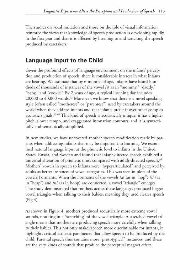
113Linguistic Experience Alters the Perception and Production of Speech
The studies on vocal imitation and those on the role of visual informationreinforce the views that knowledge of speech production is developing rapidlyin the first year and that it is affected by listening to and watching the speechproduced by caretakers.
Language Input to the Child
Given the profound effects of language environment on the infants’ percep-tion and production of speech, there is considerable interest in what infantsare hearing. We estimate that by 6 months of age, infants have heard hun-dreds of thousands of instances of the vowel /i/ as in “mommy,” “daddy,”“baby,” and “cookie.” By 2 years of age, a typical listening day includes20,000 to 40,000 words.62 Moreover, we know that there is a novel speakingstyle (often called “motherese” or “parentese”) used by caretakers around theworld when they address infants and that infants prefer it over other complexacoustic signals.63-65 This kind of speech is acoustically unique: it has a higherpitch, slower tempo, and exaggerated intonation contours, and it is syntacti-cally and semantically simplified.
In new studies, we have uncovered another speech modification made by par-ents when addressing infants that may be important to learning. We exam-ined natural language input at the phonetic level to infants in the UnitedStates, Russia, and Sweden and found that infant-directed speech exhibited auniversal alteration of phonetic units compared with adult-directed speech.66
Mothers’ vowels in speech to infants were “hyperarticulated” and perceived byadults as better instances of vowel categories. This was seen in plots of thevowel’s Formants. When the Formants of the vowels /a/ (as in “hop”) /i/ (asin “heap”) and /u/ (as in hoop) are connected, a vowel “triangle” emerges.The study demonstrated that mothers across these languages produced biggervowel triangles when talking to their babies, meaning they used clearer speech(Fig 4).
As shown in Figure 4, mothers produced acoustically more extreme vowelsounds, resulting in a “stretching” of the vowel triangle. A stretched vowel tri-angle means that mothers are producing speech more carefully when talkingto their babies. This not only makes speech more discriminable for infants, ithighlights critical acoustic parameters that allow speech to be produced by thechild. Parental speech thus contains more “prototypical” instances, and theseare the very kinds of sounds that produce the perceptual magnet effect.

114 Section 2: Auditory/Language Development
Language input to infants may therefore provide an ideal signal for construct-ing perceptual maps. The results suggest that at the phonetic level of lan-guage, linguistic input to infants provides exceptionally well-specifiedinformation about the units that form the building blocks for words.
A Theory of Speech Development
Given these findings, how might infants’ innate language predispositions pre-pare them for experience? One view is a three-step model of speech develop-ment, the Native Language Magnet (NLM) model,67 that includes the infants’innate abilities as well as changes brought about by experience with language(Fig 5).67 The model demonstrates how infants’ developing native-languagespeech representations might alter both perception and production of speech.
The following example of NLM is for vowels, although the same principlesapply to both vowel and consonant perception.
Phase 1 describes infants’ initial citizen-of-the-world abilities. At birth, theydiscriminate the phonetic distinctions of all languages. This is illustrated by ahypothetical vowel space partitioned into categories that separate the vowels
Fig 4. Infant-directed vowels (solid lines) are exaggerated, acoustically“stretching” the vowel triangle.
Reprinted with permission from Kuhl PK, Andruski JE, Chistovich IA, Chistovich LA, et al. Cross-lan-guage analysis of phonetic units in language addresses to infants. Science. 1997;277:684-686. Copyright1997 American Association for the Advancement of Science.
4000
3000
2000
1000
300 700 1100 1500 300 700 1100 1500 300 700 1100 1500
F2(H
z)
F1 (Hz) F1 (Hz) F1 (Hz)
English Russian SwedishA B C
/a/
/u/
/i/
/u/ /a/
/i/
/u/
/i/
/a/

115Linguistic Experience Alters the Perception and Production of Speech
of all languages. According to NLM, infants’ ability to hear all phonetic dif-ferences at this stage does not depend on specific language experience.
The divisions shown in Phase 1 initially structure perception in a phoneticallyrelevant way but are not likely to be due to a “language module.” This notionis buttressed by data on nonhuman animals that display abilities once thoughtto be exclusively human.5,13,68-70 This has been interpreted as evidence that theevolution of speech capitalized on acoustical distinctions well separated by themammalian auditory system.5,41
Phase 2 describes the vowel space at 6 months of age (this stage is presumedto be slightly later for consonants) for infants reared in three very differentlanguage environments, Swedish, English, and Japanese. At this stage, infantsshow evidence of language-specific magnet effects. The magnet effects stemfrom differences in the distributional properties of vowels heard by infants inthe three countries.
Fig 5. NLM posits initial boundaries (1), magnet development (2), and a restructuring of perception (3).
Natural auditory boundaries
Seco
ndFo
rman
t
First Formant
Swedish English Japanese
Phase 1
Phase 2
Phase 3 Seco
ndFo
rman
t
First Formant

116 Section 2: Auditory/Language Development
Interesting but unanswered questions about Phase 2 magnet effects are howmuch language input it takes to show these effects, and whether all languageheard by the child (including that from a radio or television) is effective inproducing this kind of learning. At present we have no idea how much lan-guage infants must hear to produce this kind of learning. Moreover, it isunclear whether language from a disembodied source (television, radio)would be sufficient. Our estimates suggest that infants have heard thousandsof instances of vowels in communication with their parents,65 but that doesnot tell us what amount is necessary. Studies presently under way will providesome information regarding these issues.
Phase 3 shows how magnet effects recursively alter the initial state of speechperception and affect the processing of foreign-language stimuli. Magneteffects stretch and shrink the perceptual space, causing certain perceptual dis-tinctions to be minimized while others are maximized. The consequence isthat some of the boundaries that initially divided the space disappear as theperceptual space is reconfigured to incorporate a language’s particular magnetplacement. Magnet effects functionally erase certain boundaries – those rele-vant to foreign but not native languages. Listeners’ auditory systems stillprocess the acoustic differences that separate these categories, but their per-ceptual maps cause them to no longer pay attention.
In Phase 3, a perceptual space once characterized by basic “auditory cuts” –boundaries that divide all speech categories – has been replaced by a warpedspace that restructures perception. It is at this phase that infants begin tounderstand and produce their first words, but become unable to discriminateforeign-language contrasts. The mapping of incoming speech has altered theacoustic differences infants respond to, producing a language-specific listenerand speaker.
What happens to infants exposed to two different languages? We are onlybeginning to study this issue, but the theory predicts that infants will developmagnet effects for the sound categories of both languages. Interestingly, pre-liminary data suggest that two sets of magnets are particularly likely to devel-op when the two languages are spoken by different speakers (mother speaksone language, father speaks another). As mentioned previously, we presumethat mapping two languages, each spoken by a different speaker, is made easi-er when infants can perceptually separate the input from the two languages.

117Linguistic Experience Alters the Perception and Production of Speech
NLM theory offers an explanation for the developmental change observed inspeech perception. A developing magnet pulls sounds that were once discrim-inable towards it, making them less discriminable. Magnet effects shouldtherefore developmentally precede changes in infants’ perception of foreign-language contrasts. The magnet effect also helps account for the results ofstudies on the perception of sounds from a foreign language by adults.16,48
For example, NLM theory may help explain Japanese listeners’ difficulty with American /r/ and /l/. The magnet effect for the Japanese /r/ categoryprototype (which is neither American /r/ nor /l/) will attract both /r/ and /l/,making the two sounds difficult for native-speaking Japanese people to dis-criminate (Kuhl et al, unpublished data). NLM theory argues that early expe-rience established a complex perceptual network (a neural “filter”) throughwhich language passes. In this view, one’s primary language and the map that results from early experience will determine how other languages are perceived. Speech production is similarly affected; the stored representationsthat altered speech perception serve as the guide to production, allowinginfants’ production of speech to become ever more precise as they focus onand imitate the patterns of spoken speech produced by adult speakers of the language.
Reinterpreting “Critical Periods”
The interaction between genetic programming and environmental stimulationis nowhere more evident than in the literature on critical periods in learn-ing.71,72 While critical periods were originally viewed as strictly timed develop-mental processes with rigid cutoff periods that restrict learning to a specifictime frame, recent studies showing that learning can be stretched by a varietyof factors has caused a shift in the terminology used to describe these periods.It is now understood that during “sensitive periods,” exposure to specifickinds of information may be more effective than at other times but that avariety of factors can alter these periods of learning. Knudsen’s work on thesound-localization system in the barn owl, for example, shows that the sensi-tive period for learning the auditory-visual map in the optic tectum can bealtered by a variety of factors that either shorten or extend the learning peri-od; the learning period closes much earlier, for instance, if experience occursin a more natural environment.73
The idea that sensitive periods define “windows of opportunity” for learning,during which environmental stimulation is highly effective in producing

118 Section 2: Auditory/Language Development
developmental change, remains well supported both in the human and theanimal literature. The ability to learn is not equivalent over time. The ques-tion is: What causes changes in the ability to learn over the lifespan?
The sensitive period denotes a process of learning that is constrained primari-ly by time or factors such as hormones that are outside the learning processitself. The work on speech, however, suggests an alternate possibility: Laterlearning may be limited by the fact that prior learning has itself altered thebrain; the brain’s altered structure may “interfere” with later learning. Forinstance, if NLM’s argument that learning involves the creation of mentalmaps for speech turns out to be correct, it would suggest that learning “com-mits” neural structure in some way. Future speech processing would be affect-ed by this neural structure. The mechanisms governing an organism’s generalability to learn may not have changed. Rather, initial learning may result in astructure that reflects environmental input, and, once committed, this struc-ture may interfere with the processing of information that does not conformto the learned pattern. On this account, initial learning alters future learningindependently of a strictly timed period.
Regarding the relationship between neural structure and interference withfuture learning, the brain’s “plasticity” could be governed from a statisticalstandpoint. When additional input does not cause the overall statistical distri-bution to change substantially, the organism becomes less sensitive to input.Hypothetically, for instance, the infant’s representation of the vowel /a/ mightnot change when the millionth token of the vowel /a/ is heard. Plasticitymight thus be independent of time but dependent on the amount and vari-ability provided by experience. At some time in the lifetime of an organism,new input no longer substantially alters the underlying distribution, and thiscould, at least in theory, reduce the system’s plasticity.
The interference theory may account for some aspects of second-languagelearning. When acquiring a second language, certain phonetic distinctions arenotoriously difficult to master both in perception and production of speech.Take the case of the /r-l/ distinction for native speakers of Japanese. Hearingthe distinction and producing it are very difficult for native speakers ofJapanese.74-76 According to NLM, this is because exposure to Japanese early inlife altered the Japanese infant’s perceptual system, resulting in magnet effectsfor the Japanese phoneme /r/ but not for American English /r/ or AmericanEnglish /l/. Once in place, Japanese magnet effects would not make it easy to

119Linguistic Experience Alters the Perception and Production of Speech
process American English. American English /r/ and /l/ would be assimilatedto Japanese /r/.
A second language learned later in life (after puberty) may require separationbetween the two systems to avoid interference. Data gathered using modernneuroimaging techniques indicate that adult bilinguals who learned both lan-guages early in life activate closer regions of the brain when processing thetwo languages than do those who learned the second language later in life. Incontrast, late bilinguals activated two very distinct regions of the brain for thetwo languages.77 This is consistent with the idea that the brain’s processing ofa primary language can interfere with the second language.
The general thesis is that acquiring new phonetic categories as adults is diffi-cult because the brain’s mental maps for speech, formed on the basis of theprimary language, are incompatible with those required for the new language;hence, interference results. Early in life, interference effects are minimal. Twonew languages can be learned because two sets of maps develop. As men-tioned earlier, limited evidence suggests that infants exposed to two languagesdo much better if each parent speaks one of the two languages, rather thanboth parents speaking both languages. This may be true because it is easier tomap two different sets of phonetic categories (one for each of the two lan-guages) if there is some way to keep them perceptually separate. Males andfemales produce speech in different frequency ranges, which could also makeit easier to maintain the separation, if they each speak a different language.
These two factors – the temporal window governed by genetic factors and theneural commitment resulting from initial learning – could both be operatingto produce constraints on learning a second language later in life. If a matura-tional process induces neural readiness at a particular time, input that missesthis timing could reduce learning. At the same time, an interference producedby initial language learning might provide an independent mechanism thatcontributes to the difficulty in readily learning a second language in adult-hood.
Future Directions
We are very interested in measurements of the brain that examine the effectsof linguistic experience. Recently, we developed a procedure for measuring inyoung infants an event-related potential, MMN (mismatched negativity).

120 Section 2: Auditory/Language Development
MMN is an auditory cortical evoked response that is triggered when a novelauditory stimulus is presented; it is especially sensitive to perceived acousticchanges. Recent studies have shown that MMN reflects experience-relatedchanges. For example, MMN increases following training on speech stimuliand it mirrors improvements in behavioral performance.78,79 Also, there arenow a number of studies, in addition to ours,80 that have successfully meas-ured MMN in young awake infants.81 Finally, the MMN response has been shown to be enhanced when a native-language phonetic prototype (as opposed to a foreign-language prototype) was used as the “mismatched”stimulus in studies of infants and adults.28,82
In a recently completed MMN study, we tested 6- to 12-month-old infants’responses to speech.80 Thirty normally developing infants were tested at twoages (6 to 7 and 11 to 12 months). The auditory stimuli were computer-synthesized syllables (/ba/ and /wa/), which differ only in the critical acousticinformation that distinguishes them. Stimuli were presented and electroen-cephalographic data acquired using Neuroscan hardware and software. Datawere collected from electrode sites using an infant-sized Electrocap (Fig 6, left panel).
The MMN was obtained by subtracting the averaged waveforms for each sub-ject under two conditions: the average of the standard and the average of themismatches in the context of the standard. The difference wave, calculated bysubtracting the two waveforms, reveals the MMN (Fig 6, right panel). The
Fig 6. Infant in Electrocap; MMN observed as a difference betweenstandard and deviant waveforms.
-12
-9
-6
-3
0
3
6
9
12
Mic
rovo
lts
-50 5 60 115 170 225 260 335 390 445 500Milliseconds
MMN

121Linguistic Experience Alters the Perception and Production of Speech
MMN was observed to be equally robust at the two ages for this native-language contrast and was shown to be stronger at electrode sites over the left than over the right hemisphere.
Conclusions
Research has shown that in the first year of life infants learn a great dealabout the perceptual characteristics of their native language. According to theNLM model, perceptual learning early in life results in the formation ofstored representations that capture native-language regularities. This theoryemphasizes the role of linguistic input (“parentese”) and the way in which itenhances speech contrasts. Infants map speech information, and the mappingcreates a perceptual space in which distances near a sound category’s mosttypical instances are shrunk and distances between categories are stretched.Perceptual maps differ in adults from different countries who have differentnative languages. The magnet effects and the mental maps they produce helpexplain how native-language speech develops, as well as our relative inabilityas adults to acquire a foreign language readily. In future studies, brain meas-ures will be used to track the development of the infant brain’s response tospeech.

122 Section 2: Auditory/Language Development
References 1. Chomsky N. A review of B.F. Skinner’s Verbal Behavior. Language. 1957;35:26-58.
2. Skinner BF. Verbal Behavior. New York, NY: Appleton-Century-Crofts, Inc; 1957.
3. Chomsky N. Aspects of the Theory of Syntax. Cambridge, Mass: MIT Press; 1965.
4. Chomsky N. Rules and Representations. New York, NY: Columbia University Press; 1981.
5. Kuhl PK. Perception, cognition, and the ontogenetic and phylogenetic emergence of human speech.In: Brauth, SE, Hall, WS, Dooling, RJ, eds. Plasticity of Development. Cambridge, Mass: MIT Press;1991:73-106.
6. Kuhl PK. The development of speech and language. In: Carew TJ, Menzel R, Shatz CJ, eds.Mechanistic Relationships Between Development and Learning. New York, NY: Wiley; 1998:53-73.
7. Gopnik A, Meltzoff AN, Kuhl PK. The Scientist in the Crib: Minds, Brains, and How Children Learn.New York, NY: William Morrow and Company, Inc; 1999.
8. Carew TJ, Menzel R, Shatz CJ. Mechanistic Relationships Between Development and Learning. NewYork, NY: Wiley; 1998.
9. Crair MC, Gillespie DC, Stryker MP. The role of visual experience in the development of columns incat visual cortex. Science. 1998;279:566-570.
10. Brainard MS, Knudsen EI. Sensitive periods for visual calibration of the auditory space map in thebarn owl optic tectum. Journal of Neuroscience. 1998;18:3929-3942.
11. Guenther FH, Gjaja MN. The perceptual magnet effect as an emergent property of neural map for-mation. Journal of the Acoustical Society of America. 1996;100:1111-1121.
12. Eimas PD, Miller JL, Jusczyk PW. On infant speech perception and the acquisition of language. In:Harnad S, ed. Categorical Perception: The Groundwork of Cognition. New York, NY: CambridgeUniversity Press; 1987:161-195.
13. Kuhl PK, Miller JD. Speech perception by the chinchilla: voiced-voiceless distinction in alveolar plo-sive consonants. Science. 1975;190:69-72.
14. Werker JF, Tees RC. Cross-language speech perception: evidence for perceptual reorganization duringthe first year of life. Infant Behavior and Development. 1984;7:49-63.
15. Strange W, ed. Speech Perception and Linguistic Experience: Issues in Cross-Language Research.Timonium, Md: York; 1995
16. Best CT. Language-specific changes in non-native speech perception: a window on early phonologicaldevelopment. In: de Boysson-Bardies B, de Schonen S, Jusczyk P, McNeilage P, Morton J, eds.Developmental Neurocognition: Speech and Face Processing in the First Year of Life. Dordrecht,Netherlands: Kluwer Academic Publishers; 1993:289-304.
17. Abramson AS, Lisker L. Discriminability along the voicing continuum: cross-language tests.Proceedings of the Sixth International Congress of Phonetic Sciences, Prague, 1967. 1970:569-573.
18. Kuhl PK, Kiritani S, Deguchi T, Hayashi A, Stevens EB, Dugger CD, Iverson P. Effects of languageexperience on speech perception: American and Japanese infants’ perception of /ra/ and /la/. Journal ofthe Acoustical Society of America. 1997;102:3135.
19. Ferguson CA, Menn L, Stoel-Gammon C, eds. Phonological Development: Models, Research,Implications. Timonium, Md: York; 1992.
20. Petitto LA. On the ontogenetic requirements for early language acquisition. In: de Boysson-Bardies B,de Schonen S, Jusczyk P, McNeilage P, Morton J, eds. Developmental Neurocognition: Speech and FaceProcessing in the First Year of Life. Dordrecht, Netherlands: Kluwer; 1993:365-383.
21. de Boysson-Bardies B. Ontogeny of language-specific syllabic productions. In: de Boysson-Bardies B,de Schonen S, Jusczyk P, McNeilage P, Morton J, eds. Developmental Neurocognition: Speech and Face Processing in the First Year of Life. Dordrecht, Netherlands: Kluwer Academic Publishers; 1993:353-363.
22. Flege JE. Factors affecting degree of perceived foreign accent in English sentences. Journal of theAcoustical Society of America. 1988;84:70-79.

123Linguistic Experience Alters the Perception and Production of Speech
23. Fromkin V, Krashen S, Curtis S, Rigler D, Rigler M. The development of language in Genie: a case oflanguage acquisition beyond the “critical period.” Brain & Language. 1974;1:81-107.
24. Curtiss S. Genie: A Psycholinguistic Study of a Modern Day “Wild Child.” New York, NY: AcademicPress; 1977.
25. Lane HL. The Wild Boy of Aveyron. Cambridge, Mass: Harvard University Press; 1976.
26. Kuhl PK. Human adults and human infants show a “perceptual magnet effect” for the prototypes ofspeech categories, monkeys do not. Perception & Psychophysics. 1991;50:93-107.
27. Kuhl PK. Infants’ perception and representation of speech: development of a new theory. In: Ohala JJ,Nearey TM, Derwing BL, Hodge MM, Wiebe GE, eds. Proceedings of the International Conference onSpoken Language Processing. Edmonton, Alberta: University of Alberta; 1992:449-456.
28. Näätänen R, Lehtokoski A, Lennes M, et al. Language-specific phoneme representations revealed byelectric and magnetic brain responses. Nature. 1997;385:432-434.
29. Willerman R, Kuhl PK. Cross-language speech perception: Swedish, English, and Spanish speakers’perception of front rounded vowels. Proceedings of the 1996 International Conference on SpokenLanguage Processing. 1996;1:442-445.
30. Iverson P, Kuhl PK. Mapping the perceptual magnet effect for speech using signal detection theoryand multidimensional scaling. Journal of the Acoustical Society of America. 1995;97:553-562.
31. Iverson P, Kuhl PK. Influences of phonetic identification and category goodness on American listeners’perception of /r/ and /l/. Journal of the Acoustical Society of America. 1996;99:1130-1140.
32. Sussman JE, Lauckner-Morano VJ. Further tests of the “perceptual magnet effect” in the perception of[i]: identification and change/no-change discrimination. Journal of the Acoustical Society of America.1995;97:539-552.
33. Lotto AJ, Kluender KR. General contrast effects in speech perception: effect of preceding liquid onstop consonant identification. Perception and Psychophysics. 1988;60:602-619.
34. Kuhl PK, Williams KA, Lacerda F, Stevens KN, Lindblom B. Linguistic experience alters phoneticperception in infants by 6 months of age. Science. 1992;255:606-608.
35. Tsao FM, Hall M, Eyraud R, Kuhl PK. Perception of American English /r/ and /l/ by Mandarinspeakers: influences of phonetic identification and category goodness. In: Kuhl PK, Crum L, eds.Proceedings 16th International Congress on Acoustics and 135th Meeting of the Acoustical Society ofAmerica. Woodbury, NY: Acoustical Society of America; 1998:2065-2066.
36. Eyraud R, Kuhl PK. Native and non-native phonetic speech perception of American and Finnish lis-teners. Journal of the Acoustical Society of America. 1996;100:2724.
37. Iverson P, Kuhl PK. Cue weighting for American English /r/ and /l/ by 7.5-month-old infants. Journalof the Acoustical Society of America. 1996;100:2725.
38. Jusczyk PW, Friederici AD, Wessels JMI, Svenkerud VY, Jusczyk AM. Infants’ sensitivity to the soundpatterns of native language words. Journal of Memory and Language. 1993;32:402-420.
39. Saffran JR, Aslin RN, Newport EL. Statistical learning by 8-month-old infants. Science. 1996;274:1926-1928.
40. Marler P. Innate learning preferences: signals for communication. Developmental Psychobiology. 1990;557:568.
41. Kuhl PK. On babies, birds, modules, and mechanisms: a comparative approach to the acquisition ofvocal communication. In: Dooling RJ, Hulse SH, eds. The Comparative Psychology of Audition:Perceiving Complex Sounds. Hillsdale, NJ: Erlbaum; 1989:379-419.
42. Konishi M. Birdsong for neurobiologists. Neuron. 1989;3:541-549.
43. Doupe A, Kuhl PK. Birdsong and speech: common themes and mechanisms. Annual Review ofNeuroscience. 1999. In press.
44. Oller DK, MacNeilage PF. Development of speech production: perspectives from natural and per-turbed speech. In: MacNeilage PF, ed. The Production of Speech. New York, NY: Springer-Verlag;1983:91-108.
45. Konishi M. Effects of deafening on song development in American robins and black-headed gros-beaks. Zeitschrift fürTierpsychologie. 1965;22:584-599.

124 Section 2: Auditory/Language Development
46. Nottebohm F. The role of sensory feedback in the development of avian vocalizations. Proceedings ofthe 14th International Ornithological Congress. 1967:265-280.
47. Locke JL, Pearson DM. Linguistic significance of babbling: evidence from a tracheostomized infant.Journal of Child Language. 1990;17:1-16.
48. Flege JE. Production and perception of a novel, second-language phonetic contrast. Journal of theAcoustical Society of America. 1993;93:1589-1608.
49. Vihman MM, de Boysson-Bardies B. The nature and origins of ambient language influence on infantvocal production and early words. Phonetica. 1994;51:159-169.
50. Kuhl PK, Meltzoff AN. The bimodal perception of speech in infancy. Science. 1982;218:1138-1141.
51. Meltzoff AN, Moore MK. Imitation of facial and manual gestures by human neonates. Science.1977;198:75-78.
52. Meltzoff AN, Moore MK. Imitation, memory, and the representation of persons. Infant Behavior andDevelopment. 1994;17:83-99.
53. Petitto LA, Marentette PF. Babbling in the manual mode: evidence for the ontogeny of language.Science. 1991;251:1493-1496.
54. Kuhl PK, Meltzoff AN. Infant vocalizations in response to speech: vocal imitation and developmentalchange. Journal of the Acoustical Society of America. 1996;100:2425-2438.
55. McGurk H, MacDonald J. Hearing lips and seeing voices. Nature. 1976;264:746-748.
56. Green KP, Kuhl PK, Meltzoff AN, Stevens EB. Integrating speech information across talkers, gender,and sensory modality: female faces and male voices in the McGurk effect. Perception & Psychophysics.1991;50:524-536.
57. Kuhl PK, Tsuzaki M, Tohkura Y, Meltzoff AN. Human processing of auditory-visual information inspeech perception: potential for multimodal human-machine interfaces. In: Proceedings of theInternational Conference on Spoken Language Processing. Tokyo: Acoustical Society of Japan; 1994:539-542.
58. Massaro DW. Speech Perception by Ear and Eye: A Paradigm for Psychological Inquiry. Hillsdale, NJ:Erlbaum; 1987.
59. MacKain K, Studdert-Kennedy M, Spieker S, Stern D. Infant intermodal speech perception is a left-hemisphere function. Science. 1983;219:1347-1349.
60. Rosenblum LD, Schmuckler MA, Johnson JA. The McGurk effect in infants. Perception &Psychophysics. 1997;59:347-357.
61. Walton GE, Bower TGR. Newborns form “prototypes” is less than 1 minute. Psychological Science.1993;4:203-205.
62. Chapman RS, Streim NW, Crais ER, Salmon D, Strand DEA, Negri NA. Child talk: assumptions ofa developmental process model for early language learning. In: Chapman RA, ed. Processes in LanguageAcquisition and Disorders. St Louis, Mo: Mosby Year Book; 1992:3-19.
63. Fernald A. Four-month-old infants prefer to listen to motherese. Infant Behavior and Development.1985;8:181-195.
64. Grieser DL, Kuhl PK. Maternal speech to infants in a tonal language: support for universal prosodicfeatures in motherese. Developmental Psychology. 1988;24:14-20.
65. Fernald A, Kuhl P. Acoustic determinants of infant preference for motherese speech. Infant Behaviorand Development. 1987;10:279-293.
66. Kuhl PK, Andruski JE, Chistovich IA, et al. Cross-language analysis of phonetic units in languageaddressed to infants. Science. 1997;277:684-686.
67. Kuhl PK. Learning and representation in speech and language. Current Opinion in Neurobiology.1994;4:812-822.
68. Kuhl PK, Miller JD. Speech perception by the chinchilla: identification functions for synthetic VOTstimuli. Journal of the Acoustical Society of America. 1978;63:905-917.
69. Kluender KR, Diehl RL, Killeen PR. Japanese quail can learn phonetic categories. Science. 1987;237:1195-1197.

125Linguistic Experience Alters the Perception and Production of Speech
70. Dooling RJ, Best CT, Brown SD. Discrimination of synthetic full-formant and sinewave /ra-la/ con-tinua by budgerigars (Melopsittacus undulatus) and zebra finches (Taeniopygia guttata). Journal of theAcoustical Society of America. 1995;97;1839-1846.
71. Thorpe WH. Bird Song: The Biology of Vocal Communication and Expression in Birds. New York, NY:Cambridge University Press; 1961.
72. Marler P. A comparative approach to vocal learning: song development in white-crowned sparrows.Journal of Comparative and Physiological Psychology. 1970;71:1-25.
73. Knudsen EI, Knudsen PF. Sensitive and critical periods for visual calibration of sound localization bybarn owls. Journal of Neuroscience. 1990;10:222-232.
74. Goto H. Auditory perception by normal Japanese adults of the sounds “l” and “r.” Neuropsychologia.1971;9:317-323.
75. Miyawaki K, Strange W, Verbrugge R, Liberman AM, Jenkins JJ, Fujimura O. An effect of linguisticexperience: the discrimination of [r] and [l] by native speakers of Japanese and English. Perception &Psychophysics. 1975;18:331-340.
76. Yamada RA, Tohkura Y. The effects of experimental variables on the perception of American English/r/ and /l/ by Japanese listeners. Perception & Psychophysics. 1992;52:376-392.
77. Kim KHS, Relkin NR, Lee KM, Hirsch J. Distinct cortical areas associated with native and secondlanguages. Nature. 1997;388:172-174.
78. Kraus N, McGee T, Carrell T, Sharma A, Nicol T. Mismatch negativity to speech stimuli in school-agechildren. In: Karmos G, et al, eds. Perspectives of Event-Related Potentials Research. Dordrecht,Netherlands: Elsevier Science B.V.; 1995:211-217.
79. Tremblay KL, Kraus N, McGee T, Zecker S. The time course of learning: neurophysiologic changesduring speech training. Journal of the Acoustical Society of America. 1998;103:2981.
80. Kuhl PK. Effects of language experience on speech perception. In: Kuhl PK, Crum L, eds. Proceedings16th International Congress on Acoustics and 135th Meeting of the Acoustical Society of America.Woodbury, NY: Acoustical Society of America; 1998;3:1601-1602.
81. Pang EW, Edmonds GE, Desjardins R, Khan SC, Trainor LJ, Taylor MJ. Mismatch negativity tospeech stimui in 8-month-old infants and adults. International Journal of Psychophysiology.1998;29:227-236.
82. Cheour M, Ceponiene R, Lehtokoski A, et al. Development of language-specific phoneme representa-tions in the infant brain. Nature Neuroscience. 1998;1:351-353.

Section 3:Cognitive Development
The Role of Early Experience in Infant Development

129
Abstracts From Section 3.Cognitive Development
Development of Cognitive Functions Is Linked to the Prefrontal CortexAdele Diamond, PhD
The prefrontal cortex is involved in “executive functions” critical for higher-level problem solving, creative thought, and focused, sustained attention.Although full maturation is achieved by puberty or early adulthood, the pre-frontal cortex makes possible important cognitive functions in infants asyoung as 9 months, and contributes to their ability to solve spatial problemsand memory problems in creative ways. Cognitive deficits related to impairedor immature prefrontal cortex function may contribute to subtle learning andbehavioral problems.
Born to Learn: What Infants Learn From Watching Us Andrew N. Meltzoff, PhD
Imitation is a powerful form of learning commonly used by children, adults,and infants. A child’s enthusiasm for imitative behavior prompts parentalattention and interaction, and provides a mechanism for transmitting appro-priate cultural and social behavior. Although simple imitative behavior is evi-dent in the postnatal period, by around 14 months infants remember andrepeat actions they observe in adults, other children, and on television.Imitation games provide early experience in mapping the similarities betweenself and other. Behavioral imitation, empathy, and moral sentiments may bepart of the same developmental pathway.
Early Experience Matters for Spatial Development(But Different Kinds at Different Times)Nora S. Newcombe, PhD
This paper reviews competing theories of cognitive/spatial development (asproposed by nativists, and followers of Piaget and Vygotsky) and discusses

130 Section 3: Cognitive Development
their salient features and shortcomings relative to each other and new researchfindings. Normal spatial development in the first year of life requires only atypical predictable environment in which children can explore and play.Beginning at 2 or 3 years of age, it is likely that children rely on culturaltransmission to acquire understanding of symbols systems and mapping.

131
Development of CognitiveFunctions Is Linked to thePrefrontal Cortex
Adele Diamond, PhD
Introduction: Babies Are Smarter Than People Thought
About a quarter of all cortex in the human brain is prefrontal cortex, locatedin the front of the brain, behind the forehead and in front of the motor areas.This is the region of the brain that has increased the most in size during thecourse of primate evolution and the region thought to be involved in “execu-tive functions” critical for higher-level problem solving and creative thought.It is needed for focused, sustained attention, working memory, and inhibitionof prepotent but inappropriate action tendencies. Not long ago, peoplethought that babies were not capable of these high-level abilities and that theprefrontal cortex did not function during the first few years of life. We nowknow, however, that even during the first year of life infants are capable ofsophisticated cognitive operations, and there is a growing body of evidencethat the prefrontal cortex makes possible important cognitive functions evenduring infancy (as early as 9 to 12 months of age).1-4
It is incorrect to assume that because the prefrontal cortex is not fully matureuntil puberty or early adulthood, it does not have any important cognitivefunctions during early life. Even though the prefrontal cortex will not be fullymature until years later, it is capable of performing cognitive operationsbefore a child’s first birthday. It would also be incorrect to assume that theprefrontal cortex is fully mature by 12 months of age in humans just becausesome of its functions begin to emerge by then. The prefrontal cortex contin-ues to mature over the next 10, or even 20, years of a person’s life, just as aperson’s cognitive development, while remarkable by 1 year, continues tounfold over the next 10 to 20 years.

132 Section 3: Cognitive Development
Infants Are Creative Problem Solvers
Creativity and ingenuity are already evident before an infant’s first birthday.For example, we all have a tendency to reach on a straight line for somethingwe see that we want. If there is a barrier in the way, we have to detour aroundit. If a toy is placed in a transparent box open on one side, babies 6 to 8months of age persist in trying to reach straight through the side of the boxthrough which they are looking, instead of detouring around to the openside. By 8.5 to 9 months of age, infants start to get the idea of reachingthrough the open side. It is quite complicated, however, for an infant to plana reach that goes off to the side and curves back around through an opening,and there is still the strong tendency to reach through the side through whichthey are looking.
She reaches in awkwardly with herLEFT hand, keeping her eye on her hand and toy through opening.
Fig 1. Example of an “awkward reach” on both sides of box.
Left side open, the young girl leans over andlooks at the toy through opening.
She reaches awkwardly with far hand.(In this way, she can continue to look inopening and keep her hand in view.)
She leans down and looks into the rightside.

133Development of Cognitive Functions Is Linked to the Prefrontal Cortex
To cope with these problems, infants of 8.5 to 9 months of age come up witha very clever and creative solution. They lean all the way over so they can lookinto the open side (Fig 1). This enables them to look along the line they willneed to reach. Once down in that position, for instance if they are leaning tolook into the right side of the box, their right arm is crumpled underneaththem and is ready to help support them should they start to fall, so again theycome up with a creative solution: They recruit their left hand to reach intothe right side of the box and, similarly, their right hand to reach into the leftside of the box. It looks very contorted and unusual (we call it the “awkwardreach”), but it is a brilliant solution to their need at this age to be lookingthrough the side into which they are reaching (AD, unpublished data).1-3
Infants Have Robust Memories
For years it was assumed that the memory support that is impaired when aperson has amnesia matures late because children less than 2 years of age per-form poorly on a test called the “delayed nonmatching to sample test,”whichrequires that memory system.5,6 But that memory system functions quite wellvery early in infancy; it is another ability required for this test that matureslate.
Earlier work with infants on the delayed matching to sample test – a classictest of the medial temporal lobe memory system that requires the ability toremember what you have just seen – revealed that toddlers cannot succeed onthe test until they are almost 2 years old.5,6 (See text box for details of thetest.)
The delayed nonmatching to sample task
Here, a sample object is presented over a shallow “well” in the center ofthe testing table. The subject being tested displaces the sample to retrievea reward from inside the well. A delay follows, then the familiar sample ispresented to one side and a new object is presented to the other side,each over a well. The correct choice is to select the novel object, ie, theobject that does not match the previously presented sample – hence thetask’s name (Fig 2). The well under the novel object contains a reward;the other well is empty. On each trial, new objects are used, and whichside the novel one appears on is varied randomly over trials.

134 Section 3: Cognitive Development
Given that success on this test appears so late in development and given thatit has been shown convincingly and repeatedly that success requires properfunctioning of the medial temporal lobe, it had been argued that the medialtemporal lobe memory system must mature late.7 The logic seemed reason-able: Here is a behavioral task that requires the functions the medial temporallobe, but children cannot succeed on this task until quite late (21 months ofage). Conclusion: This neural system must not be mature enough to subservememory until about 21 months of age. That conclusion turns out to beincorrect, however. We now know that during the first year of life, infants can remember objects or actions they have seen for minutes, hours, days, and even weeks.8-10
Fig 2. The delayed nonmatching sample test.
The test subject is expected to choose the novel stimulus object; that is, the stimulusthat does not match the sample presented in 1. The smiley face indicates thelocation of the reward.
Brief Delay
1.
2.
3.
Reward is notattached tostimulus. Childmust reach in toget reward.
4.

135Development of Cognitive Functions Is Linked to the Prefrontal Cortex
But it is important to look at the characteristics of performance, not just success or failure rates, because a given test can be failed for many differentreasons. Problems on a test can occur for various reasons, and different kindsof problems implicate different brain regions.
Similarly, developmental improvements on a task do not necessarily corre-spond to maturational changes in a particular neural region. In a broad sense,failure does not always mean a deficit in the single specific ability one washoping to measure – success usually requires multiple abilities, not just theability in which one is interested.
Consider the performance difference in human infants and in adult macaquemonkeys with damage to the medial temporal lobe on the delayed nonmatch-ing to sample test. Adult macaque monkeys with bilateral lesions of the medi-al temporal lobe (especially the rhinal cortical areas [perirhinal and entorhinalcortex]) do poorly on this task.11-15 They appear to fail because they forgetwhat the sample was. Consistent with this, their performance is better whenthere is a brief delay between sample and test (delays of 5, 10, or 15 seconds),and their performance progressively worsens as the delay increases to 60 sec-onds, 5 minutes, 10 minutes, and longer. Intact adult monkeys performextremely well even after very long delays.
In contrast, human infants do not perform well, even with brief delays of 5 or 10 seconds.5,6 Moreover, unlike adult macaques with medial temporal lobelesions, whose performance progressively worsens with longer delays, wheninfants first succeed with the 5-second delay, they also succeed with delays of 30 and 60 seconds.5
If the developmental improvement on this test were measuring an improve-ment in memory, one would expect that young infants would succeed withbrief delays and older, more mature children would succeed with longerdelays. However, once children solve the task, they succeed after both longand short delays. Thus, one problem with making the leap from “perform-ance on delayed nonmatching to sample is linked to the medial temporallobe,” to “therefore the developmental progression of performance on delayednonmatching to sample must indicate the developmental progression of themedial temporal lobe” is that the performance characteristics of intact humaninfants are quite different from the performance characteristics of adult mon-keys with damaged medial temporal lobes. Again, it is crucial to look at thecharacteristics of performance, and not just at success and failure rates.

136 Section 3: Cognitive Development
A final argument against the theory of late medial temporal lobe memory system development is the large body of evidence of robust memory in veryyoung infants. Indeed, if infants are not required to displace stimulus objectsto receive the reward in the shallow well underneath but are allowed simply toexplore the new stimulus objects themselves, infants as young as 6 months of age succeed on the task with delays as long as 3 minutes.10
Temporal vs Spatial Relationships
In being able to understand the relationship between one thing and another,a very small temporal separation can make a surprisingly large difference.
If infants have excellent memories even early in life, why then do they gener-ally fail the delayed nonmatching to sample task until almost 2 years of age?The answer appears to be that infants do not understand the relationshipbetween the reward in the shallow well and the stimulus object sitting on top of the well. For example, infants of only 9 to 12 months of age succeedon this test if the rewards are attached to the base of the sample (with Velcro®)instead of placed in wells beneath the sample (A. Diamond et al, In press,Developmental Psychology; see Fig 3). In the Velcro® condition, just as in the standard condition, the stimuli are placed atop wells, the rewards are in the wells below, and the rewards are separate objects from the stimuli; however, attaching the rewards to the stimuli with Velcro®, a seemingly minor variation in the procedure, appears to alter infants’ understanding of the task dramatically.
Something very similar was observed in chimpanzees back in 1965 byJarvik,16 who asked why it takes a smart creature like a chimpanzee 100 to200 trials to learn a simple color discrimination (eg, always choose red oralways choose blue). Color discrimination is normally tested in primates byplacing, for example, a red plaque over one shallow well and a blue plaqueover another shallow well. The left-right placement of the two plaques is varied randomly over trials, but the reward is always under the plaque of agiven color. Jarvik varied whether the reward was placed in the well underthe plaque or taped to the underside of the plaque. When the reward wasattached to the plaque, Jarvik found one trial learning.
In the Velcro® condition, the reward is physically closer to the stimulus thanin the standard procedure. Is it that spatial proximity is that critical? Perhapseven a tiny spatial separation between stimulus and reward makes a taskmuch more difficult than when there is no spatial separation at all.

137Development of Cognitive Functions Is Linked to the Prefrontal Cortex
In the classic procedure, infants pick up the stimulus and then look into thewell for the reward. There is thus a temporal gap between choosing a stimulusand seeing its reward. In the Velcro® procedure, when the rewards areattached to the base of stimuli, infants see the reward as soon as they lift andturn the stimulus. Instead of the reward remaining in the well, the rewardmoves with the stimulus. Could the critical factor be the close temporal prox-imity between displacing the stimulus and seeing the reward?
Is the Velcro® condition easy because of the close spatial proximity of thereward and stimulus or because of the close temporal proximity? To answerthat question one needs to look at evidence from additional experiments.
Fig 3. Modified delayed nonmatching to sample.
In this test, the reward is affixed to the base of the novel object with Velcro®.The infant picks up the object and then pulls off the reward.
1.
4.
3.
Brief Delay
Velcro®
2.

138 Section 3: Cognitive Development
We constructed an apparatus in which moving the correct stimulus triggereda jack-in-the-box to pop open behind it. The reward (the jack-in-the-box) wasfarther from the stimulus than in the classic procedure, but it was temporallycloser, because it popped open immediately with the slightest movement ofthe stimulus.
Infants 9 and 12 months old perform every bit as well in the jack-in-the-boxcondition as they do in the Velcro® condition (Diamond & Lee, unpublisheddata). The tight temporal coupling of “reach-for-stimulus” and “jack-pops-up”almost makes it appear as if the stimulus is a trigger that causes the jack-in-the-box to pop open. This temporal proximity may enable infants to concep-tualize the stimulus and the jack-in-the-box reward as connected, as two partsof a single unit. In both the Velcro® and jack-in-the-box conditions, thereward was temporally close to the stimulus. In the jack-in-the-box condition,however, the reward was spatially farther away from the stimulus than in eventhe standard condition. Thus, it appears that it is close temporal proximitybetween stimulus and reward that enables infants in the first year of life tobegin to grasp the relationship between stimulus and reward.
Training, Experience, and Thresholds
Training and experience often have little effect until a child is at the thresholdof acquiring a competence on his or her own. Thus, for example, Overmanand colleagues tested infants every weekday, day after day, week after week,month after month, on the delayed nonmatching to sample test from 12months of age onward.6 Those infants succeeded at the same age (21 monthson average) as the toddlers tested for the first time at 21 months by eitherOverman or Diamond, toddlers who had no prior experience with the task.5,6
On the task of reaching around a transparent barrier mentioned previously, Itried to see whether training experiences or cues would help infants succeed.17
I found that cues or training helped only when infants were at the border ofprogressing to the next higher level of performance. Cues or training helpedinfants progress to the next level perhaps 2 weeks before they would have oth-erwise. When an infant was not near the border between one level and thenext, however, nothing I tried made any difference. Infants could not profitfrom the cues or training until they were ready.

139Development of Cognitive Functions Is Linked to the Prefrontal Cortex
Measuring Function and Deficit
A global insult (such as lower or higher levels of a dietary amino acid) canhave specific effects that do not show up on standard cognitive or behavioraltests.
Global measures, such as IQ tests, are poor indices of specific cognitive func-tions and poor indicators of what particular neural system might be affected ifthere is a problem. Developmental cognitive neuropsychologists now haveprecise measures of specific cognitive functions that are sensitive to the func-tions of particular neural subsystems. These measures can help in the studyand treatment of diverse developmental disorders.
For example, cognitive deficits in children treated early and continuously forphenylketonuria (PKU) went officially unrecognized for many years becausethe children performed within the normal range on IQ tests, despite theprotestations of parents and teachers that something was wrong. The globalcognitive measures that were being used in the clinic were too imprecise.
The core problem in PKU is an inability to convert one amino acid, phenyl-alanine, into another amino acid, tyrosine. This is caused by a defect in thegene that codes for an enzyme, phenylalanine hydroxylase.18,19 When a personwho has PKU eats protein containing phenylalanine, it builds up in thebloodstream to levels that may be 20 to 30 times normal. Also, becausephenylalanine is not converted to tyrosine, tyrosine levels are low (althoughnot absent, because some tyrosine is also available in protein). Widespreadbrain damage and severe mental retardation result.20,21 The treatment forPKU consists of limiting the amount of foods containing phenylalanine oneeats. Eating a diet low in this amino acid succeeds in averting gross braindamage and produces children who have IQ scores in the normal range.22,23
This is an example of an environmental, behavioral alteration that affectsone’s biochemistry and one’s brain: Changing what children with PKU eathas a dramatic effect on the amino acid levels in their bloodstreams and,because of that, on the development of their brains.
Important cognitive problems are still evident, however.24-26 The reason forthis is that a diet low in phenylalanine rarely results in normal blood levels of this amino acid, because the diet must balance the need to minimizephenylalanine intake with the need for protein. The compromise diet reducesphenylalanine levels but not to normal concentrations, and does little to

140 Section 3: Cognitive Development
ameliorate the low blood levels of tyrosine. The consequence is that phenyl-alanine levels are mildly elevated and tyrosine levels are mildly reduced. Giventhat the levels of these amino acids in the bloodstream are not normal, it isperhaps not that surprising that there might be a problem. More surprisingwere the observations that the problems seemed to be limited to the cognitivefunctions dependent on the prefrontal cortex.27 The clinical question becamehow could an amino acid imbalance affecting the entire body produce a spe-cific effect limited to one region of the brain? The following model providesan explanation.
Because phenylalanine and tyrosine compete to cross into the brain, a moder-ate elevation in bloodstream phenylalanine results in a moderate reduction inthe amount of tyrosine that reaches the brain.28,29 Tyrosine is needed by thebrain to make the neurotransmitter dopamine. Most dopamine systems in thebrain can cope with modest changes in the level of tyrosine with no ill effects;however, the dopamine neurons that project to the prefrontal cortex are dif-ferent from most other dopamine neurons – they fire faster and turn overdopamine faster.30,31 This makes them sensitive to changes in the level of tyro-sine that are too small to affect other regions of the brain.32,33 Indeed, reduc-tions in tyrosine too small to affect other dopamine systems in other neuralregions profoundly reduce dopamine levels in the prefrontal cortex.34
Reductions in dopamine in the prefrontal cortex have severe consequences onthe cognitive functions that depend on this area of the brain.35 In fact, drasti-cally depleting the prefrontal cortex of dopamine produces cognitive deficitscomparable to those caused by destroying the prefrontal cortex altogether.36
In a large, longitudinal study of children treated early and continuously forPKU, we found that children whose blood phenylalanine levels were 6 to 10 mg/dL (3 to 5 times normal), previously considered within the acceptablerange, were impaired on tasks that required both holding information inmind (working memory) and inhibiting a dominant response (inhibitory con-trol), tasks linked to dorsolateral prefrontal cortex.4 The higher a child’s cur-rent levels of phenylalanine, the worse that child’s performance on these tests.These deficits were evident in all age groups (infants, toddlers, and youngchildren) and remained significant even after we controlled for IQ, sex, healthvariables, and background characteristics. The deficits were clear whether thechildren were compared to other PKU children with lower phenylalanine lev-els, their own siblings, matched controls, or children from the general popula-tion. Children with PKU whose blood phenylalanine levels were 3 to 5 timesnormal were not impaired on any of the large battery of control tasks, which

141Development of Cognitive Functions Is Linked to the Prefrontal Cortex
required functions dependent on other regions of the brain, such as the poste-rior parietal cortex or the medial temporal lobe. Because the functions ofthese areas were spared, the cognitive deficits appear to be selective.
How can children with serious deficits in their prefrontal cognitive abilitiesperform within the normal range on general IQ tests? It depends on how onedefines “normal.” “Within the normal range” means only IQs of 80 or better.Most children with PKU whose phenylalanine levels are 6 to 10 mg/dL haveIQ scores in the 80s and 90s – as do most patients with damage to ordestruction of the prefrontal cortex.37 The prefrontal cortex is needed, forexample, for problem solving, creativity, and manipulating several pieces ofinformation at the same time. It is needed most when changed circumstancesrequire some alteration of normal practice, or when new goals demand themodification of existing routines.38 IQ tests, however, largely measure accu-mulated or stored knowledge. They test one’s memory and past learning, andonly in small measure, one’s problem solving or creativity.
To investigate the biological mechanism more directly, my colleagues and Ideveloped and characterized the first animal model of early- and continuouslytreated PKU.39 This enabled us to study directly the effect of moderate,chronic phenylalanine elevations in the bloodstream on neurotransmitter lev-els in different brain regions. We found cognitive deficits (impaired perform-ance on a behavioral task dependent on the prefrontal cortex) and reduceddopamine in the prefrontal cortex in the PKU animal model. In contrast,other neurotransmitters and other brain regions were much less affected.
Conclusion: The Prefrontal Cortex and Behavior
Young children can sometimes get stuck in a behavioral rut from which theycannot easily extricate themselves – despite their best intentions and despiteknowing what correct performance entails. It is important to bear this inmind before mistakenly labeling a young child “bad,” “intentionally diffi-cult,” or “willful.”
It is not enough to know something or remember it; one must translate thatknowledge into behavior. Infants and young children, in whom the prefrontalcortex is not yet mature, sometimes do the wrong thing even though theyknow what they should do and are trying to do it. For example, consider achild who has just been sorting a deck of cards by color and then is instructed

142 Section 3: Cognitive Development
to sort the cards by shape but continues sorting the cards by color – eventhough on each and every trial the child correctly tells you what the new rule isand shows you where that means each card should be sorted.40 Because youngchildren can sometimes have difficulty getting their actions to reflect theirintentions, they may be labeled as “bad,” “intentionally difficult,” or “willful,”when that is not the case.
The inhibitory ability dependent on the prefrontal cortex is important formany cognitive tasks. Consider that to sustain the focused concentrationrequired for a difficult task, one needs to be able to resist distraction; to act innew ways, one needs to resist falling back into usual ways of acting or think-ing; that is, one needs the inhibitory control ability that depends upon theprefrontal cortex. To relate several ideas and facts together, one must be ableto resist focusing exclusively on just one idea or fact; to recombine ideas andfacts in new, creative ways, one needs to be able to resist repeating oldthought patterns; again, one needs the inhibitory control ability that comesfrom the prefrontal cortex.
It is easier for people to continue doing what they have been doing ratherthan to change, and it is easier to go on “automatic pilot” than to considercarefully what to do next. Sometimes, however, we need to change; some-times we need to act differently than might have been our first inclination.The ability to exercise inhibitory control, which prefrontal cortex makes pos-sible, frees us to act according to what we choose to do rather than being“unthinking” creatures of habit.
The ability to hold information in mind, which also depends on the pre-frontal cortex, enables us to consider alternatives, to bring conceptual knowl-edge (and not just perceptual input) to bear on our decisions, and to considerour remembered past and our future hopes when planning our presentactions. These two abilities, working memory and inhibitory control, make itpossible for us to be creative problem solvers and to exercise free will and self-determination. Such capabilities are not needed all the time, but when theyare needed, we would all like to be able to exercise them, and we would likethe same for our children.

143Development of Cognitive Functions Is Linked to the Prefrontal Cortex
References1. Diamond A. Differences between adult and infant cognition: is the crucial variable presence or
absence of language? In: Weiskrantz L, ed. Thought Without Language. Oxford, England: OxfordUniversity Press; 1988:337-370.
2. Diamond A. Neuropsychological insights into the meaning of object concept development. In: CareyS, Gelman R, eds. The Epigenesis of Mind: Essays on Biology and Cognition. Hillsdale, NJ: LawrenceErlbaum Associates; 1991:67-110.
3. Diamond A. Frontal lobe involvement in cognitive changes during the first year of life. In: GibsonKR, Petersen AC, eds. Brain Maturation and Cognitive Development: Comparative and Cross-CulturalPerspectives. New York, NY: Aldine de Gruyter; 1991:127-180.
4. Diamond A, Prevor M, Callender G, Druin DP. Prefrontal cortex cognitive deficits in children treatedearly and continuously for PKU. Monographs of the Society for Research in Child Development.1997;62(4):#252.
5. Diamond A, Towle C, Boyer K. Young children’s performance on a task sensitive to the memory functions of the medial temporal lobe in adults – the delayed nonmatching-to-sample task – reveals problems that are due to non-memory-related task demands. Behavioral Neuroscience. 1994;108:659-680.
6. Overman WH, Bachevalier J, Turner M, Peuster A. Object recognition versus object discrimination:comparison between human infants and infant monkeys. Behavioral Neuroscience. 1992;106:15-29.
7. Bachevalier J, Mishkin M. An early and a late developing system for learning and retention in infantmonkeys. Behavioral Neuroscience. 1984;98:770-778.
8. Meltzoff AN. Towards a developmental cognitive science: the implications of cross-modal matchingand imitation for the development of representation and memory in infancy. Annals of the New YorkAcademy of Sciences. 1990;608:1-37.
9. Rovee-Collier C. Dissociations in infant memory: rethinking the development of implicit and explicitmemory. Psychological Review. 1997;104:467-498.
10. Diamond A. Evidence of robust recognition memory early in life even when assessed by reachingbehavior. Journal of Experimental Child Psychology. 1995;59(Special Issue):419-456.
11. Mishkin M. Memory in monkeys severely impaired by combined but not separate removal of amyg-dala and hippocampus. Nature. 1978;273:297-298.
12. Meunier M, Hadfield W, Bachevalier J, Murray EA. Effects of rhinal cortex lesions combined withhippocampectomy on visual recognition memory in rhesus monkeys. Journal of Neurophysiology.1996;75:1190-1205.
13. Squire LR, Zola-Morgan S, Chen KS. Human amnesia and animal models of amnesia: performance ofamnesic patients on tests designed for the monkey. Behavioral Neuroscience. 1988;102:210-221.
14. Zola-Morgan S, Squire LR, Amaral DG. Lesions of the hippocampal formation but not lesions of thefornix or mammillary nuclei produce long-lasting memory impairment in monkeys. Journal ofNeuroscience. 1989;9:898-913.
15. Zola-Morgan S, Squire LR, Amaral DG. Lesions of the perirhinal and parahippocampal cortex thatspare the amygdala and hippocampal formation produce severe memory impairment. Journal ofNeuroscience. 1989;9:4355-4370.
16. Jarvik ME. Simple color discrimination in chimpanzees: effect of varying contiguity between cue andincentive. Journal of Comparative and Physiological Psychology. 1956;49:492-495.
17. Diamond A. Behavior changes between 6-12 months of age: what can they tell us about how themind of the infant is changing? Cambridge, Mass: Harvard University; 1983. Dissertation.
18. Woo SLC, Lidsky AS, Güttler F, Chandra T, Robson KJH. Cloned human phenylalanine hydroxylasegene allows prenatal diagnosis and carrier detection of classical phenylketonuria. Nature.1983;306:151-155.
19. DiLella AG, Marvit J, Lidsky AS, Güttler F, Woo SLC. Tight linkage between a splicing mutation anda specific DNA haplotype in phenylketonuria. Nature. 1986;322:799-803.
20. Hsia D Y-Y. Phenylketonuria and its variants. Progress in Medical Genetics. 1970;7:29-68.

144 Section 3: Cognitive Development
21. Koch R, Azen C, Friedman EG, Williamson EL. Preliminary report on the effects of diet discontinua-tion in PKU. Pediatrics. 1982;100:870-875.
22. Bickel H, Hudson FP, Woolf LI, eds. Phenylketonuria and Some Other Inborn Errors of Amino AcidMetabolism. Stuttgart, Germany: Georg Thiese Verlag; 1971.
23. Holtzman NA, Kronmal RA, van Doornink W, Azen C, Koch R. Effect of age at loss of dietary con-trol on intellectual performance and behavior of children with phenylketonuria. New England Journalof Medicine. 1986;314:593-598.
24. Dobson JC, Kushida E, Williamson ML, Friedman EG. Intellectual performance of 36 phenylke-tonuric patients and their non-affected siblings. Pediatrics. 1976;58:53-58.
25. Pennington BF, VanDoornick WJ, McCabe LL, McCabe ERB. Neuropsychological deficits in earlytreated phenylketonuric children. American Journal of Mental Deficiency. 1985;89:467-474.
26. Smith I, Beasley M. Intelligence and behaviour in children with early treated phenylketonuria.European Journal of Clinical Nutrition. 1989;43:1-5.
27. Welsh MC, Pennington BF, Ozonoff S, Rouse B, McCabe ERB. Neuropsychology of early-treatedphenylketonuria: specific executive function deficits. Child Development. 1990;61:1697-1713.
28. Oldendorf WH. Stereospecificity of blood brain barrier permeability to amino acids. American Journalof Physiology. 1973;224:967-969.
29. Pardridge W. Regulation of amino acid availability to the brain. In: Wurtman RJ, Wurtman JJ, eds.Nutrition and the Brain. New York, NY: Raven Press; 1977:141-204.
30. Thierry AM, Tassin JP, Blanc A, Stinus L, Scatton B, Glowinski J. Discovery of the mesocorticaldopaminergic system: some pharmacological and functional characteristics. Advanced BiomedicalPsychopharmacology. 1977;16:5-12.
31. Bannon MJ, Bunney EB, Roth RH. Mesocortical dopamine neurons: rapid transmitter turnover com-pared to other brain catecholamine systems. Brain Research. 1981;218:376-382.
32. Wurtman RJ, Lorin F, Mostafapour S, Fernstrom JD. Brain catechol synthesis: control by brain tyro-sine concentration. Science. 1974;185:183-184.
33. Tam S-Y, Elsworth JD, Bradberry CW, Roth RH. Mesocortical dopamine neurons: high basal firingfrequency predicts tyrosine dependence of dopamine synthesis. Journal of Neural Transmission.1990;81:97-110.
34. Bradberry CW, Karasic DH, Deutch AY, Roth RH. Regionally-specific alterations in mesotelencephal-ic dopamine synthesis in diabetic rats: association with precursor tyrosine. Journal of NeuralTransmission. 1989;78:221-229.
35. Brozoski TJ, Brown RM, Rosvold HE, Goldman PS. Cognitive deficit caused by regional depletion ofdopamine in prefrontal cortex of rhesus monkey. Science. 1979;205:929-932.
36. Sawaguchi T, Goldman-Rakic PS. D1 dopamine receptors in prefrontal cortex: involvement in work-ing memory. Science. 1991;251:947-950.
37. Stuss DT, Benson DF. The Frontal Lobes. New York, NY: Raven Press; 1986.
38. Reason J, Mycielska K. Absent-Minded? The Psychology of Mental Lapses and Everyday Errors.Englewood Cliffs, NJ: Prentice-Hall; 1982.
39. Diamond A, Ciaramitaro V, Donner E, Djali S, Robinson M. An animal model of early-treated PKU.Journal of Neuroscience. 1994;14:3072-3082.
40. Zelazo PD, Frye D, Rapus T. An age-related dissociation between knowing rules and using them.Cognitive Development. 1996;11:37-63.

145
Born to Learn: What Infants Learn From Watching Us
Andrew N. Meltzoff, PhD
Introduction
Parents are being overwhelmed with information about their role in childrear-ing. Some headlines claim that “parents don’t matter.” Others lead parents tofeel guilty because they matter too much – “early experience is destiny.”Society is asking questions about the origins of thought, emotion, language,and personality. How should developmental scientists respond?
First, we should realize that our work is in the spotlight. From the WhiteHouse to the state legislature, there is interest in research on early learning.Discoveries reported in Science and professional meetings are rapidly pickedup by the media. Research about the mental life of babies no longer creepsquietly into the professional literature.
Second, basic researchers do not have to give up their day jobs to respond tosociety’s call. Our studies of child development need not promise to cureteenage violence. There is plenty of room for those who want to stay close tothe laboratory to study the basic mechanisms of learning and psychologicaldevelopment. Today’s cutting-edge research turns into tomorrow’s applica-tions, and we can communicate that to policymakers.
Third, we can play a role in communicating the empirical discoveries to par-ents and healthcare professionals. This does not mean distorting the research.Rather, we should treat parents as intelligent consumers of information.Sharing the scientific discoveries can help them in two ways. Learning thatbabies and young children think, want, intend, and even perform their ownmini-experiments, helps people see and enjoy babies in new ways. After all,such discoveries keep scientists going late at night, so why shouldn’t it do thesame for parents?1 Also, communicating research and the scientific processcan inoculate parents against the pseudoscience that surrounds them. We may not be able to stop the emergence of institutes that claim to build betterbabies but we can intrigue parents and policymakers in the value of genuine

146 Section 3: Cognitive Development
science. If astronomers can intelligently discuss the origins of the universe innewspapers carrying astrology, we can discuss the origins of the mind despitethe pseudoscientific promises of those who claim to create super babies withpumped-up IQs and aesthetic tastes.
The goal of this paper is to organize a body of knowledge on early learningthat should be useful for healthcare providers and policymakers. To show newresearch that infants are carefully watching our actions and committing themto memory. Babies naturally do as we do, not as we say, and what they seeinfluences their behavior even after long intervals. Parents matter becausebabies are learning from us. Young children, even infants, look to us for guid-ance. This empiric research fascinates parents, and has policy implications.2
Born to Learn
Human beings are the most cognitively complex and behaviorally flexible ofanimals. Evolution has used an unlikely trick for achieving this state. Relativeto most other animals, we are born “immature” and helpless.3,4 Our extendedperiod of infantile immaturity confers us with benefits. It allows us to learnand adapt to the specific physical environment into which we are born.Instead of relying on fixed reflexes adapted for a narrow ecological niche, ourlearning capacities allow us to colonize a wide range of ecological niches, fromthe Arctic to the Equator, modifying our dress and shelter accordingly. Also,it allows us to learn about the social environment. We organize ourselves intomore different kinds of social groups, different cultures, than other species.Human cultures differ in terms of food, beliefs, and customs. Evolution’s trickis that we are born to learn. Learning is to behavioral psychology what brainplasticity is to neuroscience.
Not surprisingly, we have evolved a special and very powerful form of learn-ing. That form of learning is “imitation,” the ability to learn behavior fromobserving the actions of others. Imitation is so commonplace among adultsand children that it is often overlooked in infancy, but infants make good useof imitation. Understanding imitation in infancy changes the way we look atinfants. In so doing, it changes the way we look at ourselves, because webegin to see ourselves reflected in the behavior of our youngest children.

147Born to Learn: What Infants Learn From Watching Us
The Value of Imitative Learning in Infancy
It is obvious that infants do not have the skills of adults, so what accounts forthe developmental change? There are at least four sources of behavioralchange in infancy: maturational changes in the sensory, motor, and cognitivesystems; trial-and-error learning; independent invention and discovery; andimitative learning. The first three have been widely celebrated in the develop-mental literature, often at the expense of the fourth. A review of supporters ofthe first three reads like a Who’s Who in psychology. Maturation was celebrat-ed by Gesell in his famous studies on infancy at Yale. Trial-and-error learningwas championed by Skinner. Independent invention and solitary discovery layat the center of Piaget’s theory. Missing from this list is a strong advocate forimitative learning. Bandura has emphasized the role of social learning inschool-age children, but even he did not trace the origins of imitation back toinfancy.
Imitative learning is useful for infants. It is more flexible and responsive tocultural norms than Gesell’s maturation. It is safer than Skinnerian trial-and-error learning (who would want their own baby restricted to learning fromdangerous errors?). It is faster than relying on Piagetian solitary discoveries.Our research program has been devoted to showing the importance, power,and functional significance of imitation to preverbal children.5,6
Learning to Use Novel Tools by Imitation
Human beings are consummate tool users, and some have argued that thisplayed an important role in our evolutionary history. We use levers, wheels,and computers that enhance our natural powers. Evolutionary biologists usedto think that tool use was unique to humans. Modern studies of animalbehavior show that chimpanzees and other animals sometimes use simpletools to achieve their ends. Jane Goodall found that chimpanzees use sticks tofish for nutritious termites, for example.
Research has now shifted from simple documentation to finding out how ani-mals learn to use tools. Mother chimpanzees do not deliberately instruct theiryoung in the art of stick manufacture and use, and there is only scant evi-dence that the young learn by imitating their elders. Instead, the babies learnat first by picking up discarded sticks and enjoying a few remaining morsels.This gets them close to the termite nest while holding the tool, and trial-and-error learning and independent discovery cement the skill.7

148 Section 3: Cognitive Development
In contrast, the human dyad is composed of parents who intentionally teachand babies who are prolific imitators. Our discussion here will focus on thebabies, because they imitate even when we do not deliberately teach them.Consider the Western baby’s favorite toy – the toy telephone.
There is nothing “natural” about holding objects to our ear while we speak toinvisible people; however, our babies use toy telephones in this manner. Theyalso pretend that other objects, like bananas, are telephones. Why do Westernbabies act this way? It is not due to maturation, trial-and-error learning, orindependent discovery. It is attributable to imitative learning. Babies watch aswe drop everything and dash to pick up the ringing telephone. Some of uscarry telephones in our cars, on our belts, or in our purses. They must beimportant objects to command so much attention, so they are among thebaby’s favorite playthings.
We can conduct laboratory studies showing that very young infants observeand remember the way adults use objects. In one study, 14-month-old infantswere shown how to perform particular actions on six novel objects. Each ofthe actions was demonstrated, but the experimenter never used the words,“do what I do” or “copy me.” He simply performed the actions on the objectsand then put the objects away. The infants were not allowed to touch theobjects but were confined purely to watching what the adult did. Thisensured that there was not any reinforcement or shaping by the adult. A 1-week memory delay was then interposed to assess whether the experienceof watching the adult had an effect on the child’s behavior.8
One of the actions was intentionally designed to be quite unusual. The objectwas a flat box with a yellow top panel. The adult looked at it, and thenleaned forward and touched it with the top of his head, which made the toppanel light up (Fig 1). The experimental question was whether the infantswould imitate what they saw the adults do.
To make certain it was imitation, two other groups of infants were tested.Infants in Control-1 were not exposed to the adult model. They were simplygiven the toys to play with. This tested whether infants this age would pro-duce the target actions spontaneously or by chance. Infants in the Control-2group watched the same adult manipulate the same objects for the samelength of time as in the imitation group – however, the adult did not demon-strate the target behaviors. This group controlled for the possibility that theinfants would be “generally interested” in the toy because the adult was seen

149Born to Learn: What Infants Learn From Watching Us
manipulating it, and that production of the target behavior resulted from trialand error.
The results demonstrated imitation after the 1-week delay. Infants in the imi-tation group produced significantly more of the target acts than did infants ineither of the control groups. Moreover, infants even performed the novelaction of head touching (Fig 1). Fully 67% of the infants in the imitationgroup leaned down and touched the panel with their foreheads when theywere first presented with it after the 1-week delay. This is dramatic evidencefor the power of imitative learning. None of the 24 control infants sponta-neously leaned forward and touched the panel with their heads. This experi-ment illustrates that infants carefully watch what we do and repeat thoseactions when they are given a chance.
How Lasting Are the Effects of Early Social Experience?
What babies do, and how they behave with objects, often depends on whatthey have seen others do. Questions about how long their imitative learning
Fig 1. A 14-month-old infant imitating the novel action of touching apanel with the forehead.
Infants often react with a smile, as shown in photo 6.

150 Section 3: Cognitive Development
lasts can be addressed by studies of what is called “deferred imitation.” In onestudy, we demonstrated actions on objects for 14- or 18-month-old infantsand then imposed a 2- or 4-month memory delay.9 The infants rememberedand imitated up to the 4-month delay, and they did so regardless of their agewhen they first acquired the information.
The results taught us something interesting about infant forgetting. Infantsare not like (proverbial) elephants: They do forget. Fig 2 shows that withincreasing delay, there was a decline in imitative performance (although per-formance after the 4-month delay was still above control-group levels). Theresults also reveal that the sharper drop-off was between the immediate and 2-month delay, rather than the 2- and 4-month delays. It thus seems thatinfant memory is like adult memory in that respect. Not all events make itinto long-term memory (as shown by the large difference between the imme-diate and 2-month performance), but once an event is locked into long-termmemory, it is long lasting and only gradually fades.
The mothers who accompanied the children were truly amazed at the dura-tion of their children’s memories. By their own report, they began to realizethat what their babies saw them do made a long-lasting impression, and thisaltered their conceptions about what was going on in their babies’ minds.
Fig 2. Mean number of target acts produced as a function of memorydelay.
Infants remembered even after a 4-month delay, showing significantly higherperformance than the controls at each delay.
3.0
2.5
2.0
1.5
1.0
0.5
0
MN
o.o
fTa
rget
Act
s
No Delay 2 Months 4 Months
Length of Delay
Control
Imitation

151Born to Learn: What Infants Learn From Watching Us
Early Memories Can Be Accessed in New Contexts
Another question concerns whether early memories are highly constrained orgeneralizable. An example makes this clear. In a series of studies, Rovee-Collier showed that 3- to 6-month-old babies can learn to kick their feet tomake a mobile move.10 She also discovered that if she inserted a delay of a fewdays, the babies would remember to kick when they saw the mobile again.These young babies looked pretty sophisticated. Further research, however,showed stunning limitations. The memory was highly context bound.Everything had to remain exactly the same for the infant to demonstratememory. If infants were trained to kick in one crib and then moved to anoth-er crib where they saw the same mobile, their earlier experience had no effect.They had to learn all over again to kick to make it move. If the crib wasmoved to another room, they also failed to remember. The clinching experi-ment showed that even if babies were tested in the exact same crib in thesame room but with a different pattern on the crib liner, their performancedropped to chance. They stared blankly at the mobile as if they had neverseen it before just because they now saw it in a new context.
I wanted to test whether imitative memory was context bound. If it was,when infants learned something in the living room, they should not be ableto remember and imitate it in the kitchen. Such context-bound learningcould be interesting in the laboratory, but it would not have very far-reachingimplications in everyday life.
In our study, infants watched an adult demonstrate actions on objects withinan odd-looking tent.11 The tent was constructed of orange and white polkadots and extended from floor to ceiling, filling the infants’ entire field of view(Fig 3). Mothers were asked to wear blindfolds so that they did not see whatthe infants saw. A series of three studies were conducted with 12-month-oldinfants.
In Experiment 1, infants were randomly assigned to six independent groups.Infants in the two control groups did not see the target demonstrations sothat the likelihood that they would produce the target acts by chance couldbe assessed. In the four experimental groups, length of delay (3-minute or 1-week) was crossed with context (no change or change). The context-changeinfants saw the initial demonstrations in the polka-dot tent and were subse-quently tested in the plain laboratory room. The no-context-change infantssaw the initial demonstration in the plain room and were subsequently testedfor memory in that same room.

152 Section 3: Cognitive Development
The results were clear. Infants produced significantly more target acts afterboth the 3-minute and the 1-week delays, and they did so equally well,whether or not they changed context. Although there was a significant reduction in performance after the 1-week delay, showing forgetting, infantsin the 1-week-delay group still produced significantly more target acts thanthe controls.
In the next experiment, we instituted a larger, more ecologically valid changein context by conducting the trials in the infants’ homes. Infants in the imita-tion group saw the adult demonstrate target actions at the kitchen table, onthe living room floor, or in other familiar play spaces. Infants in the controlgroup were also visited at home by the experimenter but did not see demon-strations. After a 1-week delay, infants came into the laboratory and were tested by a different experimenter. Thus infants were tested in a novel contextby a novel adult. The results showed that infants in the imitation group
Fig 3. The polka-dot tent. Infants who learned in this context transferredtheir knowledge to more ordinary rooms.
Reprinted with permission from Klein PJ, Meltzoff AN. Long-term memory, forgetting, and deferred imi-tation in 12-month old infants. Developmental Science. 1992;2:102-113. Copyright 1999 BlackwellPublishers.

153Born to Learn: What Infants Learn From Watching Us
produced significantly more target acts than controls, again demonstratinglong-term memory and generalization across a change in context.
Finally, we increased the length of the memory delay to 4 weeks. The resultsshowed that 12-month-olds remembered the actions they saw for 4 weeks andeasily generalized across the change in context. Taken together, this researchestablished that imitative learning can be recovered in new settings. Whatinfants see adults do affects their behavior even when we are not there towatch them. They carry the lessons they learn from us wherever they go.
Learning From Peers in Day-Care Centers
The ecology of child rearing is changing in the United States. With increasingnumbers of women in the work force, infants are spending more time withpeers in day-care settings. This raises the question – Do infants learn fromand imitate their peers in day-care centers and other sites? In all previousexperiments, adults were used as models. In the next series of studies wemoved into the field, examining peer imitation in day-care centers andhomes.12
The first study developed a controlled procedure for assessing peer imitation.Fourteen-month-old infants observed “tutor infants,” 14-month-olds previ-ously trained to play with the toys in novel ways. After observing the peersplay with five objects, the “student infants” left the test room. They returned5 minutes later and were presented with the test objects in the absence of thepeers. The results showed imitation (see Fig 4). A second study used a day-care setting. The “tutor infant” was strapped into a car seat and driven to avariety of day-care sites. As the naive infants sat around a table, drinkingjuice, sucking their thumbs, and generally acting in a babylike manner, thetutor picked up and acted on novel toys in particular ways. The naive infantswere not allowed to approach or touch the toys. After a 2-day delay, a newexperimenter (not the one who had accompanied the tutor) brought a bag ofobjects to the infants’ homes and laid them out on a convenient table orfloor. Neither the parent nor this new experimenter had been present in theday-care center 2 days earlier. The only person who knew what actions hadbeen demonstrated was the “student infant” him- or herself. The resultsshowed significant imitation.
The fact that infants readily imitate actions they see performed by peers in day-care, and will bring those lessons home with them, indicates that

154 Section 3: Cognitive Development
Fig 4. (A) A “tutor infant” demonstrates how to pull apart a novel toy. (B) After a delay, the “student infant” imitates the peer’s action frommemory.
imitation may play a role beyond the laboratory. Evidently, even prelinguisticinfants are influenced by their peer groups at school.
Does It Matter That Infants Watch Television?
Of today’s American homes, 99% have at least one TV set.13 According to aNielsen report, the average 2- to 5-year-old views about 28 hours per week ofTV. There is evidence for purposive, selective, and systematic viewing by chil-dren between 2 and 3 years of age,14 and research has revealed that by 3 yearsof age, 75% of American children can name their favorite TV program.15 Nowonder that the American Academy of Pediatrics issued new recommenda-tions about children’s TV viewing in the summer of 1999.
A
B

155Born to Learn: What Infants Learn From Watching Us
Although little work has been done concerning the amount of TV viewing bychildren under 2 years of age, home observations of infants reveal TV viewinginterspersed throughout a typical day. Perhaps the most striking empiric evi-dence for the impact of TV on infants comes from work on TV exposure andearly language. In one study, a 23-month-old listening to a commercial jinglesuddenly began to croon, “Coke is it, Coke is it, Coke is it.” Another repeat-ed, “Diet Pepsi, one less calorie.”16
This suggests that the audio track of TV may be picked up by infants, but itdoes not show that the visual images have a similar effect. There is little dif-ference between “real” and “TV” speech, but this is not so with the visualmodality. Television pictures present a miniaturized, two-dimensional depic-tion of three-dimensional space – Mae West did not want to appear on TV,because she despised being reduced to anything less than full size.
Can infants relate the activities they see on a miniature, 2D screen to the real,3D world? To answer this it is not enough to know that infants are fascinatedby TV. They may be attracted to the visually changing mosaic of colors.Visual attention does not mean that they “understand” or can “decode” whatthey see.
I used infants’ tendency to reproduce events to get at this question.17 I testedimitation from TV in a total of 120 infants at two ages, 14 and 24 months.In an immediate-imitation condition, infants watched an adult’s action onTV and were allowed to copy with little delay. In the deferred-imitation con-dition, infants watched the action on TV but were not presented with the realtoy until they returned to the lab after a 24-hour delay. The results showedsignificant imitation at both ages and that 14-month-olds imitated after the1-day delay (see Fig 5).
The real objects were not in the infants’ perceptual field during the televiseddisplay, so they did not have the opportunity of looking back and forthbetween the TV depiction and the real objects. Nonetheless, the resultsshowed that infants used their memories of the TV action as a guide for howthey should behave with the objects when they were exposed to them in thereal world. In this sense, we can say that infants understood the actions theysaw on TV and mapped them to the real 3D world.

156 Section 3: Cognitive Development
Fig 5. Infants as young as14 months old imitatewhat they see on TV.
Admittedly, the test action was very simple and was purposely performed in aslow and deliberate manner, but knowing these results changes one’s attitude.It is relevant to the debate of whether we should show young children televi-sion depicting violent use of knives, guns, or other weapons that may bearound the house.18,19 At least under ideal viewing conditions such as thoseused in the laboratory experiment, infants can learn from TV and will repeatwhat they see there. It is sobering.
Good-Enough Models: Infants Understand Our Intentions
In the studies discussed so far, the adult always demonstrated a well-formedact. The adult touched his head on a panel or pulled apart an object severaltimes in plain view of the infant. In everyday interactions with children, how-ever, adults are hurried and harried, and we do not behave perfectly. We oftenmake mistakes and fail to complete our acts. The adult viewer “smooths over”
A
B
C

157Born to Learn: What Infants Learn From Watching Us
this sloppiness. We hardly see the slips and mistakes but rather “see through”to what the adult was trying to do. Similarly, in language we often resort to,“You know what I mean,” because we expect that from our interlocutor. Thefact that we expect to see through meanings is apparent when we interactwith computers. Computers take everything literally: “You stupid computer,don’t you know what I mean?” The eerie literalness of computers makes themall the more inhuman.
At what age do babies begin to understand what we “mean to do” even if wedon’t successfully do it? When can they begin to understand the intentionslying behind our bumbling behavior? To address these questions, I traded onthe infant’s proclivity for imitation, but I used it in a new, more abstract way.I was not interested in whether the infants imitated the literal surface behav-ior shown to them, but rather in whether they read beyond the literal surfacebehavior to reenact something more abstract – the aim, intention, or goal ofthe act – even if it was not seen.
In this experiment, 18-month-old infants were shown a series of unsuccessfulacts.20 For example, the adult tried to perform a behavior, but his handslipped. Thus the object was not transformed in any way, and the goal-statewas not achieved. For other acts, the adult accidentally under- or overshot histarget. To an adult, it was easy to read the actor’s intentions. The experimentalquestion was whether infants also could read through the surface behavior.The infants, who were too young to provide verbal reports, informed us howthey interpreted the event by what they imitated. The results showed thatinfants could infer the goal of the act, even though it was never seen orachieved. Most infants reenacted what the adult meant to do, not what theadult actually did do.
This experiment indicates that infants can pick up information from thefailed attempts of human actors. What if infants see the same movementsproduced by an inanimate device? A device was built that did not lookhuman but nonetheless could mimic the movements of the actor (see Fig 6).The device had pincers that “grasped” a dumbbell on each end (just as thehuman hand did) and then pulled outward. These pincers then slipped offone end (just as the human hand did). The pattern of movements and theslipping motions were closely matched to human hand movements.

158 Section 3: Cognitive Development
The infants were riveted by both displays (human and device); visual atten-tion to the displays exceeded 98% for both. They were not more frightenedby one display than by the other. The groups differed significantly, however,in their tendency to produce the target act. Infants who saw the human’sfailed attempt were six times more likely to produce the target behavior thaninfants in the other group. In fact, the infants who saw the movements of theinanimate device behaved virtually identically to infants in a baseline controlgroup.
It appears that by 18 months of age, infants are already beginning to adopt an“intentional stance” toward their fellow human beings. They make an impor-tant differentiation between the acts of humans and the movements of inani-mate devices. Human acts are not interpreted as mere movements in space,the transformations of an automaton with no deeper meaning. When they seean action they “read beyond” what was literally done and infer the goal orintention that lies behind it. This intention-reading skill we found in the lab-oratory also plays itself out in everyday life. It is probably why we feel thatnormally developing infants “get the gist” of what we are doing. Even thoughwe may not act perfectly, toddlers respond to what we mean to do, ratherthan what we literally do. I have argued that this intention reading is anessential baby step toward the development of a “theory of mind,” the ideathat other humans do not just behave but have internal thoughts, emotions,
Fig 6. Human demonstrator (top panel) and inanimate device mimickingthese movements (bottom).
Infants attributed goals to the person but not to the inanimate device.
Reprinted with permission from Meltzoff AN. Understanding the intentions of others. Re-enactment ofintended acts by 18-month old children. Developmental Psychology. 1995;31:838-850. Copyright 1995American Psychological Association.

159Born to Learn: What Infants Learn From Watching Us
and desires.21-24 In my view, the development of understanding of anotherperson’s intentions may be a crucial first step along this developmental pathway.25,26
Infants Enjoy Being Imitated: The Nature of the Imitation Game
The social-developmental literature has reported that sensitive, middle-classparents play hours of imitation games with their babies. Infants shake a rattleand parents shake back; infants vocalize and parents do likewise. In the lit-erature, the turn-taking aspect of these games is emphasized, the “rhythmicdance” between parent and child.27-30 There is more to these games, however,than the timing. There is an additional value in the similarity of form in theparticipants’ behavior. Reciprocal imitative games provide infants with specialinformation about how they are like other people and how others are likethem.
The salience of such behavior matching was tested in a series of studies with14-month-olds.31 In these studies, the infant sat across a table from twoadults. One of the adults matched everything the infant did, and the otherbusily matched the behavior of a previous infant. Thus, both adults were act-ing like perfect babies, but only one adult was acting just like the subjectbeing tested. The results showed that infants directed more visual attentionand smiled more at the person who was imitating them. They preferred anadult who was playing a matching game.
Why did they prefer the adult playing the imitation game? At issue is whetherinfants prefer people who are acting “just like they act” (structural congru-ence) or “just when they act” (temporal contingency). To distinguish thesealternatives I did another study in which both adults’ actions were equallycontingent on the infant’s. Both experimenters sat passively until the infantperformed one of the target actions on a predetermined list, and then bothexperimenters began to act in unison. One of the adults matched the infant,the other performed a mismatching response. The results again showed thatthe infant looked and smiled more at the matching adult. This proves thatinfants are sensitive to being imitated per se, not simply to the temporal con-tingencies.
This demonstration has several implications for clinical and applied work. In naturalistic interactions, parents speak in high-pitched, sing-song

160 Section 3: Cognitive Development
“Motherese.” Within the speech literature it is often remarked that the highfundamental frequency and pitch swoops may be alerting.32 Given the currentresearch, an additional reason that “Motherese” may be preferred by infants isthat it is closer to the form of their own vocal productions. From a broaderperspective, there may be deep psychological reasons that infants find mutualimitation games satisfying. It is interesting that therapists and marriage coun-selors often advise people to mirror back the thoughts and feelings of theirpartners. Patient: “I feel good about making that decision.” Therapist: “Youfeel good?” Being imitated, having one’s own behavior reflected by another, isa very salient experience for adults that facilitates communication. What wehave discovered is that for the littlest humans, mirroring back behavior is alsosalient and affectively pleasing. It is no wonder that parents and children glee-fully play mutual imitation games for long periods. If imitation is the sincer-est form of flattery, infants and young children, like adults, apparently like tobe flattered.
Innate Imitation
So far this review has discussed research in children in the second year of life – but imitation does not start there. Research in our laboratory has shownthat there is a primitive capacity to mimic the actions of others starting fromthe neonatal period. In 1977, our research showed that 2- to 3-week-olds imi-tated tongue protrusion, mouth opening, lip protrusion, and simple fingermovements (see Fig 7).33 Because these findings did not fit with classic theory,they were initially the topic of much discussion in the field. The findings ofearly behavioral matching have now been replicated and extended in thiscountry and cross-culturally in more than 24 different studies.34 The effectsare secure. The question is “how do infants do it?”
One possibility is that they learn to imitate very rapidly, during face-to-faceinteraction with their mothers in the first few weeks of life. To test this, weinvestigated newborns in a hospital setting. The average age of the infantstested was 32 hours, and the youngest infant was only 42 minutes old at thetime of the test. The results again showed successful imitation.35,36 Evidently,some primitive capacity for behavioral matching is present at birth.
Our hypothesis is that neonatal imitation is mediated by a process of “activeintermodal mapping.” The crux of this hypothesis is that imitation, even earlyimitation, is a matching-to-target process.34,37 The goal or behavioral target is

161Born to Learn: What Infants Learn From Watching Us
specified visually. Infants’ self-produced movements provide proprioceptivefeedback that can be compared with the visually specified target. Active intermodal mapping proposes that such comparison is possible because theperception and production of human movements are registered within a common “supramodal” representational system. Thus, although infants can-not see their own faces, their faces are not unperceived by them. They canmonitor their lip and tongue movements through proprioception and com-pare this felt activity to what they see. Metaphorically, we can say that per-ception and production speak the same language; there is no need for“associating” the two through prolonged learning, because they are intimatelybound at birth. In this view, infants may use imitation for subsequent learn-ing, but they do not have to learn to imitate in the first place.
Conclusion: Some Developmental Speculationson the Role of Early Social Experiences
We are now in a position to examine the innate structure infants bring totheir first encounters with other people and how this starting state is trans-formed through interpersonal interaction.
Fig 7.Imitation isnatural tobabies.
Thesephotographsshow imitativeresponses in 2- to 3-week-old infants.
Reprinted with permission from Meltzoff AN, Moore MK. Imitation of facial and manual gestures inhuman neonates. Science. 1977;198:75-78. Copyright 1977 American Association for the Advancement of Science.

162 Section 3: Cognitive Development
Let us start at the beginning. When a human act is shown to a newborn baby,it may provide a primordial “aha” experience. Human acts are especially rele-vant to infants because they look the way infants feel themselves to be andbecause they are things infants can intend. They may register: “Somethinginterpretable: That seen event is like this felt event.” On this view, it is notsimply the features of the adults that are special for infants but the way thebody moves and its relation to the self. Because human acts are seen in othersand performed by the self, the infant can represent the other as “like me.”Thus, the newborn is not a social isolate but is provided with a bridge con-necting self and other. The imitation of bodily movements is a mechanism for making a primitive connection between self and other from the beginningof infancy.
Imitation is not only an initial toehold in self-other mapping but also pro-vides a means for elaborating it. The same cognitive machinery that enablesinfants to imitate allows them to recognize when the social other is imitatingthem.
Human parents often act as good therapists, mirroring (and interpreting)infants’ thoughts, feelings, and behaviors. When parents select certain behav-iors to respond to in this way, it has significance not only because of the tem-poral contingencies involved but because infants can recognize the structuralsimilarity between the adult’s acts and their own. Imitative play thus offers aspecial channel for early communication, in which the timing and the formof the behavior give both partners an opportunity to share in the exchange.Mutual imitation produces a powerful impression in both the infant and thecaretaker that they have psychologically “made contact,” that they are in rela-tionship.
Infants’ ability to detect that something out there in the world is like themand can do what they do has cascading developmental effects. The reciprocalimitation games between parents and infants serve a didactic function,prompting infants to elaborate a sense of self and self-other correspondencesbeyond the neonatal level. The developmental progression would be from see-ing a person as someone who behaves as they do, to seeing a person whoshares deeper equivalences such as goals, desires, and intentions – and furtheralong the developmental pathway, as someone eliciting feelings of empathyand demanding moral rights equivalent to one’s own.

163Born to Learn: What Infants Learn From Watching Us
Imitation is thus thoroughly reciprocal for the baby, the parent, and theresearcher. It is a channel through which we can learn about infants’ mindsand an avenue by which they come to understand ours. Infants are born tolearn, and they learn at first by imitating us. This is why imitation is such anessential and far-reaching aspect of early development: It is not just a behaviorbut a means for learning who we are.
References1. Gopnik A, Meltzoff AN. Words, Thoughts, and Theories. Cambridge, Mass: MIT Press; 1997.
2. Gopnik A, Meltzoff AN, Kuhl PK. The Scientist in the Crib: Minds, Brains, and How Children Learn.New York, NY: William Morrow & Co; 1999.
3. Bjorklund DF. The role of immaturity in human development. Psychological Bulletin. 1997;122:153-169.
4. Bruner JS. Nature and uses of immaturity. American Psychologist. 1972;27:1-23.
5. Meltzoff AN, Moore MK. Object representation, identity, and the paradox of early permanence: stepstoward a new framework. Infant Behavior and Development. 1998;21:201-235.
6. Meltzoff AN, Moore MK. Persons and representation: why infant imitation is important for theoriesof human development. In: Nadel J, Butterworth G, eds. Imitation in Infancy. New York, NY:Cambridge University Press; 1999:9-35.
7. Tomasello M, Call J. Primate Cognition. New York, NY: Oxford University Press; 1997.
8. Meltzoff AN. Infant imitation after a 1-week delay: long-term memory for novel acts and multiplestimuli. Developmental Psychology. 1988;24:470-476.
9. Meltzoff AN. What infant memory tells us about infantile amnesia: long-term recall and deferred imitation. Journal of Experimental Child Psychology. 1995;59:497-515.
10. Rovee-Collier C. The “memory system” of prelinguistic infants. In: Diamond A, ed. Annals of theNew York Academy of Sciences: The Development and Neural Bases of Higher Cognitive Functions. NewYork, NY: New York Academy of Sciences; 1990:517-542.
11. Klein PJ, Meltzoff AN. Long-term memory, forgetting, and deferred imitation in 12-month-oldinfants. Developmental Science. 1999;2:102-113.
12. Hanna E, Meltzoff AN. Peer imitation by toddlers in laboratory, home, and day-care contexts: impli-cations for social learning and memory. Developmental Psychology. 1993;29:701-710.
13. Singer JL, Singer DG. Television, Imagination, and Aggression: A Study of Preschoolers. Hillsdale, NJ:Erlbaum; 1981.
14. Anderson DR, Lorch EP. Looking at television: action or reaction? In: Bryant J, Anderson DR, eds.Children’s Understanding of Television: Research on Attention and Comprehension. New York, NY:Academic Press; 1983:1-33.
15. Lyle J, Hoffman HR. Explorations in patterns of television viewing by preschool-age children. In:Rubinstein EA, Comstock GA, Murray JP, eds. Television and Social Behavior. Vol 4. Television in Day-to-Day Life: Patterns of Use. Washington, DC: US Government Printing Office; 1972:257-273.
16. Lemish D, Rice ML. Television as a talking picture book: a prop for language acquisition. Journal ofChild Language. 1986;13:251-274.
17. Meltzoff AN. Imitation of televised models by infants. Child Development. 1988;59:1221-1229.
18. Centerwall BS. Television and violence: the scale of the problem and where to go from here. Journalof the American Medical Association. 1992;267:3059-3063.
19. Centerwall BS. Televison and the development of the superego: pathways to violence. In: Chiland C,Young JG, eds. The Child and the Family. Northvale, NJ: Jason Aronson; 1992:176-197.

164 Section 3: Cognitive Development
20. Meltzoff AN. Understanding the intentions of others: re-enactment of intended acts by 18-month-oldchildren. Developmental Psychology. 1995;31:838-850.
21. Flavell JH. Cognitive development: children’s knowledge about the mind. Annual Review of Psychology.1999;50:21-45.
22. Flavell JH, Miller PH. Social cognition. In: Kuhn D, Siegler R, eds. Handbook of Child Psychology: Vol2. Cognition, Perception, and Language. New York, NY: John Wiley; 1998:851-898.
23. Taylor M. A theory of mind perspective on social cognitive development. In: Gelman R, Au T, eds.Handbook of Perception and Cognition: Vol 13. Perceptual and Cognitive Development. New York, NY:Academic Press; 1996:283-329.
24. Wellman HM. The Child’s Theory of Mind. Cambridge, Mass: MIT Press; 1990.
25. Meltzoff AN, Gopnik A, Repacholi BM. Toddlers’ understanding of intentions, desires, and emotions:explorations of the dark ages. In: Zelazo PD, Astington JW, Olson DR, eds. Development of Intentionand Intentional Understanding in Infancy and Early Childhood. Mahwah, NJ: Erlbaum; 1999:17-41.
26. Meltzoff AN, Moore MK. Intersubjectivity: broadening the dialogue to include intention, identity,and imitation. In: Bråten S, ed. Intersubjective Communication and Emotion in Early Ontogeny: ASourcebook. New York, NY: Cambridge University Press; 1999:47-62.
27. Brazelton TB, Koslowski B, Main M. The origins of reciprocity: the early mother-infant interaction.In: Lewis M, Rosenblum LA, eds. The Effect of the Infant on its Caregiver, New York, NY: Wiley;1974:49-76.
28. Brazelton TB, Tronick E. Preverbal communication between mothers and infants. In: Olson DR, ed.The Social Foundations of Language and Thought. New York, NY: Norton; 1980:299-315.
29. Bruner JS. Child’s Talk: Learning to Use Language. New York, NY: Norton; 1983.
30. Stern DN. The Interpersonal World of the Infant: A View From Psychoanalysis and DevelopmentalPsychology. New York, NY: Basic Books; 1985.
31. Meltzoff AN. Foundations for developing a concept of self: the role of imitation in relating self toother and the value of social mirroring, social modeling, and self practice in infancy. In: Cicchetti D,Beeghly M, eds. The Self in Transition: Infancy to Childhood. Chicago, Ill: University of Chicago Press;1990:139-164.
32. Fernald A, Kuhl PK. Acoustic determinants of infant preference for Motherese speech. Infant Behaviorand Development. 1987;10:279-293.
33. Meltzoff AN, Moore MK. Imitation of facial and manual gestures by human neonates. Science.1977;198:75-78.
34. Meltzoff AN, Moore MK. Explaining facial imitation: a theoretical model. Early Development andParenting. 1997;6:179-192.
35. Meltzoff AN, Moore MK. Newborn infants imitate adult facial gestures. Child Development. 1983;54:702-709.
36. Meltzoff AN, Moore MK. Imitation in newborn infants: exploring the range of gestures imitated andthe underlying mechanisms. Developmental Psychology. 1989;25:954-962.
37. Meltzoff AN, Moore MK. Imitation, memory, and the representation of persons. Infant Behavior andDevelopment. 1994;17:83-99.
Acknowlegment
Work on this chapter was supported by a grant from NIH (HD22514).

165
Early Experience Matters for Spatial Development(But Different Kinds at Different Times)
Nora S. Newcombe, PhD
Introduction
Everyone wants a bright, engaged, and happy child. How to achieve this endis, quite rightly, the focus of considerable professional interest and privateadvice giving, yet there is no consensus on what to do; indeed, various kindsof seemingly sage advice seem, on deeper examination, to conflict with oneanother. Consider the following two events, which might easily happen in anygiven day for a middle-class parent.
On going to buy a present for a newly arrived nephew, the parent (callhim Jack) spots a line of padded cloth toys and mobiles, made in patch-es of black and white with occasional splashes of red. The attached tagsproclaim that “science has shown” that these toys speed infants’ percep-tual development. Babies focus on black-white contrast, the tag reads, soit is good to give them something to look at that will attract and guidetheir attention.
After having acquired this development-promoting toy, Jack goes to aparents’ meeting at his children’s school. On the agenda are proposals togive minigrants to teacher-initiated curriculum ideas. One proposal isfrom a kindergarten teacher who wants to engage her children in read-ing and making maps of their community. “I don’t really like this idea,”says one parent, a former teacher. “I don’t think children in kinder-garten are developmentally ready to deal with maps.”
Jack was puzzled by the contrast between these incidents. The positionsseem to differ – the toy manufacturer has a model of development inwhich it can be accelerated and improved by stimulation, while the ex-

166 Section 3: Cognitive Development
teacher has a model of development in which stimulation is helpful onlywhen children are ready for it. One model is “pushy,” the other counselsrestraint.
Strictly speaking, both the toy manufacturer and the ex-teacher could beright, because they considered different developmental periods. In fact, thereis a middle ground between the toy manufacturer’s and the teacher’s positions,which can be called “critical-period conceptualization.” In this view, earlystimulation of infants is beneficial, indeed crucial, and careful attentionshould be paid to structuring and maximizing it. The impact of later stimula-tion is said (or implied) to be less dramatic (which means that formal educa-tion may work only when children are ready, as the teacher suggested).Critical-period conceptualization has lately attracted a good deal of attention,including coverage from the popular press, following the White House con-ference of 1997.
How can Jack decide which of these positions is scientifically supportable? A deep theoretical understanding of the nature of perceptual, cognitive, and emotional development should allow us to answer such questions.Unfortunately, more than a century of research and discussion has seeminglyled to starkly contrasting viewpoints on the nature of early development, adaunting situation to those of us who claim that a good theory has appliedvalue. Consider what we might conclude about early experience from threecompeting theories of cognitive development: nativism, Piagetian theory, anda Vygotskyan approach.
Nativism is the view that at birth all important ideas and abilities are innatelyspecified and present, at least in some form, in the infant’s nervous system.In terms of the role of early experience, this view teaches that any reasonableenvironment will lead to the inevitable unfolding of inborn potential.Nativism suggests that raising an intellectually competent child is simple,requiring nothing more than exposure to an environment that will triggerdevelopment. To use a cooking metaphor, the baby comes with a promise of“nothing to add but water.”1
Piagetian theory is based on the work of Jean Piaget. This theory can be usedin two very different ways in thinking about the role of early experience indevelopment. On one hand, some aspects of Piagetian thought emphasizethat interactions with almost any physical and social environment will allowthe unfolding of normal cognitive development. This interpretation of Piaget

167Early Experience Matters for Spatial Development
comes close to agreeing with the nativists’ “nothing to add but water” prem-ise, although from a different theoretical perspective. (The Piagetian viewplaces greater emphasis on the infant’s activities leading to development, incontrast to the nativist view that important abilities are already specified atbirth.) Theorists endorsing this style of Piagetian thinking tend to argue thatchildren should be left alone to develop naturally rather than hurried alongthe path of development with enrichment programs.2 This conclusion is simi-lar to that reached by nativists even though based on a very different under-standing of the nature of development.
On the other hand, some psychologists have argued for an interpretation ofPiaget’s theory that stresses the importance of a “match” between children’scognitive level and the experiences offered them.3 This idea has led some earlychildhood education experts to propose curricula based on Piagetian princi-ples. Such educators do not believe that there is “nothing to add but water” in children’s development – instead they feel that caretakers make importantcontributions. (To pursue the cooking metaphor, development requires thecorrect ingredients, at the proper time.)
A third way to think about cognitive development comes from the writing ofa Russian psychologist, Lev Vygotsky, who argued that the cultural transmis-sion of language and tools for thought is essential to enriching the simpleabilities present at birth.4 Thus, he assigned a very high importance to earlyexperience in determining the course of cognitive development. Vygotskycoined the phrase “zone of proximal development” to capture the idea thatadults support early cognitive development by determining the areas intowhich a child’s abilities can grow with support (or “scaffolding”), therebyallowing them to develop rapidly and easily. This idea is very similar to thatof a “match” used in one style of Piagetian thought. More recently, Greenoughhas proposed that early spatial development is “experience-expectant” and,from the age of 3 years or so, spatial development is more “experience-dependent.”5
Clearly, there are three divergent approaches to cognitive development thatlead to divergent conceptualizations of the role of early experience, and eachapproach has its own body of supportive research. Consequently eachapproach has a corps of advocates who would advise “do nothing” or “buyclinically proven toys.” Fortunately, an objective reality emerges for those willing to step back and broadly examine early infant development.

168 Section 3: Cognitive Development
This paper will discuss three areas of research, each touching on one of thethree different theoretical conceptualizations of development.
Nativism and its Shortcomings
Several investigators have suggested that the essential aspects of spatial under-standing may be innately available to infants. This is based primarily on twokinds of evidence: First, it has been argued that very young children can perform spatial analysis based on metric coding (the coding of distance anddirection), independent of visual input, based on one young blind child’s performance on simple encoding and inference tasks.6,7 Second, it has beenargued that understanding space is a modular ability, based on evidence thatyoung children rely on geometry, not landmarks, to reorient themselves fol-lowing disorientation.8-10 These observations have been widely accepted. Forinstance, Gallistel cited this work as evidence of the spatial competence ofyoung children,11 Geary used the work to justify the conclusion that certainaspects of sex differences in mathematics performance are built on innatefoundations,12 and Spelke and Newport regard the research as a foundation ofa nativist approach to spatial understanding.13 There are, however, manyproblems both with the data on these two points and with the conclusionsdrawn from them.
Early Appearance of Spatial Inference Without Visual Input
One of the cornerstones of nativism is a report by Landau and coworkers onresearch conducted with a 21⁄2-year-old blind child named Kelli.6,7 Kelli wastested in a room containing four objects, one at the midpoint of each of thefour walls. She was taught the paths between some pairs of objects but not all(see Fig 1). Given this acquisition experience, she was reportedly able to inferthe paths between novel pairs of objects. Such a performance, Landau andcolleagues argued, indicated a Cartesian understanding of space, because onlyan encoding of distances and angles for the known objects would allow forthe spatial inferences Kelli made. Furthermore, it was striking and surprisingthat Kelli was able to code information about distance and angle (and use itas a basis for inference) in the absence of vision, since her blindness preventedher from perceiving any two landmarks in the room simultaneously.
In critiquing this conclusion, it is useful to begin at the theoretical level andonly then to consider the actual empirical work. The crux of the argumentregarding Kelli’s performance is that her ability to code spatial information

169Early Experience Matters for Spatial Development
and perform spatial inference, in the absence of visual input, indicates thepresence of innate concepts of spatial metrics and Cartesian relationships.Unfortunately this argument is not clearly logical. Kelli lacks only vision; sheis competent in her auditory, olfactory, and proprioceptive senses. It is verypossible that these are sufficient for a 2-year-old to develop spatial coding andinference abilities. Perhaps such abilities can develop in the absence of theinput clearly best suited to foster them.14 That is a remarkable phenomenon,but it is hard to see how it is a proof of innateness.
Arguing about how to interpret Kelli’s performance may be somewhat prema-ture, however, because there are empiric problems with Landau’s findings.First, the data from Kelli are not in good agreement with other research on
Fig 1. Layout for task used with Kelli.
Adapted from Landau B, Spelke ES, Gleitman H. Spatial knowledge in a young blind child. Cognition.1984;16:225-260.
1 sq. = 2 ft. square Door
Mother
Pillow
Table
Basket
Test routes
Trained routes

170 Section 3: Cognitive Development
the spatial abilities of blind individuals. Congenitally blind individuals gener-ally have difficulties with spatial tasks, including quite basic spatial matterssuch as encoding distance and the angle subtended by objects, which varies asa function of distance.15 They tend to represent routes in large-scale space as aprogression of locations, rather than having an overall path representation, theformation of which may depend on being able to take in several distant loca-tions in a single glance.16 The degree of spatial impairment experienced byblind people is usually related to whether individuals had early vision.17,18
Many comparisons of congenitally blind with sighted and visually impairedindividuals indicate that some measure of visual experience, especially early inlife, is necessary for the development of normal ability to gain spatial knowl-edge about the environment and construct accurate Euclidean relations,19,20
although there may be some exceptions.14 Thus, Kelli, blind since birth,would be exceptionally spatially talented if her performance was truly compa-rable with that of sighted children.
Was her performance comparable? Methodological critiques point out thatthe Landau procedure allowed for a good deal of inadvertent cuing fromauditory and olfactory sources, as well as for self-correction by Kelli once she realized she had actually gone to the wrong goal.18,21,22 Linked to thesemethodologic problems was the fact that the primary measure of Kelli’s abilityto make spatial inferences was her position when she stopped searching. Ofcourse, prior to stopping, Kelli may have engaged in a good deal of explo-ration, self-correction, and on-line hypothesis testing. Indeed, an inspectionof Kelli’s published paths shows that they were far from direct (see Fig 2).
Although some of the methodologic flaws were corrected in later work withKelli,13 by the time these controls were used, Kelli was much older and mayhave learned to make simple spatial inferences because of her extensive experi-ence with the situation and the task.18 A recent attempt to replicate theLandau result yielded rather different findings. Even using the final-positionmeasure that may allow for self-correction, Morrongiello and coworkersfound that blind children were less accurate than age-matched sighted chil-dren.23 Sighted children younger than 4 years of age, asked to wear blindfolds,were totally unwilling to perform task. Differences between blind and sightedchildren were thus found in children considerably older than Kelli, which fur-ther undermines the argument that spacial inference is early and easy.
It is noteworthy, however, that both the study with Kelli and the data gath-ered by Morrongiello suggest that blind children and blindfolded sighted

171Early Experience Matters for Spatial Development
children have a rough knowledge of location gained from inference in theabsence of vision. For example, when blind children or sighted/blindfoldedchildren are asked to point to a location that must be inferred, rather than go to it, both perform similarly – they do better than by random chance.
Fig 2. Paths taken by Kelli.
Adapted from Landau B, Spelke ES, Gleitman H. Spatial knowledge in a young blind child. Cognition.1984;16:225-260.
Independent movementExperimenter aids child
1Route T To B
2Route B To T
3Route T To B
4Route B To T
5Route T To P
6Route P To T
7Route T To P
8Route P To T
9Route P To B
10Route B To P
11Route P To B
12Route B To P

172 Section 3: Cognitive Development
These findings are very important indicators of early competence, under-mining claims by Piaget that early spatial coding is completely nonmetric or nonconfigural. The conclusion that young children can encode metricinformation is supported by other data as well. We have observed 16- to 24-month-olds searching for small toys buried in a 5-foot-long rectangularsandbox. The children did the task with impressive accuracy, showing thatthey were capable of remembering location in continuous space.24
The existing data on spatial inference in blind children suggest that their spa-tial development proceeds more slowly than in sighted children but that theyeventually learn to construct spatial relationships on the basis of nonvisualexperience. It appears that spatial abilities increase over time in response toperceptual and motoric feedback, rather than being mature early without relevant input. That is, rather than having innate access to Cartesian spatialunderstanding, children need perceptual and motoric interaction with thephysical world to develop normal spatial skills.
Modularity
A second argument for innately based spatial understanding comes fromrecent data indicating that certain geometric principles constitute a “cognitivemodule” in the sense discussed by Fodor.25 The basic idea of a module is that it consists of a set of operations that are self-contained and that operatewithout being affected by other potentially relevant sources of information.For example, we cannot force ourselves to see green instead of red using ourwill, because color perception is, in some sense, “encapsulated.” Gallisteladvanced a modular approach to spatial coding, relying heavily on a series ofinvestigations of spatial coding in the rat conducted by Cheng.11,26 In Cheng’swork, rats were trained to dig for food in one of the corners of a rectangularenclosure. When they were disoriented, they were equally likely to dig in thecorrect corner or the diagonally opposite corner (diagonal angles are geomet-rically similar, with corresponding long and short sides). Most strikingly, therats showed this pattern of search even when strong cues existed as to whichof the two corners was actually correct, for instance, even when one of thesides of the enclosure was painted a bright color or when a distinctive odormarked the correct place. Gallistel argued that this imperviousness to cuesshowed the operation of an autonomous geometric module.
Hermer and Spelke, based on Gallistel’s theory, argued for a modular view ofearly spatial coding in humans, suggesting that this module is penetrated by

173Early Experience Matters for Spatial Development
cognition relatively late in the course of development.8,9 Basically, their studieswith toddlers paralleled those conducted by Cheng with rats. They found, ashad Cheng, that after disorientation, toddlers searched in areas defined geo-metrically, ignoring potentially helpful environmental cues (see Fig 3). Wangand Hermer replicated these findings with a child who had extensive experi-ence in a room containing three white walls and one colored wall; despite thisexperience, the child never used the colored wall to help her decide in whichcorner a toy had been hidden after she had been disoriented.27 Adults do,however, use the colored wall to find a hidden object, and Spelke suggestedthat they use linguistic coding to achieve this end.10
Fig 3. Search patterns of disoriented toddlers.
After being disoriented, toddlers searched for the toy using geometry (left side long– corner – right side short) rather than wall color. Note that they made almost thesame frequency of errors whether or not they could cue off a blue colored wall,indicating that they ignored environmental clues.
Drawn from the data in Hermer and Spelke,8,9 by permission.
Geometric Cue Only:
A white room
Far error Correct
Reversal error Near error
Far error Correct
Reversal error Near error
Toy
Geometric and EnvironmentalCues Only:
A white room with a blue wall
10% 39%
39% 12%
Toy8% 31%
49% 12%
The empiric phenomena reported by Hermer and Spelke are interesting and important. In particular, it is impressive that very young children notonly code the length of walls (as they code distance in the sandbox in theHuttenlocher studies) but also use information about the relative length ofthe walls and left-right relationships to find objects (that is, they look for ahidden object based on whether the shorter wall is to the left or the right ofthe longer wall). But do these findings support a “geometry module”? Notusing the blue wall as a landmark seems to support this claim, yet other find-

174 Section 3: Cognitive Development
ings in the literature place this conclusion in odd juxtaposition. For example,when place-learning and dead-reckoning systems conflict, the externally referenced system generally takes precedence, resetting both patterns of firingin hippocampal cells and actual spatial behavior.28,29
An alternative way to explain the lack of effect of landmarks in the Hermer-Spelke studies focuses on the kinds of landmarks they used. In some studies,the “landmarks” did not really qualify as landmarks, in that they were not stable (eg, toys such as trucks or dolls that the child had seen moved). Inother studies, the “blue wall” was a piece of fabric with which the childrenplayed peekaboo. In some cases, the fabric was not always present in theroom; children sometimes searched in an all-white room and sometimes in aroom to which the fabric had been added. In any case, the color of a wall isnot the kind of landmark typically studied in either the animal or the devel-opmental literature. Using more typical (and truly permanent-looking) land-marks (a door and a bookcase), Learmonth, Newcombe, and Huttenlocherfound that toddlers are able to use this information to find toys, even aftereffective disorientation.30 This suggests that, while children do use geometricinformation, they do not possess an impenetrable geometric module, as pro-posed by nativist theorists.
Summary
The facts that support the nativist position are actually more consistent withan “interactional” model. In this view, the environment helps shape the nerv-ous system (endowed at birth with considerable plasticity) rather than act as amere trigger for preexisting behavioral modules. Blind children are able, albeitslowly, to learn to measure their spatial environment from nonvisual cues andto construct representations that allow them to infer distance and positions.Both blind and sighted children use a variety of spatial cues to learn locationand navigate, including geometry (such as the relative length and orientationof walls), and stable permanent landmarks.
Considerable data support the idea that feedback from the environment isrequired to choose among systems when they conflict. For example, infantsappear to begin spatial learning by paying attention to contiguous cues andtheir own motor actions, preferring the latter in situations of conflict. Later inthe first year, in response both to the experiences afforded by crawling and tovisual experience, this preference is adjusted to favor the role of cues.31

175Early Experience Matters for Spatial Development
Nativists often respond to constructivists (or to environmentalists) by arguingthat neonatal starting points are “fundamental” or “foundational,” while envi-ronmental input, albeit required for normal development, is in some sensesecondary. As Carey wrote, these theorists “conjecture that ordinary, intuitivecognitive development consists only of enrichment of innate structural princi-ples.”32 Such claims are understandable as matters of emphasis or focus – onemay be more interested in beginning points or in how environmental condi-tions change – but they are not statements about what would happen if the environment were radically changed. We tend to take a normal,expectable environment for granted, forgetting that development might bequite different in a science-fiction world in which there was no gravity or inwhich solid objects could pass through each other.33 The fact that certainaspects of the environment do not normally vary widely does not mean thatthe environment does not play a crucial role in development.
Late “Natural” Development
Piaget’s approach to spatial development was quite different from thenativist’s. He held that spatial understanding was slowly constructed by thechild from interactions with the physical world. While the environmentplayed an important role in Piaget’s thinking, he was famously hostile to theidea that manipulations of the environment could meaningfully hastenchange, let alone enduringly optimize attained levels of competence.
One specific aspect of Piaget’s approach to development was the idea thatchildren take a decade to construct a mature system of spatial reference. Thisclaim was based in large part on one of Piaget’s most famous experiments. To examine whether young children can take the visual perspective of otherobservers, Piaget showed children of various ages a display consisting of threedistinctive mountains (see Fig 4) and asked them to indicate what an observerlocated at another vantage point would see of the mountains. He found thatchildren younger than 9 or 10 years old did not seem to be aware of what theother observer would see.34 Piaget argued that these data showed a fundamen-tal change in spatial representation: from an early “topological” form of spa-tial representation (in which only relations such as “closed or open” and“touching or separated” were encoded) to “projective” spatial representation(in which the orders of objects encountered along specific lines of projectionwere encoded).

176 Section 3: Cognitive Development
The idea that children have difficulty computing other observers’ viewpointsis, however, questionable. Quite young children appear to have abilities inthis area, and there is little evidence that their spatial representations are fun-damentally different from those of older individuals. Instead, developmentmay consist of the acquisition of strategies for dealing with intrinsically diffi-cult task formats, strategies that can be taught to children through appropri-ate experience.
The key difficulty in Piaget’s perspective-taking task seems to be that solvingit requires picking from a set of alternative views, or making a model, both ofwhich create a conflict between two frames of reference. Consider the classicperspective-taking situation, as shown in simplified form in Fig 5. A child sitsfacing a table, with a ball positioned on the left. The child, the table, and theball are all, in turn, situated within a room that contains a desk, a cabinet, a
Fig 4. Schematic representation of Piaget and Inhelder’s experiment.34
Children were shown a model of three mountains and were unable to indicatewhat the scene would look like from other perspectives.
a
b
c
d
b
c
d
a

177Early Experience Matters for Spatial Development
Fig 5. Example of room, array, and picture in a picture-selection task.
Diagram of original conditions
“Which picture shows where the ball is?”Correct answer is “a”
Desk
Table
Door
Cab
inet
Win
do
w
Seated child
Ball
“If you moved over here, which picture showswhere the ball is?”Correct answer is “b”
a b c d
Diagram with suggested new vantage point
Desk
Table
Door
Cab
inet
Win
do
w
Seated child
Ball Newvantagepoint
a b c d

178 Section 3: Cognitive Development
door, and a window. The most natural and powerful method of locating theball, and one in place since the end of the second year of life, is to relate it tothe external frame of reference, namely, the desk, cabinet, door, and window.
Suppose the child is asked to indicate what an observer at a new vantagepoint would see by pointing to a picture of that view. The correct pictureshows the ball facing the child. But in this picture the ball is in the wrongposition with respect to the existing frame of reference (ie, with the wrongrelationship to landmarks such as desk, cabinet, etc). Picking this picture asthe view from a new vantage point thus requires resolution of the conflictbetween the perceptually present frame of reference and the frame of refer-ence the imagined observer would have.
The conflict between actual and imagined frames of reference in Piaget’s taskcan be avoided by researchers when testing the ability to work out spatial rela-tions from another point of view. Huttenlocher and Presson used what theycalled “item questions” instead of a selection task.35 That is, they asked ques-tions about what object would occupy a specified position with respect toanother object, for example, “If you were over there, which toy would be closest to you?” They found that 9-year-old children had little difficultyanswering item questions about other perspectives, even though they showedthe usual problems with the classic task. Together with Janellen Huttenlocher, I demonstrated that children as young as 3 years old showed performance onitem questions that was substantially above chance levels.36 Preschool childrenseem to have relatively little difficulty with representing and reasoning aboutthe location of objects if given specific reference points.
Given this analysis of perspective taking, it is natural and important to askhow do children eventually become able to solve the classic perspective-takingtask. A likely possibility is that they learn to use a strategy that helps themovercome the conflict. Specifically, if one focuses on a single item in an arrayand asks oneself an item question about it (“If I were over there, what itemwould be in front?”), one can then use the answer to pick the correct pictureor model. Bialystok found that 7-year-olds used such a strategy to a very lim-ited extent but that it was consistently apparent by age 9 or 10.37
Although the present analysis highlights the nature of early competence inperspective taking, it does not deny that an important transition takes placein late childhood. The ability to deal with conflicting frames of referenceusing strategies is apparently not present until toward the end of elementary

179Early Experience Matters for Spatial Development
school. This ability is not to be dismissed; it is central to performing impor-tant spatial-cognitive tasks in the real world, as when an architect imagineshow a building will look from a variety of perspectives and draws pictures ofthese views. From a functional point of view, adults and young children arequite different, because adults, unlike young children, are able to cope withsuch situations. Although adults take longer to answer classic perspective-taking questions than to answer item questions,38 they can deal with conflictingframes of reference. Architects are happy to be able to use computer-assisteddesign to ensure swift and accurate visualization, but they did design build-ings before such aids were available.
Summary
What develops with age on the perspective-taking task is not a fundamentallydifferent form of spatial representation, as Piaget thought. Instead it is a strat-egy or set of strategies for dealing with a cognitive situation challenging toindividuals of all ages: the situation in which two or more frames of referencemust be maintained at once yet distinguished from each other and interrelat-ed. The advent of success on such tasks in late childhood is a functionally sig-nificant transformation of cognitive ability. It is possible, however, to discussperspectives with quite young children if one uses appropriate question for-mats. It is likely, although as yet undemonstrated, that exercises engaging per-spective-taking skill and teaching appropriate transformation strategies couldallow children in preschool or early elementary school to perform challengingspatial activities and accelerate the growth and eventual level of their spatialcompetence. At the least, there is no real basis for the ex-teacher’s pessimismregarding the possibility of engaging kindergarten children in a meaningfulspatial curriculum.
Spatial Symbolization
As sophisticated as human infants and toddlers are in many regards, they can-not be considered to have mature spatial competence until they can interpretspatial symbol systems. This is a uniquely human ability and one that allowsus to learn from other people’s experience as well as our own. Vygotskybelieved that the transmission of such cultural inventions lay at the heart ofcognitive development.4 From this theoretical framework, one might thinkthat appropriate teaching could lead to early development of mapping skills.However, Piaget and his adherents would say that preschool and elementary

180 Section 3: Cognitive Development
school children do not understand the concepts of proportionality and scalingnecessary to interpret maps; this formal operational ability does not developuntil early adolescence.
In truth, children are actually fairly inaccurate when they interpret maps togive distance and direction information. When asked to reproduce configura-tions of objects learned from a map, 4- and 5-year-olds reconstruct angularrelations correctly, but they are not very accurate on scale, showing bothshrinkage and expansion relative to a correct layout.39 Preschoolers were betterat scale translation when the configurations of objects were symmetric, but, of course, symmetry is uncommon on real maps. Preschoolers also have difficulty with metric placement of items, one at a time, in familiar class-rooms.40 Children of 6 and 7 years of age do better on reproduction tasksthan preschoolers, although not as well as adults – who were, nevertheless,not perfect translators of scale.39 Children as old as 7 years of age make sub-stantial errors, usually of underestimation, when asked to use a map to placeobjects in a real space, and children through the age of 10 years performworse than children of 11 to 13 years old.41
Piaget would see these findings as a vindication of his position, but Vygotskywould point out that children typically receive very little instruction in theuse of maps and that there is no real way of knowing what abilities theymight show in a supportive environment.
Our recent work has examined whether young children have some of thebasic abilities that might put them in the “zone of proximal development” forbuilding map understanding. Children’s problems with the mapping tasksreviewed above may not derive from a fundamental inability to comprehendscale but rather from the complexity of the situations in which they have beentested. We recently studied 3- and 4-year-old children’s use of a map of a 5-foot-long sandbox to acquire information about the location of a toy hid-den in the sandbox.42 This situation focuses the child’s attention on only oneobject, whose location needs to be coded along only one dimension. The chil-dren were shown a dot in a small rectangle drawn on a piece of paper andasked to use this information to find a disc that had been hidden in the sand-box. To do this successfully, the children had to be able to encode proportion-al distance. The 4-year-olds, and even a good number of the 3-year-olds (15of the 25 tested), were able to perform very well on this task, showing errorsthat averaged only 4 inches. This performance indicates an ability to scaleobserved distances in a simple situation – one in which only one object had

181Early Experience Matters for Spatial Development
to be considered at a time, in which only one dimension had to be consid-ered, and in which the map was physically available to guide the search.
Interestingly, some of the 3-year-olds (10 of the 25 tested) were unable to usethe map at all, with 6 of these 10 children simply pointing repeatedly to thesame place, indicating a lack of understanding of the task. The fact that the3-year-old group could be dichotomized easily into those who essentially didnot understand the use of the map to give distance information and thosewho did as well as the 4-year-olds suggests that what we may be seeing here isinsight. Similar changes, called “representational insight” by DeLoache,43 havebeen found in children’s understanding that an object or pictorial element canstand for something in the world.
Why do different studies show that scaling is difficult for older children butrelatively easy for younger children? The answer may lie in a combination ofchanges in task understanding, increased ability to deal with task demands,and suppression of irrelevant task strategies. First, children in some of thestudies may have been hindered by irrelevant response preferences (such asplacing items in what look like “empty spaces” on maps).40 Second, childrenmay show difficulty where there are many spatial relationships to consider,thereby placing weighty demands on their working memory. Our study delib-erately used a very simple scaling situation involving only one object and one dimension. Third, preschool children’s scaling ability may depend onrelative or proportional coding (eg, “a third of the way across”) rather than on mathematical ratios. Relative coding allows an item to be located withoutmeasurement units, because perceptually present distances from edges can becompared directly (for example, the distance from one side is twice the dis-tance from the other side). Use of measurement units and calculation ofmathematical ratios using absolute distances based on such units may be amuch later achievement.
Summary
The ability of 3- and 4-year-olds to deal with scaling shows evidence of ini-tial early insight, which is followed by steady and progressive quantitativeimprovement in the ability to use maps. During the next 5 or 10 years, map-and model-reading skills greatly improve and the interpretation of scalebecomes more precise. In addition, the ability to align the printed map andthe real world becomes easier (but never simple) as children develop strategiesfor alignment, improve their ability to mentally rotate objects, become better

182 Section 3: Cognitive Development
planners, and develop comparison skills. All of these emerging talents even-tually allow them to use maps in multistep navigation tasks. The building ofthese map skills likely requires cultural support – sequenced and guidedinstruction in the various components of map use.
Further understanding about the limits and possibilities of map instructionwill come from greater systematic manipulation of inputs, as well as fromobserving whether children can learn from various types of exposure. Forinstance, one might explore how teaching measurement techniques relates to improvement in scaling on maps or, more directly, how teaching the prin-ciples of scaling improves scaling. Precise scaling requires mastery of multipli-cation and division, so one might also examine the reciprocal relationshipbetween development of these mathematical skills and refinement of scalecontrol in maps. The fact that spatial practice and input lead to spatialgrowth makes one optimistic that education can improve map skills, even forpeople who think of themselves as having difficulty with map tasks.42,44,45
The ability to acquire spatial information from symbolic representations suchas maps (arguably what most radically distinguishes our cognitive abilitiesfrom those of other species) may not depend as completely on tutelage asVygotsky imagined. Nevertheless, real map competence is culturally transmit-ted. Thus, external input is crucial to cognitive development, which does notproceed optimally without teaching.
Conclusion
This analysis of the literature reveals there is scant support for the idea thatthere is “nothing to add but water,” and early experience does play an impor-tant role. Critical-period conceptualization, however, may be exactly back-ward, at least for spatial development.
The stimulation needed for early spatial development is adequately obtainedfrom normal environments, without the addition of tailor-made toys. All normal environments contain contour, objects, locations, and so on, and normally developing infants perceive this environment, begin to movethrough it as motor development progresses, and observe the consequences ofvarious actions within it. In Greenough’s terms, early spatial development is “experience-expectant.”5 This view agrees with both nativists and Elkind-typePiagetians that the environment is rarely variable in ways that will affect

183Early Experience Matters for Spatial Development
development but that the environment and the infant’s activity in it areregarded as crucial in development, and hence there is more agreement withPiaget than with nativism.
Variations in experience become more apparent after a few years of life. Fromthe age of about 3 years, cultural interventions and carefully engineered input(also known as good teaching) are important for optimal growth in the spatialdomain. Later spatial development is, in Greenough’s terms, more experiencedependent than experience expectant, likely because recently acquired andinvented systems of spatial symbolization come into play. Stimulation of thegrowth of spatial competence needs to be done in a way that respects chil-dren’s initial states, but we can also usefully push the envelope of children’scurrent abilities and understandings, contrary to the observation of the for-mer teacher at Jack’s parent meeting.
Our best current guess about the role of experience in spatial development isas follows: Development in the first few years of life merely requires a normalpredictable environment in which children can explore and play both percep-tually and motorically. The kind of spatial competence infants acquire in thefirst 2 years is very basic to survival and is evident in our primate relativesand, in some species-relevant form, in every mobile organism.11 It would besurprising if the development of these abilities were not deeply canalized,whereas other kinds of spatial development may be less canalized. Symbol sys-tems seem to be species specific and to have originated in humans within therelatively recent evolutionary past. In addition, their attainment is very vari-able among individuals. All of these characteristics suggest that the degree ofcanalization might be smaller and the impact of varying environment withinnormal limits greater. Beginning at 2 or 3 years of age, it is likely that chil-dren depend on cultural transmission to acquire spatial symbol systems,including language, mapping, and other symbolic forms, and they will benefitfrom enriched experience in the use of these systems.
Future research will be needed to substantiate this judgment and to settle twoissues. First, there are variations across environments in how much childrenexplore independently. Are the more restrictive of these environments (withinthe limits of normal) adequate for spatial development to proceed, or arethere degrees of restriction that impede development? Delineating the limitsof what is a normal, predictable environment and when does it change intoan abnormal, restrictive environment is a theoretically and politically crucialtask, currently the subject of heated debate.46-49 Second, calling for enriched

184 Section 3: Cognitive Development
experience with maps and graphic displays does not solve the problem of“what to do when.” Creating curricula to maximize spatial development inthe symbolic domain will require close collaboration between developmentaland educational researchers and practitioners.
The outlook for answering such questions and rising to the challenge posed atthe beginning of this paper is bright. For some time, researchers have beengathering extensive knowledge on particular domains of knowledge develop-ment. Domain-specific analysis is key to answering fundamental questionssuch as “What is the role of experience in infant development?” becauseanswers may differ across domains. If a single domain is not focused on at atime, then we cannot ask specific questions about starting points and change.There are vital differences between domains that make it difficult to involvebehavioral analysis with a discussion of neurologic substrates. Another exam-ple of “domain specificity” can be seen when comparing language develop-ment with spatial development. In language development much has beenmade of the lack of feedback to children regarding their syntactic errors.What is true of language development, however, is almost surely not true ofspatial development. Children either find a lost object or fail to find it; theyeither get lost or they do not. Thus, the mechanisms as well as the substanceof development may differ across domains.
Additionally, a view of cognitive development has been formulated in recentyears that may transcend the schisms between the existing classic theories. We have been involved for quite a few decades in fruitless debates regardingnature vs nurture and regarding models of development in which change isqualitative vs ones in which it is quantitative. We have begun to see hownature and nurture are inextricably intertwined and how development can besimultaneously quantitative and qualitative.50 Seeing human competence as anemergent property of relationships of the organism to the environment is thetheoretical context in which we can comfortably do the hard scientific workof untangling exactly how particular lines of development proceed.33

185Early Experience Matters for Spatial Development
References1. Elman JL, Bates EA, Johnson MH, Karniloff-Smith A, Parisi D, Plunket K. Rethinking Innateness.
Cambridge, Mass: MIT Press; 1996.
2. Elkind D. The Hurried Child: Growing Up Too Fast Too Soon. Perseus Press; 1989.
3. Hunt J. Intelligence and Experience. New York, NY: Ronald Press; 1961.
4. Vygotsky L. Thought and Language. Cambridge, Mass: MIT Press; 1986.
5. Greenough W, Black J, Wallace C. Experience and brain development. Child Development.1988;58:539-559.
6. Landau B, Gleitman H, Spelke ES. Spatial knowledge and geometric representation in a child blindfrom birth. Science. 1981;213:1275-1278.
7. Landau B, Spelke ES, Gleitman H. Spatial knowledge in a young blind child. Cognition. 1984;16:225-260.
8. Hermer L, Spelke ES. A geometric process for spatial reorientation in young children. Nature. 1994;370:57-59.
9. Hermer L, Spelke ES. Modularity and development. Cognition. 1996;61:195-232.
10. Spelke ES. Nativism, empiricism, and the origins of knowledge. Infant Behavior and Development.1998;21:181-200.
11. Gallistel CR. The Organization of Learning. Cambridge, Mass: MIT Press; 1990.
12. Geary DC. Reflections of evolution and culture in children’s cognition: implications for mathematicaldevelopment and instruction. American Psychologist. 1995;50:24-37.
13. Spelke EL, Newport EL. Nativism, empiricism, and the development of knowledge. In: Lerner RM,ed. Handbook of Child Psychology. Vol 1: Theoretical Models of Human Development. New York, NY:John Wiley and Sons, Inc; 1998:275-340.
14. Thinus-Blanc C, Gaunet F. Representation of space in blind persons: vision as a spatial sense?Psychological Bulletin. 1997;121:20-42.
15. Arditi A, Holtzman JD, Kosslyn SM. Mental imagery and sensory experience in congenital blindness.Neuropsychologia. 1988;26:1-12.
16. Iverson JM, Goldin-Meadow S. What’s communication got to do with it? Gesture in children blindfrom birth. Developmental Psychology. 1997;33:453-467.
17. Warren DH. Blindness and Children: An Individual Difference Approach. New York, NY: CambridgeUniversity Press; 1994.
18. Millar S. Understanding and Representing Space: Theory and Evidence From Studies With Blind andSighted Children. Oxford, UK: Clarendon Press; 1994.
19. Bigelow AE. Blind and sighted children’s spatial knowledge of their home environments. InternationalJournal of Behavioral Development. 1996;19:797-816.
20. Rieser JJ, Hill EW, Talor CR, Bradfield A, Rosen S. Visual experience, visual field size, and the devel-opment of nonvisual sensitivity to the spatial structure of outdoor neighborhoods explored by walk-ing. Journal of Experimental Psychology: General. 1992;121:210-221.
21. Liben LS. Conceptual issues in the development of spatial cognition. In: Stiles-Davis J, KritchevskyM, Bellugi U, eds. Spatial Cognition: Brain Basis and Development. Hillsdale, NJ: Erlbaum; 1988:167-194.
22. Millar S. Models of sensory deprivation: the nature/nurture dichotomy and spatial representation inthe blind. International Journal of Behavioral Development. 1988;11:69-87.
23. Morrongiello BA, Timmey B, Humphrey GK. Spatial knowledge in blind and sighted children.Journal of Experimental Child Psychology. 1995;59:211-233.
24. Huttenlocher J, Newcombe N, Sandberg EH. The coding of spatial location in young children.Cognitive Psychology. 1994;27:115-148.
25. Fodor JA. Modularity of Mind: An Essay on Faculty Psychology. Cambridge, Mass: MIT Press; 1983.
26. Cheng K. A purely geometric module in the rat’s spatial representation. Cognition. 1986;23:149-178.

186 Section 3: Cognitive Development
27. Wang RF, Hermer L. Children do not reorient using a nongeometric cue even with extensive trainingin the cue’s stability. Poster presented at the Biennial Meeting for the Society for Research on ChildDevelopment, 1997.
28. Etienne AS, Teroni E, Maurer R, Portenier V, Saucy F. Short-distance homing in a small mammal: therole of exteroceptive cues and path integration. Experientia. 1985;4:122-125.
29. Goodridge JP, Taube JS. Preferential use of the landmark navigational system by head direction cells inrats. Behavioral Neuroscience. 1995;109:49-61.
30. Learmonth AE, Newcombe NS, Huttenlocher J. Disoriented children use landmarks as well as geome-try to reorient. Poster presented at the Annual Meeting for the Psychonomic Society, 1998.
31. Newcombe NS, Huttenlocher J. Making Space: Taking Cognitive Development One Domain at a Time.Cambridge, Mass: MIT Press. In press.
32. Carey S. Knowledge acquisition: enrichment or conceptual change? In: Carey S, Gelman R, eds. TheEpigenesis of Mind: Essays on Biology and Cognition. Hillsdale, NJ: LEA; 1991:257-291.
33. Newcombe NS. Defining the ‘radical middle’: essay review of Rethinking Innateness: A ConnectionistPerspective on Development. Human Development. 1998;41:210-214.
34. Piaget J, Inhelder B. The Child’s Conception of Space. New York, NY: Norton; 1967. (Original workpublished in 1948)
35. Huttenlocher J, Presson CC. The coding and transformation of spatial information. CognitivePsychology. 1979;11:375-394.
36. Newcombe N, Huttenlocher J. Children’s early ability to solve perspective-taking problems.Developmental Psychology. 1992;28:635-643.
37. Bialystok E. Children’s mental rotations of abstract displays. Journal of Experimental Child Psychology.1989;47:47-71.
38. Presson CC. Strategies in spatial reasoning. Journal of Experimental Psychology: Learning, Memory &Cognition. 1982;8:243-251.
39. Uttal DH. Angles and distances: children’s and adult’s reconstruction and scaling of spatial configura-tions. Child Development. 1996;67:2763-2779.
40. Liben LS, Yekel CA. Preschoolers’ understanding of plan and oblique maps: the role of geometric andrepresentational correspondence. Child Development. 1996;67:2780-2796.
41. Wallace JR, Veek AL. Children’s use of maps for direction and distance estimation. Poster presented atthe Biennial Meeting of the Society for Research in Child Development, 1995.
42. Huttenlocher J, Newcombe N, Vasilyeva M. Spatial scaling in young children. Psychological Science. Inpress.
43. DeLoache JS. Young children’s understanding of models. In: Fivush R, Hudson J, eds. Knowing andRemembering in Young Children. New York, NY: Cambridge University Press; 1991:94-126.
44. Baenninger M, Newcombe N. The role of experience in spatial test performance: a meta-analysis. SexRoles. 1989;20:327-344.
45. Baenninger M, Newcombe N. Environmental input to the development of sex-related differences inspatial and mathematical ability. Learning and Individual Differences. 1995;7:363-379.
46. Baumrind D. The average expectable environment is not good enough: a response to Scarr. ChildDevelopment. 1993;64:1299-1317.
47. Jackson JF. Human behavioral genetics, Scarr’s theory and her views on interventions: a critical reviewand commentary on their implications for African American children. Child Development.1993;64:1318-1332.
48. Scarr S. Developmental theories for the 1990s: development and individual differences. ChildDevelopment. 1992;63:1-19.
49. Scarr S. Biological and cultural diversity: the legacy of Darwin for development. Child Development.1993;64:1333-1353.
50. Elman J, Bates E, Johnson M, Karmiloff-Smith A, Parisi D, Plunkett K. Rethinking Innateness: AConnectionist Perspective on Development. Cambridge, Mass: MIT Press; 1996.

Section 4:Perceptual Development
The Role of Early Experience in Infant Development

189
Abstracts From Section 4.Perceptual Development
About Functional Brain Specialization: TheDevelopment of Face RecognitionScania de Schonen, PhD
Faces convey information about social and cognitive activities that are impor-tant for developing infants and their parents. The processing of informationconveyed by faces is believed to be achieved in adults by a dedicated corticalneural system. An extensive review of the development of the face recognitionsystem in the cortex is provided herein.
The Development of Representations forPerception and ActionRick O. Gilmore, PhD
Perception of spatial relations requires the capacity to process informationfrom sources that include vision, proprioception, audition, and olfaction.Available evidence in adults suggests that the systematic combination of visualand proprioceptive information occurs in specialized areas of the occipital,parietal, and frontal cortex. However, the relative immaturity of an infant’snervous system implies spatial information is processed using a different set ofneural subsystems than adults.
The Emergence of Future-Oriented Thinking in the Early YearsMarshall M. Haith, PhD
Future events play an important role in the life of a young child; even in thefirst months of life, infants are aware of “what comes next” and they organizetheir behavior around predictable events. The development of children’s abili-ty to think about the future is supported by a cluster of behaviors referred toas future-oriented processes (FOPs). These FOPs have been characterized bylaboratory studies of infant reaction times, parental questionnaires of future-oriented thinking, and observation of future-oriented speech.

191
About Functional BrainSpecialization: The Developmentof Face Recognition
Scania de Schonen, PhD
Introduction
Studying the relationship among changes in the brain during its maturation,interactions with the environment, and the emergence (or decline) of specificcognitive abilities is a useful way to approach the neural basis of competenciesand behavior. How does the brain sort its surroundings into relevant cate-gories of objects, events, and rules? And how does it build up appropriate rep-resentations so that children living in different environments all seem to sortout the same categories of objects and events at approximately the same stageof infancy? What constraints in the brain and the environment interact soconsistently that they shape the learning processes of all members of thespecies in a similar fashion so that the competencies that eventually emergeare all organized in a similar way? How far can the developmental trajectoriesdiffer between children and groups?
This debate has been going on for a long time, but there is still relatively littleempirical data on brain maturation, differential timing of cortical networkdevelopment, the selectivity of the connections resulting from the matura-tional patterns, and the filtering of inputs and outputs during pre- and post-natal life. It is possible that organizational constraints (and other factors) thatshape the patterns of neuronal connectivity during maturation might be suffi-ciently strong to bias some groups of networks toward one kind of processingfunction and determine what their function will be. This idea is compatiblewith distributed processing, if one takes this a priori network specialization tobe a rough process, and the information will be distributed just onto somesmall parts of the cortex.
In this context, some interindividual differences may be due to timing dis-crepancies between biological signals within the brain or to systemic factors(such as sex hormones) during the maturation process. This model of

192 Section 4: Perceptual Development
development does not exclude the role of factors such as environmentalevents and personal history. There is obviously no simple, straightforwardanswer to the question of whether there exists a system specializing in onespecific competence, such as face processing. The data on cortical networklocalization, on the dissociations between postlesion deficits, and on postle-sion neuronal and functional plasticity are all in favor of a complex interac-tion between genetic determinants and environmental factors. Here,“environment” refers to the molecular environment, the cellular environment,and the neuronal network environment, as well as the child’s environment.
One approach to this question is to examine how competencies develop inchildren, looking for functionally specific building blocks along the way. Thispaper will focus on the beginning of postnatal life, the relationships that mayexist between postnatal maturation of neuronal networks involved in a givenadult competence, and the development of the behavior corresponding to thiscompetence. When and how is a group of networks allotted to a particularfunction, and when do they become irreversibly committed to that function?We have been asking these kinds of questions about the development of facerecognition in human infants. Face processing is an interesting subject,because the ability to see shapes begins at birth and because faces carry infor-mation on social and cognitive activities that are very important for child-rearing. Also, a large body of data is available on the processing of faces andtheir recognition in adults.
Arguments in Favor of a Specific Face Processing System in the Adult Brain
From several studies in adults, it has been concluded that face processingrelies on a specific neural system. This means that faces cannot be processedby another system and that this neural system processes only faces. The firstargument for this is the so-called “inversion effect.” When presented upsidedown, faces are very difficult to identify, whereas this is not the case for otherobjects.1 It has been demonstrated that the inversion effect results from anexpertise effect. People with an expert knowledge of dogs exhibit an inversioneffect in dog recognition, whereas nonexperts show no inversion effect. Theinversion effect is greater for human faces than for dogs. The existence of aninversion effect suggests that faces are processed in a different way than areother objects. The inversion effect on face processing might be a result ofexperience rather than independent of experience. It is possible, however, that

193About Functional Brain Specialization: The Development of Face Recognition
whereas the inversion effect for dogs or other animals or objects results fromexpertise, the inversion effect for faces results from a specific process inde-pendent of experience.
The second argument in favor of a neural system committed specifically toface processing is the existence of prosopagnosia. Prosopagnosic patients aresuddenly unable to recognize individual faces, even very familiar faces such asthose of their families. These patients remain able to recognize objects andpeople on the basis of other nonphysiognomic information: gait, hairline,voice, etc. Some of these patients also remain able to identify facial emotionalexpressions, as well as to read lips.
A patient described by Moscovitch et al2 showed the converse dissociation:The patient was agnosic for objects but could recognize faces, as long as thetask involved configural information and not local information. It has beenagreed that fast face recognition is based normally on configural information.It was also suggested that the inversion effect stems from a configural process-ing, whereas if a pattern or face is processed locally element by element, thereis no inversion effect.3 In this patient, configural processing was preserved,which might explain why face recognition was preserved, whereas local pro-cessing was not preserved, and, as a consequence, object processing wasdestroyed.
On one hand a cortical lesion associated with a deficit in face recognition,and on the other hand a lesion associated with a deficit in object recognition,strongly suggests that in adults face processing involves neural networks thatare different from those involved in object processing. Configural processingseems to play a crucial role in this dissociation. Several studies with normalsubjects have also shown that fast face recognition most often relies on config-ural information. The relationships between the inversion effect, the dissocia-tion between face and object, and the processing of configural informationsuggest functional specificity.
The third argument supporting a specific face-processing system in adultscomes from localization of the neural systems involved. Yin showed that theinversion effect with faces occurs only if the right hemisphere is intact.4 Thiswas confirmed by studies showing that in normal subjects, the inversion effectwas more frequent when face stimuli were projected to the right than to theleft hemisphere.5-7 Other studies have shown a right hemisphere advantage inface recognition in normal subjects. The way we process faces relies on right

194 Section 4: Perceptual Development
hemisphere specificity, and the right hemisphere advantage in face recognitionseems to be based on configural processing.
In prosopagnosic patients, observed brain damage is either bilateral or rightventrotemporal, but not unilateral left.8,9 It cannot be ruled out, however, thatthe so-called right lesions are in fact bilateral, with a left damage not visible on magnetic resonance imaging.
Greater and more frequent involvement of the right hemisphere than of theleft in face processing is confirmed by functional brain imaging. Within theright hemisphere, the fusiform gyrus seems to play a crucial role, as shown byseveral studies relying on different brain-imaging techniques: magnetoen-cephalography, functional magnetic resonance imaging, positron emissiontomography, and event-related potential recording.10-14 In a recent study,when faces were presented among either objects or flowers or nonobjects, theright fusiform gyrus (but not the left) was activated to a greater extent thanwhen those items were presented without faces.15 This does not mean thatonly the fusiform gyrus is involved in face processing. It means that among all the networks involved, there are some in the fusiform gyrus that are notinvolved in object processing. It also means that the networks involved in faceprocessing can be found in the same cortical region in the majority of peopletested so far.
Quite naturally, these findings lead to a plethora of questions. How does thisspecialization develop? Does face processing start in infancy? Is configuralprocessing involved in face processing from the beginning, or does it resultfrom experience? Is face processing localized from the beginning in the sameregion as in adults? How plastic is the system, and what happens in case of aearly cortical damage? Does face processing develop in other regions with different or similar characteristics as in normal brains? Is there only one possible developmental trajectory, or are there several?
Attention to Facedness and Individual FaceProcessing in Neonates
Newborn infants’ visual attention is spontaneously attracted toward faces.16-18
The mechanism for this, called CONSPEC by Morton & Johnson (for theconspecific recognition of our own species),18 underlies the particular auto-

195About Functional Brain Specialization: The Development of Face Recognition
matic attention for a facelike structure. It constitutes one of the earliest con-straints on visual attention that might shape the perceptual processes. Aninfant’s gaze is attracted by faces for slightly longer periods than it is by otherobjects, as long as the spatial arrangement can be detected by the infant.19,20
Since infants’ vision is limited, faces can play an important role only undersome conditions. They must, for instance, be quite near the subject (within arange of about 20 cm). People looking after infants tend to be about this dis-tance away when they turn their faces towards an infant and speak to him orher, which presumably reinforces the attraction exerted by the target face. Inaddition, infants at birth are known to react by turning their visual attentiontoward any source of noise. The conditions are therefore highly favorable forinfants to acquire representations of individual faces.
Whether subcortical or cortical, it is plausible that CONSPEC guides thecortical activity toward face acquisition because it orients visual fixation.Acquiring representations of individual faces, however, requires cortical participation. Three-day-old infants were said to recognize their mothers’faces on the basis of purely visual information21: they looked longer at theirmothers’ than at strangers’ heads and faces. It was also shown that neonates,like 3-month-old infants, preferred a new face to a familiar face even when a2-minute interval is interposed between the end of the familiarization periodand the preference test.22 In amnesic patients (with damage to their hip-pocampal and perihippocampal systems), the preference for a new face is notpreserved after even a few seconds.23 The study of newborns’ memories there-fore was interpreted as showing that some of the relationships between thecortex and the hippocampal and perihippocampal systems are functional atbirth. Neonates are able to build, within about 1 to 2 minutes (a familiariza-tion period), representations of complex stimuli such as a face that can last at least 2 minutes and then help orient attention to new faces.
These studies21,22 could lead to the conclusion that the neonatal cortex is the site in which infants learn faces and that face processing might developthroughout this learning period. Things are not so simple, however. Otherdata suggest that between the sixth and the ninth weeks of life, some corticalreorganization occurs that impacts on face processing.
First, it was shown that 3-day-olds recognize their mothers’ faces on the basisof the outer contour of the head and hairline rather than on the basis of facial configuration.24 When a scarf is put on a woman’s head (hiding the

196 Section 4: Perceptual Development
Fig 1. Durations of visual fixation on mothers’ and strangers’ faces by 3-day-old infants according to whether the outer contour of the head and hairline are masked.
.7
.6
.5
.4
.3
.2
.1
0
Fixa
tio
nTi
me
%
.66
.48
.31
.5
Mother Stranger
Hairline Visible Hairline Masked
hair, hairline, and hair/forehead) a newborn does not show a preference foreither its mother or a stranger (Fig 1). At about age 6 to 10 weeks infantsbegin to look longer at their mothers when the hairline is masked by a scarf.25
Some prosopagnosic patients say they use the shape of the hairline to identifypeople.26 It has also been established in several studies on healthy adults thatthe internal configuration (eyes, nose, mouth, etc.) is necessary for a personto be able to recognize familiar faces, whereas with new faces, the head con-tour can be used as a cue.27-29
Second, the preference for a simple right-side-up face-like model (over itsinverted version) vanishes at the age of 6 weeks, and other kinds of face mod-els that involve cortical activity become preferred.18-20,30-33
Third, the recognition memory and preference for a new face after a delay of 2 minutes observed in neonates seems to be absent at the age of 9 to 10weeks (Pascalis, Fourreau, de Schonen, unpublished data) but is present againat the age of 3 months.22,34
The fact that neonates process the outer contour of the head during the firstfew weeks of life, the existence of a change in the representation of facedness,

197About Functional Brain Specialization: The Development of Face Recognition
and the disappearance of the recognition memory capacity around the age of6 to 9 weeks suggest a reorganization of cortical activity during this period.This is not to say that from the age of 9 weeks onwards face processing isadultlike. It has been shown, for instance, that infants do not become able torecognize a face from a point of view they have not yet experienced before theage of 3 to 4 months.34 Nevertheless, it can be assumed that at about the endof the second month, a cortical system able to process and learn how to dif-ferentiate and recognize individual faces starts to develop.
Where in the Infant Cortex Are Faces Processed?
Is face processing localized from the beginning in the same region as it is inadults? In general, this question is answered from data on the long-termeffects of neonatal brain lesions. For instance, it was observed that right andleft hemisphere damage at birth slows the development of language in a simi-lar manner.35 From this observation, it was concluded that language developsin both hemispheres. The part played by the right hemisphere, however, isnot clear. Damage in one hemisphere might affect the contralateral hemi-sphere by inducing some neural reorganization that (depending on the stateof maturation) might or might not be functional.
Functional brain imaging, rather than long-term effects of brain damage, canprovide better information on where in the cortex the networks involved in agiven competence are. We performed a positron emission tomography studyusing H2
15O as marker on 2-month-old infants born at risk. Our goal was todevelop a system of diagnosis and prognosis that would make it possible toapply suitable aid and rehabilitation procedures at an early age, before theonset of a behavioral deficit.36 These infants were not under medication at thetime of the study, and their neurological signs were mild. The regional pat-terns of brain activation were compared in two situations. In the first, thecontrol situation, a visual stimulus was presented, consisting of a small circu-lar set of red and green light-emitting diodes that blinked at a speed that var-ied every 4 seconds. In the second situation, color slides of women’s facesunder the same lighting conditions, with neutral expressions and wearingscarves, were presented for 4 seconds each.
At the age of 2 months, the density of the synapses in the human cortex isincreasing rapidly,37-41 but dendritic arborizations are still poorly developed.42
Therefore, the presentation of blinking lights or a human face might seemunlikely to cause regional differences in cortical metabolic activity. Similarly, a

198 Section 4: Perceptual Development
visual stimulus such as a face may seem likely to trigger only a diffuse increasein the overall synaptic activity. Indeed, the results showed that cortical activitywas relatively poor, but subcortical activity was high. Nevertheless, a signifi-cant level of activation in the right fusiform gyrus was revealed when theactivity associated with the blinking lights was subtracted from the activityassociated with the face.
In other words, when infants are just beginning to process and represent indi-vidual faces but still show poor face-recognition competence (for instance,recognition of a face from different viewpoints does not occur until the age of3 to 4 months), the right fusiform gyrus is activated by the presentation offaces as it is in adults. Given that we do not know which structures would beactivated by complex nonface stimuli, we cannot conclude that there exists aspecific cortical system that processes faces in 2-month-olds. We do know thatsome constraints on the anatomical functional cortical organization are pres-ent at the age of 2 months that can explain that face processing is allotted tosome particular region of the cortex and will develop there.
At an age when facial information processing is undergoing changes ininfants, local patterns of activity occur that involve the same associative corti-cal areas as adults. This suggests that the cortical maturation process does notencompass one large cortical region after another, but rather groups of net-works after others, according to a scheme where the emphasis is a more func-tional one, as if cortical maturation proceeds by successive waves in the wholecortex. Future research will show what kind of constraints can act to bias thedevelopment of a given competence in a given set of associative regions.
Some cortical regional activations were observed in infants that are notobserved with face stimuli in adults. The additional regional activities ininfants might be due to a weak control situation (the blinking circle of lights),but more likely to the existence of neural connections between regions thathave a functional role (for instance, active relationships between faces, objects,and speech might be necessary for the development of speech). These rela-tionships might be progressively inhibited or partially lost during develop-ment (for instance, the relationship between speech and the visual worldmight change once representation of the visual world by speech is achieved).Therefore, the architecture of the connections between networks involved inface or object processing and speech in adults might be different from thoseexisting during the beginning of life when speech is to be developed.

199About Functional Brain Specialization: The Development of Face Recognition
Postlesional Functional Plasticity
A right hemisphere advantage in face processing was observed during the firstyear of life. Infants recognize their mothers’ faces faster with their right hemi-sphere than with their left; and they discriminate between two faces betterwith their right hemisphere, even with presentation as short as 300 to 350 msec.43,44 The right hemisphere advantage is not because the left hemi-sphere cannot recognize what is a face and what is not (both hemispheres areable to discriminate between facelike patterns and non-facelike patterns).45
The right hemisphere advantage is also not due to a general advantage in pat-tern processing. Rather, it is likely due to the two hemispheres’ processing dif-ferent visual information.
Like adults, infants process configural visual information with their righthemisphere and more local information with their left hemisphere.46,47 This is also true with faces; configural information within faces is processed by theright hemisphere.48 The fact that the right hemisphere has a face-processingadvantage might be because the right hemisphere is particularly suited forrepresenting and recognizing patterns and objects as complex as individualhuman faces. The information processed by the left hemisphere may beinvariant throughout smaller numbers or different kinds of transformations,but may be more adequate for efficiently detecting small local changes in patterns, such as the changes in the shape of the mouth that occur when aperson is speaking.49 What can be said quite definitely is that the two hemi-spheres do not extract the same information from the environment, or ratherthat they do not aggregate in the same way the lower-level pieces of infor-mation extracted from the environment. These early hemispheric charac-teristics are obviously not imposed by the external environment. They takeshape because of the way in which the neuronal networks are organized. Wedo not know yet what cortical neuronal pathways may be responsible forthese differences.
The right hemisphere advantage is all the more noteworthy in that it seems tobe impossible for facial coding information to be transferred from the righthemisphere to the left. It was observed that a categorial learning rule that dis-criminates between facelike and non-facelike patterns can be transmitted fromone hemisphere to the other in 19- to 26-week-old infants.45 But no visualinformation about individual faces or patterns, however, seems to be transmit-ted between hemispheres during the first year of life.44,46 It seems that it isonly around the age of 24 months that visual information on patterns can betransferred from one hemisphere to the other.11,50,51

200 Section 4: Perceptual Development
From at least their fourth or fifth month of life, infants process the configuralinformation within faces with their right hemispheres, and the right hemi-sphere has an advantage in face recognition over the left hemisphere. Ininfants, as in adults, the left hemisphere is capable of encoding informationthat can be used to distinguish and recognize faces. The information pro-cessed by the left hemisphere is, however, not the same as that processed bythe right, and the information processed by the left seems to be relevant to face recognition only under some conditions. Also, 2-month-olds showactivation of the right fusiform gyrus when presented with faces. As soon asthe ability develops to process the truly physiognomical information providedby faces, a functional anatomic feature of the adult brain (the right hemi-sphere advantage with involvement of the right fusiform gyrus) can be said to exist in infants.
Given these data, it is interesting to investigate whether early right posteriordamage would preclude normal development of face processing or whetherfunctional plasticity is the rule. Can face processing develop in the left hemisphere alone, and does it develop on the basis of local or configuralinformation?
We studied face-processing competencies in six children aged between 6 and14 years who had congenital right (n=3) or left (n=3) hemispheric damage.52
Some patients had huge deficits in some of the competencies studied relativeto control subjects. The patients did not show deficit in all the face tasks,indicating that some tasks involve a kind of processing that is preserved orthat can be rescued by a different kind of processing. Also, the level of face-processing competency was independent of IQ level. In other words, years ofdaily exposure to faces is not sufficient to develop normal face-processingcompetencies. Similarly, Stiles and colleagues showed that postlesional plastic-ity seems to be poorer in the domain of visuospatial competence than that oflanguage.35,53-55
In another study we investigated whether children with deficits in face processing also have deficits in other object processing and in configural processing. Patients with unilateral ischemic damage at birth (six with lefthemisphere damage, five with right hemisphere damage) were divided intotwo groups: those whose performance on face-processing tasks was signifi-cantly lower than that of matched controls and those who did not differ frommatched controls.

201About Functional Brain Specialization: The Development of Face Recognition
First we observed that posterior but not anterior brain damage was associatedwith a face-processing deficit. We confirmed the fact that damage to eitherhemisphere can be associated with a long-term deficit in face processing,despite the fact that the right has an advantage over the left in the first year oflife. Next, we observed that patients who had a deficit in face processing alsohad a deficit in other object processing (no deficit specific to face processingwas observed), and some patients had no deficit in face processing but adeficit in other object processing. More significant was the finding concerningconfigural processing. Children who had a face-processing deficit were moreinclined to use local processing to recognize geometric patterns than weretheir matched controls, whereas patients without a face-processing deficit didnot differ from their matched controls. This association between a deficit inface processing and configural processing implies that a deficit in configuralprocessing can generate a deficit in face processing and that the face compe-tencies developed by these patients rely on local processing. We have thus farobserved that children can develop face processing with local processing; how-ever, their level of performance is not as high as that of normal children. Insummary, despite early cortical localization of the neural networks involved inface processing, a certain level of competence can develop in other corticalregions that uses different kinds of information than normal.
Given the very early right hemisphere localization observed in other studies, itis surprising that left hemisphere damage can cause deficits in face-processingand configural-processing. There are two possible explanations. First, the exis-tence of a right hemisphere advantage does not rule out the possibility thatprocessing by both hemispheres is necessary to develop a normal level of com-petence, even though only the right is necessary for face processing in adults.We have seen that in 2-month-olds, different cortical regions are activatedthan in adults; as suggested earlier, it might be that during development rela-tionships between the activities of several regions are necessary for one oranother competence to develop and for them to communicate later on.56
The second possible explanation is that early left hemisphere damage haseffects on right cortical development. The corpus callosum does not seem ableto coordinate perceptual information, but insult to its neurons can result insome contralateral reorganization that might not always be functional. Thisreorganization could result in a deficit in some perceptual competenciesdepending on the region affected. Currently, both theories seem to have equalsupport in the literature.

202 Section 4: Perceptual Development
Functional Plasticity After Early Deprivation
Another way to investigate how much cortical localization and characteristicsof face processing depend on the interaction between a given state of organi-zation of the cortex and early visual experience is to study the effect of earlyvisual deprivation. Maurer and collaborators studied patients deprived of patterned input during early infancy caused by bilateral congenital opaquecataracts. These patients were treated, at age 2 to 22 months, by surgicalremoval of cataracts and fitting of contact lenses. All patients were tested afterage 10 and compared with 6- and 10-year-old normal children and adults.Compared with normal controls, the patients made more errors on tasksinvolving identity despite changes in head orientation, but they performednormally on other tasks (matching identity despite change in expression,matching expression, lipreading, gaze direction).57 The results of tasks inves-tigating whether the patients were using local, global (outer contour), or configural information suggest that they use global but not configural infor-mation regardless of how long they were visually deprived. It might be pos-sible that recognizing a face across different orientations requires moreconfigural information than recognizing faces across different expressions orrecognizing different expressions. The results show that visual deprivationduring the first 2 years of life modifies the characteristics of face processingwithout damaging it severely.
Conclusions
Some years ago, we proposed a scenario by which some neural networksbecome specialized for face processing by the convergence of several fac-tors.58,59 These factors are summarized below.
1. The associative cortical regions (those located in the temporal cortex),which receive the information about faces, objects, and patterns, mightbecome functional at an earlier age in the right hemisphere than in theleft.59-63
2. When functional networks become sufficiently large or numerous in thisregion (at about 6 to 9 weeks of age), they receive visual information thatis conveyed mainly by the low-spatial-frequency channels.64-67

203About Functional Brain Specialization: The Development of Face Recognition
3. The networks capable of dealing with face processing, which can processdifferences between two individual faces (“capable” means that these net-works receive the information required for face processing from the low-level visual-processing systems; these networks can be “capable” of without“specializing” in it) and perform other kinds of processing (such as voice),may first start doing so in the right hemisphere, mainly on the basis oflow-spatial-visual-frequency information.
4. The advantage of this early processing system is not due only to the factthat the right temporal cortex matures earlier than the left. It is also dueto the advantage of a configural encoding of faces. When a face is charac-terized and encoded through the configural information it carries, it mightmore frequently offer an opportunity for the same encoding despitechanges in lighting or points of view, and therefore provide more opportu-nities for recognition. This might be one of the reasons the system persistseven after a local processing system has developed and even after some leftneural networks have become functional.
This scenario, and the data collected under its assumptions, underscore sever-al points. First, early cortical maturation proceeds by waves, with differentnetworks maturing before others and becoming progressively more special-ized. Second, early cortical functional localization of a competence related toan adult competence does not mean that the infant’s activated region containsa reduced model of the adult competence. The existence in infants of a local-ized cortical set of neural networks involved in face processing coincides witha very poor face processing competence that does not yet have all the charac-teristics of adult face processing. Some change in the processing and in thestructure of the neural networks has to occur before a child’s face processingbecomes similar to an adult’s. The relevant changes might occur through envi-ronmental effects (stimulation and learning), but some neural changes thatare independent of environmental stimulation may also be involved.
Third, early cortical functional localization does not exclude the possibility of other developmental trajectories, if some constraints are not present at theproper time (in the case of early deprivation as well as of early brain lesion,some constraints normally present are lacking). Early localized cortical activitydoes not imply that only one region has the required characteristics to per-form a given kind of computation. On the other hand, the existence of otherpossible developmental trajectories (and functional plasticity) does notexclude the existence of constraints on cortical localization and functional

204 Section 4: Perceptual Development
specialization. The development of competence probably results from theactivity of several networks in several regions (for example, PET studies showthat several cortical regions are activated by face presentations in 2-month-oldinfants, whereas these regions are not all activated in adults). If one region isdamaged, the functioning of the other networks will differ from normal, butthe preservation of these networks will probably constrain and bias the func-tioning of other preserved networks so that competence not totally similar tonormal will develop. This would result in functional plasticity: Children whohad early unilateral posterior damage have a deficit in face processing but arenot prosopagnosic. We have also seen that the rescuing systems do not per-form exactly the same kind of processing as the normal (most common) one:Children with early unilateral posterior damage develop face-processing com-petency based more on local processing than it is in normal children. As predicted by the scenario, timing of the input seems to be crucial: Childrendeprived of vision for a relatively short period after birth develop some faceprocessing, but they use more local processing than do normal children.
All this suggests that there exists one most-efficient developmental pathway,and, in case some constraints normally present are lacking at the proper time,some variations around the normal pathway can preserve the general capacity(recognizing faces, expressions, etc) as long as a different kind of informationprocessing can be used (local instead of configural information, for instance).If the preserved constraints are not strong enough, however, the outcomemight be a severe deficit. The scope of the deficit might be very important if neural reorganization takes place that has no way to be functional.
References1. Yin RK. Looking at upside-down faces. Journal of Experimental Psychology. 1969;81:141-145.
2. Moscovitch M, Winocur G, Behrmann M. What is special about face recognition? Nineteen experi-ments on a person with visual object agnosia and dyslexia but normal face recognition. Journal ofCognitive Neuroscience. 1997;9:555-604.
3. Farah MJ, Tanaka JW, Drain HM. What causes the face inversion effect. Journal of Experimental-Psychology: Human Perception and Performance. 1995;21:628-634.
4. Yin RK. Face recognition by brain-injured patients: a dissociable ability. Neuropsychologia. 1970;8:395-402.
5. Valentine T. Upside-down faces: a review of the effect of inversion upon face recognition. BritishJournal of Psychology. 1988;79:471-491.
6. Leehey SC, Carey S, Diamond R, Cahn A. Upright and inverted faces: the right hemisphere knowsthe difference. Cortex. 1978;14:411-419.
7. Hillger LA, Koenig O. Separable mechanisms in face processing: evidence from hemispheric special-ization. Journal of Cognitive Neuroscience. 1991;3:42-58.

205About Functional Brain Specialization: The Development of Face Recognition
8. De Renzi E. Current issues in prosopagnosia. In: Ellis HD, Jeeves MA, Newcombe F, Young A, eds.Aspects of Face Processing. Dordrecht, NL, Boston, Mass, London, UK: Kluwer; 1986:199-210;243-252.
9. De Renzi E, Prabi D, Carlesimo GA, Siveri MC, Fazio F. Prosopagnosia can be associated with dam-age confined to the right hemisphere – an MRI and PET study and a review of the literature.Neuropsychologia. 1994;32:893-902.
10. Lu ST, Hämäläinen MS, Hari R, Ilmoniemi RJ, Lounasamaa OV, Sams M, Vilkman V. Seeing facesactivates three separate areas outside the occipital visual cortex in man. Neuroscience. 1991;43:287-290.
11. Sergent J, Ohta S, MacDonald B. Functional neuroanatomy of face and object processing. A positronemission tomography study. Brain. 1992;115:15-36.
12. Haxby JV, Grady CL, Horwitz B, et al. Dissociation of object and spatial visual processing pathwaysin human extrastriate cortex. Proceedings of the National Academy of Sciences USA. 1991;88:1621-1625.
13. Haxby JV, Horwitz B, Ungerleider LG, Maisog A, Pietrini O, Grady CL. The functional organizationof human extrastriate cortex: a PET-rCBF study of selective attention to faces and locations. Journal ofNeuroscience. 1994;14:6336-6353.
14. Kanwisher N, McDermott J, Chun MM. A module for the visual representation of faces? Neuroimage.1996;3:S361.
15. McCarthy G, Puce A, Gore JC, Allison T. Face specific processing in the human fusiform gyrus.Journal of Cognitive Neuroscience. 1997;9:605-610.
16. Goren CC, Sarty M, Wu PYK. Visual following and pattern discrimination of face-like stimuli bynewborn infants. Pediatrics. 1975;56:544-549.
17. Johnson MH, Dziurawiec S, Ellis HD, Morton J. Newborns’ preferential tracking of face-like stimuliand its subsequent decline. Cognition. 1991;40:1-19.
18. Johnson M, Morton J. Biology and Cognitive Development: The Case of Face Recognition. Oxford,England: Blackwells; 1991.
19. Mondloch C, Lewis TL, Budreau DR, et al. Face perception during early infancy. Psychological Science.In press.
20. Kleiner KA. Specific versus non-specific face recognition device? In: de Boysson-Bardies B, deSchonen S, Jusczyk P, MacNeilage P, Morton J, eds. Developmental Neurocognition: Speech and FaceProcessing in the First Year of Life. Dordrecht, NL, Boston, Mass, London, UK: Kluwer; 1993:125-134.
21. Bushnell IWR, Sai F, Mullin JT. Neonatal recognition of the mother’s face. British Journal ofDevelopmental Psychology. 1989;7:3-15.
22. Pascalis O, de Schonen S. Recognition memory in 3-4 day-old human infants. NeuroReport. 1995;5:1721-1724.
23. McKee RD, Squire LR. On the development of declarative memory. Journal of ExperimentalPsychology: Learning, Memory and Cognition. 1993;19:397-404.
24. Pascalis O, de Schonen S, Morton J, Deruelle C, Fabre-Grenet M. Mother’s face recognition byneonates: a replication and an extension. Infant Behavior and Development. 1995;18:79-85.
25. Bartrip J, de Schonen S, Morton J. Infant responses to the mother’s face. British Journal ofDevelopmental Psychology. In press.
26. Davidoff J, Matthews WB, Newcombe F. Observations on a case of prosopagnosia. In: Ellis HD,Jeeves MA, Newcombe F, Young A, eds. Aspects of Face Processing. Dordrecht, NL, Boston, Mass,Lancaster, UK: Martinus Nijhoff Publishers; 1986:279-290.
27. Ellis HD, Sheperd JW, Davies JM. Identification of familiar and unfamiliar faces from internal andexternal features: some implications for theories of face recognition. Perception. 1979;8:431-439.
28. Endo M, Takahashi K, Maruyama K. The effects of observer’s attitude on the familiarity of faces:using the difference in cue value between central and peripheral facial elements as an index of familiar-ity. Tohoku Psychologica Folia. 1984;43:23-24.

206 Section 4: Perceptual Development
29. de Haan EHF, Hay DC. The matching of famous faces, given either the internal or the external fea-tures: a study on patients with unilateral brain lesions. In: Ellis HD, Jeeves MA, Newcombe F, YoungA, eds. Aspects of Face Processing. Dordrecht, NL, Boston, Mass, Lancaster, UK: Martinus NijhoffPublishers; 1986:302-309.
30. Dannemiller JL, Stephens BR. A critical test of infant preference models. Child Development. 1988;59:210-216.
31. Kleiner KA, Banks M. Stimulus energy does not account for two-month-old infants face preferences.Journal of Experimental Psychology: Human Perception and Performance. 1987;13:594-600.
32. Maurer D. Infant’s perceptions of facedness. In: Filed TF, Fox N, eds. Social Perception in Infants.Norwood, USA: Ablex Publishing Corp; 1985:73-100.
33. Morton J, Johnson MH, Maurer D. On the reasons for newborns’ responses to faces. Infant Behaviorand Development. 1990;13:99-103.
34. Pascalis O, de Haan M, Nelson C, de Schonen S. Long term recognition memory for faces assessed byvisual paired comparison in 3- and 6-month-old infants. Journal of Experimental Psychology: LearningMemory and Cognition. 1998;24:249-260.
35. Bates E, Thal D, Trauner D, Fenson J, Aram D, Eisele J, Nass R. From first words to grammar inchildren with focal brain injury. Developmental Neurospychology. 1997;13:275-344.
36. Tzourio N, de Schonen S, Mazoyer B, et al. Regional cerebral blood flow in two-month-old alertinfants. Society for Neuroscience Abstracts. 1992;18(2):1121.
37. Huttenlocher PR. Synaptic density in human frontal cortex – developmental changes and effects ofaging. Brain Research. 1979;163:195-205.
38. Huttenlocher PR. Morphometric study of human cerebral cortex development. Neuropsychologia.1990;28:517-527.
39. Huttenlocher PR. Synaptogenesis, synapse elimination and neural plasticity in human cerebral cortex.In: Nelson CA, ed. Threats to Optimal Development: Integrating Biological, Psychological, and SocialRisk Factors. Minnesota Symposium on Child Psychology, vol 27. Hillsdale, NJ: Lawrence ErlbaumAssociates; 1994:35-54.
40. Huttenlocher PR, de Courten C. The development of synapses in striate cortex of man. HumanNeurobiology. 1987;6:1-19.
41. Huttenlocher PR, de Courten C, Garey L, van der Loos H. Synaptogenesis in human visual cortex:evidence for synapse elimination during normal development. Neuroscience Letters. 1982;33:247-252.
42. Scheibel A. Dendritic structure and language development. In: de Boysson-Bardies B, de Schonen S,Jusczyk P, MacNeilage P, Morton J, eds. Developmental Neurocognition: Speech and Face Processing inthe First Year of Life. Dordrecht, NL, Boston, Mass, London, UK: Kluwer; 1993:51-62.
43. de Schonen S, Gil de Diaz M, Mathivet E. Hemispheric asymmetry in face processing in infancy. In:Ellis HD, Jeeves MA, Newcombe F, Young A, eds. Aspects of Face Processing. Dordrecht, NL, Boston,Mass, London, UK: Martinus Nijhoff; 1986:199- 210.
44. de Schonen S, Mathivet E. Hemispheric asymmetry in a face discrimination task in infants. ChildDevelopment. 1990;61:1192-1205.
45. de Schonen S, Bry I. Interhemispheric communication of visual learning: a developmental study in 3-6-month-old infants. Neuropsychologia. 1987;25:601-612.
46. Deruelle C, de Schonen, S. Hemispheric asymmetries in visual pattern processing in infancy. Brainand Cognition. 1991;16:151-179.
47. Deruelle C, de Schonen S. Pattern processing in infancy: shape and location of components are notprocessed by the same hemisphere. Infant Behavior and Development. 1995;18:123-132.
48. Deruelle C, de Schonen S. Do the right and the left hemispheres attend to the same visuo-spatialinformation within a face in infancy? Developmental Neuropsychology. 1998;14:535-554.
49. Campbell R, Landis T, Regard M. Face recognition and lipreading: a neurological dissociation. Brain.1986;109:509-521.
50. Liegeois F, de Schonen S. Simultaneous attention in the two visual hemifields and inter-hemisphericintegration: a developmental study on 20- to 26-month-old infants. Neuropsychologia. 1997;35:381-385.

207About Functional Brain Specialization: The Development of Face Recognition
51. Liegeois F, Bentejac L, de Schonen S. Interhemispheric integration of visual events: a developmentalstudy on 19-28 month-old infants. Data on file.
52. Mancini J, de Schonen S, Deruelle C, Massoulier A. Face recognition in children with early right orleft brain damage. Developmental Medicine and Child Neurology. 1994;36:156-166.
53. Stiles J, Nass R. Spatial grouping ability in young children with congenital right or left hemisphereinjury. Brain and Cognition. 1991;15:201-222.
54. Stiles-Davis J. Developmental change in young children’s spatial grouping activity. DevelopmentalPsychology. 1988;24(4):522-531.
55. Stiles-Davis J, Janowski J, Engel M, Nass R. Drawing ability in four young children with congenitalunilateral brain lesions. Neuropsychologia. 1988;26:359-371.
56. de Schonen S. Comments on “Developmental Cognitive Neuroscience. An Introduction” by MHJohnson. Early Development and Parenting. 1998;7:133-139.
57. Mondloch C, Geldart S, Maurer D, de Schonen S, Lewis L, Brent HP. The importance of early visualexperience for the development of face processing. Poster presented at the European ResearchConference: “Brain Development and Cognition in Human Infants.” San Feliu de Guixols, France,September 1998.
58. de Schonen S. Some reflections on brain specialisation in facedness and physiognomy processing. In:Young A, Ellis HD, eds. Handbook of Research on Face Processing. Amsterdam, The Netherlands:North-Holland; 1989:379-389.
59. de Schonen S, Mathivet E. First come first serve: a scenario about the development of hemisphericspecialization in face processing in infancy. European Bulletin of Cognitive Psychology (CPC). 1989;9:3-44.
60. Turkewitz G. Face processing as a fundamental feature of development. In: Young A, Ellis HD, eds.Handbook of Research on Face Processing. Amsterdam, The Netherlands: North-Holland; 1989:401-404.
61. Turkewitz G. The origin of differential hemispheric strategies for information processing in the rela-tionships between voice and face perception. In: de Boysson-Bardies B, de Schonen S, Jusczyk P,MacNeilage P, Morton J, eds. Developmental Neurocognition: Speech and Face Processing in the FirstYear of Life. Dordrecht, NL, Boston, Mass, London, UK: Kluwer; 1993:165-170.
62. de Schonen S, Deruelle C, Mancini J, Pascalis O. Hemispheric differences in face processing andbrain maturation. In: de Boysson-Bardies B, de Schonen S, Jusczyk P, MacNeilage P, Morton J, eds.Developmental Neurocognition: Speech and Face Processing in the First Year of Life. Dordrecht, NL,Boston, Mass, London, UK: Kluwer; 1993:149-163.
63. Atkinson J, Braddick O. Development of basic visual functions. In: Slater A, Bremner G, eds. InfantDevelopment. London, England: Lawrence Erlbaum; 1989.
64. de Schonen S, Liegeois F, Mancini J. About functional cortical specialization: the development of facerecognition. In: Simion F, Butterworth G. The Development of Sensory, Motor and Cognitive Capacitiesin Early Infancy: From Perception to Cognition. Sussex, England: Erlbaum; 1998:99-115.
65. Banks MS, Stephens BR, Hartmann EE. The development of basic mechanisms of pattern vision.Spatial frequency channels. Journal of Experimental Child Psychology. 1985;40:501-527.
66. Banks MS, Dannemiller JL. Infant visual psychophysics. In: Salapatek P, Cohen L, eds. Handbook ofInfant Perception, vol 1. Orlando, Fla: Academic Press; 1987:115-184.
67. Slater AM. Visual perceptual abilities at birth: implications for face perception. In de Boysson-BardiesB, de Schonen S, Jusczyk P, MacNeilage P, Morton J, eds. Developmental Neurocognition: Speech andFace Processing in the First Year of Life. Dordrecht, NL, Boston, Mass, London, UK; 1993:125-134.
Acknowledgements
This paper was supported by Grant RG-33/95B from the Human FrontierScience and Grant BMH4-CT97-2032 from the Biomedical Program of theEuropean Community.

209
The Development ofRepresentations for Perception and Action
Rick O. Gilmore, PhD
Introduction
How do babies know where things are? This is a central question in develop-mental psychology for which we do not yet have a complete and satisfyinganswer. Indeed, perceiving directions and distances of objects in the environ-ment poses a fundamental problem for all animals, not just human infants.The perception of spatial relations demands the capacity to detect and repre-sent information derived from many perceptual sources including vision, pro-prioception, audition, and olfaction. Moreover, this information mustultimately be made available in a form that can be used to guide a variety ofdifferent actions including looking, reaching, and locomotion.
Several lines of research have been brought to bear on the question of howinfants perceive the world and plan to act within it. Recordings from neuronsin the cerebral cortex have suggested that the nervous system uses a numberof different representations of location, each centered on different parts of the body, such as the retina, head, trunk, and arm.1-3 Behavioral studies ofbrain-injured adults have revealed how normal and injured brains process spa-tial information.4,5 Computational models have suggested how spatial process-ing might occur in the brain.6-9 Finally, developmental evidence has suggestedthat the numerous flexible ways in which adults represent space emerge grad-ually from much simpler forms.10-13
The work described herein attempts to incorporate evidence from all of theseperspectives by exploring the development of spatial perception and action interms of the neural systems and computational processes that support them.The motivation for the approach is straightforward. The human central nerv-ous system develops in a rapid and dramatic fashion at a time early in lifewhen there are equally rapid and dramatic changes in perception, cognition,and behavior. Consequently, patterns of change in the nervous system and itsinformation-processing capabilities may provide fundamental constraints on

210 Section 4: Perceptual Development
infants’ abilities to perceive, represent, and act on spatial information.Moreover, understanding the development of perception and action systemsearly in life may be essential to comprehension of how these mechanismsoperate in adults.
The focus of my current research is on the joint role of visual and propriocep-tive information in the perception of spatial relations and action planning.Perceptual and motor limitations over the first several months of life severelyconstrain the range and accuracy of spatial information available to younginfants from vision and visually related motor behavior. As a consequence,infants’ immature visual systems do not represent spatial relations in the flexi-ble ways as those of older children and adults do. Rather the ability to detectand integrate spatial information from visual and proprioceptive sourcesimproves significantly during the first year of life. The result is that maturerepresentations for action emerge from simpler, more primitive forms andthat spatial processing engages a different set of functional systems in theinfant nervous system than in that of the adult.
Representations for Perception and Action in Adults
A simple model of action planning, perhaps the simplest possible model, hastwo components. The first involves determining the location or trajectory of atarget in space. The second involves planning an action (or series of actions)that moves some part of the body into a position that will intercept the tar-get. The first component, specifically the determination of direction, isreviewed below.
Typically, our abilities to perceive location proceed so effortlessly that we areoften unaware of the complexity of the neural machinery that underlies ourapparent skill. In fact, both perception and action planning involve represen-tations of location that are coded in multiple frames of reference or coordi-nate frames (see Fig 1) that are defined relative to multiple points on thebody or even features of the environment, depending on the desired behav-ioral goal.1,14 Consider the predicament of a soccer player who sees a ballapproach. The player may require an eye-based representation – in which theball is located relative to the current line of gaze to move the eyes to fixate the ball. In addition, the player may require a hand-based representation (tocatch the ball), a head-based representation (to deflect the ball), or even afoot-based representation (to kick the ball).

211The Development of Representations for Perception and Action
These representations, or frames, seem to follow a logical hierarchy, onebuilding upon another. The simplest possible representation of location, aneye-centered, or retinocentric, representation, could be derived from the topo-graphic, point-to-point projection of the visual image onto the retina. At firstglance, this would appear to be all the information needed to move the eyestoward a target in the periphery, but this is not strictly the case. Moving on,we see that a head-based representation would combine visual information(retinal position) and eye position relative to the head; a hand-based represen-tation would combine retinal position, eye position relative to the head, headposition relative to the trunk, and hand position relative to the trunk. Otherframes of reference for actions involving other parts of the body can bederived in a similar fashion. What should be apparent is that specifying a target location in coordinates relative to any body part other than the retinarequires the systematic integration of visual information, a target’s position
Fig 1. Two types of coordinate frames useful in perception and actionplanning: body-relative frames anchored to different sense organs oreffectors and environment-relative frames anchored to objects or physicalfeatures of the landscape.
Up
North
East
Body-Relative Frames Environment-Relative Frames

212 Section 4: Perceptual Development
on the retina, and proprioceptive information, such as eye, head, or arm posi-tion. Consequently, for most practical purposes, determining the location of avisible object is not strictly a visual task.
von Helmholtz observed more than a century ago that the visual system mustcombine visual and nonvisual signals to maintain visual stability despite rapidchanges of eye and head position.15 More recent perceptual and neurologicalevidence supports von Helmholtz’s insight that perceiving locations and plan-ning actions engages representations in the brain that combine both visualand nonvisual sources of information. For example, information about eyeposition and eye movement influences the perception of the relative direc-tions of visual targets, eye-movement planning, and the localization of coinci-dent lights and sounds.16-20 Neurophysiological studies have revealed theexistence of neurons in the occipital, parietal, and frontal lobes of the primatecerebral cortex that appear to code visual target locations in retina-, head-,trunk-, and arm-based coordinates.1,2 Evidence from patients who have sustained brain injuries suggests that damage to the parietal cortex causes avariety of deficits in spatial-oriention, cognition, and action-planning. Thesedeficits appear to reflect the separate influence of multiple body-centered andeven object-based spatial frames of reference.4,5,21 The challenge for a develop-mental researcher is to determine how these representations emerge and fromwhat initial form.
Constraints on the Development of Representations for Perception and Action
There are several reasons to believe that young infants are guided by far simpler representations of location than adults. Three interrelated factors constrain the capacity of the developing visual system to integrate visual andproprioceptive information into elaborate, flexible representations of space:
Poor visual acuity and contrast sensitivity. Young infants see poorly. At 1 monthof age, infants’ visual acuity and contrast sensitivity are an order of magnitudeworse than adults’.22 Infants’ visual acuity and contrast sensitivity improverapidly in the first several months of life,22 but diminished visual capacitieshave potentially widespread impact on spatial perception and action planningearly in life. Poor visual acuity limits the spatial resolution of retinal informa-tion about the position and velocity of visual targets.

213The Development of Representations for Perception and Action
Poor eye and head control. Because accurate information about eye and headposition is essential for the head- and body-based representations of locationsthat characterize adult action planning, the ability to make accurate, preciseeye and head movements appears crucial for mature spatial processing. Theavailable data suggest, however, that accurate eye and head control does notemerge until several months after birth.
Patterns of development in infants’ saccade, pursuit, and vergence behaviorprovide evidence that infants have poor control over eye position.23,24 Forexample, newborns make saccades in the general direction (left/right, up/down) of peripheral stimuli, but they make large errors in amplitude com-pared with adults,25 and their saccades are less sensitive to visual feedbackthan those of adults.26 Studies of infant smooth pursuit suggest that intermit-tent smooth pursuit of moving visual targets appears between 6 and 8 weeksof age,25,27 but eye position often lags target movement, and corrective sac-cades are common.28 By 12 weeks, however, many infants anticipate thefuture location of a moving stimulus by making tracking responses thatexceed the velocity of the moving object.28 Similarly, changes in the magni-tude and direction of eye convergence suggest that information regarding relative angular deviation of the eyes is unreliable until 3 to 5 months ofage,23 a period in which stereopsis commonly emerges.29,30
Head-position control develops along similar lines. Newborns do not reliablyhold their heads erect without support until several weeks after birth; how-ever, some eye movements related to gaze stabilization suggest that sensitivityto head movement may emerge early. Head rotation helps compensate forimmature vestibulo-ocular responses by 1 month of age.31 This behaviorclosely approximates adult head rotation by approximately 4 months of age.32
Similarly, without head support “help,” infants from 11 to 28 weeks of ageshowed steady improvements in matching the velocity of their heads to themotions of a visual target but much slower improvement in matching eyevelocity to target velocity.33
In summary, young infants appear to have initially poor control of their eyesand heads. Eye- and head-movement control improves rapidly, but in theinterim, eye- and head-position signals almost certainly provide less reliablesources of spatial information. This would further constrain the accuracy of head- or body-centered representations for actions that depend on thisinformation. If visual and motor commands require a period of postnatal calibration to compensate for growth in the eye and migration of retinal pho-

214 Section 4: Perceptual Development
toreceptors,34 then neonates’ eye-position signals may not provide reliableinformation until the fine-tuning process is complete. The duration of thecalibration period is unknown. Consequently, the first several months of lifeappear to consist of rapid improvements in infants’ capacities to sense wherethe eyes and head are positioned.
Development of visual cortex. Extensive behavioral and neurological evidencesuggests that much of the cortical circuitry related to high-level vision, includ-ing those regions thought to be crucial for spatial processing and action plan-ning, is immature at birth.24,35-38 For example, Huttenlocher showed that totalsynaptic densities in the neonate visual cortex are initially two to three timessmaller than adult levels.37 The number and density of synapses begin to riserapidly between 2 and 6 months of age before peaking at twice the adult aver-age in the second year of life. Synaptic numbers and densities subsequentlydecline, slowly reaching adult levels by approximately age 10 years. Similarly,Chugani and colleagues showed that resting glucose metabolic rates in theinfant brain do not reach adult levels (or even approximate the adult distribu-tion of activity) until the end of the first year.38 These levels of activity even-tually reach twice the adult average, by approximately age 9 years, beforedeclining slowly to adult levels.
While the evidence is limited, other regions of the brain implicated in spatialprocessing appear to mature later and more slowly than primary visual cortex.For example, only minimal changes in metabolic activity in the parietal cortex were observed at 2 months of age compared with marked changes at 3 months.38 Part of the limitation on early parietal activity may be due toincoherent or insufficiently strong inputs from primary visual cortex to othervisual regions.24 Also, several behavioral measures suggest that parietal cir-cuitry does not strongly influence human-infant visual behavior until 2 to 4 months of age.24,39
Based on these data and others concerning patterns of relative immaturity ofthe infant brain, several authors have argued that visual perception in the firstseveral postnatal months involves a gradual shift from subcortical to corticalcontrol.24,35,36 This implies that young infants’ representations of location arelimited by subcortical visual circuitry that develops early. Furthermore, untilseveral months after birth, gradual postnatal development of the cortex limitsthe availability of flexible representations for perception and action. The available evidence is not conclusive, but it suggests that the subcortical cir-cuits involved in visual perception do not combine visual and nonvisual signals in the same way that cortical circuits do.

215The Development of Representations for Perception and Action
Implications of Early Visual Perception
Neonates’ limited visual acuity diminishes the resolution and accuracy of spa-tial information derived from vision. Changes in the accuracy of eye and headcontrol suggest that young infants do not have precise information about thepositions of their eyes and heads. Neurological evidence suggests that circuitryin the neonatal cortex devoted to spatial perception and action planning isinitially synaptically sparse and metabolically inactive, but shows rapid synap-tic proliferation and metabolic increases in the first several months after birth.Adult visual perception and oculomotor behavior involve the systematic inte-gration of visual and proprioceptive sources of spatial information that permithead- or body-based frames of reference to guide behavior. Consequently, themonths following birth may involve fundamental changes in the representa-tions that govern visual perception and action planning because of changesin visual acuity, eye- and head-movement control, and cortical circuitry thatcombine visual and proprioceptive sources of information about spatial loca-tion. That intriguing possibility motivated the studies discussed below.
How Do Spatial Representations Change During Development?
Based largely on improvements in infants’ capacities to reach for and retrievehidden objects, Piaget suggested that during the first several years of lifeinfants’ representations of spatial relations necessary for problem solving areinitially egocentric (body centered), then shift to being essentially allocentric(environment centered) several years after birth.12,13 This account has beencriticized for relying too heavily on infants’ behavior toward hidden objectsand for overlooking their early competence in using other measures to gainknowledge about object and spatial properties.37 Nonetheless, several relatedlines of research have confirmed that infants do shift from egocentric to allocentric representations gradually and at an earlier age than Piaget predict-ed. In fact, both egocentric and allocentric information influence behaviorthroughout the first year of life.10,11 The transition from one representationscheme to the other appears to mark infants’ enhanced abilities to integratemental representations of spatial location with perceptual information thatspecifies movement within the environment.
Recently, Mark Johnson and I sought to explore how body-centered rep-resentations of location develop prior to 6 months of age.39,40 We adapted a
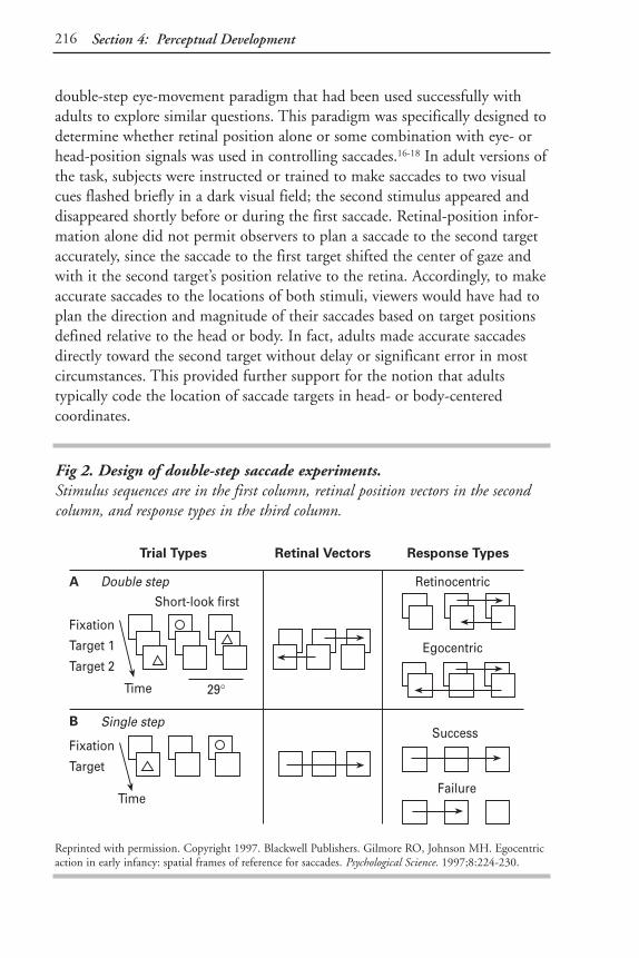
216 Section 4: Perceptual Development
double-step eye-movement paradigm that had been used successfully withadults to explore similar questions. This paradigm was specifically designed todetermine whether retinal position alone or some combination with eye- orhead-position signals was used in controlling saccades.16-18 In adult versions ofthe task, subjects were instructed or trained to make saccades to two visualcues flashed briefly in a dark visual field; the second stimulus appeared anddisappeared shortly before or during the first saccade. Retinal-position infor-mation alone did not permit observers to plan a saccade to the second targetaccurately, since the saccade to the first target shifted the center of gaze andwith it the second target’s position relative to the retina. Accordingly, to makeaccurate saccades to the locations of both stimuli, viewers would have had toplan the direction and magnitude of their saccades based on target positionsdefined relative to the head or body. In fact, adults made accurate saccadesdirectly toward the second target without delay or significant error in mostcircumstances. This provided further support for the notion that adultstypically code the location of saccade targets in head- or body-centered coordinates.
Fig 2. Design of double-step saccade experiments. Stimulus sequences are in the first column, retinal position vectors in the secondcolumn, and response types in the third column.
Reprinted with permission. Copyright 1997. Blackwell Publishers. Gilmore RO, Johnson MH. Egocentricaction in early infancy: spatial frames of reference for saccades. Psychological Science. 1997;8:224-230.
Trial Types Retinal Vectors Response Types
Double step Retinocentric
Egocentric
Fixation
Target 1
Target 2
Short-look firstA
Time 29°
Single step
Fixation
Target
Time
Success
Failure
B

217The Development of Representations for Perception and Action
Our adaptation of the double-step paradigm for infants sought to determinewhat spatial representations guide behavior from the earliest months of lifeusing a behavior that babies could perform from birth: saccadic eye move-ments (Fig 2). We hypothesized that young infants would be more likely thanolder infants to code the locations of the visual targets in strictly eye-centeredcoordinates. Our prediction was based on the presumption that coding thetargets’ locations in head- or body-centered coordinates would not be possiblebecause it would require accurate integration of visual and proprioceptiveinformation that depend on immature circuitry in the cerebral cortex.Specifically, we predicted that visually based representations of location woulddominate saccade planning initially; later, during the first several months oflife body-centered representations would gradually strengthen in influence.
In our first set of experiments, 4- to 6-month-old infants viewed a display ona central computer monitor that was followed by a sequence of two visualcues flashed to the left or right of the center of fixation one after the other, ina randomly selected left-right or right-left pattern (see Fig 3 top).39 In bothexperiments, the targets elicited sequences of eye movements that wererecorded on videotape and subsequently coded for direction and latency.Sequences were classified as retinocentric if the second saccade of the sequenceended at the midpoint of the center screen, or egocentric if the sequence endedat the midpoint of the screen where the second target had appeared (see Fig 3A top). Single-target trials (Fig 3 bottom) were presented to control forthe possibility that 4- and 6-month-olds could differ in their capacity to makelong eye movements independent of the specific form of spatial representation.
Fig 3 summarizes the results of the two studies. In both experiments, the ratio of egocentric to retinocentric sequences was significantly smaller inyounger infants. A marginally significant difference in the ratio of long looksto short ones was found in the first study but not in the second. The smallerproportion of egocentric saccades to the target location in the younger groupsuggested that younger infants more often relied on retinocentric coding ofvisual-target position in planning their saccade sequences. The fact that themean proportion of successful long saccades in the control trials did not differsignificantly argues against an alternative explanation in terms of differencesin the ability to make long saccades, but we conducted a second experimentto address this possibility directly.

218 Section 4: Perceptual Development
In a second study, we examined the patterns of saccade sequences made by 3- and 7-month-old infants performing a more challenging two-dimensionalversion of the double-step task.40 In this experiment (see Fig 4), separategroups of 3- and 7-month-old infants observed double-step visual events. Ineach trial, a fixation stimulus appeared in one of four positions on the screen(up, down, left, or right). Subsequently, two targets appeared on the screen,one following the other, with no period of overlap. The amount of time eachtarget was on the screen was chosen so that on most trials, both targets wouldhave disappeared prior to the onset of the first saccade. A 1.5-second responseinterval followed each double-step trial. Saccade sequences were coded asindicated in Fig 4B.
Fig 3. Results of two double-step saccade experiments. The mean relative ratio of egocentric to retinocentric saccades is plotted for theexperimental trials in the first experiment, and the ratio of total egocentric toretinocentric sequences is plotted for the second experiment. The mean relativeratio of successful long looks to failures is plotted for the control trials.
Reprinted with permission. Copyright 1997. Blackwell Publishers. Gilmore RO, Johnson MH. Egocentricaction in early infancy: spatial frames of reference for saccades. Psychological Science. 1997;8:224-230.
Display
Fixation
Target 1
Target 2
Time
Fixation
Target
Time
4 mos 6 mos 4-5 mos 6 mos
Results
1
0.75
0.5
0.25
0Egoc
entr
icSe
quen
ces
Experiment 1 Experiment 2
P<.001 P<.01
1
0.75
0.5
0.25
0
Long
Look
s P<.09n.s.
4 mos 6 mos 4-5 mos 6 mos

219The Development of Representations for Perception and Action
The results of this study are shown in Fig 5. On trials in which babies madeinitial saccades to the first target, younger babies made fewer second saccadesoverall (3 months old, 60%; 7 months old, 71%). When they did respond, 3-month-olds made more second saccades (Fig 5B) to the retinocentric screen location where no stimulus flashed (3 months old, 43% of sequences;7 months old, 25%) and fewer body-centered sequences to the actual targetlocations (3 months old, 8% of sequences; 7 months old, 42%). Several additional analyses ruled out alternative hypotheses that the results stemmedfrom improved accuracy or memory for the target locations.38
Thus, in three separate experiments, we observed an increase with age in the prevalence of body-centered saccade sequences. The combined results
Fig 4. Design of infant double-step saccade experiment. (A) Stimulus sequence. (B) Response classification. An egocentric sequenceconsisted of two saccades, one to each target location, and a retinocentric sequenceconsisted of two saccades, each equivalent to the positions of the targets at the timeof presentation relative to the retina.
Reprinted from Cognition. Vol 65. Gilmore RO, Johnson MH. Body-centered representations for visually-guided action emerge in early infancy. B1-B9. Copyright 1997, with permission from Elsevier Science.
A. Stimulus sequence
Fixation
Target
Target
24°
4°
B. Response classification
Egocentric Retinocentric

220 Section 4: Perceptual Development
supported our initial hypothesis that the representations of the location ofvisual targets for saccades shifts from being eye centered to head or body cen-tered in the first several postnatal months. This coincides with a period ofrapid development in the brain’s circuitry for action planning.
We interpreted these results to mean that a retinocentric representation ofspace usually dominates saccade planning in the youngest of infants, but
Fig 5. Results of infant double-step saccade experiment. (A) Proportion of initial eye movements directed to each of the nine screenlocations. T1 and T2 indicate the location of the first and second targetsrespectively. (B) Proportion of second saccades that followed an initial look to T1. Most second saccades were directed toward either the retinocentric oregocentric (T2/Body) location.
ADisplay & location grid Mean % valid first saccades
3 mo 7 mo
+ 2 (1) + 2 (1) 1 (1) + 0 (0)
T1 T2 50 (5) 15 (3) 22 (3) 59 (5) 5 (2) 30 (4)
4 (1) 2 (1) 4 (1) 2 (1) 1 (0) 1 (1)
BFirst saccade to T1 Mean % valid second saccades
3 mo 7 mo
1 (1) 5 (2) 1 (1) 4 (2) 5 (2) 1 (1)+
15 (6) 8 (3) 10 (3) 42 (7)T1 T2/Body
15 (4) 43 (8) 13 (3) 3 (2) 24 (5) 11 (4)Retino-centric
Reprinted from Cognition. Vol 65. Gilmore RO, Johnson MH. Body-centered representations for visually-guided action emerge in early infancy. B1-B9. Copyright 1997, with permission from Elsevier Science.

221The Development of Representations for Perception and Action
gradual development of cortical circuitry permits functional body-centeredrepresentations to emerge subsequently. (These circuits are likely in the pari-etal lobe because it is associated with processing information about movementand retina and body position.) Our data cannot rule out the possibility, how-ever, that gradual development in subcortical components of the oculomotorsystem could contribute to the observed shift in representation,41 nor, becausehead movements were neither eliminated entirely nor systematically varied,can we distinguish between the possibly separate influences of head- or body-centered coordinates in planning saccades.
Conclusion: The Development of Representationsfor Perception and Action
Even the simplest visually guided actions depend on representations of loca-tions in which visual and nonvisual sources of information are combined systematically. Available evidence in adults suggests that the systematic combi-nation of visual and proprioceptive sources of information occurs in circuitsof the cerebral cortex, particularly in perception- and action-planning areas of the occipital, parietal, and frontal lobes. However, perceptual, motor, andneurological factors suggest that young infants’ spatial-processing systems areless highly tuned than in adults. Results of three experiments provided evi-dence for a gradual shift in the representation of visual location between 3and 7 months of age. It appears that at least some forms of spatial behaviorearly in life are guided by simple, visually dominated representations of spatiallocation and that more stable and flexible body- and environment-centeredrepresentations emerge gradually in the first year. Consequently, while eye-,head-, trunk-, arm-, and even object-based representations underlie visuallyguided action in adults, far simpler frames of reference may be most influen-tial early in life. The development of mature representations for action maybe driven both by the infant’s perceptual and movement experiences and bythe development of circuitry in the brain devoted to action planning.
This evidence pertains directly to the development of spatial information forsaccadic eye movements, and it is possible that the shift from eye- to head- or body-centered representations of visual locations does not easily generalizeto other perception-action systems. The gradual emergence of body-centeredrepresentations to control saccades (during the first 6 months of life) is con-sistent with the development of other visually guided behaviors that emergearound this age and that may also depend on multimodal, body-centered

222 Section 4: Perceptual Development
representations of space. For example, infants between 2 and 6 months of ageshow increasing sensitivity to self- versus object-patterns of motion,42 accuracyin orienting toward the location of sounds,43 and precision in reaching withvisual feedback or without it.44,45 From 6 to 12 months of age, spatial process-ing continues to develop – infants show increasing sensitivity to landmarkswhen searching for hidden objects or predicting events. Determining howinfants develop integrated representations of space that combine visual, audi-tory, olfactory, and proprioceptive sources of information remains a challeng-ing and exciting topic for future research.
Another central problem that future research will have to consider concernsthe role of early experience. A number of sources suggest that self-producedmovements play a powerful role in the normal development of perceptionand action systems.46,47 Moreover, a number of factors argue for the impor-tance of experience-dependent plasticity in human infants’ visual perceptionsystems, including postnatal changes in the size of the eyes and head, themigration of retinal photoreceptors toward the fovea, and improvements ineye movement control.23,34 We are beginning to assess individual differencesin experiential factors that may contribute to the development of representa-tions for perception and action. At this time, however, we cannot yet say withcertainty whether one form of visual experience or another is critical for thedevelopment of mature, flexible representations for visually guided action.
It is tempting to speculate that the simple spatial representations that seem toinfluence eye-movement planning in young infants are truly adaptive. Thesesimple sensorimotor systems may provide the neonate with opportunities togather information from experience – by looking at a rich, patterned visualworld. This experience may be necessary to calibrate the relationship betweenvisual information and motor commands, such as those that drive saccades,and, ultimately, to contribute to the construction of mature representations of space. These representations are subsequently encoded in the newly matur-ing, powerfully flexible cerebral cortex, and, in turn, they provide new oppor-tunities for action and learning. While this view is speculative, I believe that it will help guide future research into the earliest underpinnings of infants’visuomotor behavior, the results of which promise to illustrate the nature ofinfants’ changing spatial representations and the processes that transformthem.

223The Development of Representations for Perception and Action
References1. Andersen RA, Snyder LH, Li CS, Stricanne B. Coordinate transformations in the representation of
spatial information. Current Opinion in Neurobiology. 1993;3:171-176.
2. Graziano MSA, Yap GS, Gross CG. Coding of visual space by premotor neurons. Science. 1994;266:1054-1057.
3. Stein JF. The representation of egocentric space in the posterior parietal cortex. Behavioral and BrainSciences. 1992;15:691-700.
4. Farah MJ, Brunn JL, Wong AB, Wallace MA, Carpenter PA. Frames of reference for allocating atten-tion to space: evidence from the neglect syndrome. Neuropsychologia. 1990;28(4):335-347.
5. Moscovitch M, Behrmann M. Coding of spatial information in the somatosensory system: evidencefrom patients with neglect following parietal lobe damage. Journal of Cognitive Neuroscience. 1994;6(2):151-155.
6. Goodman SJ, Andersen RA. Microstimulation of a neural network model for visually guided saccades.Journal of Cognitive Neuroscience. 1989;1:317-326.
7. Guenther FH, Bullock D, Greve D, Grossberg S. Neural representations for sensorimotor control. III.Learning a body-centered representation of a three-dimensional target position. Journal of CognitiveNeuroscience. 1994;6(4):341-358.
8. Olson CR, Hanson SJ. Spatial representation of the body. In: Hanson SJ, Olson CR, eds.Connectionist Modeling and Brain Function: The Developing Interface. Cambridge, Mass: MIT Press;1990:193-254.
9. Zipser D, Andersen RA. A back-propagation programmed network that simulates response propertiesof a subset of posterior parietal neurons. Nature. 1988;331:679-684.
10. Acredolo L. Behavioral approaches to spatial orientation in infancy. Annals of the New York Academy ofSciences. 1990;608:596-612.
11. Bremner JG. Egocentric versus allocentric spatial coding in nine-month-old infants: factors influenc-ing the choice of code. Developmental Psychology. 1978;14:346-355.
12. Piaget J; Cook M, trans. The Construction of Reality in the Child. New York, NY: Basic Books; 1954.
13. Piaget J, Inhelder B; Landon FJ, Lunzer JL, trans. The Child’s Conception of Space. London, England:Routledge & Kegan Paul; 1948.
14. Marr D. Vision. San Francisco, Calif: W Freeman; 1982.
15. von Helmholtz H. Treatise of Physiological Optics. New York, NY: Dover; 1925.
16. Becker W, Jürgens R. An analysis of the saccadic system by means of double step stimuli. VisionResearch. 1979;19:967-983.
17. Hallett PE, Lightstone AD. Saccadic eye movements to flashed targets. Vision Research. 1976;114:107-114.
18. Hallett PE, Lightstone AD. Saccadic eye movements towards stimuli triggered by prior saccades.Vision Research. 1976;16:99-106.
19. Matin L, Pearce DG. Visual perception of direction for stimuli flashed during voluntary saccadic eyemovements. Science. 1965;148:1485-1488.
20. Matin L, Stevens JK, Picoult E. Perceptual consequences of experimental extraocular muscle paralysis.In: Paillard J, ed. Brain and Space. Oxford, England: Oxford University Press; 1991.
21. Behrmann M, Tipper SP. Object-based attentional mechanisms: evidence from patients with unilateralneglect. In: Umilta C, Moscovitch M, eds. Attention and Performance XV: Conscious and NonconsciousInformation Processing. Cambridge, Mass: MIT Press; 1994:351-375.
22. Banks MS, Dannemiller JL. Infant visual psychophysics. In: Salapatek P, Cohen LB, eds. Handbook ofInfant Perception: From Sensation to Perception. New York, NY: Academic Press; 1987:115-184.
23. Aslin RN. Motor aspects of visual development in infancy. In: Salapatek P, Cohen LB, eds. Handbookof Infant Perception: From Sensation to Perception. New York, NY: Academic Press; 1987:43-113.

224 Section 4: Perceptual Development
24. Johnson MH. Cortical maturation and the development of visual attention in early infancy. Journal ofCognitive Neuroscience. 1990;2(2):81-95.
25. Aslin RN, Salapatek P. Saccadic localization of visual targets by very young infants. Perception andPsychophysics. 1975;17:293-302.
26. Salapatek P, Aslin RN, Simonson J, Pulos E. Infant saccadic eye movements to visible and previouslyvisible targets. Child Development. 1980;51:1090-1094.
27. Hainline L. Oculomotor control in human infants. In: Groner R, McConkie GW, Menz C, eds. EyeMovements and Human Information Processing. Amsterdam, Netherlands: Elsevier; 1985.
28. Aslin RN. Development of smooth pursuit in human infants. In: Fisher DF, Monty RA, Senders JW,eds. Eye Movements: Cognition and Visual Perception. Hillsdale, NJ: Erlbaum; 1981:31-51.
29. Gwiazda J, Bauer J, Held R. From visual acuity to hyperacuity: a 10-year update. Canadian Journal ofPsychology. 1989;43:109-120.
30. Birch EE, Gwiazda J, Held R. Stereoacuity development for crossed and uncrossed disparities inhuman infants. Vision Research. 1982;22:507-513.
31. Regal DM, Ashmead DH, Salapatek P. The coordination of eye and head movements during earlyinfancy: a selective review. Behavioral and Brain Research. 1983;10:125-132.
32. Reisman JE, Anderson JH. Compensatory eye movements during head and body rotation in infants.Brain Research. 1989;484(1-2):119-129.
33. Daniel BM, Lee DN. Development of looking with head and eyes. Journal of Experimental ChildPsychology. 1990;50(2):200-216.
34. Aslin RN. Perception of visual direction in human infants. In: Granrud CE, ed. Visual Perception andCognition in Infancy. Hillsdale, NJ: Lawrence Erlbaum Associates; 1993.
35. Atkinson J. Human visual development over the first six months of life: a review and a hypothesis.Human Neurobiology. 1984;3:61-74.
36. Bronson GW. The postnatal growth of visual capacity. Child Development. 1994;45:873-890.
37. Huttenlocher PR. Morphometric study of human cerebral cortex development. Neuropsychologia.1990;28:517-527.
38. Chugani HT, Phelps ME, Mazziotta JC. Positron emission tomography study of human brain func-tional development. Annals of Neurology. 1987;22:487-497.
39. Gilmore RO, Johnson MH. Egocentric action in early infancy: spatial frames of reference for saccades.Psychological Science. 1997;8:224-230.
40. Gilmore RO, Johnson MH. Body-centered representations for visually-guided action emerge in earlyinfancy. Cognition. 1997;65:B1-B9.
41. Mays LE, Sparks DL. Dissociation of visual and saccade related responses in superior colliculus neu-rons. Journal of Neurophysiology. 1980;43:207-232.
42. Kellman PJ, von Hofsten C. The world of the moving infant: perception of motion, stability, andspace. In: Rovee-Collier C, Lipsitt LP, eds. Advances in Infancy Research. Vol 7. Norwood, NJ: AblexPublishing Corp; 1995:147-185.
43. Muir DW, Field J. Newborn infants orient to sounds. Child Development. 1979;50:431-436.
44. von Hofsten C. Developmental changes in the organisation of prereaching movements. DevelopmentalPsychology. 1984;20:378-388.
45. Clifton RK, Muir DW, Ashmead DH, Clarkson MG. Is visually guided reaching in early infancy amyth? Child Development. 1993;61(1):1098-1110.
46. Bertenthal B, Campos J, Barrett K. Self-produced locomotion: an organizer of emotional, cognitive,and social development in infancy. In: Emde R, Harmon R, eds. Continuities and Discontinuities inDevelopment. New York, NY: Plenum Press; 1984:175-210.
47. Held R, Hein A. Movement produced stimulation in the development of visually guided behavior.Journal of Comparative Physiological Psychology. 1963;56(5):872-876.

225
The Emergence of Future-OrientedThinking in the Early Years
Marshall M. Haith, PhD
Introduction
This paper will address a cluster of psychological processes in infants andyoung children that I believe underlies the development of children’s ability tothink about the future. This cluster is referred to as future-oriented processes(FOPs), a term that includes such processes as perceptual set, prospectivemotor processes, expectations, forecasting, intentionality, planning, and goalsetting – any process that is oriented to events that have not yet occurred.My colleagues and I have taken three approaches to this phenomenon (Table1). The first is an experimental approach with infants who are usually in thefirst 3 months of life but may be as old as 7 months. The second involves aninterview and questionnaire to obtain parents’ reports of their children’sunderstanding about the future, when the children are 9 to 42 months of age.The third consists of longitudinal analyses of transcripts of parents’ utteranceswith their children to examine to what extent parents talk to their childrenabout the past, present, and future at 14, 20, and 32 months of age.
If one were to ask most people how much time they spend thinking about the past, present, and future, the most common answer would be that future-oriented thought occupies an enormous amount of mental energy, typicallymuch more than past thought. A few examples are your thoughts about whatyou are going to wear in the morning, with whom you will have lunch, whenyou plan to work on a report, how you can set aside enough money for retire-ment, and when you should call your physician to make an appointment.
Nonetheless, psychology has paid very little attention to future-orientedthought, certainly in comparison to past-oriented thought, or what is referredto as memory. For memory, there are several cognitive, neurophysiological,and biochemical models, a highly developed terminology (eg, short- andlong-term memory, working memory, episodic memory, and semantic memo-ry), and a large number of accepted paradigms for studying these domains.This is not true for future-oriented thought. That is not to say that people

226 Section 4: Perceptual Development
have ignored phenomena that involve future-oriented thinking; future think-ing is embedded in virtually everything we do. Research on planning andproblem solving are prime examples. Nevertheless, the spotlight has not beenturned on FOPs in the same way that it has illuminated research on memory;there has not been a focus on future-oriented thought processes per se.
This neglect is even more severe in the area of child development, so my col-leagues and I have spent the last decade or so doing what we can to bring thisissue into focus. Our belief is that FOPs are crucial to adaptation of anyorganism to the world. In fact, the irony is that the adaptive function ofmemory is to foster better future-oriented action. Memory benefits us byenabling more effective functioning in future situations; to paraphrase theBoy Scouts, “Preparation is all.”
Where do FOPs come from? To address this question, we have been conduct-ing a research program with infants mostly in the first 3 months of life. Atthis age, infants’ motor control is not sophisticated enough to permit them
Table 1. Three Approaches to Studying the Development of Future-Oriented Processes
ExperimentalInitial findings with 3-month-olds
Expectations for nonalternating events
Adaptive function of expectations
Role of constraints in infant expectations
Individual differences in expectation formation and prediction of later cognitive functioning
QuestionnaireDimensions of future-oriented thinking
Development from 9 months to 3 years in understanding
Parents’ report of their talk about the future
ObservationalCategorizing time talk
Percent of parents’ utterances about past, present, and future

227The Emergence of Future-Oriented Thinking in the Early Years
to press buttons or pull levers and, even if they could, their ability to under-stand instructions is nonexistent. Therefore, we engineered studies to takeadvantage of their visual behavior and eye movements to tell us what thebrain is doing, specifically the expectations they form for what comes next.Eye movements are well controlled by alert infants almost from birth; in fact,this infant motor system more closely parallels that of the adult than anyother action system.
The Basic Visual Expectation Paradigm and Initial Findings
The video setup that we use to record infant eye movements is shown in Fig 1. Infants lie on their backs and watch displays on the television screenreflected in a mirror angled at 45°, while we record the image of their eyewith infrared light through the mirror. Attractive pictures appear to the leftand the right of center, for example, checkerboards, faces, and bull’s-eyes.
Fig 1. Infant recording apparatus.
Copyright ©1991 by the American Psychological Association. Reprinted with permission. Canfield RL,Haith MM. Active expectations in 2- and 3-month-old infants: complex event sequences. DevelopmentalPsychology. 1991;27:198-208.

228 Section 4: Perceptual Development
Fig 2 shows typical timing for an alternating sequence with, for example, apicture on the left for 700 msec, followed by an interpicture interval (IPI) of1000 msec, then a picture on the right for 700 msec, and so on. Later, wedecode the infants’ eye orientation, frame by frame, 30 times each second, todetermine where they are looking at each moment. We do this either manual-ly or through a custom-built computerized eye tracker.
Fig 2. Timeline for a typical study.
Left
IPI IPI IPI IPIIPI
Right Right
1000 700 700 700 7001000 1000 1000 1000
Milliseconds
AnticipationWindow
RT Window
Timeline for Alternating Sequence
We use these important concepts in these studies: expectation, anticipation,and facilitation. Expectation is a construct that we infer from specific behav-iors of the infant. We cannot see expectations, but we can see their effect.The most obvious effect is anticipation, which we define as an eye movementto the location where an event is going to happen before it begins – duringthe interpicture interval or so quickly after a picture appears that the eyemovement must have been programmed beforehand. Facilitation is anotherindicator of expectation – most of the time, infants do not actually anticipatethe next event, but their reaction times to picture onsets are so quick thatthey appeared ready for them to occur, as though they forecast what was com-ing next. We calculate response time (RT) from the time of picture onset tothe beginning of a reactive eye movement. With proper control conditions,one can ask whether infants form expectations from their RT data – their efficiency of response indicates preparedness just as the efficiency of an adult’sdriving behavior (braking) is enabled by seeing a school-zone sign before seeing a ball bounce into the street.
IPI = interpicture interval.

229The Emergence of Future-Oriented Thinking in the Early Years
Fig 3. Infant visualtracking record. Shown here is a sequenceof picture onsets, indica-ted by the rectangularboxes, that are separatedby blank displayinterpicture intervals;time proceeds from top to bottom. Overlaid onthis picture sequence arean infant’s fixations andeye movements (dottedline). One can see that forthe first several pictures of the eye-tracking recordof the 3-month-old infant,the eye movements arereactive. For the fifth andsixth pictures, the infantanticipates picture onsetand also does so forseveral other pictures. On average, infants whoshow any anticipations at all make their firstanticipation within 17seconds of the beginningof the series.
Adapted from Haith MM. The early development of visual expectations. In Granrud C (ed), Carnegie-Mellon Symposium on Visual Perception and Cognition in Infancy. Lawrence Erlbaum Associates; 1993.Courtesy of the author.
RightstimulusBaby’stracking
Leftstimulus
Time
700MS
1100MS

230 Section 4: Perceptual Development
Table 2 displays some RT and anticipation data for the experiment.1 We typi-cally present 10 baseline pictures at the beginning in randomly sequencedlocations to get an indication of infants’ “raw” RT before they have learnedanything about the patterns. These baseline RTs were longer than RTs in the subsequent predictable, alternating series. For infants who saw a nonpre-dictable, irregular series, however, the RTs did not decrease following baseline.Infants in the alternating group also displayed significantly more fixationshifts to the opposite side during the interpicture interval than did those in the irregular group.
Table 2. Median Reaction Times and Percent of Anticipations.
Predictable NonpredictableAlternating Irregular
Display Display
Reaction Time: Baseline 468 msec 462 msec
Reaction Time: Experimental 391 msec 462 msec
Anticipations 22% 11.1%
Adapted from Haith MM, Hazan C, Goodman GS. Expectation and anticipation of dynamic visual eventsby 3.5 month-old babies. Child Development. 1988;59:467-479.
Expectations for More Complex Event Sequences
One of the limitations of this experiment is its simplicity. One might supposethat infants somehow become trained to a pendulum-like sequence and forsome reason just sped up a bit, eventually moving ahead of each pictureappearance, displaying a kind of mental short-circuiting. Digging deeper,Richard Canfield and I carried out a separate experiment that showed whythis was not the case.2 Fig 4 shows the four groups that were used. One groupsaw an alternating series (as in the previous study [the 1/1 group]), a secondgroup saw a repeating series of two pictures on the left side, followed by oneon the right side (the 2/1 group), and a third group saw a repeating series ofthree pictures on the left, followed by one on the right (the 3/1 group). Afourth, control, group saw a random series with embedded 1/1, 2/1, and 3/1series, but the location of the next picture at any moment was unpredictable.

231The Emergence of Future-Oriented Thinking in the Early Years
Fig 4. Complex event sequences schematic conditions.
Copyright ©1991 by the American Psychological Association. Reprinted with permission. Canfield RL,Haith MM. Active expectations in 2- and 3-month-old infants: complex event sequences. DevelopmentalPsychology. 1991;27:198-208.
1/1 2/1 3/1homeside
homeside
homeside
targetside
targetside
targetside
700 ms onfixation shift
after one
(correct antic.)
700 ms onfixation shift
after one
(error antic.)1000 ms
IPI1000 ms
IPI
fixation shiftafter one
(error antic.)
fixation shift
(correct antic.)
fixation shiftafter two
(correct antic.)
fixation shift
(correct antic.)
fixation shiftafter two
(error antic.)
fixation shiftafter three
(correct antic.)
fixation shift
(correct antic.)
Com
paris
onSe
tfor
1/1
Com
paris
onSe
tfor
2/1
Com
paris
onSe
tfor
3/1
fixation shiftafter one
(same as return)
fixation shiftafter one
(same as return)
fixation shiftafter two
returnfixation shift
(same as one)
fixation shiftafter one
(same as return)
fixation shiftafter two
fixation shiftafter three
Irregular
EXPERIMENTAL CONDITIONS COMPARISONCONDITION

232 Section 4: Perceptual Development
Fig 5. Infant visualtracking record for the 2/1 sequence.
Adapted from Haith MM. The early development of visual expectations. In Granrud C (ed), Carnegie-Mellon Symposium on Visual Perception and Cognition in Infancy. Lawrence Erlbaum Associates;1993.Courtesy of the author.
Time
700MS
1000MS
= 200 msec
Left stimulus
Left stimulus
Baby’s tracking
Right stimulus

233The Emergence of Future-Oriented Thinking in the Early Years
Fig 5 displays a tracking sequence for an infant in the 2/1 condition. One cansee again that the infant first only reacts to pictures as they are presented butthen quickly begins to anticipate forthcoming pictures (eg, pictures 9 and12). Fig 6 shows the data for the whole experiment in terms of RT declinefrom baseline. For 3-month-olds, as the complexity of the sequence increased,the RT savings diminished. Infants in the 2/1 condition had a reliably greaterdecline than did control infants, and they had more frequent anticipations.The RT of the 2-month-old group did not benefit from predictability in anyof the series.
This study demonstrates that, by 3 months of age, infants not only formexpectations for sequences that are nonpendular but also that they are capableof quickly forming expectations when different numbers of events occur inone location than another.
Fig 6. Change in reaction times and sequence complexity.
The simple sequences had shorter reaction times.
Copyright ©1991 by the American Psychological Association. Reprinted with permission. Canfield RL,Haith MM. Active expectations in 2- and 3-month-old infants: complex event sequences. DevelopmentalPsychology. 1991;27:198-208.
25
20
15
10
5
0
−5
1/1 2/1 3/1 IRREG
Per
cen
tD
rop
Complexity of Sequence
2-months-olds 3-months-olds

234 Section 4: Perceptual Development
As stated earlier, the benefit of memory is more efficient performance infuture situations to which experience can be applied. The above experimentshave only demonstrated that an infant can learn in a single session to formexpectations for what comes next in that situation. Can infants utilize expec-tations they form for situations in which they later find themselves?
The Adaptive Function of Expectations
Denise Arehart and I observed 3-month-olds in two sessions, separated byabout 1 week.3 On the first visit, two groups saw different sequences; Group1 saw a regular alternating sequence, Group 2 saw a random irregularsequence. On the second visit, both groups saw a regular alternatingsequence. The data presented here are for only the second visit, when bothgroups saw the same display. As shown in Fig 7, the group that saw the alter-nating sequence on week 1 was much faster to respond in the first blocks of 10 pictures than the group that saw the random series. After three blocksof trials, the irregular group caught up, but they slowed down in blocks 5 and 6. Fig 8 shows the data for anticipations. Again, the group that saw thealternating sequence produced more anticipations at the beginning and, infact, throughout the test session. This study illustrates that infants can usetheir experience as an aid for forming expectations for similar situations at a later time.
Fig 7. Median reaction times on Visit 2.
Adapted from Haith MM, Wentworth N, Canfield RL. The formation of expectations in early infancy. In: Rovee-Collier C, Lipsitt LP (eds) Advances in Infancy Research. Norwood, NJ; Ablex: 1993.
500
400
300
Med
ian
Rea
ctio
nTi
me
1 2 3 4 5 6 7Block
RegularIrregular

235The Emergence of Future-Oriented Thinking in the Early Years
The situations discussed so far are fairly limited. After all, how often is it thatwe know exactly what will happen next? In the non-laboratory world, wetend to form expectations for classes of events rather than for specific thingsto happen. In listening or reading, for example, when we hear the sentence“Jane picked up the…” we know that the next word will be a noun and thatit will be something that is graspable (ie, not a football field, fire, water, air,etc), even though we do not know exactly what the object will be. We useconstraints such as these in language to facilitate both reading and listening.Are babies only capable of benefitting from expectations for something thatwill certainly happen, or can they benefit also from expectations for eventsthat are only likely to happen? Stated differently, are infants able to benefitfrom knowing what can happen as well as exactly what will happen?
The Role of Constraints in Infant Expectation
Thomas Dougherty addressed this question in an experiment that modifiedour usual paradigm.4 Instead of using a continuing flow of pictures, he usedwhat looks like a more traditional trial format. Each trial started with anattractive picture that appeared in the center of the screen to get the infant’sattention. Infants participated in one of three groups at 7 months of age (see Fig 9). For the perfectly predictable group (0 bits, in information-theoryterminology), the center picture disappeared, and, a variable time later, a
Fig 8. Percent of anticipation on Visit 2.
Adapted from Haith MM, Wentworth N, Canfield RL. The formation of expectations in early infancy. InRovee-Collier C, Lipsitt LP, eds. Advances in Infancy Research. Norwood, NJ: Ablex; 1993.
30
20
10
0
%A
nti
cip
atio
ns
1 2 3 4 5 6 7Block
RegularIrregular

236 Section 4: Perceptual Development
peripheral target picture appeared, always in the same place. For differentinfants, this target picture could appear in one of four locations – up-left, up-right, down-left, or down-right. For the two-location group, the target picturecould appear in either of two places (either up-left or down-right, for exam-ple), but on any trial the infant had no way to predict which would occur.Thus, this group had constraints on where things could happen, but infantsdid not know exactly where something would appear next. For the four-location group, a picture could appear in any of four locations, up-left, up-right, down-left, or down-right, again unpredictably.
The results were consistent with the hypothesis that infants can use con-straints on what can happen even when they cannot predict exactly what itwill be. Response times improved (difference between baseline and postbase-line; see Fig 10) when the infants were more constrained and had fewer possi-
Fig 9. Constraints ininfant expectations,schematic of conditions.
1 Target1 Possible Location
FixationField
TargetField
1 Target2 Possible Locations
1 Target4 Possible Locations
Time

237The Emergence of Future-Oriented Thinking in the Early Years
bilities that could occur. Importantly, there was significantly greater facilita-tion for the two-location than for the four-location condition, even thoughprecise predictability was not present for either. Data from anticipatory fixa-tions supported the RT data. In the two-location group, when infants didanticipate the next picture, they usually looked at a possible location ratherthan at an impossible location; this was true for eight of nine infants, indicat-ing that they were sensitive to where things could happen even if they did notknow exactly when. Thus, as for adults, young infants can use constraints inthe environment to form expectations for likely but not certain events.
Individual Differences
The question always arises about whether we are really tapping cognitivefunctioning with this paradigm and, if so, whether there are individual differ-ences in performance. The quick answer is “yes” to both questions. In onestudy, we brought 3-month-old infants back to the lab a little less than 1 week after they had seen an alternating left-right sequence and showedthem the same sequence again.5 We obtained both stable within-session andbetween-session performance for RTs, with between-session correlations typi-cally in the .60s. Individual stability was somewhat less evident for anticipa-tions, with the between-session correlation falling in the mid-.30s.
Fig 10. Facilitation of reaction times for the three conditions.
70
60
50
40
30
20
10
0
−10
−20
−30
Mea
nR
eact
ion
Tim
eD
eclin
eFr
om
Bas
elin
e(m
sec)
n=14PS1 target (0)
Groups (Bits)
n=14PS2 targets (1)
n=13PS4 targets (2)

238 Section 4: Perceptual Development
Indications that we are tapping a cognitive process and that infant perform-ance is predictive of later cognitive functioning come from three studies. Oneyielded stable correlations between performance on the Visual ExpectationParadigm at 8 months of age and performance on the Stanford-Binet at 3 years of age, and a second study produced a similar finding between infantperformance and midparent IQ (as a proxy for the eventual infants’ IQ).6,7
More recently, Thomas Dougherty and I followed up infants whom we hadseen at 3 months of age to assess their RT and intellectual performance ataround 4 years of age.8 We obtained the first somewhat surprising evidencethat there is a fairly strong relation between infant RT and childhood RT but,more relevant, that RT and percent of anticipations correlated with infants’full-scale IQ at 4 years of age (see Fig 11). We also examined whether vari-ability in infants’ RTs was predictive and found that the amount of variabilitycorrelated reliably with childhood full-scale IQ.
It is not clear what processes are responsible for the continuity that we found between early infancy and childhood. Our best guess is that RT taps
Fig 11. Relation between infant performance in the Visual ExpectationParadigm and childhood RT, anticipations, and IQ.
FSIQ
.43 −.44 −.46.51
ChiVRT
InfANT InfRT InfSD
FSIQ = Full Scale IQChiVRT = Childhood Visual Reaction TimeInfANT = Infant Percent AnticipationsInfRT = Infant Reaction Time (eye movement)InfSD = Infant Standard Deviation of Reaction Times

239The Emergence of Future-Oriented Thinking in the Early Years
processing speed, anticipations tap memory, and RT variance taps mainte-nance of attention. These are all processes that have been implicated in intelli-gence tests, but we are speculating at this point, and a lot more research mustbe done to validate or refute these possibilities.
Summary: Reaction Time Studies
We have seen that infants form visual expectations as early as 3 months ofage, and they do so very quickly – typically within only a few seconds ofexposure to a regular spatiotemporal visual display. This ability can bedemonstrated not only for simple left-right sequences but also for sequencesthat require the infant to remember a complete spatial pattern and where the infant is in that pattern at each moment (eg, the 2/1 sequence). Thus, itseems quite easy and natural for infants to form expectations for predictableevents. Moreover, we have seen that infants can remember the events forwhich they formed expectations and can utilize that stored information in alater, similar task. They also appear to be able to detect constraints on whatcan happen next, so they are not limited to forming expectations only for per-fectly certain events. Finally, there are reliable individual differences betweeninfants both in RT and in their tendency to form expectations. These individ-ual differences are predictive of moderate variance in cognitive performancein later childhood, so it appears that the Visual Expectation Paradigm is tap-ping processes that are important for cognitive development. We believe thatcandidate processes are processing speed, memory, and the ability to sustainattention. Regardless, the roots of future-oriented processing seem to beestablished in the very early months of life.
The Questionnaire Approach
While the approximately 20 years that we have put into developing andexploiting the Visual Expectation Paradigm have been fruitful in tapping adomain of future-oriented processing in early childhood, we also knew thatwe were missing many other aspects of future thinking that occur outside thelab in everyday life. To get a feeling for what these domains might be, weadopted a very different approach, consisting initially of interviews withmothers of infants which led to developing and using questionnaires. Theeffort was enlightening both in terms of the difficulty of conceptual boot-strapping in an undeveloped area and in terms of how much effort is requiredto develop a useful, understandable, and reliable instrument from scratch.9

240 Section 4: Perceptual Development
We are close to submitting a more complete report of our findings from follow-up studies.
The questionnnaire consists of two parts (see Table 3). The first part dealswith family-context factors – parents’ beliefs about what their children under-stand about the future, parent theories about how children learn about thefuture, family practices that promote future understanding (regular routines,traditions, etc), and how the parents were raised along these dimensions. Thesecond part involves the domains of future-oriented processing that representour initial approach to conceptualizing the dimensions of future thinking inyoung children. These domains are Order (ordering of events), Routines(knowing what follows what in a routine), Planning, Expectation, Time, andProblem Solving. Examples of these domains are shown in Table 4. In laterwork, we added a section that we call an Emotional/Compliance domain,which addresses social negotiation in rule situations (eg, “if you jump on thebed, you’ll go to time out”).
Table 3. The Future-Oriented Processes Questionnaire (9-36 Months)Demographic Information.9
Part I: Contextual Influences
• Parental beliefs: what child knows about near- vs far-term future (4-point scale: Understands Well – Does Not Understand)
• Parental theories: how child knows about the future (4-point scale: Importance Ranking)
• Family practices (4-pt scale: Almost Always – Almost Never)
• Precedent family practices (4-pt scale: Almost Always – Almost Never)
Part II: Domains (and Subdomains) of Future Orientation (4-point scale: Very True – Not at All True)
• Order
• Routine
• Planning
• Expectation (cues; violations; language; comprehension; production)
• Time (terms; duration; verbs; tools)
• Problem solving (flexibility; goal-orientation; social)

241The Emergence of Future-Oriented Thinking in the Early Years
In our first study, 68 parents participated who had children ranging in agefrom 9 to 36 months. We carried out an additional study with 286 more par-ents of 12- to 42-month-olds with a revised survey, but the initial findings arenot materially different from the earlier findings, which are reported here.
First, we ran alpha statistical tests to give us some indication of the coherenceof questions within domains and their separability from other domains, andthese turned out satisfactorily, ranging from the mid-.60s to the mid-.90s(Table 5).
Table 4. Example Questions for the Six Domains.9
(Possible Answers: Very true, Sometimes true, Once in a while, Not at alltrue)• Order
My child understands that some things must happen before other things.(Example: “You must wash before eating” means wash your hands andthen eat lunch.)
• RoutineMy child wants some things done the same way every time.(Example: Having a snack before bedtime.)
• PlanningMy child does things that show preparation for the future.(Example: My child gets a toy to take to Grandma’s.)
• Expectation (cues; violation; language; comprehension; production)My child has expectations for things that happen.(Example: My child expects someone to enter the house when hearingthe doorbell.)
• Time (terms; duration; verbs; tools)My child understands time if I compare it to something familiar.(Example: “It will take as long as ‘Sesame Street’ for the cookies to be ready.”)
• Problem-solving (goal orientation; flexibility; social agent)My child tries to get help from others to get something done.(Example: Putting my hand on the music box to make it play.)

Second, there was more clear development for some of the domains than forothers, as shown in Fig 12. We worried about whether parents’ judgmentwould be influenced by their children’s language development, but the changein domains does not parallel language changes very closely. The level of per-formance for some domains is high throughout the age range; for others, thelevel changes dramatically, and for still others performance remains relativelylow while yet improving with age. Thus, the different domains of future ori-entation do not follow the same developmental course.
Third, very surprisingly, parents’ reports of how much they talked to theirchildren about the future did not vary much across the age range of 9 monthsto 36 months, a wide developmental span. The reports at least seemed to havesome validity, because parents gave sensible answers when asked how muchthey talked about the future as a function of time. That is, they reported lesstalk about more distant future events (eg, months away) than about moreproximal events (eg, tomorrow; see Fig 13).
This does not seem reasonable, however. We all know that infants understandmore of what one says to them when they are 3 years old than when they are9 months old. Are parents really unaware of what their own children under-stand? Fig 14 shows what parents think their children understand about whatthey are being told; I have superimposed the data from Fig 13 on this graph.In fact, parents know very well that their 9-month-olds do not understandwhat they are saying and that their children understand more as they growolder. This knowledge does not seem to affect how much they talk to theirchildren about the future – at least, that is what they report.
242 Section 4: Perceptual Development
Table 5. Item Internal Consistency (Alpha) for Future-OrientationDomains.9
DOMAIN N ALPHA # OF ITEMS
Order 66 .85 2Routine 68 .67 2Planning 66 .80 2Expectation 61 .92 13Time 63 .86 8Problem solving 66 .79 8

243The Emergence of Future-Oriented Thinking in the Early Years
Fig 12. Age trends for the major domains of the Future-Oriented ProcessesQuestionnaire.
Adapted from Benson JB. The origins of future orientation in the everyday lives of 9- to 36-month-oldinfants. In Haith MM, Benson JB, Roberts RJ Jr, Pennington BF, eds. The Development of Future-OrientedProcesses. Chicago, IL: University of Chicago Press; 1994.
4.0
3.0
2.0
1.0Rep
ort
edP
erfo
rman
ce
9 12 18 24 30 36Age in Months
Age Trends: All Domains(Mean Domain Score)
RoutineProb. Solve
Expectation
Order
Planning
Time
LanguageProduction
Fig 13. Future time talk by parents to their children at different ages.
Adapted from Benson JB. The origins of future orientation in the everyday lives of 9- to 36-month-oldinfants. In Haith MM, Benson JB, Roberts RJ Jr, Pennington BF, eds. The Development of Future-OrientedProcesses. Chicago, IL: University of Chicago Press; 1994.
4.0
3.0
2.0
1.0
Freq
uenc
yof
Futu
reTi
me
Talk
9 12 18 24 30 36Age in Months
TomorrowDays Away
Weeks AwayMonths Away
Family Practices: Future Time Talk

244 Section 4: Perceptual Development
There appear to be two factors in operation here. One is that parents do notknow precisely what their infants understand, but they do know that theirunderstanding about the future will grow. They talk to their children aboutfuture events, because they know that understanding emerges gradually andthat their children will come to grasp what is going to happen next as theyconnect language to action – hearing about something that will happen andthen actually seeing it happen. In brief, the parent scaffolds the child’s accu-mulating grasp of future events through talk; like chicken soup for the com-mon cold, future talk may not help, but it can’t hurt. The second factor isthat the parent is constantly thinking about what she has to do next or aboutplanning for future events and is simply talking out loud, about events suchas errands, dinner, or picking up an older sibling from school. Perhaps adultswould do the same if we had only a dog as a companion. Nevertheless, thepotential scaffolding effect is the same.
This branch of our research effort has broadened our horizons about the vari-ous planes on which future thinking develops. There are three conclusions to
Fig 14. Parental beliefs about their children’s understanding of time talk.
Adapted from Benson JB. The origins of future orientation in the everyday lives of 9- to 36-month-oldinfants. In Haith MM, Benson JB, Roberts RJ Jr, Pennington BF, eds. The Development of Future-OrientedProcesses. Chicago, IL: University of Chicago Press; 1994.
4.0
3.0
2.0
1.0
Freq
uen
cyFu
ture
Tim
eTa
lk/
Ch
ildU
nd
erst
and
ing
9 12 18 24 30 36Age in Months
Tomorrow
Days Away
Tomorrow
Weeks AwayMonths Away
A Day/A WeekA Month
Family Practices & Parental Beliefs:Future Time Talk and Child Understanding

245The Emergence of Future-Oriented Thinking in the Early Years
be drawn from this brief description. First, the span of FOPs is quite broad inearly childhood, certainly a lot broader than expectation formation in the labin young infants. Second, not all domains advance at the same pace. Third,the scaffolding of infants’ learning about the future can be quite extensive,with parent speech being a major component.
Observation of Future-Oriented Talk
We were troubled by the lack of direct empirical support for the informationwe were getting from parents. Do parents really talk to their children aboutthe future as much as they say? Wouldn’t we expect parent talk to be mostlyabout the present and the past, leveraging on young children’s direct experi-ence? If so, perhaps future-oriented talk just slips in as a minor fraction ofparent-child dialogue, because, in fact, it is important only to the parent.
To address this question, we used the Child Language Data Exchange System,which was pioneered by Brian MacWhinney at Carnegie-Mellon University.This is a large database of coded transcripts of parent-child narratives. Weanalyzed data from a longitudinal project headed by Catherine Snow basedon mother-infant dialogues when the child was 14, 20, and 32 months ofage.10,11 The setting consisted of free play and the presentation of several playobjects. Each of the mother’s utterances was categorized in terms of orienta-tion to the past, the present, or the future (with more emphasis on event timethan on tense; see Table 6 for examples). Utterances that were categorized asnaming, imperatives, or ambiguous were not included. Thus our analysis forfuture events is somewhat conservative, as other investigators consider imper-atives to be future oriented.
We expected a small change in future talk with age, consistent with the ques-tionnaire data we collected. We also thought that talk about the past wouldincrease with age as the child’s memory improved and that overall time talkwould increase also.
Fig 15 displays the results of our analyses. Across all ages, we found thatutterances most often could be categorized as dealing with the present, but wealso found a substantial fraction of utterances that dealt with the future, andthese increased with age. Counterintuitive to what most people might expect,

246 Section 4: Perceptual Development
there were many more utterances that referenced the future than the past.These data support what parents have been telling us. Parents spend a signifi-cant amount of time talking to their children about the future. Of all utter-ances that we categorized as time talk, around 30% were about the futureover all ages and close to 5% were about the past. Again, we did not includeimperatives; if we had, the discrepancy between the percentages of past andfuture talk would be even greater.
Table 6. Sample Adult Utterances, Coding Category, and Rationale.
Adult Utterance Coding Category RationaleType
Time Talk:
“We danced at ballet classyesterday.”“I put that on chicken whenI make some dishes.”
past event, past tense
past event, current tensePast
“Now here we are.”“You’ve got too much in it.” Current
current event, current tensecurrent event, current tense
“We need to write it down.”“Let me see if I can do thatmyself.”
Futurefuture event, future tensefuture event, future tense
Other Talk:
“This is a duck.”“Elmo is playing soccer.”
Naming naming an objectnaming an action
“Put the chicken to bed.”“Let me see your finger.”
Imperative command statement
“What?”“Hmmm.”
Ambiguous unsure of the reference ofthe utterance

247The Emergence of Future-Oriented Thinking in the Early Years
Conclusions
Future events play an important role in the life of the young child. Even inthe first months of life, infants are aware of what comes next, and they organ-ize their mental activity and behavior around what is going to happen for pre-dictable events rather than just reacting to them. After the fact, one can askhow it could be otherwise. A world without expectations would be chaotic.We have shown several aspects of this future-oriented process, including itsimportance in maximizing efficiency for later behavior, its status as a cogni-tive process, and its relation to stable processes that underlie intellectual performance in the early years. The questionnaire work has helped us todimensionalize the domains that constitute early future-oriented thinkingbeyond the laboratory into everyday life. Finally, the language analyses sup-port our intuitions and parents’ reports that at least the children in our sam-ple are surrounded by influences, both linguistic and nonlinguistic, thatsupport their learning about the future.
It is not difficult to make the case that future thinking during childhood, inadolescence, and, indeed, throughout life is critical to one’s development, wel-fare, and successful adaptation to the world. One hears repeated claims abouthow critical it is for children to understand the future consequences of theircurrent action, whether we talk about morals, smoking, saving money, or risk
Fig 15. Changes with age in percent of parent time categorized as orientedto the past, present, or future.
Time Talk CategoriesCurrentFuturePast
40
35
30
25
20
15
10
5
0Prop
ortio
nof
Adu
ltU
tter
ance
s
14 20 32Age (in months)

248 Section 4: Perceptual Development
avoidance. We do not really know what the effects on the development offuture thinking might be of growing up in a relatively chaotic home whereevents are less predictable and less supportive of future thinking than in themiddle-class samples we studied, nor do we know what the effects might beof limited opportunity for children to discuss future events with their parents.We do believe that a child’s ability to think about the future depends on envi-ronmental regularity, such as family routines and rituals, and parental scaf-folding through talk and action.
We have just scratched the surface in developing a scheme for conceptualizingthis important area of inquiry and how contextual factors could affect itsdevelopment. More fundamentally, the recognition that future-orientedprocesses are central to cognitive development is a necessary first step formaking progress in this area. We hope that the work discussed here will helpfoster that recognition and form a foundation for further investigation.
References1. Haith MM, Hazan C, Goodman GS. Expectation and anticipation of dynamic visual events by 3.5
month-old babies. Child Development. 1988;59:467-479.
2. Canfield RL, Haith MM. Active expectations in 2- and 3-month-old infants: complex eventsequences. Developmental Psychology. 1991;27:198-208.
3. Arehart DM, Haith MM. Memory for space-time rules in the infant visual expectation paradigm.Poster presented at the International Conference on Infant Studies, Montreal, Canada, April 1990.
4. Dougherty T. Processing Speed and Working Memory in Infancy. Denver, Colo: University of Denver;1998. Dissertation.
5. Haith MM, McCarty M. Stability of visual expectations at 3.0 months of age. DevelopmentalPsychology. 1990;26:68-74.
6. Benson JB, Cherny SS, Haith MM, Fulker DW. Rapid assessment of infant predictors of adult IQ:midtwin/midparent analyses. Developmental Psychology. 1993;29:434-447.
7. DiLalla LF, Thompson LA, Plomin R, et al. Infant predictors of preschool and adult I.Q.: a study ofinfant twins and their parents. Developmental Psychology. 1990;26:759-769.
8. Dougherty T, Haith MM. Infant expectations and reaction time as predictors of childhood speed ofprocessing and IQ. Developmental Psychology. 1997;33:146-155.
9. Benson JB. The origins of future orientation in the everyday lives of 9- to 36-month-old infants. In:Haith MM, Benson JB, Roberts RJ Jr, Pennington BF, eds. The Development of Future-OrientedProcesses. Chicago, Ill: University of Chicago Press; 1994.
10. Snow CE. Imitativeness: a trait or a skill? In: Speidel G, Nelson K, eds. The Many Faces of Imitation.New York, NY: Reidel; 1989.
11. Benson JB, Talmi A, Haith MM. Analyzing adult time talk to infants and toddlers. Poster presented atthe International Society for the Study of Behavioral Development, Atlanta, Ga, April 1998.

Section 5:Biosocial Development
The Role of Early Experience in Infant Development

251
Abstracts From Section 5.Biosocial Development
Hormones May Influence Both Social Attachment and Reactivity to StressC. Sue Carter, PhD
Social attachments, especially pair bonds, are important to survival and repro-duction. They provide a sense of safety, reduce anxiety, and facilitate maternalbehaviors. Hormonal processes can be causally important in the formation ofthese attachments. A dynamic interaction between oxytocin and vasopressin ispart of a larger system that integrates neuroendocrine and autonomic changesassociated with social behaviors.
The Role of Early Experience in InfantDevelopment – Enhancing Outcome After Extremely Preterm Birth Neena Modi, MB, CHB, MD, FRCP, FRCPCH
One of the major challenges confronting a preterm infant is immaturity ofthe neonatal brain, responsible for a number of immediate and long-termimpediments to normal development. It is plausible that morbidity frompreterm birth can be either reduced or increased by the infant’s early experi-ences, including the nature of environmental stimuli. Ongoing clinicalresearch seeks to find the optimal treatment strategies that will ensure survivalwithout disability.
Effects on Mother and Infant of OxytocinReleased in the Postpartum PeriodKerstin Uvnäs-Moberg, MD, PhD
The interaction between mothers and infants immediately following birth has been shown to cause mutual behavioral and physiologic effects. Theseeffects, including breastfeeding and attachment behavior, may involve endo-genous oxytocinergic mechanisms. Routine clinical care in the perinatal peri-od can affect oxytocin secretion and activity, and thereby influence behavior.

253
Hormones May Influence BothSocial Attachment and Reactivityto Stress
C. Sue Carter, PhD
Introduction
Social bonds are of major importance to survival and reproduction. Socialattachments, and especially pair bonds, provide a sense of safety, reduce anxi-ety or stress, and facilitate sexual and maternal behaviors. However, selectivesocial behaviors, including those that comprise social bonds, are uncommonin laboratory animals. Nonhuman primates and some domestic animals doexhibit selective social behaviors, but are difficult to study in the laboratory.In addition, it is sometimes assumed that the development of selective socialbonds is based on complex or “higher” cognitive processes. For these reasonsthe study of social attachments has remained largely within the domain ofsocial and behavioral sciences.
There is accumulating evidence that biological, and specifically hormonal,processes can be causally important in social behavior in general, and morespecifically in the formation of social attachments. One purpose of this paperis to describe an animal model that is now helping to advance our under-standing of the endocrinology of social attachment. Research in animals hashighlighted the hypothesis that hormones, and specifically oxytocin and vaso-pressin, may act within the nervous system to facilitate social bonding. Socialbonds and other positive social experiences may buffer the response to stres-sors and convey a survival advantage. Hormones (especially oxytocin) that arecapable of influencing social bonding also have been implicated in the regula-tion of hypothalamic-pituitary-adrenal (HPA) or “stress” axis.
Gender differences exist in the processes that regulate social bonding and theeffects of stress on the development of attachments. In addition, studies oflactating females with high oxytocin levels (including humans) support thehypothesis that the hormonal systems that regulate social attachment also maycoordinate the activities of the HPA axis. The effects of oxytocin are facilitat-ed by estrogen and may be more pronounced in females. Oxytocin facilitates

254 Section 5: Biosocial Development
positive social behaviors and also may coordinate the behavioral and healthbenefits of positive behaviors by its additional capacity to down-regulate theHPA axis. Vasopressin synthesis is androgen-dependent; vasopressin can facili-tate alertness and territorial aggression and does not down-regulate the HPAaxis. Differential effects of oxytocin and vasopressin may contribute to male-female differences in the vulnerability to stress-related illnesses, includingthose that are influenced by the social environment.
Animal Models and Analysis of the Endocrinology of Social Bonding
Monogamy in Prairie Voles
Monogamous rodents, such as prairie voles (Microtus ochrogaster), form long-lasting pair bonds with many of the characteristics of human socialattachments. Prairie voles are small, mouse-sized rodents from the midwesternUnited States. Males and females of this species live in pairs in nature andshow well-defined behavioral preferences for their familiar partners.1,2 Pairbonding in this species is assessed by allowing an experimental animal tochose between a stimulus animal made familiar by association or cohabitationand a comparable stranger.3 Within about 24 hours following the onset ofmating, male prairie voles become very aggressive to strangers.4 Aggression infemales takes longer to appear (8 to 12 days) and is not directly affected bymating (Bowler and Carter, unpublished data). The study of prairie voles hasoffered a unique opportunity to examine mechanisms underlying the forma-tion of social bonds. This research has revealed that specific peptide hor-mones, including oxytocin and vasopressin, may help regulate pair bonding.
Neuropeptides: Structure and Synthesis
Oxytocin and vasopressin are small neuropeptides, consisting of nine aminoacids configured as a six-amino acid ring with a three-amino acid tail asshown in Fig 1. These two peptides are identical with the exception of oneamino acid in the ring and one in the tail of the molecules. Oxytocin andvasopressin are synthesized primarily within large, magnocellular neurons inthe supraoptic nucleus and paraventricular nucleus; peptides manufactured inthese cells are released into the systemic circulation at the posterior pituitarygland. In addition, both peptides are released within the central nervous sys-tem (CNS) from smaller, parvocellular neurons located in the paraventricularnucleus and other brain areas. Cervical stimulation during parturition and

255Hormones May Influence Both Social Attachment and Reactivity to Stress
nipple stimulation during nursing are proximate stimuli for the release of oxy-tocin. Social signals, including tactile and olfactory cues that may be presentin positive social interactions, play a major role in the release of oxytocin.Vasopressin is released by various stressors, including dehydration, and maybe released with a novel social partner during the early stages of pair bonding.The release of peptides within the CNS and posterior pituitary can occurindependently, although central and peripheral release patterns also may becoordinated.5
Within the CNS, oxytocin and vasopressin receptors are found in the olfacto-ry system, limbic-hypothalamic system, brainstem, and spinal cord areas thatregulate reproductive and autonomic functions. The distribution of the recep-tors for these peptides within the CNS varies across development and amongmammalian species. The densities and binding patterns for oxytocin andvasopressin also can be influenced by steroid hormones, including estrogen,progesterone, androgens, and glucocorticoids.6 Some of the major effects ofoxytocin and vasopressin are summarized in Table 1.
Fig 1. Oxytocin and vasopressin.
Cys
Tyr
Ile
Gln
Asn
Cys
Pro
Leu
Gly NH2
oxytocin
S
S
Cys
Tyr
Phe
Gln
Asn
Cys
Pro
Arg
Gly NH2
vasopressin
S
S

256 Section 5: Biosocial Development
Oxytocin, Vasopressin, and Pair Bonding
Reliable partner preferences, a core component of pair bonding, can developfollowing a period of nonsexual cohabitation, but preferences occur morequickly when a male and female are allowed to mate. Mating, or vaginal/cer-vical stimulation, is known to cause the release of oxytocin. In addition, wehave observed increased social contact in female prairie voles treated withvasopressin or oxytocin (by intracerebroventricular injection, ICV). We ini-tially hypothesized that treatment of females with oxytocin or of males withvasopressin might facilitate pair bonding, and this hypothesis was confirmed.In addition, we found that in both males and females, either peptide at highICV doses could hasten the onset of partner preferences. Conversely, oxytocinantagonists or vasopressin antagonists (for the V1a receptor) interfered withpartner-preference formation following either oxytocin or vasopressin treat-ments in both sexes. These results suggested that at high doses both peptidescould facilitate pair bonding in both sexes, and that blocking the centralreceptors for either oxytocin or vasopressin interfered with pair bonding.7
These unexpected findings suggest that both oxytocin and vasopressin canaffect pair bonding in both sexes. The effects of steroids on binding patternsfor both receptors, however, are species specific and, especially for oxytocin,gender specific. In addition, these same peptide hormones play a role in theregulation of the hypothalamic-pituitary-adrenal (HPA) axis.
Stress Responses and Pair Bonds
The most abundant and commonly measured “stress” hormones are the glucocorticoid steroids, cortisol (predominant in primates), and corticoster-one (predominant in rodents), herein described collectively as “CORT.”Glucocorticoids are synthesized in the adrenal cortex and usually released inresponse to psychological and physical challenges. Basal levels of CORT varywidely among different species and may differ among individuals withinspecies. Furthermore, reactions to stressful experiences differ among speciesand individuals. In highly social or monogamous mammals, social separationor isolation may be followed by increased release of CORT, indirectly suggest-ing that such experiences are stressful.8,9
Stressful experiences or injections of CORT can directly promote social bondsin prairie voles. Positive social behaviors and social bonds in turn may reduceHPA axis activity or have “antistress” effects. In socially naive prairie voles,treatment with exogenous oxytocin or vasopressin is capable of increasing

257Hormones May Influence Both Social Attachment and Reactivity to Stress
positive social behaviors and the formation of pair bonds. Both oxytocin andsocial interactions reduce activity in the HPA axis. Central vasopressin isandrogen dependent, and vasopressin increases aggression in male prairievoles. Vasopressin does not significantly inhibit or increase HPA axis activity,as measured by CORT secretion.
Male prairie voles form new partner preferences quickly following eitherexposure to a stressor (3 minutes of swimming) or corticosterone injections.Under comparable conditions, stressed females did not form new pair bondswith males but did develop preferences for females that were present immedi-ately following exposure to the stressor. Prairie voles are both monogamousand communal. Males have no opportunity to mate within their natal fami-lies, and must leave their families to reproduce. For female prairie voles, conditions of environmental stress could produce physiologic signals encour-aging a return to the security of the family nest; unlike males, females mayproduce litters within the family nest. Thus, stress may encourage the rapidformation of social preferences in both sexes, even when the object of theattachment is not necessarily a member of the opposite sex.
Table 1. Oxytocin and Vasopressin: Mediators of Social Interaction.
Oxytocin
Vasopressin
Major Effects
“Social”attachment,pair bonding
“Defensive”arousal,agitation,vigilance
Triggers
Tactile, olfac-tory
Stress,dehydration
Sex HormoneInteraction
Augmented by estrogen
Androgendependent
HPA AxisEffects
• ↓HPA axis• InhibitsCORTsecretion
• Nodocumentedeffect on HPAor CORTsecretion• Possiblyreleases CORTand ACTH
Pair Bonding
• High doses↑bonding inmales andfemales• Antagonistsblock bonding
• High doses↑bonding inmales andfemales• Antagonistsblock bonding

258 Section 5: Biosocial Development
Species Variation in Adrenal Corticoids
Species differences in the tendency to form selective social bonds may berelated in part to variations in HPA axis activity. For example, prairie voles areglucocorticoid resistant and have serum levels of adrenal corticoids that are 5to 10 times higher than those found in other vole species that are nonmonog-amous. In many cases, exceptionally high levels of activity in the HPA axis,measured as increased CORT secretion, are associated with or precede well-defined patterns of social attachment. Correlations between HPA axis activityand attachment offer further support for the role of HPA axis hormones, andspecifically CORT, in the events that eventually lead to pair bonding.
When previously unpaired and unfamiliar male and female prairie voles arefirst introduced to each other, both sexes show a rapid decline in serum levelsof CORT (Fig 2a). This change occurs slightly more quickly in males andlasts slightly longer in females. Since mice and other rodents show increasedHPA axis activity (higher levels of CORT) under conditions of social novelty,we did not expect this change. Similar socially induced declines in HPA activ-ity have been observed in monogamous primates (common marmosets),which are also glucocorticoid resistant.
Oxytocin Reduces HPA Axis Activity in Voles
Intracerebrovascular injections of oxytocin (but not vasopressin) inhibitCORT secretion in both male and female prairie voles (Fig 2b). The effects ofoxytocin are similar to those observed during pairing. In addition, pairing nolonger reduces CORT levels if males or females are pretreated with oxytocinantagonists, suggesting that the antistress effects of pairing are mediated byoxytocin.
Steroid Effects on Peptides
Steroid hormones can influence oxytocin-receptor binding in the CNS, particularly in the olfactory-limbic-hypothalamic area, which has been impli-cated in social and sexual behaviors. For example, there is evidence from theresearch of Zingg and associates that progesterone is capable of binding to therat oxytocin receptor, potentially inhibiting the functions of oxytocin. Theconcurrent presence of multiple steroid and peptide receptors in a given neu-ral system (or within the same cell) offers mechanisms through which steroidhormones may regulate peptide functions. Gonadal and/or adrenal steroids

259Hormones May Influence Both Social Attachment and Reactivity to Stress
Fig 2. The interrelation of oxytocin, corticosterone, and pair bonding.
Adapted from Carter CS. Neuroendocrine perspectives on social attachment and love.Psychoneuroendocrinology. 1998;23:779-818.
A. Cohabitation involvedcaging with an unfamiliarmember of the opposite sex for 60 min prior to bloodcollection. Both cohabitation or oxytocin inhibitedcorticosterone secretion in maleand female prairie voles.
B. In a separate study, oxytocinor a control injection wasadministered 60 min prior to blood collection. Animalsthat received oxytocin hadsignificantly lower levels ofcorticosterone than controls(P<.05).
C. Prairie voles were injectedwith oxytocin or control andallowed to cohabit with anassigned partner of the oppositesex for 1 hour. Subsequently,the animals were given a 3-hour preference test in whichthey could elect to spend timewith either the familiar partneror an unfamiliar but otherwisecomparable stranger. Animalsreceiving oxytocin showed asignificant preference for thefamiliar partner (P<.05), andcontrol-injected animals wereequally likely to select thefamiliar animal or a stranger.
900
600
300
0M
ean
Ser
um
Co
rtic
ost
ero
ne
(ng
/mL)
P<.05
P<.05
Control Cohabitation Control Cohabitation
Male Female
A Effect of Cohabitation on Serum Corticosterone
900
600
300
0
Mea
nC
ort
ico
ster
on
e(n
g/m
L)
P<.05
P<.05
Control Oxytocin Control Oxytocin
Male Female
B Effect of Oxytocin on Serum Corticosterone
100
75
50
25
0Mea
nTi
me
inPh
ysic
alC
onta
ct(m
inu
tes)
P<.05
P<.05
Control Oxytocin Control Oxytocin
Male Female
C Effect of Oxytocin on Pair Bonding
Partner Stranger

260 Section 5: Biosocial Development
also can cause site-specific changes in peptide activity. Modulation of peptidebinding by specific steroid hormones or combinations of steroid hormonescould account for at least some of the regulatory effects of stress on behavior.
A Working Model for the Role of Peptides andAdrenal Steroids in Pair Bonding
Stressful experiences (such as pregnancy and parturition), anxiety, neophobia,and isolation often precede the formation of social attachments. These cir-cumstances may increase social drive or motivation and subsequent socialinteraction.10 Positive social interaction in turn could be rewarding, and, inspecies that form attachments, positive social bonds would follow. Both posi-tive social interaction and social bonds could function to provide a sense ofsafety and reduce anxiety or the negative feelings associated with stressfulexperiences. The stress-reducing effects of social interactions may be especiallyimportant for females.
Traits
The tendency to form attachments is a species-typical trait. Based on some-what limited mammalian data, it appears that social bonding is particularlycommon in species that are glucocorticoid resistant and thus capable of pro-ducing exceptionally high serum levels of CORT. At present, the best-knownanimal models of glucocorticoid resistance are New World primates androdents, many of which, such as prairie voles or marmosets, also are capableof developing selective social attachments or pair bonds.
States
Attachment formation and other forms of social behavior also are statedependent. For example, mother-infant attachment usually follows theextreme physiologic challenges associated with pregnancy and parturition.Metabolically demanding behaviors, such as sexual activity or exercise, mayprecede attachment formation. In addition, both glucocorticoids and oxy-tocin can be anxiolytic in rats. High levels of steroids and/or oxytocin mayinduce physiologic processes that cause motivation, and increase the probabil-ity of social interaction. Our research with prairie voles indicates that oxy-tocin increases both social contact and social preferences.

261Hormones May Influence Both Social Attachment and Reactivity to Stress
Positive social interaction is associated with increased oxytocin secretion and astate-dependent decline in HPA axis activity. Oxytocin is capable of facilitat-ing both social contact and social attachment, at least in prairie voles. In addition, positive social behaviors, perhaps mediated in part through oxy-tocin, may feed back to inhibit HPA axis activity and reduce sensations ofanxiety or fear. There is a recurrent association between increased activity inthe HPA axis and the subsequent expression of social behaviors and attach-ments. The HPA axis and adrenal steroids are particularly responsive to socialand environmental demands. Under certain conditions, stressful experiencesand HPA axis activity are followed by increased sexual, parental, and socialbehaviors and the formation of social bonds. Adrenal steroid-neuropeptideinteractions involving oxytocin or oxytocin receptors may regulate the devel-opment of social attachments while concurrently modulating the HPA axis.Positive social behaviors, perhaps mediated through a central oxytocinergicsystem, may modulate the activity of the HPA axis and the autonomic nerv-ous system, accounting for health benefits that are attributed to attachment.
Gender and Social Attachment
Research with prairie voles suggests that the neuroendocrinology of socialattachment is sexually dimorphic, with males more likely than females toform heterosexual pair bonds in the presence of a stressful experience or highlevels of HPA activity. In male prairie voles the induction of aggressiontoward strangers, which accompanies mating and pair bonding, is regulatedby a second neuropeptide, vasopressin. Vasopressin shares some functionswith oxytocin, including the induction of pair bonds, but in other contextsvasopressin has effects that oppose those of oxytocin. For example, vasopressindoes not inhibit the HPA axis, and there is evidence in rats (not yet studiedin voles) that vasopressin releases corticotropin (ACTH) and CORT. Thefact that vasopressin synthesis, especially in the limbic system, is androgendependent makes vasopressin a particularly likely candidate for social process-es that are sexually dimorphic. Male voles remain capable of forming pairbonds under stress and following treatments with CORT.
Although both males and females can respond to exogenous oxytocin, sex differences exist in the endogenous regulation of both social behavior and the HPA axis. Sex differences are adaptive in an evolutionary sense, and malesand females are under different selection pressures. Sex differences in neu-ropeptides may permit males to form new pair bonds and mate in the face ofenvironmental challenges but also may account in part for gender differencesin vulnerability to stress-related illnesses, which tends to be higher in males.

262 Section 5: Biosocial Development
Summary: Pair Bonding, Neuropeptides, Adrenal Steroids
Studies of monogamous prairie voles provide an opportunity to examine theneuroendocrinology of both social behavior and the HPA axis. Neuropep-tides, including oxytocin and vasopressin, have emerged as central regulatorsof both systems, playing an important role in the adaptive coordination ofsocial behavior and physiologic systems such as the HPA axis. The analysis ofpositive social behaviors suggests new insights regarding the mechanismsresponsible for both stress and antistress. The activity of the HPA axis hasbeen implicated in a variety of mental and physical illnesses.11 Understandingof the role of social behaviors and neuropeptides in control of the HPA axismay be particularly important in understanding sex differences in vulnerabili-ty to stress-related illnesses.
Peptides and Human Behavior
Peptide hormones, including oxytocin and vasopressin, do not readily crossthe blood-brain barrier and must be administered centrally (ICV) to reach thebrain. Nasal sprays have been used to promote milk letdown and have beenused in a few behavioral studies, but the extent to which such compoundsreach the brain is not known. Therefore, virtually nothing is known regardingthe effects in humans of centrally administered oxytocin.
Naturally Occurring Changes in Peptides
Two aspects of mammalian life, birth and breast-feeding, are clearly associatedwith the release of oxytocin. The naturally occurring changes in peptides asso-ciated with birth and lactation offer opportunities to correlate behavioral andhormonal events. Among the neuroendocrine adaptations that accompanyboth birth and subsequent lactation are hormonal changes that may promoteselective social interactions, including maternal behaviors and high levels ofphysical contact.
Birth and Breast-feeding
Birth is a difficult-to-study hormonally complex event, and giving birthundoubtedly plays a role in the particularly strong form of social bondingthat is recognized as mother love. Studies in sheep have demonstrated (at leastfor sheep) a major role for oxytocin in maternal bonding.12 Although theexperiences associated with birth may play a role in maternal bonding in

263Hormones May Influence Both Social Attachment and Reactivity to Stress
humans, studies of the physiology of mother-infant interactions are at presentprimarily correlational.13,14
Lactation is the defining characteristic of mammalian existence and oxytocinhas a central role in lactation and milk letdown.15 The study of human lacta-tion, in conjunction with animal research, provides an opportunity to begindeveloping viable hypotheses regarding the behavioral effects of oxytocin(Table 2). It has been found that lactating women interact more positivelywith their babies, touching and smiling at their infants more than bottle-feeding mothers. It also is reported that nursing mothers are more likely than bottle-feeding women to describe positive mood states.
Table 2. Some Effects of Oxytocin/Lactation on Behavior in Mothers.
↑ Smiling toward infant
↑ Touching infant
↑ Positive moods
↓ Response to stress
↓ ACTH and CORT concentrations
↓ Symptoms of panic disorder
Wiesenfield and associates measured reduced autonomic reactivity in responseto infant cries in lactating women.16 Both skin conductance and heart rateshowed indications of lower levels of sympathetic arousal in lactating than in nonlactating mothers. The reduced responsivity to stressful experiencesassociated with lactation may be viewed as an adaptive response that protectsa nursing woman from overreacting to stressful stimuli and promotes success-ful lactation.
In general, neuroendocrine reactivity of the adrenal axis is reduced during lac-tation.17 Successful lactation involves the nervous system and neuroendocrineadaptations of the HPA and gonadal axes. Oxytocin plays a pivotal role in theintegration of the behavioral and physiological processes unique to femalephysiology. Based primarily on research with animals, we speculate that oxy-tocin and vasopressin, and interactions between these hormones and steroidhormones, might regulate dynamic behavioral states, including the capacity of

264 Section 5: Biosocial Development
an individual to respond to both social and physical challenges. It has beenknown since the early 1970s that lactating female rodents showed reducedadrenal reactivity, often indexed by reduced corticosterone secretion, follow-ing exposure to stressors such as ether, surgical trauma, and electric shock. Inrats, injections of hypertonic saline normally are considered stressful and areexpected to cause glucocorticoid release. During lactation, however, there isselective inhibition of normal hypothalamic stress responses. In women, bothACTH and CORT levels fall during a period of breast-feeding. There also isevidence that peripheral injections of oxytocin can inhibit ACTH and CORTrelease in both men and women. In addition, oxytocin injections can inhibitthe release of ACTH and/or CORT, which normally follows treatments withACTH-releasing hormone, vasopressin plus ACTH-releasing hormone, orexercise.
Carter and Altemus examined the effects of physical stress in lactating versusrecently delivered, bottle-feeding women.17 In that study, women were giventreadmill exercise to 90% of their maximum oxygen consumption. The twogroups were matched in age and weeks postpartum. The peak blood lactatelevel, a measure of exercise intensity, was similar in both groups, and lactatingand nonlactating women had similar basal levels of ACTH and CORT.ACTH, CORT, and vasopressin increased following exercise in bottle-feedingwomen, as would be expected in normal controls. The magnitude of theincrease in ACTH, CORT, and vasopressin in response to exercise stress wasblunted in the lactating women. Thus, lactating women show a marked inhi-bition of stress hormone secretion in response to exercise, which was not seenin postpartum women who bottle-fed their infants. Taken together, thesestudies suggest, for humans and other mammals, that lactation and/or oxy-tocin can reduce physiologic reactivity to various stressors.
Lactation also influences the activity of other neural systems that have beenimplicated in the management of psychological stress. For example, cate-cholamine responses to stress are reduced in lactating rats. Suckling alsoincreases central production of gamma-aminobutyric acid (GABA) in rats andsheep. GABA is an inhibitory neurotransmitter known to play an importantrole in the regulation of anxiety and behavioral reactivity. Lactating femalesdo not show the expected neuronal activation in cortical neurons followingexposure to an excitatory amino acid, suggesting that the functional modifica-tions associated with lactation extend beyond the hypothalamus to includecortical functions.

265Hormones May Influence Both Social Attachment and Reactivity to Stress
Since oxytocin and lactation may alter adrenal function and glucocorticoidsecretion, it is to be expected that this might have immunologic conse-quences. There is not much recent research on the effects of lactation onimmunologic parameters in animals or humans. Among the few studies avail-able is work indicating that lactation is associated with enhanced inflammato-ry reactions to endotoxin and ozone in rats. We recently observed that theresponses of lymphocytes (an important component of the immune system)to mitogen stimulation are altered in lactating women and rats (Redwine etal, unpublished data).
Clinical research also indicates that biologic changes associated with pregnan-cy and lactation may protect some women from mental disorders. In womenwith histories of panic disorder, panic symptoms tend to decline in pregnancyand remain low during lactation. These results suggest that patterns of infantfeeding may influence a mother’s mental health and thus her ability to dealwith the demands of child rearing.
For mammalian reproduction to succeed, self-defense and reactivity to stressmust be subjugated to positive social behaviors, at least during birth, lacta-tion, and sexual behavior. Perhaps the important task of regulating interactionbetween social behaviors is managed, in part, by interactions between tworelated neurochemical systems that incorporate oxytocin and vasopressin intheir functions.
Conclusions
The neuropeptides oxytocin and vasopressin participate in important repro-ductive functions, such as parturition and lactation, and in homeostaticresponses, including modulation of the HPA axis. Recent evidence also impli-cates these hormones in social behaviors. Natural conditions associated withhigh levels of oxytocin may facilitate both social contact and selective socialinteractions associated with social attachment, pair bonding, and reducedHPA reactivity. Vasopressin, in contrast, is associated with behaviors thatmight be broadly classified as “defensive,” including enhanced arousal, atten-tion or vigilance, increased aggressive behavior, and a general increase in HPAreactivity. Based on the literature regarding the functions of these hormonesand on our own recent findings, we propose that dynamic interactionsbetween oxytocin and vasopressin are components of a larger system that inte-grates neuroendocrine and autonomic changes associated with mammaliansocial behaviors and the concurrent regulation of the stress axis. In addition,

266 Section 5: Biosocial Development
studies of lactating females provide a valuable model for understanding themore general neuroendocrinology of the stress axis.
References1. Getz LL, Carter CS. Prairie-vole partnerships. American Science. 1996;84:56-62.
2. Carter CS, DeVries AC, Getz LL. Physiological substrates of mammalian monogamy: the prairie volemodel. Neuroscience Biobehavioral Reviews. 1995;19:303-314.
3. Carter CS, Getz LL. Monogamy and the prairie vole. Scientific American. 1993;268:100-106.
4. Winslow JT, Hastings N, Carter CS, Harbaugh CR, Insel TR. Selective aggression and affiliationincrease following mating in a monogamous mammal: a role for central vasopressin in pair bonding.Nature. 1993;365:545-548.
5. Englemann M, Wotjak CT, Neumann I, Ludwig M, Landgraf R. Behavioral consequences of intra-cerebral vasopressin and oxytocin: focus on learning and memory. Neuroscience Biobehavioral Reviews.1996;20:341-358.
6. Insel TR. A neurobiological basis of social attachment. American Journal of Psychiatry. 1997;154:726-735.
7. Cho MM, DeVries AC, Williams JR, Carter CS. The effects of oxytocin and vasopressin on partnerpreferences in male and female prairie voles (Microtus ochrogaster). Behavioral Neuroscience. In press.
8. Keverne EB, Nevison CM, Martel FL. Early learning and the social bond. Annals of the New YorkAcademy of Science. 1997;807:203-209.
9. Hennessy MB. Hyppothalamic-pituitary-adrenal responses to brief social separation. NeuroscienceBiobehavioral Reviews. 1997;21:11-29.
10. Carter CS. Neuroendocrine perspectives on social attachment and love. Psychoneuroendocrinology.1998;23:779-818.
11. Chrousos GP, McCarty R, Pacak K, Cizza G, Sternberg E, Gold PW, Kvetnansky R. Stress basicmechanisms and clinical implications. Annals of the New York Academy of Science. 1995; volume 771.
12. Keverne EB, Nevison CM, Martel FL. Early learning and the social bond. Annals of the New YorkAcademy of Science. 1997;807:329-339.
13. Uvnas-Moberg K, Windstrom AM, Nissen E, Bjorvell H. Personality traits in women 4 days post par-tum and their correlation with plasma levels of oxytocin and prolactin. Journal of PsychosomaticObstetrics Gynaecology. 1990;11:261-272.
14. Uvnas-Moberg K. Oxytocin may mediate the benefits of positive social interaction and emotions.Psychoneuroendrocriniology. 1998;23:819-835.
15. Russell JA, Leng G. Sex, parturition and motherhood without oxytocin? Journal of Endocrinology.1998;157:343-359.
16. Wiesenfield AR, Malatesta CZ, Whitman PB, Grannose C, Vile R. Psychosocial response of breast-and bottle-feeding mothers to their infant’s signals. Psychophysiology. 1985;22:79-86.
17. Carter CS, Altemus M. Integrative functions of lactational hormones in social behavior and stressmanagement. Annals of the New York Academy of Science. 1997;807:164-174.
Acknowledgements.
Support from the Department of Defense, National Science Foundation(BNS 7925713, 8506727, 8719748), the National Institutes of Health,including the Institute of Child Health and Human Development (HD16679), and the National Institute of Mental Health (MH 45836) has beenessential to this research.

267
The Role of Early Experience in Infant Development – Enhancing Outcome AfterExtremely Preterm Birth
Neena Modi, MB, ChB, MD, FRCP, FRCPCH
Introduction
Survival rates for extremely immature infants have improved steadily over thelast decades, and more than 80% of infants born weighing less than 1500 gnow survive.1 In the early days of neonatal intensive care in the late 1970sand early 1980s, survival was a major goal. Survival as a key objective, how-ever, has been overtaken by the goal of survival without disability. There wasgreat optimism at the beginning of the 1990s that the rapidly falling rates of major cerebral injury, so prevalent in the previous decade, would lead toimproved quality of survival. In our practice, extensive periventricular hem-orrhagic infarction, porencephaly, multicystic periventricular and subcorticalleucomalacia, and obstructive ventriculomegaly are now rarely seen. Yet in thedeveloped world, disability rates among very-low-birth-weight survivors haveremained disappointingly static. Currently, between 5% and 15% of very-low-birth-weight survivors develop cerebral palsy.1
There are also growing concerns about the substantial proportion of theseinfants who develop subtle deficits in the absence of markers of cerebralinjury. It has been estimated that between 25% and 50% of very-low-birth-weight survivors develop abnormalities of cognition and behavior with result-ing learning difficulties.2 Additionally, we now recognize that these infants are at greater risk for other long-term problems, including poor growth inchildhood, increased anxiety, psychological morbidity, and, in some cases,increased risk of cardiovascular and other diseases in adult life.3,4 A majorfocus of research is the cause of these often subtle yet frequently disablingconditions. There is great interest in the possible role of early experience and environmental influences.5,6 Certainly, better understanding of theseissues would enable us to move forward to a new goal for the next decade
268 Section 5: Biosocial Development
of neonatal intensive care, namely survival not only without disability but alsowith preservation of full potential.
Development of the Preterm Brain
The human brain undergoes remarkable organizational changes duringintrauterine and early postnatal development and generates many more neu-rons than will be needed in adult life. The final population of neurons in theadult brain is due to both production and programmed cell death. A surplusof interneuronal connections is similarly pruned during infancy. For example,there are approximately twice as many synapses present in parts of the cortexduring early postnatal life than during adult life.7 Certain of these changesnot only create particular patterns of vulnerability but also underpin the con-cept of plasticity of the developing brain.8 This long-established concept hasbeen given a firm basis in recent years via techniques such as transcranial focalmagnetic brain stimulation.9 When damage is sustained early in development,remarkable brain reorganization is possible. Clearly, there is great potential forboth adverse and beneficial influences during this period of rapid cerebralorganization.
The healthy preterm baby upon reaching full term is not the same as a babyborn at full term; this gives credence to the possibility that the postnatal environment has affected brain development. Healthy preterm babies havebeen found to differ from their full-term counterparts in structural braindevelopment, assessed via magnetic resonance imaging, as well as in neurobe-havioral parameters. Hüppi et al studied 18 healthy preterm babies born at amean gestational age of 32 weeks.10 At full term, comparison was made with13 infants born at term. The preterm infants at term showed significantly less gray/white matter differentiation and myelination as well as significantlypoorer neurobehavioral performance. My colleagues at HammersmithHospital have preliminary evidence that cortical folding is reduced in preterminfants (born at gestational ages from 24 to 29 weeks) on reaching term compared with infants born at term.11 At term, the overwhelming majority of these infants also have evidence of subtle white matter disease or whitematter loss in the absence of major markers of brain injury.12

269Enhancing Outcome After Extremely Preterm Birth
Evidence for Early Environmental ExperienceAffecting Neural Development
The evidence that early experiences may affect future neural development inboth beneficial and detrimental ways comes from animal studies and a smallbut increasing amount of human research. Attention has focused on bothintrauterine and early postnatal influences.
In rats, born at a stage equivalent to the late fetus in humans, early postnatalhandling by humans results in altered endocrine and behavioral responses to stress throughout life.13-15 Handling during the first postpartum week has greater long-term effects than handling in the subsequent 2 weeks.14
Handling increases glucocorticoid receptor gene transcription in the hip-pocampus,16 an effect that in turn appears to be mediated by an increase inserotonin activity.17 The resulting permanent increase in hippocampal gluco-corticoid receptors enhances negative feedback inhibition of subsequent glu-cocorticoid secretion and so protects the animal from glucocorticoidhypersecretion in later life.18 In these rat models, the effect appears to beinduced not by human handling itself but because human handling results ina much greater amount of licking and grooming by the mother. Tactile stimu-lation by mother rats also maintains growth hormone and ornithine decar-boxylase levels in rat pups. Ornithine decarboxylase is thought to be a keyregulator of cell proliferation and differentiation.19 In contrast, rat pups subjected to the severe stress of prolonged maternal deprivation show anincreased sensitivity to stress during adult life,14,20 persistent changes in corticotropin-releasing factor,21 impaired tissue growth,22 altered circadianrhythms,23 and deficits in learning ability and memory.24
The extent to which these observations may be transposed to the human condition remains unclear. Environmental-enrichment programs, applied predominantly beyond the period of hospitalization, have not appeared toprovide benefit other than to children from socioeconomically deprived backgrounds.25
There is limited evidence for adverse effects of very early experience inhumans. A prospective study of male babies circumcised in the immediateneonatal period found that they cried more and exhibited greater distress inresponse to routine immunization at 4 months than those who had not beencircumcised.26 Ramsay and Lewis observed differences in the cortisol responseto routine immmunization at 2, 4, and 6 months of age between infants who

270 Section 5: Biosocial Development
had been in either “optimum” or “nonoptimum” condition at birth.27
Stevenson and Aynsley-Green studied twin pairs discordant for surgery beforethe age of 5.5 years.28 A twin who had experienced surgery had higher meanimpulsivity and activity than his or her counterpart, although the authorspointed out that this study could not differentiate between the effects of dif-ferent components of the experience of surgery on later behavior.29 Grunau et al compared 36 extremely low-birth-weight infants with matched full-term controls at the age of 4.5 years.30 The extremely low-birth-weight infants had had prolonged hospitalization and numerous painful interventions. Theyshowed higher somatization (frequency of stomachaches, headaches, and legpains) than the full-term controls.30,31 Of particular interest was that withinthe extremely low-birth-weight group, the children with high levels of soma-tization were not more likely to be those experiencing continuing medicalproblems.
The Environment of Neonatal Intensive Care andEarly Attempts at Manipulation
A striking feature of neonatal intensive care is the extent to which the envi-ronment differs from the popular view of what is appropriate for a newbornbaby. Soft lights, soothing sounds, comforting touch, and gentle swaddlingare not in great evidence in neonatal intensive care units. For almost 3decades, researchers have attempted to investigate whether the neonatal intensive care unit environment affects outcome.
In 1993, Lacy and Ohlsson assessed all peer-reviewed “developmental” inter-ventions published between 1980 and 1990 that attempted to reduce adversebiological and developmental effects on infants weighing less than 2250 g atbirth.32 Of 81 papers initially retrieved, 29 were finally accepted for inclusionin their review. Inclusion criteria were that the intervention both started andended during hospitalization and that there was an appropriate control group.The interventions included a range of vestibular, auditory, and tactile stimula-tion, positioning, decreased handling, and decreased visual stimulation.Twenty-six of the 29 studies ascribed short-term benefit to the intervention,but all had serious methodological flaws. The three studies showing no benefit were also those with the largest sample sizes and the best design.Information on follow-up was disappointing, with only a single study report-ing outcome at 2 years. This was a randomized, controlled trial of auditorystimulation and a rocking bed.33 Eighty-seven infants were enrolled into four

271Enhancing Outcome After Extremely Preterm Birth
groups: control, stimulation at fixed intervals, stimulation after 90 secondswithout activity by the infant, or stimulation only once per hour. The authorsreported improved cognitive scores at 24 months in all groups, but concernshave been voiced that there may have been problems in the randomizationprocess, because the fourth group, comprising only 10 infants, was formed bya mistake in the settings of the stimulation switches.
Five of the studies reviewed demonstrated improved weight gain over obser-vational periods ranging from 4 days to 9 months, but the extent to whichnutritional intake was controlled was largely unclear. More recently, Osbornand Henderson-Smart reviewed the effects of kinesthetic stimulation inpreterm infants.34 They found no evidence of benefit in development at 1 year. Massage therapy for preterm infants has also been subjected to system-atic review.35 Massage improves daily weight gain by 5 g (95% CI 3.5, 6.7)and appears to have a slight beneficial effect on weight at 4 to 6 months;however, evidence that massage improves developmental outcome for preterm infants remains weak.
Als and coworkers conducted a randomized, controlled trial of “individualizeddevelopmental care” involving 38 infants born at less than 30 weeks’ gesta-tion.36 Specially trained nurses made detailed observations of the infants toconstruct so-called developmental-care plans. These involved the use of proce-dures such as gentle positioning, supportive holding, dark/light cycling, lowsound levels, and parent support. The infants in the experimental group weresaid to have a significantly shorter duration of mechanical ventilation; earlierprogression to oral feeding; reduced incidences of intraventricular hemor-rhage, chronic lung disease, and pneumothorax; and enhanced neurodevelop-mental outcome at 9 months. This study was not without problems, many ofwhich have been discussed in correspondence columns. Morbidity rates in thecontrol group were surprisingly high (for example, pneumothorax: 33% andgrade III/IV intraventricular hemorrhage: 33%), suggesting that the twogroups were not well matched for disease severity. Although randomizationassignment was not revealed to the staff, it is difficult to understand howblinding could be achieved, given both the nature of the interventions andthat each assessment took 3 to 4 hours to complete. Though not explicitlystated, the study also appeared to have been conducted prior to 1990, and theenvironment of neonatal intensive care has changed considerably since then.These effects have not been reproduced in recent research, and it is probablyfair to say that so far, formal environmental manipulation has not impactedappreciably on neonatal intensive care.

272 Section 5: Biosocial Development
How Might the Effects of Early Experience BeMediated?
The early studies attempted to create an environment that conformed insome way to the popular notion of what was suitable for a newborn baby.There was little focus on the biological mechanisms that might underlie pos-sible effects, adverse or otherwise, of early experience and thereby allow moreprecise targeting of interventions. What might these mediators be?
Programming
The concept of programming has influenced our understanding of how earlyexperiences may have an impact on later well-being. Programming has beendefined as the process whereby a stimulus, applied at a critical or sensitiveperiod, causes long-lasting effects on structure or function.37 Evidence in sup-port of programming as an influence on outcome is mounting. Programmingin fetal life may permanently affect blood pressure, glucose tolerance, and therisk of hyperlipidemia and coronary artery disease.3,4,38 In babies born at term,reduced birth weight is associated with higher blood pressure in childhoodand adult life, as well as glucose intolerance. The hypothesis linking fetalexperience with later disease involves programming of the hypothalamic-pituitary-adrenal (HPA) axis resulting in glucocorticoid hypersecretion thatpersists into later life. These associations reflect powerful effects; for example,a small baby with a large placenta has a relative risk of adult hypertensionapproximately three times that of a large baby with a normal placenta,3
whereas, in contrast, the angiotensinogen M235T genotype, a molecularmarker of adult hypertension, is associated with a relative risk of only 1.6.39
The extent to which preterm birth programs future events is an exciting and expanding area of research.
Nutrition
Early nutritional influences also appear to have long-lasting effects on neu-rodevelopment. Follow-up, so far to 8 years, of preterm infants recruited into randomized trials begun in the early 1980s has shown that a brief periodof dietary manipulation involving human milk and/or nutrient enrichmentimproves mental and motor outcome.40-43 In these studies, 424 preterminfants weighing less than 1850 g at birth were randomized to receive a nutrient-enriched preterm formula or standard infant formula, either as solediet or as a supplement to maternal milk, for a mean of 1 month. Diet was

273Enhancing Outcome After Extremely Preterm Birth
shown to have an important impact on outcome, and there was a major gen-der difference. At 71⁄ 2 to 8 years, boys who had received standard formula assole diet had a 12.2-point disadvantage in verbal IQ (95% CI 3.7, 20.6). Inthe group that received no maternal milk, cerebral palsy was significantlymore common in those fed standard rather than preterm formula (8/67 vs1/66, P=.03).43 Higher IQs were found in children who received diets thatincluded their own mothers’ milk.41 The authors suggested that their findingssupport the hypothesis that nutrition during critical periods of growth canhave a permanent effect on cognitive outcome. They speculated that becausecerebral palsy in many preterm children is believed to originate antenatally,the brain’s ability to achieve functional compensation might depend on theavailability of adequate and appropriate nutritional substrates during the peri-od of rapid growth and development. For example, human milk contains arange of polyunsaturated fatty acids, notably docosahexanoic acid (DHA,22:6ω3). The brain and retina are rich in DHA,44 and the addition of long-chain polyunsaturates to infant diets improves retinal function.45 Breast milk also contains a number of growth factors and other biologically activepolypeptides.
Mother/Infant Interaction
Children who were born preterm are at increased risk for anxiety and behav-ioral problems46; not surprisingly, mother/infant interaction during the firstyear after preterm birth is poorer than after birth at full term.47 Mothers ofpreterm infants are more likely to develop mental health problems,48 includ-ing postnatal depression, which is a risk factor for suboptimal mother/infantinteraction.49
Part of the reason for this may be biochemical. At birth, the human adreno-cortical system is highly responsive, and alterations in circulating cortisol canbe demonstrated after even minor manipulations. At about 3 months afterfull-term birth, a marked decrease in responsivity occurs; in other words,HPA axis activity is buffered, with the result that the infant is protected fromexposure to persistently elevated glucocorticoids. In human infants, the secu-rity of mother/infant attachment appears to dictate the degree of buffering of HPA axis activity. Spangler and Grossman demonstrated that adrenocort-ical activation during Ainsworth’s strange situation paradigm occurred only ininsecurely attached infants.50 In contrast, securely attached infants appropri-ately utilized the strategy of maternal contact and showed no adrenocorticalresponse. Similarly, 18-month-old infants who were wary or fearful of new

274 Section 5: Biosocial Development
experiences, such as approach by a clown, did not show elevation in cortisol iftheir attachment to their parents was secure; however, similarly fearful infantswhose attachment was insecure showed significant elevations in cortisol.51 Theextent to which facilitating mother/infant interaction after extremely pretermbirth might affect long-term outcome has not been systematically examined.
Neonatal Intensive Care and Hormonal Stress Responses
A common denominator for the experiences that together make up neonatalintensive care is that they impose stress on infants. The most immature ofinfants, including a fetus,52 is able to mount a metabolic and endocrine stressresponse. Acutely stressful procedures appear to have the potential to affectoutcome adversely. The observation that preterm infants mount considerablestress responses when undergoing surgery and that the administration of anesthesia during operations improves outcome had a landmark impact onphysicians’ attitudes to the stress of painful procedures during neonatal inten-sive care.53
Neonatal intensive care in its entirety might with reason be regarded as amodel for chronic stress. To the stress of chronic illness is added that of a dis-ordered environmental milieu, repeated painful procedures, lack of dark/lightcycling, and over- or possibly understimulation. There is persuasive evidencenot only that chronic stress is damaging but also that stress experienced earlyin development may program the HPA axis for life (for example, the risks ofdeveloping cardiovascular disease, hypertension, and adult glucose intolerancein infants experiencing intrauterine stress reviewed earlier). The rapid devel-opment of invasive techniques for the treatment and investigation of the fetushas meant that many now experience repeated intrauterine procedures.Chronically stressed fetuses, such as those who are anemic, tolerate vascularpuncture less well than those with normal hematocrits.54 In addition, pretermbirth, with its associated prolonged period of intensive care, is a commonsequel to such a clinical course. To our knowledge there have been no studiesof neurobehavioral outcome after repeated fetal intervention.
Antenatal stress may also impact on the baby via maternal stress. Prenatalstress in pregnant rhesus monkeys has been shown to have long-term effectson the offspring, augmenting their hormonal and behavioral responses to new stressors.55-59

275Enhancing Outcome After Extremely Preterm Birth
Adrenal glucocorticoid hormones represent the end product of stimulation ofthe HPA axis. This axis is largely controlled by corticotrophin-releasing factor,a peptide secreted by the neurons located in the paraventricular nuclei of thehypothalamus. The glucocorticoids comprise a major component of themammalian stress response and are essential for survival. Excessive HPA activ-ity is harmful, however, and an efficient response to stress is one that is effec-tively ended or “turned off ” when no longer required.
We have observed a very wide range of cortisol output in timed urine samples(20 to 2000 nmol/L) and plasma samples (15 to 1500 nmol/L) in pretermbabies receiving intensive care (unpublished observations). The highest levelswere found in the most ill babies, but there was considerable variation in bothwell and unwell groups. Others have reported similar levels in well prematurebabies (31 to 306 nmol/L in the first 8 postnatal weeks), with a significantfall during this period.60 Extremely high levels, up to 6000 nmol/L, have alsobeen reported in very sick babies.61 To our knowledge, the pattern of gluco-corticoid secretion over time in such babies (to determine whether babieswith high early output still have above-average output after recovery) has not been studied. There is evidence that preterm babies excrete relativelygreater amounts of adrenal steroids other than cortisol (eg, 11-deoxycortisol,17-hydroxypregnenolone) than do full-term babies,62 probably because therelevant synthesizing enzymes have not reached mature levels, but there hasbeen little research on this in infants at less than 33 weeks’ gestation.
Glucocorticoid Toxicity
Attention has focused on the glucocorticoid steroids as possible mediators ofthe damaging effects of the stress response. Glucocorticoids are neurotoxic,predominantly to formation of the hippocampus. This is an area of the brainthat plays an important role in learning and memory. A consistent link hasbeen demonstrated in humans between structural changes in the hippocam-pus and impairment of memory. The hippocampus contains the highest con-centration of glucocorticoid receptors in the brain and, as discussed earlier, isan important extrahypothalamic inhibitor of HPA axis activity.
Sustained social stress in primates results in marked hippocampal degenera-tion, multiple gastric ulcers, and hyperplastic adrenal cortices, suggestive ofsustained glucocorticoid release.63 At the extreme of social stress, cerebral atrophy has been described in human torture victims. Exposure of pregnantrats to hydrocortisone for only 3 days affects the long-term development and

276 Section 5: Biosocial Development
behavior of the offspring, with precise effects appearing to depend on thetime of administration.64 Rats exposed to high concentrations of glucocorti-coids before birth respond to stress in later life with increased glucocorticoidsecretion.
In a study designed to mimic clinical practice, treatment of pregnant Rhesusmonkeys with dexamethasone resulted in reduced hippocampal cell num-bers and pronounced degeneration in the hippocampal CA3 region in their fetuses.65 Degeneration was dose dependent but occurred even as low as 0.5 mg/kg, which is comparable to the dose used to enhance fetal lung maturation in humans. Long-term effects were studied in rhesus monkeysreceiving dexamethasone 5 mg/kg. At 20 months of age, magnetic resonanceimaging of the brain showed an approximately 30% reduction in hippocam-pal volume in treated compared with control offspring.66
In animals, including primates, prolonged exposure to high cortisol levelsleads to hippocampal cell death and loss of hippocampal volume.67 Aging ischaracterized by, among other things, a decrease in glucocorticoid receptorsand neurons in the hippocampus and an increase in basal glucocorticoid lev-els. Exposure to high concentrations of glucocorticoids in the perinatal periodresults in premature brain aging,68 but these effects may be attenuated byearly postnatal handling.69 In adults, chronic hypercortisolemia is associatedwith reduced hippocampal volume and memory dysfunction.70 Reduction in hippocampal volume has been demonstrated in adults with depression,71
Cushing’s syndrome,72 and posttraumatic stress disorder.73 Correlations have been reported between hypercortisolemia and the severity of hippo-campal atrophy in patients with Alzheimer’s disease.74 Hippocampal damagesustained in early life was recently described in patients with anterogradeamnesia.75
In rodent models, a short period of stress or glucocorticoid exposure compro-mises hippocampal neurons, impairing their ability to withstand seizures andhypoxic insults.76 After more prolonged exposure, there is permanent loss ofhippocampal neurons. Glucocorticoids compromise glucose utilization andpotentiate hypoxic-ischemic brain injury,77,78 which is a major cause of neu-rodevelopmental impairment in preterm infants, raising the possibility thatglucocorticoids may exacerbate other neurological injury.
Glucocorticoids are used extensively for therapeutic purposes in perinatalmedicine. They are used to accelerate fetal lung maturation in threatened

277Enhancing Outcome After Extremely Preterm Birth
preterm delivery, and a short course of treatment prior to preterm delivery hasundoubtedly beneficial effects on neonatal outcome. On the other hand,recent experience with exogenous glucocorticoids administered immediatelyafter preterm birth in an attempt to reduce the risk of chronic respiratory dis-ease suggests that this exacerbates neurological injury, increasing the occur-rence of periventricular leucomalacia,79 neuromotor dysfunction at 2 years,80
and, in boys, somatic growth at 2 years. We are currently researching theeffects of repeated antenatal glucocorticoid exposure on brain growth and differentiation.
Overall, preterm infants are at risk for substantial exogenous and endogenousglucocorticoid exposure. Areas other than the hippocampus, such as the cin-gulate gyrus and amygdala, as well as regions of the frontal lobes, are alsoknown to be rich in glucocorticoid receptors. Given the role of glucocorti-coids in compromising the ability of neurons to withstand hypoxic, ischemic,and excitotoxic insults, it appears a plausible and worrying hypothesis thatglucocorticoids may be neurogenerative to the developing brain and furtheradd to the risk of adverse outcome in these already vulnerable infants.
Current Research
To our knowledge, no study involving extremely preterm human newbornshas attempted to influence outcome by reducing the glucocorticoid stressresponse associated with neonatal intensive care, using nonpharmacologicinterventions. We have studied the effects of massage,81 auditory stimulation(a recording of the uterine bruit),82 and mother/infant skin-to-skin contact on cortisol responses in infants born at less than 32 weeks’ gestation.83 Whenperformed by an experienced nurse, at times considered suitable for theinfant, massage resulted in a significant reduction in serum cortisol.81 Whenmassage was performed by mothers, without heed to the state of the infant,no consistent pattern of response was seen (unpublished observations). Sound stimulation did not result in a significant change in either cortisol or ß-endorphin.82
In our initial skin-to-skin study,83 both serum cortisol and ß-endorphin fellsignificantly after the skin-to-skin session (cortisol: geometric mean change66%, P=.008; ß-endorphin: geometric mean change 74%, P=.002). Therewas also a significant fall in cortisol levels during the control session, in con-trast to ß-endorphin levels, in which there was no significant change. Analysis

278 Section 5: Biosocial Development
of variance showed that the fall in ß-endorphin during the skin-to-skin ses-sion was significant compared with the control session but that there was nosignificant difference in the degree of fall in cortisol between the two sessions(ß-endorphin: F [1,14]=5.70, P=.02; cortisol: F [1,14]=0.04, P=.8). We havesince repeated this study using measurements of salivary cortisol and shownskin-to-skin contact results in a significant reduction (Gitau et al, unpub-lished data). During skin-to-skin contact, all infants maintained physiologicstability, and in oxygen-dependent infants, a reduced flow rate was required.All the mothers said they found the experience pleasurable. We have thereforechosen this intervention to test in a randomized, controlled trial in infantsborn at less than 32 weeks’ gestation, involving 120 mother/infants pairs.Outcome assessments include both short-term indices of biological well-beingand long-term assessments, initially at 2 years.
Future Directions
In the United Kingdom, the environment and nature of neonatal intensivecare have changed markedly during the last 2 decades. The limits of viabilityare now at 22 to 23 weeks’ gestation, and infants born at less than 29 weeks’gestation form the major part of the neonatal intensive care workload.Technological and medical advances have meant that physiologic stability iswell maintained, even in infants weighing less than 1000 g. Nursery sizes aresmall and rarely exceed 20 isolettes in total, usually made up of a combina-tion of smaller configurations. Family visiting is open and encouraged.Nursing routines are individualized, and analgesics are used liberally, but theimpact this has had on outcome is uncertain. Attempts to improve outcomefor extremely preterm newborns through specific environment manipulationshave met with only limited measurable success, although the recognition ofbreast-feeding and nutrition as determinants of cognitive and neuromotoroutcome has been a major advance. If interventions are to be better targetedin the future, more research on possible mediators of putative effects of earlyexperiences is necessary. Attention has recently focused on the role of thestress response and on neonatal intensive care as a model of chronic stress.Acutely painful procedures experienced during early development have beenshown to result in measurable long-term effects, and adequate anesthesia dur-ing surgical procedures clearly improves at least short-term outcome. Concernregarding the provision of pain relief to preterm infants represents a signifi-cant change in neonatal intensive care practice; however, the use of opiates

279Enhancing Outcome After Extremely Preterm Birth
and other analgesics and sedatives for prolonged periods during neonatalintensive care is entering into widespread use without evidence of either bene-fit or harm, although randomized, controlled trials are currently under way.Other, nonpharmacologic methods of stress reduction are also being evaluat-ed. From our perspective at present, it seems plausible that the most potentinterventions for enhancing outcome will ultimately prove to be those thatnot only have direct effects but that initiate a positive feedback loop, such as through enhanced maternal interaction, and so continue to exert theireffects after discharge from the hospital, through infancy and childhood intoadult life.
References1. Volpe JJ. Neurologic outcome of prematurity. Archives of Neurology. 1998;55:297-300.
2. Volpe JJ. Brain injury in the premature infant: neuropathology, clinical aspects, pathogenesis, and pre-vention. Clinical Perinatalogy. 1997;24:567-587.
3. Barker DJP, Gluckman PD, Godfrey KM, et al. Fetal nutrition and cardiovascular disease in adult life.Lancet. 1993;341:938-941.
4. Clark PM. Programming of the hypothalamo-pituitary-adrenal axis and the fetal origins of adult dis-ease hypothesis. European Journal of Pediatrics. 1998;157(suppl 1):S7-S10.
5. Nathanielsz PW. Fetal and neonatal environment has influence on brain development. Lancet.1996;347:314.
6. Winburg J. Do neonatal pain and stress program the brain’s response to future stimuli? ActaPaediatrica. 1998;87:723-725.
7. Huttenlocher PR, Courten C. The development of synapses in the striate cortex of man. HumanNeurobiology. 1987;6:1-9.
8. Johnston MV. Neurotransmitters and vulnerability of the developing brain. Brain & Development.1995;17:301-306.
9. Singh KD, Hamdy S, Aziz Q, Thompson DG. Topographic mapping of trans-cranial magnetic stimu-lation data on surface rendered MR images of the brain. Electroencephalography and ClinicalNeurophysiology. 1997;105:345-351.
10. Hüppi PS, Schuknecht B, Boesch C, et al. Structural and neurobehavioral delay in postnatal braindevelopment of preterm infants. Pediatric Research. 1996;39:895-901.
11. Ajayi-Obe M, Saeed N, Rutherfors M, et al. Quantification of cortical folding by magnetic resonanceusing image segmentation and cortical contour folding (abstract). Early Human Development.1999;1:81-95.
12. Maalouf EF, Duggan PJ, Rutherford MA, et al. Magnetic resonance of the brain in extremely preterminfants: normal and abnormal findings from birth to term. Journal of Paediatrics. In press.
13. Meaney M, Aitken DH. The effects of early postnatal handling on hippocampal glucocorticoid recep-tor concentrations: temporal parameters. Developmental Brain Research. 1985;22:301-304.
14. Meaney M, Bhatnagar S, Diorio J, et al. Molecular basis for the development of individual differencesin the hypothalamic-pituitary-adrenal stress response. Cellular and Molecular Neurobiology.1993;13:321-347.
15. Sapolsky RM. Potential behavioral modification of glucocorticoid damage to the hippocampus.Behavioral Brain Research. 1993;57:175-182.
16. Liu D, Diorio J, Tannenbaum B, et al. Maternal care, hippocampal glucocorticoid receptors and HPAresponses to stress. Science. 1997;277:1659-1662.

280 Section 5: Biosocial Development
17. Smythe JW, Rowe WB, Meaney M. Neonatal handling alters serotonin (5-HT) turnover and 5-HT2receptor binding in selected brain regions: relationship to the handling effect on glucocorticoid recep-tor expression. Developmental Brain Research. 1994;80:183-189.
18. Sapolsky RM. The importance of a well groomed child. Science. 1997;277:1620-1621.
19. Lau C, Cameron AM, Antolick LL, Stanton ME. Repeated maternal separation in the neonatal rat:cellular mechanisms contributing to brain growth sparing. Journal of Developmental Physiology.1992;17:265-276.
20. Plotsky PM, Meaney MJ. Early postnatal experience alters hypothalamic corticotropin-releasing factor(CRF) mRNA, median eminence CRF content and stress induced release in adult rats. MolecularBrain Research. 1993;18:195-200.
21. Ladd CO, Owens MJ, Nemeroff CB. Persistent changes in corticotropin-releasing factor neuronal sys-tems induced by maternal deprivation. Endocrinology. 1996;137:1212-1218.
22. Diaz J, Moore E, Petracca F, Stamper C. Somatic and central nervous system growth in artificiallyreared rat pups. Brain Research Bulletin. 1983;11:643-647.
23. Shimoda K, Hanada K, Yamada N, et al. Periodic exposure to mother is a potent zeitgeber of rat pup’srhythm. Physiology and Behavior. 1986;36:723-730.
24. Tonjes R, Hecht K, Brautzsch M, et al. Behavioral changes in adult rats produced by early postnatalmaternal deprivation and treatment with choline chloride. Experimental and Clinical Endocrinology.1986;88:151-157.
25. Hack M, Klein NK, Taylor HG. Long term developmental outcomes of low birth weight infants.Future Child. 1995;5:176-196.
26. Taddio A, Katz J, Ilersich AL, Koren G. Effect of neonatal circumcision on pain response during sub-sequent routine vaccination. Lancet. 1997;349:599-603.
27. Ramsay DS, Lewis M. The effects of birth condition on infants’ cortisol response to stress. Pediatrics.1995;95:546-549.
28. Stevenson J, Aynsley-Green A. The long term behavioural sequelae of surgery studied in young twins.Proceedings of the British Psychology Society. 1995;3:59.
29. Stevenson J. Long term sequelae of acute stress in early life. Balliere’s Clinical Paediatrics. 1995;3:619-631.
30. Grunau RVE, Whitfield MF, Petrie JH, Fryer EL. Early pain experience, child and family factors, asprecursors of somatization: a prospective study of extremely premature and full term children. Pain.1994;56:353-359.
31. Grunau RVE, Whitfield MF, Petrie JH. Pain sensitivity and temperament in extremely low birthweight premature toddlers and preterm and full term controls. Pain. 1994;58:341-346.
32. Lacy JB, Ohlsson A. Behavioral outcomes of environmental or care giving hospital based interventionsfor preterm infants: a critical overview. Acta Paediatrica. 1993;82:408-415.
33. Barnard KE, Bee HL. The impact of temporally patterned stimulation on the development of preterminfants. Child Development. 1983;54:1156-1167.
34. Osborn DA, Henderson-Smart DJ. Kinaesthetic stimulation versus theophylline for apnea in preterminfants at risk (Cochrane Review). In: The Cochrane Library 1999 Issue 2. Oxford, Update Software.CD-ROM.
35. Vickers A, Ohlsson A, Lacy JB, Horsley A. Massage to promote development in preterm and/or lowbirth weight infants. (Cochrane Review). In: The Cochrane Library 1999 Issue 2. Oxford, UpdateSoftware. CD-ROM
36. Als H, Lawhon G, Duffy FH, et al. Individualized developmental care for the very low birth weightpreterm infant. Journal of the American Medical Association. 1994;272:853-858.
37. Lucas A. Programming by early nutrition in man. In: Bock GR, Whelan J, eds. The ChildhoodEnvironment and Adult Disease. Chichester, UK: Wiley; 1991:38-55.
38. Seckl JR, Benediktsson R, Lindsay RS, Brown RW. Placental 11ß-hydroxysteroid dehydrogenase andthe programming of hypertension. Journal of Steroid Biochemistry and Molecular Biology. 1995;55:447-455.

281Enhancing Outcome After Extremely Preterm Birth
39. Jeunemaitre X, Soubrier F, Kotelevtsev Y, et al. Molecular basis of human hypertension: role ofangiotensinogen. Cell. 1992;71:169-180.
40. Lucas A, Morley R, Cole TJ, et al. Early diet in preterm babies and developmental status at 18months. Lancet. 1990;335:1477-1481.
41. Lucas A, Morley R, Cole TJ, et al. Breast milk and subsequent intelligence quotient in children bornpreterm. Lancet. 1992;339:261-264.
42. Morley R. The influence of early diet on later development. Journal of Biosocial Science. 1996;28:481-487.
43. Lucas A, Morley R, Cole TJ. Randomised trial of early diet in preterm babies and later intelligencequotient. British Medical Journal. 1998;317:1481-1487.
44. Innis SM. Essential fatty acids in growth and development. Progress in Lipid Research. 1991;30:39-103.
45. Birch DG, Birch EE, Hoffman DR, Uauy RD. Retinal development in very low birth weight infantsfed diets differing in omega-3 fatty acids. Investigative Ophthalmology and Visual Science. 1992;33:2365-2376.
46. Sykes DS, Hoy EA, Bill JM, McClure BG, Halliday HL, Reid M. Behavioural adjustment in schoolof very low birthweight children. Journal of Child Psychology and Psychiatry. 1997;38:315-325.
47. Barnard KE, Bee HL, Hammond MA. Developmental changes in maternal interactions with term andpreterm infants. Infant Behaviour & Development. 1984;7:101-113.
48. Gennaro S, Yori R, Brooten D. Anxiety and depression in mothers of low birthweight and very lowbirthweight infants: birth through 5 months. Issues Comp Pediatric Nursing. 1990;13:97-109.
49. Murray L. The impact of postnatal depression on infant development. Journal of Child Psychology andPsychiatry. 1992;33:543-561.
50. Spangler G, Grossman KE. Biobehavioral organisation in securely and insecurely attached infants.Child Development. 1993;64:1439-1450.
51. Nachmias M, Gunnar M, Mangelsdorf S, et al. Behavioral inhibition and stress reactivity: moderatingrole of attachment security. Child Development. 1996;67:508-522.
52. Giannakoulopoulos X, Sepulveda W, Kourtis P, Glover V, Fisk N. Fetal plasma cortisol and ß-endor-phin response to intrauterine needling. Lancet. 1994;344:77-81.
53. Anand KJS, Sippell WG, Aynsley-Green A. Randomised trial of fentanyl anaesthesia in preterm babiesundergoing surgery: effects on the stress response. Lancet. 1987;1:243-248.
54. Moise KJ, Schumacher B. Anaemia. In: Fisk NM, Moise KL, eds. Fetal Therapy: Invasive andTransplacental. Cambridge, England: Cambridge University Press; 1997:147.
55. Schneider M. The effect of mild stress during pregnancy on birthweight and neuromotor maturationin Rhesus monkey infants. Infant Behaviour and Development. 1992;15:389-403.
56. Clarke AS, Schneider ML. Effects of prenatal stress on behaviour in adolescent rhesus monkeys.Annals of the New York Academy of Science. 1997;807:490-491.
57. Clarke AS, Schneider ML. Long-lasting effects of prenatal stress on emotional regulation in juvenilerhesus monkeys. Infant Behaviour and Development. 1996;19:19.
58. Clarke AS, Soto A, Bergholz T, Schneider ML. Maternal gestational stress alters adaptive and socialbehaviour in adolescent rhesus monkey offspring. Infant Behaviour and Development. 1996;19:451-456.
59. Clarke AS, Wittwer DJ, Abbott DH, Schneider ML. Long term effects of prenatal stress on HPA reac-tivity in juvenile rhesus monkeys. Developmental Psychobiology. 1994;27:257-269.
60. Wittekind C, Arnold J, Leslie G, et al. Longitudinal study of plasma ACTH and cortisol in very lowbirth weight infants in the first 8 weeks of life. Early Human Development. 1993;33:191-200.
61. Hughes D, Murphy J, Dyas J, et al. Blood spot glucocorticoid concentrations in ill preterm infants.Archives of Disease in Childhood. 1987;62:1014-1018.
62. Lee M, Rajagopalan L, Berg G, Moshang T. Serum adrenal steroid concentrations in prematureinfants. Journal of Endocrinology and Metabolism. 1989;69:1133-1136.

282 Section 5: Biosocial Development
63. Uno H, Tarara R, Else JG, et al. Hippocampal damage associated with prolonged and fatal stress inprimates. Journal of Neuroscience. 1989;9:1705-1711.
64. Fuji T, Horinaka M, Hata M. Functional effects of glucocorticoid exposure during fetal life. Progressin Neuro-Psychopharmacology and Biological Psychiatry. 1993;17:279-293.
65. Uno H, Lohmiller L, Thieme C, et al. Brain damage induced by prenatal exposure to dexamethasonein fetal rhesus macaques. 1. Hippocampus. Developmental Brain Research. 1990;53:157-167.
66. Uno H, Eisele S, Sakai A, et al. Neurotoxicity of glucocorticoids in the primate brain. Hormones andBehavior. 1994;28:336-348.
67. Sapolsky RM, Uno H, Rebert CS, Finch CE. Hippocampal damage associated with prolonged gluco-corticoid exposure in primates. Journal of Neuroscience. 1990;10:2897-2902.
68. Sapolsky RM, Krey LC, McEwan BS. The neuroendocrinology of stress and aging: the glucocorticoidcascade hypothesis. Endocrine Reviews. 1986;7:284-301.
69. Meaney MJ, Aitken DH, Bhatnagar S, Sapolsky R. Postnatal handling attenuates certain neuroen-docrine, anatomical and cognitive dysfunctions associated with aging in female rats. Neurobiology ofAging. 1991;12:31-38.
70. Sapolsky RM. Why stress is bad for your brain. Science. 1996;273:749-750.
71. Axelson DA, Doraiswamy PM, McDonald WM, et al. Hypercortisolaemia and hippocampal changesin depression. Psychiatric Research. 1993;47:163-173.
72. Starkman M, Gebarski S, Berent S, et al. Hippocampal formation volume, memory dysfunction andcortisol levels in patients with Cushing’s syndrome. Biological Psychiatry. 1992;32:756-765.
73. Bremner JD, Randall P, Scott TM, et al. MRI-based measurement of hippocampal volume in patientswith combat related posttraumatic stress disorder. American Journal of Psychiatry. 1995;152:973-981.
74. De Leon MJ, McCrae T, Tsai JR, et al. Abnormal cortisol response in Alzheimer’s disease linked tohippocampal atrophy. Lancet. 1988;2:391-392.
75. Vargha-Khadem F, Gadian D, Watkins KE, et al. Differential effects of early hippocampal pathologyon episodic and semantic memory. Science. 1997;277:376-380.
76. McIntosh L, Sapolsky R. Glucocorticoids may enhance oxygen radical mediated neurotoxicity.Neurotoxicology. 1996;17:873-882.
77. Tombaugh GC, Yang SH, Swanson RA, Sapolsky RM. Glucocorticoids exacerbate hypoxic and hypo-glycaemic hippocampal injury in vitro: biochemical correlates and a role for astrocytes. Journal ofNeurochemistry. 1992;59:137-146.
78. Stein-Behrens BA, Lin WJ, Sapolsky RM. Physiological elevations of glucocorticoids potentiate gluta-mate accumulation in the hippocampus. Journal of Neurochemistry. 1994;63:596-602.
79. Yeh TF, Lin YJ, Huang CC, et al. Early dexamethasone therapy in preterm infants: a follow up study.Pediatrics. 1998;101(5). Abstract.
80. Baud O, Zupan V. Neurological adverse effects of early postnatal dexamethasone in very preterminfants. (Letter) Archives of Disease in Childhood. 1999;80(2):F159.
81. Acolet D, Modi N, Giannakoulopoulos X, Bond C, Glover V. Changes in plasma cortisol and cate-cholamine levels in response to massage in preterm babies. Archives of Disease in Childhood.1993;68:29-31.
82. Giannakoulopoulos X, Murthy S, Modi N, Glover V. Change in circulating ß-endorphin and cortisolin preterm infants: lack of association with intrauterine-like sound stimulation. Journal of Reproductiveand Infant Psychology. 1995;13:33-39.
83. Mooncey S, Giannakoulopoulos X, Glover V, Acolet D, Modi N. The effect of mother-infant skin-to-skin contact on plasma cortisol and ß-endorphin concentrations in preterm neonates. InfantBehavior and Development. 1997;20:553-557.

283
Effects on Mother and Infant of Oxytocin Released in thePostpartum Period
Kerstin Uvnäs-Moberg, MD, PhD
Introduction
The interactions between a mother and her infant immediately after birth,including breast-feeding, have been shown to cause behavioral and physiolog-ic effects in both of them. These effects may involve activation of endogenousoxytocinergic mechanisms. Consequently, anything that affects this interac-tion may also affect the development of these oytocin-related adaptations.
Spontaneous Maternal and Infant Interaction
Mammals, by definition, give birth to offspring who receive milk from mam-mary glands. Parallel with milk production, most mammalian females have aninborn behavioral program to care for their offspring, often referred to asmaternal behavior. Of course, humans do not express preprogrammed mater-nal behavior as do other mammals. However, some innate programs formaternal behavior and attachment may be discerned if the mother is allowedto express them and if they are sought by the infant. It has been found, forexample, that a new mother approaches her baby in a very regular way. Shestarts to examine her baby with her fingertips, speaks in a high-pitched voice,and maintains eye-to-eye contact.1,2
Mammalian young are also programmed to approach their mothers’ nipplesand to start suckling immediately after birth. Newborn human babies (ifdelivered without anesthesia) may start a sequential, innate behavior to reachthe nipple and start sucking when put skin to skin on their mothers’ chests.In this “breast crawl” for the first 15 minutes after birth, infants are immobileand slightly unresponsive to environmental stimuli. Thereafter, they start tomake rooting and sucking movements as well as hand-to-breast and hand-

284 Section 5: Biosocial Development
to-mouth movements, which develop over the first hour until the baby hasfound the nipple and started sucking. After sucking at the breast, they nor-mally fall asleep.3,4
Effects of Early Skin-to-Skin Contact
Maternal Effects
When mothers are put skin to skin with their babies immediately after birth,they report themselves to be warmer, sleepier, and thirstier than motherswithout such contact (Widström et al, unpublished data). Surprisingly, suchskin-to-skin contact during the immediate postpartum period may also causelong-term effects on the future mother/infant relationship. These effectsinclude such different behaviors as fondling, cuddling, and soothing; timespent with the baby; and other physically affectionate behaviors.5-7 The dura-tion of breast-feeding has also been observed to be prolonged in somestudies.8 There is reason to believe that suckling immediately postpartumstrengthens these effects. In a study in which mothers immediately after birthwere either allowed skin-to-skin contact or skin-to-skin contact with breast-feeding, it was found that mothers in the breast-feeding group spent moretime and interacted more with their babies than mothers in the skin-to-skingroup. The mothers who breastfed and held their infants skin-to-skin also hada different hormonal pattern. Their gastrin levels were lower during breast-feeding, and correlated with the time the infants spent in the nursery. Sincegastrin levels are controlled by the vagal nerve, these data indicate that vagalnerve activity and, therefore, digestion and metabolism may also be influ-enced by contact between mother and baby in the early postpartum period.9
Each suckling session is followed by a release of oxytocin, prolactin and somevagally controlled gastrointestinal hormones; in addition, cortisol levels andblood pressure fall. After a while, these effects become permanent – thus,breast-feeding is related to lower blood pressure and cortisol levels.10-13
The personality profiles of breast-feeding women differ from those of nonpregnant, nonlactating women of the same age. As measured by theKarolinska Scales of Personality, the mothers describe themselves as moreopen, more interactive, and calmer than nonpregnant, nonbreast-feedingwomen. This temporary shift in personality begins a few days after deliveryand lasts as long as breast-feeding is continued. It is in part dependent onskin-to-skin contact after birth and is reinforced by breast-feeding.14,15

285Effects on Mother and Infant of Oxytocin Released in the Postpartum Period
A major effect on personality can be seen in studies that documenteddecreased infant abandonment after the introduction of early contact betweenmother and baby, rooming in at the maternity ward, and subsequent breast-feeding.16,17
Infant Effects
Much less is known about the effects of early skin-to-skin contact on infants.We do know that babies held skin-to-skin cry less than other infants. Also,skin-to-skin contact helps regulate and warm the newborn’s skin temperature,particularly distal areas such as the feet. This thermoregulation persists for 2 to 3 days and is mediated by increased cutaneous circulation due to sympatholytic activity.18,19 Whether the release of gastrointestinal hormones is influenced by skin-to-skin contact is not presently known. However, pre-mature babies kept in the kangaroo position on their mothers’ chests havelower levels of cortisol and cholecystokinin than babies kept in incubators.20
In addition, suckling in infants is followed by a release of gastrointestinal hormones.21,22
Together these results indicate that closeness between mother and infantimmediately after birth (as well as in connection with breast-feeding) seem tobe important to induce certain behavioral and physiologic effects. Both moth-er and infant become calmer, activity in the sympathoadrenal axis decreases,and some aspects of vagal nerve activity linked to increased gastrointestinalfunction and anabolic metabolism are enhanced.
Oxytocin
Oxytocin is produced in the supraoptic and paraventricular nuclei of thehypothalamus. The magnocellular neurons send projections to the neurohy-pophysis, from which the peptide is released into the circulation. In addition,parvocellular neurons from the paraventricular nuclei project to multiple areaswithin the central nervous system such as the median eminence, amygdala,hippocampus, ventral tegmental area, frontal cortex, brain stem, pons, medulla, and spinal cord.23
In addition to causing milk ejection, oxytocin has been shown to facilitateattachment between mother and offspring as well as induce maternal behaviorin rats and sheep.24-26 Oxytocin also promotes social interaction and induces

286 Section 5: Biosocial Development
physiologic changes in rats.27 Oxytocin has anxiolytic and sedative effects and increases vagal-nerve activity, thereby influencing the levels of some gastrointestinal hormones. It has antistress effects (reduces blood pressure and cortisol levels) which can last for more than 1 week, especially afterrepeated injections.28-32 In rat pups, the effects induced by oxytocin maybecome permanent.33
It is well established that oxytocin is released during labor and breast-feeding.Oxytocin is also released during the postpartum period if the baby is allowedskin-to-skin contact with the mother. Suckling and the intensity of massage-like hand movements that babies perform on mothers’ breasts have beendemonstrated to release maternal oxytocin; it is highly probable that skin-to-skin contact by itself stimulates the oxytocin release.4,34 In support of this,various tactile stimuli such as touch, stroking, massage, and pleasant warmtemperature have been shown to cause a release of oxytocin in the centralnervous system of animal experiments.35,36 For the same reasons, oxytocin isalso likely to be released in response to skin-to-skin contact in a baby even ifit is not detected by increased blood levels. Furthermore, sucking itself maybe linked to a release of oxytocin in babies. For example in calves, suckling,but not drinking from a bucket, causes a marked increase in oxytocin levels.37
Another factor that may contribute to the effects of skin-to-skin contact is a special kind of sensory fiber that projects from the skin in the mammarygland area to the nucleus tractus solitarius. These vagally mediated sensoryfibers use vasoactive intestinal polypeptide as a neurotransmitter. Such fibershave been found to occur in rats and humans. In further support of a func-tional role of these fibers, suckling in rats increases C-fos activity in the nucle-us tractus solitarius.38,39
Some correlational data in women also support a role for oxytocin in mater-nal adaptations. As mentioned above, breast-feeding mothers are calmer andmore social than age-matched nonlactating women. When oxytocin levels inresponse to breast-feeding were analyzed, basal levels were related to calmperiods, whereas pulsatile oxytocin levels were related to social interests andthe amount of milk transferred to the baby during breast-feeding.14,15
It is possible that effects induced by oxytocin during the postpartum periodwill be augmented by the high levels of estrogen present during pregnancy –estrogen enhances the binding of oxytocin to its receptors. These effects maydiminish because estrogen levels drop quickly after the placenta is expelled,

287Effects on Mother and Infant of Oxytocin Released in the Postpartum Period
and even large amounts of oxytocin will lack the synergistic effects of estrogenand oxytocin.
Influence of Immediate Postdelivery Interactions on Expression of Oxytocin-Related Effects
In the early postpartum period, oxytocin has considerable impact on theimmediate and long-term behavior and physiology of both mothers and theirinfants. It is important to describe and characterize the release pattern andeffects of oxytocin in normal mother/infant interaction and identify externalfactors that may influence oxytocin release. Some clinical situations in whichoxytocin release may be influenced by interactions in early life are discussedbelow.
Rat models. If rat pups are separated from their mothers, they immediatelyrespond by emitting ultrasound distress calls, which cease as soon as they arereunited. When the separation is prolonged, the rats are stressed and theircortisol levels rise (this may even result in permanent overactivity of the hypo-thalamic-pituitary axis).40-42 Conversely, extra handling or increased maternalattention has been shown to relieve stress and result in offspring that weighmore, are calmer, and have lower HPA axis activity when they mature thannonhandled controls.43,44 Rat pups given repeated injections of oxytocindemonstrate a similar response; they weigh more, have higher insulin andcholecystokinin levels, and higher pain thresholds than saline-treatedcontrols.33 It is reasonable that the effects of extra handling of rat pups arelikely to be due to a release of endogenous oxytocin.
Maternal-infant separation. If oxytocin is released in both mother andinfant postpartum when they are allowed to stay in skin-to-skin contact, separation most likely leads to reduced release of oxytocin. Such decreasedoxytocin concentrations in the postpartum period are likely to induce short-and long-term effects on maternal and infant behavior and physiology.
Interventions. Technical interventions may influence the spontaneous releaseof oxytocin. Epidural anesthesia given to mothers in labor to relieve pain hasbeen shown to blunt oxytocin release.45 Mothers who received epidurals alsoreported themselves to be less calm and less close to their babies the day afterbirth. These effects may be related to the lower amounts of oxytocin releasedin these mothers (Uvnäs-Moberg, unpublished data).

288 Section 5: Biosocial Development
Cesarean sections have been shown to influence the development of breast-feeding in mothers which may be related to oxytocin secretion. Mothers whohad a cesarean had relatively flat oxytocin levels, compared to the pulsatilepattern observed in the absence of a cesarean. Because the number of oxy-tocin pulses correlates with maternal social skills and with the amount of milkejected, the less pulsatile oxytocin pattern may be related to delayed develop-ment of breast-feeding following cesarean section. The lower oxytocin levelsmay have been because the mothers did not experience the second stage oflabor and they lacked early skin-to-skin contact with their infants.
The differences between breast-feeding and bottle-feeding may also includethe development of oxytocin-related effects in both mother and baby. Forexample, when a calf drinks its mother’s milk from a bucket, it releases only asmall amount of oxytocin; in contrast, suckling causes a much larger releaseof oxytocin.37 If we assume that similar mechanisms exist in human babies aswell, the quality of sucking and the frequency of breast-feeding may influencethe physiology and behavior of the baby.
Conclusion
Oxytocin release may be triggered by the interaction between mother andbaby in the postpartum period as well as by breast-feeding. Oxytocin releasedin these situations may have calmative, stress-reducing, and growth-promot-ing effects in mother and child. These effects may be acute and perhaps alsomore long-lasting. Professionals attending infants and mothers in the perina-tal period should be aware that clinical routines can affect oxytocin secretionand activity.

289Effects on Mother and Infant of Oxytocin Released in the Postpartum Period
References1. Klaus MH, Kennel JH, Plumb N, Zuehlke, S. Human maternal behavior at first contact with young.
Pediatrics. 1970;46:187-192.
2. Kennel JH, Jerauld R, Wolfe H, et al. Maternal behaviour one year after early and extended contact.Developmental Medicine and Child Neurology. 1974;16:172-179.
3. Widström AM, Ransjö-Arvidson A-B, Christensson K, Matthiesen A-S, Winberg J, Uvnäs-Moberg K.Gastric suction in healthy newborn infants. Effects on circulation and developing feeding behaviour.Acta Paediatrica Scandinavica. 1987;76:566-572.
4. Ransjö-Arvidsson A-B. Childbirth Care in Affluence and Poverty. Maternity Care Routines in Swedenand Zambia. Stockholm, Sweden: Karolinska Institute; 1998. Dissertation.
5. Klaus MH, Jerauld R, Kreger N, McAlpine W, Steffa M, Kennel JH. Maternal attachment; impor-tance of the first postpartum days. New England Journal of Medicine. 1972;286:460-463.
6. Grossman K, Thane K, Grossmann KE. Maternal tactual contact of the newborn after various post-partum conditions of mother-infant contact. Developmental Psychology. 1983;17:158-169.
7. Hales DJ, Lozoff B, Sosa R, Kennell JH. Defining the limits of the maternal sensitive period.Developmental Medicine and Child Neurology. 1977;19:454-461.
8. Salariya EM, Easton PM, Cater JI. Infant feeding. Duration of breastfeeding after early initiation andfrequent feeding. Lancet. 1978;II:1141-1143.
9. Widström AM, Wahlberg V, Matthiesen AS, et al. Short-term effects of early suckling and touch ofthe nipple on maternal behaviour. Early Human Development. 1990;21:153-163.
10. Amico JA, Johnston JM, Vagnucci AH. Sucking induced attenuation of plasma cortisol concentrationsin post partum lactating women. Endocrine Research. 1994;20:79-87.
11. Nissen E, Uvnäs-Moberg K, Svensson K, Stock S, Widström AM, Winberg J. Different patterns ofoxytocin, prolactin but not cortisol release during breastfeeding in women delivered by caesarean sec-tion or by the vaginal route. Early Human Development. 1996;45:103-118.
12. Altemus M, Carter CS. Integrative functions of lactational hormones in social behavior and stressmanagement. Annals of the New York Academy of Science. 1997;807:164-174.
13. Uvnäs-Moberg K. Neuroendocrinology of the mother-child interaction. Trends in Endocrinology andMetabolism. 1996;7:126-131.
14. Uvnäs-Moberg K, Widström A M, Nissen E, Björvell H. Personality traits in women 4 days postpar-tum and their correlation with plasma levels of oxytocin and prolactin. Journal of PsychosomaticObstetrics and Gynaecology. 1990;11:261-273.
15. Nissen E, Gustavsson P, Widström A-M, Uvnäs-Moberg K. Oxytocin, prolactin, milk production andtheir relationship with personality traits in women after vaginal delivery or caesarean section. Journalof Psychosomatic Obstetrics and Gynaecology. 1998;19:49-58.
16. Buranasin B. The effects of rooming-in on the success of breastfeeding and the decline in abandon-ment of children. Asia-Pacific Journal of Public Health. 1990;5:217-220.
17. Klaus MH, Kennell JH, Robertson SS, Sosa R. Effects of social support during parturition on mater-nal and infant morbidity. British Medical Journal. 1986;293:585-587.
18. Christensson K. Care of the Newborn Infant: Satisfying the Need for Comfort and Energy Conservation.Stockholm, Sweden: Karolinska Institute; 1994. Dissertation.
19. Christensson K, Cabrera T, Christensson E, Uvnäs-Moberg K, Winberg J. Separation distress call inthe human neonate in the absence of maternal body contact. Acta Paediatrica. 1995;84:468-473.
20. Törnhage CJ, Serenius F, Uvnäs-Moberg K, Lindberg T. Plasma somatostatin and cholecystokinin lev-els in preterm infants during kangaroo care with and without nasogastric tubefeeding. Journal ofPediatric Endocrinology and Metabolism. 1998;11:645-651.
21. Uvnäs-Moberg K, Widström AM, Marchini G, Winberg J. Release of GI hormones in mother andinfant by sensory stimulation. Acta Paediatrica Scandinavica. 1987;76:851-860.
22. Uvnäs-Moberg K, Marchini G, Winberg J. Plasma cholecystokinin concentrations after breast feedingin healthy 4 day old infants. Applied Animal Behavioral Science. 1993;68:46-48.

290 Section 5: Biosocial Development
23. Sofroniew MW. Vasopressin and oxytocin in mammalian brain and spinal cord. Trends inNeurosciences. 1983;6:467-472.
24. Kendrick K, Keverne EB, Baldwin BA. Intracerebroventricular oxytocin stimulate maternal behaviourin sheep. Neuroendocrinology. 1987;46:56-61.
25. Keverne EB, Lévy F, Poindron P, Lindsay DR. Vaginal stimulation: an important determinant ofmaternal bonding in sheep. Science. 1983;219:81-83.
26. Pedersen CA, Ascher JA, Monroe YL, Prange AJ Jr. Oxytocin induced maternal behavior in virginmaternal rats. Science. 1982;216:648-649.
27. Witt DM, Winslow JT, Insel TR. Enhanced social interactions in rats following chronic, centrallyinfused oxytocin. Pharmacology, Biochemistry and Behavior. 1992;43:855-861.
28. Petersson M, Alster P, Lundeberg T, Uvnäs-Moberg K. Oxytocin causes a long-term decrease of bloodpressure in female and male rats. Physiology and Behavior. 1996;60:1311-1315.
29. Uvnäs-Moberg K. Antistress pattern induced by oxytocin. News Physiol Sci. 1998;13:22-26.
30. Uvnäs-Moberg K. Oxytocin-linked antistress effects – the relaxation and growth response. ActaPhysiologica Scandinavica. 1997;161(suppl 640):38-42.
31. Uvnäs-Moberg, K. Physiological and endocrine effects of social contact. Annals of the New YorkAcademy of Science. 1997;807:146-163.
32. Uvnäs-Moberg K. Oxytocin may mediate the benefits of positive social interaction and emotions.Psychoneuroendocrinology. 1998;23:819-835.
33. Uvnäs-Moberg K, Alster P, Petersson M, Sohlström A, Björkstrand E. Postnatal oxytocin injectionscause sustained weight gain and increased nociceptive thresholds in male and female rats. PediatricResearch. 1998;43:344-348.
34. Nissen E, Lilja G, Widström AM, Uvnäs-Moberg K. Elevation of oxytocin levels early post partum inwomen. Acta Obstetrica & Gynecologica Scandinavica. 1995;74:530-533.
35. Stock S, Uvnäs-Moberg K. Increased plasma levels of oxytocin in response to afferent electrical stimu-lation of the sciatic and vagal nerves and in response to touch and pinch in anaesthetized rats. ActaPhysiologica Scandinavica. 1988;132:29-34.
36. Uvnäs-Moberg K, Bruzelius G, Alster P, Lundeberg T. The antinociceptive effect of non-noxious sen-sory stimulation is partly mediated through oxytocinergic mechanisms. Acta Physiolica Scandinavica.1993;149:199-204.
37. Samuelsson B. Importance of Milking and Feeding Routines in Dairy Cattle. Uppsala, Sweden: SwedishUniversity for Agriculture: 1996. Dissertation.
38. Eriksson M, Lindh B, Uvnäs-Moberg K, Hökfelt T. Distribution and origin of peptide-containingnerve fibers in the rat and human mammary gland. Neuroscience. 1996;1:227-245.
39. Uvnäs-Moberg K, Eriksson M. Breastfeeding: physiological, endocrine and behavioural adaptationscaused by oxytocin and local neurogenic activity in the nipple and the mammary gland. ActaPaediatrica. 1996;85:525-530.
40. Hofer MA. Early relationships as regulators of infant physiology and behaviour. Acta Paediatrica.1994;397(suppl):9-19.
41. Hofer MA, Shair H, Murowchick E. Isolation distress and maternal comfort response of two-week-oldrat pups reared in social isolation. Developmental Psychobiology. 1989;22(6):553-566.
42. Levine S. Plasmafree corticosteroid response to electric shock in rats stimulated in infancy. Science.1962;135:795-796.
43. Wakshlak A, Weinstock M. Neonatal handling reverses behavioral abnormalities induced by prenatalstress. Physiology and Behavior. 1990;48(2):289-292.
44. Carden SE, Hofer MA. Effect of social companion on ultrasonic vocalizations and contact responsesin 3-day-old rat pups. Behavioral Neuroscience. 1992;106(2):271-279.
45. Goodfellow CF, Hull MGR, Swaab DF, Dogterom J, Buijs RM. Oxytocin deficiency at delivery withepidural analgesia. British Journal of Obstetrics and Gynaecology. 1983;90:214-219.

Section 6:Discussion
The Role of Early Experience in Infant Development

293
Abstract From Section 6.Discussion
Nathan Fox, PhD, Betsy Lozoff, MD, Lewis Leavitt, MD, Ronald Barr, MD, Ann C. Stadtler, MSN, CPNP, Rosemary White-Traut, DNSc, RN
Discussions at the Pediatric Round Table conference covered a spectrum ofissues related to the role of early experience in infant development. The con-sensus of those exchanges is presented in this final chapter.

295
Discussion
Nathan Fox, PhD, Betsy Lozoff, MD, Lewis Leavitt, MD, Ronald Barr, MD, Ann C. Stadtler, MSN, CPNP, Rosemary White-Traut, DNSc, RN
The faculty of the 1999 Johnson & Johnson Pediatric Round Table: The Role ofEarly Experience in Infant Development are among the world’s leaders in thefield of infant development. Each brought to the table years of experiencefrom a variety of disciplines. Together with a discussant in clinical practice,presenters from each session were charged with summarizing the most salientissues for healthcare professionals and parents, and identifying directions forfuture research. The highlights of these discussions are presented in the fol-lowing chapter.
Salient Findings
Brain development. In many ways the neurologic underpinnings of braindevelopment in infancy set the stage for an infant’s development in all otherrealms: cognitive, social, perceptual, language, and motor. Recent technicaladvances have made it possible to analyze the developmental progress of neu-rons in the central nervous system under a variety of conditions. This newinformation is expected to clarify several misconceptions that have evolved inthe popular “science and medicine” press. For example, the eloquently statedconcept that “. . . the years 0 to 3 is a critical period, and if you do not stimu-late your child in precisely the correct way, at exactly the right time, his brainis toast. . . ” was soundly refuted by the faculty for a number of reasons.
Human brain development is an asynchronous process with different brainsystems becoming competent at different times. Sensory systems tend todevelop earlier than most other brain areas, so incoming visual, auditory, andtactile information will have effects sooner rather than later. “Sensitive peri-ods” or “critical periods” do exist within the overall envelope of brain develop-ment, but these periods differ for different functional systems, and persistbeyond 3 years. For example the critical period for binocular vision ends atabout 6 months of age, but the period for learning a second language accent-free ends years later.

296 Section 6: Discussion
Brain development also entails dendritic arborization and the creation of anexcess number of synapses, which is followed over a number of years by selec-tive pruning and loss of synapses. The process of synapse loss is normal andnecessary. Synaptic pruning can be regarded as the body’s way of fine tuningthe wiring of the nervous system to best meet the needs imposed by the envi-ronment. In fact, deficient pruning (or excessive synapses) has been linked topathologic conditions such as fragile X mental retardation. Thus attempts topreserve synapses at all costs are ill-advised.
Synapses form and persist in greater numbers in the context of a rich environ-ment than in a stimulus-poor environment. These observations indicate thatthe structure of the brain is modified by experience. This neural flexibility isgreatest in the very young, but synaptic plasticity is a lifelong process inwhich learning and recovery from injury can occur throughout life. Thebrain’s inherent plasticity is such that the same experience will cause aninjured brain to react differently from an intact brain, creating the possibilityfor experience-based therapy following brain trauma.
Language development. The auditory system is mature at birth and ready toprocess signals that are important for language development. During the firstyear of life infants interact with their parents and caregivers to develop impor-tant language-related skills, such as the ability to identify native from non-native speech, recognize word segmentation, and distinguish stress-based cues.Continued interaction with language input during the first several years isassociated with developing skills needed to “find” words in the languagestream and learn the grammar of a language.
Experience with language molds the developing nervous system. These experi-ences modify an infant’s ability to perceive and produce speech, as theybecome more fluent in their native language. For example, Japanese infantsactually lose the ability to distinguish the sound of “r” from “l” and also for-feit the ability to pronounce distinct “r” and “l” sounds because they neverhear an “r”sound in the Japanese language. However, the brain’s speech centeris sufficiently adaptable that children growing up in a bilingual environmentare able to achieve typical development in two languages while keeping thelanguage skills separate.
Language production in the first 3 years is clearly related to the amount oflanguage input; the more one speaks to a child, the greater is the child’svocabulary. This effect is proportional to the amount of speech, not the size

297Discussion
of the parent’s vocabulary. However, the content of what caregivers say isrelated to growth in syntax; greater syntactic growth is observed in the chil-dren of parents who frequently expand on what their infants say. Nonetheless,children can achieve typical language development across a wide range ofinput, although the precise boundaries of that range are presently unknown.
Research on deaf, blind, neurologically atypical, and neglected children hasshown that early experience is very important for language development. Inchildren with language impairments, some remediation is possible throughintervention, due to the brain’s inherent plasticity, even after the first severalyears of life. Research in these populations and on sign language developmentsuggests that there are special mechanisms common to communication skillsthat are a fundamental part of the human nervous system.
The faculty also advocated mandatory hearing screening for infants andschool-age children as a clinically important and cost-effective method of preventing learning disabilities that may stem from undiagnosed hearingproblems.
Cognitive development. Cognitive development does indeed depend on experience; while many remarkable examples of that experience have beendescribed in the literature, simply hearing words, moving around in an ordi-nary physical environment, and interacting with people are sufficient for nor-mal cognitive development for most infants. Infants who are exposed to thisexperience can benefit from it much earlier than is often assumed, expected,or imagined. However, it is clear that well-intentioned caregivers can pushinfants excessively in an attempt to advance the experience-learning curve,and that these experiences can be frustrating (and even counterproductive) for the infant. For this reason, people who interact with infants should beattentive to the infants’ cues to engage and disengage; with careful observa-tion, an infant can literally tell you what sorts of activities are within theboundaries that they are ready, willing, and able to experience and benefitfrom.
Human beings pass information from generation to generation in a socialcontext; we learn from the experience of others. We are more imitative thanother species, and a substantial portion of our learning comes from observingand imitating the actions of others. Infants understand the intentions of adultactions and can “smooth over” mistakes or incomplete actions. In so doingthey can extract from imperfect experience the important points of that

298 Section 6: Discussion
experience for their own benefit. For example, by about 18 months, childrenwho observe an adult unsuccessfully attempt an action are able to successfullyperform that act without ever having seen it done correctly.
Cognitive development can be affected in very specific ways by generalizedinsults to the entire body. For example, children with phenylketonuria, agenetic defect that alters tyrosine and phenylalanine levels throughout thebody, perform within the normal range on global IQ tests, but show impairedworking memory and inhibitory control. Measurement of these and otherchanges are being made possible by a new generation of specific cognitivetests. Although currently research tools, these evaluations may have broaderclinical application in the near future.
Perceptual development. Infants are energetic people that enter the world withthe tools they need to interact with the world around them. They are bornwith the ability to perceive human facial information, enabling them to rapid-ly recognize parents and caregivers. Face processing originates in specific corti-cal regions and follows an established developmental pathway. Yet the brain isplastic, and if this path is blocked by deprivation or brain lesions, other path-ways are activated that are less efficient, but still facilitate face recognition.
Infants perceive spatial relationships in simpler terms than adults, and theirunderstanding of space develops rapidly in the first several months of life.Changes in their perception of space coincides with periods of rapid develop-ment in the cerebral cortex. The specific role of experience in development ofspatial perception is not precisely known, but there is little reason to doubtthat everyday visual experiences are sufficient for normal development.
Infants are sensitive to predictability and regularity of their environment atan early age; infants can form expectations of what comes next as early as 2 months of age. Experience with predictable events fosters the development of expectations and future-oriented thinking. Parents can facilitate forwardthinking by including in their usual discourse with their children commentsabout what they will be doing, rather than what they are doing.
Biosocial development. To maximize the potential development of each infant,including those born preterm or otherwise challenged, the postnatal environ-ment should be designed to facilitate optimal development. This is because an infant’s experience within that environment has the potential to alterdevelopment.

299Discussion
The effects of experience are mediated by the nervous system. Sensory path-ways that provide input from environmental stimuli are capable of either pro-moting or inhibiting growth, restoration, and development, depending on thetype of stimulus and the behavioral and physiologic state of the infant. Manyexperiences are context dependent, and each infant is unique, therefore care-givers must be sensitive to the infant’s cues and the overall setting in whichthe infant receives care. Among these experiences, breastfeeding is a powerfulexample of how behavior influences biology with a wide range of long-termbeneficial effects.
Research Perspectives
Exciting research is ongoing in many aspects of infant development beyondthose discussed at this Pediatric Round Table. In the area of developmentalneurobiology, investigation at the molecular level of neurotrophins, and thesubstrates of synapse addition and loss, will add to the understanding of braindynamics. Noninvasive imaging techniques will continue to add new infor-mation about brain metabolism and its relation to synapse arborization andpruning. Additional resources should probably be allocated to develop animalmodels that mimic relevant human clinical conditions. And, observing thatmore research has been performed on sensory systems than some other rele-vant systems, the faculty suggested that further study is needed on the effectsof experience on other functional brain systems.
Newer imaging techniques also offer a richer understanding of the relation-ship among language skills and brain structure and function. Longitudinalstudies are needed to discern the implications of individual differences in lan-guage reception and production, as well as to reveal the effects of differencesin language input. Both cross-sectional and longitudinal studies should beperformed to gain insight into special populations such as children withWilliams Syndrome, Down’s Syndrome, autism, deafness, and blindness.Similar studies would also be beneficial in addressing issues related to bilin-gual language exposure as well as the development of dialectical differences.
Understanding individual differences is also one of the growing areas of inter-est in cognitive development. Ongoing research will lead to a better apprecia-tion of the boundary conditions that define meaningful experiences forindividual infants. This research will involve careful observation of behavioralphenomena from which robust definitions of infant competencies can be

300 Section 6: Discussion
drawn. Such information should lead to more effective interventions for cog-nitive, perceptual, and other developmental challenges.
The faculty proposed several unanswered questions in the area of perceptualdevelopment that were ripe for additional research. For example, how doesearly passive experience while being carried influence an infant’s perception ofspace? What is the effect if infants are carried facing outward or inward?Does early experience make a difference in how a child understands and con-structs the future through planning and problem solving?
In the area of biosocial development, the faculty recommended that furtherinvestigation be directed toward defining the components, mechanisms, andprocesses associated with healing, especially for preterm infants.
Summary
Early experience has a profound impact on infant development and the verystructure of the human brain. The extent of the impact has very often beendocumented by extreme outliers in terms of normal experience, such asdeprived Romanian orphans, those with congenital cataracts, or those raisedin the absence of language input. Fortunately for parents and others who carefor children, the range of “normal” is quite broad, and as long as infants aresupplied with affectionate social interaction, an environment containingobjects to see, touch, and manipulate, and caregivers who speak to them,most infants will develop normally. And although windows of opportunityand critical periods do exist, most normal environments contain enough keysto open those windows without special toys or enrichment classes.

PRT99-256
Call 1-877-JNJ-LINK (565-5465)Fax 1-877-656-3299
jnjPediatricInstitute.com